Embed Size (px)
Citation preview

THE JOURNAL OF B I O ~ I C A L CHEMISTRY 0 1994 by The American Society for Biochemistry and Molecular Biology, Inc.
Vol. 269, No. 7, Issue of February 18, pp. 5303-5312, 1994 Printed in U.S.A.
Role of the NH2-terminal Domain of Angiotensin I1 (ANG 11) and [SarllAngiotensin I1 on Conformation and Activity NMR EVIDENCE FOR AROMATIC RING CLUSTERING AND PEPTIDE BACKBONE FOLDING COMPARED WITH [des-l,2,31ANGIOTENSIN 11*
(Received for publication, March 24, 1993, and in revised form, July 29, 1993)
John M. MatsoUkasi**, John Hondrelisi, Maria Keramidai, Thomas Mavromoustakos8, Alexandros Makriyannis8, Raghav Yamdagnifl, Qiao WuR and Graham J. Moorell** From the $Department of Chemistry, University of Patras, Patras 26000, Greece, the $National Hellenic Research Foundation, Institute of Organic and Pharmaceutical Chemistry, Athens 11635, Greece, and the Departments of YChemistry and I(Pharmaco1ogy & Therapeutics, University of Calgary, Calgary, Alberta T2N 4N1, Canada
The role of the N H 2 termini of angiotensin I1 (ANG 11) and [Sar’IANG 11 on conformation and activity were ex- amined by proton NMR two-dimensional-J-correlated spectroscopy and one-dimensional nuclear Overhauser effect studies in the relatively nonpolar “receptor-simu- lating“ environment provided by dimethyl sulfoxide-d6, using the biologically inactive COOH-terminal penta- peptide [desl,2,3lANG I1 as control. Irradiation of CaH, C2H, and C4H proton resonances in ANG I1 and [Sar’IANG I1 resulted in enhancements of Tyr and Phe ring proton resonances, indicating that the three aro- matic rings cluster together. Very strong enhancements (17-22%) of the Cay proton resonance in ANG I1 and [Sar’IANG I1 upon irradiation of the CaH proton reso- nance, and vice versa, revealed that a Tyr-ne-His bend is a predominant feature of the conformation of the two agonists. In contrast, saturation of the CaH and Cay proton resonances in the control pentapeptide [des- 1,2,3]ANG I1 did not produce, respectively, any Cay or CaH proton nuclear Overhauser effect enhancement, il- lustrating the absence of a Tyr-ne-His bend in the trun- cated ANG I1 peptide. The present findings indicate that the NH2-terminal domain of ANG I1 appears to have an essential role in generating the biologically active charge relay conformation of the hormone.
As a result of its important biological activities (11, the pep- tide hormone angiotensin I1 (Asp-Arg-Val-Tyr-Ile-His-Pro-Phe) has been the subject of a large number of diverse studies. Struc- ture-activity studies on ANG 11’ have illustrated the impor- tance of the NHz-terminal domain for expression of maximum agonist or antagonist activity during the ligand-receptor inter- action (2). Replacement of Asp with Sar in ANG I1 results in a superagonist, while replacement of Sar in slowly reversing Type I (Sarilesin) and competitive Type I1 (Sarmesin) antago- nists, reduces and abolishes, respectively, antagonist activity (3). On the basis of physicochemical and spectroscopic investi-
Squibb, the Alberta Heritage Foundation for Medical Research, the * This work was supported by research grants from Bristol-Myers
Greek Ministry of Research and Technology, and Commission of the European Communities Grant BIOMED PL 920038. The costs of pub- lication of this article were defrayed in part by the payment of page charges. This article must therefore be hereby marked “advertisement” in accordance with 18 U.S.C. Section 1734 solely to indicate this fact.
The abbreviations used are: ANG 11, angiotensin 11; COSY, J-corre- lated spectroscopy; ROESY, rotating frame nuclear Overhauser spec- troscopy; Sar, sarcosine (N-methyl glycine); SAR, structure-activity re- lationships; 1D-NOE, one-dimensional nuclear Overhauser effect; HPLC, high performance liquid chromatography; ppm, partdmillion.
** To whom correspondence should be addressed.
gations, several diverse conformational models for ANG I1 have been proposed in the last three decades, including conforma- tions which contain an a-helix (4), a random coil (51, /3 struc- ture, y-turn, P-turn (6, 7), an ion-dipole interaction (81, and a number of other models (9, 10). However, in all these studies little has been said about the role of the NH2-terminal domain on the conformation of ANG I1 and [Sar’IANG 11. We have recently suggested a conformational model for ANG I1 charac- terized by clustering of the three aromatic rings and a charge relay system involving the triad Tyr hydroxyl-His imidazole- Phe carboxylate (11). A turn in the NHz-terminal region has also been suggested to bring the Sar residue close to the ring cluster (12, 13). This model was based on structure-activity studies, and proton NMR spectroscopy (1D-NOE and 2D- ROESY) and fluorescence lifetime studies in receptor-simulat- ing environments (12-14). Recent investigations have located ligand binding sites in the transmembrane domains of recep- tors (15) and have emphasized the role of lipid-induced peptide folding in peptide-receptor interactions (16). Thus, as the pep- tide enters the nonpolar receptor environment, loss of solvation by water molecules is compensated for by the formation of intramolecular electrostatic interactions between oppositely charged groups, resulting in folding of the molecule into a com- pact conformation. For this reason we have used dimethyl sulf- oxide-& as the solvent for NMR on ANG I1 and analogues, because its dielectric constant is lower (-45) than that of H20 (-80), permitting a more ordered peptide structure. In addi- tion, dimethyl sulfoxide binds water molecules and acts as a dehydrating agent for the peptide. Furthermore the higher vis- cosity of dimethyl sulfoxide allows a stronger build up of the NOE, which can be used to advantage in conformational inves- tigations (17). With the intent of understanding the role of NH2-terminal domain on the bioactive conformation ofANG 11, and its relationship with the ring cluster, we have compared, by proton NMR spectroscopy, the solution conformation of ANG I1 and the superagonist [SarlIANG 11, with that of the inactive COOH-terminal pentapeptide [des-l,2,3lANG 11. Our results, based on two-dimensional COSY and one-dimensional NOE experiments, provide evidence for folding of the NH2-terminal toward the ring cluster, resulting in stabilization of the charge relay system and the tyrosinate species which triggers activity (14). These data have demonstrated clearly a Tyr-Ile-His bend in the peptide backbone of ANG I1 and [SarlIANG I1 not pre- viously reported and confirm previous NMR studies which have proposed interactions of the aromatic rings of ANG 11.
EXPERIMENTAL PROCEDURES Materials-ANG I1 and [Sar’IANG I1 were purchased from Sigma.
Purity was verified by reverse-phase HPLC and were used for NMR
5303
5304 Conformation of Angiotensin II studies without further treatment. [Des-l,2,31ANG I1 was purchased from Bachem Inc. Alternatively, the truncated pentapeptide was syn- thesized by Fmoc solid-phase methodology utilizing the acid labile choro tritylchloride resin as solid support as described by Barlos et al. (18,19). Attachment of the first amino acid (Phe) to the resin was achieved by a simple, fast, and racemization free reaction using di-isopropylethyl- amine in dichloromethane solvent a t room temperature. The histidine imidazole ring was protected with the trityl (trt) group and the tyrosine hydroxyl was protected with the t-butyl (t-bu) group. All Fmoc-amino acids used (Tyr (t-bu), Ile, His (trt), Pro, Phe) for the synthesis of the pentapeptide were purchased from Bachem Inc. Stepwise synthesis of the pentapeptide was carried out with DCC/HOBt for the coupling step and with 20% piperidine in dimethylformamide for the Fmoc deprotec- tion step. Cleavage of the finished peptide from the resin, with simul- taneous deprotection, was effected using 30% trifluoroacetic acid in dichloromethane. Purification of [des-l,2,3]ANG I1 was camed out by reverse-phase HPLC by methods previously described (20).
NMR Methods-NMR experiments were carried out using a Brucker 400 MHz NMR spectrometer. Five mg of peptide was dissolved in 0.35 ml of dimethyl sulfoxide-d6. Chemical shifts were reported relative to the undeuterated fraction of the methyl group of dimethyl sulfoxide-d6 at 2.50 ppm with respect to tetramethylsilane. Acquisition and process- ing of data were controlled by an Aspect 3000 computer equipped with an array processor using 1988 DISNMR software.
width and 16 K data points (zero filled to 32 K data points). A total of 60 One-dimensional proton spectra were recorded with a 4500 Hz sweep
scans were accumulated to obtain a good signal-to-noise ratio. Methods used were similar to those described previously (21, 22).
The COSY (two-dimensional correlated spectroscopy) experiments were carried out using parameters previously described (12). Pulse sequences incorporated a decoupler presaturation interval of lower power level to suppress the water signal. The pulse sequence for the COSY experiment was, D1-9O0-D0-45"-FID. The relaxation delay (Dl) was 1.0 s. D0 was the evolution time. 90" pulse in our experiments was 8.6 ps long. Acquisition time for each scan was -0.1 8. Number of acquisitions was -300. Number of data points used for each acquisition was 512. For symmetric display we used a zero fill in the second di- mension giving a number of 256 data points. The resulting 2D matrices were displayed and slight phase adjustment in both dimensions were usually necessary to obtain the best data representation. Because only coupling connectivities were of interest, we found it useful to record the magnitude spectrum. Ridges along t l were considerably reduced by methods earlier described (21).
One-dimensional NOE enhancement measurements were camed out in the difference mode using multiple irradiation. This procedure used a very low decoupler power setting (typically 10 dB lower than for a standard NOE experiment) so that it was possible to avoid partial saturation of resonances in close proximity. The selected lines were irradiated 20 times for 100 ms (total irradiation time 2.0 8) . To monitor the NOE build-up, other irradiation times (0.2,0.5,1, and 5 6 ) were also employed in some experiments. Each line required a total of 1000 scans and the relaxation time was 2 s. Time of acquisition for each transient was -3 s. Total experimental time was about 12-15 h for irradiating 10-15 groups of multiplets. NOE enhancements were measured as the point increase in signal size/proton after saturation of a distinct proton. We used NOE experimental conditions (low power, different T preirra- diation times, saturation of control areas) so that spin diffusion and partial saturation would be visibly minimized for the discussed inter- actions.
Molecular Modeling Methods-Theoretical calculations were per- formed on a Silicon Graphics 4D/35 workstation. The structure of [Sar'IANG I1 was built using the Sequence Builder of Quanta 3.3 ver- sion (Molecular Simulations Incorporation, MSI). It was then mini- mized using a combination of Steepest Descent and Adopted-Basis New- ton Raphson Algorithms and finally Dynamics with minimization was applied to get the lowest energy structures utilizing the default param- eters. Distance constraints were obtained from fluorescence (14) and 1D-NOE data described herein, The experiment was run at 300 "K in dimethyl sulfoxide environment ( E = 45). The time frames for the three steps of the experiment were: heating 1,020 ps, equilibration 2.020 ps, and simulation 2.020 ps (time interval for each step was 0.010 ps).
RESULTS Figs. 1 , 2 , and 3 show reference and 1D-NOE difference spec-
tra for [Sar'IANG 11, ANG 11, and [des-l,2,3IANG I1 taken in dimethyl sulfoxided6. The relative intensities of double reso- nances for the C2H and C4H protons in the reference spectra
illustrate the presence of cisltrans isomers due to restricted rotation about the His6-Pro7 bond. Thus for [Sar'IANG I1 a 4: l ratio is observed resulting in a complex 'H NMR sprectrum, while for ANG I1 a 1 O : l ratio is observed affording a better resolved sprectrum. The difference in the cidtrans ratio be- tween ANG I1 and [Sar'lANG 11, observed in dimethyl sulfox- ide-& and dimethyl sulfoxide-d&lzO (7/1) suggests a role of the NHz-terminal residue in determining the His6-Pro7 cis/ trans equilibrium. The one-dimensional NMR spectrum of [Sar'IANG I1 was characterized by a complex downfield region with broad overlapping NH resonances attributable to fast ex- change and the presence of cidtrans isomers. A simplified spec- t rum of [Sar'IANG I1 was obtained after addition of DzO, shift- ing the cidtrans equilibrium toward the trans conformer and facilitating backbone assignment (12). For [des-l,2,31ANG I1 a very well resolved spectrum was obtained revealing a major cidtrans isomer as judged by the relative intensities of the C2H and C4H proton resonances (Fig. 3); enhancement of the C6P proton resonances upon irradiation of the CaH proton revealed that this isomer is the trans. This experiment was carried out in dimethyl ~u1foxide-d~ before and after the addition of four drops of DzO, the latter system allowing a clear inspection of the C6P enhancements at 6 = 3.56 ppm and St= 3.32 ppm (Table 111).
In contrast to our previous NMR studies (121, in the present study we conducted 2D-COSY and 1D-NOE experiments mostly in dimethyl sulfoxide-& (without DZO) since we were interested in possible distance information for the backbone C a and amide protons. Tables I, 11, and I11 show assignments and NOE enhancements for backbone and aromatic protons of [SarllANG 11, ANG 11, and [des-l,2,31ANG I1 using dimethyl su1f0xide-d~ as solvent. Assignment was possible by combining information from COSY spectra, NOE experiments, and on the basis of previous assignments of resonances in ANG I1 and analogues (2, 12, 13).
Folding of the NHz-terminal toward the ring cluster was indicated by NOE experiments involving irradiation of the Sar residue protons. While saturation of the Sar NCH3 proton at 6 = 2.26 ppm in [Sar'IANG I1 did not produce any intraresidue NOE enhancements, irradiation of the CaSar proton at 3.11 ppm (overlapped with the C6R proton at 3.05 ppm) resulted in enhancement of the ColH proton at 6 = 4.68 ppm (0.9%) and C2H proton at 6 = 7.47 ppm (1.2%) (Fig. L4). A resonance at 6 = 8.05 ppm which also was enhanced (0.8%) is tentatively as- signed to the His NH proton. The NHz-terminal folding toward the ring cluster was also indicated by NOE experiments involv- ing irradiation of the Arg guanido protons in ANG 11. Satura- tion of the Arg Ne proton at 6 = 8.90 ppm resulted in enhance- ments of the Tyr ortho (4.31%), Tyr meta (3.75%), Phe (5.06%), and C4H (2.72%) proton resonances, revealing proximity of the guanido group and the ring cluster.
Saturation of the CaH proton in [SarllANG 11, ANG 11, and [des-l,2,3]ANG I1 gave valuable information concerning a m4- Ile5-His6 bend, a His6-Pro7 trans configuration, and ring clus- tering. Thus C a H proton saturation in [SarlIANG I1 resulted in strong NOE enhancements of the C a y and C6P protons, and weak enhancements of Tyr ortho, meta protons, the Phe ring proton(s), and the CPH protons. In particular, saturation of the CaH proton at 6 = 4.68 pprn of [SarllANG I1 (Fig. lL3) resulted in enhancements of the C a y proton at 6 = 4.51 ppm (22.4%), indicating a backbone bend. The strong NOE between CaH and C a y protons was found to be a reverse phenomenon; thus satu- ration of CaY proton (Table I) produced enhancement of CaH proton (18%), confirming the presence of a Tyr-Ile-His bend. Furthermore, enhancement of the C6P protons at 6 = 3.15 pprn (8.4%) and 3.48 ppm (12.6%) resulting from CaH proton satu- ration, indicated the presence of the His6-Pro7 trans conformer

Conformation of Angiotensin I1 5305
NOE difference spectra of [Sar'IANG FIG. 1. Reference spectrum ( E ) and
I1 in dimethyl sulfoxide-& obtained upon saturation of proton lines for Sar CaH/Arg CaH (A), CaH ( B ) , Phe ring H (C), and C2H ( D ) .
(A) at 6 = 1 . 1 1 p p Saturation of Sar C,n/rrg C6H
8.0 7.0 6.0 5.0 4.0 3.0 2.0 1.0 /I PPM
(') at 6 = 7.15 ppm Saturation of Phe ring
I I
PPM
8.0 7.5 7.0 6.5 6.0 5.5 5.0 4.5 4.0 3.5 3.0 2.5 2.0 1.5 PPM
, "" 8.5 8.0 7.5 7.0 6.5 6.0 5.5 5.0 4.5 4.0 3.5 3.0 2.5
, ~
PPM

5306 Conformation of Angiotensin ZZ
NOE difference spectra for ANG I1 in FIG. 2. Reference spectrum ( B ) and
dimethyl sulfoxide-& obtained upon saturation of proton lines for C d (A), C4H (B) , Tyr mH ( 0 , and Phe ring H (D).
I
(e) Saturation of Tyr-mH atS=7.02ppm
i I P H I , p
(D) Saturation of Phe ring atS-7.15ppm

Conformation of Angiotensin I1 5307

5308 Conformation of Angiotensin 11 TABLE I
NOE enhancements for [Sar'lANG II in dimethyl sulfoxide-d6 TABLE I1
NOE enhancements for ANG II in dimethyl sulfoxide-d6
saturated Proton(s) Ch:Epl Enhancement ch:jp' % proton
saturated Proton(s) ch:&cal Enhancement ch:jAcal % proton
Tyr-m 6.99 Tyr-0 6.61 Phe ring 7.15 Phe ring C2H
7.15
C2H 7.47 7.47
C4H 6.87 C4H 6.87 C4H 6.87 CaH 4.68 CaH 4.68 CaH 4.68 CaH 4.68 CaH 4.68 CaH CaY
4.68 4.51
cay 4.51 cay 4.51 CaY 4.51 CaY 4.51 CaR CaR
4.40 4.40
CaR 4.40 CaP 4.26 VaVIle, C 4.12 VaVIle, C 4.12 CaF 4.08 CSIUSar Ca 3.11 CSWSar Ca 3.11 CSWSar Ca 3.11
Tyr-0
Tyr-0 Tyr-m
Tyr-m Tyr-0 Tyr-m Tyr-0 Tyr-m Phe ring cay CaP CPH CSP CS'P CSWSar Ca CaH CPY CaP CSWSar Ca Sar Me CSIUSar Ca Arg aNH Arg NH CPP VaVIle Me VaVIle aNH CPF CaH C2H His aNH
6.61 6.99 6.61 6.99 6.61 6.99 6.61 6.99 7.15 4.51 4.26 2.90 3.19 3.55 3.10 4.68 2.85 4.26 3.10 2.23 3.11 8.49 7.99 1.80 0.85 7.90 3.02 4.68 7.49 8.05
21.2 13.5 2.6 2.2 0.8 0.7 3.2 2.4
22.4 2.8
4.8 6.2
12.6 8.4 1.6
18.6 7.3 6.5 2.3 0.6 0.6 1.2 1.8 2.6 6.4 3.8 1.2 0.9 1.2 0.8
(as the major one) while the observed CaP enhancement at 6 = 4.26 ppm (4.8%) originates from the cis His6-Pro7 conformer. CaH saturation-induced enhancements of the Tyr ortho (6.61 ppm, 1.2%), meta (6.99 ppm, 0.9%) and Phe ring (7.15 ppm, 1.3%) proton(s) indicated ring clustering; although these NOE enhancements were weak, their validity could be demonstrated using the control pentapeptide, [des-l,2,3]ANG I1 which was deprived of such enhancements. Thus, saturation of the CaH proton at 6 = 4.60 ppm of [des-l,2,3]ANG I1 (Fig. 3G), using the same conditions did not produce any enhancements for the Cay, Tyr ortho, meta, and Phe ring protons, indicating the absence of any backbone bend or folding, and validating our results for ISarlIANG I1 and ANG 11.
Saturation of the C2H, C4H, Tyr ortho, meta, and Phe ring protons in [SarlIANG I1 resulted in aromatic ring proton NOE enhancements illustrating proximity of the rings. In particular, saturation of the C4H proton at 6 = 6.87 ppm resulted in en- hancements of Tyr ortho (6 = 6.61 ppm, 3.2%), Tyr meta (6 = 6.99 ppm, 2.4%), and the Phe ring (6 = 7.15 ppm, 2.8%) protons indicating proximity of the His ring with the Tyr and Phe rings. Weaker enhancements for the Tyr ortho (0.8%) and Tyr meta (0.7%) resulted upon saturation of the C2H proton indicating distant interaction (Fig. ID). Saturation of the Phe ring pro- ton(s) at 6 = 7.15 ppm in [Sar'IANG I1 (Fig. 1C) resulted in relatively strong enhancement of the Tyr ortho (2.6%) and Tyr meta (2.2%) protons, again illustrating proximity of the Tyr and Phe rings. Saturation of Phe ring protons of ANG I1 resulted also in strong Tyr ortho and meta proton enhancements. Al- though the interactions within the ring cluster may vary in the two agonists (Tables I and 111, accounting for their difference in expression of activity, the predominant conformation for ANG 11, like [Sar'IANG 11, is characterized by a Tyr-Ile-His back- bone bend. This is demonstrated by strong enhancements of the Cay (16.71%) and CaH (18.17%) proton resonances, upon satu- ration of the CaH (6 = 4.65 ppm) and Cay (8= 4.45 ppm) proton resonances respectively. Saturation of the CaH proton resulted in enhancements of the C6P protons at 6 = 3.16 ppm (16.84%)
C2H 7.46 Tyr-0 6.61 C2H 7.46 C2H
Tyr-m 7.02 7.46
C4H 6.86 Phe ring 7.17
C4H Tyr-0 6.61
6.86 C4H
Tyr-m 7.02 6.86
C4H Phe ring 7.12
6.86 C2H 7.46 Tyr-0 6.61 Tyr-m Tyr-0 6.61 C4H 6.86
7.02
""0 6.61 Phe ring 7.12 Tyr-m 7.02 Tyr-0 6.61 Tyr-m 7.02 C2H 7.46 m - m 7.02 C4H 6.86 %"m 7.02 Phe ring 7.15 Phe ring 7.15 Tyr-0 6.61 Phe ring 7.15 Tyr-m 7.02 Phe ring 7.15 C2H 7.46 Phe ring 7.15 C4H 6.86 CaH 4.65 CaY 4.45 CaH 4.65 CaPICaR 4.28 CaH 4.65 c SP 3.16 CaH 4.65 CS'P 3.54 CaH 4.65 CPH 2.87 cay 4.45 Tyr NH 8.12 CCiY 4.45 Tyr-0 6.61 CaY 4.45 %"m 7.02 cay 4.45 CaH 4.65 cay 4.45 CaPICaR 4.28 CaY 4.45 CPY 2.85
Arm' 4.28 4.28 4.28 Arg NH 8.41
1.45 A r g / p r o , Ca CPP 1.85 CaI 4.11 Ile NH 8.04 CaV 4.04 Arg NH 8.41 cav 4.04 Val NH 8.05 Arg N. 8.90 CaH 7.46 Arg N. 8.90 Phe ring 7.13 k g N. 8.90 Tyr-0 6.61 Arg N . 8.90 M - m 7.02
CPR
TABLE I11 NOE enhancements for [des-1.2, 3lAhG II in
dimethyl sulfoxide-d6 + D,O
saturated F'roton(s) Chemical Enhancement Chemical
Shift Shift
2.4 3.2 7.8 4.7 4.9 5.5 1.3
19.1 1.3 3.6
24.0 0.8 2.4 9.7 5.6 6.7 2.6 2.1
16. 4.0
16.8 14.5 6.1 7.6 1.2 4.2
18.1 4.2 8.4 3.7 5.7 3.7 6.1 4.5 3.6 2.7 5.1 4.3 3.7
% proton
~~
Tyr-m Tyr-0 CaH CaH CaH CaH CaH CaF CaF CaF CaF CaF CaF CaF CaP CaP CaP CaI CaI cay CaY cay cay cay cay
6.94 6.62 4.60 4.60 4.60 4.60 4.60 4.42 4.42 4.42 4.42 4.42 4.42 4.42 4.30 4.30 4.30
4.22 4.22
3.86 3.86 3.86 3.86 3.86 3.86
Tyr-0 Tyr-m CaF CSP CS'P CPH CPH CaH CaP Tyr-0 Tyr-m C4H Phe ring Phe NH CuF
Phe NH CaY
His NH Ile NH CaP Tyr-0 Tyr-m C4H Phe ring Ile NH
6.62 6.94 4.42 3.56 3.32 2.79 2.94 4.60 4.30 6.62 6.94 6.83 7.25 8.40 4.42 3.86 8.40 8.28 8.35 4.30 6.62
6.83 6.94
7.25 8.35
17.1 11.6 2.7
12.0 11.0 7.0 3.0 1.0 6.3 0.4 0.6 0.5 3.2
4.3 1.2
2.1 2.7 4.4 1.1 9.7 4.5 1.4 0.4 0.9 2.7
and 6 = 3.54 ppm (14.51%), indicating that the major conformer is the His6-Pro7 trans conformer. Since the cis component rep- resents only a small fraction (l/lO - 1/12) of the trandcis equi-

Conformation of Angiotensin II 5309
librium, the relatively strong enhancement (4.07%) at 6 = 4.28 ppm for the resonance assigned to overlapping C,P/CaR pro- tons, probably originates from the Arg rather than the Pro residue (Fig. 2, Table 11). This is in agreement with the fact that the Arg guanido do' protons have the option of interacting with the Asp p-carboxyl oxygen in ANG I1 thus placing the CaR proton close to the Cay and C,H protons (Table 11).
For the inactive control pentapeptide [des-l,2,31ANG 11, saturation of aromatic ring protons (as in [SarlIANG I1 and ANG 11) did not produce any visible NOE enhancement for other ring protons, thus excluding the possibility of a ring clus- ter in this truncated peptide. In particular, saturation of C2H or C4H, Tyr ortho, or meta, and Phe ring protons of the control peptide afforded clean NOE difference spectra in the aromatic regions under question (Fig. 3, A and B ) . Furthermore satura- tion of the CaH proton of 6 = 4.60 ppm of the pentapeptide did not produce enhancement for the Cay proton resonance and vice versa, excluding also the possibility of the backbone Tyr- Ile-His bend in [des-l,2,3]ANG I1 (Fig. 3G) present in the oc- tapeptides. Enhancement of the C6P (3.56 ppm, 12%) and C6'P (3.32 ppm, 11%) protons upon saturation of the CaH proton revealed a trans His-Pro amide bond configuration in the trun- cated peptide. Saturation of either the Cay proton at 6 = 3.86 ppm or the CaF proton at 6 = 4.42 ppm resulted in relatively strong enhancements of the CaP proton resonance at 6 = 4.30 ppm (9.76 and 6.30%, respectively). The NOES between Cay and CaP protons, as well as between Cap and CaF protons, were found to be reverse phenomena (Fig. 3 and Table 111). These interactions, not seen in [Sar'lANG I1 and ANG I1 (Tables I and 111, suggest that the pentapeptide backbone curves around to create a near-cyclic structure in this trun- cated peptide, which cannot accommodate the charge relay sys- tem proposed for ANG I1 and active analogues. In contrast to the situation with Tyr, Phe, and Pro, saturation of the CaI proton at 6 = 4.22 ppm, did not produce any Ca proton enhance- ment (Fig. 30). As expected saturation of the Tyr ortho proton resulted in enhancement of the Tyr meta proton (11.6%) and vice versa (17.1%). These control experiments, carried out un- der the same conditions, while revealing a substantially differ- ent conformation for the pentapeptide, further validate the NOE findings for [SarlIANG I1 and ANG 11.
DISCUSSION In the last two decades the biologically active conformation of
ANG I1 has been the subject of many investigations utilizing a variety of different spectral techniques. These studies have been largely prompted by the well known role of ANG I1 in blood pressure regulation and in the development and mainte- nance of hypertension. We have recently proposed that cluster- ing of the aromatic rings in ANG I1 provides for a charge relay system within the molecule involving the triad Tyr OH-His imidazole-Phe COO- (11,12,14). The proposed hydrogen bond between the Tyr OH and His imidazole is thought to be impor- tant for the expression of agonist activity since its absence in Sarmesin produces a competitive antagonist (3, 20). In the present study we focused our attention on the role of the NHz- terminal domain of ANG I1 on conformation and biological ac- tivity. In particular it was of interest to find out the position of the NH2 terminus in relation to the Tyr/His/Phe ring cluster, and to assess the overall effect of the NHz-terminal domain on conformation.
Previous NMR studies on angiotensin using 1D-NOE and 2D-ROESY techniques have shown that this octapeptide hor- mone takes up a folded structure in neutral solvents of low dielectric constant (12, 13). The solvent is an important consid- eration when studying the conformation of a biologically active peptide (14). Solvents of low or intermediate polarity such as
dimethyl sulfoxide are representative of the environment which the peptide will encounter at its membrane receptor, forcing the peptide to take up an ordered and folded conforma- tion.
Due to restricted rotation about the Hise-Pro7 amide bond, two isomers (cidtrans) are present in equilibrium for ANG I1 and [SarlIANG 11, in dimethyl sulfoxide-de, as illustrated by the relative intensities of the pairs of C2H and C4H proton resonances (Figs. 1 and 2). For [SarlIANG I1 a 4:l ratio for the two isomers is observed, whereas for ANG I1 this ratio is 1O:l. Previous studies on ANG I1 using 13C NMR spectroscopy (23), as well as recent 2D-ROESY (12) and 1D-NOE (13) studies on ANG I1 analogues, have shown that the major isomer is the trans. The difference in the cis/trans ratio between ANG I1 and [SarlIANG I1 suggests a role for Sar at the NH2 terminus in determining the Hise-Pro7 cidtrans equilibrium, and the long range effect of Sar may be attributable to interactions of the N H z terminus with the aromatic ring cluster. As a result of the cidtrans equilibrium and the chemical
exchange taking place, the lH NMR spectrum of neutral [SarlIANG I1 in dimethyl SUlfOxide-& was complicated with broad and overlapped NH proton resonances (Fig. 1). There- fore, our attempts to assign all NHs of this peptide for our NOE studies were only partially successful. While all aromatic ring and Ca proton resonances of the major trans isomer could be individually assigned by combined COSY and NOE studies, overlapping of the NHs did not permit saturation and distance information. However, the Arg Ne proton resonance could be unequivocally assigned at 6 = 8.0 ppm, even though it is over- lapped with other NHs, through COSY cross-peaks with the C6R proton at 3.05 ppm. Saturation of the C6R proton reso- nance, overlapped with the Sar CaH resonance, resulted in enhancements of CaH, C2H, and Arg N , protons (Fig. L4). Proximity of Sar and/or Arg to His is therefore indicated. In this regard, previous NOE studies on [Sar1,Aze71ANG 11, wherein the Sar Ca and C6R resonances were not overlapped, have shown proximity of the C6R proton to the Tyr ortho protons only (28). This may indicate that enhancement of CaH and C2 protons in this study was due to saturation of Sar CaH (rather than C6R).
Assignment of broad NH resonances ofANG I1 by COSY and 1D-NOE experiments was less complicated compared to [Sar'lANG 11, since the trans conformer was of greater pre- dominance (Fig. 2). The low field signal at 6 = 8.90 ppm for ANG I1 was assigned to Arg Ne, as a result of COSY cross-peaks with the well established Arg C6 proton at 6 = 3.00 ppm (Fig. 2). The 6-E cross-peaks ofArg could only be identified at a lower contour level. The Arg Ne assignment in ANG I1 (8.90 ppm) is also consistent with the Arg N , proton resonance (6 = 9.2 ppm) of the control derivative Ac-Arg-OCH, taken in dimethyl sulfoxide-de for the purpose of chemical shift assignment and for compari- son reasons (Fig. 4). An attempt to saturate the ANG I1 Arg N. proton at 6 = 8.90 pprn resulted in enhancements of the Tyr ortho (4.31%), Tyr meta (3.75%), Phe (5.06%), and C4H (2.72%) proton resonances. Proximity of the Arg guanido group to the ring cluster is therefore indicated. The higher field Arg N, proton resonance of [SarlIANG I1 (8.00 ppm), compared with ANG I1 (Arg Ne resonance at 8.90 ppm) may be related to interaction of the guanido group with the Asp carboxylate in the latter. In ANG I1 the Arg aNH proton was tentatively as- signed to the only unassigned peak of the spectrum of ANG I1 at 8.4 ppm; this peak could not be unequivocally assigned be- cause there was no observable cross-peak with Arg aCH (prob- ably because the dihedral angle is close to 90"). In another study of ANG I1 conformation carried out in HzO, the behavior of the Arg backbone and side chain proton resonances has been

5310 Conformation of Angiotensin II
suggested to reflect Arg side chain mobility and accessibility (24).
Saturation of the CaH proton resonance in [Sar'IANG I1 and ANG I1 (Figs. 1B and 2 A ) resulted in enhancements of the aromatic Phe and Tyr ring proton resonances as well as of the Cay and CSP protons (Tables I and 11). Enhancements of the Phe and Tyr ring protons indicate proximity of the Tyr and Phe rings with the His residue, while enhancement of the Cay proton reveals a Tyr-Ile-His bend which brings the Tyr and His rings into close proximity. The CaWCaY proton interaction was a strong reverse NOE phenomenon (Figs. 1B and 2.41, with very high NOE enhancements of 17-22% in ANG I1 and [SarlIANG 11, which has also been observed in other active ANG I1 analogues: revealing that this bend is a predominant, and possibly exclusive, conformational characteristic of angio- tensin peptides in dimethyl sulfoxide. Enhancement of the C6P protons upon irradiation of the CaH proton resonance in ANG 11, [Sar'IANG 11, and [des-l,2,3lANG I1 revealed that the His6- Pro7 amide bond of the major isomer is trans in each case.
Saturation of the C2H and C4H proton resonances in [Sar'IANG I1 resulted in enhancements of Tyr and Phe ring protons (Table I). Similarly, saturation of the Phe ring proton(s) resulted in enhancements of the Tyr ortho and meta protons. The present data illustrate a cluster of aromatic rings in [Sar'lANG I1 and in ANG 11, and a bend of the Tyr-Ile-His sequence in which the Cay and CaH backbone protons are close in both agonists (Tables I and 11). Interestingly, no such enhancements were observed in [des-l,2,31ANG I1 upon satu- ration of CaH and Cay proton resonances, indicating a differ- ent structure for the pentapeptide (Table 111). The observed interactions between backbone Cay and Cap protons as well as between CaF and CaP protons may indicate a cyclic conforma-
J. M. Matsoukas, J. Hondrelis, M. Keramida, T. Mavromoustakos, A. Makriyiannis, R. Yamdagni, Q. Wu, and G. J. Moore, unpublished results.
tion maintained by a terminal amino to carboxyl ionic bond, which would be favored in the nonpolar aprotic environment provided by dimethyl sulfoxide. This cyclic conformation could not accommodate a charge relay system involving the Tyr hy- droxyl-His imidazole-Phe carboxylate triad. Our finding that the CaF proton at 6 = 4.42 ppm of the pentapeptide was com- pletely exchanged upon addition of D20 was unexpected. This unusual lability for the CaF proton must be due to factors which affect its acidity. Enhanced acidity of the CaF proton when Phe is located at the COOH terminus of a peptide chain has been reported (27) and may result from inductive and reso- nance electronic effects of the COOH-terminal carboxylate and the Phe ring conjugate system, combined with other through space effects of neighboring groups. In [des-l,2,31ANG 11, the spatial location of the CaF proton close to the basic His imid- azole nitrogen and the NH, terminus residue as indicated by NOE interactions (Phe CaWHis C4H and Phe CaWTyr mH) may in particular increase the acidity of this proton and have a bearing in the unprecedented lability of the Ca proton in the truncated pentapeptide.
In contrast to ANG I1 and [Sar'IANG 11, the Phe ring protons of the pentapeptide appear as a singlet at lower field (6 = 7.25 ppm) indicating free rotation (25). The observed interresidue sequential CH (i) - NH (i + 1) interactions, i.e. Tyr CaHiIle NH (2.7%), Ile CaWHis NH (4.41%) and Pro CaH/Phe NH (2.7%), suggests that the pentapeptide spends time in an extended backbone conformation, which is in equilibrium with the c w e d form. The extended conformation is also evidenced by CaH-NH coupling constants found to be: JC~H-NH (Phe) = 8.06 Hz, JCuH-NH (Ile) = 8.88 Hz, JC-H-NH (His) = 6.80 Hz (26). Lack of enhancements of the aromatic proton resonances observed for [des-l,2,3]ANG 11, upon saturation of C2H, C4H, Phe and Tyr ring proton resonances, indicate that peptide backbone folding and aromatic ring clustering in ANG I1 and [Sar'lANG I1 is directed by the NH,-terminal domain of these octapep-

Conformation of Angiotensin II 5311
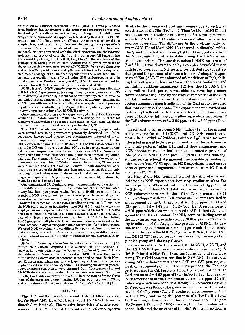
F~G. 5. A family of structures for [Sar'] ANG I1 generated by distance geometry and molecular dynamics. Distance constraints were obtained from 1D-NOE data and fluorescence lifetime studies.
tides. The NOE experiments on [des-l,2,3lANG I1 also serve as a control experiment to show the minimal contribution of par- tial saturation to the NOE enhancements of ring protons in [Sar'IANG 11, thereby validating the integrity of the NOE re- sults for ANG I1 and [Sar'IANG I1 (Tables I and 11).
Recent fluorescence studies on ANG I1 analogues in receptor- simulating environments have provided convincing support for the existence of a charge relay system, comprised of Tyr OH- imidazole-carboxylate in the bioactive conformation of ANG 11, and for the involvement of the tyrosinate species so generated in the receptor triggering mechanism (14). The present findings demonstrate that the associated processes of aromatic ring clustering and charge relay formation in ANG I1 only occur in the presence of the NHz-terminal domain, and presumably de- rive from interaction of the positively charged amino and gua- nidino groups with the negative charge dispersed within the charge relay triad. In nonpolar environments where solvation of charged groups by water is not possible, it is expected that oppositely charged groups in the peptide will come together in the manner observed herein; such considerations presumably also apply in the nonpolar transmembrane receptor environ- ment where the ligand binds (15).
To test the proposed model several constrained linear and cyclic ANG I1 analogues have been recently synthesized by us using novel Fmoc solid-phase peptide methods and special res- ins developed in our group (18). A series of linear ANG I1 ana- logues in which position 7 was occupied by secondary cyclic amino acids of variable ring size ( h e , Pro, Pip) supported the proposed model (28). Another series of constraint cyclic amide- linked ANG I1 analogues have been also recently synthesized by us and by novel strategies to test furthermore the ring
clustering and the proposed charge relay bioactive conforma- tion. These analogues were synthesized by connecting side chain amino and carboxyl groups at positions 1 and 8,2 and 8.3, and 8,3 and 5, MIz-terminal amino and COOH-terminal car- boxyl groups at positions 1 and 8,2, and 8,4 and 8 and side chain amino to COOH-terminal carboxyl groups at positions 1 and 8. All these analogues were biologically inactive except for cyclic [Sar1,Asp3,Lys51ANG I1 which had high contractile activ- ity in the rat uterus assay (30% of ANG 11). Molecular modeling suggests that the ring structure of the potent analogue can be accommodated.in the charge relay conformation proposed for
Since the charge relay triad in ANG I1 essentially relocates the negative charge originating on the COOH-terminal carbox- ylate to the imidazole ring and/or Tyr OH, proximity of the positively charged Ar2 guanidino group and NHz-terminal amino groups to the TyrEIis side chains might be expected. Indeed, recent energy calculations3 suggest that 1) folding of the MIz-terminal domain locates the NHz-terminal amino group near the His ring and 2) formation, stability, and spatial location of the tyrosinate pharmacophore generated by the charge relay interactions in ANG I1 is fostered by the Arg guanidino group.
Fig. 5 shows a superposition of family structures for [Sar'IANG I1 generated by distance geometry and molecular dynamics. Distance geometries have been calculated based on NOE intensities using the interproton distances Tyr dm (2.5 and available alp information (2.3 A min-3.0 A max) for cali- bration. Thus, % proton values in the range 11.6-24% (aver-
ANG 11.3
J. M. Matsoukas and G. J. Moore, unpublished results.

5312 Conformation of Angiotensin I1 TABLE lV
Chemical shifts and multiplicities for Ac-Arg-OCH, Distinct minor peaks at 6 = 7.35 ppm and 6 = 6.65 (Fig. 4) are assigned
to guanido w and w' protons of a minor conformer with side chain- restricted rotation.
Proton(s) Chemical shiR Multiplicity
(6) ClY 4.23 d t CP9 Y 1.74-1.51 C6 3.10
m dt
N€ 9.28 d w ' 7.90
Broad Broad
Ne 8.58 d CH3CO- 1.89 s CH30- 3.66 S
age !7.8%) for Tyr o/m (Tables 1-111) represent a distance of 2.5 A. The maximum possibleodistance between a and p pro- tons of an amino acid is 3.0 A, and since the lowest percent proton observed (Phe alp in Table !) was 1.2%, this value is taken to represent a distance of 3.0 A. Values less than 1% are considered insignificant for the present purposes and were not incorporated into models generated by molecular dynamics simulations. Optimized geometries for the superagonist [Sar'IANG I1 are shown in Figs. 5 and 6. The charge relay conformation for [SarlIANG I1 represents a local energy mini- mum in the potential energy surface obtained by searching from extended conformations and is a valid candidate for the biologically active conformation of ANG I1 and [Sar'IANG 11. The proposed conformation is stabilized by guanidinium (Are) ...p henolate (Tyr4) electrostatic and hydrogen bonding interaction as well as by hydrogen bonds between imidazole (His6) ... carboxylate (Phe8) and imidazole (His6) ... hydroxylate (Tyr4). The family of structures (Fig. 5) for [Sar'IANG I1 was generated based on distance constraints obtained from 1D- NOE and fluorescence data. In particular the distance con- straints from NOE data used were those for the ring cluster, the Tyr-Ile-His bend, and the proximity of the NH2-terminal domain to the ring cluster. The charge relay system con- straints used in this study are supported by fluorescence life- time studies (14). Energy minimization calculations illustrate that the absence of Asp' carboxylate in [Sar'lANG I1 results in a tighter association of the Are guanidinium with the Tyr4
oxygen, due to the availability of a biforcated interaction of the guanidinium with tyrosinate, and that the resulting in- creased stabilization of the tyrosinate pharmacophore may ex- plain the superagonist activity of [Sar'IANG 11. Removal of the NH2-terminal dipeptide in ANG I1 substantially reduces the biological activity. The present calculations suggest that the NH2-terminal domain has no formal role in the proton transfer, except that it is located close to the ring cluster sta- bilizing the receptor bound form. Folding of the NH2-terminal domain appears to be largely driven by electrostatic attrac- tions between the positively charged NH2 terminus and the negative charge dispersed within the charge relay triad. The relative positioning of the NH2-terminal domain brings the Ar$ guanidinium group into a favorable location for interac- tion with the tyrosinate anion generated by the charge relay system in ANG 11, thereby stabilizing the tyrosinate species. The auxiliary role of the NH2-terminal dipeptide of angioten- sin in the proton transfer mechanism as revealed by AM1 ge- ometry optimization, is supported by our NMR studies which clearly indicate proximity of the NH2-terminal domain to the cluster (28).
This model has been also used as a basis for the design
FIG. 6. Model of [Sar'lANG 11.
synthesis of non-peptide drugs. The imidazole based synthe- sized compounds were found to act as angiotensin I1 inhibitors furthermore validating our suggestion (29).
REFERENCES 1. Ondetti, M. A,, and Cushman, D. W. (1981) Annu. Reu. Biochem. 51,283-308 2. Smeby, R. R., and Fermandjian. S . (1976) in Amino Acids. Peptides and Pro-
3. Matsoukas, J. M., Goghari, M. H., Scanlon, M. N., Franklin, K. J., and Moore,
4. Smeby, R. R., Arakawa, K., Bumpus, F. M., and Marsh, M. M. (1962) Biochim.
5. Paiva, T. B., Paiva, A. C. M., and Scheraga, H. A. (1963) Biochemistry 2,
6. Fermandjian, S., Morgat, J. L., and Fromageot, P. (1971) Eur. J. Biochem. 24.
teins (Weinstein, N., ed) pp. 117-162, Marcel Dekker New York
G. J. (1985) J. Med. Chem. 28,780-783
Biophys. Acta 58,550-557
1327-1334
252-258 7. Printz, M. P.. Williams, H. P., and Craig, L. C. (1972) Proc. Natl. Acad. Sci.
"_ ".
U. S. A. 69.378-382 8. Weinkam. R. J.: and Jorgensen. E. C. (1971) J. Am. Chem. Soc. 93,7033-7038
9. Deslauriers, R., Ralston, E., and Somojai, R. L. (1977) J. Mol. Biol. 113,
10. Lenkinski, R. E., Stephens, R. L., and Krishna, N. R. (1981) Biochemistry 20,
11. Moore, G. J., and Matsoukas, J. M. (1985) Bio. Sci. Rep. 4,407-416 12. Matsoukas, J. M., Bigam, G., Zhou, N.. and Moore, G. J. (1990) Peptides 11,
13. Matsoukas, J. M., Yamdagni, R., and Moore, G. J. (1990) Peptides 11,367-374 14. Turner, R. J., Matsoukas, J. M., and Moore, G. J. (199l)Biochim. Biophys. Acta
15. Dixon. R. A. F., Strader. C. 0.. and Sigal, I. S . (1988)An. Rep. Med. Chem. 23,
16. Sargent, D. F., and Schwyzer, R. (1986) Proc. Natl. Acad. Sei. U. S.A. 83,
17. Noggle, J. H., and Shirmer, R. E. (1971) The Nuclear Overhauser Effect, pp.
18. Barlos, K., Gatos. D., Hondrelis, J., Matsoukas, J. M., Moore, G. J., Schiifer. W.,
19. Barlos, K., Gatos, D., and Schafer, W. (1991)Angew. Chem. Int. Ed. Engl. 30,
20. Matsoukas, J. M.. Cordopatis, P.. Belte, U., Goghari, M. H.. Ganter. R. C.,
21. Otter, A,, Scott, P. G., and Kotovych, G . (1988) Biochemistry 27,3560-3567 22. Marion, D. (1985) FEES L e f t . 192,99-I03 23. Deslauriers, R., Paiva, A. C. M., Schaumburg, K., and Smith, I . C. P. (1975)
24. Zhou, N., Moore, G. J.. and Vogel, H. J. (1991) J. Prof. Chem. IO, 333-343 25. Matsoukas, J. M., and Moore, G . J. (1984) Biochem. Biophys. Res. Commun.
26. Otter,A., Scott. P. G., Maccioni, R. B., and Kotovych. G. (1991)Biopolymers31, 122,434-438
27. Gross, E. and Meienholer, J. (eds) (1983) The Peptides. Vol. 5, pp. 257-284, 449-458
28. Matsoukas, J. M., Agelis, G., Hondrelis, J., Yamdagni, R., Wu, Q.. Ganter. R., Academic Press Inc., New York
29. Wahhab, A,, Smith, J. R., Ganter, R. C., Moore, D. M., Hondrelis. J.. Mat- Smith, J. R. Moore, D., and Moore, G. J. (1993) J. Med. Chem. 36,904-911
soukas, J., and Moore, G. J. (1994) Drug Res. 43, 1157-1168
and 7038-7044
697-710
3122-3126
359-366
1065.21-28
221-233
5774-5778
76-95, Academic Press, New York
and Sotiriou, P. (1989) Liebigs. Ann. Chem. 951-955
590-593
Franklin, K. J., and Moore, G. J. (1988) J. Med. Chem. 31, 1418-1421
Biochemistry 14,878-886