Embed Size (px)
Citation preview

68
Redescription of Monorcholepis dujardini (Krabbe, 1869) and M. passerellae (Webster, 1952) (Cestoda: Cyclophyllidea: Aploparaksidae) in passerine birds from the Holarctic Region
Svetlana Bondarenko and Jurijus Komisarovas
Institute of Ecology, Vilnius University, Akademijos 2, LT-08412 Vilnius-21, Lithuania
Key words: Cestoda, Cyclophyllidea, Aploparaksidae, Monorcholepis, Passeriformes, Holarctic
Abstract. Two species of avian tapeworms, Мonorcholepis dujardini (Krabbe, 1869) and M. passerellae (Webster, 1952), of the cyclophyllidean family Aploparaksidae Mayhew, 1925 (earlier included in the Hymenolepididae) are redescribed. Relative to congeners, the morphology of the strobila of both species shows strong similarities including a unique form of the cirrus. Separa-tion of these species, however, can be based on the number (40–53 and 25–31, respectively) and size (18–25 µm and 14–18 µm) of the rostellar hooks, although their shape in specimens of both species varies considerably. We examined specimens of Monor-cholepis dujardini in Turdus iliacus L., T. philomelos Brehm and T. merula L. from the Curonian Spit (Baltic Sea, Russia), T. naumanni Temminck from Chukotka, Motacilla alba L. from the Kuril Islands (Russia) and Passerella iliaca iliaca (Merrem) from Alaska (USA). Specimens in T. iliacus from the collection of O. Fuhrmann (Natural History Museum, Geneva, Switzer-land) were re-studied. The type material (holotype and paratypes) designated as M. dujardini neoarctica (Webster, 1955) in Ixo-reus naevius naevius (Gmelin) from Douglas Island, USA was re-examined and validity of this subspecies was rejected. A rede-scription of M. passerellae (Webster, 1952) was based on material in P. iliaca iliaca from Wisconsin (type specimen), and Point Barrow, Alaska, and on the one specimen recorded for the first time, in Turdus iliacus from the Palaearctic (Curonian Spit). Spe-cies of the genus Monorcholepis Oshmarin, 1963 and subgenus Aploparaksis (Tanureria) Spassky et Yurpalova, 1968 are char-acterized by similar topography of the gonads. These generic taxa and the interrelationships of constituent species are discussed.
The genus Моnorcholepis Oshmarin, 1963 (Cestoda: Cyclophyllidea, Aploparaksidae) with the type species M. dujardini (Krabbe, 1869) includes parasites of Passeriformes. Most of them were originally described as members of Aploparaksis Clerc, 1903. Clerc (1903) placed into this genus cestodes with one testis per proglottis, including А. dujardini (Krabbe, 1869). The morphological characters of A. dujardini, a parasite of passeriform birds, and Aploparaksis spp., parasites of charadriiform birds, corresponded well, with the only one exception being the number of the rostellar hooks. The rostellum of Aploparaksis spp. is armed with 10 hooks, whereas the number of hooks of the A. dujardini is much higher (40–46, according to Krabbe 1869). Since establishment of the genus Aploparaksis, addi-tional species with more than 10 rostellar hooks have been described primarily in passerine birds from the Nearctic: Aploparaksis passerellae Webster, 1952 in Passerella iliaca iliaca (Merrem), A. dujardini neoarc-ticus Webster, 1955 in Ixoreus naevius naevius (Gmelin), A. turdi Williamson et Rausch, 1965 in Turdus migratorius L. and Ixoreus naevius, and A. picae Todd, 1967 in Pica pica hudsonia (Sabine). In addition, Monorcholepis sobolevi Tsimbalyuk, Andronova et Ku-likov in Spasskaya, 1966 was described based on specimens in five species of passerine birds from Karaginskiy and Komandorskiy Islands (Russia, Far East). Tsimbalyuk, Andronova and Kulikov (1968) re-
corded M. sobolevi as an obligate parasite of Calcarius lapponica (L.).
The diagnosis of Monorcholepis was based on the characters of M. dujardini recorded in thrushes and Monorcholepis sp. in Vulpes vulpes from the Primorskiy Kray, Russia (Oshmarin 1963). This author did not ad-dress the taxonomic status of North-American species of Aploparaksis with more than 10 rostellar hooks. Spassky and Freze (1961) and Spassky (1963) excluded A. dujardini from Aploparaksis and placed it in Моnorcholepis referring to “Oshmarin (1961, in print)”; i.e., before the original generic diagnosis was published by Oshmarin (1963), bringing confusion in the date for erection of the genus. Later, A. dujardini neoarctica, A. passerellae and A. turdi were transferred into Мо-norcholepis (Spassky 1963, Spassky and Spasskаyа 1972). Concurrently, M. dujardini neoarctica was con-sidered as a taxon of uncertain status because of incom-plete original description (Spassky and Spasskaya 1972), although Williamson and Rausch (1965) consid-ered this subspecies as a morphological variation of A. dujardini.
In total, five species and one subspecies of Aplo-paraksis correspond well to the diagnosis of Мо-norcholepis. Among them only M. turdi clearly differs in the shape and length of the rostellar hooks, which are characterized by a long handle, and in the morphology of the copulatory apparatus (long and thick cylindrical
FOLIA PARASITOLOGICA 54: 68–80, 2007
Address for correspondence: J. Komisarovas, Laboratory of Parasitology, Institute of Ecology, Vilnius University, Akademijos 2, LT-08412 Vilnius-21, Lithuania. Phone: ++370 527 29 249; Fax: ++370 527 29 352; E-mail: [email protected]
Bondarenko, Komisarovas: Redescription of Monorcholepis spp.
69
cirrus). With the exception of M. dujardini, all other species of the genus Monorcholepis are known only from the original descriptions.
Recently, Bondarenko and Kontrimavichus (2006) have found M. sobolevi, a species characterized by 20 rostellar hooks (16 hooks reported in the original de-scription), as a typical parasite of passerine birds from Chukotka. In the same locality they found an additional species of this genus, M. atrashkevichi Bondarenko et Kontrimavichus, 2006, which is very similar to M. sobolevi, but distinguished from it by the number of rostellar hooks (40). It is difficult to separate these spe-cies on the basis of morphology, as both are character-ized by a relatively short, cylindrical to conical cirrus and similar shape of the rostellar hooks. A parallel situa-tion is observed with two other morphologically similar species, М. dujardini and M. passerellae, which are characterized by uniformity in the structure of the cir-rus.
The aim of the present study is to redescribe М. du-jardini on the basis of specimens from Europe, Asia and North America, and M. passerellae, based on of the type specimen and new findings from Alaska and the Curonian Spit (Baltic Sea, Russia).
MATERIALS AND METHODS
We examined the following specimens: Monorcholepis dujardini, Collection of the Natural History
Museum, Geneva (MHNG), Collection Neuchâtel (O. Fuhr-mann – J. Baer), No. 62/1–13, in T. iliacus (syn. T. musicus), originally labeled as a lot received from the Vienna Museum, number 19 (series of transverse and longitudinal sections of different part of strobila; scolex is lacking).
Monorcholepis dujardini neoarctica (Webster, 1955), U.S. National Parasite Collection (USNPC), holotype and paratypes in Ixoreus naevius naevius. USNPC 46648.02 – slide contain-ing the holotype (‘type’ – immature specimen) and five para-types (scoleces and neck only); the holotype is designated by a circle on the coverslip. USNPC 46648.01 – remnants of mate-rial from the type host and locality preserved in ethanol, con-taining one fragment of a premature specimen and 5 scoleces; the latter were studied in polyvinyl alcohol for detailed exami-nation of their rostellar hooks. According to Webster (1955), the site, date, locality, and collector are unknown; however, the following information is included in the USNPC cata-logue: USA, Alaska, Douglas Island, June 1949, collector Williams, R.B., identifier Webster, J.D.
Monorcholepis dujardini, 12 specimens in Turdus iliacus from the Curonian Spit, Baltic Sea, Kaliningrad Region, Rus-sia, collected by J. Komisarovas, 9–12 October 2004; one specimen in T. merula from the Curonian Spit, collected by J. Komisarovas, 20 October 2003; two specimens in T. philo-melos from the Curonian Spit, collected by J. Komisarovas, 11 October 2005. Materials from the Curonian Spit are held in the collection of J. Komisarovas, except for one specimen which is deposited in the collection of the MHNG, INVE 47854 (C126/84).
Monorcholepis dujardini in Motacilla alba from Para-mushir Island, Far East Russia (the vicinity of the city North Kuril’sk, 27 July 1965); one slide which was kindly provided for our investigation by Prof. O.I. Belogurov, Far East State University, Vladivostok, Russia; held in the collection of S.K. Bondarenko.
Monorcholepis dujardini, two specimens in one Turdus naumanni from West Chukotka, collected by S. Bondarenko, 9 July 1979; held in collection of S.K. Bondarenko.
Monorcholepis spp., 4 unidentified specimens and frag-ments in one Passerella iliaca iliaca from Point Barrow, Alaska on 29 May 1968 from the collection of Dr. R.L. Rausch (field number DWN B-14), which he kindly provided for our investigation; held in collection of S.K. Bondarenko.
Monorcholepis passerellae (Webster, 1952), USNPC 37365, holotype and paratypes in fox sparrow, Passerella iliaca iliaca from Wisconsin, USA.
Monorcholepis passerellae, one specimen in T. iliacus, col-lected by J. Komisarovas on the Curonian Spit, 12 October 2004; deposited in the collection of the MHNG, INVE 47855 (C126/85).
The fixed tapeworms were stained with Ehrlich’s haema-toxylin, dehydrated in an ascending alcohol series, cleared in clove oil and mounted in Canada balsam. The details of the morphology of the hooks, cirri and embryophores were also studied in specimens mounted in polyvinyl alcohol and in resin using the method of Steedmann (1958). Four scoleces and one fragment of the paratypes of M. dujardini neoarctica were mounted in polyvinyl alcohol because the available whole Canada-balsam mount did not allow exact examination. The measurements are given in micrometres (µm) unless oth-erwise stated. The descriptions and redescriptions are given with our own illustrations with the exception of hooks from the type specimens of M. dujardini, which were redrawn from the original description as presented by Krabbe (1869).
RESULTS
Monorcholepis dujardini (Krabbe, 1869) Oshmarin, 1963 Syns.: Taenia dujardini Krabbe, 1869; Monorchis du-jardini (Krabbe, 1869) Clerc, 1902; Aploparaksis du-jardini (Krabbe, 1869) Clerc, 1903; Haploparaksis dujardini (Krabbe, 1869) Yamaguti, 1935; Aploparaksis dujardini neoarctica Webster, 1955
Redescription. Based on a series of transverse and longitudinal sections of different parts of strobila origi-nally examined by O. Fuhrmann in MHNG (Figs. 1–3).
Proglottides 22–40 × 190–860. Ventral osmoregula-tory canals 33 wide, with anastomosis in each proglot-tis; dorsal canals about 3 wide. Scolex and rostellar hooks not seen. Male genital system dorsal, consisting of single testis, vas deferens, cirrus-sac and armed cir-rus. Testis 53–73 × 36–46 situated in aporal part of proglottis, near to lateral osmoregulatory canals. Cirrus- sac 200–207 long and 29–31 wide, may attain aporal osmoregulatory canals. External seminal vesicle elon-gate, 92 × 26, overlapping cirrus-sac dorsally. Internal

70
Figs. 1–3. Monorcholepis dujardini (Krabbe, 1869) drawn from Fuhrmann’s (1895) specimens (MHNG, slides 62/2, 6). Fig. 1. Transverse section of the proglottis showing the position of longitudinal muscles of the strobila, male organs and seminal recep-tacle. Abbreviations: BM – bundle of muscles; CS – cirrus-sac; DOC – dorsal osmoregulatory canal; ESV – external seminal vesicle; ISV – internal seminal vesicle; T – testis; SR – seminal receptacle; VOC – ventral osmoregulatory canal. Fig. 2. Em-bryophore and oncosphere (median pair of embryonal hooks are shorter than lateral pairs due to retraction). Fig. 3. Cirrus. Scale bars: Fig. 1 = 100 µm; Figs. 2, 3 = 20 µm.
seminal vesicle 132 long, occupies 2/3 of cirrus-sac length. Evaginated cirrus 67 long, distinctive shape; basal part of cirrus dilated (6 in diameter), then diameter of cirrus narrows to 4 and again expands, forming me-dian fusiform part with maximal diameter 5; diameter of distal part 3. Proximal part of cirrus armed with small spines; spines differ in size. Largest spines cover basal swelling; distinctly smaller spines cover fusiform part; distal part of cirrus smooth. Female genital organs situ-ated along transverse axis of proglottis. Part of ovary (observed in transverse sections) situated in centre of proglottis, ventral to male system. Vitellarium antiporal to ovary. Seminal receptacle elongate to oval, 89 × 40, ventral to cirrus-sac. Vagina tubular, 60 long. Gravid proglottides almost entirely filled with sac-shaped uterus. Embryophore 18–22 × 20–26, with thick (thick-ness 3–4) smooth walls; oncosphere 14 long and 16–18 wide; embryonic hooks 10–11 long.
● Description of the specimen in Turdus philomelos from the Curonian Spit Figs. 4–8
Strobila 67 mm long, with a maximum width of 930. Scolex 246 × 218. Suckers rounded, 73–82 in diameter. Rostellum rounded, 102 in diameter. Rostellar sheath 272 × 136. Rostellar hooks 40 in number, situated in
single row. Hooks with blade about as long as guard. Length of hooks 17.5–18 (blade 8.3–10, base 17.5–18, guard 10–11.6). Genital pores unilateral, dextral; genital ducts dorsal to longitudinal osmoregulatory canals. Ventral osmoregulatory canals 20–33 wide, with rare anastomoses, dorsal osmoregulatory canals 4 wide. Male genital system with single testis, 102–110 × 61–65, antiporal. Cirrus-sac, 190–210 × 20–23, reaching midline of proglottis or passing it. Internal seminal vesi-cle occupies 2/3 of cirrus-sac. External seminal vesicle large, ovoid, near middle of proglottides opposite anti-poral region of cirrus-sac or overlying its dorsal surface. Evaginated cirrus 103–110 long. At basal part, cirrus with dilatation, 10 in diameter, then a little slender part with diameter 5.6–7 and fusiform median part with 7.4–8 in diameter. Proximal part of cirrus covered with fine spines. Female system with trilobed ovary. Vitellarium compact, oval, 49–65 × 40–49, antiporal to ovarium. Vagina tubular. Seminal receptacle oval, 82–90 × 61–65, in poral half of median field or in middle. Short duct leading to triangular-shaped Mehlis’ gland, 60–62 in diameter. Gravid uterus sacciform, occupies almost en-tire proglottis. Eggs numerous, rounded, 53–54 in di-ameter. Embryophore 25–30 in diameter; wall smooth,

Bondarenko, Komisarovas: Redescription of Monorcholepis spp.
71
Figs. 4–8. Monorcholepis dujardini in Turdus philomelos (present study, Curonian Spit) (MHNG INVE 47854, C126/84). Fig. 4. Scolex. Fig. 5. Hooks. Fig. 6. Egg. Fig. 7. Mature proglottis. Fig. 8. Gravid proglottis. Scale bars: Figs. 4, 7, 8 = 100 µm; Fig. 6 = 50 µm; Fig. 5 = 20 µm.
4–5 in thickness, without polar thickenings. Oncosphere 15–17 in diameter, embryonic hooks of central and lat-eral pairs identical in shape and length, 10–11 long.
Summarized measurements of specimens in Turdus spp. collected from Curonian Spit are given in Table 1. The morphology of the hooks of specimens in T. iliacus and T. merula from the same locality is represented in Figs. 9–16. ● Description of specimens in Passerella iliaca from Point Barrow, Alaska (2 of 4 specimens collected by R.L. Rausch) Figs. 17–19
Strobila 35 × 0.70–0.76 mm. Rostellum rounded, armed with 50 and 51 hooks, blade of same length or little longer than guard. Length of hooks, measured from intact rostellum: on superior surface of a slide 21.5–23 in length, whereas 18 in length (blade 11.6–13, base 17–20, guard 8–9) when measured on bottom sur-face. Male system with single testis, 62–70 × 106–119. Cirrus-sac 193–291 × 26–29 (cavity diameter 21–25), reaching midline of proglottis or passing it. Internal seminal vesicle 120–160 × 21–24. Evaginated cirrus 103–106 long; basal part of cirrus with dilatation, 8–10 in diameter, then a little slender part with diameter 5,
Figs. 9–16. Rostellar hooks of Monorcholepis dujardini in different thrushes in Curonian Spit (present study). Figs. 9–15. Hooks of specimens in Turdus iliacus. Fig. 16. Hook of specimen from Turdus merula. Scale bar = 20 µm. and fusiform median part up to 8 (with spines). Proxi- mal part of cirrus covered with small spines. Spines on basal and median part slightly larger than those on slen-der part. Distal part of cirrus 62 long, unarmed. Female

72
Figs. 17–19. Monorcholepis dujardini in Passerella iliaca from Point Barrow, Alaska. Fig. 17. Hooks. Fig. 18. Cirrus. Fig. 19. Egg. Scale bars: Figs. 17, 18 = 20 µm; Figs. 19 = 30 µm. Figs. 20–23. Monorcholepis dujardini in Turdus naumanni from Chukotka and Motacilla alba from Paramushir Island (Kuril Islands). Fig. 20. Scolex. Fig. 21. Hooks. Fig. 22. Cirrus. Fig. 23. Egg. Scale bars: Fig. 20 = 100 µm; Figs. 21, 22 = 20 µm; Fig. 23 = 50 µm.
Bondarenko, Komisarovas: Redescription of Monorcholepis spp.
73
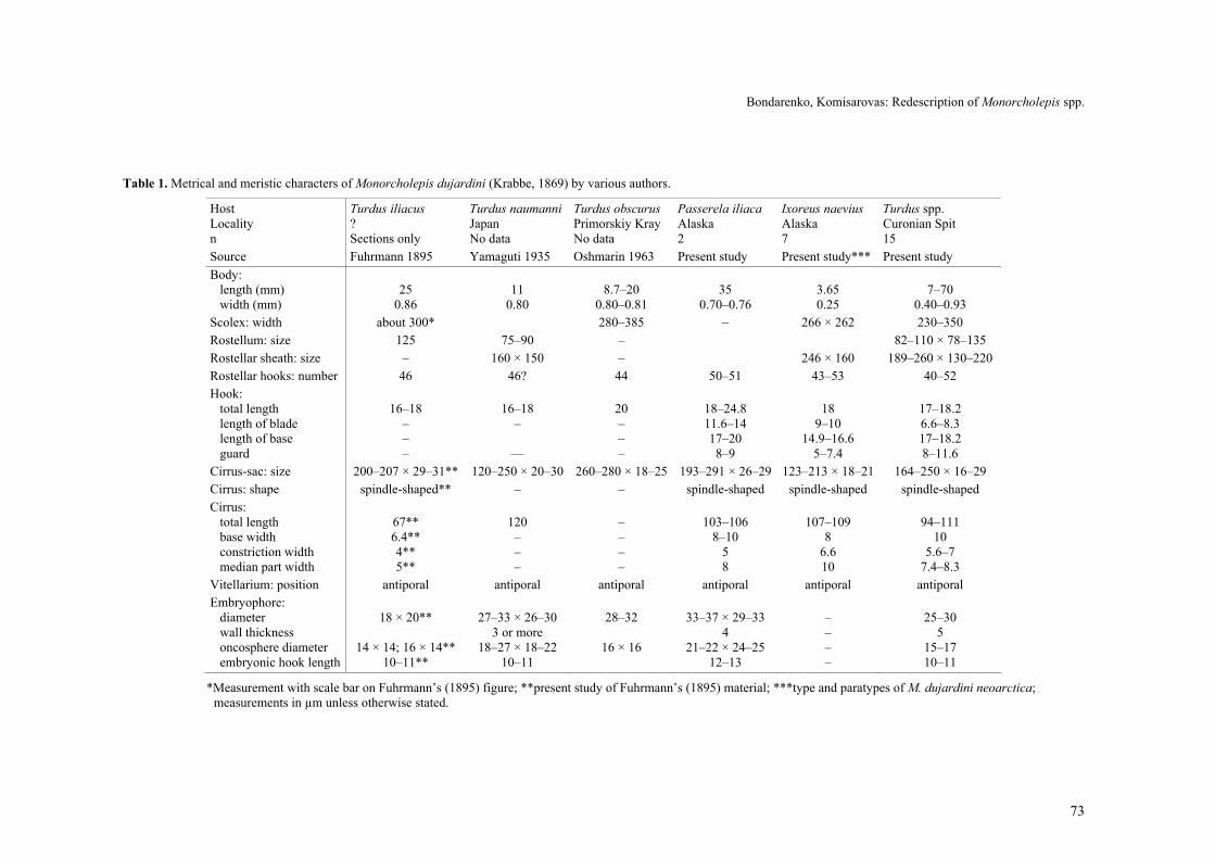
Table 1. Metrical and meristic characters of Monorcholepis dujardini (Krabbe, 1869) by various authors.
Host Turdus iliacus Turdus naumanni Turdus obscurus Passerela iliaca Ixoreus naevius Turdus spp. Locality ? Japan Primorskiy Kray Alaska Alaska Curonian Spit n Sections only No data No data 2 7 15 Source Fuhrmann 1895 Yamaguti 1935 Oshmarin 1963 Present study Present study*** Present study Body: length (mm) width (mm)
25
0.86
11
0.80
8.7–20
0.80–0.81
35
0.70–0.76
3.65 0.25
7–70
0.40–0.93 Scolex: width about 300* 280–385 – 266 × 262 230–350 Rostellum: size 125 75–90 – 82–110 × 78–135 Rostellar sheath: size – 160 × 150 – 246 × 160 189–260 × 130–220 Rostellar hooks: number 46 46? 44 50–51 43–53 40–52 Hook: total length length of blade length of base guard
16–18
– – –
16–18
–
––
20 – – –
18–24.8 11.6–14 17–20 8–9
18
9–10 14.9–16.6
5–7.4
17–18.2 6.6–8.3 17–18.2 8–11.6
Cirrus-sac: size 200–207 × 29–31** 120–250 × 20–30 260–280 × 18–25 193–291 × 26–29 123–213 × 18–21 164–250 × 16–29 Cirrus: shape spindle-shaped** – – spindle-shaped spindle-shaped spindle-shaped Cirrus: total length base width constriction width median part width
67** 6.4** 4** 5**
120 – – –
– – – –
103–106
8–10 5 8
107–109
8 6.6 10
94–111
10 5.6–7
7.4–8.3 Vitellarium: position antiporal antiporal antiporal antiporal antiporal antiporal Embryophore: diameter wall thickness oncosphere diameter embryonic hook length
18 × 20**
14 × 14; 16 × 14**
10–11**
27–33 × 26–30
3 or more 18–27 × 18–22
10–11
28–32
16 × 16
33–37 × 29–33
4 21–22 × 24–25
12–13
– – – –
25–30
5 15–17 10–11
*Measurement with scale bar on Fuhrmann’s (1895) figure; **present study of Fuhrmann’s (1895) material; ***type and paratypes of M. dujardini neoarctica; measurements in µm unless otherwise stated.

74
gonads poorly delineated. Uterus fills most space of each proglottis. Gravid eggs 41–55 in diameter. Em-bryophore 33–37 × 29–33, wall smooth, 4 in thickness, without polar thickenings. Oncosphere 21–22 × 24–25, closely adjoins to walls of embryophore; when free of embryophore, 25 × 29. Embryonic hooks 12–13 long.
● Description of the specimens in Turdus naumanni from Chukotka and Motacilla alba from Paramushir Island Figs. 20–23
Mature specimens 11–20 mm long and 0.8 mm maxi-mum width at level of gravid proglottides. Scolex 330 in diameter. Suckers 100 in diameter. Rostellum 150 × 120. Rostellar sheath 340 × 175. Rostellar hooks 46 (possible 48) with very short handle; blade short but pointed, shorter than guard. Length of hooks 17–18 (length of blade 6.6–8, length of base 17–18, length of guard 8–10; width of hook in profile 10, width of base in full face 5). In male system, cirrus-sac 168 × 18, reaches median line and crosses antiporal border of tes-tis. Fully-evaginated cirrus 115–129 long; basal part of cirrus with dilatation, about 10 in diameter, then a little slender part with diameter 5–6, and fusiform median part up to 8–10 (with spines). Distal part of cirrus up to 62 in length, unarmed. Female system with trilobed ovary, in poral part of median field. Vitellarium com-pact, oval, antiporal to ovary, at level of testis, ventral to it. Uterus sacciform. Gravid eggs 45–49 in diameter. Embryophore rounded, 25–26 in diameter, wall smooth 4–6 in thickness, without polar thickenings. Oncosphere 16–16.5 in diameter. Embryonic hooks of central and lateral pairs identical in shape and length, 10 long. Remarks
Yamaguti (1935) described М. dujardini in T. naumanni from Japan but did not present any illustra-tions. He mentioned that his material was identical to Fuhrmann’s 1895 description. The differences were only in the shape of the ovary, described as lobed by Yamaguti (1935). Data on metrical and meristic charac-ters of specimen described by him and our data of specimens in the same host from Chukotka and in M. alba from Paramushir Island coincide. Measurements of M. dujardini as reported by various authors and ob-servations from the present study are summarized in Table 1.
Monorcholepis dujardini neoarctica Webster, 1955 [=Monorcholepis dujardini (Krabbe, 1869)]
Redescription. Based on the type material (holotype and paratypes) (Figs. 24–27).
Length of strobila about 2 mm, maximum width 0.l6 mm in holotype. Scolex 164 × 139, rostellar sheath sac-like, 106 at the level of rostellum. Rostellum 60 × 86, number of hooks 42. Proglottides juvenile, without genital organs. Scolex of paratypes 266–272 × 240–262. Rostellum 98 × 86–131. Rostellar sheath 170–246 long and 110–160 wide. Rostellar hooks 42–53, of aplo-
paraksoid shape; blade as long as guard. Length of hooks 17–18 (blade 9–10, base 15–16, guard 5–7). Neck 127 wide immediately posterior to scolex. Genital atrium dextral, opens in middle of lateral proglottis margin or slightly anterior. Ventral osmoregulatory ca-nals with transverse anastomoses (not always distinct). Male system with oval testis, slightly antiporal, 80 × 60–80. Cirrus-sac 123–213 × 17–21, its antiporal region reaches antiporal osmoregulatory canals. Internal semi-nal vesicle 103 long, occupies 2/3 of cirrus-sac length. External seminal vesicle rounded, opposite to antiporal region of cirrus-sac, crossing antiporal osmoregulatory canals. Evaginated cirrus (Fig. 29) 109 long. Basal part of cirrus slightly dilated, 10 in diameter; in distal direc-tion, diameter narrows to 7 and then expands to 8 in middle of median fusiform part. Basal swelling and middle part of cirrus armed with small spines arranged in diagonal rows. Distal part of cirrus about 20 long, smooth. Female system with trilobed ovary, in centre of median field. Vitellarium rounded, compact, antiporal to ovary, ventral to testis. Seminal receptacle oval, 25 long and 30 wide, in poral half of median field. Vagina tubu-lar, 85 long and 3 wide. Remarks
Webster (1955) believed that this subspecies differed from the nominotypical A. dujardini dujardini (Krabbe, 1869) described by Fuhrmann (1895) and Yamaguti (1935) by the following characters: the cirrus-sac is smaller, both absolutely and relative to the proglottis width, the rostellar hooks are slightly longer and the body size is smaller. However, our study showed that specimens of Webster’s are very young. The length of hooks is 17–18 (i.e. the same as described by Krabbe 1869), the cirrus sac (Fig. 26) is long and reaches to the antiporal osmoregulatory canals. Therefore, there are no reasons to place it to the separate subspecies because it corresponds well with the characters of М. dujardini (see Table 1).
In the description of Taenia dujardini (syn. Monor-cholepis dujardini), Krabbe (1869) summarized data on specimens in Turdus musicus (syn. T. iliacus) from two localities on the coast of the Baltic Sea, Pomerania and Schleswig. These specimens differed by the number and shape of the rostellar hooks. The specimen from Pom-erania was 15 mm long and 1 mm wide and its rostel-lum was armed with 40 hooks 16–18 long. The hooks (as shown in Fig. 28) have a short blade (as long as the guard); the cirrus was short, 63 long and 4 wide, cylin-drical; and embryonic hooks were 10 long. The total length of the specimen from Schleswig was not noted; its rostellar hooks were the same length (17), but the number of the hooks was 46 and their shape was some-what different, with a blade slender and shorter than the guard (Fig. 29); the cirrus was slightly longer (88), but twice thicker (8) than in the specimen from Pomerania; the embryophore was round, thick-walled. In spite of the fact that the subsequent descriptions of M. dujardini

Bondarenko, Komisarovas: Redescription of Monorcholepis spp.
75
Figs. 24–27. Monorcholepis dujardini (= M. dujardini neoarctica; paratype, USNPC 46648) in Ixoreus naevius from Alaska. Fig. 24. Scolex. Fig. 25. Hooks. Fig. 26. Mature proglottis. Fig. 27. Cirrus. Scale bars: Fig. 24 = 100 µm; Figs. 25, 27 = 20 µm; Fig. 26 = 50 µm.
Figs. 28, 29. Rostellar hooks of Monorcholepis dujardini re-drawn from Krabbe (1869). Fig. 28. Hook of specimen in Turdus iliacus from Pomerania. Fig. 29. Hook of specimen in T. iliacus from Schleswig. Scale bar = 20 µm.
by Fuhrmann (1895) and Yamaguti (1935) were more detailed, they did not contain data about rostellar hooks except for their length. In the majority of later publica-tions (Fuhrmann 1932, Joyeux and Baer 1936, Skrjabin and Matevossian 1945, Spasskaya 1966), descriptions of
M. dujardini were shown with illustrations of hooks of specimens from Pomerania and Schleswig redrawn from Krabbe (1869).
The morphology of the specimens in T. naumanni (Chukotka) and M. alba (Paramushir Island) corre-sponds with specimens described by Yamaguti (1935). On the basis of the present results, we can assume that the number of the hooks of the specimens from Euro-pean populations of definitive hosts ranges between 40 and 52, from Eastern populations between 46 and 51, and from the North American population between 42 and 53. Demshin (1975) described metacestodes of M. dujardini in naturally infected oligochaetes from Pri-morskiy Kray (Russian Far East) with 46–62 rostellar hooks, 22 long; however, he illustrated a single row consisting of less than 30 hooks.
76
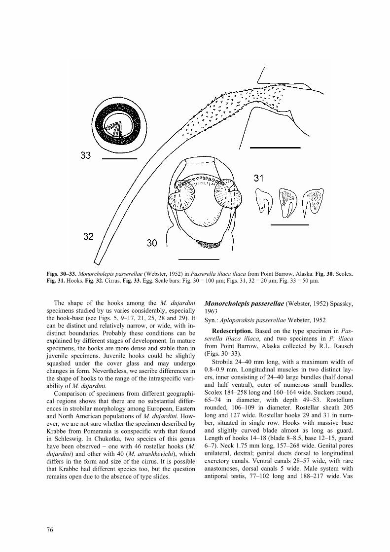
Figs. 30–33. Monorcholepis passerellae (Webster, 1952) in Passerella iliaca iliaca from Point Barrow, Alaska. Fig. 30. Scolex. Fig. 31. Hooks. Fig. 32. Cirrus. Fig. 33. Egg. Scale bars: Fig. 30 = 100 µm; Figs. 31, 32 = 20 µm; Fig. 33 = 50 µm.
The shape of the hooks among the M. dujardini
specimens studied by us varies considerably, especially the hook-base (see Figs. 5, 9–17, 21, 25, 28 and 29). It can be distinct and relatively narrow, or wide, with in-distinct boundaries. Probably these conditions can be explained by different stages of development. In mature specimens, the hooks are more dense and stable than in juvenile specimens. Juvenile hooks could be slightly squashed under the cover glass and may undergo changes in form. Nevertheless, we ascribe differences in the shape of hooks to the range of the intraspecific vari-ability of M. dujardini.
Comparison of specimens from different geographi-cal regions shows that there are no substantial differ-ences in strobilar morphology among European, Eastern and North American populations of M. dujardini. How-ever, we are not sure whether the specimen described by Krabbe from Pomerania is conspecific with that found in Schleswig. In Chukotka, two species of this genus have been observed – one with 46 rostellar hooks (M. dujardini) and other with 40 (M. atrashkevichi), which differs in the form and size of the cirrus. It is possible that Krabbe had different species too, but the question remains open due to the absence of type slides.
Monorcholepis passerellae (Webster, 1952) Spassky, 1963 Syn.: Aploparaksis passerellae Webster, 1952
Redescription. Based on the type specimen in Pas-serella iliaca iliaca, and two specimens in P. iliaca from Point Barrow, Alaska collected by R.L. Rausch (Figs. 30–33).
Strobila 24–40 mm long, with a maximum width of 0.8–0.9 mm. Longitudinal muscles in two distinct lay-ers, inner consisting of 24–40 large bundles (half dorsal and half ventral), outer of numerous small bundles. Scolex 184–258 long and 160–164 wide. Suckers round, 65–74 in diameter, with depth 49–53. Rostellum rounded, 106–109 in diameter. Rostellar sheath 205 long and 127 wide. Rostellar hooks 29 and 31 in num-ber, situated in single row. Hooks with massive base and slightly curved blade almost as long as guard. Length of hooks 14–18 (blade 8–8.5, base 12–15, guard 6–7). Neck 1.75 mm long, 157–268 wide. Genital pores unilateral, dextral; genital ducts dorsal to longitudinal excretory canals. Ventral canals 28–57 wide, with rare anastomoses, dorsal canals 5 wide. Male system with antiporal testis, 77–102 long and 188–217 wide. Vas

Bondarenko, Komisarovas: Redescription of Monorcholepis spp.
77
Figs. 34–37. Monorcholepis passerellae in Turdus iliacus, (present study, Curonian Spit) (MHNG INVE 47855, C126/85). Fig. 34. Scolex. Fig. 35. Hooks. Fig. 36. Cirrus. Fig. 37. Mature proglottis. Scale bars: Figs. 34, 37 = 100 µm; Fig. 35 = 20 µm; Fig. 36 = 50 µm.
deferens slender, leading from testis to large, ovoid, medially-placed external seminal vesicle. Cirrus-sac 190–287 × 30–45, reaching almost to midline of pro-glottis. Internal seminal vesicle occupies 2/3 of cirrus-sac. Cirrus up to 128 long. At basal part, cirrus with dilatation, 8–9 in diameter, then slightly slender part with diameter 5 and fusiform median part with same diameter as base. Proximal part of cirrus covered with tiny spines (spinous part 50 long). Female system with trilobed ovary. Vitellarium oval, 60 in diameter, antipo-ral to ovarium and ventral to testis. Oval seminal recep-tacle 30–40 wide, situated antero-poral to median line. Vagina tube-shaped, opens anterior to cirrus-sac. Uterus sac-like, entirely filling all proglottis. Eggs rounded, 42–50 × 63. Embryophore 26–34 × 24–31, with thick smooth walls (thickness 4–6), without polar thickenings. Oncosphere 17–21 × 21–23. Embryonic hooks 12–12.5 long. ● Description of the specimen in Turdus iliacus from the Curonian Spit Figs. 34–37
Strobila 25 mm long, with a maximum width of 320. Scolex, 260 × 210. Suckers rounded, 86–90 in diameter. Rostellum rounded, 78 in diameter. Rostellar sheath 217
× 140. Rostellar hooks 31 in number, situated in single row. Hooks with weakly curved blade, slightly shorter than guard. Length of hooks 18.2 (blade 8.3, base18.2, guard 11.6–12). Neck 900 long. Genital atrium dextral, opens in middle of lateral proglottis margin. Genital ducts pass dorsally across poral longitudinal osmoregu-latory canals. Ventral osmoregulatory canals 18–28 wide, with rare transverse canals, dorsal osmoregulatory canals 4 wide, without anastomoses. Male system with antiporal testis, 94–120 long × 75–92 wide. Vas defer-ens slender, leading from testis to large, ovoid, medi-ally-placed external seminal vesicle. Cirrus-sac 205–217 × 16–21, reaches and crosses median line of proglottis. Internal seminal vesicle occupies 2/3 of cirrus-sac. Cir-rus 78–98 long. At basal part, cirrus with dilatation 10–11.6 in diameter, then little slender part with diame-ter5.6–6.6 and fusiform median part 7.4–8.3 in diame-ter. Proximal part of cirrus covered with fine spines. Female system with trilobed ovary. Vitellarium oval, 33–53 × 29–41, antiporal to ovarium. Vagina tubular. Oval seminal receptacle 70–98 × 66–102, situated poral to median line. The specimen is not gravid; therefore, structure of uterus and eggs was not studied.

78
Table 2. Metrical and meristic characters of Monorcholepis passerellae (Webster, 1952).
Host Passerella iliaca Passerella iliaca Passerella iliaca Turdus iliacus Locality North America
(Madison) North America (Madison)
North America (Point Barrow, Alaska)
Curonian Spit (Baltic Sea)
n 2 1 2 1 Source Webster 1952 Present study* Present study Present study Total length (mm) 24 22 40 25 Maximum width (mm) 0.80 0.74 0.88 0.32 Scolex: size ? × 140–160 ? × 160 258 × 164 260 × 210 Rostellum: size 109 – 106 × 106 82 × 78 Rostellar sheath: size – 111 205 × 127 217 × 140 Rostellar hooks: number 25 25–30? 29–31 31 total length 13 – 14–18 18.2 length of blade 8–8.5 8.3 length of base 12–14.9 18.2 guard 6.6–7 11.6–12 Cirrus-sac: size 190–220 300 × 33 190–287 × 30–45 205–217 × 16–21 Cirrus: shape – – spindle-shaped spindle-shaped total length – – 109–128 78–98 base width – – 8–9 10–11.6 constriction width – – 5–6 5.6–6.6 median part width – – 8–9 7.4–8.3 Vitellarium: position – – antiporal antiporal Embryophore: size 24–26 × 26–30 29 × 32 26–34 × 24–31 – wall thickness – – 4–6 – oncosphere size 17–21 × 19–24 21 × 21 17–21 × 21–23 – embryonic hook length – 12 12–12.5 –
*Re-study of the type specimen; measurements in µm unless otherwise stated.
Remarks
Webster (1952) based his description upon two specimens, one complete (deposited in USNPC as No. 37365) and one sectioned. The morphology of the stro-bila was reconstructed from serial sections. The rostellar hooks were described as 25 in number, 13 long; their illustration was a free-hand sketch. The cirrus was de-scribed as stout and unarmed. No data on the ovary were presented. Webster (1952) differentiated M. passerellae from A. dujardini by the number and the size of the rostellar hooks only.
The re-examination of the holotype of M. passerellae was not very useful from the morphological point of view because of its poor condition. Determination of a precise number of hooks in the holotype was not possi-ble, however, it was seen that no more than 30 are pre-sent. The two specimens from Alaska provided addi-tional data about the characters not described in the original description. Our specimens had 29 and 31 ros-tellar hooks (instead of 25 in the original description). The length of the hooks was 14–18 (instead of 13) and their shape (Fig. 31) varied in relation to their position in crown. The comparison between M. dujardini (Figs. 19–21) and M. passerellae (Figs. 30–33) from the same bird confirms the validity of the latter species. The spe-cies differ by the number and the length of the rostellar hooks: 50–51 hooks with the length of 18–25 in M. du-jardini and 29–31 hooks with the length of 14–18 in M. passerellae. The shape of the hooks is also slightly dif-
ferent (compare Figs. 17 and 31). Additionally, speci-mens in T. iliacus collected on the Curonian Spit (Figs. 34–37) are similar to those of M. passerellae, based on the number of rostellar hooks (Table 2), but differ by their shape, i.e. the guard is slightly longer than the blade.
DISCUSSION
The genus Monorcholepis has been characterized by the number of rostellar hooks, which is always higher than 10, the presence of a single testis and the antiporal position of the vitellarium in relation to the ovary (Oshmarin 1963). When this genus was erected by Oshmarin, other species of Aploparaksis with ten rostel-lar hooks and the same topography of the gonads were unknown. Spassky and Jurpalova (1968) erected the subgenus Tanureria when they described A. (Tanureria) lateralis. The following diagnosis for the subgenus was provided: “Aploparaksis. Vitellarium compact, antiporal to ovary. Cirrus cylindrical with small basal swelling. Type species – A. (Tanureria) secessivus Gubanov et Mamaev in Spassky, 1963.”
According to Bondarenko and Kontrimavichus (2006), eight species of Aploparaksis correspond to the diagnosis of this subgenus. These are: A. (Tanureria) se-cessivus, A. (Tanureria) borealis Bondarenko et Rausch, 1977, A. (Tanureria) diagonalis Spassky et Bobova, 1961, A. (Tanureria) galli Rausch, 1951, A.

Bondarenko, Komisarovas: Redescription of Monorcholepis spp.
79
(Tanureria) lateralis Spassky et Yurpalova, 1968, A. (Tanureria) pseudosecessivus Belopolskaja, 1969, A. (Tanureria) vanelli Kornyushin, Bondarenko et Greben, 2006 and A. (Tanureria) regelae Bondarenko et Kon-trimavichus (2006).
The genera Aploparaksis and Monorcholepis are considered as members of the subfamily Aploparaksinae Mayhew, 1925 (Hymenolepididae) (Spassky and Spas-skaya 1972). However, Spassky (1992) elevated Aplo-paraksinae Mayhew, 1925 to the family rank. Bon-darenko and Kontrimavichus (2006) supported this opinion, based on characters of the life cycles of these cestodes. In contrast to hymenolepidid tapeworms, in-termediate hosts of aploparaksids are annelids. More-over, six unique morphological modifications of cysti-cercoid (not observed among hymenolepidids) are rec-ognized.
The new data on the morphology of M. dujardini from various parts of the Holarctic (Table 1) show sig-nificant differences in the number, length and shape of the hooks, but the morphology of proglottides in the both species is identical, first of all the shape and length of the cirrus. The variability of the number of the rostel-lar hooks in M. passerellae is lower than that of M. du-jardini, probably because of the small number of speci-mens available. There is a substantial difference be-tween the number of the hooks of the two species, 25–31 and 40–53. However, a range of the number of ros-tellar hooks between 25 and 53 (or even 62, as de-scribed by Demshin, 1975) is considerably beyond the limits of intraspecific variability as recognized among any species of hymenolepidoidean cestodes (see Skry-abin and Mathevossian 1945, Spasskaya 1966).
Additional support for the validity of M. passerellae and M. dujardini is related to the data by Bondarenko and Kontrimavichus (2006) concerning variability in the number and form of hooks observed between two other species, M. sobolevi and M. atrashkevichi, in passerine birds from Chukotka. As mentioned above, these two species differ in the number of rostellar hooks, 20 and 40, respectively. No specimen with an intermediate number of hooks was found either from naturally in-fected definitive hosts or from experimentally infected intermediate hosts (Enchytraeidae). However, Aplo-paraksis (Tanureria) borealis, M. sobolevi and M. atrashkevichi can be found in the intestine of the same passeriform bird. They differ mainly in the number of rostellar hooks (10, 20 and 40, respectively). This could be interpreted as a mode of speciation associated with the increase in the number of fixation organs. In M. sobolevi and M. atrashkevichi, the hooks may appear to be arranged in pairs on the rostellum; the cirrus is simi-lar in having a cylindrical form and is of moderate di-mensions.
Spassky and Spasskaya (1972) mentioned the inter-mediate morphological position for species of the sub-genus Tanureria relative to those of Aploparaksis and
Monorcholepis. They questioned the validity of Monor-cholepis and discussed its possible position as a subge-nus of Aploparaksis. Bondarenko and Kontrimavichus (2002) returned to this question in the discussion of some unresolved taxonomic problems of the Aploparak-sidae. Tanureria and Monorcholepis share similarities in morphological characters (similar topography of go-nads in adult tapeworms) and ontogeny of metacestodes (Bondarenko and Kontrimavichus 2002, 2006).
Life cycles and post-embryonal development of four species of Tanureria and three species of Monorchole-pis were studied under natural and experimental condi-tions in the northern regions of Eastern Siberia; annelids were determined to be the intermediate hosts (Bon-darenko and Kontrimavichus 2002, 2004, 2006). Pat-terns of metacestode development and the morphology of mature cysticercoids among Tanureria and Monor-cholepis indicate the presence of a unique morphologi-cal modification termed an “autotomicercus”, which is absent among species of the nominal subgenus Aplo-paraksis. Species of the latter subgroup are further dif-ferentiated by five attributes that are unique among the Aploparaksidae (Bondarenko and Kontrimavichus 1976, 2002, 2006). The development of an autotomicercus is accompanied by formation of a very large primary la-cuna. Separated fragments of the tail of the mature autotomicercus can exist independently in the coelom of intermediate hosts for extended periods (several months). Similar metacestodes identified as belonging to M. dujardini have also been found in naturally in-fected oligochaetes from England (Harper 1930) and from Primorskiy Kray (Demshin 1975).
Bondarenko and Kontrimavichus (2006) mentioned, however, that the data on the morphological characters, life cycles and development in the final host are not currently sufficient for proposing an explicit hypothesis about the phylogenetic relations between the species with an antiporal position of the vitellarium and the other Aploparaksidae. Perhaps, results of comparative molecular studies of A. (Tanureria) spp. and Monorcho-lepis spp. will help resolve this problem in the future.
Acknowledgements. Thanks are due to the following col-leagues for providing material for this study: Claude Vaucher, Muséum d’Histoire Naturelle, Geneva; Eric Hoberg and Patricia Pilitt, United States National Parasite Collection, Beltsville, Maryland, USA. We are grateful to Prof. R. Rausch and our colleagues from the Far East State University (FESU) – Prof. O.I. Belogurov and A.K. Tsimbalyuk, Vladivostok, Russia. The help with fieldwork provided by colleagues at the Biological Station of the Zoological Institute, Russian Acad-emy of Sciences is also acknowledged. We are grateful to Boyko B. Georgiev, Eric Hoberg and two anonymous referees for making some useful comments and for help with the lan-guage. This work was completed with the financial support of the International Science Foundation (grant LE 3000) and the Lithuanian State Science and Studies Foundation (grant No. C-06/2004(1)).

80
REFERENCES
BONDARENKO S.K., KONTRIMAVICHUS V.L. 1976: Polymor-phism of larvae of the genus Aploparaksis Clerc, 1903 (Hy-menolepididae). Folia Parasitol. 23: 39–44.
BONDARENKO S.K., KONTRIMAVICHUS V.L. 1979: [Life cycle and post-embryonic development of Aploparaksis (Tanureria) diagonalis Spassky & Jurpalova, 1968 – parasite of Charadri-iformes.] In: M.D. Sonin (Ed.), [Ecology and Morphology of Helminths of Vertebrates of Chukotka.] Nauka, Moscow, pp. 29–37. (In Russian.)
BONDARENKO S.K., KONTRIMAVICHUS V.L. 2002: Cestodes of the genus Aploparaksis Clerc, 1903 (Hymenolepididae): re-sults of research and unsolved issues. In: A.F. Alimov (Ed.), Problems of Cestodology. Vol. 2. Zoological Institute, Rus-sian Academy of Sciences, St. Petersburg, pp. 47–63. (In Rus-sian.)
BONDARENKO S.K., KONTRIMAVICHUS V.L. 2006: Aploparaksi-dae of Wild and Domesticated Birds. Fundamentals of Cestodology. Vol. 14. Nauka, Moscow, 443 pp.
BONDARENKO S.K., RAUSCH R.L. 1977: Aploparaksis borealis sp. n. (Cestoda: Hymenolepididae) from passeriform and cha-radriiform birds in Chukotka and Alaska. J. Parasitol. 63: 96–98.
CLERC W. 1903: Contribution a l’etude de la faune hel-minthologique de l’Oural. Rev. Suisse Zool. 11: 241–368.
DEMSHIN N.I. 1975: Oligochaeta and Hirudinea as Intermediate Hosts of Helminths. Nauka, Novosibirsk, 189 pp. (In Rusian.)
FUHRMANN O. 1895: Beitrag zur Kenntnis der Vögeltaenien. I. Rev. Suisse Zool. 4: 433–458.
FUHRMANN O. 1932: Les tenias des Oiseaux. Memoires de l’Université de Neuchâtel. 8: 383 pp.
HAPPER W.F. 1930: On some British larval cestodes from land and fresh-water invertebrate hosts. Parasitology 22: 202–213.
JOYEUX C., BAER J.G. 1936: Faune de France. 30. Cestodes. Office Central de Faunistique, Paris, 613 pp.
KRABBE H. 1869: Bidrag til Kundskab om Fuglenes Baen-delorme. Dansk. Vid. Selsk. Skr., Natur. Mat. Afd. 8: 249–363.
OSHMARIN P.G. 1963: [Parasitic worms of mammals and birds from the Primorskiy Kray.] Nauka, Moscow, 323 pp. (In Rus-sian.)
SKRYABIN K.I., MATEVOSYAN E.M. 1945: Hymenolepididae of birds. Sel’chozgiz, Moscow, 479 pp. (In Russian.)
SPASSKAYA L.P. 1966: [Hymenolepididae of birds of the USSR.] Nauka, Moscow, 698 pp. (In Russian.)
SPASSKY A.A. 1963: [Hymenolepidids – helminths of wild and domesticated birds.] Part 1. Osnovy Tcestodologii, Vol. 2. Nauka, Moscow, 418 pp. (In Russian.)
SPASSKY A.A. 1992: Two new tribes and the structure of super-family Hymenolepidea (Cestoda: Cyclophyllidea). Helmin-thologia 29: 167–170.
SPASSKY A.A., FREZE V.I. 1961: [Review of the genus Aplo-paraksis Clerc, 1903 (Cestoda: Hymenolepididae).] Česko-slovenská parasitologie 8: 385–389. (In Russian.)
SPASSKY A.A., SPASSKAYA L.P. 1972: [Genera Monorcholepis Oshmarin, 1961, Chimaerolepis, gen. n., and subfamily Aplo-paraksinae (Cestoda: Hymenolepididae).] Parazity zhivotnykh i rasteniy, Kishinev, 8: 69–75. (In Russian.)
SPASSKY A.A., YURPALOVA N.M. 1968: [Aploparaksis lateralis, n.sp. (Cestoda; Hymenolepididae) – new species from shore-birds – and substantiation a subgenus Tanureria, n. subgen.] Parazity zhivotnykh i rasteniy, Kishinev, 3: 30–37. (In Rus-sian.)
STEEDMANN H.F. 1958: Dimethyl hydantoin formaldehyde: a new water-soluble resin for use as a mounting medium. Quart. J. Microsc. Sci. 99: 451–452.
TSIMBALYUK A.K., ANDRONOVA K.Y., KULIKOV V.V. 1968: [Monorcholepis sobolevi sp. nov. (Hymenolepididae) and Similuncinus leonovi sp. nov. (Choanotaeniidae) – two new species from the bird of islands of the Bering Sea.] Soob-scheniya Dal’nevostochnogo filiala AN SSSR. Helminths of Far East and Pacific ocean, Vladivostok, 26: 126–131. (In Russian.)
WEBSTER J.D. 1952: A new hymenolepidid tapeworm from the fox sparrow. Proc. Indiana Acad. Sci. 61: 305–307.
WEBSTER J.D. 1955: Three new forms of Aploparaksis (Cestoda: Hymenolepididae). Trans. Am. Microsc. Soc. 74: 45–51.
WILLIAMSON F.S., RAUSCH R.L. 1965: Studies on the helminth fauna of Alaska. 62. Aploparaksis turdi sp. n., a hymeno-lepidid cestode from thrushes. J. Parasitol. 51: 249–252.
YAMAGUTI S. 1935: Studies on the helminth fauna of Japan. Part 6. Cestodes of birds. I. Jpn. J. Zool. 6: 183–232.
Received 13 March 2006 Accepted 30 August 2006