Embed Size (px)
Citation preview
TWO REMARKABLE EVENTS IN THE FIELDOF INTRAOCULAR FOREIGN BODY: (1) THEREVERSAL OF SIDEROSIS BULBI (2) THESPONTANEOUS EXTRUSION OF AN
INTRAOCULAR COPPER FOREIGN BODY
BY Robert B. Welch, MD
Tmil- VAST NUMBER OF REFEREN(CES IN TliE LITERATURE ON TflE SUBJECT OF INTRA-
ocular foreign body is indeed a numerical testimony to the interestin this subject. Goldsmith' presented a comprehensive survey of theintraocular foreign body literature in 1965, and noted 746 articles on thesubject from 1933-1963, (331 from 1933-1943, 224 from 1944-1953, and191 from 1954-1963). To bring the tally up to date, I have found anadditional 324 articles from 1964-1974, thus illustrating the fact that thisentity is neither losing its appeal for study nor diminishing in interestas a subject for publication in the literature.
In spite of the large number of papers on this subject, it must be re-membered that the occurrence of an intraocular foreign body as anophthalmic entity is unusual when viewed in relation to ophthalmicproblems in the population as a whole; yet in highly industrializedareas of the world, its recognition is commonplace. Much of our informa-tion concerning this type of injury is to be found in the British literaturein the excellent reviews by Cridland,2 Roper-Hall,3 and Percival.4 Inthis country, our own Harvey Thorpe is considered the "Dean of theForeign Body" and has published articles on this subject since the early1930's. 5To summarize the general subject, we may state that the majority of
intraocular foreign bodies are metallic and magnetic, that many non-magnetic foreign bodies contain copper, and that the air pellet gun ismaking its presence known as a cause of intraocular foreign body. Themost commoni cause of the intraocular foreign body is still the hand-wielded hammer and chisel.
In spite of the vast literature on this subject, the present reportdeals with two unusual events stemming from intraocular foreign bodies
Tii A\1 O(irii i So() vol. LXXIII, 1975
which, although reported in the literature, are generally unknown to theophthalmologist. Since reports of both situations were presented atthe American Ophthalmological Society in the remote past, it is of interestto revisit these subjects, provide photographic documentation, and to onceagain re-emphasize the old adage that there is really nothing new inmedicine.
SIDERoSIS BULBI WN'ITH I)ILATED INACTIVE PUPIL. RECOVERY AFTER REMOVAL OF
FOREIGN BoDY'
In 1923, at the American Ophthalmological Society, Dr Nelson M. Blackof Milwaukee, Wisconsin, was introduced by the president, Dr WilliamHolland Wilmer, to present his paper on Siderosis Bulbi with DilatedInactive Pupil. Recovery.6 It is perhaps of only historical interest thatDr Wilmer introduced the speaker, yet I mention it today since thisyear marks the Wilmer Institute's 50th anniversary and our presentpresident, Dr Elliott Randolph, is one of our Institute's finest products.
In Black's report, a 22-year-old white male reported that he hadrecently noted, while shaving, that his left pupil was dilated. On testinghis vision, he found that his acuity in the left eye was reduced. Thisrecalled to his mind that six months previously something had struekhim in the left eye while he was using a hammer and punch. Becauseof pain and slight bleeding, he had reported to the hospital where hisleft eye was dressed. No evidence of significant injury or foreignbody was noted, so he was sent home. Following this, he had ex-perienced no difficulty until the present observation. On examination,the right eye was normal with a light, blue-gray iris, while the left irisappeared yellowish-gray with an immobile 7 mm pupil. The visioIn inthe left eye was reduced to 6/10. A foreign body was found by roentgen-ography and removed with a magnet. Two weeks later, he was noted tohave a retinal detachment. It is of historic interest that he was admitted tothe hospital for treatment of his detachment. The treatment consistedof "rest in bed, compress, bandage, atropine, dionin, and pilocarpinesweats." In spite of persistence of his detachment, his pupils and iriscolor were equal in the two eyes three months later.When Black presented his case, a review of the literature revealed
that mydriasis with intraocular foreign body was not generallly recognized;although reports by Clegg,7 Vossius,8 and others9 did report thisphenomenon. It is of note that Dr William M. Sweet (of Sweetlocalization fame) stated, "I do not recall ever having seeni a refelelnceto a similar case," yet a year later he too was able to report on stuch (aphenomenon.10
188 Welch
Intraocular Foreign Body
Von Graefe,jt in 1860, was the first to call attention to the discolora-tion of the tissue of the eye from retained metallic particles; and Bunge, 12in 1890, was the first to apply the name, siderosis bulbi. In 1908de Schweinitz13 reported a case where the iris discoloration and wreathof yellowish-brown spots beneath the lens capsule disappeared followingremoval of the foreign body, but there was no reference to the state of thepupil.
Since Black's classic presentation, occasional reports have occurredin the literature. Thus, in 1940, at the Colorado OphthalmologicalSociety, Danielson and Long14 reported three cases of delayed removalof intraocular steel in which the only definite sign of siderosis was adilated pupil. After removal of the foreign body, the pupils returned tonormal. Unfortunately, in their report they do not mention the color ofthe iris.The case report today, photographically demonstrates the sign which led
to the diagnosis of an unsuspected intraocular foreign body - a dilatedinactive pupil with sideroisis iridis - and further documents the clinicalreversal of these findings following removal of the foreign body. To myknowledge, this is the first photographic documentation of such a train ofevents, although its recognition dates back for more than fifty years.
(CASE REPORT
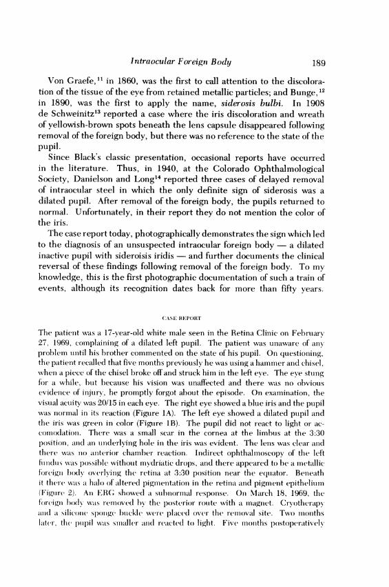
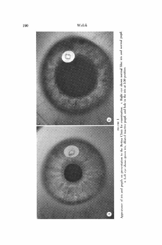
The patient was a 17-year-old white male seen in the Retina Clinic oni February27, 1969, complaininlg of a dilated left pupil. The patient was unaware of any,prollem until his hrother commented on the state of his pupil. On questioninlg,the patient recalled that five months previously he was using a hammer and chisel,when a piece of the chisel broke off and struck him in the left eye. The eve stUnlgfor a while, hut because his vision was unaffected and there was no obviousevidenice of injury, he promptly forgot about the episode. On exalmlination, thevisual acuity was 20/15 in each eye. The right eye showed a blue iris and the pupilwas normal in its reaction (Figure IA). The left eye showed a dilated pupil andthe iris was green in color (FigLre IB). The pupil did not react to light or ac-comodattion. There was a smaall scar in the cornea at the limlbus at the 3:30position, an ain undilerlving hole in the iris was evident. The lens was clear anldthere was nlo aniterior chamber reaction. Indirect ophthalmoscopv of the lefttiili(Cis wats possible xwithout mvdriatic drops, acnd there appeared to be ac metallicf0reigni body overlying the retinta tt 3:30 position near the e(qtuttor. Benieathit there was t halo of altered pigmiienitationi in the retinia and pigment epitheliium(Figurc 2). Al] ERG showed t subnormal responise. On1 March 18, 1969 thefireign lbodl w\vas remnove(l by the posterior rouite with at imalgnet. Crvotherapvalnd t siliconse sponge buckle were placed over the removal site. Two monthslater, th( pupil wats sialler a(wl raetedtl to light. Five months postoperatively
189
Welch190
C-5
-2z co
tx
ct
X Z
12
Intraocular Foreign Body
FIGURE 2Intraocular foreign body lying just over the retina. Note the halo of altered pigmentation
in the retina and pigment epithelium.
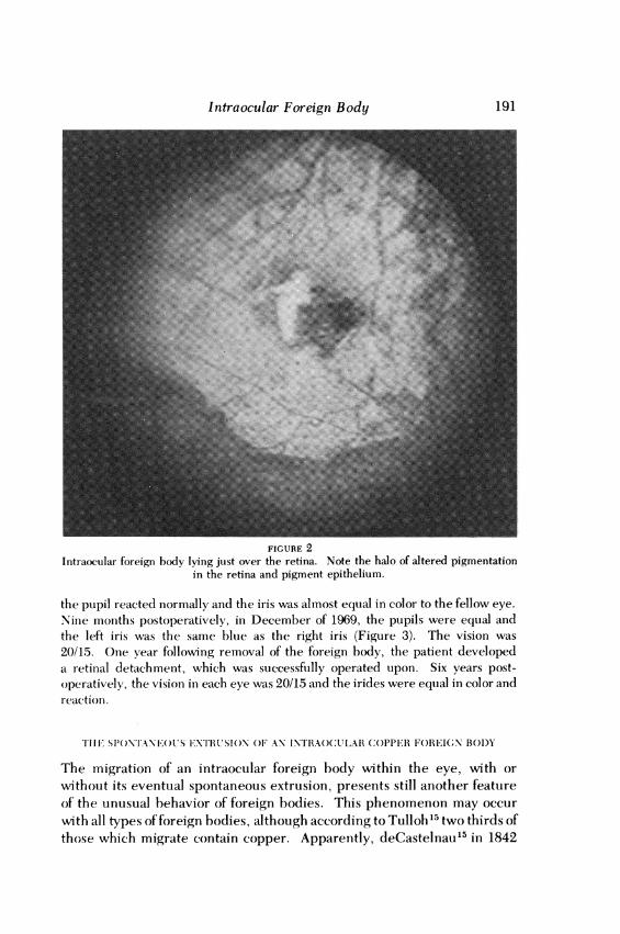
the pupil reacted normally and the iris was almost equal in color to the fellow eye.Nine months postoperatively, in December of 1969, the pupils were equal andthe left iris was the same blue as the right iris (Figure 3). The vision was20/15. One year following removal of the foreign body, the patient developeda retinal detachment, which was successfully operated upon. Six years post-operatively, the vision in each eye was 20/15 and the irides were equal in color andreaction.
THFj SPONTANM'US XTHRUSION OF AN INTRAOCULAR COPPER FOREICN BODY
The migration of an intraocular foreign body within the eye, with orwithout its eventual spontaneous extrusion, presents still another featureof the unusual behavior of foreign bodies. This phenomenon may occurwith all types of foreign bodies, although according to Tulloh 15 two thirds ofthose which migrate contain copper. Apparently, deCastelnau15 in 1842
191
Welch
FIGURE 3Photograph of both eyes shows return of left iris to normal color and pupillary function
nine months after removal of intraocular iron foreign body.
reported the first case of spontaneous expulsion of a foreign body. Thisforeign body was iron. Our own Charles Kipp reported the spontaneousexpulsion through the cornea of a large piece of copper foreign body in ablind eye in 1884,16 and mentions this and an additional case before oursociety in 1901.17 Goldsmith in his survey of the foreign body literatuirewas especially interested in the spontaneous migration and extrusion offoreign bodies and presents an excellent review of the literature. 1
(CASE REPORT
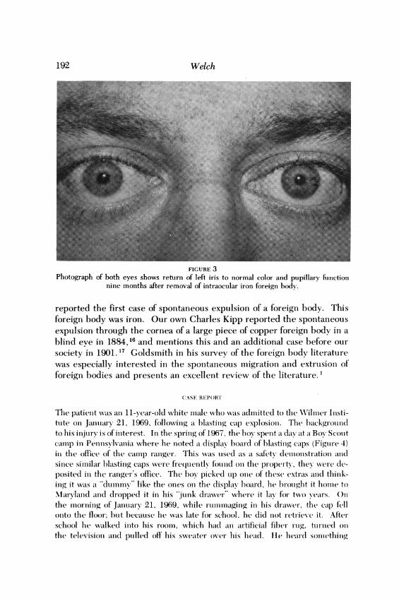
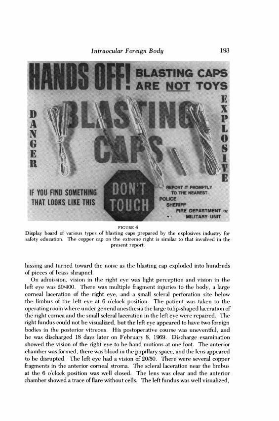
The patienit was ani 11-year-old white male who was admitted to the Wilmer Inisti-tute on Januiary 21, 1969, f'olloxving a blasting cap explosion. The backgroundto his injury is of initerest. In the spring of 1967, the boy spenit a day at a Boy Scoultcamiip in Pennsvlvania where he inoted a display board of blasting calps (Figuil-e 4)in the office of the camiip ranger. This was uised as a safety demlonistriation andlsinice similar- blasting caps were firequently foundi(l onl the property, thex were de-posited in the ranger's office. The box picked uip onie of these extras ainld thinik-ing it was a "dummy" like the onies oni the display board, lbe l)rollght it lhomile toMarylaand anid dropped it in his 'Junk dratwer" where it lax' f'or two y'ears. Onthe moriniilg of Januiary 21, 1969, while rutimimial.wginig in his diraiwel, the ciap fellonto the floor; hut l)becaulse he was late for school, he did niot retrieve it. Afterschool he wailked inlto his roomil, wkhich had a ii artificial fiber itig, tutrine(d onlthe televisioni aniid ptlledl ofl' hiis sweater over hiis hleadl. He hear(l somiething
192
Intraocular Foreign Body
IF YOU FIND S'ID 1)B
THAT LOOKS LI.KE. Til
.........
or
FIGURE 4Display board of various types of blasting caps prepared by the explosives industry forsafety education. The copper cap on the extreme right is similar to that involved in the
present report.
hissing and turned toward the noise as the blasting cap exploded into hundredsof pieces of brass shrapnel.
Oni admission, vision in the right eye was light perception and vision in theleft eye was 20/400. There was multiple fragmenit inijuries to the body, a largecorneal laceration of the right eye, and a small scleral perforationi site belowthe limbus of the left eye at 6 o'clock position. The patient was taken to theoperating room where under general anesthesia the large tulip-shaped laceration ofthe right cornea and the small scleral laceration in the left eye were repaired. Theright fiundus could not be visualized, but the left eye appeared to have two foreignbodies in the posterior vitreous. His postoperative course was uneventful, andhe was discharged 18 days later on February 8, 1969. Discharge examinationshowed the vision of the right eye to be hand motions at one foot. The anteriorchamber was formed, there was blood in the pupillary space, and the lens appearedto be disrupted. The left eye had a vision of 20/50. There were several copperfragments in the anterior corneal stroma. The scleral laceration near the limbusat the 6 o'clock position was well closed. The lens was clear and the anteriorchamber showed a trace of flare without cells. The left fundus was well visualized,
193
Welch
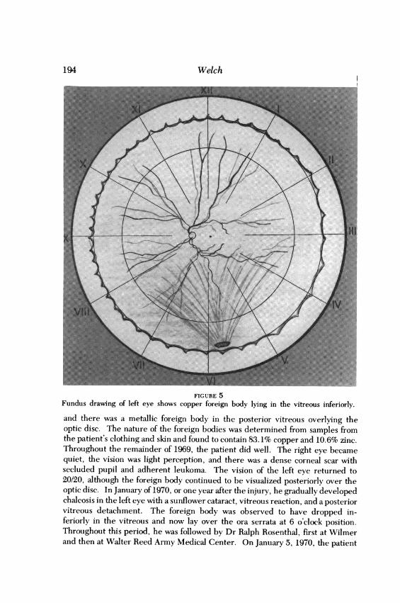
FIGURE 5Fundus drawing of left eye shows copper foreign body lying in the vitreous inferiorly.and there was a metallic foreign body in the posterior vitreous overlying theoptic disc. The nature of the foreign bodies was determined from samples fromthe patient's clothing and skin and found to contain 83.1% copper and 10.6% zinc.Throughout the remainder of 1969, the patient did well. The right eye becamequiet, the vision was light perception, and there was a dense corneal scar withsecluded pupil and adherent leukoma. The vision of the left eye returned to20/20, although the foreign body continued to be visualized posteriorly over theoptic disc. In January of 1970, or one year after the injury, he gradually developedchalcosis in the left eye with a sunflower cataract, vitreous reaction, and a posteriorvitreous detachment. The foreign body was observed to have dropped in-feriorly in the vitreous and now lay over the ora serrata at 6 o'clock position.Throughout this period, he was followed by Dr Ralph Rosenthal, first at Wilmerand then at Walter Reed Army Medical Center. On January 5, 1970, the patient
194
Intraocular Foreign Body
77
.:: 4
*:.f; :i..: ,.s.: .:_.. _ -N
/1 Ii./:IJJ
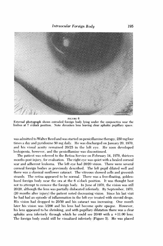
FIGURE 6External photograph shows extruded foreign body lying under the conjunctiva near thelimbus at 7 o'clock position. Note shrunken lens leaving clear aphakic pupillary space.
was admitted to Walter Reed and was started on penicillamine therapy, 250 mg fourtimes a day and pyridoxine 50 mg daily. He was discharged on January 20, 1970,and his visual acuity remained 20/25 in the left eye. He soon developedleukopenia, however, and the penicillamine was discontinued.The patient was referred to the Retina Service on February 14, 1970, thirteen
months post injury, for evaluation. The right eye was quiet with a healed cornealscar and adherent leukoma. The left eye had 20/20 vision. There were severalcorneal foreign bodies as previously described. The left pupil dilated well andthere was a classical sunflower cataract. The vitreous showed cells and greenishstrands. The retina appeared to be normal. There was a free-floating, golden-hued foreign body near the ora at the 6 o'clock position. It was thought bestnot to attempt to remove the foreign body. In June of 1970, the vision was still20/20, although the lens was partially dislocated inferiorly. By September, 1970,(20 months after injury) the patient noted decreasing vision. Since his last visithe had had an episode of inflammation in the left eye treated with steroid drops.His vision had dropped to 20/50 and his cataract was increasing. One monthlater his vision was 3/200 and his lens had become quite opaque. However,his lens appeared to be shrinking, and with pupillary dilatation there was a clearaphakic area inferiorly through which he could see 20/40 with a +11.00 lens.The foreign body could still be visualized inferiorly (Figure 5). He was placed
195
Welch
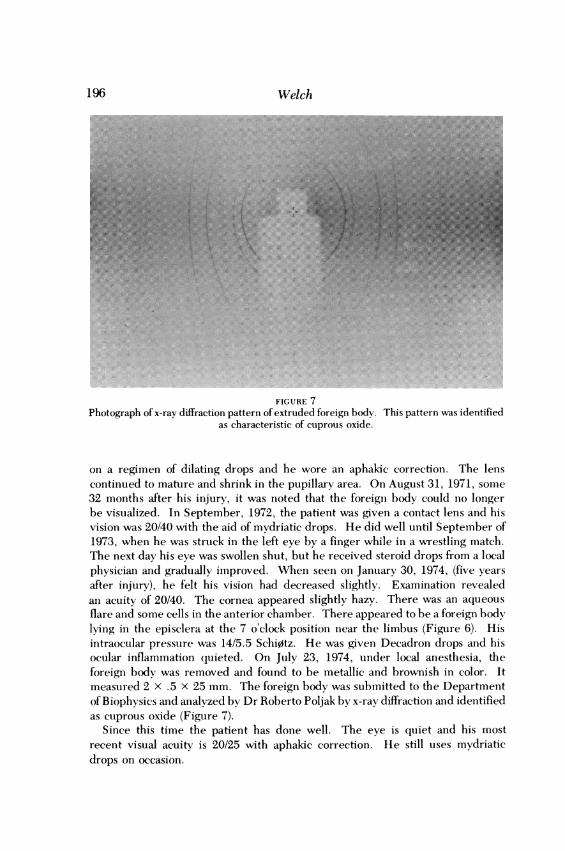
FIGURE 7Photograph of x-ray diffraction pattern of extruded foreign body. This pattern was identified
as characteristic of cuprous oxide.
on a regimen of dilating drops and he wore an aphakic correction. The lenscontinued to mature and shrink in the pupillary area. On August 31, 1971, some32 months after his injury, it was noted that the foreign body could no longerbe visualized. In September, 1972, the patient was given a contact lens and hisvision was 20/40 with the aid of mydriatic drops. He did well until September of1973, when he was struck in the left eye by a finger while in a wrestling match.The next day his eye was swollen shut, but he received steroid drops from a localphysician and gradually improved. When seen on January 30, 1974, (five yearsafter injury), he felt his vision had decreased slightly. Examination revealedan acuity of 20/40. The cornea appeared slightly hazy. There was an aqueousflare and some cells in the anterior chamber. There appeared to be a foreign bodylying in the episclera at the 7 o'clock position near the limbus (Figure 6). Hisintraocular pressure was 14/5.5 Schi0tz. He was giveni Decadron drops and hisocular inflammation quieted. On July 23, 1974, under local anesthesia, theforeign body was removed and found to be metallic and brownish in color. Itmeasured 2 x .5 x 25 mm. The foreign body was submitted to the Departmentof Biophysics and analyzed by Dr Roberto Poljak by x-ray diffraction and identifiedas cuprous oxide (Figure 7).
Since this time the patient has done well. The eye is quiet and his mostrecent visual acuity is 20/25 with aphakic correction. He still uses mydriaticdrops on occasion.
196
...
... .... .......... .....
Intraocular Foreign Body
DISCUSSION
The first case illustrates the phenomenon of an occult intraocular ironforeign body. The incidence of such cases must be rare, yet its recognitionis ofparamount importance, since removal of the foreign body may reversethe clinical picture of siderosis bulbi and permit the long term retentionof a functioning eye. Since most foreign bodies are metallic and iron-containing, the picture of siderosis is a primary clue to the presence of suchan event. Thus the eye becomes stained from the iron pigment and thisis most obvious in the iris. According to Wise, 18 the iron is released fromthe metallic foreign body as free ions which may become loosely boundto the acid mucopolysaccharides (AMPS) in the eye. These ions or com-plexes diffuse throughout the eye to become bound to the enzymes of cells.A particular cell may either detoxify and store the ions or, if sufficientintracellular enzymes are destroyed, the cell may degenerate and die,releasing the iron pigment which is picked up by macrophages. There isgreat epithelial affinity for the iron deposition; thus, retinal ganglion cells,retinal pigment epithelium, the iris muscles, and the epithelium of thelens are especially susceptible to the iron.A second clue to unsuspected intraocular foreign body is iridoplegia
with mydriasis. Verhoeff19 felt that the impaired motility of the iris wasdue to the impairment of the function of the muscle from the iron deposi-tion, rather than from a selective action on the nerve terminals as suggestedby Tuckett.9 The picture of clinical reversal of siderosis as seen in thiscase by return of normal iris color and pupillary function would seemto indicate that if the source of iron is removed in time before the cellularenzymes are poisoned, the process may be reversed and normal functionreturn. The present case does not answer the questions as to whethersiderosis of the retina may be reversible. An initial ERG showed reduc-tion. Three years after removal of the foreign body the ERG was still50-75% reduced but we must remember that there had been an inter-vening retinal detachment. Nevertheless, the visual field is full bynormal testing. We must consider that a portion of the retinal cell func-tion had been destroyed by the iron foreign body.The second case illustrates the phenomenon of spontaneous expulsion
of a copper foreign body and points up the fact that intraocular copperdoes not necessarily spell the death knell of any eye. The factors con-cerned in the migration and extrusion of copper particles from the eyeare well summarized in Tulloh's article of 1956.15 A copper foreign bodyabhors entrapment and whether because of its chemical nature, its non-magnetic characteristics which favor its retention, its size and shape, or theforces of gravity and intraocular pressure, it heads for the surface and may
197
be expelled. The conservative approach to this case should be viewed inthe light of present day advances in the techniques of surgery of thevitreous and one wonders how a surgeon skilled in such surgery would havehandled the problem when the vision was 3/200, there was a cataractpresent, and the foreign body could still be seen inferiorly.
SUMMARY
Two unusual events concerning intraocular foreign bodies are presented.The first patient had an occult or unsuspected intraocular foreign body.
He showed iridoplegia with mydriasis, siderosis iridis, and an intraocularpiece of iron lying posteriorly near the retina. The foreign body wasremoved and the patient regained normal iris color and pupillary activity.His vision remains 20/15 six years postoperatively despite ensuing retinaldetachment one year after removal of the foreign body.The second patient was a young boy injured by a blasting cap explosion.
He lost one eye from the injury and had a piece of intraocular brass inhis left eye. In spite ofthe development of chalcosis and a mature cataract,the lens gradually shrank in the pupillary space permitting a clearaphakic area and 20/25 vision. The brass fragment migrated forwardand inferiorly and was finally extruded under the conjunctiva five yearslater, where it was removed and chemically analyzed by x-ray diffraction.
REFERENCES
1. Goldsmith MO: Survey of intraocular foreign body literature with special reference tospontaneous migration and extrusion. J Int Coll Surg 43:630-640, 1965.
2. Cridland AB: Investigation on the aftermath of cases of intraocular foreign body.Trans Ophthalmol Soc UK 53:438-465, 1933.
3. Roper-Hall MJ: Review of 555 cases of intraocular foreign body with special referenceto prognosis. Br J Ophthalmol 38:65-99, 1954.
4. Percival SPB: A decade of intraocular foreign bodies. Br J Ophthalmol 56:454-461,1972.
5. Thorpe HE: Ocular endoscope (An instrument for the removal of intravitreousnon-magnetic foreign bodies) Trans Am Acad Ophthalmol Otolaryngol 39:422-424,1934.
6. Black NM: Siderosis bulbi with dilated inactive pupil. Recovery. Am J Ophthalmol6:990-995, 1923.
7. Clegg JG: Siderosis of the eye, with notes of seven cases. Ophthalmoscope 13:501-507, 1915.
8. Vossius A: Ueber die Siderosis Bulbi. Ber Dtsch Ophthalmol Ges 29:170-180, 1901.9. Tuckett IL: A case of siderosis affecting the innervation of the pupil. BrJ Ophthalmol
2:79-82, 1918.10. Sweet WM: Siderosis with dilated inactive pupil. Am J Ophthalmol 7:871-872, 1924.11. von Graefe A: Cataracta traumatica und chronische Chorioioditis durch einen fremden
Korper in der Linse bedingt. Graefe's Arch 6:134, 1860.
Welch198
Intraocular Foreign Body
12. Bunge G: Ueber die Siderosis Bulbi. Verhandlungen des X InternationalenMedicinischen Congress, Berlin, 1890, Bc iv, p 151.
13. de Schweinitz GE: Concerning the disappearance of the coloration in siderosis bulbi.Ophthalmol Rec 18:155-156, 1909.
14. Danielson RW, Long JC: Dilated pupils with retained intraocular steel. Am JOphthalmol 24:330-331, 1941.
15. Tulloh EG: Migration of intraocular foreign bodies. Br J Ophthalmol 40:173-177,1956.
16. Kipp CJ: Clinical notes of cases of foreign bodies lodged in or on the iris; and in theanterior chamber. Am J Ophthalmol 1:103-133, 1884.
17. Kipp CJ: Discussion of Hubbell's paper. Trans Am Ophthalmol Soc 9:352, 1900-1902.18. Wise JB: Treatment of experimental siderosis bulbi; vitreous hemorrhage, and corneal
bloodstaining with deferoxamine. Arch Ophthalmol 75:698-707, 1966.19. Verhoeff FH: Siderosis bulbi. BrJ Ophthalmol 2:571-573, 1918.
DISCUSSION
DR TRYGVE GUNDERSEN. I feel honored and very happy to have been called on toopen the discussion of this fascinating paper by Robert B. Welch on SiderosisBulbi and Spontaneous Extrusion of a Copper Intraocular Foreign Body. Itgives me a chance to further glorify the great name of Wilmer and that of mycherished friend, our president, M. E. Randolph. There is nothing that I canadd nor detract from Doctor Welch's well-written and well-documented reporton these unusual cases. The spontaneous extrusion of an intraocular foreign bodyis certainly unusual although not unique. I have surely never witnessed suchan amazing case. Still, as early as 1881, Landmann reported on the spontaneousextrusion of an intraocular foreign body, and there have been at least nine or tenreports since that time. The reversal of siderosis has been documented morefrequently. Even I have witnessed its occurrence in two patients and one findsmany references to it in Duke-Elder's System of Ophthalmology, Volume 14,Part 1. I have wondered how I might add any information or stimulate interestin this fascinating subject.
In one of my two patients who had an early siderosis from a tiny intraocularforeign body, I noticed that in addition to the indirect or generalized siderosis,there was a finely pigmented ring on the anterior lens capsule. I was struckby the fact that its size corresponded exactly to the average size of the 19 Vossiusrings which I reported before this Society at the November meeting in 1945. When Itold of this observation to D. G. Cogan, he told me that he had seen such a ringof that size on the lens of a patient with a mild anterior uveitis.Why should I bring up the name of Vossius and his famous ring in connection
with this paper? Well, because I found one in a patient with typical siderosis bulbi,many months after he sustained a subclinical perforation of the globe with a minuteferrous foreign body. Certainly it was not a forced imprint of the iris on the lenscapsule. This is still the generally accepted theory ofits formation. Again, I refer toDuke-Elder's text.My previous observations on the Vossius ring were made on soldiers wounded
in battle, chiefly by shell fragments or from fragments of exploding land mines.
199
As mentioned, these rings were always of the same size, 2.25 to 2.75 mm indiameter. This was strange since the injuries sometimes occurred at midday undera bright Italian sun or at midnight in starlight illumination. Surely the pupilswere not of equal size during these times of day. Secondly, no ring was seenearlier than four days after injury. Nor has anyone ever reported seeing a ringimmediately after an injury. Furthermore, the ring has a sharp border at itsperiphery and fades towards its center. This is contrary to what one might expectfrom an imprint of the posterior surface of the iris. Finally, it is important tonote that the Vossius ring does not occur except in youthful individuals.
These arguments then enforce my conviction that the "abklatch theory" ofVossius regarding the formation of his ring is fallacious. Bear in mind that thethinnest part of the anterior lens capsule is where the lamellar portion ends and inall probability it is just in this ring-like zone where fine pigment particles failto penetrate and become deposited on its surface. The fact that a typical Vossiusring can occur in siderosis as well as in anterior uveitis is worth recording and shouldonce and forever negate the "abklatch theory" of its formation.
DR DAVID HARRINGTON. Mr President. I congratulate Dr Welch for his documen-tation of these cases and would like to present two additional cases ofchalcosis.
In 1926, Dr Frederick Cordes examined a 6-year-old child with bilateral intra-ocular metallic foreign bodies from a percussion cap explosion. The right lenswas cataractous but X-ray localization revealed foreign bodies in the vitreousofboth eyes and a metallic splinter was visible ophthalmoscopically in the left eye.The patient was seen again in 1928, 1932, and 1934 at which time Dr Cordes
and I reported the findings (Am J Ophthalmol 18:348, 1935). Vision in the righteye was light perception. The cataract had absorbed but there was a capsularmembrane. Vision in the left eye was 20/30. The anterior surface of the leftlens showed a brownish, metallic disciform opacity directly beneath the anteriorcapsule. The fundus was easily seen. No foreign body could be found and repeatedX-rays revealed no evidence of foreign body in either eye.The lack of severe reaction to an intraocular foreign body is unusual. The
appearance of typical chalcosis in the left eye would indicate that the foreignbody contained copper and that it had been retained for a long time and wasgradually absorbed rather than extruded.The second case which I would like to call to your attention was reported by
Dr William Delaney (Ann Ophthalmol 7:378, 1975). His patient had wellestablished chalcosis with heavy macular metal deposition. The foreign body wasremoved; the intraretinal metal disappeared within fourteen months and visionof 6/6 was retained.
DR BRENDAN D. LEAHEY. I would like to add another case of spontaneous ab-sorption of copper deposits on the lens and probably the absorption of theintraocular foreign bodies.
Welch200
Intraocular Foreign Body
In March 1947, a 15-year-old boy was injured by a dynamite cap explosionwith two perforations at the limbus at the 4:30 and 7:30 o'clock positions.Roentgenograms were negative. When the vitreous cleared, two very minuteshiny foreign bodies could be seen well back in the lower vitreous.The roentgenograms were rechecked and were again reported as negative.Vision slowly returned to 20/20 and no surgery was performed.
Six months later he began to develop a slight greenish sheen on the sur-face of the lens in the pupillary area. There were faint peripheral lensopacities near the 4:30 position, but there were no aqueous cells. A fewmonths later the green copper deposit had increased slightly. Vision was20/20, and both tiny gold-colored specks could still be seen in the lowervitreous. We insisted there were foreign bodies present, but new roentgeno-grams were again negative. No soft tissue roentgenograms were taken,however.Three and a half years after the injury, the greenish copper deposit was
very marked. There was a sunflower pattern and there was also slight increasein the lens opacities. Vision had fallen to 20/40.
Four years after the injury he developed very severe iridocyclitis. Theanterior chamber was full of fibrin and cells and the vision was reduced toshadows. After five months of intermittent recurrent inflammation, theiridocyclitis cleared. We were amazed to find the copper deposits on thelens surface had entirely disappeared. Vision returned to 20/50. Thevitreous foreign bodies could not be found.
In 1969, 22 years after injury, vision in this eye had fallen to 10/200 dueto the cataract. There was no copper deposit. The lens was removedintracapsularly with a resulting vision of 20/20. There were a few vitreousopacities, but we were unable to find either of the tiny foreign bodies.
Absorption of copper from Descemets membrane in Wilson's disease hasbeen achieved many times by prolonged use of penicillamine as a chelatingagent (Sternlieb I, Scheinberg IH; Penicillamine therapy for hepatolenticulardegeneration. JAMA 189:748, 1948). No such treatment was used in this case,however, and it is probable that the acute intraocular inflammation was a veryimportant factor in absorbing the heavy copper deposit on the lens as well asthe tiny foreign bodies themselves. The iridocyclitis occurred before the days ofcorticosteroids. Since the patient had prolonged foreign protein therapy, thismay also have been a factor.
DR A. EDWARD MAUMENEE. I would like to thank Dr Welch for brining these twovery interesting cases to our attention. Since the question of pigmentation inthe lens has been brought up, I would like to briefly mention a case that I haveseen recently. This patient had an iron containing intraocular foreign body whichhis physician had attempted to remove but was unable to obtain. He referredthe patient to Wilmer Institute approximately a year after he had recieved hisintraocular foreign body. He had definte heterochromia, depression of hiselectroretinogram, and typical siderosis. He had some focal pigmentation of his
201
202Weclens, but the remainder of the lens was entirely clear. The intraocular foreignbody was removed, and after about six months a cataract progressed to the pointwhere he had a cataract extraction. There were pigment deposits that appearedto be on the surface of the lens capsule. However, on histopathologic examina-tion it was found that these pigment deposits were iron deposits in nodules ofproliferated lens epithelial cells under the capsule. This appears to be the areawhere iron accumulates in the lens.Thank you.
DR JOSEPH DLXON. We know that when a dynamite cap explodes it sprays largenumbers of small particles of copper, I would like to ask Doctor Welch how hecan be sure that this one he recovered from the conjunctiva is the same one hesaw in the fundus.
DR ROBERT BROCKHURST. Mr President. Members and Guests. First I wouldlike to congratulate Dr Welch on his excellent presentation. I noticed that in one ofhis slides of the patient with siderosis there was a notation that the ERG wassubnormal. I recently saw a patient with unilateral uveitis who had been treatedfor nine months with cyclophosphamide. Heterochromia was present and aperforation of the iris was noted. The ERG was found to be quite subnormal.One year after removal of a magnetic foreign body the ERG returned to normal.Although the ERG has been an important tool in the diagnosis of degenerativedisease, this represents one practical clinical application of the ERG first todocument deficient retinal function in the presence of a foreign body. Afterremoval, it gives us a guide as to the prognosis. I would like to ask if theelectroretinogram was repeated following removal ofthe foreign body, and if so, didit show improvement.
DR ROBERT B. WELCH. First of all I would like to thank all the discussers of thispaper for their interesting comments. It certainly brings out the old adage thatnot only is there nothing new in medicine, but that everything starts at theAOS. I especially would like to thank Dr Gundersen for discussing the paperand for providing us with the information as to the cause of the Vossius ring,which most of us were taught was due to the imprintation of the iris on the lenscapsule from blunt trauma. His clarification of the phenomenon is most valuable.I would like to thank Dr Harrrington and Dr Leahey for bringing out the factthat perhaps the copper foreign bodies in their cases absorbed rather thanmigrated, although in the light of the present report migration must be kept inmind. Certainly, foreign bodies can absorb and this brings up still anotherinteresting aspect of the behavior of foreign bodies. Dr Maumenee's reportof the case of siderosis with removal of the lens and histological documentationprovides further information on siderotic involvement of the eye. I knew someonewas going to ask me how I could be sure that the foreign body under the con-junctiva was the same one that had been seen in the fundus, and Dr Dixon did it.Don't think for a minute that I didn't ask myself the same question. Yet
202 Welch
Intraocular Foreign Body 203
the facts were there - I had observed the large foreign body in the vitreoussince its inception, had watched it sink inferiorly and had noted its disappearance31 months post injury. At this time I wondered about its absorption and hadmentioned this fact to several colleagues who wondered if I'd lost my grip in doingindirect ophthalmoscopy. When the patient presented with the foreign bodyunder the conjunctiva, I wondered if this was something that had been groundinto his eye (he's a champion scholastic wrestler) and that is why I removed itand sent it to Dr Poljak for analysis by x-ray diffraction. When it came back copperI was convinced. In response to Dr Brockhurst, I did mention that the ERG wassubnormal in the case with siderosis. I repeated the ERG three years afterremoval of the foreign body and it was still 50% reduced, but we must rememberthat the patient had had an intervening retinal detachment. Nevertheless,the retina was reattached and the Goldmann fields were normal. I do not thinkwe can say that we reversed the posterior siderosis in spite of the dramatic clinicalchange in the anterior segment.