Embed Size (px)
Citation preview
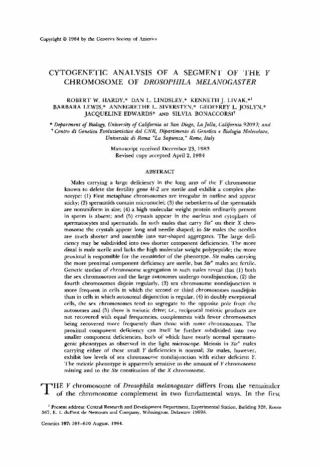
Copyright 0 1984 by the Genetics Society of America
CYTOGENETIC ANALYSIS OF A SEGMENT OF T H E Y CHROMOSOME OF DROSOPHILA MELANOGASTER
ROBERT W. HARDY,* DAN L. LINDSLEY,* KENNETH J. LIVAK,*'
JACQUELINE EDWARDS* AND SILVIA BONACCORSIt
* Department of Biology, University of Calfornia at San Diego, La Jolla, Calfornia 92093; and t Centro di Genetica Euoluzionistica del CNR, Dipartimento di Genetica e Biologia Molecolare,
BARBARA LEWIS,* ANNEGRETHE L. SIVERSTEN,* GEOFFREY L. JOSLYN,*
Universita di Roma "La Sapienza, Rome, Italy
Manuscript received December 23, 1983 Revised copy accepted April 2, 1984
ABSTRACT
Males carrying a large deficiency in the long arm of the Y chromosome known to delete the fertility gene kl-2 are sterile and exhibit a complex phe- notype: (1) First metaphase chromosomes are irregular in outline and appear sticky; (2) spermatids contain micronuclei; (3) the nebenkerns of the spermatids are nonuniform in size; (4) a high molecular weight protein ordinarily present in sperm is absent; and ( 5 ) crystals appear in the nucleus and cytoplasm of spermatocytes and spermatids. In such males that carry Ste+ on their X chro- mosome the crystals appear long and needle shaped; in Ste males the needles are much shorter and assemble into star-shaped aggregates. The large defi- ciency may be subdivided into two shorter component deficiencies. The more distal is male sterile and lacks the high molecular weight polypeptide; the more proximal is responsible for the remainder of the phenotype. Ste males carrying the more proximal component deficiency are sterile, but Ste+ males are fertile. Genetic studies of chromosome segregation in such males reveal that (1) both the sex chromosomes and the large autosomes undergo nondisjunction, (2) the fourth chromosomes disjoin regularly, (3) sex chromosome nondisjunction is more frequent in cells in which the second or third chromosomes nondisjoin than in cells in which autosomal disjunction is regular, (4) in doubly exceptional cells, the sex chromosomes tend to segregate to the opposite pole from the autosomes and (5) there is meiotic drive; i.e., reciprocal meiotic products are not recovered with equal frequencies, complements with fewer chromosomes being recovered more frequently than those with more chromosomes. The proximal component deficiency can itself be further subdivided into two smaller component deficiencies, both of which have nearly normal spermato- genic phenotypes as observed in the light microscope. Meiosis in Ste+ males carrying either of these small Y deficiencies is normal; Ste males, however, exhibit low levels of sex chromosome nondisjunction with either deficient Y. The meiotic phenotype is apparently sensitive to the amount of Y chromosome missing and to the Ste constitution of the X chromosome.
HE Y chromosome of Drosophila melanogaster differs from the remainder T of the chromosome complement in two fundamental ways. In the first
' Present address: Central Research and Development Department, Experimental Station, Building 328, Room 367, E. 1. duPont de Nemours and Company, Wilmington, Delaware 19898.
Genetics 107: 591-610 August, 1984.
592 R. W. HARDY ET AL.
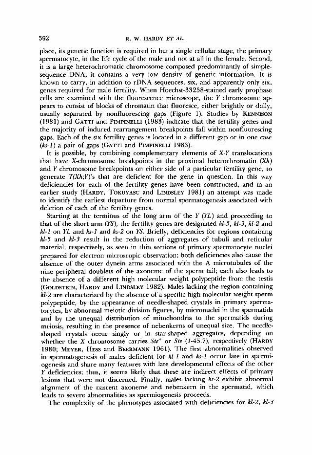
place, its genetic function is required in but a single cellular stage, the primary spermatocyte, in the life cycle of the male and not at all in the female. Second, it is a large heterochromatic chromosome composed predominantly of simple- sequence DNA; it contains a very low density of genetic information. It is known to carry, in addition to rDNA sequences, six, and apparently only six, genes required for male fertility. When Hoechst-33258-stained early prophase cells are examined with the fluorescence microscope, the Y chromosome ap- pears to consist of blocks of chromatin that fluoresce, either brightly or dully, usually separated by nonfluorescing gaps (Figure 1). Studies by KENNISON (1981) and GATTI and PIMPINELLI (1983) indicate that the fertility genes and the majority of induced rearrangement breakpoints fall within nonfluorescing gaps. Each of the six fertility genes is located in a different gap or in one case (ks-1) a pair of gaps (GATTI and PIMPINELLI 1983).
It is possible, by combining complementary elements of X-Y translocations that have X-chromosome breakpoints in the proximal heterochromatin (Xh) and Y chromosome breakpoints on either side of a particular fertility gene, to generate T(Xh;Y)'s that are deficient for the gene in question. In this way deficiencies for each of the fertility genes have been constructed, and in an earlier study (HARDY, TOKUYASU and LINDSLEY 1981) an attempt was made to identify the earliest departure from normal spermatogenesis associated with deletion of each of the fertility genes.
Starting at the terminus of the long arm of the Y (YL) and proceeding to that of the short arm (YS), the fertility genes are designated kl-5, kl-3, kl-2 and kl-1 on YL and ks-1 and ks-2 on YS. Briefly, deficiencies for regions containing kl-5 and kl-3 result in the reduction of aggregates of tubuli and reticular material, respectively, as seen in thin sections of primary spermatocyte nuclei prepared for electron microscopic observation; both deficiencies also cause the absence of the outer dynein arms associated with the A microtubules of the nine peripheral doublets of the axoneme of the sperm tail; each also leads to the absence of a different high molecular weight polypeptide from the testis (GOLDSTEIN, HARDY and LINDSLEY 1982). Males lacking the region containing kl-2 are characterized by the absence of a specific high molecular weight sperm polypeptide, by the appearance of needle-shaped crystals in primary sperma- tocytes, by abnormal meiotic division figures, by micronuclei in the spermatids and by the unequal distribution of mitochondria to the spermatids during meiosis, resulting in the presence of nebenkerns of unequal size. The needle- shaped crystals occur singly or in star-shaped aggregates, depending on whether the X chromosome carries Ste+ or Ste (1-45.7), respectively (HARDY 1980; MEYER, H ~ s s and BEERMANN 1961). The first abnormalities observed in spermatogenesis of males deficient for kl-1 and ks-1 occur late in spermi- ogenesis and share many features with late developmental effects of the other Y deficiencies; thus, it seems likely that these are indirect effects of primary lesions that were not discerned. Finally, males lacking ks-2 exhibit abnormal alignment of the nascent axoneme and nebenkern in the spermatid, which leads to severe abnormalities as spermiogenesis proceeds.
The complexity of the phenotypes associated with deficiencies for kl-2, kl-3
ANALYSIS OF T H E Y CHROMOSOME 593
I BSXhi 5 6 7 12 13 17 18 22 23 250
t c FIGURE 1 .-Cytological map of the dYy’ chromosome (after GAITI and PIMPINELLI 1983). T h e
upper figure is a fluorescence microscope photograph of a Hoechst-33258-stained prophase Y chromosome. YL is to the left. T h e lower figure represents the linear differentiation of the Y chromosome as determined by CAW and PIMPINELLI (1983) utilizing a variety of staining tech- niques coupled with genetic analysis. T h e arrowheads on each figure delimit the Y chromosome region presented in greater detail in Figure 3.
and &I-5 are conceivably explicable in terms of pleiotropism; however, the apparent unrelatedness of the different aspects of the phenotypes suggests genetic complexity within the segments deleted. In this communication we describe experiments in which we subdivide the original RI-2 deletion into smaller component deficiencies; we show that the determinant(s) of male fer- tility and the sperm protein is separable from and distal to that of crystal formation, meiosis and mitochondrial distribution.
MATERIALS AND METHODS
Male-sterile Y chromosomes: Y chromosomes bearing male-sterile mutations constitute useful ma- terial for the investigation of Y chromosome orpni7ation. For this study we have chosen male- sterile Y chromosomes [ms(Y)] that a re defective in hl-2 (see Table 1). ms(Y)’s initially selected as defective in hl-2 alone a re designated E 5 . E 6 . E7 , ER. C 7 . G 8 , L37 and SIR; of these, E5 and E7 subsequently lost hl-3 and hl-5, and in addition E5 lost the YL terminus marked by B‘. E6 and ER lost hl-5; E17 . which was recovered as h 1 - Y . subsequently lost hl-2. Y chromosomes simultaneously defective for both hl-2 and h1-3 are G I 9 and G20 and S16; ms(Y)’s defective for hl-2, hl-3 and h1- 5 a re G21. G22, G23 and G24. ms(Y)’s prefixed with letters E and G were induced in B‘Yy+ by ethyl methanesulfonate (EMS) or y-irradiation, respectively (KENNISON 1983). ms(Y)L37 is one of Brosseau’s (1960) original mutations induced in y+Y by irradiation; S I 6 and SIR were induced in y+Y with y rays by MARCIA .%HWARTZ. S I 6 carries two paracentric inversions: /n(YL)F16 = /n(YL)h7;hII + /n (YL)h /3 ;h17 . and S I R is a pericentric inversion: /n(YLS)SIR = /n(YLS)h10-ll;h20 (GAI-I-I and PIMPINELLI 1983).
Male fertile Xh-Y franslocahns: T h e X-Y translocations employed in this study have breakpoints in the proximal heterochromatin of the X chromosome and in the long arm of the Y. T h e two elements of these translocations a re PX‘, the terminus of YL appended to the heterochromatic centric portion of the X. and X”y, the entire euchromatin plus a portion of the proximal heter- ochromatin of the X appended to the centric portion of the Y consisting of the portion of YL proximal to the translocation breakpoint and YS. Since ou r interest in this communication is in YL, henceforth we will frequently abbreviate p X p as Y” and X”Yp as Yp.
In the study of the ultrastructural lesions associated with deficiencies for individual fertility
594 R. W. HARDY E T A L .
kl- 1 ks-2 y+ Yp
0- U
Y o Bs kl-5
0-0
0- 0
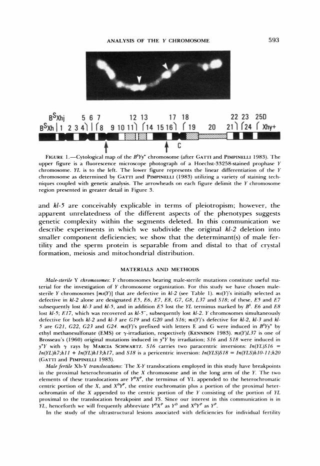
FIGURE 2.-Translocations used in the production of deficiencies of the kl-2 region by the method of segmental aneuploidy. The upper translocation is T(Xh;YL)E15 and the lower one T(Xh;YL;3R)W27. In the past (e.g., HARDY, TOKUYASIJ and LINDSLEY 1981) W27 was considered a simple reciprocal Xh-YL translocation; however, it is actually a four-break rearrangement with the following new order: 1-20Flh20-hlOlh20-Xhy+; 20F191A-100; Xh@-h9191A-61. (The segment be- tween the arrows is inverted, although the inverted sequence is omitted from the diagram). Males carrying P 3 R p and 3RDXp from W27 in combination with XDy from E15 are specifically deficient for kl-2 and flanking sequences. Solid wide lines, Y chromosome; open wide lines, X heterochro- matin; hatching, ribosomal cistrons; thin solid line, X euchromatin; thin dashed line, third chro- mosome. Chromosome arms are not represented in proportion to their lengths. Among the other translocations employed, VI 7 and V63 also involve autosomes. T(Xh;YL;3L)Vl7 = T(XY;3)20F;h13- 14;75F-76A; new order: 1-20FI hl4-Xhy+; 20FI 75F-61; X@-hl3176A-100. T(Xh;YL;A)V63 = T(XY;A)20F;h13-14; A, new order: l-20FIh14-Xhy+;20FIAD;h131AP. In segmental deficiencies derived from simple reciprocal translocations, an autosome is not involved, and P A p + ADXP is replaced by PXp. In this communication P A p + ADXP of complex translocations are treated as if they were PXp, since the autosomal involvement does not affect the results.
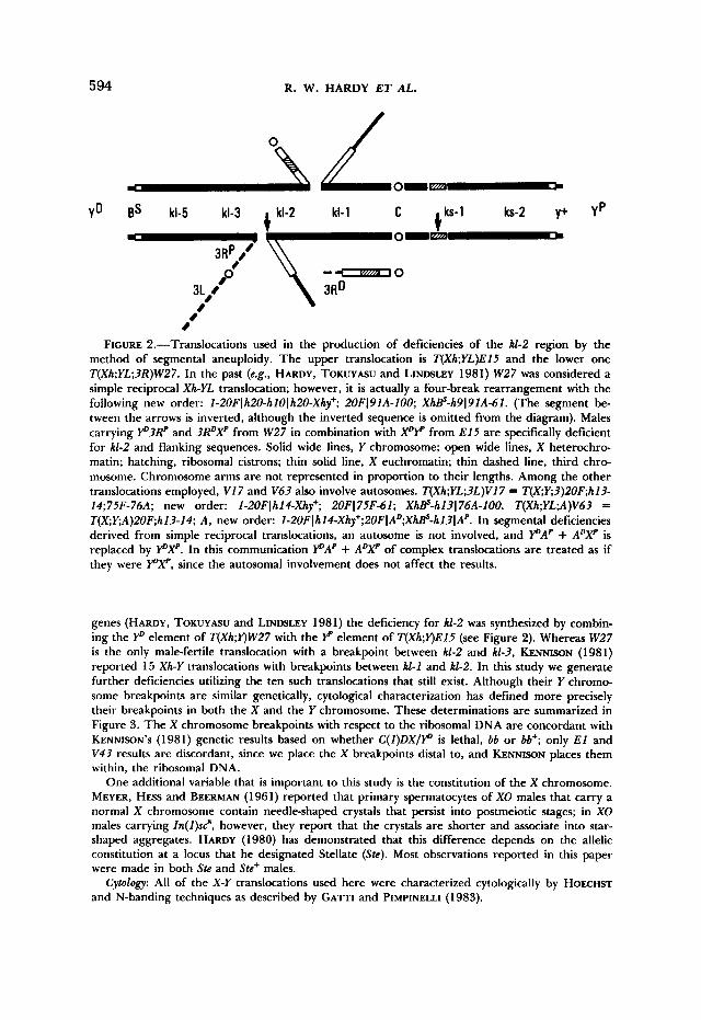
genes (HARDY, TOKUYASIJ and LINDSLEY 1981) the deficiency for kl-2 was synthesized by combin- ing the P element of T(Xh;Y)W27 with the element of T(Xh;Y)E15 (see Figure 2). Whereas W27 is the only male-fertile translocation with a breakpoint between RI-2 and kl-3, KENNISON (1981) reported 15 Xh-Y translocations with breakpoints between kl-1 and kl-2. In this study we generate further deficiencies utilizing the ten such translocations that still exist. Although their Y chromo- some breakpoints are similar genetically, cytological characterization has defined more precisely their breakpoints in both the X and the Y chromosome. These determinations are summarized in Figure 3. The X chromosome breakpoints with respect to the ribosomal DNA are concordant with KENNISON'S (1981) genetic results based on whether C(l)DX/r" is lethal, bb or bb+; only E l and V43 results are discordant, since we place the X breakpoints distal to, and KENNWN places them within, the ribosomal DNA.
One additional variable that is important to this study is the constitution of the X chromosome. MEYER, H ~ s s and BEERMAN (1961) reported that primary spermatocytes of XO males that carry a normal X chromosome contain needle-shaped crystals that persist into postmeiotic stages; in XO males carrying ln(l)scs, however, they report that the crystals are shorter and associate into star- shaped aggregates. HARDY (1980) has demonstrated that this difference depends on the allelic constitution at a locus that he designated Stellate (Ste). Most observations reported in this paper were made in both Ste and Ste+ males.
Cytology: All of the X-Y translocations used here were characterized cytologically by HOECHST and N-banding techniques as described by GATTI and PIMPINELLI (1983).
ANALYSIS OF THE Y CHROMOSOME
2L1 H 1 h9 h10 h l l h12 h13 h14 h15 4
595
b
NO
W27 0
P7 N29 0 0
V17 0
V3 1 0
V63 E15 0 .
0
E l 68 625 0 0 .
v43
FIGURE 3.-Distribution of heterochromatic breakpoints of Xh-YL translocations. The X heter- ochromatin is represented on the ordinate, and the segment of Y bounded by arrows in Figure 1 is represented on the abscissa. Solid segments fluoresce brightly, and hatched segments fluoresce dully in Hoechst-33258-stained preparations. The line labeled 2L1 indicates the location of DNA sequences described in DISCUSSION and by LIVAK (1984). C, centromere; NO, ribosomal cistrons; Xe, X euchromatin.
RESULTS
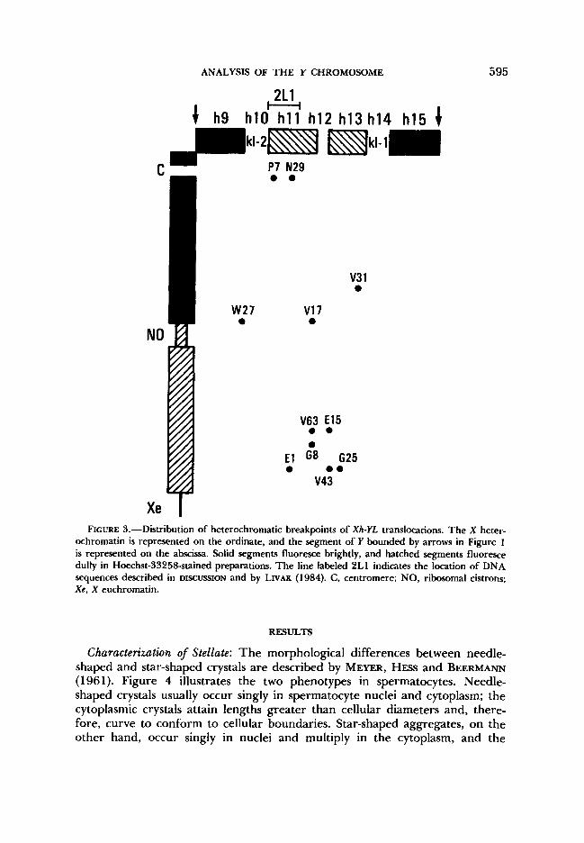
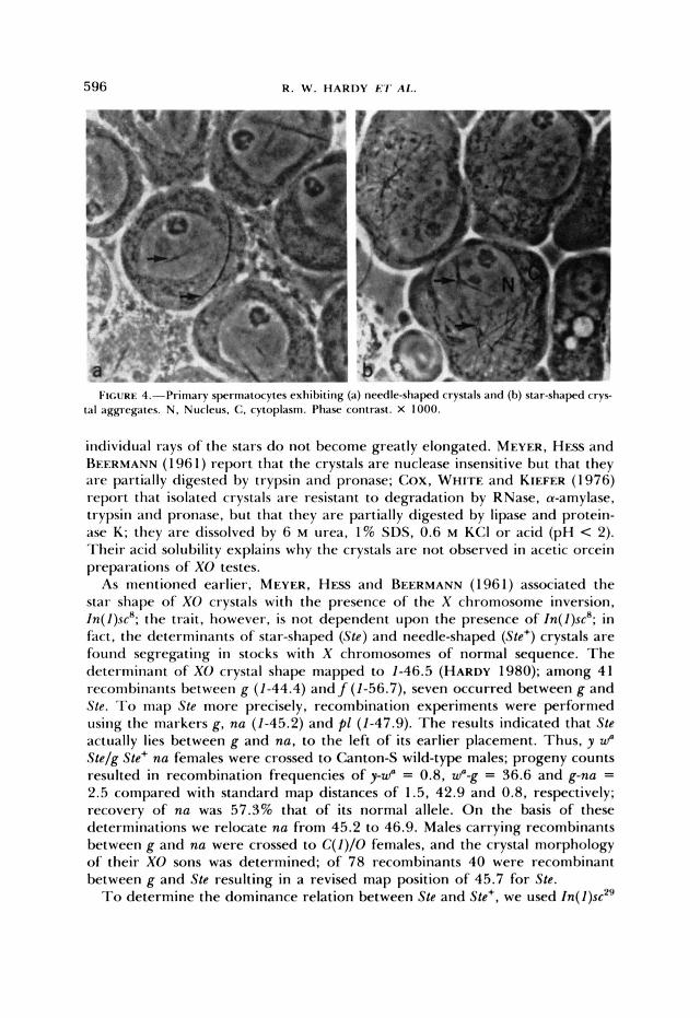
Characterization of Stellate: The morphological differences between needle- shaped and star-shaped crystals are described by MEYER, HESS and BEERMANN (1 96 1). Figure 4 illustrates the two phenotypes in spermatocytes. Needle- shaped crystals usually occur singly in spermatocyte nuclei and cytoplasm; the cytoplasmic crystals attain lengths greater than cellular diameters and, there- fore, curve to conform to cellular boundaries. Star-shaped aggregates, on the other hand, occur singly in nuclei and multiply in the cytoplasm, and the
596 R. W . HARDY E?' AI..
I ~ U R E 4.-hiniary spermitocytcs exhit>iting (a) needle-shaped crystals and (b) star-shapcd c'rvs- tal aggregates. N . Nucleus, C, cytoplasm. I'hasC' contrast. X 1000.
individual rays of the stars do not become greatly elongated. MEYER. HESS and REERMANN (1 96 1) report that the crystals a re nuclease insensitive but that they are partially digested by trypsin and pronase; Cox, WHITE and KIEFER ( 1 976) report that isolated crystals are resistant to degradation by RNase, a-amylase, trypsin and pronase, but that they are partially digested by lipase and protein- ase K; they a re dissolved by 6 M urea, 1% SDS, 0.6 M KCI or acid (pH < 2). Their acid solubility explains why the crystals are not observed in acetic orcein preparations of XO testes.
As mentioned earlier, MEYER, H ~ s s and BEERMANN (1961) associated the star shape of XO crystals with the presence of the X chromosome inversion, In(1)sc"; the trait, however, is not dependent upon the presence of In(1)sc'; in fact. the determinants of star-shaped (Ste) and needle-shaped (.$le+) crystals a re found segregating in stocks with X chromosomes of normal sequence. T h e determinant of XO crystal shape mapped to 1-46.5 (HARDY 1980); among 41 recombinants between g (1-44.4) and f (1-56.7), seven occurred between g and Ste. To map Ste more precisely, recombination experiments were performed using the markers g, nu (1-45.2) and p l (1-47.9). T h e results indicated that Ste actually lies between g and nu, to the left of its earlier placement. Thus, y WO Stc/g Ste+ na females were crossed to Canton-S wild-type males; progeny counts resulted in recombination frequencies of y-w" = 0.8, WO-g = 36.6 and g-nu = 2.5 compared with standard map distances of 1.5. 42.9 and 0.8, respectively; recovery of nu was 57.3% that of its normal allele. O n the basis of these determinations we relocate nu from 45.2 to 46.9. Males carrying recombinants between g and nu were crossed to C ( I ) / O females, and the crystal morphology of their XO sons was determined; of 78 recombinants 40 were recombinant between g and Ste resulting in a revised map position of 45.7 for Stc.
To determine the dominance relation between Ste and Stc+, we used In( I ) S C * ~
ANALYSIS OF THE Y CHROMOSOME 597
= Zn(Z)ZB;23A2-5 to construct a free duplication carrying the g-nu region. X- ray-induced deletion of most of the euchromatin results in a centric fragment containing the tip of the X marked with y+, the region around 13A, and X heterochromatin. Thus, Zn(2)sc2'/Y males were irradiated with 4000 r and crossed to YSX-YL, Zn(Z)EN, y B females. Exceptional y+ sons were crossed individually to YSX-YL, Zn(Z)EN, y B females to establish stocks and then to g nu/FA43 females to test for the presence of g+ and nu+. Of 12 y+ males, one transmitted a duplication designated Dp(ZfiLJ9 that carries both g" and nu+. (Two other duplications were also generated; Dp( 2;flLJ4 carries na+, and Dp( 2;f)LJ8 carries nu+ and produces variegation for the g phenotype.) The original Zn(I)sc2' chromosome carries Ste+, the determinant for needle-shaped crystals; thus, Dp( 2;f )LJ9 presumably carries Ste+. This was confirmed by using two deficiencies: DflI)HA92 = Dfl2)22A7;22D3, which uncovers g, and Dfl2)KAB = Dfl2)22E1;23A5, which uncovers nu. Males of genetic constitution DflZ)HA92/Dp(ZfiLJ9 and DflI)KA9/Dp( I fiLJ9 both survive and have needle- shaped crystals in their spermatocytes, indicating that Dp( 2;j)LJ9 carries Ste+, assuming one of the deficiencies uncovers Ste. Cytological analysis of polytene chromosomes confirms that Dp(ZfiLJ9 consists of the region 12A6-10 to 13A2- 5 capped by the tip of the X. Thus, the full designation of the duplication is Dp(l;f)LJ9, y+ sc2' g+ Ste+ nu+ = Dp(2fi2B;22A6-2O;I3A2-5;20, and the new order is ZA-ZBl23A2-22A20120. With regard to the dominance relation, XO males of genetic constitution g2 Ste+ sd f/Dp(Z;f)LJ9, Ste+ and y w" Stel Dp(l;f)LJ9, Ste+ were examined. It was found that Ste+/Ste+ produces needle- shaped crystals in XO spermatocytes and Ste/Ste+ produces star-shaped crystals. Thus, Ste is dominant to Ste+.
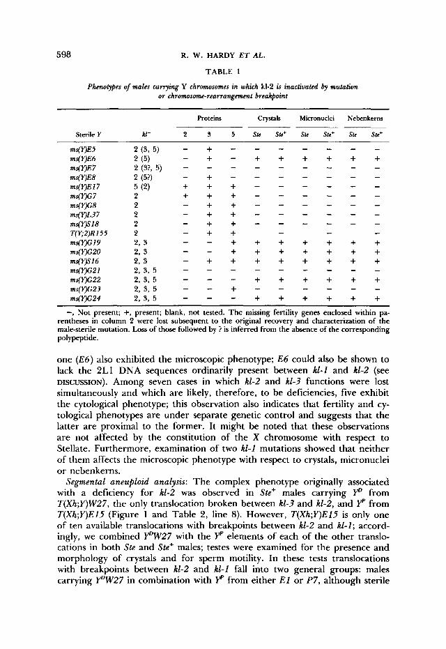
Analysis of kl-2-defctive Y chromosomes: Each of the kZ-2-defective Y chromo- somes described in MATERIALS AND METHODS was combined with X chromo- somes marked with y w Ste+ and y w" Ste. Testes of the resultng males were examined for the presence of high molecular weight polypeptides by polyac- rylamide electrophoresis as described in GOLDSTEIN, HARDY and LINDSLEY (1 982) and by phase-contrast microscopy of living testes in saline for the pres- ence of crystals in the primary spermatocytes and for micronuclei and nebenk- erns of nonuniform size in spermatids. The results of these determinations are presented in Table 1. Three features of the data are noteworthy: First, the majority of kZ-2 mutants lack the kZ-2 polypeptide but do not exhibit crystals, micronuclei or nonuniform nebenkerns; thus, the kZ-2 polypeptide is mutation- ally separable from crystals, micronuclei and nonuniform nebenkerns. The second notable feature is the complete correlation among the three aspects of the microscopic phenotype and the strong correlation between sterility and the absence of kZ-2 polypeptide. These correlations indicate a commonality of cause. We infer that the kl-2 polypeptide is necessary for male fertility and alteration (postulated in the cases of E27 and G7) or elimination of the poly- peptide leads to male sterility; furthermore, crystals, micronuclei and nonuni- form nebenkerns seem to result from a lesion in a single genetic determinant. Finally, we note that among ten cases in which kZ-2 was inactivated by itself and which are, therefore, the most likely candidates for point mutations, only
598 R. W. HARDY E T AL.
TABLE 1
Phenotypes of males currying Y chromosomes in which kl-2 is inactivated by mutation or chromosom-rearrangement breakpoint
Proteins Crystals Micronuclei Nebenkerns
Sterile Y kl- 2 3 5 Ste Ste+ Ste Ste+ Ste Ste+
- - - - - - “ E 5 2 ( 3 , 5 ) - + - “ E 6 2 ( 5 ) ms(VE7 ms(VE8 2 (5?) - + - ms(VEl7 5 (2) ms( Y)G 7 2 + + + - ms(Y)c8 2 - + + - - - - - - ms(VL37 2 - + + - - - - - - ms(Y)SlS 2 T(Y;2)R155 2 - + + - - - ms(r)G19 2 , 3 - - + + + + + + + ms(Y)G20 2, 3 - - + + + + + + +
2, 3 - + + + + + + + + “ W 1 6 2, 3, 5
ms(k7G22 2, 3 , 5 - - - + + + + + + m s ( w 2 1
ms(qG23 2, 3 , 5 ms(Y)c24 2, 3, 5 - - - + + + + + +
- + - + + + + + + - - - - - - - - 2 (3?, 5 ) -
- - - - - - + + + - - - - - -
- - - - -
- + + - - - - - -
- - - - - - - - -
- - + - - - - - -
-, Not present; +, present; blank, not tested. The missing fertility genes enclosed within pa- rentheses in column 2 were lost subsequent to the original recovery and characterization of the male-sterile mutation. Loss of those followed by ? is inferred from the absence of the corresponding polypeptide.
one ( E 6 ) also exhibited the microscopic phenotype; E 6 could also be shown to lack the 2L1 DNA sequences ordinarily present between kl-1 and k1-2 (see DISCUSSION). Among seven cases in which k1-2 and kl-3 functions were lost simultaneously and which are likely, therefore, to be deficiencies, five exhibit the cytological phenotype; this observation also indicates that fertility and cy- tological phenotypes are under separate genetic control and suggests that the latter are proximal to the former. It might be noted that these observations are not affected by the constitution of the X chromosome with respect to Stellate. Furthermore, examination of two kl-1 mutations showed that neither of them affects the microscopic phenotype with respect to crystals, micronuclei or nebenkerns.
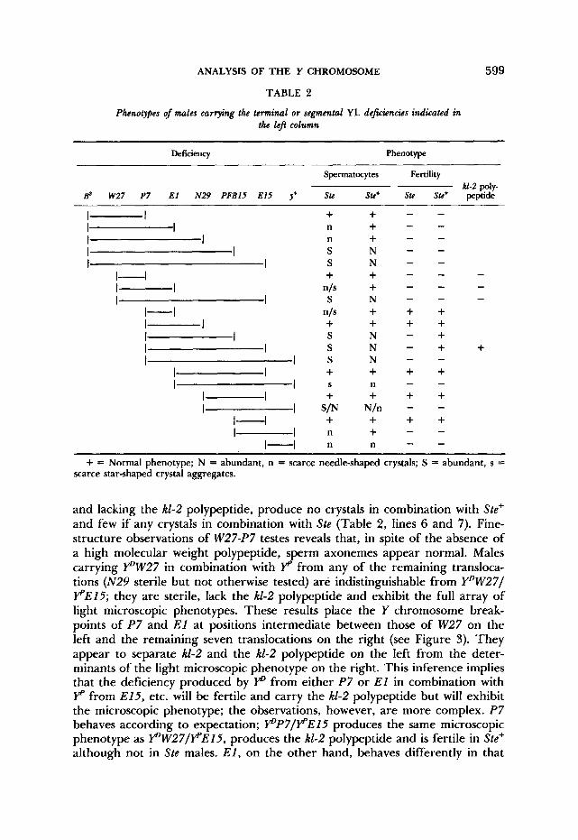
Segmental aneuploid analysis: The complex phenotype originally associated with a deficiency for kl-2 was observed in Ste+ males carrying p from T(Xh;Y)W27, the only translocation broken between kl-3 and kl-2, and YP from T(Xh;Y)ElS (Figure 1 and Table 2, line 8). However, T ( X h ; Y ) E l S is only one of ten available translocations with breakpoints between kl-2 and k1-I; accord- ingly, we combined p W 2 7 with the YP elements of each of the other translo- cations in both Ste and Ste+ males; testes were examined for the presence and morphology of crystals and for sperm motility. In these tests translocations with breakpoints between kl-2 and kl-1 fall into two general groups: males carrying Y“W27 in combination with YP from either E l or P7, although sterile
ANALYSIS OF THE Y CHROMOSOME
TABLE 2
Phenotypes of males carrying the terminal or segmental YL deficiencies indicated in the l e j column
599
Deficiency Phenotype
Spermatocytes Fertility kl-2 poly-
Bs W27 P7 E l N29 PFBlS E15 J+ Stc Ste+ Stc Stc+ peptide
I I I I I I I I I I
1-1 I I I I
1-1 I I I I I I I I
I I I I
I I I I
I 4 I I
1-1
+ n n S S + S
+ S S S + + + n n
n/s
n/s
S
SIN
- - - + N + + + + + + N - + N N + + + n + + + + + +
- - -
+ + - - -
- -
N/n - -
+ = Normal phenotype; N = abundant, n = scarce needle-shaped crystals; S = abundant, s = scarce star-shaped crystal aggregates.
and lacking the kl-2 polypeptide, produce no crystals in combination with Ste’ and few if any crystals in combination with Ste (Table 2 , lines 6 and 7). Fine- structure observations of W27-P7 testes reveals that, in spite of the absence of a high molecular weight polypeptide, sperm axonemes appear normal. Males carrying PW27 in combination with YP from any of the remaining transloca- tions (N29 sterile but not otherwise tested) are indistinguishable from Y”W27I PE15; they are sterile, lack the k1-2 polypeptide and exhibit the full array of light microscopic phenotypes. These results place the Y chromosome break- points of P7 and El at positions intermediate between those of W27 on the left and the remaining seven translocations on the right (see Figure 3). They appear to separate kl-2 and the kl-2 polypeptide on the left from the deter- minants of the light microscopic phenotype on the right. This inference implies that the deficiency produced by YD from either P7 or E l in combination with YP from E15, etc. will be fertile and carry the AI-2 polypeptide but will exhibit the microscopic phenotype; the observations, however, are more complex. P7 behaves according to expectation; pP7IFE15 produces the same microscopic phenotype as pW27IFEl5 , produces the kl-2 polypeptide and is fertile in Ste+ although not in Ste males. E l , on the other hand, behaves differently in that
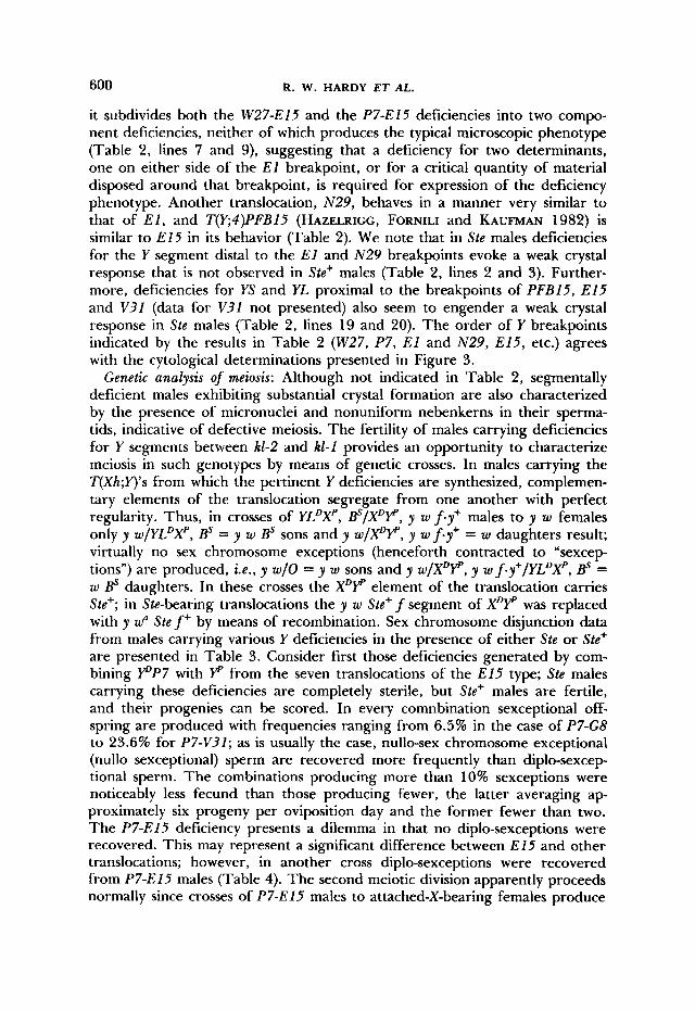
600 R. W. HARDY ET AL.
it subdivides both the W27-El5 and the P7-El5 deficiencies into two compo- nent deficiencies, neither of which produces the typical microscopic phenotype (Table 2, lines 7 and 9), suggesting that a deficiency for two determinants, one on either side of the E l breakpoint, or for a critical quantity of material disposed around that breakpoint, is required for expression of the deficiency phenotype. Another translocation, N29, behaves in a manner very similar to that of E l , and T(E4)PFBlS (HAZELRIGG, FORNILI and KAUFMAN 1982) is similar to E15 in its behavior (Table 2). We note that in Ste males deficiencies for the Y segment distal to the E l and N 2 9 breakpoints evoke a weak crystal response that is not observed in Ste+ males (Table 2 , lines 2 and 3). Further- more, deficiencies for YS and YL proximal to the breakpoints of PFBl5 , E15 and V31 (data for V32 not presented) also seem to engender a weak crystal response in Ste males (Table 2 , lines 19 and 20). The order of Y breakpoints indicated by the results in Table 2 (W27, P7 , E l and N29, E15, etc.) agrees with the cytological determinations presented in Figure 3.
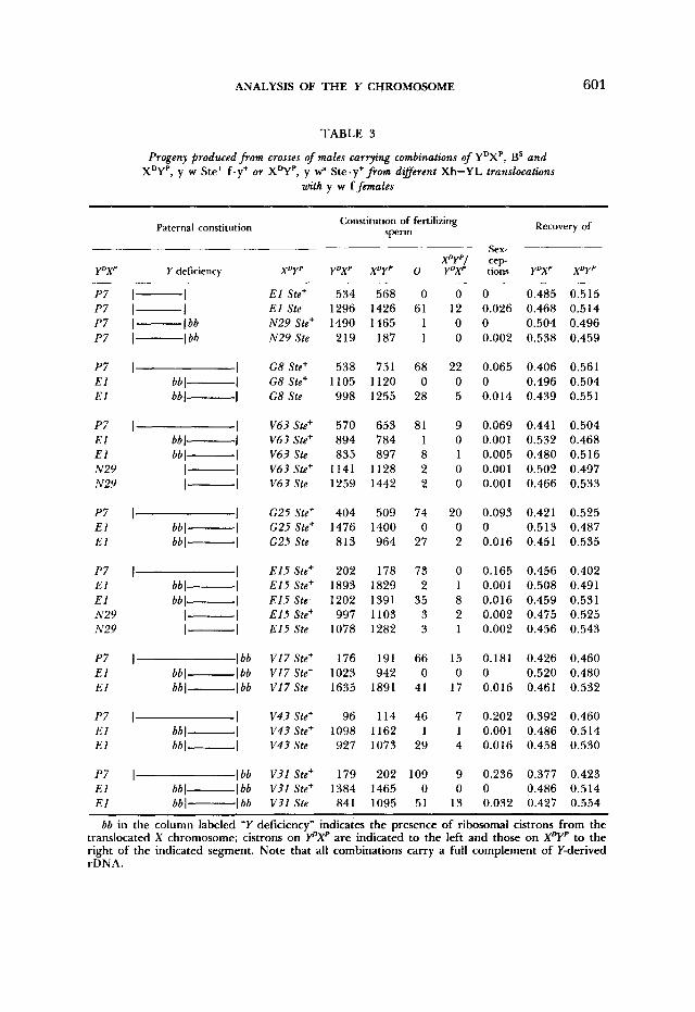
Genetic analysis of meiosis: Although not indicated in Table 2, segmentally deficient males exhibiting substantial crystal formation are also characterized by the presence of micronuclei and nonuniform nebenkerns in their sperma- tids, indicative of defective meiosis. The fertility of males carrying deficiencies for Y segments between kl-2 and k1-1 provides an opportunity to characterize meiosis in such genotypes by means of genetic crosses. In males carrying the T(Xh;Y)'s from which the pertinent Y deficiencies are synthesized, complemen- tary elements of the translocation segregate from one another with perfect regularity. Thus, in crosses of YLDXp, @ / X D y , y w fey' males to y w females only y w/YLDXp, BS = y w BS sons and y w/XDy, y w f.y+ = w daughters result; virtually no sex chromosome exceptions (henceforth contracted to "sexcep- tions") are produced, i .e. , y w/O = y w sons and y w/XDF, y w f-y+/YLDXp, @ = w BS daughters. In these crosses the XDY" element of the translocation carries Ste+; in Ste-bearing translocations the y w Ste+ f segment of XDy was replaced with y w" S t e f + by means of recombination. Sex chromosome disjunction data from males carrying various Y deficiencies in the presence of either Ste or Ste+ are presented in Table 3. Consider first those deficiencies generated by com- bining P P 7 with YP from the seven translocations of the E15 type; Ste males carrying these deficiencies are completely sterile, but Ste+ males are fertile, and their progenies can be scored. In every comnbination sexceptional off- spring are produced with frequencies ranging from 6.5% in the case of P7-G8 to 23.6% for P7-V31; as is usually the case, nullo-sex chromosome exceptional (nullo sexceptional) sperm are recovered more frequently than diplo-sexcep tional sperm. The combinations producing more than 10% sexceptions were noticeably less fecund than those producing fewer, the latter averaging a p proximately six progeny per oviposition day and the former fewer than two. The P7-El5 deficiency presents a dilemma in that no diplo-sexceptions were recovered. This may represent a significant difference between E15 and other translocations; however, in another cross diplo-sexceptions were recovered from P7-El5 males (Table 4). The second meiotic division apparently proceeds normally since crosses of P7-El5 males to attached-X-bearing females produce
ANALYSIS OF THE Y CHROMOSOME 601
TABLE 3
Progeny produced from crosses of males carrying combinations of YDXP, BS and XDYP, y w Ste+ f.y+ or XDYP, y wa Ste.y+ from dafferent Xh-YL translocations
with y w f females
Paternal constitution Constitution of fertilizing
sperm Recovery of
Sex-
Y D X P
P7 P7 P7 P7
P7 E l E l
P7 E l E l N 2 9 N 2 9
P7 E l E l
P7 E l E l N 2 9 N 2 9
P7 E l E l
P7 E l E l
P7 E l E l
~
I- I l- I
I bb I- I bb I-
I I bbl- I bbl- I
I I bb I- I bbl- I
I- I I- I
I I bbl- I bbl- I
I I bb 1- I bbl- I
I- I l- I
E l Ste+ E l Ste N 2 9 Ste+ N 2 9 Ste
G 8 Ste+ G 8 Ste+ G 8 Ste
V6? Ste' V 6 3 Ste' V 6 3 Ste V 6 3 Ste' V 6 3 Ste
G25 Ste+ G25 Ste' G 2 5 Ste
E 15 Ste+ E 15 Ste+ E15 Ste E15 Ste+ E15 Ste
V17 Ste+ VI 7 Ste+ V17 Ste
V 4 3 Ste+ V4? Ste+ 1143 Ste
V31 Ste+ V31 Ste+ V31 Ste
534 568 0 1296 1426 61 1490 1465 1 219 187 1
538 751 68 1105 1120 0 998 1255 28
570 653 81 894 784 1 835 897 8
1141 1128 2 1259 1442 2
404 509 74 1476 1400 0 813 964 27
202 178 73 1893 1829 2 1202 1391 35 997 1103 3
1078 1282 3
176 191 66 1023 942 0 1635 1891 41
96 114 46 1098 1162 1 927 1073 29
179 202 109 1384 1465 0 841 1095 51
0 12 0 0
22 0 5
9 0 1 0 0
20 0 2
0 1 8 2 1
15 0
17
7 1 4
9 0
13
0 0.485 0.515 0.026 0.468 0.514 0 0.504 0.496 0.002 0.538 0.459
0.065 0.406 0.561 0 0.496 0.504 0.014 0.439 0.551
0.069 0.441 0.504 0.001 0.532 0.468 0.005 0.480 0.516 0.001 0.502 0.497 0.001 0.466 0.533
0.093 0.421 0.525 0 0.513 0.487 0.016 0.451 0.535
0.165 0.456 0.402 0.001 0.508 0.491 0.016 0.459 0.531 0.002 0.475 0.525 0.002 0.456 0.543
0.181 0.426 0.460 0 0.520 0.480 0.016 0.461 0.532
0.202 0.392 0.460 0.001 0.486 0.514 0.016 0.458 0.530
0.236 0.377 0.423 0 0.486 0.514 0.032 0.427 0.554
bb in the column labeled "Y deficiency" indicates the presence of ribosomal cistrons from the translocated X chromosome; cistrons on Y"Xp are indicated to the left and those on XDYp to the right of the indicated segment. Note that all combinations carry a full complement of Y-derived rDNA.
602 R. W. HARDY ET AL.
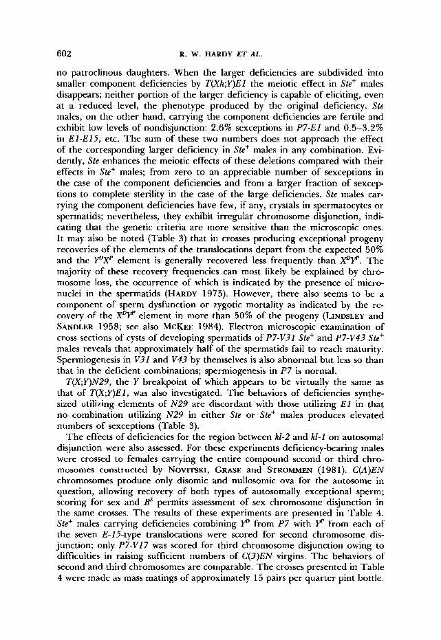
no patroclinous daughters. When the larger deficiencies are subdivided into smaller component deficiencies by T(Xh;Y)El the meiotic effect in Ste+ males disappears; neither portion of the larger deficiency is capable of eliciting, even at a reduced level, the phenotype produced by the original deficiency. Ste males, on the other hand, carrying the component deficiencies are fertile and exhibit low levels of nondisjunction: 2.6% sexceptions in P7-El and 0.5-3.2% in El-E15, etc. The sum of these two numbers does not approach the effect of the corresponding larger deficiency in Ste+ males in any combination. Evi- dently, Ste enhances the meiotic effects of these deletions compared with their effects in Ste+ males; from zero to an appreciable number of sexceptions in the case of the component deficiencies and from a larger fraction of sexcep- tions to complete sterility in the case of the large deficiencies. Ste males car- rying the component deficiencies have few, if any, crystals in spermatocytes or spermatids; nevertheless, they exhibit irregular chromosome disjunction, indi- cating that the genetic criteria are more sensitive than the microscopic ones. It may also be noted (Table 3) that in crosses producing exceptional progeny recoveries of the elements of the translocations depart from the expected 50% and the PXp element is generally recovered less frequently than XDP. The majority of these recovery frequencies can most likely be explained by chro- mosome loss, the occurrence of which is indicated by the presence of micro- nuclei in the spermatids (HARDY 1975). However, there also seems to be a component of sperm dysfunction or zygotic mortality as indicated by the re- covery of the XDY element in more than 50% of the progeny (LINDSLEY and SANDLER 1958; see also MCKEE 1984). Electron microscopic examination of cross sections of cysts of developing spermatids of P7-V31 Ste+ and P7-V43 Ste+ males reveals that approximately half of the spermatids fail to reach maturity. Spermiogenesis in V31 and V43 by themselves is also abnormal but less so than that in the deficient combinations; spermiogenesis in P7 is normal.
T(XY)N29 , the Y breakpoint of which appears to be virtually the same as that of T ( X Y ) E l , was also investigated. The behaviors of deficiencies synthe- sized utilizing elements of N 2 9 are discordant with those utilizing E l in that no combination utilizing N29 in either Ste or Ste+ males produces elevated numbers of sexceptions (Table 3).
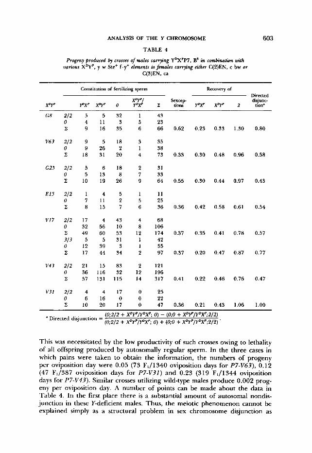
The effects of deficiencies for the region between kl-2 and kl-1 on autosomal disjunction were also assessed. For these experiments deficiency-bearing males were crossed to females carrying the entire compound second or third chro- mosomes constructed by NOVITSKI, GRASE and STROMMEN (1 98 1). C(A)EN chromosomes produce only disomic and nullosomic ova for the autosome in question, allowing recovery of both types of autosomally exceptional sperm; scoring for sex and @ permits assessment of sex chromosome disjunction in the same crosses. The results of these experiments are presented in Table 4. Ste+ males carrying deficiencies combining P from P7 with P from each of the seven E- 15-type translocations were scored for second chromosome dis- junction; only P7-Vl7 was scored for third chromosome disjunction owing to difficulties in raising sufficient numbers of C(3)EN virgins. The behaviors of second and third chromosomes are comparable. The crosses presented in Table 4 were made as mass matings of approximately 15 pairs per quarter pint bottle.
ANALYSIS OF THE Y CHROMOSOME
TABLE 4
Progeny produced by crosses of males carrying YDXPP7, Bs in combination with various X q p , y w Ste’ f.y+ elements to fmales carrying either C(2)EN, c bw or
C(3)EN, ca
603
Constitution of fertilizing sperm Recovery of Directed
xv-J Sexcep disjunc- XDYP P X p xDYp 0 Y D P z: tions YDXp x q p 2 tion’
G 8 212 0 z
V63 212 0 z
G25 212 0 z
E15 212 0 z
V I 7 212 0 z
0 z
313
v43 212 0 z
V 3 l 21.2 0 z
5 4 9
9 9
18
5 5
10
1 7 8
17 32 49 5
12 17
21 36 57
4 6
10
5 11 16
5 26 31
6 13 19
4 11 15
4 56 60
5 39 44
15 116 131
4 16 20
32 3
35
18 2
20
18 8
26
5 2 7
43 10 53 31 3
34
83 32
115
17 0
17
1 5 6
3 1 4
2 7 9
1 5 6
4 8
12 1 1 2
2 12 14
0 0 0
43 23 66 0.62 0.23 0.33 1.30 0.80
35 38 73 0.33 0.30 0.48 0.96 0.58
31 33 64 0.55 0.30 0.44 0.97 0.43
11 25 36 0.36 0.42 0.58 0.61 0.54
68 106 174 0.37 0.35 0.41 0.78 0.5’7 42 55 97 0.37 0.20 0.47 0.87 0.77
121 196 317 0.41 0.22 0.46 0.76 0.47
25 22 47 0.36 0.21 0.43 1.06 1.00
(0;2/2 + XDYP/YDXP; 0) - (0;O + XDrP/YDXP;2/2) (0;2/2 + XDYP/YDXP; 0) + (0;O + XDYP/YDXP;2/2) . Directed disjunction =
This was necessitated by the low productivity of such crosses owing to lethality of all offspring produced by autosomally regular sperm. In the three cases in which pains were taken to obtain the information, the numbers of progeny per oviposition day were 0.05 (73 FJ1340 oviposition days for P7-V63), 0.12 (47 F1/387 oviposition days for P7-V3I ) and 0.23 (319 F1/1344 oviposition days for P7-V43). Similar crosses utilizing wild-type males produce 0.002 prog- eny per oviposition day. A number of points can be made about the data in Table 4. In the first place there is a substantial amount of autosomal nondis- junction in these Y-deficient males. Thus, the meiotic phenomenon cannot be explained simply as a structural problem in sex chromosome disjunction as
604 R. W. HARDY ET AL.
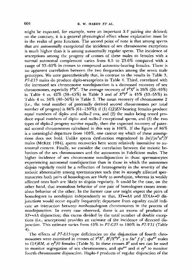
might be expected, for example, were an important X-Y pairing site deleted; on the contrary, it is a general physiological effect whose explanation must lie in the realm of gene function. The second point of note is that among sperm that are autosomally exceptional the incidence of sex chromosome exceptions is much higher than it is among autosomally regular sperm. The incidence of sexceptions among the progeny of crosses of these males to females with a normal autosomal complement varies from 6.5 to 23.6% compared with a range of 33-62% in crosses to compound autosome-bearing females. There is no apparent correlation between the two frequencies among the seven male genotypes. We note parenthetically that, in contrast to the results in Table 3, P7-El5 males do produce diplo-sexceptions in Table 4. Third, correlated with the increased sex chromosome nondisjunction is a decreased recovery of sex chromosomes, especially pXp. The average recovery of pXp is 26% (20-40%) in Table 4 us. 42% (38-45%) in Table 3 and of XDp is 45% (33-55%) in Table 4 vs. 50% (40-56%) in Table 3. The mean recovery of chromosome 2 (i.e., the total number of paternally derived second chromosomes per total number of progeny) is 86% (61-130%); if (1 ) C(2)EN-bearing females produce equal numbers of diplo- and nullo-2 ova, and (2) the males being tested pro- duce equal numbers of diplo- and nullo-2 exceptional sperm, and (3) the two types of diplo-2 progeny survive equally, then the expected recovery of pater- nal second chromosomes calculated in this way is 100%. If the figure of 86% is a meaningful departure from loo%, one cannot say which of these assump- tions does not hold. Unlike sperm dysfunction engendered in Zn(l)sc4scs/Y males (MCKEE 1984), sperm recoveries here seem relatively insensitive to au- tosomal content. Finally, we consider the correlation between the meiotic be- haviors of the sex chromosomes and the autosomes in Y-deficient males. The higher incidence of sex chromosome nondisjunction in those spermatocytes experiencing autosomal nondisjunction than in those in which the autosomes disjoin regularly could be a reflection of heterogeneity in the severity of the meiotic abnormality among spermatocytes such that in strongly affected sper- matocytes both pairs of homologues are likely to nondisjoin, whereas in weakly affected ones both are likely to disjoin regularly. It could be the case, on the other hand, that anomalous behavior of one pair of homologues causes anom- alous behavior of the other. In the former case one might expect the pairs of homologues to nondisjoin independently so that, XY-AA and XYAAHO dis- junctions would occur equally frequently; departure from equality could indi- cate an interaction between nonhomologous chromosomes in the process of nondisjunction. In every case observed, there is an excess of products of XY-AA disjunction; this excess divided by the total number of double excep- tions (i .e. , sexceptions) provides an estimate of the incidence of directed dis- junction. This estimate varies from 43% in P7-G25 to 100% in P7-V31 (Table
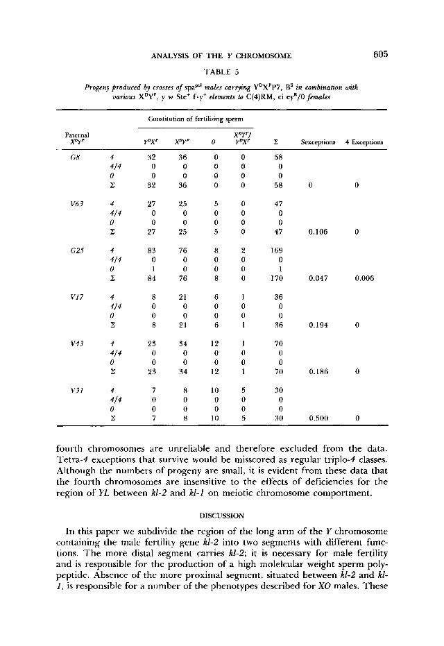
The effects of P7-ElS-type deficiencies on the disjunction of fourth chro- mosomes were examined in crosses of pXp, p/XDp, y w &+fey+; spaPo' males to C(4)RM, ci eyR/O females (Table 5). In these crosses BS and sex can be used to monitor segregation of sex chromosomes, and sPaP'" and ci ef to monitor fourth chromosome disjunction. Haplo-4 products of regular disjunction of the
4).
ANALYSIS OF THE Y CHROMOSOME 605
TABLE 5
Progeny produced by crosses of spap' males carrying YDXPP7, BS in combination with various XDYP, y w Ste+ f .y+ elements to C(4)RM, ci eyR/O females
Constitution of fertilizing sperm
Paternal XDY'I X D Y P Y D X P X D Y P 0 Y D X P z Sexceptions 4 Exceptions
G8
V 6 3
G25
VI 7
v 4 3
V31
4 414 0 z
4 414 0 z
4 414 0 z
4 414 0 z
4
0 z
4
0 z
4 / 4
414
32 0 0
32
27 0 0
27
83 0 1
84
8 0 0 8
23 0 0
23
7 0 0 7
36 0 0
36
25 0 0
25
76 0 0
76
21 0 0
21
34 0 0
34
8 0 0 8
0 0 0 0
5 0 0 5
8 0 0 8
6 0 0 6
12 0 0
12
10 0 0
10
0 0 0 0
0 0 0 0
2 0 0 0
1 0 0 1
1 0 0 1
5 0 0 5
58 0 0
58
47 0 0
47
169 0 1
170
36 0 0
36
70 0 0
70
30 0 0
30
0
0.106
0.047
0.194
0.186
0.500
0
0
0.006
0
0
0
fourth chromosomes are unreliable and therefore excluded from the data. Tetra-4 exceptions that survive would be misscored as regular triplo-4 classes. Although the numbers of progeny are small, it is evident from these data that the fourth chromosomes are insensitive to the effects of deficiencies for the region of YL between kl-2 and kl-1 on meiotic chromosome comportment.
DISCUSSION
In this paper we subdivide the region of the long arm of the Y chromosome containing the male fertility gene kl-2 into two segments with different func- tions. The more distal segment carries kl-2; it is necessary for male fertility and is responsible for the production of a high molelcular weight sperm poly- peptide. Absence of the more proximal segment, situated between kl-2 and k1- 1 , is responsible for a number of the phenotypes described for XO males. These
606 R. W. HARDY ET AL.
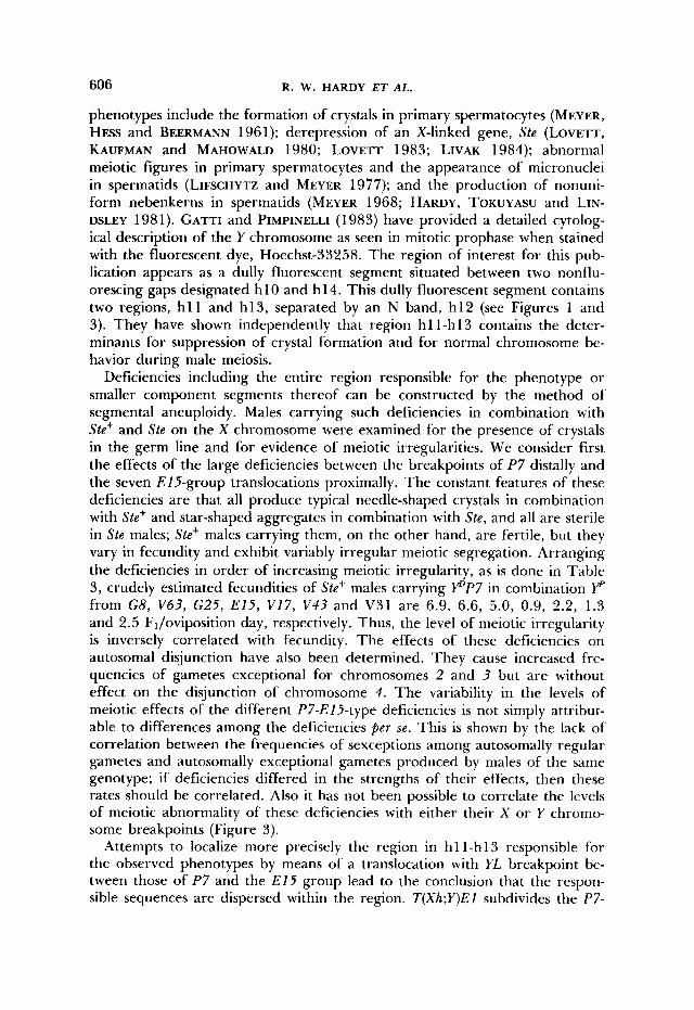
phenotypes include the formation of crystals in primary spermatocytes (MEYER, HESS and BEERMANN 1961); derepression of an X-linked gene, Ste (LOVETT, KAUFMAN and MAHOWALD 1980; LOVETT 1983; LIVAK 1984); abnormal meiotic figures in primary spermatocytes and the appearance of micronuclei in spermatids (LIFSCHYTZ and MEYER 1977); and the production of nonuni- form nebenkerns in spermatids (MEYER 1968; HARDY, TOKUYASU and LIN- DSLEY 1981). GATTI and PIMPINELLI (1983) have provided a detailed cytolog- ical description of the Y chromosome as seen in mitotic prophase when stained with the fluorescent dye, Hoechst-33258. The region of interest for this pub- lication appears as a dully fluorescent segment situated between two nonflu- orescing gaps designated h 10 and h 14. This dully fluorescent segment contains two regions, h l l and h13, separated by an N band, h12 (see Figures 1 and 3). They have shown independently that region hl l-h13 contains the deter- minants for suppression of crystal formation and for normal chromosome be- havior during male meiosis.
Deficiencies including the entire region responsible for the phenotype or smaller component segments thereof can be constructed by the method of segmental aneuploidy. Males carrying such deficiencies in combination with Ste+ and Ste on the X chromosome were examined for the presence of crystals in the germ line and for evidence of meiotic irregularities. We consider first the effects of the large deficiencies between the breakpoints of P7 distally and the seven E15-group translocations proximally. The constant features of these deficiencies are that all produce typical needle-shaped crystals in combination with Ste+ and star-shaped aggregates in combination with Ste, and all are sterile in Ste males; Ste+ males carrying them, on the other hand, are fertile, but they vary in fecundity and exhibit variably irregular meiotic segregation. Arranging the deficiencies in order of increasing meiotic irregularity, as is done in Table 3, crudely estimated fecundities of Ste+ males carrying p P 7 in combination YP from G 8 , V63 , G25 , E15, V17, V 4 3 and V31 are 6.9, 6.6, 5.0, 0.9, 2.2, 1.3 and 2.5 FJoviposition day, respectively. Thus, the level of meiotic irregularity is inversely correlated with fecundity. The effects of these deficiencies on autosomal disjunction have also been determined. They cause increased fre- quencies of gametes exceptional for chromosomes 2 and 3 but are without effect on the disjunction of chromosome 4 . The variability in the levels of meiotic effects of the different P7-EI5-type deficiencies is not simply attribut- able to differences among the deficiencies p e r se. This is shown by the lack of correlation between the frequencies of sexceptions among autosomally regular gametes and autosomally exceptional gametes produced by males of the same genotype; if deficiencies differed in the strengths of their effects, then these rates should be correlated. Also it has not been possible to correlate the levels of meiotic abnormality of these deficiencies with either their X or Y chromo- some breakpoints (Figure 3).
Attempts to localize more precisely the region in hl l-h13 responsible for the observed phenotypes by means of a translocation with YL breakpoint be- tween those of P7 and the E15 group lead to the conclusion that the respon- sible sequences are dispersed within the region. T(Xh;Y)EI subdivides the P7-
ANALYSIS OF THE Y CHROMOSOME 607
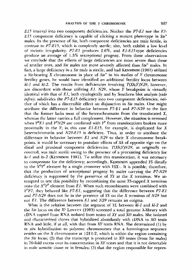
E15 interval into two component deficiencies. Neither the P7-El nor the E l - E15 component deficiency is capable of eliciting a mutant phenotype in Ste+ males. In the presence of Ste, both component deficiencies are male fertile, in contrast to P7-El5 , which is completely sterile; also, both exhibit a low level of meiotic irregularity. P7-El produces 2.6%, and El-EIS-type deficiencies produce an average of 1.5% sexceptional progeny. From these observations we conclude that the effects of large deficiencies are more severe than those of smaller ones, and Ste males are more severely affected than Ste+ males. In fact, a large deficiency in a Ste male is sterile, and had KENNISON (1981) utilized a Ste-bearing X chromosome in place of Ste+ in his studies of Y chromosome fertility genes, he would have identified an additional fertility locus between kl -1 and k1-2. The results from deficiencies involving T(Xh;Y)N29, however, are discordant with those utilizing E l . N29 , whose Y breakpoint is virtually identical with that of E l , both cytologically and by Southern blot analysis (vide infra), subdivides the P7-El5 deficiency into two component deficiencies, nei- ther of which has a discernible effect on disjunction in Ste males. One might attribute the difference in behavior between P7-El and P7-N29 to the fact that the former lacks most of the heterochromatin from the translocated X , whereas the latter carries a full complement. However, the situation is reversed when YDEl and P N 2 9 are combined with Yp from translocations broken more proximally in the Y; in this case El-E15, for example, is duplicated for X heterochromatin and N29-El5 is deficient. Thus, in order to attribute the difference in behavior between E l and N 2 9 to their X chromosome break- points, it would be necessary to postulate effects of Xh of opposite sign on the distal and proximal component deficiencies. T(Xh;Y)N29, as originally re- covered, was male sterile owing to the presence of a preexisting deficiency for ks-1 and ks-2 (KENNISON 1981). To utilize this translocation, it was necessary to compensate for the deficiency; accordingly, KENNISON appended YS distally to the XDF element by a single crossover with YSX.. It is possible, therefore, that the production of sexceptional progeny by males carrying the P7-N29 deficiency is suppressed by the presernce of YS at the X terminus. We at- tempted to test this possibility by recombining the same YS-capped X terminus onto the XDF element from E l . When such recombinants were combined with YDP7, they behaved like P7-E1, suggesting that the difference between P7-El and P7-N29 does not lie in the presence of YS on the X terminus of N 2 9 but not E l . The difference between E l and N29 remains an enigma.
What is the relation between the segment of YL between k1-1 and kl-2 and the Ste locus on the X? LOVETT (1983) screened a total genome A-library with cDNA copied from RNA isolated from testes of XY and XO males. She isolated and characterized clones that hybridized abundantly with cDNA to XO testis RNA and little, if at all, with that from XY testis RNA. She determined (1) by in situ hybridization to polytene chromosomes that a homologous sequence resides on the X chromosome at 12F1-2, which is within the region containing the Ste locus; (2) that the transcript is produced in XO testes (from Ste males) in 70-fold excess over its concentration in XY testes and that it is not detectable in male somatic tissue or in females; (3) that the region responsible for repres-
608 R. W. HARDY E T A L .
sion of transcription of this sequence occupies a position between kl-1 and kl- 2 of the Y chromosome; and (4) that a 17,000-dalton polypeptide is translated from the transcript in an in v i t ro protein-synthesizing system. One of LOVETT’S clones (XDm2L1) has been studied in greater detail by LIVAK (1984). It has been shown to comprise at least eight copies of a 1250-base pair sequence arranged in tandem. This sequence is referred to as the 2L1 sequence. Se- quences homologous to 2L1 are present in approximately 200 copies in Ste- bearing X chromosomes and an approximately tenfold lower copy number in Ste+-bearing Xs; homologous sequences are also carried in at least 80 copies on the Y (LIVAK 1984). The restriction map of the Y-linked sequences differs from that of the X-linked sequences, and the map of the majority of the Ste X sequences differs from that of the Ste’ sequences. Interestingly, we are unable to detect the 200-kb difference between Ste and Ste+ in orcein-stained polytene chromosomes of heterozygotes. By means of Southern blots of CfoI digests of DNA isolated from aneuploid segregants from T(XY) heterozygotes, LIVAK has mapped the 2L1 sequences on the Y. As indicated in Figure 3 all copies of the sequence lie between the Y chromosome breakpoints of W27 distally and E15 [as well as T(Y;4)PFBl5] proximally; perhaps 10% of the 2L1 sequences are distal to the P 7 breakpoint, 60-70% lie between P7 and E l and 20-30% are proximal to the E l (and N29) breakpoint (see Figures 9 and 10 of LIVAK 1984).
The chromosome segments that carry 2L 1 -type sequences are concordant with those responsible for the phenotypes discussed in this communication. The results of LOVETT (1983) and LIVAK (1984) indicate that the P7-El5 deficiency leads to derepression of sequences on the X chromosome that almost certainly reside at the Ste locus; as a consequence, there is excessive production of a transcript that produces a 17-kilodalton polypeptide in an in v i t ro trans- lation system. I t is a logical inference that this polypeptide is overproduced in P 7 - E l 5 deficiency-bearing males and that its overproduction is responsible in some way for the observed phenotype. If the meiotic phenotype is the conse- quence of the overproduction of the Stellate gene product, then we must inquire into possible mechanisms. It might be supposed that at high concen- trations this polypeptide participates in crystal formation and further adheres to chromosomes, imparting to them a sticky aspect and interfering with the orderly progression of meiosis in a nonspecific mechanical way. The difference in meiotic phenotype and crystal morphology between Ste+/Df and S t e / D f males may be attributable either to the fact that Ste has many more sequences than Ste+ and, therefore, more product results, or to the existence of slight differ- ences between the coding sequences of Ste and Ste+ as indicated by the restric- tion map differences, resulting in the overproduction of slightly different pro- teins. There is also reason to believe that reducing the amount of material deleted from the Y reduces the expression of Stellate. The P7-EI5 deficiency may be subdivided into two component deficiences, P7-El and El-E15 . Neither of these elaborates the crystals characteristic of the larger deficiency, nor does either of them cause nondisjunction of the sex chromosomes in Ste+ males as does the parent deficiency. In Ste males, on the other hand, both are fertile and produce low numbers of exceptions, whereas P7-El5 Ste males are sterile.
ANALYSIS OF THE Y CHROMOSOME 609
A nonspecific derangement of meiosis becomes less plausable when the meiotic phenotypes are examined. Meiosis in P7-E15-type deficiences shares many features in common with mutants affecting meiosis in males, and the latter are presumably interfering with a specific reaction. Those features are as follows. (1) Nondisjunction of the sex chromosomes is largely confined to the first meiotic division; (2) nondisjunction involves chromosomes 2 and 3 as well as the sex chromosomes; this argues against a specific geometrical problem with sex chromosome pairing and segregation and in favor of a more general physiological effect on the control of meiosis; (3) chromosome 4 segregates regularly; this is the converse of the effect of m e i S 8 in which only chromosome 4 fails to segregate regularly in males (SANDLER et al. 1968); (4) different pairs of homologues respond to the deficiency in a correlated manner, sex chro- mosome nondisjunction being considerably higher in spermatocytes in which one pair of autosomes fails to disjoin than in those in which the autosomes disjoin regularly. ( 5 ) In double exceptions, there is a marked tendency for the chromosomes of one bivalent to proceed to one pole and those of the other bivalent to proceed to the other. There are no indications of such directed segregation of nonhomologous chromosomes (i .e. , distributive disjunction) in normal males (GRELL 1976). However, directed segregation in double excep- tions has also been reported in a male meiotic mutant (GETHMANN 1974). The majority of the aforementioned meiotic features are found in one or the other of the male meiotic mutants characterized by IVY (1980). The exact mechanism by which the overproduction of the Stellate gene product engenders the spe- cific array of meiotic abnormalities observed must await further experimenta- tion.
This research was supported by National Institutes of Health (NIH) grant GM 26810; K. J. L. was supported by a Damon Runyon, Walter Winchell Cancer Fund Fellowship DRG351; G. L. J. and B. L. were supported by NIH training grant GM 07240; A. L. S. was supported by the Danish Science Research Council. We thank JAMFS KENNISON and MAURIZIO GATTI for thoughtful com- ments on the manuscript.
LITERATURE CITED BROSSEAU, G. E., JR., 1960
COX, G. N., J. D. WHITE and B. I. KIEFER, 1976
GATTI, M. and S. PIMPINELLI, 1983
Genetic analysis of the male fertility factors on the Y chromosome of Drosophila melanogaster. Genetics 45: 257-274.
Isolation and partial characterization of the XO spermatocyte crystals in Drosophila melanogaster. Genetics 83(Suppl.): sl7.
Cytological and genetic analysis of the Y chromosome of Drosophila melanogaster. I. Organization of the fertility factors. Chromosoma (Berl.) 8 8 349- 373.
Meiosis in male Drosophila melanogaster. 1. Isolation and characterization of meiotic mutants affecting second chromosome disjunction. Genetics 7 8 1127-1 142.
Structural genes on the Y chro- mosome of Drosophila melanogaster. Proc. Natl. Acad. Sci. USA 7 9 7404-7409.
Distributive pairing. pp. 435-486. In: The Genetics and Biology of Drosophila, Vol. l a , Edited by M. ASHBURNER and E. NOVITSKI. Academic Press, New York.
T h e influence of chromosome content on the size and shape of sperm heads in Drosophila melanogaster and the demonstration of chromosome loss during spermi- ogenesis. Genetics 79: 23 1-264.
GETHMANN, R. C., 1974
GOLDSTEIN, L. S. B., R. W. HARDY and D. L. LINDSLEY, 1982
GRELL, R. F., 1976
HARDY, R. W., 1975
610
HARDY, R. W., 1980 Crystal aggregates in the primary spermatocytes of XO males in Drosophila melanogaster. Drosophila Inform. Serv. 55: 54-55.
HARDY, R. W., K. T. TOKUYASU and D. L. LINDSLEY, 1981 Analysis of spermatogenesis in Drosophila melanogaster bearing deletions for Y-chromosome fertility genes. Chromosoma (Berl.) 83: 593-617.
HAZELRIGG, T., P. FORNILI and T. C. KAUFMAN, 1982 A cytogenetic analysis of X-ray induced male steriles on the Y chromosome of Drosophila melanogaster. Chromosoma (Berl.) 87: 535- 559.
Mutations that disrupt meiosis in males of Drosophila melanogaster. Ph.D. Thesis,
The genetic and cytological organization of the Y chromosome of Droso-
Analysis of Y-linked mutations to male sterility in Drosophila melanogaster.
Characterization of male-sterile mutations: the genetic control of meiotic divisions and gametogensis. Chromosoma (Berl.) 64: 37 1-392.
The meiotic behavior of grossly deleted X chromosomes in Drosophila melanogaster. Genetics 43: 547-563.
Organization and mapping of a sequence on the Drosophila melanogaster X and Y chromosomes that is transcribed during spermatogensis. Genetics 107: 61 1-634.
Molecular aspects of Y-chromosome function in Drosophila melanogaster sper- miogenesis. Ph.D. Thesis, Indiana University, Bloomington, Indiana.
A locus on the X chromosome appar- ently controlled by the Y chromosome during spermatogenesis in Drosophila melanogaster. Eur. J. Cell Biol. 22: 49.
MCKEE, B., 1984 Sex chromosome meiotic drive in Drosophila melanogaster males. Genetics 106:
Spermiogenese in normalen und Y-defizienten Mannchen von Drosophila melanogaster und D. hydei. 2. Zellforsch. Mikrosk. Anat. 84: 141-175.
Phasenspezifische Funktionsstrukturen in Sper- matocytenkernen von Drosophila melanogaster und ihre Abhangigkeit von Y-Chromosom. Chro- mosoma (Berl.) 12: 676-716.
The entire compound autosomes of Drosophila melanogaster. Genetics 98: 257-273.
Mutants affecting meiosis in natural populations of Drosophila melanogaster. Genetics 6 0 525-558.
Corresponding editor: T . C. KAUFMAN
R. W. HARDY E T AL.
IVY, J. M., 1980
KENNISON, J. A., 1981
KENNISON, J. A., 1983
LIFSCHYTZ, E. and G. F. MEYER, 1977
LINDSLEY, D. L. and L. SANDLER, 1958
University of California, San Diego, California.
phila melanogaster. Genetics 98: 529-548.
Genetics 103: 219-234.
LIVAK, K. J., 1984
LOVEIT, J., 1983
LOVETT, J., T. C. KAUFMAN and A. P. MAHOWALD, 1980
403-422.
MEYER, G. F., 1968
MEYER, G. F., 0. H ~ s s and W. BEERMANN, 1961
NOVITSKI, E., D. GRASE and C. STROMMEN, 1981
SANDLER, L., D. L. LINDSLEY, B. NICOLETTI and G. TRIPPA, 1968