Embed Size (px)
Citation preview
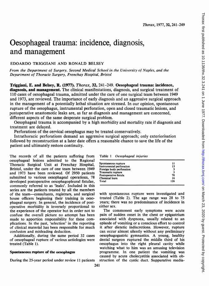
Thorax, 1977, 32, 241-249
Oesophageal trauma: incidence, diagnosis,and managementEDOARDO TRIGGIANI AND RONALD BELSEY
From the Department of Surgery, Second Medical School in the University of Naples, and theDepartment of Thoracic Surgery, Frenchay Hospital, Bristol
Triggiani, E. and Belsey, R. (1977). Thorax, 32, 241-249. Oesophageal trauma: incidence,diagnosis, and management. The clinical manifestations, diagnosis, and surgical treatment of110 cases of oesophageal trauma, admitted under the care of one surgical team between 1949and 1973, are reviewed. The importance of early diagnosis and an aggressive surgical approachin the management of a potentially lethal situation are stressed. In our opinion, spontaneousrupture of the oesophagus, instrumental perforation, open and closed traumatic lesions, andpostoperative anastomotic leaks are, as far as diagnosis and management are concerned,different aspects of the same desperate surgical problem.
Oesophageal trauma is accompanied by a high morbidity and mortality rate if diagnosis andtreatment are delayed.
Perforations of the cervical oesophagus may be treated conservatively.Intrathoracic perforations demand an aggressive surgical approach; only exteriorisation
followed by reconstruction at a later date offers a reasonable chance to save the life of thepatient and ultimately restore continuity.
The records of all the patients suffering fromoesophageal lesions admitted to the RegionalThoracic Surgical Unit at Frenchay Hospital,Bristol, under the care of one team between 1949and 1973 have been reviewed. Of 2950 patientssubmitted to various oesophageal operations, 78developed postoperative oesophagopleural fistulae,commonly referred to as 'leaks'. Included in thisseries are the patients treated by all the membersof the team-consultants, registrars, and surgicalhouse officers beginning their training in oeso-phageal surgery. In general, the incidence of post-operative morbidity is inversely proportional tothe experience of the operator but in order not toconfuse the overall picture no attempt has beenmade to apportion responsibility for these com-plications. In the past, 'selection' in the reportingof clinical material has been responsible for muchconfusion and misleading deduction.
Additionally, during the same period 32 casesof oesophageal rupture of various aetiologies weretreated (Table 1).
Spontaneous rupture of the oesophagus
During the 25-year period under review 11 patients
Table 1 Oesophageal injuries
Spontaneous rupture 11Instrumental perforation 15Foreign body perforation 3Traumatic rupture 3Postoperative fistula 78Chemical burn 16Total 126
with spontaneous rupture were investigated andtreated (Table 2). The age range was 28 to 75years; there was no predominance of incidence ineither sex.The commonest early symptoms were acute
pain of sudden onset in the chest or epigastriumassociated with dyspnoea, usually related to anepisode of vomiting or a conscious effort to controlit after dietetic indiscretions. However, rupturecan occur almost silently without any preliminaryoesophagogastric gymnastics. A young, healthydental surgeon ruptured the middle third of hisoesophagus into the right pleural cavity whilewatching what to him was an amusing televisionprogramme. In one patient the vomiting wascaused by acute cholecystitis associated with ob-struction of the cystic duct. Suppurative media-
241
on March 23, 2020 by guest. P
rotected by copyright.http://thorax.bm
j.com/
Thorax: first published as 10.1136/thx.32.3.241 on 1 June 1977. D
ownloaded from

242
Table 2 Spontaneous rupture: 6 males; 5 females
SymptomChest or epigastric painDyspnoeaDysphagiaShoulder tip pain
SignSurgical emphysema (neck)PneumothoraxShockPleural effusionFeverEmpyemaCyanosisSepticaemia
RadiologySurgical emphysema (neck)PneumothoraxPleural effusionPositive oesophagogramMediastinal emphysemaPneumoperitoneum
10105
4
1110996322
1110109
7
stinitis was already present on admission. Totaloesophagectomy, with cervical oesophagostomyand feeding jejunostomy, was performed togetherwith drainage of the gall bladder; four monthslater cholecystectomy and total oesophageal re-
construction with left colon were performed suc-cessfully. This case demonstrates the necessity fora complete, accurate, preoperative diagnosis.The important diagnostic sign is surgical emphy-
sema in the neck, secondary to mediastinal emphy-sema. This sign was present in every patient at thetime of admission to hospital. Unfortunately, it isfrequently missed, especially after the 'silent' orless dramatic onset of symptoms when the patienthas usually been admitted with an erroneous diag-nosis of myocardial infarction. In these circum-stances there may be a delay of several days beforethe correct diagnosis is reached, by which timesuppurative mediastinitis and empyema will haveoccurred.A chest radiograph will reveal a pleural effusion
and probably a pneumothorax on the side intowhich the oesophagus has ruptured. In six patientsthe rupture was in the lower third into the leftpleural cavity, but in three patients the rupturewas in the n*ddle third and involved the rightpleural cavity. In one of these, an over confidentdiagnosis had been made, the chest radiograph hadbeen omitted, and an abortive left thoracotomywas performed.An oesophagogram with contrast medium, pre-
ferably iodised oil, is obligatory to confirm thediagnosis and locate the site of the rupture. Innine of 10 patients thus examined the leak was
clearly demonstrated.Early diagnosis is essential to the success of con-
servative surgical treatment. In six patients the
E. Triggiani and R. Belsey
correct diagnosis was made within 24 hours and infour of these immediate thoracotomy and primaryrepair of the oesophagus were carried out. Intwo patients the rupture was so extensive thatno repair was possible; in one an oesophagogastricresection was followed by an intrathoracicoesophagogastrostomy, and in the other patientexteriorisation and secondary reconstruction withleft colon at a later date was necessary. Three ofthe four patients treated by primary suture of therupture developed postoperative complications-recurrence of the leak with subsequent chronicempyema in two patients and a broncho-oesopha-geal fistula in one. Of the five patients whosediagnosis had been delayed for 24 hours or longer,three were treated by total resection and exterior-isation followed by a secondary interposition pro-cedure with left colon two months later withexcellent results. 'Conservative' procedures,pleural drainage, and feeding jejunostomy werecarried out in two patients with unsatisfactoryresults; one patient died of sepsis a week later andthe other developed a chronic empyema (Table 3).Our experience suggests that if diagnosis is
delayed, if the rupture is extensive or the oeso-phageal tissues are infected, oedematous, andfriable, or if suppurative mediastinitis is alreadypresent, then the only treatment likely to save thepatient's life is exteriorisation followed bysecondary reconstruction at a later date. By'exteriorisation' is indicated total resection of theintrathoracic oesophagus, leaving the patient witha cervical oesophagostomy to prevent the aspira-tion of saliva, and a feeding gastrostomy, thecardia having been closed. The results have fullyjustified this extended or staged surgical pro-
Table 3 Treatment of spontaneous rupture
Diagnosis' Result2Treatment
A B Recovered Comp. Died
Primary repair bysuture 4 1 3 (recur-
rent leaks)Resection, oesophago-gastrostomy 1 1Exteriorisation, stagedreconstruction 1 3 4'Conservativetreatment' 2 1
1 Diagnosis: A (early < 24 hours); B (late > 24 hours).2 Recovered: Uneventful convalescence.Complicated: Ultimate recovery after convalescence with
complications.Site of rupture: Thoracic-Middle third 3 (right side)
-Lower third 6 (left side)Abdominal INot localised I
on March 23, 2020 by guest. P
rotected by copyright.http://thorax.bm
j.com/
Thorax: first published as 10.1136/thx.32.3.241 on 1 June 1977. D
ownloaded from

Oesophageal trauma: incidence, diagnosis, and management
gramme for a condition that until recently hadbeen regarded as invariably fatal.
Instrumental perforation of the oesophagus
The common cause of instrumental perforation ofthe oesophagus is the passage of some form ofoesophagoscope or dilator; less commonly theorgan may be damaged during an operation onthe mediastinum or hilum of the lung when diffi-culty is encountered during the dissection. It isimpossible to ascertain the global incidence of in-strumental perforation as few of these catastro-phies are reported, for obvious reasons.
This report concerns 15 cases of instrumentalperforation. In the Regional Thoracic SurgicalUnit at Frenchay Hospital, between 1949 and1973, there were 12 cases of instrumental perfora-tion in a consecutive series of 5900 oesophago-scopies, an incidence of 0-2%. Three patients werereferred from other hospitals with an establisheddiagnosis of instrumental perforation. In all casesthe rigid, open-ended oesophagoscope (Belsey type)was used. Many of the operations were performedby junior surgical staff in training. Although localanaesthesia was employed in the great majorityof these 5900 examinations, it may be significantthat, in nine of the 12 cases perforated, generalanaesthesia had been used, possibly reflecting thegreater need for gentleness in the unconsciouspatient. In eight patients the perforation occurredduring a diagnostic examination and in seven casesduring the dilatation of a stricture (Table 4). Thestricture was malignant in four patients and benign
Table 4 Instrumental perforations
Total cases 15 Frenchay Hospital 12 (oesophagoscopies 5900;incidence 0-2 %°)
Other hospitals 3
Anaesthesia: Local 6General 9
Oesophageal lesion:Neoplasm 4Hiatus hernia 3Diverticulum 3Benign stricture 3Other 2
Procedure: DiagnosticTherapeutic
Oesophagogram:PositiveNegativeNot performed
Treatment:
Cervical-conservativeThoracic
Repair by sutureEmergency resection and
reconstructionExteriorisation, staged
reconstruction
924
Site:CervicalMid thirdLower third
Recovered Com- Diedplicatedresult
4 2
2
2 2
3
in three, including one case of fibrous stenosissecondary to epidermolysis bullosa and anothersecondary to scleroderma. In three patients theperforation occurred at the site of a pharyngealdiverticulum. Since it became apparent as a resultof this experience that oesophagoscopic examina-tion carries an additional risk in this situation, theprocedure is now contraindicated when a knowndiverticulum is present or when the bariumswallow examination fails to reveal the state of thecervical oesophagus.
During diagnostic oesophagoscopy the commonsites for the perforation were the cervical oeso-phagus (40%) or the lower end in the regionof the inferior sphincter (40%). Early diag-nosis is both necessary and possible, even duringthe course of the examination. If the operatorbecomes 'lost' and is confronted by unfamiliarappearances, then he is almost certainly outsidethe oesophagus. The instrument is withdrawn im-mediately and the diagnosis is confirmed by anoesophagogram with iodised oil. The rapid appear-ance of pain in the chest, tachycardia, surgicalemphysema in the neck, pneumothorax, dyspnoea,and upper abdominal rigidity secondary to medi-astinal irritation give the typical clinical picture ofperforation. The oesophagogram was positive innine of 11 cases; this investigation was not per-formed in four other patients in whom the siteof the perforation was obvious at the time of theexamination.
Treatment of instrumental perforations isdictated by three factors-the site of the perfora-tion, the underlying pathology calling foroesophagoscopy, and the speed of diagnosis. Per-foration of the cervical oesophagus occurredduring diagnostic examinations. All six patientswere treated by 'conservative' measures-nil bymouth, intravenous fluids, and broad-spectrumantibiotics. In four patients spontaneous healingoccurred uneventfully; in two, a local uppermediastinal abscess required drainage through theneck and was followed by a transient salivaryfistula which closed within a few days.
Intrathoracic perforations call for emergencythoracotomy. Immediately the diagnosis has beenconfirmed the patient is prepared for thoracotomy.Ideally, a one-stage repair of the perforation, withinterrupted inverting sutures of stainless steel wire,is combined with the originally planned operationfor the underlying lesion. In two cases, repair oflower-third perforations was combined with repairsof the hiatal hernias. In four patients with neo-plasms of the middle third, the perforation re-sulted from attempted bouginage; an emergency
243
on March 23, 2020 by guest. P
rotected by copyright.http://thorax.bm
j.com/
Thorax: first published as 10.1136/thx.32.3.241 on 1 June 1977. D
ownloaded from
244
oesophagectomy and one-stage reconstruction withintrathoracic oesophagogastric anastomosis was
performed in all four cases. Two patients recovered(one is alive, well, and free from recurrence fouryears later) but two succumbed, one from coronary
thrombosis and the other from anastomotic prob-lems. In three patients with benign strictures inwhom one-stage reconstruction was contrain-dicated by gross mediastinitis despite early diagno-sis, exteriorisation of the oesophagus was followedby left colon interposition at a later date, withsatisfactory results.
In retrospect, all the cases of intrathoracic per-
foration with gross underlying oesophageal path-ology causing stenosis would have been treatedmore satisfactorily and more safely by exteriorisa-tion and staged reconstruction with left colon. Theprognosis following conservative management ofintrathoracic perforations, by pleural drainage andfeeding jejunostomy, has been observed to be very
poor in terms of survival.
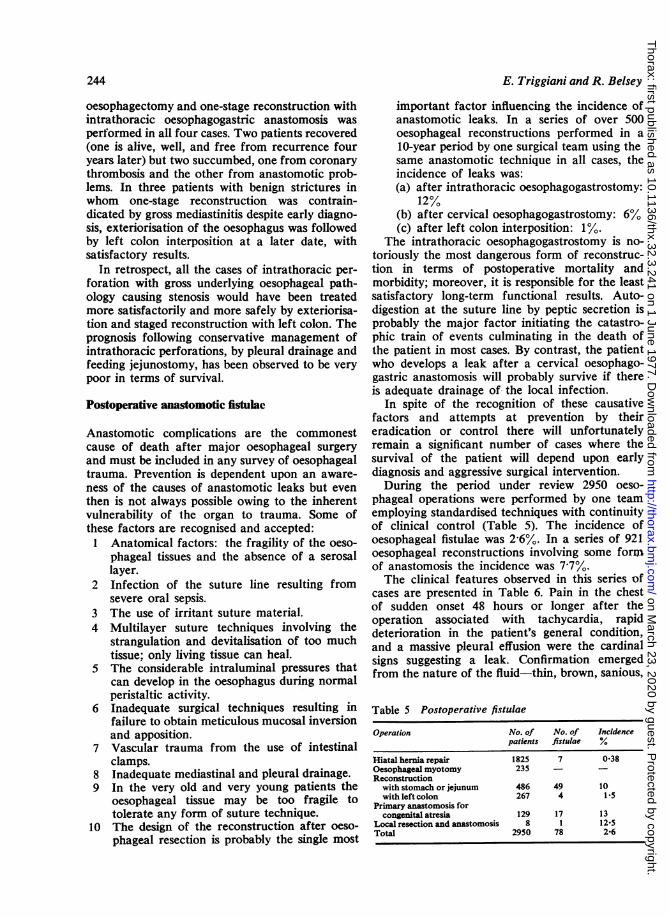
Postoperative anastomotic fistulac
Anastomotic complications are the commonestcause of death after major oesophageal surgery
and must be included in any survey of oesophagealtrauma. Prevention is dependent upon an aware-
ness of the causes of anastomotic leaks but even
then is not always possible owing to the inherentvulnerability of the organ to trauma. Some ofthese factors are recognised and accepted:
1 Anatomical factors: the fragility of the oeso-
phageal tissues and the absence of a serosallayer.
2 Infection of the suture line resulting fromsevere oral sepsis.
3 The use of irritant suture material.4 Multilayer suture techniques involving the
strangulation and devitalisation of too muchtissue; only living tissue can heal.
5 The considerable intraluminal pressures thatcan develop in the oesophagus during normalperistaltic activity.
6 Inadequate surgical techniques resulting infailure to obtain meticulous mucosal inversionand apposition.
7 Vascular trauma from the use of intestinalclamps.
8 Inadequate mediastinal and pleural drainage.9 In the very old and very young patients the
oesophageal tissue may be too fragile totolerate any form of suture technique.
10 The design of the reconstruction after oeso-
phageal resection is probably the single most
E. Triggiani and R. Belsey
important factor influencing the incidence ofanastomotic leaks. In a series of over 500oesophageal reconstructions performed in a10-year period by one surgical team using thesame anastomotic technique in all cases, theincidence of leaks was:(a) after intrathoracic oesophagogastrostomy:
12%(b) after cervical oesophagogastrostomy: 6%(c) after left colon interposition: 1%.
The intrathoracic oesophagogastrostomy is no-toriously the most dangerous form of reconstruc-tion in terms of postoperative mortality andmorbidity; moreover, it is responsible for the leastsatisfactory long-term functional results. Auto-digestion at the suture line by peptic secretion isprobably the major factor initiating the catastro-phic train of events culminating in the death ofthe patient in most cases. By contrast, the patientwho develops a leak after a cervical oesophago-gastric anastomosis will probably survive if thereis adequate drainage of the local infection.
In spite of the recognition of these causativefactors and attempts at prevention by theireradication or control there will unfortunatelyremain a significant number of cases where thesurvival of the patient will depend upon earlydiagnosis and aggressive surgical intervention.During the period under review 2950 oeso-
phageal operations were performed by one teamemploying standardised techniques with continuityof clinical control (Table 5). The incidence ofoesophageal fistulae was 2-6%. In a series of 921oesophageal reconstructions involving some formof anastomosis the incidence was 7-7%.The clinical features observed in this series of
cases are presented in Table 6. Pain in the chestof sudden onset 48 hours or longer after theoperation associated with tachycardia, rapiddeterioration in the patient's general condition,and a massive pleural effusion were the cardinalsigns suggesting a leak. Confirmation emergedfrom the nature of the fluid-thin, brown, sanious,
Table 5 Postoperative fistulae
Operation No. of No. of Incidencepatients fistulae %
Hiatal hernia repair 1825 7 0-38Oesophageal myotomy 235 - -
Reconstructionwith stomach or jejunum 486 49 10with left colon 267 4 1.5
Primary anastomosis forcongenital atresia 129 17 13
Local resection and anastomosis 8 1 12-5Total 2950 78 2-6
on March 23, 2020 by guest. P
rotected by copyright.http://thorax.bm
j.com/
Thorax: first published as 10.1136/thx.32.3.241 on 1 June 1977. D
ownloaded from

Oesophageal trauma: incidence, diagnosis, and management
Table 6 Postoperative fistulae
No. ofcases
Clinical signsFeverChest painPersistent tachycardiaShortness of breathPleural infectionUpper abdominal painAcute respiratory failurePericarditisDysphagiaCervical abscessSalivary fistulaAcute shockNo clinical evidence (occult leak)
Chest radiographAtelectasisPleural effusionPneumothoraxMediastinal emphysemaMediastinal widening, pericardial effusion
OesophagogramPositiveNegativeNot performed (fistulae revealed at routine
postoperative barium swallow)
6860585728121077643
7
5553311824
4416
7
foul-smelling-and the radiographic demonstra-tion of a fistula by an oesophagogram with iodisedoil. This examination is obligatory whenever a leakis suspected, even when the signs are insidious inonset and less dramatic in intensity. The lipiodolswallow was positive in 44 of the 60 patients inwhom it was used. Rarely, a leak may remainsilent or 'occult' and be discovered in a routinepostoperative barium examination. This manifesta-tion is of clinical interest but little importance asspontaneous healing without further treatment isusual (Table 7).
Treatment of postoperative cervical 'leaks'
There were eight instances of anastomotic prob-lems after cervical oesophagogastrostomy; all weretreated conservatively with antibiotics and tem-porary intravenous feeding. In six of the eightpatients spontaneous healing occurred unevent-fully except for mild local infection. One patient
developed a temporary salivary fistula; anothersuccumbed to acute mediastinitis.
Treatment of intrathoracic 'leaks'
By contrast, the 'conservative' management ofleaks after intrathoracic oesophagogastrostomyproved disastrous. In 40 patients treated by pleuraldrainage, by either catheter or rib resection, feed-ing jejunostomy or gastrostomy, and wide-spectrum antibiotic therapy, the mortality ratewas 50%. In 15% a chronic empyema developed,and in only 35% was healing ultimately obtainedafter long periods in hospital and numerous re-visions of the drainage.
In five patients in whom the diagnosis had beenmade early, before gross mediastinitis had oc-curred, an emergency thoracotomy was carriedout with the object of closing the leak. The resultswere equally disappointing and in only one of thefive was satisfactory repair achieved. In fourpatients the leak recurred within a few days.
In view of these recurring failures a moreaggressive policy was adopted, and in six patients,where early diagnosis of the complication appearedto justify the procedure, the entire anastomosiswas resected, all devitalised tissue was removed,and the anastomosis was reconstructed. In onlyone patient was a satisfactory result obtained; inthe remainder the leak recurred with a fatal out-come in 50%. The pathology of this catastrophiccomplication remains baffling. In most cases thesize of the pleural effusion and the fulminatingnature of the mediastinitis were out of all propor-tion to the size of the anastomotic dehiscence,usually a minute defect due apparently to thefailure of a single suture. The use of multilayeranastomotic techniques does not reduce the riskof this complication. In view of the findings atthoracotomy and the apparent technical simplicityof the problem, the unacceptable results and highmortality and morbidity are doubly disappointing.
In a further series of 12 cases where late diag-nosis and the presence of severe suppurative medi-
Table 7 Treatment of postoperative fistulae
Type and site ofleak No. ofcases Treatment Recovered Complicated Died
'Occult': Cervical 1 None 1Thoracic 5 None 5Abdominal 1 None 1 (100%)
Obvious: Cervical 8 Conservative 6 1 1r 5 Repair of fistula 1 1 3
Thoracic 6 Resection of anastomosis and reanastomosis 1 2 3T 12 Exteriorisation and staged reconstruction 7 3 2t40 Drainage and feeding gastrostomy or jejunostomy 14 6 20
245
on March 23, 2020 by guest. P
rotected by copyright.http://thorax.bm
j.com/
Thorax: first published as 10.1136/thx.32.3.241 on 1 June 1977. D
ownloaded from

246
astinitis appeared to contraindicate any attemptat repair or reconstruction of the anastomosis,exteriorisation of the remainder of the oesophagus,cervical oesophagostomy, closure of the gastricremnant, and feeding gastrostomy were performed,followed by staged reconstruction with left coloninterposition two to three months later. Sur-prisingly, the results in this group were the bestobtained with a mortality rate reduced to 17%.
In retrospect the probable answer to this prob-lem is to avoid an intrathoracic oesophagogastricanastomosis whenever an alternative form of re-construction is technically possible. If a leakoccurs, then our experience suggests that the onlyway of saving the patient's life is by exteriorisationand staged reconstruction.
Oesophageal perforations associated with retainedforeign bodies
In three patients a retained intraluminal foreignbody was directly or indirectly responsible for aperforation (Table 8). In one patient a middlethird perforation, diagnosed within four hours ofits occurrence, was treated successfully by primarysuture. A second patient in whom spasm of thelower sphincter secondary to gastro-oesophagealreflux had caused impaction of a piece of muttonbone and subsequently perforation in the regionof the cardia, was subjected to early thoracotomy,suture of the perforation, and synchronous repairof the hiatal hernia; convalescence was uneventful.In the third patient the perforation had occurredduring attempts to remove a piece of bone fromthe lower oesophagus at another hospital 48 hourspreviously. The patient, aged 64, also had a known
E. Triggiani and R. Belsey
hiatal hernia; mediastinitis was present on admis-sion. The oesophagus was exteriorised and a recon-
struction with left colon was performed twomonths later with a satisfactory final result. Thispatient would probably have succumbed to a more
'conservative' surgical approach.
Oesophageal rupture due to trauma
Of the three patients included in this group (Table9), two were transferred to the unit with per-
forations resulting from intraoperative accidents,a cervical laceration occurring during subtotalthyroidectomy, and an abdominal perforation com-plicating vagotomy. The first patient developed a
cervical abscess and salivary fistula but spontane-ous healing followed conservative management.The second patient, a man of 59 years, developedacute abdominal pain soon after the operation; aradiological examination after an iodised oilswallow revealed a leak into the mediastinum andperitoneal cavity. An emergency left thoracotomyand oesophagogastrectomy with intrathoracicanastomosis was followed by an uneventful con-
valescence and recovery.The third patient, a young man aged 21 years,
sustained a steering wheel injury during a caraccident. The extent of the intrathoracic traumadid not become apparent for 48 hours when aniodised oil swallow revealed an oesophagotrachealfistula. Emergency right thoracotomy disclosedan 8 cm rent in the membranous part of thetrachea and virtual destruction of the upper thirdof the oesophagus. The trachea was repaired andthe oesophagus resected with cervical oesophago-stomy and feeding gastrostomy. One month later
Table 8 Foreign body perforations
Sex Age Foreign body Other lesions Level Delay in Treatment Resultdiagnosis
F 56 Denture Mid third 4 h Primary suture RecoveredM 36 Bone Hiatus hernia Lower third 6 h Primary suture and hiatus hernia repair RecoveredF 64 Bone Hiatus hernia Lower third 48 h Exteriorisation and staged
reconstruction Recovered
Table 9 Oesophageal rupture due to trauma
Sex Age Cause Clinical signs Oesophagogram Treatment Result
M 21 Car accident Surgical emphysema; Tracheo-oesophageal Tracheal repair; exteriorisation; staged Recovereddysphagia; aspiration fistula reconstruction
F 51 Thyroid operation Cervical abscess; Leak from cervical Conservative Recoveredsalivary fistula oesophagus
M 59 Vagotomy Acute abdomen Leak in mediastinum Oesophagogastrectomy Recoveredand peritoneum
on March 23, 2020 by guest. P
rotected by copyright.http://thorax.bm
j.com/
Thorax: first published as 10.1136/thx.32.3.241 on 1 June 1977. D
ownloaded from

Oesophageal trauma: incidence, diagnosis, and management
the oesophagus was reconstructed by a long iso-peristaltic transplant of left colon. The final resultwas satisfactory.
Corrosive burns of the oesophagus
During the period under review 16 cases of severestenosis resulting from chemical burns of theoesophagus were admitted for radical surgicaltreatment. Six of the children and three of theadults had swallowed caustic soda accidentally; inthe remaining seven adults the trauma resultedfrom abortive attempts to commit suicide. In everycase repeated attempts had been made to dilatethe stricture without success so these cases mustbe regarded as a selected group; obstruction wassevere or complete in all. The treatment adoptedwas resection of the stenosed segment with syn-chronous reconstruction of the oesophagus by anisoperistaltic transplant of left colon; in most casesthe entire oesophagus had to be replaced owingto the extent of the stenosis. There was oneoperative death due to anoxia resulting from ananaesthetic error; in the remaining 15 patientsconvalescence was uneventful, and normalswallowing was restored.
Discussion
The successful management of spontaneous rup-ture of the oesophagus depends upon two factors-early diagnosis and complete diagnosis, not onlyof the location and extent of the rupture but alsoof the cause of the vomiting that precipitated therupture. Regrettably, the common mistaken diag-nosis of myocardial infarction stems from failureto examine the patient for surgical emphysema inthe neck and for a pleural effusion, failure torequest an immediate radiograph of the chest, andfailure to perform an iodised oil swallow examina-tion of the oesophagus. Suppurative mediastinitiswill be present within 12 hours of the accident,more especially in the presence of dental sepsis,which plays an important role in prognosis as faras survival is concerned. The ideal treatment con-sists of early diagnosis and location of the rupture,and emergency thoracotomy with primary repairof the rupture within 12 hours of the event. In thepresence of established mediastinitis, severe dentalsepsis, and severe inflammatory oedema in the wallof the oesophagus adjacent to the rupture, primarysuture will fail and exteriorisation followed bystaged reconstruction offers the only opportunityfor saving the patient's life. So-called 'conserva-tive' treatment by catheter drainage of the pleura
and antibiotic therapy carries a 50% mortalityrate in our experience, with the alternative of pro-longed hospital treatment for pleural suppurationand feeding by jejunostomy. There have been nofatalities after staged reconstruction.The treatment of instrumental perforations is
determined largely by their position. The incidenceof cervical perforations can be reduced by re-fraining from performing oesophagoscopies onpatients with pharyngeal diverticula, and by pre-operative barium examination of the cervical oeso-phagus to exclude the presence of 'silent' pouches.The mortality rate from perforations can be re-duced by the routine elimination of all dentalsepsis before the examination.
Cervical perforations can be treated conserva-tively-no oral feeding, intravenous alimentation,and antibiotics. Eighty per cent will heal spontane-ously; in 20% a localised cervical abscess willdevelop and require open drainage. Perforationsof the intrathoracic oesophagus demand immediatethoracotomy, repair of the perforation, and syn-chronous correction of the lesion for which theexamination was being performed. For example,if the perforation occurred at the cardia in apatient with an hiatal hernia, then an immediatedefinitive repair should follow closure of the de-fect, the fundoplication buttressing the suture line.When the perforation occurs during the dilatationof a stricture, an emergency resection of thestricture and immediate reconstruction by themethod of choice according to the pathology ofthe stricture has been found to provide the mostsatisfactory results.The prevention of postoperative anastomotic
fistulae depends to a greater extent on the designof the reconstruction than on the anastomotictechnique employed. The incidence of fistulaeafter the three forms of reconstruction mainlyemployed in this series was 12% after intrathoracicoesophagogastrostomy; 6% after cervical oeso-phagogastrostomy; and 1% after interposition ofleft colon. In all three techniques a similar single-layer anastomosis with non-absorbable suturematerial, in this case monofilament stainless steelwire, was used. The high risk of 'leaks' and post-operative mortality after intrathoracic oesophago-gastrostomy has prompted the abandoning of thistechnique in favour of the other two techniqueswhenever possible. The probable cause of the leakis autodigestion of oesophageal tissue at the sutureline when gastric secretion seeps along a sutureunder the influence of the negative intrathoracicpressure. The low incidence after the interpositionof left colon reflects the benign, non-erosive nature
247
on March 23, 2020 by guest. P
rotected by copyright.http://thorax.bm
j.com/
Thorax: first published as 10.1136/thx.32.3.241 on 1 June 1977. D
ownloaded from
248
of the mucus secreted by the colon.The established leak when occurring within the
chest presents a desperate clinical situation towhich the patient commonly succumbs. Emerg-ency thoracotomy, suture of the fistula, or re-
fashioning of the anastomosis is rarely successfulin salvaging the patient's life. In the authors'experience the only hope for the recovery of thepatient from this complication rests in an aggres-
sive approach to the problem-exteriorisation ofthe remaining oesophagus, closure of the stomachand its return to the abdomen, cervical oesopha-gostomy, feeding gastrostomy, and staged recon-
struction at a later date with left colon. Thispolicy is justified only when the prognosis for sur-
vival from the original pathology of the obstruct-ing lesion is hopeful.
In view of the lower operative mortality andmorbidity, and in spite of its greater complexity,the left colon interposition procedure should now
replace intrathoracic oesophagogastrostomy as thereconstruction of choice even after resections formalignant strictures where the long-term prognosisis always in doubt. The cervical oesophagogastros-tomy can be reserved for those patients in whomthe use of colon is contraindicated by intrinsiccolonic disease or mesenteric endarteritis as-
sociated with systemic hypertension. Fistulae froma cervical anastomosis frequently heal spontane-ously after drainage of the local infection.
In the case of neglected or undiagnosed fistulaewhere the patient's condition is critical, nothingcan be done other than pleural drainage and feed-ing jejunostomy. Few patients will survive thissituation.
Oesophageal perforations associated with re-
tained foreign bodies result either from failure todetect non-radio-opaque foreign bodies or attemptsat endoscopic removal of very irregular articles.These may be partial dentures or, in the case ofchildren, the sharp-edged metal obturators tornfrom the tops of beer cans, which now litter thecountryside. If endoscopic removal cannot beeffected safely and without undue manipulation,then emergency thoracotomy and removal byoesophagotomy before infection has supervened isthe safer course of action.
Accidental injuries to the oesophagus duringthe course of any operation on the mediastinumshould be identified and repaired immediately, anda feeding jejunostomy added if doubt exists re-garding the efficiency of the repair. Owing to thefrequency of incompetence of the lower oeso-
phageal sphincter in apparently normal people,feeding gastrostomies are contraindicated in any
E. Triggiani and R. Belsey
situation where it is deemed advisable to 'rest' adamaged oesophagus while healing occurs. Un-suspected oesophageal perforations will be quicklyrevealed by the early appearance of mediastinalemphysema or pneumothorax in the immediatepostoperative period.
Extensive ruptures of the oesophagus can com-plicate closed chest injuries as a result of suddencompression between the sternum and thoracicspine. Early diagnosis may be obscured by theseverity of the associated injuries, but if the pos-sibility is considered the diagnosis can be quicklyand easily confirmed by chest radiography afterthe swallowing of 10 to 20 ml of iodised oil. Un-like barium, residual oil in the mediastinum causesno complications. Staged reconstruction may beindicated by the severity of the oesophagealtrauma if the patient survives the other injuries.In the case of penetrating wounds of the thorax aperforation may be suspected from the trajectoryof the bullet or knife. As with all oesophagealinjuries, early diagnosis, before the inevitablemediastinitis, and early repair are the key to thesurvival of the patient. The risk of fibrous stric-tures secondary to wounds of the oesophagus isproportional to the number of surgical assaults onthe mediastinum in attempts to restore continuity;the first, if correctly performed, is that most likelyto succeed.
Corrosive burns of the oesophagus result fromthe accidental or suicidal ingestion of caustic soda,ammonia, or concentrated acid solutions; in bothsituations, prevention is difficult and beyond thescope of this report. The prognosis is determinedwithin seconds of the injury by the depth of theburn, as in the case of thermal injuries to the skin.No first-aid treatment is likely to influence thecourse of the inflammatory response; the outcomevaries from minor, commonly multiple stricturesamenable to eventual dilatation to complete oeso-phageal stenosis. There is no conclusive evidencethat steroid therapy influences the subsequentfibrosis. Two practical problems ensue-the value,if any, of early dilatation, and the choice of resec-tion and reconstruction or bypass for the severe,undilatable stricture. Too early attempts to passdilators through an acutely inflamed, ulceratedoesophagus are merely adding further trauma toexisting trauma and are illogical. The full extentof the damage will not become apparent for twoto three weeks; this is the time to oesophagoscopethe patient and decide whether dilatation is pos-sible and likely to relieve the dysphagia, or
whether radical surgical treatment is indicatedafter preliminary gastrostomy. Contemporary at-
on March 23, 2020 by guest. P
rotected by copyright.http://thorax.bm
j.com/
Thorax: first published as 10.1136/thx.32.3.241 on 1 June 1977. D
ownloaded from

Oesophageal trauma: incidence, diagnosis, and management
tempts to prevent stenosis by the early use ofplastic stents still await evaluation.The outstanding problem remains the choice
between resection and bypass in the managementof severe, undilatable, corrosive strictures. Thedisturbing factor is the increase in reports ofmalignant degeneration in these strictures, mostlyemanating from Eastern Europe. The availableevidence to date favours one-stage synchronous re-section and reconstruction in preference to bypass.Contrary to previously held views, resection of thecorrosive stricture is technically easier thanthe resection of the chronic peptic stricture as thefibrosis is confined to the mucosa and submucosa;perioesophagitis and mediastinitis are present onlyin those patients in whom ill-advised and over-aggressive attempts to dilate the stricture haveresulted in rupture of the oesophagus.
Bibliography
Abbott, 0. A., Mansour, K. A., Logan, W. D.,Hatcher, C. R., and Symbas, P. N. (1970). Atrau-matic so-called 'spontaneous' rupture of the eso-phagus. Journal of Thoracic and CardiovascularSurgery, 59, 67-83.
Barrett, N. R. (1946). Spontaneous perforation of theoesophagus: review of the literature and report of3 new cases. Thorax, 1, 48-70.
Bertelsen, S. (1971). Traumatic perforations of theoesophagus. Scandinavian Journal of Thoracic andCardiovascular Surgery, 5, 103-110.
Collis, J. L., Humphreys, D. R., and Bond, W. H.(1944). Spontaneous rupture of the oesophagus.Lancet, 2, 179-180.
Keen, G. (1968). The surgical management of oldesophageal perforations. Journal of Thoracic andCardiovascular Surgery, 56, 603-606.
Keighley, M. R. B., Girdwood, R. W., Wooler, G. H.,and Ionescu, M. I. (1972). Morbidity and mortalityof oesophageal perforation. Thorax, 27, 353-358.
Loop, F. D. and Groves, L. K. (1970). Esophageal per-forations. Annals of Thoracic Surgery, 10, 571-587.
McBurney, R. P. (1969). Perforation of the esophagus:A complication of vagotomy or hiatal hernia repair.Annals of Surgery, 169, 851-856.
Diagnostic et traitement des m6diastinites aigues parl6sions oesophagiennes (essentiellement)-Colloquedirige par J. M. Dor. (1969). Annales de Chirurgie,23, 631-673.
Requests for reprints to: Dr. Edoardo Triggiani, 2ndFaculty of Medicine and Surgery, University of Naples,Cappella Cangiani, Naples, Italy.
249
on March 23, 2020 by guest. P
rotected by copyright.http://thorax.bm
j.com/
Thorax: first published as 10.1136/thx.32.3.241 on 1 June 1977. D
ownloaded from