Embed Size (px)
Citation preview

THE EFFECT OF GRADED AND SHORT-TERM, HIGH-INTENSITY
EXERCISE ON EXFIRATORY MUSCLE FERFORMANCE
by
Lisa Jacqueline Wilkins
Thesis submitted to the faculty of
Virginia Polytechnic Institute and State University
in partial fulfillment of the requirements for the degree of
MASTER OF SCIENCE IN EDUCATION
in
Exercise Physiclogy
APFROVED: | f / Ps
| LL Dr. Jay H. as
ods il Batti Ve R. ‘Sebolt
May, ilgos
Rlacksburg, Virginia

LD 5655
VIS 19792
cZ
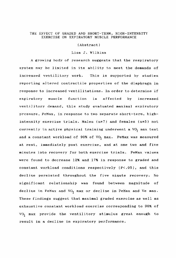
THE EFFECT OF GRADED AND SHORT-TERM, HIGH-INTENSITY EXERCISE ON EXPIRATORY MUSCLE PERFORMANCE
(Abstract)
Lisa J. Wilkins
A growing body of research suggests that the respiratory
system may be limited in its ability to meet the demands of
increased ventilitory work. This is supported by studies
reporting altered contractile properties of the diaphragm in
response to increased ventilitations. In order to determine if
expiratory muscle function is affected by increased
ventilitory demand, this study evaluated maximal expiratory
pressure, PeMax, in response to two separate short-term, high-
intensity exercise trials. Males (n=7) and females (n=5) not
currently in active physical training underwent a VO, max test
and a constant workload of 90% of vO, max. PeMax was measured
at rest, immediately post exercise, and at one two and five
minutes into recovery for both exercise trials. PeMax values
were found to decrease 12% and 17% in response to graded and
constant workload conditions respectively (P<.05), and this
decline persisted throughout the five minute recovery. No
Significant relationship was found between magnitude of
decline in PeMax and VO, max or decline in PeMax and Ve max.
These findings suggest that maximal graded exercise as well as
exhaustive constant workload exercise corresponding to 90% of
vo, max provide the ventilitory stimulus great enough to
result in a decline in expiratory performance.

ACKNOWLEDGEMENTS
I would like to sincerely thank the following individuals
for their time and effort contributed towards the completion
of this research project:
Dr. Jay H. Williams, chairman of my committee, for your
fuidance and support throughout this project: and Dr. Don
Sebolt and Dr. Charles Baffi for serving on my committee.
Kevin Davy, for your guidance and technical assistance;
as well as those individuals involved in the data collection
process, including Lee Pierson, Laurie Miller, Tim Rose and
Allison Chouinard.
In addition I wish to thank my parents, Mrs. Marjorie
Wilkins and Mr. Douglas Wilkins for their continued support
and encouragement throughout my academic career.

ACKNOWLEDGEMENTS... eee eee eee
TABLE OF CONTENTS
LIST OF TABLES... . ssc verr eve seers voevreserevecene
LIST OF FIGURES. eee # @ @ @ @ @ @ @ @ © 8 ee we eH He He
Chapter I:
CHAPTER ITI:
CHAPTER III:
INTRODUCTION......-+. es 8 €¢ © © Oe 8 @ 8
STATEMENT OF THE PROBLEM.
SIGNIFICANCE OF THE STUDY RESEARCH HYPOTHESIS....
DELIMITATIONS......e00-
LIMITATIONS... cee eeeee
BASIC ASSUMPTIONS
DEFINITION
SUMMARY ee 8 2» @ @ 8 @
see #@ @ 6
AND SYMBOLS.
REVIEW OF LITERATURE.......
RESPIRATORY SYSTEM... eee eee oes
INSPIRATORY MUSCLE PERFORMANCE...
Chronic Obstructive Pulmonary
° €
DISEASE. cre revvessevevecsececse
External Loaded Breathing.....
Exercise StudieS...-e.ee-
Effect on Subsequent Performance
Untrained Subjects. Trained vs.
EXPIRATORY MUSCLE PERFORMANCE....
Measurement of Performance....
Chronic Obstructive Pulmonary
DiSEC€ASE. cee ee
External Loaded Breathing.
Exercise Studies..
SUMMARY. ef
JOURNAL MANUSCRIPT..
Abstract......
INTRODUCTION....
METHODS....-+00%
Subjects......
+
General Protocol.....
Measurement of PeMax.
Graded Exercise Test.
Constant Workload Exercise.
StatisticS..ccccsevevervvvece
RESULTS... ce vce cere veers eees
DISCUSSION... ces. cvenvvvves
Comparison with PiMax....
Critique of Methodology..
Comparisons to other studies.
iw
e
6
e
*
ii
iy
ed.
OCsAYMH Hoon
hd

CHAPTER
REFERENCES....+.55
Appendix
Appendix
Appendix
Appendix
Appendix
VITA. ee ¢ © @ © © & @ © ee © © © ee ee ee ee ©
IV:
A
B
C:
D
E:
Relationship with VO2 max
MAXs. cc ceer serv nvsessrcccveees
SUMMALY. cs eee es
REFERENCES......-.
LIST OF CAPTIONS..
SUMMARY owe @ © @ @ © © eee eH eo
DISCUSSION...
ee 8
o> @ 8 @
e ¢ 6 6
# ¢@ @ @
eo * 8 @
RESEARCH IMPLICATIONS...
RECOMMENDATIONS FOR FURTHER
RESEARCH. eens ef © @ @ 86 @ 8 8 © © 8 6
METHODOLOGY........
INFORMED CONSENT...
QUESTIONNAIRE......
RESULTS OF REPEATED
POST HOC TESTS...
RAW DATA.....
MEA SURES
*. 6¢ 6 8
ee 6 @
e #¢ @# @
ee @# 6 @
ee 6
ANOVA
e
AND
xo ¢ 8 © © @ © @ © 8 # © © © © © & © Fe ee © ee Bo
44 44 46 48
53 54 59
60
62
68 74 76
78 82
84

Table
LIST OF TABLES
Mean and Standard Error for Age, Height, Body
Weight, vo, max, Ve max, and FEV) oe eee eee e ees 79
Analysis of Variance Between Measurement
Periods for the 12 Subjects during both the
Graded and 90% VO, max TrialS..eeeevevrvsevssee 80
Duncan’s Post-Hoc Analyses of Graded and 90%
VO, max TrialS..r.cccvecersveevrsvevcveeveseresevvsees 81
vi

Figure
LIST OF FIGURES
PeMax vs. Measurement Period; Incremental Trial..
PeMax vs. Measurement Period; 90% vo, max Trial..
Percent Decline PeMax measured after 90% VO, wees
max vs. Ve max
Percent Decline PeMax measured after 90% VOse ees
max vs. VO2 max
vii
49
50
51

Chapter I
INTRODUCTION
The physical limits of human performance have always been
of interest. This is evident by the number and variety of
sporting events which test these physical limits. The
Physiological processes behind these physical limitations are
important not only from the standpoint of the athlete trying
to maximize performance, but also in order to identify
abnormal conditions in the clinical setting. Without a clear
understanding of the normal condition, it is difficult to
identify or fully understand the abnormal condition.
Maximal oxygen consumption, (VO, max), iS considered the
primary limitation on aerobic capacity inman. The ability to
adequately transport oxygen and utilize it in the locomotor
skeletal muscles is thought to be the ultimate factor which
limits aerobic performance. Therefore, factors such as stroke
volume, cardiac output, and oxidative capacity of the
locomotor skeletal muscles are looked upon as the "weak links"
in human performance.
Recent studies suggest that there may be other "weak
links" within the pulmonary system which may limit it’s
ability to perform maximally. The pulmonary system is a
highly efficient system which can readily adjust to
substantial workloads effectively. Added demand on _ the
1

2
pulmonary system causes an increase in the work of the
respiratory muscles, which may result in an increase in the
force of their contraction. The increased work of the
respiratory muscles expand the chest wall, creating the
pressure necessary to increase tidal volume. Feedback
mechanisms, such as chemoreceptors, which can detect the
decline in pH or arterial oxygen saturation associated with
exercise, and mechanoreceptors located on the chest wall, all
work to regulate the activity of the respiratory muscles, in
order to ensure that the pulmonary system is functioning as
efficiently as possible.
Current research suggests that despite the capabilities
of the pulmonary system, there may be a limit to the amount of
work that the respiratory muscles can perform in order to meet
increased metabolic demands. Research examining the
inspiratory muscles during resistive loaded breathing, (Mador
and Acededo, 1991) as well as studies looking at inspiratory
muscles during endurance exercise, (Bye et al, 1984) suggest
that the respiratory muscles reach a point at which thay are
unable to meet the demands of higher workloads.
STATEMENT OF THE PROBLEM
Researchers investigating the pulmonary system as a
source of limitation on human performance have largely focused
their efforts on the inspiratory muscles. These individuals

3
have demonstrated limitations in meeting the increased
metabolic demands necessary during resistive, loaded breathing
as well as exercise. Studies involving the inspiratory
muscles have reported significant declines in inspiratory
pressures generated after loaded breathing, long term
endurance exercise, as well as short-term, high-intensity
bouts . This decline in inspiratory pressure has been shown
to alter inspiratory muscle performance, (Mador and Acevedo,
1991). Conversly, very little has been done to investigate
the possibility of expiratory muscle fatigue.
Since force development and length of the muscles cannot
be measured directly, expiratory muscle performance is
characteristically evaluated by measuring maximum expiratory
pressure (Pemax), (Rochester et al., 1979). PeMax, a
measurement of the overall strength of the expiratory muscles
is considered the optimal measurement to determine muscle
performance. Studies using expiratory resistvie loaded
breathing as a stressor on the expiratory muscles’ have
demonstrated that a point is reached at which maximum
expiratory pressure declines. Suzuki et al., (1991), reported
a Significant decrease in Pemax for up to sixty minutes after
loaded expiratory breathing, In this study, loaded breathing
caused a decline in breathing frequency and an increase in
tidal volume, which is consistant with other studies of this
nature (Garrard and Lane, 1978). This suggests that the
4
expiratory muscles fatigue as a result of increased resistance
rather than increased respiratory rate.
Although resistive breathing studies suggest that
expiratory muscles fatigue in response to increased workloads,
the question remains as to whether increased respiratory
rates, such as those seen during exercise, could ever become
a cause of fatigue in the expiratory muscles resulting in a
limitation of VO» and subsequent exercise performance,
Studies analyzing maximal expiratory pressures in runners,
bofore and after a marathon have reported a significant
decline in pressure with a prolonged exercise bout (Loke et
al., 1982). This indicates that prolonged exercise requires
ventilitory demands great enough to limit expiratory muslce
function in these athletes. It is unknown, however, whether
ventilitory demands could ever become great enough during
short-term, high-intensity exercise to cause significant
respiratory fatigue. Although this type of exercise has been
reported to decrease inspiratory pressures (Bye et al., 1984),
it is not clear if expiratory pressures decline.
The purpose of this study was to determine whether a
short-term, high-intensity exercise bout and graded exercise
bout would elicit significant changes in Pemax to the muscles
of expiration.
This study evaluated the maximum expiratory pressures
generated during two different high intensity bouts.

5
Measurements of PeMax were compared before and after both
maximal graded exercise, as well as after a short-term, high-
intensity bout. This protocol allowed for comparisons in
expiratory pressure generation which could indicate whether
there was a significant change in muscle performance.
SIGNIFICANCE OF THE STUDY
Results of this study could help to provide more
information as to what conditions are necessary to compromise
expiratory muscle performance. Previous studies have
suggested that exercise at high levels of VO, for extended
periods of time, such as that seen in marathon runners may
cause ventilitory demands great enough for expiratory muscle
performance to decline. This investigation allowed expiratory
muscle performance to be evaluated prior to and following
separate short duration exercise bouts at a greater percentage
of VO, max, as well as graded exercise in subjects who are not
endurance athletes. This could help to determine whether an
intense, exhaustive bout is sufficient to fatigue the
expiratory muscles or if a more extended stimulus is
necessary.
RESEARCH HYPOTHESES
The following null hypotheses were developed to define
the objectives of the present study:
6
1. Ho: There was no change in Pemax following a graded
exercise test to exhaustion
2. Ho: There was no change in Pemax following an exhaustive
constant workload bout at 90% of vO, max.
DELIMITATIONS
The following delimitations were incorporated into the
study by the investigator:
1. The subject population was delimited to male (N=7) and
female (N=5) age 19 to 24 years, who were apparently healthy,
nonsmokers not currently in active physical training.
2. The evaluation of expiratory muscle performance in this
study was delimited to the measurement of maximum expiratory
pressure (PeMax).
3. This evaluation was delimited to the muscles of
expiration.
4. This investigation was delimited to two short-term, high-
intensity conditions, including maximal graded exercise, and
constant workload exercise corresponding to 90% vO, max.
5. The results of this study are delimited to the ability of
the expiratory muscles to generate maximum pressure.
LIMITATIONS
The investigatory acknowledged the following limitations:
1. Due to the specific population evaluated in this study,

7
the results are limited to apparently healthy college age
individuals not in active physical training.
BASIC ASSUMPTIONS
The following assumptions were made at the onset of this
investigation:
1. The subjects performed a true maximal effort in all
pressure measurements.
2. The subjects performed a true maximal effort during both
exhaustive exercise trials.
3. It was assumed that changes in Pemax were due to changes
in the function of the expiratory muscles.
4. It was assumed that no significant changes in VO, max
occurred between the two conditions.
DEFINITIONS AND SYMBOLS
Active physical training- Performing steady state aerobic
exercise for a period of at least twenty minutes, three days
a week
Forced expiratory volume (FEV, ))- The volume of air that
can be forceably exhaled in the first second after a maximal
inhalation.
Maximum expiratory pressure (PeMax)- The maximum amount
of pressure that can be generated after a maximal inhalation.
Maximum inspiratory pressure (PiMax)- The maximum amount

8
of pressure that can be generated after a maximal exhalation.
Tidal volume- The volume of gas inspired or expired
during each respiratory cycle.
Residual volume- The volume of gas remaining in the lungs
at the end of a maximal expiration.
Vital capacity- The largest volume measured on complete
expiration after the deepest inspiration without forced or
rapid effort.
SUMMARY
The pulmonary system is considered to be a highly
efficient system which is able to meet increased metabolic
demands effectively. However, recent studies suggest that a
point may be reached at which the respiratory muscles are
unable to meet the demands placed on them. It has been
demonstrated that inspiratory muscle performance declines in
response to an increased resistance, (Mador and Acevedo, 1991)
as well as a prolonged, (Bye et al.) or high ventilitory
stimulus, (Mador and Acevedo, 1991).
It has been hypothesized that there may be limitations in
expiratory muscle function as well. Studies evaluating the
maximum pressure generated by the expiratory muscles have
reported declines in pressure after resistive, loaded
breathing, (Suzuki et al., 1991), as well as after prolonged
ventilitory demands, such as those associated with marathon

running, (Loke et al, 1982). It is unknown, however, if a
short-term, high-intensity type of exercise could cause
ventilitory demands great enough to alter expiratory pressure.
The purpose of this study was to evaluate the maximum
expiratory pressure generated during both a maximal graded
exercise test and a short-term, high-intensity exercise bout
to provide insight as to whether this type of stimulus could
alter expiratory muscle performance.

Chapter II
REVIEW OF LITERATURE
Recent literature evaluating maximal exercise performance
has implicated the respiratory system as a possible source of
limitation at high exercise intensities. In order to identify
possible limitations on human performance, this review
evaluates the ability of the respiratory system to adequately
meet the increased demands associated with obstructive
disease, external loaded breathing and exercise. This review
focuses on the muscles of respiration, both the inspiratory
and expiratory muscles, and their fatigue as a possible source
of limitation on exercise performance.
RESPIRATORY SYSTEM
The pulmonary system is considered to be a highly
efficient system which effectively meets the added metabolic
demands of steady-state exercise. Characteristically, factors
such as cardiac output and oxidative capacity of the locomotor
skeletal muscles have been thought to limit human performance
rather than the pulmonary system’s ability to adequately
oxygenate the blood. This is supported by research which
indicates that at maximal exercise, maximal ventilitory
volumes are not reached, ventilation to perfusion ratios are
almost uniform, and diffusion capacity of the lung is in
10
11
excess of that required to maintain hemoglobin saturation of
end-pulmonary capillary blood (Shepherd, 1958; Bryan et al.,
1964 and Dempsey et al., 1977).
However, some research inthis area suggests that some of
the above pulmonary "reserves" may actually be surpassable and
that information regarding the homeostatic regulation of these
capacities may be inaccurate (Dempsey et al., 1984). It is
well established that alveolar to arterial oxygen partial
pressure difference widens linearly in response to oxygen
uptake, (Asmussen and Nielson, 1960) and that arterial to
venous oxygen content widens as well, (Dempsey et al., 1977)
suggesting the possiblility of an eventual limit on oxygen
transport. In addition, a few studies have reported
Significant reductions in arterial oxygen partial pressures as
well as oxygen saturation in highly trained athletes working
at high intensities (Rowell et al., 1964, and Gledhill et al.,
1980). These findings suggest the possibility of a
theoretical limit to the capabilities of the pulmonary system.
Dempsey et al., (1984) observed that the typical
hyperventilitory response observed during high intensity
exercise was not seen in highly trained individuals who were
exercising at extremely high intensities. This
hyperventilation, commonly seen in response to metabolic
acidosis, hypoxemia and exercise was reported to be absent,
minimal, or uncorrelated with the magnitude of metabolic

12
acidosis or hypoxemia at these very high workloads. Dempsey
et al. (1984), attributed these findings to mechanical
limitations present within the pulmonary system during periods
of increased demand. This idea is further supported by a study
which implemented helium, a gas less dense than nitrogen,
breathing to reduce the mechanical work required by the
muscles of respiration in trained subjects at high-intensity
exercise (Nattie and Tenney, 1970). Nattie and Tenney (1970)
reported an increased hyperventilitory response to high
intensity exercise with helium breathing suggesting that the
muscles of respiration may be unable to keep up with the added
demands placed on them during periods of increased work.
These studies strongly suggest that a limit is reached
regarding the respiratory system’s ability to meet the
metabolic demands associated with high intensity exercise.
They suggest that mechanical factors ultimately prevent the
respiratory system from keeping pace with increased exercise
intensities, thus limiting exercise performance. Since
mechanical factors are thought to play an important role in
this limitation of respiratory system performance, respiratory
muscle fatigue was examined in this review.
INSPIRATORY MUSCLE PERFORMANCE
The muscles of inspiration, which include the external
intercostals, internal obliques, and diaphragn, are

13
responsible for approximately two thirds of the work
associated with respiration. Upon contraction, the inspiratory
muscles increase the thoracic volume which decreases thoracic
pressure, causing air to flow into the lungs. Since these
muscles are constantly in use, they have great oxidative
capacity, which predisposes them to meet metabolic demands
effectively (Dempsey, 1984). However, recent investigations
have reported that under some conditions a point is reached at
which these muscles are unable to keep up with the demands
placed on them. The result is a decline in inspiratory
pressure and, in some cases, a subsequent decline in exercise
performance (Mador and Acevedo, 1991).
Chronic Obstructive Pulmonary Disease (COPD)
Inspiratory muscle performance was first evaluated in
patients with advanced chronic obstructive pulmonary disease,
in an effort to determine the role that the inspiratory
muscles play in respiratory failure. Respiratory failure is
a complex phenemenon in which numerous factors interact to
ultimately cause failure; a phenomemon which encompasses
hypoxemia and acidemia, hyperinlflation of the lungs, impaired
respiratory drive and increased resistance to airflow.
Several investigators have documented respiratory fatigue in
this disease, as evidenced by marked reductions in PiMax
(Braun and Rochester, 1977), as well as diaphragmatic and

14
intercostal electromyographic changes (Cohen et al, 1982).
Decreases in Pimax have been shown to correspond with
hypoventilation and hypoxemia which suggest that inspiratory
muscle fatigue plays an important role in the respiratory
failure which occurs in the later stages of COPD. (Cohen et
al., 1982). Although the exact mechansism of respiratory
failure is not clear, it is hypothesized that the increased
airway resistance associated with COPD places added demands on
the muscles of inspiration at a time when metabolic factors
such as hypoxemia and acidemia cause the muscles to work
inefficiently. Mechanical factors also seemto play a role in
inspiratory muscle fatigue and subsequent respiratory failure.
Braun and Rochester, (1977) reported a high correlation
between progressive shortening of the diaphragmatic muscle as
measured roentgenographically, and decreases in PiMax in COPD
patients. Furthermore, mechanical breathing abnormalities
such as respiratory alterans and abdominal paradox are widely
observed in patients with respiratory failure and is
considered a reliable clinical sign of inspiratory muscle
fatigue (Cohen et al, 1982). All of these factors interplay
to result in inspiratory muscle fatigue, which results in a
downward spiral towards respiratory failure (Rochester et al.,
1987).
15
External loaded breathing
In order to determine if inspiratory muscle fatigue
could be induced in apparently healthy individuals, a model of
external resistive breathing was devised to mimic the
resistance experienced in COPD patients. By analyzing
transdiaphragmatic pressures as well as electromyogaphic
patterns of the diaphragm, it was reported that the diaphragm
could be fatigued using external loaded breathing in healthy
individuals (Roussos and Macklem, 1977). Roussos and Macklem
(1977), estimated that the threshold for diaphragmatic fatigue
was a pressure equal to 40% of maximum pressure generated.
They observed that the diaphragm could generate pressures
under this threshold indefinitly, but pressures greater than
40% of maximum pressure would result in eventual fatigue. This
is supported by Gross et al., (1979), who report that at 25%
of maximum diaphragmatic pressure, a fatiguing pattern was not
seen upon electromyographic analysis, but a pattern of fatigue
was reported at 50% and 75% of maximum diaphragmatic pressure.
In contrast to the previously mentioned studies, Bellmare
and Grassino (1982), reported an electromyographic pattern of
fatigue at a workload of 15% of maximum diaphragmatic
pressure. The reason for the discrepancy between
investigations may be attributed to the inspiratory time.
Bellmare and Grassino (1982) discovered that the ratio of
inspiratory time to the time of the total tidal cycle has an

16
impact on the rate of fatigue in the diaphragm. They
determined that the greater the inspiratory time in relation
to the total tidal cycle, the earlier the onset of inspiratory
muscle fatigue. This would explain the descrepancies between
the study by Gross et al. (1979), which reported a ratio of
-6, and the investigation by Bellmare and Grassino (1982),
which reported a ratio of 1.0. Further investigation revealed
that this ratio was different in the conflicting studies
(Bellmare and Grassino, 1982).
Although the immediate physiological response to loaded
breathing is highly variable, (Axen and Sperber-Haas, 1979;
Read et al, 1974), prolonged reponse to steady-state loaded
breathing is well established. It has been widely reported
that the inspiratory muscles work to preserve tidal volume by
avariety of possible mechanisms (Hof et al., 1986). Although
the primary mechanism behind this preservation of tidal volume
has not yet been identified, an increase in the duration of
inspiration, a decrease in the frequency of breathing and a
slight decrease in ventilation have all been widely observed
(Hof et al, 1986 and Axen et al, 1983). Ironically, a
prolongation of inspiratory time to preserve tidal volume
appears counterproductive in that it is not the most efficient
way to maintain mean inspiratory pressure and therefore
minimize the cost of breathing (Rochester and Bettini, 1976
and Younes and Riddle, 1984). However, this mechanism allows

17
for a slight decrease in ventilation which may be important in
the reduction of sensory disturbances associated with the
load. (Hof et al., 1986).
Studies evaluating inspiratory muscle performance in COPD
patients as well as external loaded breathing trials indicate
that the muscles of inspiration do indeed fatigue in response
to an increased workload. Declines in Pimax seen in
association with hypoventilation and hypoxemia in COPD
patients suggest that inspiratory muscle fatigue is an
important factor in the mechanism of respiratory failure. In
addition, external loaded breathing studies indicate that this
fatigue occurs in apparently healthy populations as well, with
the threshold for fatigue dependant upon the pattern of
ventilation.
Exercise Studies
Since the pattern of ventilation seems to play an
important role in inspiratory muscle fatigue with external
loaded breathing studies, the question arises as to situations
where ventilitory demands are increased dramatically such as
during exercise. The importance of preserving ventilation
during loaded breathing could suggest that an increased
ventilitory demand could be fatiguing to the inspiratory
muscles, and may subsequently affect exercise performance.

18
Research in the past has ruled out the idea that
increased ventilitory demands during exercise place a
limitation on the respiratory muscles and subsequent exercise
performance because of investigations that suggested that the
pulmonary system did not reach its maximal capacity during
high intensity exercise. This is supported by the following
investigations. First, Shephard et al, (1967), demonstrated
that during maximal exercise, subjects were able to
voluntarily increase minute ventilation, suggesting that
maximal ventilations were not reached during maximal exercise,
and a large respiratory reserve prohibited the respiratory
muscles from ever becoming a limiting factor on exercise
performance. Second, Hughes et al., (1968), in an effort to
determine the limiting factors at maximal exercise, compared
cardiovascular, respiratory, and metabolic adaptations to
increased workloads under various concentrations of inspired
oxygen. They reported that maximal ventilitory volume is not
normally reached in high intensity exercise.
Although these studies suggest that maximal ventilations
are not reached at high levels of exercise, recent research
suggests that the ventilitory demands experienced during
exercise may be great enough to fatigue the inspiratory
muscles. Although maximum ventilitory values are not acheived,
prolonged periods of time at a high percentage of maximum
ventilitory values may result in fatigue. A study by Loke et

19
al., (1982) reported a 16.5% decline in PiMax, as well as a
19.7% decline in transdiaphragmatic pressure at inspiratory
capacity following a marathon race. This decline in
performance of the inspiratory muscles suggests that this
endurance exercise caused a certain amount of fatigue to the
muscles of inspiration. This is further supported by a study
by Fregosi et al. (1987), which reported a significant
increase in intracellular lactate levels in rat diaphragm
after running to exhaustion. In addition, post exercise MVV
measures have been shown to be significantly lower than
controls both after a 60 minute exhaustive running bout and a
marathon (Loke et al., 1982).
Although MVV values have been reported to decline after
marathon running and other long term exhaustive exercise,
short-term, high-intensity exercise does not appear to elicit
a decline in this parameter (Bender and Martin, 1984). This
raises the possibility that a short, intensive exercise
stimulus may not provide a ventilitory demand great enough to
cause fatigue of the inspiratory muscles. In contrast, Coast
et al., (1990) reported a 17% decline in PiMax after maximal
graded exercise. This is further supported by Bye et al.,
(1984), who reported a decline in Pdi as well as a 20% decline
in the ratio of high to low frequency components of the
diaphragm EMG after a short-term, high-intensity constant
workload exercise bout. This suggests that this exercise

20
stimulus was indeed great enough to evoke diaphragm fatigue.
Bye et al., (1984) also found that substituting room air with
40% oxygen reduced ventilation, and delayed the onset of
diaphragmatic fatigue. This indicates that the ventilitory
demand plays a primary role in the fatigue of the inspiratory
muscles during short-term exercise.
Effect on Subsequent Performance
An important question which arises from the previous
studies reporting inspiratory muscle fatigue, is the impact
that this fatigue has on exercise performance. Recent
investigations suggest that the declines in inspiratory
pressures reported may cause a limitation on subsequent
exercise performance. In other words, the changes seen in
inspiratory muscle performance may actually result in a
limitation on an individual’s exercise capacity. For example,
Mador and Acevedo (1991), had subjects inspire against a load
equivalent to 80% of their maximum mouth pressure until they
were unable to generate this target pressure. Subjects then
cycled at 90% of their vo, max until volitional exhaustion.
Following loaded breathing, exercise time was decreased, as
well as vO, and heart rate during the last minute of exercise.
Additionally, ventilation and subjective ratings of
respiratory effort were greater after the induction of
fatigue. This supports earlier findings by Martin et al.

21
(1982), reporting that individuals who were subjected to prior
ventilitory work, (150 min of sustained MVV) experienced a
decline in time to exhaustion during short-term maximal
running. Subjects stopped at a lower ventilation, heart rate
and vO, after prior ventilitory work. These investigations
demonstrate that inspiratory muscle fatigue results in a
decline in subsequent exercise performance and may be an
important limiting factor on human performance.
Trained versus Untrained Subjects
In light of research reporting that PiMax and Pdi decline
in response to increased ventilitory demand and that this
decrease in pressure may have a negative impact on exercise
performance, efforts have been made to determine whether this
affects trained and untrained individuals equally. Dempsey
(1986), hypothesized that a greater decrease in inspiratory
muscle performance would be observed in highly trained
individuals. He reasoned that trained individuals had
acquired the adaptations in vO, max and skeletal muscle
oxidative capacity necessary to reach ventiliations high
enough to limit the capabilities of the pulmonary system.
However, a recent study by Coast et al (1990), demonstrated
that untrained subjects showed significant declines in PiMax
for up to two minutes following maximal exercise while a group
of highly trained cross country skiers showed no significant

22
declines in PiMax. Furthermore, the 12% decrease in PiMax
found in the untrained subjects in this study are similar to
declines seen in a similar study by Bye et al. (1984), using
subjects who were not in active physical training.
One reason for the lack of inspiratory muscle dysfunction
in the cross country skiers in Coast’s study may be due to a
training effect on the muscles of inspiration. Several
investigations suggest that a training effect on the muscles
of respiration is possible. Three pieces of evidence support
this idea. First, respiratory muscle training techniques have
been shown to increase maximal sustained ventilation, (Bradley
and Leith, 1978 and Morgan et al., 1987), as well as pressure
generating capacities of the respiratory muscles {Leith and
Bradley, 1976). However, the benefits accrued were very
specific to the type of training used and did not result in an
increase in exercise capacity. Second, artificially
increasing the work of breathing has been reported to result
in a training effect of the respiratory muscles. Farkas and
Roussos, (1974) as well as Keens et al., (1978) reported
increases in marker enzymes for aerobic metabolism in the
diaphragm of animals with artificially-induced airway
resistance. This suggests that the diaphragm did indeed
undergo a training effect in response to an increased work of
breathing. Third, some studies have reported increases in the
level of diaphragm succinate dehydrogenase (Leiberman et al.,

23
1972) and other enzymes of beta oxidation (Moore and Gollnick,
1982; Ianuzzo et al, 1982) in response to dynamic endurance
exercise training, demonstrating that whole body exercise may
also bring about a training effect of the respiratory muscles.
However, others have found no change in enzymatic composition
of the diaphragm after high-intensity exercise training
(Metzger and Fitts, 1986), suggesting that endurance training
may be more effective in eliciting a training effect.
EXPIRATORY MUSCLE PERFORMANCE
Although many investigations have examined the role of
inspiratory muscle fatigue on the limitations of the pulmonary
system, very little research is available regarding the
expiratory muscles, their role in respiratory muscle fatigue
and possible effects on subsequent exercise performance.
The muscles of expiration, which include the internal
intercostals, rectus abdominus, external and internal obliques
and transversus abdominus, do not contribute to respiration
during periods of rest or passive breathing (Campbell and
Green, 1955; Gilbert and Auchincloss, 1969). These muscles
are recruited only during periods of increased respiratory
work in an effort to preserve functional residual capacity
(FRC). Electromyographical analysis has demonstrated that the
muscles of expiration become actively recruited at
ventilations of 40 L/min (Campbell and Green, 1955; and Strohl

24
et al., 1981). This value is in agreement with investigations
using other techniques for determining expiratory muscle
recruitment. Grimby et al. (1976) reported a pressure volume
displacement of the abdominal muscles at ventilations of 40-80
L/min, indicating that expiratory muscle recruitment was
initiated at this threshold. Henke et al. (1988) reported
expiratory muscle recruitment at 60 L/min by using a helium
dilution technique to measure end expiratory lung volume.
Measurement of Expiratory Muscle Performance
The measurement most commonly used to assess”- the
performance of the expiratory muscles is maximal expiratory
pressure (PeMax) measured at the mouth. This measurement,
obtained by acheiving a maximal expiration after inhaling to
total lung capacity, is considered a simple, reproducible
indicator of expiratory muscle performance. Black and Hyatt,
(1969) measured PeMax in 120 subjects and reported a
coefficient of variation for duplicate measurements of 9% and
no significnat differences between measurements obtained over
three consecutive days. The values reported by Black and
Hyatt, (1969) are in argeement with other maximum expiratory
pressures obtained using other techniques, including
intraesophageal pressure measurements (Hyatt, as referenced in
Black and Hyatt, 1969). Furthermore, the coefficient of
variation reported is in agreement with results obtained from

25
other muscle groups tested, (Ringqvist, 1966; and Tornvall,
1963), suggesting that this is an acceptable amount of
variation. Statistical analysis yeilded no significant impact
on age below the age of 55 and found that the pressures
obtained in females were 65%-&0% lower than those obtained by
males (Blackk and Hyatt, 1969).
Expiratory muscle fatigue was first examined in patients
with COPD, where respiratory muscle fatigue is thought to play
an important role in respiratory failure. Several
investigations evaluating inspiratory muscle performance have
reported declines in inspiratory muscle strength which are
believed to play an important role in the hypoventilation and
hypoxemia observed in respiratory failure. Although
inspiratory muscle fatigue is well documented in this disease,
(Rochester et al., 1987) it is uncertain as to whether the
resistive loading associated with COPD causes’ sufficient
expiratory muscle activity to elicit fatigue. Byrd and Hyatt,
(1968) report no difference in the strength of the expiratory
muscles in patients with COPD compared with healthy
individuals. However, in their study, they did not correct
their data to reflect the abnormal static recoil seen in
patients with COPD. When correction for abnormal recoil is
employed (Rochester et al., 1987), declines in PeMax and FEV1
26
are observed, reflecting a decline in expiratory muscle
performance. Furthermore, they reported that this expiratory
weakness may contribute to impairment of inspiratory muscle
function.
External Loaded Breathing
In order to further investigate whether an increase in
the work of breathing can cause expiratory muscle fatigue,
Suzuki et al., (1991) used a model of external loaded
breathing to evaluate respiratory muscle strength in normal,
healthy individuals. After subjects breathed against an
expiratory resistance for 60 minutes declines in Pemax of 16%
were observed, and these declines persisted for up to an hour
after loaded breathing. Suzuki et al., (1991) further
evaluated the effect of resistive loading on the expiratory
muscles by performing EMG analysis on the rectus abdominus
muscle. EMG analysis confirmed that the rectus abdominus was
fatigued by expiratory loading.
In this study, expiratory loading resulted in a decline
in breathing frequency (55%) and an increase in tidal volume
(60%) which is in agreement with other studies of expiratory
loading (Garrard and Lane, 1987). In addition functional
residual capacity was increased presumably to increase the
expiratory force generated by optimizing the pressure-volume
relationship (Suzuki et al., 1991).

27
Exercise Studies
Although the resuits reported by Suzuki et al., (1991)
suggest that expiratory muscle fatigue is possible in
apparently healthy individuals in response to an increased
workload, it does not clarify whether expiratory muscle
fatigue could occur in response to an increased ventilitory
demand. Inspiratory muscles have been shown to fatigue in
response to incresed ventilitory demands associated with high
intensity exercise which suggests that expiratory muscles may
fatigue as well. Recent studies have investigated whether
increased ventilitory demands such as those seen during
exercise are great enough to fatigue the expiratory muscles,
as they do the inspiratory muscles. Loke et al., (1982)
report a 27.9% decline in PeMax in runners after a marathon
race, suggesting that this endurance activity provided a
ventilitory demand great enough to induce expiratory muscle
fatigue. Other parameters assessing pulmonary function in
marathon runners were evaluated in a study by Maron et al.
Maron et al., (1979) reported a 8.6% reduction in forced vital
capacity immediately after a marathon. This agrees with an
early study by Gordon et al. (1924), which reports a 17%
reduction in forced vital capacity after a marathon. Total
lung capacity was reported to be unchanged suggesting that the
decline in forced vital capacity resulted from expiratory
limitations. Loke et al., (1982) reported a significnat

28
decline in PeMax, suggesting that expiratory muscle strength
decreased after a marathon.
Very few investigations have examined expiratory muscle
performance during a short-term, high-intensity exercise bout.
It is still unclear as to whether this type of exercise
stimulus could provide the ventilitory demands great enough to
fatigue the expiratory muslces. Although relatively few
studies have directly addressed this question, research in
this area support the possibility that expiratory muscle
fatigue may occur in response to short-term, high-intensity
exercise. First, as mentioned previously, expiratory muscle
recruitment has been shown to occur at rather modest exercise
intensities. Ventilations of 40 L/min, which correspond to
approximately 50% of VO2 max are easily surpassed during short
term exhaustive exercise and can be maintained for several
minutes, suggesting that the ventilitory demands may be
adequate for fatigue. Second, a decline in PeMax has been
reported with short-term, high-intensity exercise. O’Kroy et
al., (1991) reported a significant decrease in PeMax after
both a seven minute bout at 90% of vo, max and a thirty minute
bout at 60% of vO, max. These values for PeMax were
significantly lower than pre test values at 5, 10, and 30
minutes post exercise for both trials. Finally, forced vital
capacity has been reported to decrease significantly following
a short-term, high-intensity bout at 90% of vO, max (O’Kroy et

29
al., 1991). This decline in forced vital capacity is also
thought to reflect a decline in performance of the expiratory
muscles, resulting in an inability to exhale to residual
volume.
These previously mentioned studies suggest that the
muscles of expiration may fatigue during periods of increased
metabolic demand. Studies evaluating expiratory muscle
performance in patients with COPD, as well as healthy
individuals breathing against an expiratory resistance show a
decline in expiratory pressure as well as a fatiguing EMG
pattern, suggesting that an increased workload causes fatigue
of the expiratory muscles. Furthermore, exercise studies
demonstrate that the increased ventilitory demand associated
with marathon running and high-intensity exercise are great
enough to elicit expiratory muscle fatigue.
SUMMARY
The objective of this review was to examine the role of
the pulmonary system, or more specifically the respiratory
muscles on the limitation of human performance. Both the
inspiratory and expiratory muscles were reviewed, in order to
determine their function during periods of increased metabolic
demand, including airway obstruction, and increased
ventilition.

30
Although the pulmonary system has characteristically been
thought to have an adequate reserve to meet increased
metabolic demands effectively, recent evidence suggests that
this reserve may have been overestimated and that mechanical
factors may ultimately limit the ability of the pulmonary
system to meet the incresed demands of high intensity exercise
(Dempsey et al., 1984). This is supported by studies which
reported a deline in arterial oxygen saturations, (Glendhill
et al., 1980) and a widening of arterial to venous oxygen
partial pressures (Demspey et al., 1977) at high exercise
intensities. It is further supported by an uncorrelation of
compensatory hyperventilation in response to acidosis or
hypoxemia in highly trained athletes during high intensity
exercise (Dempsey et al., 1984).
In support of the above notion, the inspiratory muscles
appear to fatigue in response to loaded breathing. This has
been demonstrated in patients with COPD (Rochester et al.,
1987), as well as in apparently healthy individuals in
response to external loaded breathing (Roussos and Mackienm,
1977). In addition, increased ventilitory demands during
exercise cause a decline in inspiratory muscle performance.
For example, maximum inspiratory pressures decline after
marathon running (Loke et al., 1982), and during short-tern,
high-intensity exercise (Bye et al., 1984). Furthermore, this
change in performance of the inspiratory muscles has been

31
reported to affect subsequent exercise performance (Mador and
Acevedo, 1991).
The expiratory muscles also exhibit functional decline in
response to increased workloads and ventilation.
Investigations demonstrate that expiratory muscles are
recrited at rather modest ventilations (Strohl et al., 1981).
In addition, maximum expiratory pressures decline in reponse
to increased expiratory resistance in both COPD patients,
(Rochester et al., 1987) and apparently healthy individuals
(Suzuki et al, 1991). This decline is also demonstrated
following marathon running (Loke et al., 1984). Although very
little research has been done to establish expiratory muscle
fatigue in response to short-term, high-intensity exercise,
declines in PeMax as well as forced vital capacity following
this type of stimulus suggest that a decline in expiratory
muscle performance may occur (O’Kroy et al., 1991). This is
further supported by investigations reported that expiratory
muscles are recruited at rather modest ventilations (Strohl et
al., 1981).

Chapter III
THE EFFECT OF GRADED AND SHORT-TERM, HIGH-INTENSITY EXERCISE
ON EXPIRATORY MUSCLE PERFORMANCE
Lisa J. Wilkins
Department of Health and Physical Education
Virginia Polytechnic and State University
32
33
ABSTRACT
A growing body of research suggests that the respiratory
system may be limited in its ability to meet the demands of
increased ventilitory work. This is supported by studies
reporting altered contractile properties of the diaphragm in
response to increased ventilitations. In order to determine if
expiratory muscle function is affected by increased
ventilitory demand, this study evaluated maximal expiratory
pressure, PeMax, in response to two separate short-term, high-
intensity exercise trials. Males (n=7) and females (n=5) not
currently in active physical training underwent a VO, max test
and a constant workload of 90% of VO, max. PeMax was measured
at rest, immediately post exercise, and at one two and five
minutes into recovery for both exercise trials. PeMax values
were found to decrease 12% and 17% in response to graded and
constant workload conditions respectively (P<.05), and this
decline persisted throughout the five minute recovery. No
significant relationship was found between magnitude of
decline in PeMax and vO, max or decline in PeMax and Ve max.
These findings suggest that maximal graded exercise as well as
exhaustive constant workload exercise corresponding to 90% of
vo, max provide the ventilitory stimulus great enough to
result in a decline in expiratory performance.
INTRODUCTION
When evaluating possible limitations on exercise
performance, the pulmonary system has largely been overlooked.
Due to research indicating that at maximal exercise, maximal
ventilitory volumes are not reached, and ventilation to
perfusion ratios are almost uniform, the pulmonary system has
conventionally been considered to have more than adequate
reserves to meet the demands of maximal exercise effectively
(Shepherd, 1958; Bryan et al., 1964). However, recent
investigations reporting reductions in arterial oxygen partial
pressures as well as declines in oxygen saturation in highly
trained athletes during maximal exercise suggest that there
may be a limit to the capabilities of the pulmonary system
(Rowell et al., 1964, Gledhill et al., 1980; Dempsey et al.,
1977; Williams et al., 1986).
Research examining mechanical limitations within the
respiratory system during periods of increased ventilation
have demonstrated a decline in inspiratory muscle performance
in response to marathon running as well as short-term, high-
intensity exercise (Loke et al., 1982; Bye et al., 1984). In
contrast, very little research is available regarding
expiratory muscle function. Studies evaluating expiratory
muscle performance using a model of resistive loaded breathing
report declines in PeMax suggesting that expiratory muscle
performance declines in response to an increase in the work of
34

35
breathing (Suzuki et al., 1991). However it is still unclear
whether PeMax is affected by increased ventilitory work.
Expiratory muscle performance has been reported to decline in
response to marathon running (Loke et al., 1982), but it is
still unclear whether short-term, high-intensity exercise
provides the ventilitory stimulus great enough to effect
expiratory muscle performance.
In this investigation, maximal expiratory pressure,
(PeMax), was measured in order to evaluate expiratory muscle
performance. PeMax was measured before and after both maximal
graded exercise and short-term, constant workload exercise in
order to determine whether these two short-term, intensive
exercise bouts provide the ventilitory stimulus great enough
to cause a decline in expiratory muscle performance.
METHODS
Subjects
Apparently healthy college aged male (n=7), and female
(n=5) volunteers were used for this investigation. In order
to qualify for participation in this study, individuals had to
be nonsmokers, have no history of chronic pulmonary disease
and have had no recent upper respiratory infections. In
addition, only individuals who had not participated in any
regular aerobic training for three months prior to _ the
investigation were selected to participate. Forced expiratory

36
volume during the first second of expiration, (FEV, ) was
measured both before and after exercise to eliminate any
subjects with pulmonary dysfunction.
General Protocol
Subjects performed a graded exercise test on a cycle
ergometer in order to determine VO, max, and returned on a
second day for a short-term, high-intensity cycling bout at a
workload corresponding to 90% of vO, max. Each test required
subjects to exercise to voluntary exhaustion. Measurements of
PeMax were obtained before each trial, immediate post-exercise
and during recovery.
Measurement of PeMax
Maximal expiratory pressure was measured in this study
through the use of a resistance tube attached to a mouthpiece
which led into a pressure transducer (Omega Engineering, 0-250
mmHg). A small leak was present in the tube in order to
prevent glottis closure. Pressure signals were amplified
(Grass P511 low-level DC, 0-30 Hz) and displayed on a strip-
chart recorder (Kipp and Zonen). Prior to each test, the
transducer was calibrated by applying known pressures
(sphyngomanometer) and monitoring the pen displacement on the
strip chart recorder.
Subjects, wearing a noseclip, were first directed to

37
inhale to total lung capacity. The "airway" was closed and
each subject was instructed to exhale maximally against the
resistance tube. A minimum of three resting trials were
obtained before each exercise bout. More than three trials
were performed if variation existed between the second and
third trials. Pilot data revealed a correlation of .9 between
resting measurements. Measurements were also taken
immediately post exercise (IPE) within 20 seconds of test
termination), and one, two and five minutes into recovery.
Subjects were verbally encouraged to give a maximal effort
during each trial.
Graded Exercise Test
For the graded exercise test, males started at a workload
of 50 Watts, and females at a workload of 25 Watts. Workload
was increased 25 Watts every two minutes until the subject was
unable to continue despite verbal encouragement. During the
final 30 sec. of each stage, heart rate, blood pressure,
perceived exertion, ventilation and expired gas fractions were
determined. Measurement of VO, , VCO, ; and Ve were made by the
open circuit technique using a Rayfield dry gas meter to
measure ventilation and a Metex S-3A oxygen analyzer and a
Metex CD-3A carbon dioxide analyzer to determine VO, and VCO, .
Subjects breathed through a low resistance non-rebreathing
valve, with the expired gases passing into a 5-1 mixing

38
chamber. The gas analyzers were calibrated before each test
using standardized gases. Criteria for assessment of vo, max
included a respiratory exchange ratio greater than 1.15, a
heart rate + 10 bpm of age predicted maximum, and
identification of a plateau in VO, with an increase in
exercise intensity. If two of the three criteria were met,
then the highest VO, recorded was chosen as the subject’s VO,
max. All subjects successfully met this criteria. PeMax
measurements were taken both before and after the test as
described previously.
Constant Workload Exercise
Subjects were brought into the lab on a second day in
order to perform a short-term, high-intensity, constant
workload exercise bout. After a two minute warm up period,
subjects cycled at a workload corresponding to 90% of their
VO, max. They cycled at this workload until they were unable
to continue despite verbal encouragement. During this
exercise condition, heart rate, ventilation, and expired gas
fractions were determined as described above. PeMax
measurements were performed after the test as described
previously.

39
Statistics
To determine the effects of exercise protocol and
measurement period on PeMax, a two-way ANOVA, adjusted for
repeated measures was performed. Where indicated, a Duncan’s
post-hoc exam was used to determine individual differences
between measurement periods.
RESULTS
Subjects cycled for a mean duration of 13 min on the
graded protocol to a mean VO, max of 40 + 2.3 ml/kg/min.
Changes in PeMax following graded exercise are shown in Fig.
1. PeMax was found to be significantly lower immediatly post
exercise, (12%), as compared to resting values. This decline
in PeMax persisted throughout the five minute recovery.
PeMax was also found to decline after the constant
workload, high-intensity bout (Fig. 2). Subjects cycled for a
mean duration of 5.5 min. at a workload corresponding to 90%
of their predetermined vo, max. PeMax dropped 17% from
resting measurements and this decline persisted throughout the
five minute recovery.
No significant difference in PeMax was found between the
two conditions. In addition, no significant relationship was
found between the decline in PeMax and vo; max (Fig. 4), or
decline in PeMax and Ve max (Fig. 3).
40
DISCUSSION
The present study demonstrates that graded exercise and
constant-load, high-intensity exercise results ina decline in
expiratory muscle performance. This suggests that these two
exercise protocols elicited a ventilitory stimulus great
enough to affect expiratory muscle function in apparently
healthy individuals. This finding is in agreement with other
research in this area which reports declines in both
inspiratory and expiratory muscle function in response to
intensive exercise.
Studies evaluating expiratory muscle performance suggest
that the expiratory muscles fatigue in response to increased
ventilitory demand. Research demonstrates that although the
muscles of expiration do not contribute to the work of passive
breathing, these muscles are recruited at ventilations of 40
L/min (Campbell and Green, 1955; and Strohl et al, 1981). The
present study found a mean maximal ventilation of 82 + 6.5
L/min. This would indicate that prolonged activity at
moderate to high ventilations may be capable of causing
expiratory fatigue. This is supported by studies evaluating
marathon runners after a race, which report declines in PeMax
of 27.9% as well as reductions in FVC (Loke et al., 1982).
The present investigation supports the idea that
expiratory muscle dysfunction may occur in response to short,
intensive exercise. Although very few investigations have

4]
examined expiratory muscle performance using this type of
exercise stimulus, research in this area support’ the
possibility that it may occur. Declines in PeMax have been
reported in response to a seven minute bout at 90% of vO, max
and significant declines in FVC have also been reported using
this protocol (O’Kroy et al., 1991). The present study found
a decline in PeMax following both maximal graded exercise and
a constant workload bout at 90% of vO, max which supports
previous findings (O’Kroy et al., 1991).
Comparisons with PiMax
The magnitude of decline in PeMax found in this study is
Similar to that seen in studies evaluating inspiratory muscle
performance after exercise. Although patterns of recruitment
are different between these two muscle groups, such a
comparison is relevant because together they share the
increased work of breathing associated with high ventilations
(Dempsey et al., 1982). Bye et al., (1984) reported a 28%
decline in transdiaphamatic pressure, (Pdi), as well as a 20%
decline in the ratio of high to low frequency components of
the diaphragm EMG after a short-term, high-intensity constant
workload exercise bout. In addition, Coast et al., (1990)
reported a 17% decline in PiMax in untrained subjects after
maximal graded exercise. These studies are in agreement with
the mean 14.5% decline in PeMax found in the present study.

42
Taken together, these results suggest that both inspiratory
and expiratory muscle performance declines in response to
short-term, high-intensity exercise, and the magnitude of
decline is similar between the two muscle groups.
Critique of Methodology
The possibility exists that the declines in PeMax found
in this study may be attributed to a lack of motivation to
perform maximal breathing maneuvers after intensive exercise.
If this were the situation, the declines in PeMax would
reflect a generalized fatigue associated with the discomfort
of exercise rather than fatigue of the expiratory muscles.
This is unlikely due to several reasons. First, as previously
mentioned, the magnitude of decline in PeMax in this study is
consistent with other studies evaluating respiratory muscle
function in response to short-term, high-intensity exercise.
Second, the magnitude of decline in PeMax was not
Significantly different between the two conditions. It is
unlikely that a lack of motivation would be so consistent in
response to two different conditions that were of different
duration on separate days. Finally, declines in PeMax
persisted throughout the five minute recovery for both
exercise trials. If the discomfort associated with high
intensity exercise resulted in a a lack of motivation to
exhale maximally, it would be expected that PeMax would return

43
to resting values once the subject had a chance to recover
from the discomfort of exercise. Since PeMax values were
still depressed five minutes into recovery, factors other than
motivation most likely were causing the decline. This
suggests that the declines seen in this investigation actually
represent expiratory muscle dysfunction rather than a lack of
motivation to perform maximally in these particular subjects.
Comparisons to other investigations
The decline in PeMax throughout recovery seen in this
investigation is consistant with other studies in this area.
The declines in PiMax found by Coast et al. (1990), after
maximal graded exercise persisted throughout a five minute
recovery. These findings are in agreement with O’Kroy et al.
(1991), who reported declines in PeMax five minutes after a
seven minute bout at 90% of VO, max and a 30 min bout at 60%
of vO, max. Additionally, Suzuki et al., (1991) reported a
decline in PeMax for up to 60 min following expiratory
resistive loaded breathing but did not find a corresponding
EMG pattern of fatigue. Although the mechanism behind this
decline in PeMax is not known, it lends support to the idea
that the declines in expiratory muscle performance may be
persistant enough to affect subsequent exercise performance.

44
Relationship with VO, _max and Ve max
The present study did not find a strong correlation
between the magnitude of decline in PeMax and VO, max. This
conflicts with research which found that highly trained
subjects have lesser changes in PiMax following maximal
exercise than untrained subjects (Coast et al., 1990),
possibly due to a training effect on the respiratory muscles.
One reason for a failure to see a relationship between PeMax
and vO, max, as well as a relationship between PeMax and Ve
max in this study might be due to the rather homogeneous
population of untrained subjects. This present study
consisted of 12 subjects with a mean vO, max of 40 + 2.3
ml/kg/min with a coefficient of variation of .58. In addition,
none of the subjects in the present study would be considered
to be trained and thus may not consistently reach ventilations
high enough in their daily activities to result ina training
effect of the respiratory muscles. It is possible that
inclusion of trained subjects in the present study might have
resulted in results similar to Coast et al., (1990).
Summary
The primary objective of this study was to evaluate
expiratory muscle performance during two short-term, high-
intensity exercise protocols. The declines in PeMax observed
in response to both graded and constant-load exercise suggest

45
that this type of exercise stimulus provides the ventilitory
demands great enough to cause a decline in expiratory muscle
performance.
These data provide a framework for further research in
order to determine if expiratory muscle dysfunction is
ultimatly a limiting factor on exercise performance. This
could have implications not only for the athlete, training to
optimize exercise performance, but also in the diagnostic and
functional assessment of VO, max of sedentary populations.
Since greater declines in respiratory muscle performance are
seen in sedentary people, respiratory system limitations may
occur in this population that currently have been attributed
to limitations in VO, max. In addition, recent research in the
area of respiratory muscle function suggests that a training
effect on the muscles of respiration may be possible in
response to repeated exposure to increased ventilations. If
this were to be confirmed, athletes may alter their training
to optimize these potential benefits. This also may have
implications for exercise training in patients with COPD and
efforts to improve their ability to perform activities of
daily living.

46
REFERENCES
Bryan A., Bentivaglio, L., Beerel, F., MacLeish, H . ;
Zidulka, A., and Bates, D. (1964). Factors affecting
regional distribution of ventilation and perfusion in
the lung. Journal of Applied Physiology, 19, 395-402,
Bye, P., Esau, S., Walley, K., Macklem, P., and Pardy, R.
(1984). Ventilitory muscles during exercise in air and
oxygen in normal men. Journal of Applied Physiology,
96(2), 464-471.
Campbell, E., and Green, J. (1955). The behaviour of the
abdominal muscles and the intra-abdominal pressure during
quiet breathing and increased pulmonary ventilation. A
study in man. Journal of Physiology London, 127, 423-
426,
Coast, J., Clifford, P., Henrich, T., Stray-Gundersen, J.,
and Johnson, R. (1990). Maximal inspiratory pressure
following maximal exercise in trained and untrained
subjects. Medicine and Science in Sports and Exercise,
22(6), 811-815.
Dempsey, J., Gledhill, N., Reddan, W., Forester, H., Hanson,
P., and Claremont, A. (1977). Pulmonary adaptation to
exercise: effects of exercise type and duration, chronic
hypoxia and physical training. Annals of the New York
Academy of Sciences, 301, 243-261.
Dempsey, J., Hanson, P., and Henderson, K. (1984).
Exercise-induced arterial hypoxemia in healthy human
subjects at sea level. Journal of Physiology, 355, 161-
175.
Dempsey, J. (1986). Is the lung built for exercise?
Medicine and Science in Sports and Exercise, 18(2),
143-155.
Gledhill, P., Phillips, D., and Meyers, E. (1980). Acid-
base status with induced erythrocythemia and its
influence on arterial oxygenation during heavy exercise.
Medicine and Science in Sports and Exercise, 12, 122
(Abstract).
Loke, J., Mahler, D., and Virgulto, J. (1982). Respiratory
muscle fatigue after marathon running. Journal of
Applied Physiology, 52(4), 821-824.
47
O’Kroy, J., Loy, R., Story, S., and Coast, J. (1991). Pulmonary function changes following treadmill running.
Medicine and Science in Sports and Exercise, 24(5), 583
(Abstract).
Rowell, L., Taylor, H., Wang, Y., and Carlson, W. (1964).
Saturation of arterial blood with oxygen during maximal
exercise. Journal of Applied Physiology, 2, 29-41.
Shepherd, R. (1958). Effect of pulmonary diffusing
capacity on exercise tolerance, Journal of Applied
Physiology, 12-13, 487-488.
Strohl, K., Mead, J., Banzett, R., Loring, S., and Kosch, P.
(1981). Regional differences in abdominal muscle
activity during various maneuvers in humans. Journal of
Applied Physiology, 51, 1471-1476.
Suzuki, S., Suzuki, J., and Okubo, T. (1991). Expiratory
muscle fatigue in normal subjects. Journal of Applied
Physiology, 70(6), 2632-2639.
Williams, J.,; Powers, S.4 and Stuart, K. (1986).
Hemoglobin desaturation in highly tained athletes during
heavy exercise. Medicine and Science in Sports and
Exercise, 18(2), 168-173.

48
LIST OF CAPTIONS
Figure
1 PeMax vs. Measurement Period; Incremental Trial
* p<0.05
2 PeMax vs. Measurement Period; 90% VO, max Trial
* p<0.05
3 Percent Decline PeMax measured after 90% vo, max vs.
Ve max
4 Percent Decline PeMax measured after 90% vO, max VS.
VO, max

Pema
x (m
m Hg)
75
70
65
60
55
50
45
49
i INCREMENTAL
L
* *
m *
EXERCISE
LC) ! L l L L L l J
REST IPE 1 2 3 4 5
MEASUREMENT PERIOD (min)
Figure 1

50
75 -
90% VO,max
~ om
xr
& £
TZ
x< 0
£ lil
oO
EXERCISE
45 LC | LL I | l ] | ]
REST IPE 1 2 3 4 5
MEASUREMENT PERIOD (min)
Figure 2

-1
Vmax
(L
min
)
120
105
90
75
60
45
30 0
r
5
51
~.29 0
10 15 20 25 30 35 40
PRESSURE DECREASE (%)
Figure 3

VO,m
ax
(mi kg
| mi
n’)
35
50
45
35
30
25 A
a)
IO
O
P96
O
O O O
O O O
O O 30
{ i _ i i 1 |
5 10 15 20 25 30 35 40
PRESSURE DECREASE (%)
Figure 4

aR
Chapter IV
SUMMARY
Recent literature evaluating maximal and _ short-term
exercise performance has implicated expiratory muscle fatigue
as a possible source of limitation at high exercise
intensities (O’Kroy et al., 1991). The present investigation
evaluated PeMax following two different short-term, intensive
exercise bouts to determine whether this type of exercise
stimulus provides the ventilitory demands great enough to
result in a decline in expiratory muscle performance.
Twelve apparently healthy subjects not currently in
active physical training underwent two different exercise
protocols; a graded exercise test on a cycle ergometer and an
exercise bout to exhaustion at a workload corresponding to 90%
of VO, max. PeMax was measured before the exercise trials as
well as immediate post, one, two and five minutes post
exercise.
Subjects cycled for a mean duration of 13 min on the
graded protocol to a mean VO, max of 40 + 2.3 ml/kg/min with
a coefficient of variation of .58. PeMax was found to be
Significantly lower immediatly post exercise, (53.9 + 4.3
mmHg), aS compared to resting values (61.4 + 4.3 mmHg). This
decline in PeMax persisted throughout the five minute recovery
with PeMax values of 53.1 + 3.1 mmHg, 54.2 + 4.5
53

54
mmHg, and 53.6 + 4.3 mmHg at one, two and five minutes
respectively.
PeMax was also found to decline after the constant
workload, high intensity bout. Subjects cycled for a mean
duration of 5.5 min. at a workload corresponding to 90% of
their predetermined vo, Max. PeMax dropped from a mean
resting value of 65.75 + 4.7 mmHg to 54.28 + 4.4 mmHg
immediately post exercise. This decline in PeMax persisted
throughout the five minute recovery with values of 57.84 + 4.6
mmHg, 58.29 + 5.3 mmHg, and 58.16 + 5.7 mmHg at one, two and
five minutes respectively.
No significant difference in the pattern of PeMax change
was found between the two trials. In addition, no significant
relationship was found between the decline in PeMax and VO,
max or decline in PeMax and Ve max.
DISCUSSION
The present study demonstrated that both the graded
maximal exercise and constant workload high intensity exercise
resulted ina decline in expiratory muscle performance. This
suggests) that these two exercise protocols caused a
ventilitory stimulus great enough to affect expiratory muscle
function in apparently healthy college aged individuals not in
active physical training. This finding is in agreement with

55
other research in this area reporting declines in both
inspiratory and expiratory muscle function in response to
intense exercise.
Studies evaluating expiratory muscle performance suggest
that the expiratory muscles are recruited in response to
increased ventilitory demand (Loke et al., 1982). Research
demonstrates that although the muscles of expiration do not
contribute to the work of passive breathing, these muscles are
recruited at ventilations of 40 L/min (Campbell and Green,
1955; Strohl et al, 1981). The present study found a mean
maximal ventilation of 82 + 6.5 L/min. This would indicate
that prolonged activity at moderate to high ventilations may
be capable of causing expiratory fatigue. This is supported
by studies evaluating marathon runners after a race, which
report declines in PeMax of 27.9% as well as reductions in FVC
(Loke et al., 1982).
The present investigation supports the idea that
expiratory muscle dysfunction may occur in response to short,
intensive exercise. Although very few investigations have
examined expiratory muscle performance using this type of
exercise stimulus, research in this area support’ the
possibility that it may occur. Declines in PeMax have been
reported in response to a seven minute bout at 90% of vo, max
and significant declines in FVC have also been reported using
this protocol (O’Kroy et al., 1991). The present study found
56
a decline in PeMax following both maximal graded exercise and
a constant workload bout at 90% of vo, max which supports the
findings of O’Kroy et al. (1991).
The magnitude of decline in PeMax found in this study is
in agreement with declines seen in studies evaluating
inspiratory muscle performance. Although patterns of
recruitment are different between these two muscle groups, it
is relevant to compare them because together they share the
increased work of breathing associated with high ventilations.
Bye et al., (1984) reported a 28% decline in Pdi as well as a
20% decline in the ratio of high to low frequency components
of the diaphragm EMG after short term high intensity constant
workload exercise bout. In addition, Coast et al., (1990)
reported a 17% decline in PiMax in untrained subjects after
maximal graded exercise. These studies are in agreement with
the mean 14.5% decline in PeMax found in the present study.
These investigations suggest that both inspiratory and
expiratory muscle performance declines in response to short-
term, high-intensity exercise, and the magnitude of decline is
Similar between the two muscle groups.
The possibility exists that the declines in PeMax found
in this study may be attributed to a lack of motivation to
perform maximal breathing maneuvers after intensive exercise.
If this were the situation, the declines in PeMax would
reflect a generalized fatigue associated with the discomfort

57
of exercise rather than fatigue of the expiratory muscles.
This is unlikely due to Several reasons. First, as previoulsy
mentioned, the magnitude of decline in PeMax in this study is
consistent with other studies evaluating respiratory muscle
function in response to short-term, high-intensity exercise.
Second, the magnitude of decline in PeMax was not
Significantly different between the two trials in this study.
It is unlikely that a lack of motivation would be _ so
consistent in response to two different trials that were of
different duration on separate days. Finally, declines in
PeMax persisted throughout the five minute recovery for both
exercise trials in this study. If the discomfort associated
with high intensity exercise resulted in a a lack of
motivation to exhale maximally, it would be expected that
PeMax would return to resting values once the subject had a
chance to recover from the discomfort of exercise. Since
PeMax values were still depressed five minutes into recovery,
factors other than motivation were causing the decline. Thus,
the declines in PeMax seen in this investigation represent
expiratory muscle dysfunction rather than a lack of motivation
to perform maximally in these particular subjects.
The decline in PeMax throughout recovery seen in this
investigation is consistant with other studies in this area.
The declines in PiMax found by Coast et al. (1990), after
maximal graded exercise persisted throughout a five minute

58
recovery. These findings are in agreement with O’Kroy et al.
(1991), who reported declines in PeMax five minutes after a
seven minute bout at 90% of vo, max and a 30 min bout at 60%
of vO, max. Additionally, Suzuki et al., (1991) reported a
decline in PeMax for up to 60 min following expiratory
resistive loaded breathing but did not find a corresponding
EMG pattern of fatigue. Although the machanism behind this
decline in PeMax is not known, it is possible that the
declines in expiratory muscle performance may be persistant
enough to affect subsequent exercise performance.
The present study did not find a strong correlation
between the magnitude of decline in PeMax and VO, max. This
conflicts with research which found that highly trained
subjects have lesser changes in PiMax following maximal
exercise than untrained subjects (Coast et al., 1990). The
different changes in PiMax betweem trained and untrained
subjects reported in this study were attributed to a possible
training of the respiratory muscles. One reason for a failure
to see a relationship between PeMax and vO, max, as well as a
relationship between PeMax and Ve max in this study could be
due to the homogeneous population of untrained subjects. This
present study consisted of subjects with a mean vO, max of 40
ml/kg/min + 2.3 with a coefficient of variation of .58. In
addition, none of the subjects in the present study would be
considered to be trained and thus may not consistently reach

59
ventilations high enough in their daily activities to result
in a training effect of the respiratory muscles.
RESEARCH IMPLICATIONS
The primary objective of this study was to evaluate
expiratory muscle performance during two short-term, high-
intensity exercise protocols. The declines in PeMax observed
in response to both graded and constant workload maximal
exercise suggest that this type of exercise stimulus provides
the ventilitory demands great enough to cause a decline in
expiratory muscle performance.
This finding provides a framework for further research in
order to determine if expiratory muscle dysfunction is
ultimatly a limiting factor on exercise performance. This
could have implications not only for the athlete, training to
optimize exercise performance, but also in the diagnostic and
functional assessment of vo, max of sedentary populations.
Since greater declines in respiratory muscle performance are
seen in sedentary people, respiratory system limitations may
eccur in this population that currently have been attributed
to limitations in Vo2 max. In addition, recent research in the
area of respiratory muscle function suggests that a training
effect on the muscles of respiration may be possible in
response to repeated exposure to increased ventilations. If
this were to be confirmed, athletes may alter their training

60
to optimize these potential benefits. This also may have
implications for exercise training in patients with COPD and
efforts to improve their ability to perform activities of
daily living.
RECOMMENDATIONS FOR FURTHER RESEARCH
In order to fully understand the significance of the
decline in PeMax found in this investigation, further research
is necessary. The following are suggestions for futher
research relating to expiratory muscle performance, as well as
subsequent exercise performance.
1) Since research indicates that PiMax declines to a
Sreater extent in trained subjects rather than untrained
subjects, it would be beneficial to repeat the present
investigation using highly trained athletes. By comparing the
magnitude of decline found in trained and untrained subjects,
more information could be gathered regarding the possibility
of a training effect of the expiratory muscles.
2) Another important research question in the area of
expiratory muscle function is whether the declines seen in
PeMax have an effect on exercise performance. The answer to
this question could help to determine the importance of
further study in the area of expiratory muscle fatigue.
Therefore, it would be beneficial to evaluate exercise
performance after inducing a decline in PeMax perhaps by

61
external loaded breathing. A decline in subsequent exercise
performance has been demonstrated after the induction of
inspiratory muscle fatigue, which suggests that it may occur
in the expiratory muscles.
3) Another important area of investigation in order to
determine the magnitude of expiratory muscle dysfunction is to
determine the point at which expiratory muscle performance
begins to decline. Therefore, it would be valuable to repeat
the present investigation using an intense, but not exhaustive
protocol in order to determine if this stimulus would be
sufficient to cause a decline in PeMax.
4) Finally, it is important to investigate possible
mechanisms behind the decline in expiratory muscle
performance. Although it is postulated that the declines seen
in PeMax are a result of an increased work of breathing
associated with exercise, this has yet to be investigated
fully. Therefore, in order to confirm whether the decline in
PeMax during intensive exercise is due to an increased
mechanical work of breathing, studies utilizing a lower
density gas would be beneficial. <A lower density gas such as
helium would decrease the work required by the expiratory
muscles. This would allow researchers to examine the effects
of a decreased workload on PeMax at exercise.

REFERENCES
Asmussen, E., and Nielsen, M. (1964). Studies in the
regulation of respiration in heavy work. Acta Physiologica
Scandinavica, 12, 171-188.
Axen, K., and Sperber-Haas, S. (1979). Range of first breath
ventilitory responses to added mechanical loads in naive
men. Journal of Applied Physiology, 46, 743-751.
Axen, K., Sperber-Haas, S., Gaudino, D., and Haas, A. (1983).
Ventilitory adjustments during sustained mechanical loading
in conscious humans. Journal of Applied Physiology, 55,
1211-1218.
Bellmare, F., and A. Grassino (1982). Effect of pressure ad
timing of contraction on human diaphragm fatigue. Journal
of Applied Physiology, 53(5), 1190-1195.
Bender, P., and Martin, B. (1985). Maximal ventilation aftr
exhausting exercise. Medicine and Science in Sports and
Exercise, 17(1), 164-167.
Black, L., and Hyatt, R. (1969). Maximal respiratory
pressures: normal values and relationship to age and sex.
American Review of Respiratory Disease, 99, 696-702.
Bradley, M., and Leith, D. (1978). Ventilitory muscle
training and the oxygen cost of sustained hyperpnea.
Journal of Applied Physiology, 55, 885-892.
Braun, N., and Rochester, D. (1977). Respiratory muscle
strength in obstructive lung disease. American Review of
Respiratory Disease, 115(4), 91.
Bryan, A., Bentivaglio, L., Beerel, F., MacLeish, H., Zidulka,
A., and Bates, D. (1964). Factors affecting regional
distribution of ventilation and perfusion in the lung.
Journal of Applied Physiology, 19, 395-402.
Buono, M., Constable, S., Morton, A., Rotkis, T., Stanforth,
P., and Wilmore, J. (1981). The effect of an acute bout
of exercise on selected pulmonary function measurements.
Medicine and Science in Sports and Exercise, 13(5), 290-
293.
Bye, P., Esau, S., Walley, K., Macklem, P., and Pardy, R.
(1984). Ventilitory muscles during exercise in air and
62

63
oxygen in normal men. Journal of Applied Physiology, 26(2), 464-471.
Byrd, R., and Hyatt, R. (1968). Maximum respiratory
pressures in chronic obstructive lung disease. American
Review of Respiratory Disease, 98, 848.
Campbell, E., and Green, J. (1955). The behaviour of the
abdominal muscles and the intra-abdominal pressure during
quiet breathing and increased pulmonary ventilation. A
study in man. Journal of Physiology London, 27, 423-4
Coast, J., Clifford, P., Henrich, T., Stray-Gundersen, J., a
Johnson, PR, (1990). Maximal inspiratory pressure
following maximal exercise in trained and untrained
subjects. Medicine and Science in Sports and Exercise,
22(6), 811-815.
Cohen, C., Zagelbaum, G., Gross, D., Roussos, C., and Mecklen,
P. (1982). Clinical manifestations of inspiratory muscle
fatigue. American Journal of Medicine, 73, 308-316.
Dempsey, J., Gledhill, N., Reddan, W., Forester, H., Hanson,
P., and Claremont, A. (1977). Pulmonary adaptation to
exercise: effects of exercise type and duration, chronic
hypoxia and physical training. Annals of the New York
Academy of Sciences, 301, 243-261.
Dempsey, J., Hanson, P., and Henderson, K. (1984). Exercise-
induced arterial hypoxemia in healthy human subjects at sea
level. Journal of Physiology, 355, 161-175.
Dempsey, J. (1986). Is the lung built for exercise?
Medicine and Science in Sports and Exercise, 18(2), 143-
155.
Farkas, G., and Roussos, C. (1984). Histochemical and
biochemical correlates of ventilitory muscle fatigue in
emphysematous hamsters. Journal of Clinical Investigation,
74, 1214-1220.
Fregosi, R., Sanjak, M., and Paulson, D. (1987). Endurance
training does not affect diaphragm mitochondrial
respiration. Respiratory Physiology, 77, 225-237.
Garrard, C., and Lane, D. (1987). The pattern of stimulated breathing in man during non-elastic expiratory loading.
Journal of Physiology London, 279, 17-29.

64
Gilbert, R., and J. Auchincloss. (1969). Mechanics) of
breathing in normal subjects during brief severe exercise.
Journal of Laboratory Clinical Medicine, 73, 439-450.
Gledhill, N., Spriet, L., Froese, A., Wilkes, D., and Meyers,
E. (1980). Acid-base status with induced erythrocythemia
and its influence on arterial oxygenation during heavy
exercise. Medicine and Science in Sports and Exercise,
12, 122(Abstract).
Gordon, B., Levine, S., and Wilmaers, A. (1924). Observations
on a group of marathon runners with special reference to
the circulation. Archives of Internal Medicine, 33, 425-
434,
Gross, D., Grassino, A., Ross, W., and Macklem, P. (1979).
Flectromyographic pattern of diaphragm fatigue. Journal
of Applied Physiology, 46, 1-7.
Grimby, G., Goldman, M., and Mead, J. (1976). Respiratory
muscle action inferred from ribcage and abdominal V-P
partitioning. Journal of Applied Physiolgy, 41, 739-751.
Henke, K., Sharratt, M., Pefelow, D., and Dempsey, J. (1988).
Regulation of end-expiratory lung volume during exercise.
Journal of Applied Physiology, 64, 135-146.
Hof, V., West, P., and Younes, M. (1986). Steady-state
response of normal subjects to inspiratory resistive load.
Journal of Applied Physiology, 60(5), 1471-1481.
Hughes, R., Clode, M., Edwards, R., Goodwin, T., and Jones,
N. (1968). Effect of inspired oxygen on cardiopulmonary
and metabolic responses to exercise in man. Journal of
Applied Physiology, 24(3), 336-347.
Tanuzzo, C., Noble, E., Hamilton, N., and Dabrowski, B.
(1982). Effects of streptozotocin diabetes, insulin
treatment and training on the diaphragn. Journal of
Applied Physiology, 52, 1471-1475.
Keens, T., Chien, V., Patel, P., O’Brien, P., Levison, H., @
Ianuzzo, C. (1978). Cellular adaptations of the
ventilitory muscles to a chronic increased respiratory
load. Journal of Applied Physiology, 44, 905-908.
Lieberman, D., Maxwell, C., and Faulkner, J. (1972).
Adaptation of guinea pig diaphragm to aging and endurance
training. American Journal of Physiology, 222, 556-560.

65
Leith, D., and Bradley, M. (1976). Ventilitory muscle
strength and endurance training. Journal of Applied
Physiology, 4i, 508-516.
Loke, J., Mahler, D., and Virgulto, J. (1982). Respiratory
muscle fatigue after marathon running. Journal of Applied
Physiology, 52(4), 821-824.
Mador, M. (1991). Respiratory muscle fatigue and breathing pattern. Chest, 100, 1430-1435.
Mador, M., and Acevedo, F. (1991). Effect of respiratory
muscle fatigue on breathing pattern during incremental
exercise. American Review of Respiratory Disease, 143,
462-468.
Mador, M., and Acevedo, F. (1991). Effect of respiratory
muscle fatigue on subsequent exercise performance. Jarral
of Applied Physiology, 70(5), 2059-2065.
Maron, M., Hamilton, L., and Maksud, M. (1979). Alterations
in pulmonary function consequent to competitive marathon
running. Medicine and Science in Sports, 11(3), 244-249.
Martin, B., Heintzelman, M., and Chen, H. (1982). Exercise
performance after ventilitory work. Journal of Applied
Physiology, 52(6), 1581-1585.
Metzger, J., and Fitts, R. (1986). Contractile and biochemical properties of diaphragm: effects of exercise
training and fatigue. Journal of Applied Physiology,
60(5), 1752-1758.
Moore, R., and Gollnick, P. (1982). Response of ventilitory
muscles of the rat to endurance training. Pflugers
Archives, 392, 268-271.
Morgan, D., Kohrt, W., Bates, B., and Skinner, S. (1987).
Effects of respiratory muscle endurance training on
ventilitory and endurance performance of moderately trained
cyclists. International Journal of Sports Medicine, 8,
88-93.
Nattie, E., and Tenney, S. (1975). Lung water volume at wet
and exercise in dogs. Journal of Applied Physiology, 39,
7-8,
O’Kroy, J., Loy, R., Story, S., and Coast J. (1991). Pulmonary function changes following treadmill running.

66
Medicine and Science in Sports and Exercise, 24(5),
583,(Abstract).
Read, D., Freedman, S., and Kafer, R. (1974). Pressures
developed by loaded inspiratory muscles in conscious and
anesthetized man. Journal of Applied Physiology, 37, a¥-
218.
Ringgqvist, T. (1966). The ventilitory capacity in healthy
subjects: An analysis of causal factors with special
reference to the respiratory forces. Scandinavian Journal
of Clinical Laboratory Investigation, 18, 1.
Rochester, D., and Bettini, G. (1976). Diaphragmatic blood
flow and energy expenditure in the dog. Effects of
inspiratory airflow resistance and hypercapnia. Journal
of Clinical Investigation, 57, 661-672.
Rochester, D., Braun, N., and Arora, N. (1987). Respiratory
muscle strength in chronic obstructive pulmonary disease.
American Review of Respiratory Disease, 14, 151-154.
Roussos, C., and Macklem, P. (1977). Diaphragmatic fatigue
in man. Journal of Applied Physiology, 43, 189-197.
Rowell, L., Taylor, H., Wang, Y., and Carlson, W. (1964).
Saturation of arterial blood with oxygen during maximal
exercise. Journal of Applied Physiology, 42, 29-41.
Shephard, R. (1967). The maximum sustained voluntary
ventilation in exercise. Clinical Science, 32, 167-176.
Shepherd, R. (1958). Effect of pulmonary diffusing capacity
on exercise tolerance. Journal of Applied Physiology, 12-
13, 487-488.
Strohl, K., Mead, J., Banzett, R., Loring, S., and Kosch, P.
(1981). Regional differences in abdominal muscle activity
during various maneuvers in humans. Journal of Applied
Physiology, 51, 1471-1476.
Stubbing, D., Pengelly, L., Morse, J., and Jones, N. (1980). Pulmonary mechanics during exercise in normal males
Journal of Applied Physiology, 49(3), 506-510.
Suzuki, S., Suzuki, J., and Okubo, T. (1991). Expiratory
muscle fatigue in normal subjects. Journal of Applied
Physiology, 70(6}), 2632-2639.

67
Tornvall, G. (1963). Assessment of physical capabilities:
With special reference to the evaluation of maximal
voluntary isometric muscle strength and maximal working
capacity: An experimental study on civilian and military
subject groups. Acta Physiology Scaninavia, 58(201),
1 e
Williams, J., Powers, S., and Stuart, K,. (1986). Hemoglobin
desaturation in highly tained athletes during h e av y
exercise. Medicine and Science in Sports and Exercise,
18(2), 168-173.
Younes, M., and Kivinen, G. (1984). Respiratory mechanics
and breathing pattern during and following maximal
exercise, Journal of Applied Physiology, 57(6), 1773-
1782.
Younes, M., and Riddle, W. (1984). Relation between
respiratory output and tidal volume. Journal of Applied
Physiology, 56, 1110-1119.

Appendix A
METHODOLOGY
68

69
METHODOLOGY
Subjects
Apparently healthy college aged male (n=7), and female
(n=5) volunteers were used for this investigation. In order
to qualify for participation in this study, individuals had to
be nonsmokers, have no history of chronic pulmonary disease
and have had no recent upper respiratory infections. In
addition, only individuals who had not participated in any
regular aerobic training for three months’ prior to the
investigation were selected to participate. Subjects were
screened by completing the questionnaire presented in Appendix
Cc. FEV, values were measured both before and after exercise to
eliminate any subjects with present pulmonary dysfunction.
General Protocol
Subjects performed a graded exercise test on a cycle
ergometer in order to determine vO, max, and returned on a
second day for a short-term, high-intensity cycling bout at a
workload corresponding to 90% of vO, max. Each test required
subjects to exercise to voluntary exhaustion. Measurements of
PeMax were obtained before each trial, immediate post-exercise
and during recovery. Trials were separated by two to ten
days.

70
Measurement of PeMax
Maximal expiratory pressure, PeMax, was measured in this
study through the use of a resistance tube attached to a
mouthpiece which led into a pressure transducer (Omega
Engineering, 0-250 mmHg). A small leak was present in the tube
in order to prevent glottis closure. Pressure signals were
amplified (Grass P511 low-level DC, 0-30 Hz) and displayed on
a strip-chart recorder (Kipp and Zonen). Prior to each test,
the transducer was calibrated by applying known pressures
(sphyngomanometer) and monitoring the pen displacement on the
strip chart recorder.
Subjects, wearing a noseclip, were first directed to
inhale to total lung capacity. The "airway" was closed and
each subject was instructed to exhale maximally against the
resistance tube. A minimum of three resting trials were
obtained before each exercise bout. More than three trials
were performed if significant variation existed between the
second and third trials. Measurements were also taken after
both exercise trials, including immediate post (taken within
twenty seconds of test termination), one, two and five minute
recovery measurements. Subjects were verbally encouraged to
give a maximal effort during each trial. Pilot data revealed
a high reproducability between trials.

71
Measurement of FEV) 4
Forced expiratory volume during the first second of
exhalation was measured both before and five to seven minutes
after the graded exercise trial. Subjects were directed to
inhale to total lung capacity and then exhale as forcefully as
possible into a mouthpiece connected to a spirometer (Timed
vitalometer, Warren E. Collins Inc). Three tests before and
after the exercise bouts were administered on each subject, in
order to screen for any unknown respiratory disorders, such as
exercise induced asthma. Posttest measurements were obtained
within ten minutes of the completion of the vO, max test.
Graded Exercise Test
For the graded exercise test, males started at a workload
of 1.0 kg, and females at a workload of 0.5 kg at a constant
rate of 50 revolutions per minute. Workload was increased 0.5
kg every two minutes until the subject was unable to continue
despite verbal encouragement. During the final 30 sec. of each
stage, heart rate, blood pressure, perceived exertion,
ventilation and expired gas fractions were determined.
Measurement of VO,, VCO, , and Ve were made by the open circuit
technique using a Parkinson-Cowen dry gas meter to measure
ventilation and a Metex S-3A oxygen analyzer and a Metex CD-
3A carbon dioxide analyzer to compute VO, and VCO, . Subjects
breathed through a low resistance non-rebreathing valve, with

72
the expired gases passing into a 5-1 mixing chamber. The gas
analyzers were calibrated before each test using standardized
gases. Criteria for assessment of vo, max included a
respiratory exchange ratio greater than 1.15, a heart rate +
10 bpm of age predicted maximum, and identification of a
plateau in VO, with an increase in exercise intensity. If two
of the three criteria were met, then the highest vO, recorded
was chosen as the subject’s VO» max. All subjects
successfully met this criteria. PeMax measurements were taken
both before and after the test by the technique described
previously.
Constant Workload Exercise
Subjects reported to the lab on a second day in order to
perform a_e short-term, high-intensity, constant workload
exercise bout. After a two minute warm up period, subjects
cycled at a workload corresponding to 90% of their Vo, max.
They cycled at this workload until they were unable to
continue despite verbal encouragement. During this exercise
trial, heart rate, ventilation, and expired gas fractions were
determined. PeMax measurements were performed as described
previously.

73
Statistics
To determine the effects of exercise protocol and
measurement period on PeMax, a two-way ANOVA, adjusted for
repeated measures was performed. Where indicated, a Duncan’s
post-hoc exam was used to determine individual differences.

Appendix B
INFORMED CONSENT
74

75
Informed Consent for Participation in this Study
1. Explanation of the Exercise Test
You will perform an exercise test on a cycle ergometer.
The exercise intensity will begin at a level you can easily
accomplish and will be advanced in stages depending on your
fitness level. We may stop the test at any time because of
Signs of fatigue or you may stop when you wish because of
personal feelings of fatigue or discomfort.
2. Risks and Discomforts
There exists the possibility of certain changes occurring
during the test. These include abnormal blood pressure,
fainting, disorder of heart beat, and in rare instances, heart
attack, stroke or death. Every effort will be made to
minimize these risks by evaluation of preliminary information
relating to your health and fitness and by observations during
testing.
3. Responsibilities of the Participant
Information you possess about your health status or
previous experiences of unusual feelings with physical effort
may affect the safety and value of your exercise test. Your
prompt reporting of feelings of effort during the exercise
test itself are also of great importnace. You are responsible
to fully disclose such information when requested by the
testing staff.
4. Benefits to be Expected
The results obtained from the exercise test may assist
the diagnosis of an illness or in evaluating what type of
Physical activities you might do with low risk of harm.
5. Inquiries
Any questions about the procedures used in the exercise
test or in the estimation of functional capacity are
encouraged. If you have any doubts or questions, please ask us
for further explanations.
6. Freedom from Consent
Your permission to perform this exercise test is
voluntary. You are free to deny consent or stop the test at
any point if you so desire.
I have read this form and I understand the test procedures
that I will perform. I consent to participate in this test.
Signature of Witness Signature of Subject

Appendix C
QUESTIONNAIRE
76
77
Name Date
Age
Subject number
1. Have you had any recent illness, such as the flu or any
respiratory infections?
2. Do you suffer from any respiratory disorders, such as
asthma, that you know of?
3. Do you smoke?
4, Do you currently have any medical problems that would
limit you from participating in high intensity exercise?
5. Do you have any orthopedic problems that would impair your
ability to ride a stationary bike?
6. Are you currently participating in any regular aerobic
exercise? If yes, please indicate type of exercise and
frequency.

Appendix D
RESULTS OF REPEATED MEASURES ANOVA AND POST HOC TESTS
78

79
Table 1
Mean and Standard Error for Age, Height, Body Weight, Vo,
max, Ve max, and FEV, 4:
Variable Mean S.E.
Age (yrs) 20.5 0.23
Height (cm) 168.46 2.50
Weight (kg) 67.13 2.42
vO, max (ml/kg/min) 40.05 2.34
Ve max (L/min) 82.04 6.50
FEV) 4 (L) 3.42 0.11

Table 2
Analysis of Variance Between Measurements Periods for the 12
Subjects during both the Graded and 90% VO, max Trials.
Source DF Sum of Squares Mean Square F Ratio Prob>F
Test 13 93.50 393.50 0.36 NS
Subj(A) 22 24251.52 1102.34
Time 4 1341.77 335.44 9.31 P<.05
Test(B) 4 79.91 19.98 0.55 NS
Error 88 3169.01 36.01
Total 119 29235.71
A=Test
B=Time

“1

Appendix E
RAW DATA
82

Table 3
SUBJECT
OO OO AE
O ON
&
& AA NNR
TE a o
NMR
N FMR
NFP
PRP NR MR DR YR NR DERN
1 REST
7.5
79.4
71.88
83.24.
87.39
71.88
57.81
56.25
39.38
51.47
32.81
40.63
63.23
66.18
62.5
66.25
63.75
63.75
86.76
94.12
51.47
46.09
62.5
64.71
83
RAW DATA
IPE
$0
67.65 79.69
73.53
45.31
43.75
48.44
51.56
36.88
44.12
235.44
26.56
70.59
7.35
62.5
51.88
53.75
7.5
63.24
82.35
2.94
42.19
60
52.94
POST1L
63.75
67.65 51.56
67.65
54.69
59.38
50.78
43.22
40
44.12
27.34
25.78
61.76
57.35
60.9
60
53.13
75
66.18
86.76
50
45.31
$7.5
55.88
POST2
53.75
64.71 54.69
7.65
43.44
59.38
3.13
52.34
58.75
47.06
29.69
26.56
58.82
61.76
65.23
63.13
60
60
92.65
101.47
50
35.16
45
60.29
POSTS5
53.75
70.59
65.63
79.41
43.44
60.16
53.13
49.22
37.5
38.24
28.13
25.78
61.76
61.76
60.9
62.5
55
60
86.76
97.06
47.06
34.38
45

VITA
Lisa Jacqueline Wilkins was born July, 2, 1968 in
Denville, New Jersey to Mrs. Marjorie Wilkins and Mr.Douglas
Wilkins. She has one older sister, Debbie, who is currently
teaching middle school in Hillsborough, New Jersey, and one
younger brother, Gordon, who will graduate from Washington and
Lee University in Lexington, Virginia, this June. Lisa
graduated from Mountain Lakes High School in 1986, and from
there went on to attend Furman University in Greenville, South
Carolina. At Furman, Lisa majored in biology, but became
interested in exercise science and took several supporting
courses in this area. She obtained a B.S. degree in Biology
from Furman in 1990, and began courses toward her Master of
Science Degree in exercise physiology at Virginia Polytechnic
Institute and State University in Blacksburg, Virginia later
that same year. While a graduate student, Lisa worked in the
Virginia Tech Cardiac Rehabilitation Program. In addition,
Lisa worked in the Cardio-Pulmonary Department at Montgomery
Regional Hospital as a cardiac rehabilitation educator,
assisting in exercise testing, phase I, and phase II cardiac
rehabilitation. After obtaining her masters degreee, Lisa
would like to work in a hospital based cardiac rehabilitation
Nien program. vung WN
ro