Embed Size (px)
Citation preview

1
1
Ocular Trauma: Triage &
Treatment
COPE ID # 21554-SD
Ernest L. Bowling, O.D., M.S., F.A.A.O., Dipl.
Private Optometric Practice
Gadsden, AL
Chief Optometric Editor
Optometry Times magazine1
Speaker Disclosures
� Alcon Speakers Bureau
� InSpire Speakers Bureau
� Bausch & Lomb Speakers Bureau
� Clinical Investigator, Bausch &
Lomb
2
33
44
5
Significance
� According to Gallup polls, eye injury is the most feared disability
� Approx. 75% of information from the outside world comes from our eyes
� Half of the human cortex is dedicated to vision
� Prevention is much more effective than treatment
5
6
7
Why Should We Care?
U.S. Eye Injury Registry:
� 2.4 million eye injuries yearly in the U.S.
� Leading cause of monocular blindness
� Second leading cause of visual impairment (cataract 1st)
� One third of eye injuries in children →permanent visual deficit
7USEIR, September, 2008
8
Epidemiology of Ocular Trauma
� Socio-economic factors
� Rural/Agricultural – 32% result in legal blindness
� Alcohol abuse
� Age
� Young > Old, (Av age 30); blindness greater in old
� Gender
� Males 80% (4.6:1 males:females)
� Education
� Inversely proportional
8Kuhn & Pieramici, Ocular Trauma, 2002
9
Epidemiology of Ocular Trauma
� 25% eye injuries occur in the workplace
� cost over $450 billion annually
� 2001 workman’s comp claims due to
eye injury… $924,840,000
� 90+% result from not wearing proper
eye protection at the time of injury
9US Bureau of Labor Statistics, May 5, 2008

2
10
Cornea
� Small irregularities can result in significant loss of vision function
� Cornea is involved in >50% of all serious ocular trauma reported in the U.S.
� 83% corneal injuries involve males
� 52% corneal injuries involve full-
thickness lacerations
10
11
12
What’s Our Job?
� Recognize full extent of the injury
� Triage appropriately
� Initiate appropriate care/counsel/referral� Correct and complete diagnosis
� Patient education, prognosis
� Timely referral
12
13
Telephone Triage
� Your front office staff MUST be able to “triage” complaints
� Documentation !
14
CATAGORIES
� True Ocular Emergencies
� Requires care in minutes to hours
� Acute Urgencies
� Requires care within 6-12 hours same day
� Subacute Urgencies
� Requires care within 12-24 hours
� ASAP
� Care within 24 hours
15
TRUE EMERGENCIES !� Requiring care within minutes to hours to save the eye and/or vision
� Chemical/alkaline burns
� CRAO
� Sudden loss of vision with or w/o trauma
� Flashes and floaters
� Post-surgical red eye/VA loss/pain
� High velocity projectile injury
� Orbital cellulitis
16
17
18
URGENCIES
� Endophthalmitis
� Penetrating injuries
� AACG
� Pupillary block glaucoma
� Orbital cellulitis
� Cavernous sinus thrombosis
� Corneal ulcer
� K foreign body
� K abrasion
� Acute anterior uveitis
� Acute retinal tear
� Acute RD
� Hyphema
� Lid laceration
� Acute vitreous hemorrhage
3
19
Acute Urgencies
� Requires care within 6-12 hours same day
� AACG
� Blunt ocular trauma with or without
vision loss
� Severe corneal pain associated with
CL wear
20
21
22
Subacute Urgencies
� Requires care within 12 to 24 hours
� K abrasion (vs. K laceration)
� K foreign body
� Must r/o penetrating injury
� Dull aching pain without vision loss
23
24
ASAP
� Painful lesion
� Insidious painful eye
� New onset diplopia
25
Retinal Detachment
� Macula on or off?
� If macula on, stat refer to retinal surgeon� Instruct patient NPO
� If macula off� How long has the macula been off?
� Good outcome for good VA is off <24 h
� Macula off longer than 1 week – no real sense of urgency
� Refer within 24 – 48 hours
26
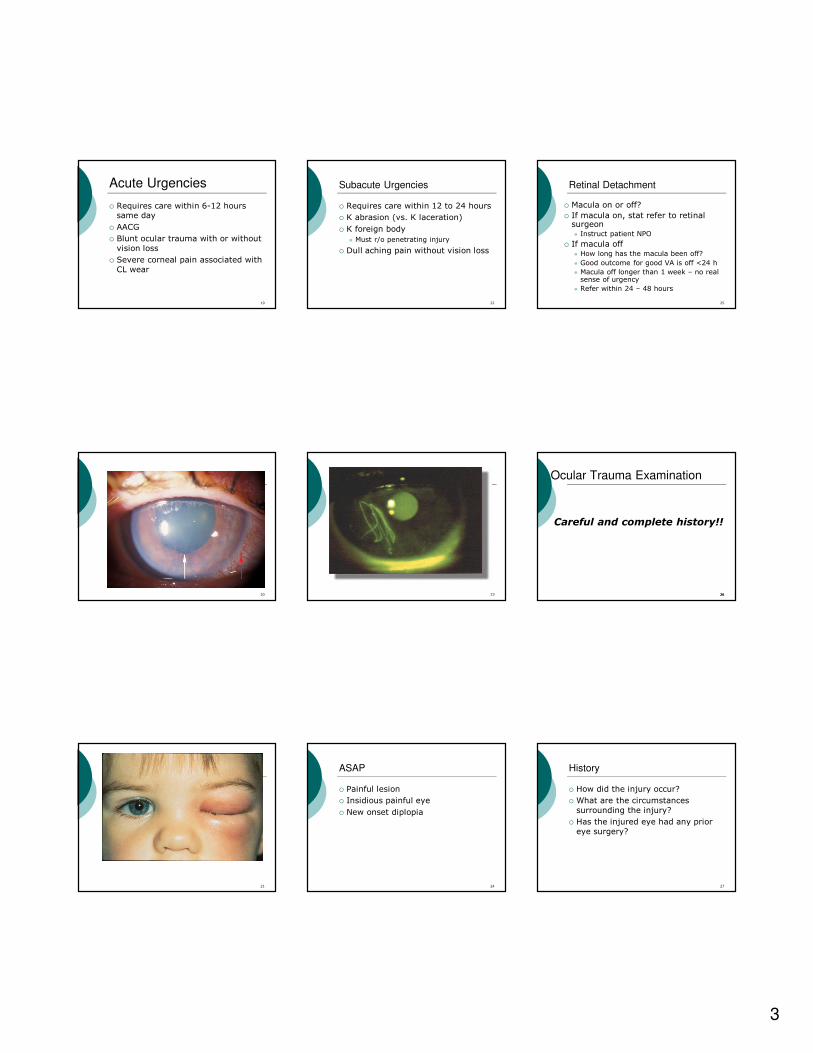
Ocular Trauma Examination
Careful and complete history!!
26
History
� How did the injury occur?
� What are the circumstances
surrounding the injury?
� Has the injured eye had any prior
eye surgery?
27

4
28
Ocular Trauma Examination
� Physical Examination
� Gross exam of head, eyes, ears, nose, face
� Blood pressure and pulse rate
� Review of systems
� Medical history �including tetanus vaccine status
� HPI �changes in vision, eye pain, swelling, discharge, etc.
28
29
Ocular Trauma Examination
� Unaided and pinhole acuities in BOTH eyes
� Topical anesthetic if necessary
� If chemical exposure � begin emergency treatment before VA
� Pupils and EOMs
� Lids and orbits
� Anterior segment
� IOP � unless ruptured globe
� DFE
� B-scan� if indicated
29
30
Methods to Reduce Anxietyo Reassure the Patient you can help him/her
o Explain procedures in advance, esp. to children
o Never patch both eyes unless both eyes are injured
o Place the Patient in a calm, quiet setting
o Be mindful of Patient’s privacy/modesty
o Be realistic, but not overly pessimistic about potential outcomes; impart a sense of hope and optimism (but never lie!)
o Explain in such a way the Patient understands the extent of injury; deliver the message with compassion and candor
30
31
Types of Ocular Trauma
�Mechanical� Superficial corneal/conj abrasions
� Corneal/conj/scleral foreign bodies
� Blunt and penetrating injuries
�Chemical
� Thermal
�Combination
31
32
Birmingham EyeTrauma
Terminology System (BETTS)
� Eyewall: Sclera and cornea
� Closed globe injury: No full-thickness
wound of eyewall
� Lamellar laceration: Partial-thickness wound of eyewall, created by a sharp object
� Contusion: tissue damage created by energy of blunt force trauma
� Superficial foreign body32
33
BETTS
� Open globe injury: Full-thickness wound of eyewall
� Laceration: Full-thickness wound of eyewall, caused by sharp object
�wound at impact site, “outside-in” mechanism
� Penetrating injury� Entrance wound only – corneal foreign body
� Perforating injury� Entrance and exit wound, both caused by same agent – IOFB
33
34
BETTS Terminology
�Rupture: Full-thickness wound of eyewall, caused by blunt object
�eyewall yields at its weakest point to momentary increase in IOP, “inside-out” mechanism
34
35
Birmingham Eye Trauma Terminology System
35
36
Classification of Ocular Trauma
4 categories of globe injury at initial
examination:
� Type
� Grade
� Presence or absence of APD
� Extent (zone) of injury
36

5
37
Classification of Ocular Trauma
37
38
Classification of Ocular Trauma
38
39
Classification of Ocular Trauma
39
40
Classification of Ocular Trauma
40
41
Recommended Timing of Intervention
ECH = Expulsive Choroidal Hemorrhage 41
42
Mechanical Injuries
42
43
Corneal Abrasion
� One of the most common globe injuries� 10% of new patient ER visits
� Frequently accompanies deeper ocular trauma
� Results when basal epithelial cells are removed from the basement membrane
� Scarring occurs if Bowman’s layer is breached
� Source of corneal epithelial restoration is believed to be limbal stem cells at the corneoscleral junction � damage to these cells result in healing problems
43
44
Corneal Abrasion
� Symptoms appear out of proportion to the severity of the injury
� Photokeratitis (UV-induced corneal damage, welder’s burn) presents with similar symptomology to abrasion, but symptoms are delayed 6-12 hours post-exposure
� Topical antibiosis (4GFQ), NSAID and cycloplegia the preferred tx. PO analgesia. Bandage CL ?
� RTC 24 h 44
45

6
46
47
Corneal Abrasion
47
Man vs. Weedeater
48
Man vs. weedeater
49
50
Recurrent Corneal Erosion
� 7-8% of corneal abrasions result in RCE
� RCE represents abnormal adhesion in
the base of the epithelial defect
� Especially common if injury involves:
� Fingernail
� Paper cut
� Classic AM syndrome
50
51
Recurrent Corneal Erosion
51
52
RCE Flowchart
3052
53
Corneal Foreign Bodies
� Represent 40% of eye injuries
� Strong association with high-risk activities without safety Rx – hammering, welding, grinding
� R/O any intraocular material
� Remove superficial FB with FB spud or 30-
gauge needle, cycloplege, topical Ab and NSAID
53
54
Corneal Foreign Bodies
�Symptoms are frequently out of proportion to
severity of injury
� Determine depth with thin optic section, esp. for transparent FB – glass or plastic . R/O self-
sealing lacerations
� To ensure no corneal perforation/IOFB:
Seidel’s, Gonioscopy, DFE
� Deep stromal FB: Leave in place if inert, small, non-toxic/antigenic, non-vegetative
54

7
Full-Thickness Corneal Foreign
Body
55
56
Corneal Foreign Bodies
3356
57
58
59
Corneal Foreign Bodies
59
60
Don’t Forget the Conjunctiva !
61
Intraocular Foreign Body
� Intraocularly retained projectiles
� History is crucial diagnostic tool
� Primary purpose in detection is to prevent associated conditions
(endophthalmitis, RD)
� MRI safety with metallic foreign bodies is still controversial; CT preferred
imaging modality61
62
Intraocular Foreign Bodies
62
63Photo courtesy Mamta Somaiya, MD

8
64
Intraocular Foreign Bodies
64
65
Corneal Lacerations
� Determine partial or full-thickness
� Check IOP, if possible
� If cannot check IOP, evaluate AC depth compared to fellow eye
65
66
Corneal Lacerations
�Small, self-sealing� topical antibiotic
� Large, self-sealing� bandage CL or corneal glue + Ab
� suture if high risk of reopening
� Flaps� in place →
bandage CL + Ab
� displaced →flap repositioned, sutured in place
� if epithelial ingrowth →flap debridement + BCL
66
67
68
69
70
Corneal Lacerations
Lens opacification suggests injury to deeper intraocular tissues
70
Corneal Laceration with Iris
Damage
71
72
Scleral and Corneoscleral
Injuries
�Traumatic corneoscleral defects occur:� acutely from traumatic event
� secondarily from tissue necrosis of post-traumatic inflammation/infection
� most always require surgical intervention �Suturing or patching
�Management goals:� restore integrity of globe
� avoid further injury to ocular tissues
� prevent corneal scarring and astigmatism
72

9
73
Case #1
� 32 year-old mechanic
� Fan belt broke and hit OD 15 min.
ago (no safety glasses)
� VA OD 20/200 PHNI, OS 20/20
� SLE: Corneal, scleral and lid
lacerations, distorted pupil OD
� What do you want to know next?
� Presence or absence of APD OD
73
7474
75
76
Man vs. Fishhook
77
Scleral and Corneoscleral Injuries
Role of the O.D.:
� Recognize the full extent of injury
� If questionable, treat as open globe� tissue prolapse is diagnostic
� If appropriate, exclude or confirm presence of IOFB
� Institute medical therapy if indicated prior to surgical evaluation
Open Globe Injury
� Signs that suggest the presence, or possibility of open globe trauma
include:
� Obvious open wound
� Collapsed or severely distorted eye
� Prolapsed uveal tissue
� Peaked pupil
� SCH with shallowing, or deepening
of the AC
� Ocular hypotony78
79
81
Global Rupture (Open Globe Injury)
If global rupture is suspected, protect globe and orbital adnexa… Never patch!
81

10
82
Global Rupture (Open Globe Injury)
o Protect the eye with a rigid cover (not a patch!)
oDo not instill any eye medications before evaluation by oculo-plastic specialist
oOculo-plastic specialist will order imaging studies
oX-ray with Caldwell, Waters and Lateral views
oCT scan with axial, coronal and sagittal views
82
83
Caldwell (Coronal) View
83
84
Waters View
84
85
Lateral (Sagittal) View
85
86
Axial View
86
87
Blunt Force Trauma
87
88
Blunt Force Trauma Eye Injuries
� Eye is struck with a solid object
� Extent of injury is dependent on size and speed of object
�Smaller the object, greater the velocity
�Small solid objects traveling at high speeds can cause global rupture
(e.g., BB’s, paintballs)88
89
Case # 2
� 34-year-old attorney attending New
Year’s Eve party
� His law partner shouts, “Watch this!”
� Young attorney turns to “watch this”…
89
90
Case # 2
90

11
91
Case # 2
91
92
Sub-Conjunctival Hemorrhages
�SCH w/ superficial conj abrasion, good VA� antibiotics and analgesics
�SCH w/ superficial conj laceration, good VA � reorganization of conj, possible suture with topical antibiotic
�Suspect posterior globe rupture if conjunctivalabrasion/laceration accompanied by:
� lid swelling� extensive SCH (“jelly roll”)� deepened AC� poor VA� APD
92
93
94
95
Mechanisms of Blunt Ocular
Trauma
� Coup� Initial force produced at point of impact
� Contracoup� Shock wave transmission through ocular structures
� Equatorial expansion� Equator expands and distorts normal ocular architecture
� Global repositioning� Compression and indentation at moment of impact, damaging internal ocular structures
95
96
COUP
contrecoup
Equatorial expansion
97
Blunt Force Trauma Eye Injuries
97
98
Traumatic Iritis
� Inflammatory reaction of the iris or CB
� Commonly seen after blunt trauma
� Pain, photophobia, epiphora
� May not present until 2-3 days post-
trauma
98
99
Traumatic Iritis
99

12
100
101
Iridodialysis
102
Iridodialysis
103
Iris Sphincter Rupture
104
Iridodialysis
105
106
Case # 3
� 12-year-old boy gets paintball gun for Christmas
� Parents ignore Dr. Mason’s article in Southern Medical Journal and allow Junior to play war games in wooded area behind their home
� Moderate discomfort, photophobia OS
� Entering unaided acuities: � OD 20/20 � OS 20/100 PHNI
� SLE OS reveals… 106
107
Case # 3
107
108
Hyphema

13
109
110
111
112
Management Guidelines -
Traumatic Hyphema
� Limited activity or bedrest w/ bathroom privileges
� Elevate head 30 degrees. Fox shield r-t-c !
� Atropine 1% t.i.d., or b.i.d. if microhyphema
� No ASA or NSAIDs; mild analgesics only (acetaminophen). No sedatives !
� If traumatic iritis develops (usu. 2-3 days after trauma), add prednisolone acetate 1% 4-8x daily
� For elevated IOP: beta blocker 1st; then alpha agonist. Avoid prostaglandin analogs & miotics
112
113
Management Guidelines -
Traumatic Hyphema
� Office visit daily for 3 days post-trauma/rebleed
� Check VA, IOP, SLE (check for new bleeds)
� Glasses or eye shield during the day and at night for 2 weeks post-trauma/rebleed
� No strenuous activities (including Valsalva) for 2 weeks after initial trauma/rebleed
� Perform gonioscopy one month post-trauma/rebleed
� Treat increased IOP as indicated
114Case CW – woman vs. fish lead sinker
115
Traumatic Glaucoma
� Early onset
�TM obstruction, inflammation
�TM disruption
�Hyphema
� Delayed onset
�Angle recession
�Lens-Associated� Phacomorphic, phacolytic, lens particle
115
116
Traumatic Glaucoma
Commonly injured intraocular structures following contusion: 1. Iris sphincter 2. Iris root (iridodialysis) 3. Between CB
longitudinal and circular m. (angle recession) 4. CB insertion
(cyclodialysis) 5. TM 6. Zonules (subluxated/dislocated lens) 7. Retinal tears/dialysis
116
Angle Recession
117

14
Angle Recession
118
119
Flowchart for Management of
Traumatic Glaucoma
119
120
Chemical Eye Injuries
� Devastating injuries that may lead to blindness
� Alkaline agents penetrate ocular tissues rapidly and continue to do so,
causing severe damage
� Acids cause proteins to coagulate on
corneal epithelium and stroma, and acids precipitate out quickly, which limits ocular penetration
120
121
Pathophysiology of Chemical
Eye Injury
� Both alkalines & acids cause ocular surface epithelial cells to die upon contact
� Retained particulate matter in superior fornix can cause continued exposure
� Penetration of alkalines and acids into corneal stroma result in keratocyte death & loss of stromal clarity
� Hydration of collagen fibrils lead to thickening of TM and increase in IOP
� Time of penetration into AC varies (immediate for ammonia) & can result in secondary glaucoma, cataract, CB damage, hypotony, and phthisis bulbi with prolonged pH >11.5
121
122
Pathophysiology of Chemical
Eye Injury
� Complete corneal epithelial injury requires epithelium from the limbus, where stem cells of corneal epithelium reside
� The recovery of an intact and phenotypically normal corneal epithelium is the most important determinant of a favorable outcome following chemical eye injury
� With extensive corneal and limbal epithelial injury, the surrounding conjunctival epithelium provides the only source of epithelial regeneration
122
123
Alkaline agents commonly associated
with eye injuries
�Ammonia*�fertilizers, household cleaning agents
�Lye*
�drain cleaners
�Magnesium hydroxide �sparklers
�produce thermal and alkali burns
�Lime �plaster, mortar, cement, whitewash
123
124
Alkaline Eye Injury
124
Case SM - unresolved chemical burn s/p 4 weeks
Case SM - s/p 4 weeks chemical burn
15
Case SM - Chemical burn to conjunctiva s/p 4 weeks
Case RC - Chemical Burn OS s/p 3 days
Case RC - chemical burn OS s/p 3 days
130
Case RC – amniotic membrane graft s/p 2 w chemical burn
Case RC - s/p 1 m Chemical Burn OS – Note K re-epithelizing
Case RC – s/p 1 m chemical burn OS. K repithelizing but w/ defect remaining
Case RC – s/p 1 month chemical burn OS
134
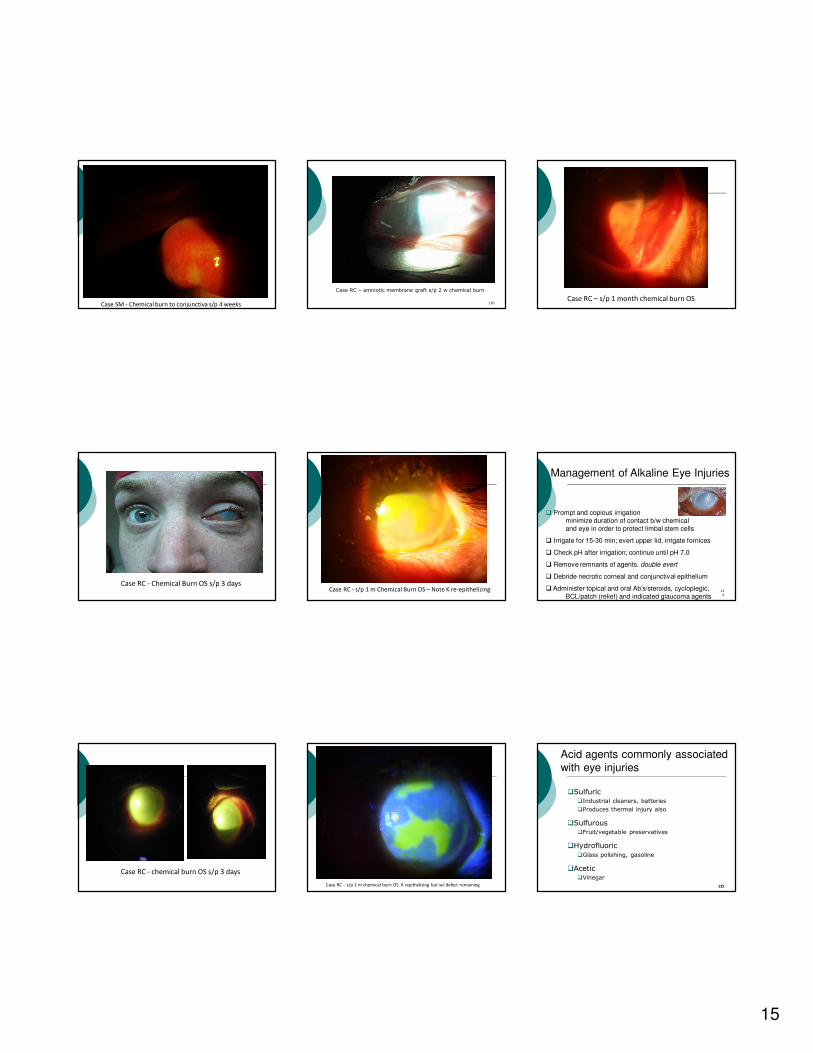
Management of Alkaline Eye Injuries
� Prompt and copious irrigation minimize duration of contact b/w chemical and eye in order to protect limbal stem cells
� Irrigate for 15-30 min; evert upper lid, irrigate fornices
� Check pH after irrigation; continue until pH 7.0
� Remove remnants of agents. double evert
� Debride necrotic corneal and conjunctival epithelium
� Administer topical and oral Ab’s/steroids, cycloplegic, BCL/patch (relief) and indicated glaucoma agents
135
Acid agents commonly associated
with eye injuries
�Sulfuric �Industrial cleaners, batteries
�Produces thermal injury also
�Sulfurous �Fruit/vegetable preservatives
�Hydrofluoric
�Glass polishing, gasoline
�Acetic �Vinegar
135

16
136
Acid Eye Injury
136
137
Management of Acid Eye Injuries
� Prompt and copious irrigation
�minimize duration of contact b/w chemical & eye in order to protect limbal stem cells
� Irrigate for 15-30 min; evert upper lid, irrigate fornices
� Check pH after irrigation; continue until pH 7.0
� Remove remnants of agents. double evert
� Debride necrotic corneal and conjunctival epithelium
� Administer topical & oral Ab’s/steroids, cycloplegic, BCL/patch (relief) and indicated glaucoma agents
137
Management of Chemical Eye
Injuries
� Bandage contact lens
� 4GFQ: 1 gtt 4-6x/day (prevents infection)
� Prednisolone phosphate: 1 gtt q 1-2 hr while awake
(reduces inflammation)
� Vitamin C: 1-2 gm po QD (reduces corneal thinning/ulceration)
� 10% sodium citrate: 1 gtt q 2 hr while awake (chelates
Ca++ and impairs PMN chemotaxis)
� Scopolamine 0.25%: 1 gtt TID (reduces pain/scarring with AC inflammation)
� 10% Mucomyst (n-acetyl-cysteine): 1 gtt 6x/day
(mucolytic agent and collagenase inhibitor)
� Doxycycline 100 mg po bid (collagenase inhibitor)
� Glaucoma gtts/oral diamox if IOP elevated
138
Morgan Lens
139
Morgan Lens Instrumentation
� Morgan Lens
� Molded Scleral lens with an aqueous lock that is attached to an IV bag
� IV bag with sterile 0.9% saline/lactated
Ringer’s
� Litmus paper
� Emesis basin or fluid management system
� Topical anesthetic
140
Morgan Lens: Procedure
� Instill topical anesthetic
141
Morgan Lens: Procedure
� Attach IV infusion tubing to the lens & start a minimal flow
142
Morgan Lens: Procedure
• Have the patient look down and insert the Morgan lens under the upper lid
• Position the lens horizontally as the patient looks straight ahead
143
Morgan Lens: Procedure
� Adjust the flow to the desired rate
144

17
Curling Iron Burn
Photo courtesy Sandra M. Brown, MD
Corneal Thermal Burn
146
UV Keratitis
� welder’s flash, tanning booth-eye
� luckily these risk factors seldom co-exist
� sx - pain, tearing, photophobia
� 6-12 h after exposure
� slit lamp + fluorescein
� superficial punctate keratitis = microdots
� cycloplegia, eryth. ung., pressure patch o/n
� consider oral narcotic. It hurts!
148
149
Tissue Prolapse
� Defined as extrusion of intraocular content outside its normal compartment
� Classified as intrabulbar or extrabulbar
� Intraocular tissue prolapse should be
suspected in all open globe injuries
149
150
Iris Prolapse through Limbal
Surgical Wound (Extrabulbar)
150
151
Uveal Prolapse through Corneal
Laceration (Extrabulbar)
151
152
Vitreous Prolapse into Anterior
Chamber (Intrabulbar)
152
Lens Injury
153

18
154
Lens Injury
� Blunt trauma can break zonules
� If 25% or more zonules are broken, the lens produces iridodonesis (trembling of the iris)
� If enough zonules are disrupted, the lens may:� dislocate into the AC
� occlude pupillary space (pupillary block glaucoma)
� subluxate into PC
� be expulsed altogether
� Contusion injuries can cause immediate traumatic cataracts
154
155
Lens Subluxation into Posterior
Chamber
155
156
Lens Subluxated into Posterior
Chamber
156
157
Reference: Rappon
158
159
160
161
162
Penetrating Eye Injuries
o Eye is pierced by sharp object or high velocity missile (BB, glass, metal-on-metal)
o Patient should be admitted to hospital for broad-spectrum IV antibiotics within 6 hours of injuryo Cephazolin or fortified vancomycin plus
o Gatifloxacin or moxifloxacin
o Tetanus update
o Endophthalmitis may develop, leading to permanent blindness
o Nearly 25% of eyes that suffer penetrating wounds are eventually enucleated 162

19
16375
Penetrating Eye Injuries
163
164
Ruptured Globe
�Nearly 20% of patients with ruptured globes do not have apparent signs of perforation
�Vision may be excellent and the most important clue to occult rupture may be…
� what the patient was doing at the time of injury !
164
165
Ruptured Globe
� If rupture suspected from clinical presentation, findings or history, apply metal shield or other protective covering
� Never patch!
� Slightest manipulation of a ruptured globe may compound an already serious problem
165
166
Flowchart for Evaluation of
Possible Open Globe Injury
166
167
Ruptured Globe
Fireworks injury
167
168
Ruptured Globe
Traumatic retina and vitreous
hemorrhage with choroidal rupture
168Photo courtesy Robert Morris, MD
169
Ruptured Globe
Traumatic Choroidal Rupture
169Photo courtesy Robert Morris, MD
170
Ruptured Globe
170
Orbital Blow-Out Fracture
� A patient presenting with an orbital blow out fracture has a history of
blunt trauma to orbit
� Example: fist, baseball, beer bottle
171

20
Signs of Blow-out Fracture
� Restricted globe movement, esp. on elevation
� Orbital crepitus (subcutaneous emphysema)
� Lid edema & ecchymosis
� hypoesthesia of the ipsilateral
cheek, due to entrapment of the infraorbital nerve
172
173
Blowout Fracture
173
174
Blowout Fracture
174
175
176176
177177
178178
Shaken Baby Syndrome
� 15% mortality rate
� Typical victims are male < 6 moa, who
is alone with the perpetrator at the time of injury
� Incidence unrelated to race, socio-economic status or education
� Presenting sign is eye-related in 4 to 6% of cases
� Retinal hemorrhages in 50% - 80% of shaken babies 179
Shaken Baby Syndrome
180

21
Shaken Baby Syndrome
181
182
Predicting Functional Prognosis
o Global rupture and endophthalmitis
carry poor prognosis for vision recovery
o Variables such as age, extent of wound, hyphema, initial VA, intraocular FB, lens
injury, RD – controversial
182
183
Predicting Functional Prognosis
o More pathology = less vision recovery
o Strongly associated with poor vision outcomes:
o Vitreous hemorrhage
o Absence of lens
o Severe distortion of eyewall
183
Predicting Functional Prognosis –
The OTS
184
Predicting Functional Prognosis -
The OTS Score
185
In Conclusion:
� Have a low threshold for an open globe injury
� Never use anesthetics except to examine a patient
� Be extra vigilant with contact lens wearers
� Always do a fundoscopy if you suspect child abuse
� Encourage trauma prevention with safety glasses 186
187
188
Disclosures
� I have no financial interest in any products mentioned in this
presentation. I wish I did. I have 2 kids in college …
188
References
� Kuhn F, Pieramici DJ, eds. Ocular Trauma: Principles and
Practice. New York: Thieme Medical Publishers, 2002.
� Rappon JM. Ocular trauma management for the primary care provider. 2004. Pacific University College of Optometry On-Line
CE. Available at: http://www.opt.pacificu.edu/ce/catalog/10310-SD/Triage.html
� Naidu K. The injured eye: practical management guidelines and
referral criteria for the rural doctor. SA Fam Pract 2006; 48 (7): 39-45.
� Pramanik S. Assessment and Management of Ocular Trauma.
University of Iowa, 2007. http://webeye.ophth.uiowa.edu/eyeforum/trauma.htm
o Ferrera PC (ed) et al. Trauma Management, An Emergency
Medicine Approach. Mosby Inc, 2001: 201-215.
189

22
190
Thank you !!!
190