Embed Size (px)
Citation preview

8/3/2019 Ocular Trauma 09
http://slidepdf.com/reader/full/ocular-trauma-09 1/38
State of IllinoisTrauma Nurse Specialist Program
OCULAR TRAUMA
Connie J. Mattera, M.S., R.N., TNS
Time allotment: 1 hour
OBJECTIVES:
Upon completion the participant will
1. identify the anatomical structures of the eye and describe the corresponding physiological functions.
2. explain the assessment maneuvers for evaluating the eye and adnexal areas.
3. describe and define cardinal signs and symptoms of ocular injury.
4. interpret assessment findings to formulate nursing diagnoses.
5. establish a nursing care plan based on the nursing diagnoses.
6. describe the correct method for removing contact lenses, irrigating eyes, applying eye shields or
pressure patches.
7. discuss the etiology, pathophysiology, clinical presentation assessment findings, emergency
management, and possible complications for specific ocular injuries including orbital fractures, lidlacerations, chemical and thermal burns, UV exposure, corneal/conjunctival foreign bodies, corneal
abrasion/laceration, subconjunctival hemorrhage, globe penetration/perforation, hyphema, lens
dislocation/subluxation, retinal detachment, optic nerve injury and enucleation.
CJM: 1/09
8/3/2019 Ocular Trauma 09
http://slidepdf.com/reader/full/ocular-trauma-09 2/38
8/3/2019 Ocular Trauma 09
http://slidepdf.com/reader/full/ocular-trauma-09 3/38
State of Illinois TNS Program page 2
Ocular Trauma
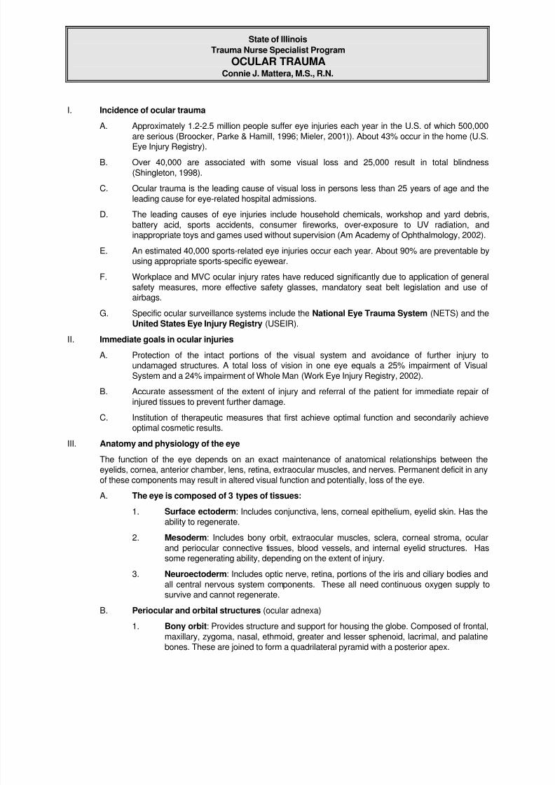
Damage to these bones can cause globeinjury. Since the globe and orbit are in close
approximation to many other important non-ocular structures, serious ocular injury is
often seen in the context of serious non-ocular injury.
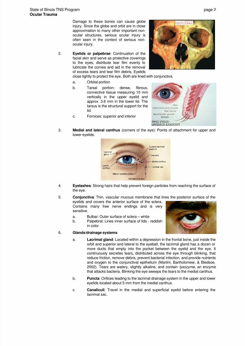
2. Eyelids or palpebrae: Continuation of the
facial skin and serve as protective coveringsto the eyes, distribute tear film evenly to
lubricate the cornea and aid in the removalof excess tears and tear film debris. Eyelids
close tightly to protect the eye. Both are lined with conjunctiva.
a. Orbital portion
b. Tarsal portion: dense, fibrous,connective tissue measuring 10 mm
vertically in the upper eyelid andapprox. 3.6 mm in the lower lid. The
tarsus is the structural support for thelid.
c. Fornices: superior and inferior
3. Medial and lateral canthus (corners of the eye): Points of attachment for upper and
lower eyelids.
4. Eyelashes: Strong hairs that help prevent foreign particles from reaching the surface of
the eye.
5. Conjunctiva: Thin, vascular mucous membrane that lines the posterior surface of the
eyelids and covers the anterior surface of the sclera.
Contains many free nerve endings and is verysensitive.
a. Bulbar: Outer surface of sclera – whiteb. Palpebral: Lines inner surface of lids - reddish
in color
6. Glands/drainage systems
a. Lacrimal gland: Located within a depression in the frontal bone, just inside the
orbit and superior and lateral to the eyeball, the lacrimal gland has a dozen or
more ducts that empty into the pocket between the eyelid and the eye. Itcontinuously secretes tears, distributed across the eye through blinking, that
reduce friction, remove debris, prevent bacterial infection, and provide nutrientsand oxygen to the conjunctival epithelium (Martini, Bartholomew, & Bledsoe,
2002). Tears are watery, slightly alkaline, and contain lysozyme , an enzymethat attacks bacteria. Blinking the eye sweeps the tears to the medial canthus.
b. Puncta: Orifices leading to the lacrimal drainage system in the upper and lower
eyelids located about 5 mm from the medial canthus.
c. Canaliculi: Travel in the medial and superficial eyelid before entering the
lacrimal sac.

8/3/2019 Ocular Trauma 09
http://slidepdf.com/reader/full/ocular-trauma-09 4/38
State of Illinois TNS Program page 3
Ocular Trauma
d. Sebaceous glands: Associated with the eyelashes as with other hair follicles.
Secrete a lipid-rich substance that keeps lids from sticking together.
e. Lacrimal caruncle: Soft mass of tissue located at the medial canthus
containing glands that produce thick secretions that contribute to the gritty
deposits sometimes found after a night's sleep.
C. Extrinsic eye (oculomotor) muscles: Originate on the surface of the orbit and control the
position of the eye.
1. Medial rectus: Eye rotates toward the nose CN III2. Inferior rectus: Eye looks downward CN III
3. Inferior oblique: Eye looks upward and to the side CN III4. Superior rectus: Eye looks upward CN III
5. Superior oblique: Eye looks down and to the side CN IV6. Lateral rectus: Eye rotates laterally away from nose CN VI
D. Eyeball (Globe)
1. Each eye is roughly spherical with a diameter of about 2.5 cm (1 inch). The length from
the apex of the cornea to the point at which the optic nerve exits the sclera isapproximately 24.5 mm. The globe weighs 7.5-8.0 g and has a volume of 6.5 ml. Itoccupies 1/5th of the orbital volume. It shares space with the extrinsic eye muscles, the
lacrimal gland, the various cranial nerves and blood vessels that service the eye andadjacent areas of the orbit and face, and orbital fat that provides padding and insulation
(Martini, Bartholomew, & Bledsoe, 2002).
8/3/2019 Ocular Trauma 09
http://slidepdf.com/reader/full/ocular-trauma-09 5/38

8/3/2019 Ocular Trauma 09
http://slidepdf.com/reader/full/ocular-trauma-09 6/38
State of Illinois TNS Program page 5
Ocular Trauma
(3) Secretes and reabsorbs aqueous humor that circulates within the eye(4) Controls the shape of the lens (Martini et al, 2002)
b. Iris: Circular, contractile muscular disc that is an extension of the ciliary body
that is located anterior to the lens. It contains pigment cells that produce thecolor of the eye and two layers of smooth muscle. The iris controls the amount
of light reaching the retina by dilating and constricting its muscles to changepupillary size in response to impulses mediated by CNs II & III. When there are
no pigment cells in the iris, light passes through it and bounces off its inner
surface of pigmented epithelium. The eye then appears blue. In order, personswith gray, brown, or black eyes have increasing numbers of pigment cells in thebody and surface of the iris (Martini, Bartholomew, & Bledsoe, 2002).
c. Pupil: Typically is a round central opening in the iris
(1) Parasympathetic stimulation: Rapid reflex constriction of the pupil in
response to bright light.
(2) Sympathetic stimulation: Slower pupillary dilation in response to a
reduction in light levels
d. Ciliary body: Along its outer edge, the iris attaches to the anterior portion of
the ciliary body which is composed primarily of a ring of ciliary muscle that
projects into the interior of the eye. It begins at the junction between the corneaand sclera and extends to the anterior edge of the retina. Posterior to the iris,the suspensory ligaments of the lens attach to folds called the ciliary
processes. These fibers position the lens so light passing through the pupil
goes through the center of the lens. Responsible for producing aqueous humorand for changing the shape of the lens.
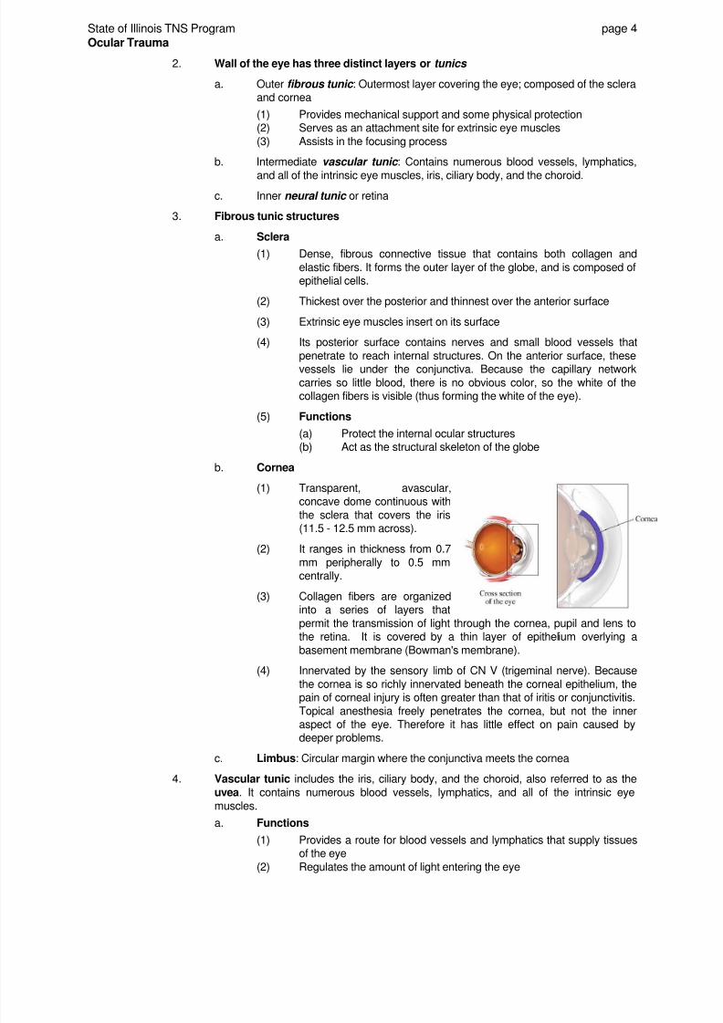
e. Lens: Disc-shaped structure approximately 9 mm in diameter and 4 mm thick,
containing transparent crystalline matter, suspended immediately behind theiris and anterior to the vitreous by suspensory ligaments that connect it to the
wedge-shaped ciliary body. The lens is highly elastic and contraction orrelaxation of the ciliary body changes its thickness and shape, thereby
permitting images from varied distances to be focused on the retina.
Its shape changes for near and far vision, becoming flatter for far vision. Theability to change shape deteriorates with age explaining need for readingglasses after 40...
8/3/2019 Ocular Trauma 09
http://slidepdf.com/reader/full/ocular-trauma-09 7/38
State of Illinois TNS Program page 6
Ocular Trauma
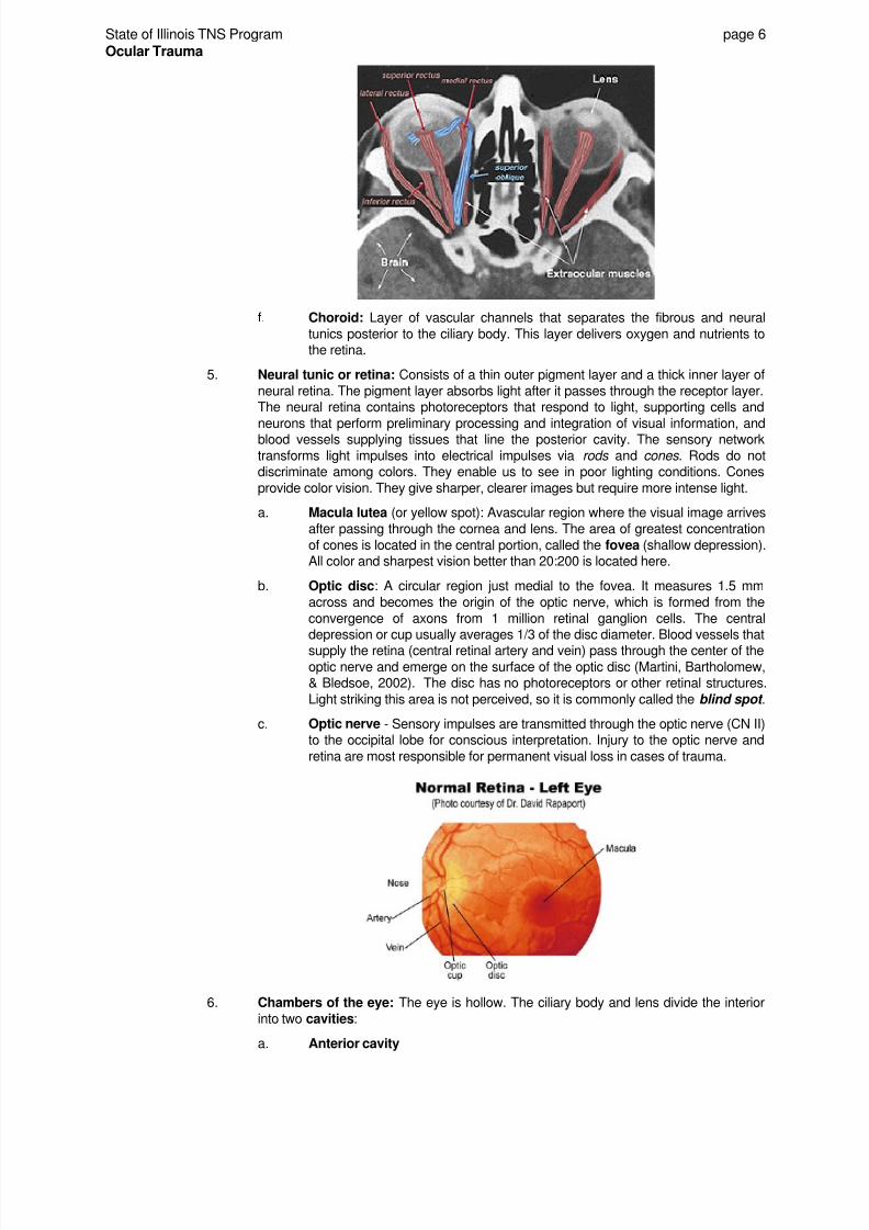
f. Choroid: Layer of vascular channels that separates the fibrous and neural
tunics posterior to the ciliary body. This layer delivers oxygen and nutrients tothe retina.
5. Neural tunic or retina: Consists of a thin outer pigment layer and a thick inner layer of
neural retina. The pigment layer absorbs light after it passes through the receptor layer.The neural retina contains photoreceptors that respond to light, supporting cells and
neurons that perform preliminary processing and integration of visual information, andblood vessels supplying tissues that line the posterior cavity. The sensory network
transforms light impulses into electrical impulses via rods and cones . Rods do notdiscriminate among colors. They enable us to see in poor lighting conditions. Cones
provide color vision. They give sharper, clearer images but require more intense light.
a. Macula lutea (or yellow spot): Avascular region where the visual image arrives
after passing through the cornea and lens. The area of greatest concentration
of cones is located in the central portion, called the fovea (shallow depression).All color and sharpest vision better than 20:200 is located here.
b. Optic disc: A circular region just medial to the fovea. It measures 1.5 mm
across and becomes the origin of the optic nerve, which is formed from the
convergence of axons from 1 million retinal ganglion cells. The centraldepression or cup usually averages 1/3 of the disc diameter. Blood vessels that
supply the retina (central retinal artery and vein) pass through the center of theoptic nerve and emerge on the surface of the optic disc (Martini, Bartholomew,& Bledsoe, 2002). The disc has no photoreceptors or other retinal structures.
Light striking this area is not perceived, so it is commonly called the blind spot .
c. Optic nerve - Sensory impulses are transmitted through the optic nerve (CN II)to the occipital lobe for conscious interpretation. Injury to the optic nerve and
retina are most responsible for permanent visual loss in cases of trauma.
6. Chambers of the eye: The eye is hollow. The ciliary body and lens divide the interior
into two cavities:
a. Anterior cavity

8/3/2019 Ocular Trauma 09
http://slidepdf.com/reader/full/ocular-trauma-09 8/38
State of Illinois TNS Program page 7
Ocular Trauma
(1) Divided into two chambers
(a) Anterior chamber: Extends from the cornea to the iris
(b) Posterior chamber: Extends from the iris to the ciliary body
and lens.
(2) Contents: Filled with aqueous humor produced by the ciliary
processes and secreted into the posterior chamber. The fluid
circulates within the anterior cavity, passing from the posterior to theanterior chamber through the pupil. It leaves the anterior chamber
near the edge of the iris through the canal of Schlemm that returns thefluid to the venous system. The anterior cavity holds about 125
microliters of fluid. Interference with secretion, circulation, orabsorption can change the pressure within the eye. Glaucoma iscaused by an elevation in ocular pressure and can produce blindness
by distorting the retina and the optic disc.
b. Posterior cavity or vitreous chamber contains the gelatinous vitreous body.Helps to maintain the shape of the eye and holds the retina against the choroid.
7. Vascular supply
a. Arterial: The ophthalmic (central retinal) artery is the main vessel that supplies
blood to the globe and orbit and is the first branch off of the internal carotid
after it enters the cranial cavity. Emboli from the internal carotid artery canenter the ophthalmic artery, producing visual problems. Facial and maxillary
arteries feed the lower lid, medial canthus and inferior orbit.
b. Venous: Blood returns through the inferior and superior ophthalmic veins.
Venous draining through the cavernous sinus is an important route for the
intracranial spread of infection. There are no lymphatics in the orbit, but the eyelid has a rich lymphatic system.
IV. Assessment and management of ocular trauma
Many patients presenting with ocular trauma have other associated trauma to the head and neck withfacial and skull injuries that may delay recognition of ocular damage. While understandable, an early
consult with ophthalmology is desirable (Broocker et al, 1996).
A. Chief complaint/history of present illness: Because the nature of eye emergencies ranges
from minor to severe, history taking may be done simultaneously with the eye exam. Whenever
time permits, obtaining an accurate, detailed history can aid in the rapid treatment and evaluationof the patient. Important details to include:
1. Nature of complaint: traumatic vs. non-traumatic
a. Non-traumatic: Divided into c/o of vision, appearance or sensation
b. Traumatic: Mechanism of injury - blunt, penetrating, or explosive; thermal, UV,
chemical materials involved. If chemical exposure was involved ask if it wastoxic, acid or alkali.
2. HPI - Detailed description of chief complaint: Obtain OPQRST details about onset,
provocation (what object or item caused the injury - magnetic?)/ palliation, quality,region/recurrence, severity, and date and time of injury.
a. Visual changes from baseline: Blurred, double, or deficits in acuity or visual
fields
b. Is one or are both eyes affected?
c. Diplopia (double vision)
(1) Diplopia may indicate trauma to the globe with muscle entrapment ornerve deficit, peripheral or central (Cuculino & DiMarco, 2002)
(2) Monocular diplopia usually indicates a refractive error in the eye itself
(3) Binocular diplopia, present only when both eyes are open, is the resultof a deficiency in the movement of the eye
8/3/2019 Ocular Trauma 09
http://slidepdf.com/reader/full/ocular-trauma-09 9/38
State of Illinois TNS Program page 8
Ocular Trauma
d. Rednesse. Tearing, drainage
f. Paing. Note associated complaints/symptoms other than decreased vision
B. SAMPLE History
1. Allergies
2. Medications: Regular use of eye medications or over-the-counter remedies. Use of
anticoagulants or aspirin. Include licit and illicit drug use, as they can affect pupillary sizeand reactivity.
3. Past ocular history
a. Any ocular or visual problems - past or present change in, or loss of, vision;ocular pain
(1) Blurred vision that does not improve with blinking(2) Diplopia
(3) Sectorial visual loss(4) Spots before eyes, curtain over the visual field?
(5) Halos or rings around lights(6) Photophobia
b. Baseline visual acuity; ever worn glasses or contact lenses? How long?
If contacts are worn: hard, soft, extended wear? For reading, driving, distance?c. Eye injury or surgery? When?
d. Excessive tearing, crusting, discharge?
e. Burning (inflammation)
f. Relevant occupational hazards; dust, chemicals, metalwork
(1) Use of protective devices
(2) Use of safety glasses, if traumatic injury
4. Past medical history in self or family
a. Heart disease, hypertensionb. Diabetesc. Sickle cell disease
d. Liver diseasee. Vascular disorders
f. Cataracts, glaucoma
5. Last oral intake; tetanus
6. Events surrounding the incident: Details regarding probable size, velocity, and
chemical constituency of pellets or projectiles; determine treatment rendered prior toarrival.
C. Physical exam
1. The physician may require the following for an adequate exam:
a. Near vision card, pinhole occluder (for vision < 20/30), loose +2.75 spherespectacle lens for the presbyopic patient; occluder for assessing each eyeseparately.
b. Penlight with cobalt blue filterc. Schiotz tonometer, tonopen
d. Ophthalmoscopee. Topical anesthetic: Research by a group of New Mexico physicians suggests
that a patient's response to local anesthesia may predict the complexity of a
corneal lesion. Those that receive significant pain relief probably have anuncomplicated corneal lesion.
Patients with a diagnosis of conjunctivitis, iritis, corneal ulcer, hyphema,glaucoma, or subconjunctival hemorrhage typically had less pain initially and
got little relief from proparacaine (Sklar et al, 1990).
f. Lid retractor or bent paper clip retractors

8/3/2019 Ocular Trauma 09
http://slidepdf.com/reader/full/ocular-trauma-09 10/38
State of Illinois TNS Program page 9
Ocular Trauma
g. Sodium fluorescein test strips to evaluate corneal epithelial loss
h. Short-acting dilating (mydriatic) drops; use with caution in patients with
concomitant intracranial injury
i. Slit lamp: Aids in the evaluation of the conjunctiva, cornea, anterior chamber,
iris, lens and anterior vitreous cavity. It can detect the presence of blood in the
anterior chamber, inflammatory cells, and pus.
2. Remove and store contact lenses: Lenses should usually be removed as part of
trauma assessment/management
a. Hard contact lenses: Remove with suction cup lens remover. Place in 0.9%
NS and label containers left and right.
b. Soft contact lenses: Easily removed by pinching the lens together and pulling
away from the eye. Place immediately in 0.9% NS and label containers left and
right.
3. External structures - Inspect/palpate
Order: top to bottom; outside to inside (cornea to fundus); medial to lateral (inner
canthus to outer canthus)
a. Orbit and periorbital structures: Often, one needs only to visually inspect the
patient's face to suspect injury. However, bony palpation of the orbital rim candetect point tenderness, deformity or crepitus. Orbital rim should be smooth,without deformities or irregularities. Insect/palpate for the following:
(1) Deformity, contusion, abrasion, punctures, laceration
(2) Ecchymoses, bleeding, hemorrhage, hematoma, F/B(3) Periorbital edema, masses, asymmetry or missing tissue
(4) Tenderness, instability, crepitus: may indicate a sinus fracture(5) Also examine for paresthesias possibly indicating injury to an
infraorbital sensory nerve
b. Eyebrow size and extension
c. Lids: examine closed and open
(1) Inspect integrity of the structure, symmetry; ecchymoses
(2) Lacerations/lesions: Document whether the laceration involves the
lid margins or loss of tissue. Lacerations to the medial 1/3 pose a riskfor disrupting the lacrimal apparatus and require plastic surgery or
ophthalmology for closure (Cuculino & DiMarco, 2002). While seriousthemselves, can also be associated with penetrating trauma to the
globe. Look for prolapse of orbital fat.
(3) Edema, inflammation
(4) Ability to spontaneously lift and close lid to cover the superior limbus ofthe iris by 1 mm - 3 mm. Note the following:
(a) Ptosis: If the lid cannot lift up and covers more of the iris than
the other side or extends over the iris, suspect a congenital oracquired weakness of the levator (plus Muller's) muscle or
paresis of the third cranial nerve.
(b) If the lid cannot close, suspect dysfunction of CN VII whichinnervates the orbicularis muscle.
(5) Surface growths/internal masses; never palpate an injured eye
d. Lashes, sebaceous glands, and drainage system
(1) Presence or absence of discharge or matting(2) Scaling or lesions
(3) Hair loss(4) Pus, blood, CSF from puncta
e. Position of globe within the orbit and in comparison with the ocular structures
on the opposite side
8/3/2019 Ocular Trauma 09
http://slidepdf.com/reader/full/ocular-trauma-09 11/38
State of Illinois TNS Program page 10
Ocular Trauma
(1) Proptosis or exophthalmos: Bulging or protrusion of the eye
(2) Enophthalmos: Backward displacement of the eyeball causing it to
recede within the orbit
(3) Pupils should be on same horizontal plane. If one eye is lower than
the other suspect orbital floor fracture. Eyes should be in line with topof ear. Is gaze focused on you?
4. Anterior segment: This examination includes the conjunctiva, cornea, anterior
chamber, iris and lens and is done by the physician using a slit lamp or penlight withblue filter. The slit lamp allows for magnification of these structures to provide improved
detail and visualization.
a. Conjunctiva: Penlight or light from ophthalmoscope is acceptable if no slitlamp is available. Evert eyelids to fully assess if necessary.
Inspect for
(1) degree and depth of redness/injection;(2) foreign body, embedded material, laceration;
(3) chemosis or hemorrhage;(4) rust ring;(5) black or brown defect (choroid prolapse); and
(6) pus.
b. Sclera
(1) Should be white. Abnormal colors: jaundice, bluish (Marfan'ssyndrome), injected.
(2) Wounds, hemorrhage, ocular Battle sign?
c. Cornea: Should be smooth, round, clear, glistening and free of lesions. To
assess for a corneal defect, instill sodium fluorescein. Wet the tip of a
fluorescein strip with a commercial irrigating solution, touch to the conjunctiva
in the inferior cul-de-sac. Blinking will disperse the fluorescein in the preoculartear film. Fluorescein adheres to and stains any exposed portion of the cornealbasement membrane denuded of epithelium. Excess dye can be removed with
an irrigating solution to provide contrast between the lesion and surroundingareas. Physicians will view under a cobalt blue light to inspect for:
(1) foreign body;
(2) clouding/opacity; could indicate acute glaucoma, edema from trauma,
foreign body in the anterior chamber, or infection;
(3) abnormal pigment;
(4) irregular light reflection if irregular in size or shape;
(5) abrasion/laceration: defects will appear bright green;
(6) positive Seidel test: Positive if the fluorescein appears to be
streaming away from the cornea, forming a pool in the cul-de-sac. Thestreaming is caused by the aqueous humor as it flows out of the
defect (Cuculino & DiMarco, 2002). Indicates an open globe injury.
(7) Anterior chamber: Should be clear and deep with the iris well
separated from the posterior corneal surface. The depth of the anteriorchamber is important to note, especially prior to instilling dilating drops(Cuculino & DiMarco, 2002). Shine a penlight across the cornea from
the lateral to medial direction. With normal depth, the light will shineevenly across the iris. A shallow chamber will show a shadow on the
medial aspect of the iris caused by the iris as it bulges outward intothe anterior chamber. Patients with a shallow anterior chamber may
be at risk for acute angle closure glaucoma if dilated. It may alsoindicate a globe rupture with loss of aqueous humor. If blood(hyphema) or white cells (hypopyon) are present, serious injury has
occurred.

8/3/2019 Ocular Trauma 09
http://slidepdf.com/reader/full/ocular-trauma-09 12/38
State of Illinois TNS Program page 11
Ocular Trauma
d. Intraocular pressure: This should always be measured by a physician when
there is suspicion of hyphema, or any other condition that would affect theintraocular pressure (IOP). It should be deferred if a penetrating globe injury is
suspected (Cuculino & DiMarco, 2002). Normal IOP ranges from 8 to 21mmHg (Varma, 1997). Low intraocular pressures may be found withinflammatory conditions (except iritis which may elevate pressures) and globe
rupture. Instruments used to measure IOP are the Goldmann Applicationtonometer (attached to the slit lamp), the Schiotz tonometer, and the Tonopen.
IOP can also be measured using puff bursts of air blown against the cornea.Two readings are obtained using this process.
(1) Instill topical anesthetic in each eye if a sensor is placed on thecornea. If puffs of air are used, no anesthetic is necessary.
(2) Have patient look forward with eyes open in a dimly lit room. If AMS,
hold eyelids open, supporting upper lid against superior rim and thelower lid against inferior rim of the orbit. Do not push on eyeball!
(3) The Tonopen is an automated, hand-held machine. After calibration,
tap the covered tip gently against the anesthetized eye with the patient
in a supine position. An average of three readings is taken.
(4) Schiotz
(a) Place 5.5 G wt on tonometer
(b) Have pt. look up and rest foot-plate of the tonometer on thecentral portion of the cornea. Read the scale.
(c) If the reading is less than 5 apply the 7.5 wt on top of the 5 Gwt. Continue to add weights until the scale reads between 5
and 20.
(d) The scale value is translated into an intraocular pressure
using a special table. Normal pressure is ≤ 21. Dangerouslyhigh pressures are > 30 mmHg and indicate angle closureglaucoma, pupillary block glaucoma or a retrobulbar
hemorrhage. This is an emergency and requires immediatetreatment by an ophthalmologist.
(5) Goldmann: Instill a drop of fluorescein, and at the slit lamp, a
tonometer tip is laid against the cornea under a cobalt blue light. Thedial is adjusted so that the inside borders of the split prism are aligned.
e. Iris: Should be flat and smooth with regular margins and a centrally located,
round pupil. In trauma, may show tears, holes, or eccentric margins. If torn,suspect ruptured globe.
5. Lens: Should be transparent. Not uncommon to have post-traumatic cataracts develop
which may be visible on penlight exam. In severe trauma, lens may subluxate ordislocate to float free in the vitreous or settle on the retina.
6. Cranial nerve assessment
a. II - Optic: Transmits sensory impulses from the retina to the occipital lobe for
processing.
Assess visual acuity: Vital sign of the eye. Obtain on everyone presentingwith an ocular complaint. Usually done as part of the initial assessment in the
triage area, however, never delay ocular irrigation for chemical burns in order
to obtain visual acuity. This is vital from both a medical and legal standpoint.
(1) Test each eye separately; shield eye that is not being tested with an
occluder. Assess with (cc) and without (sc) correction.
(2) If patient normally wears corrective lenses and they are not available,use a pin hole occlusive device. If one is not available, use an indexcard with a small hole. The pinhole acts as a refractive device by
aligning the beam of light as it enters the eye (Cuculino & DiMarco,2002).

8/3/2019 Ocular Trauma 09
http://slidepdf.com/reader/full/ocular-trauma-09 13/38
State of Illinois TNS Program page 12
Ocular Trauma
(3) Record results from both eyes using the following abbreviations:
(a) OU: Both eyes
(b) OD: Oculus dexter = R
(c) OS: Oculus sinister = L
(4) Note their best vision using one or more of the following:
(a) Snellen eye chart at 20 feet. Record best vision as a fraction.The numerator (20) always represents the distance in feet
between the patient and the eye chart. The denominatordepends on the smallest line of letters the patient is able to
read without straining or squinting. The larger thedenominator, the poorer the vision. Ex: Normal 20/20; very
poor 20/200.
(b) Rosenbaum "near" card held at 14 inches: Note smallest line
of print that can be clearly read.
(c) If patient cannot see largest print on eye chart, determine if
they can count fingers or perceive hand motion. Documentthe distance at which they can see the stimulus.
(d) If patient is unable to see hand motion, document if they canperceive light with or without the ability to determine the
direction from which it is projecting.
(e) If they cannot detect light, record the results as "No light
perception" (NLP) or blind eye.
(f) If pain is a significant factor in achieving patient cooperation
for the exam, instill a topical ophthalmic anesthetic asprescribed.
(g) For children who cannot read, use a picture card, such as anAllen chart, or cards with the letter "E" pointing in various
directions.
(5) Partial differential diagnosis of post-traumatic loss of vision
(a) Lid swelling; blood or foreign material covering cornea;
corneal damage
(b) Hyphema; vitreous hemorrhage
(c) Traumatic cataract; luxation of lens
(d) Central retinal artery or vein occlusion (from markedly
increased orbital pressure or embolus)
(e) Traumatic retinal edema and hemorrhages of retina from
direct or contrecoup blow to head
(f) Retinal detachment
(g) Avulsion of optic nerve by trauma of lateral orbital wall orcontrecoup blow to head
(h) Indirect trauma to optic nerves and/or chiasm (traumatic opticneuritis)
(i) Intracranial interruption of visual pathways
(hemorrhage/foreign body)(j) Cortical blindness from hematoma, ischemia, or anoxia
(patient may be unaware of blindness)
(k) Acute angle-closure glaucoma precipitated by emotionalstress of recent trauma or from intumescent lens, etc.
(l) Hysteria
(m) Malingering
(n) Dislocation or loss of contact lens
(6) Visual fields; peripheral vision

8/3/2019 Ocular Trauma 09
http://slidepdf.com/reader/full/ocular-trauma-09 14/38
State of Illinois TNS Program page 13
Ocular Trauma
b. CN III – Oculomotor
(1) Pupil size, shape, equality: Assess before installation of eye drops.
Have the patient look at a distant object. This prevents distortion fromaccommodation (pupillary constriction that occurs when one looks at a
near object). Compare one side to the other. Pupils should be round,symmetric, midpoint, and equal in size. The size, shape and reactivity
of pupils can be affected by trauma, medications, previous ocularsurgery, and central neurological events as well as by ocular
processes. Obtaining an accurate history is essential.
(a) MIOSIS: Possible etiologies of constricted or small pupils:
(i) Drugs: Acetylcholine, Histamine, Picrotoxin,
Mecholyl, Physostigmine, Morphine (narcotics),
Pilocarpine, Neostigmine, Ergotamine, Diisopropyl &related drugs.
(ii) Physiological from: Sleep, forced closure of lid
(Wesphal-Piltz), near reaction, old age; iris atrophy,direct light stimulation, congenital miosis with no
dilator muscle, consensual light stimulation.
(iii) Pathological from: Areflexia of diabetes mellitus,
meningitis, blind eye, alcoholism, Argyll-Robertsonpupil, carbon dioxide poisoning, Horner's syndromewith ptosis, pontine lesions, encephalitis.
(b) Mydriasis: Possible etiologies of enlarged pupils:
(i) Drugs: Adrenalin, Paradrine, Atropine,
Euphthalmine, Benzedrine, Homatropine, Canabis,Cocaine, Scopolamine, Ammonium chloride, Tetra-
ethyl and related drugs.
(ii) Physiologically normal: Fright reaction, darkness,
strong voluntary efforts to diverge, age - young
myopes.
(iii) Pathological
(a) One eye: Apoplexy, tonic pupil (Adie), brain
shift with pressure on 3rd nerve.
(b) Both eyes: Diphtheria, ciliary gangliamitis
(retrobulbar neuritis), juvenile paresis,pineal tumors, hysterical, botulism,
generalized paresis, catatonia, herniationsyndromes.
(c) Causes of pupil asymmetry
(i) Traumatic mydriasis or miosis from direct blow
(ii) Iridodialysis or rupture of iris sphincter(iii) Unilateral use of topical drugs
(iv) Intraorbital trauma to ciliary nerves or ganglia(v) Acute intraocular inflammation with spasm or atony(vi) Horner's syndrome; third-nerve palsy
(vii) Iritis; Adies pupil(viii) Unilateral blindness
(ix) Stroke; Pontine lesions(x) Lues (Argyll-Robertson)
(d) Eccentric pupils may indicate globe rupture. As the anteriorchamber contents are lost, the iris may be pulled into the
defect, causing the pupil to look asymmetrical and tear dropshaped. (Cuculino & DiMarco, 2002).
8/3/2019 Ocular Trauma 09
http://slidepdf.com/reader/full/ocular-trauma-09 15/38
State of Illinois TNS Program page 14
Ocular Trauma
c. CN II & III: Pupil reactivity to light; direct and consensual. Bring the light in
from the side, observe the direct and consensual light reflex in each eye. Theresponse should be brisk and symmetrical. (Assessing the sensory limb of CN
II and the motor limb of CN III.)
(1) Direct response: Each pupil should react by constricting briskly to a
bright hand light.
(2) Consensual response: The opposite eye's pupil should also
constrict, even in the absence of direct illumination.
(3) Afferent defect: Shine a bright light directly at the healthy eye for 30
seconds and quickly swing it to the other side. Note the first movement
of the pupil. Repeat: swing the light to the other side. Both eyes shouldnormally constrict or remain constricted in a consensual response.
Any disease that blocks light from reaching the optic nerve or blockstransmission of the optic nerve will cause an afferent pupillary defect
where the affected pupil dilates when light is shone into it. That eye isreceiving less light than normal and dilates in an attempt to bring inmore (Cuculino & DiMarco, 2002). This test can be performed even if
only one pupil can be assessed.
In ambient light, even if the optic nerve of one eye has beencompletely transected, the pupils should be equal because the
consensual response of the damaged eye to the good eye will result inequal pupils. Therefore, the swinging light test is essential.
d. Mobility of the globe (EOMs): CN III, IV, VI
(1) Have the patient fixate on an object as it is moved up, down, left andright to evaluate nerve and muscle function. The eyes should move an
equal amount in each gaze direction. Assess for range of each eye,symmetry, dysconjugate gaze, gaze palsies, or pain on movement.
Defer this part of the assessment if a ruptured globe is suspected.
(2) EOMs may be altered in cases of trauma, neurological events,
infections, and tumors. Common causes include orbital edema,muscle entrapment and cranial nerve damage.
(3) Inspect for nystagmus - vertical or horizontal
e. CN V: Trigeminal: Three branches (ophthalmic, maxillary, mandibular) supplysensation to the forehead, upper and lower eyelid, nose, cornea, cheek, upper
lip and gums. Fractures of the orbit can cause loss of sensation to these areas.
7. Fundus/ophthalmoscopic examination: Includes inspection of the optic nerve, disc,
macula, retina and blood vessels. Done through the use of an ophthalmoscope.Visualization is easier with dilation of the pupils (instillation of mydriatic drops), but is notnecessary. The drop effects may last for hours to days and should be documented
carefully, especially in trauma patients.
Examiner should note the following:
a. Red reflex: Present bilaterally; look for opacities
b. Optic nerve: Head should be somewhat yellow with sharply defined marginsc. Discs: Note shape and color. Should be well delineated; disc/cup ratio 1:3
(glaucoma increases size of cup); light yellow to pink; lighter than retinal
background.
d. Retinal vessels: Central retinal artery and vein. Should have no A-V nicking;
A-V ratio 4:5; no papilledema. Veins = no light reflex, arteries have a light
reflex.
e. Background: Light red/orange, no hemorrhages, exudate, changes in
pigmentation, or retinal detachment. Normal background color comes from the
underlying retinal pigment epithelium and the vascular choroid. Retinal edema(Berlin's edema) is common after ocular trauma. Retina will appear milky white.
8/3/2019 Ocular Trauma 09
http://slidepdf.com/reader/full/ocular-trauma-09 16/38
State of Illinois TNS Program page 15
Ocular Trauma
f. Macula: No scarring. Inspect last, will cause tearing. In central retinal artery
occlusion, the macula will present as a "Cherry Red Spot".
g. Vitreous abnormalities: Hemorrhages
D. Diagnostic studies
1. X-ray: Not used as often any more
2. CT scan: C-T scanning is essential to delineate completely the extent of orbital and
optic canal fractures as well as intraocular and orbital foreign bodies.
3. Orbital and intraocular ultrasonography will aid in the evaluation of retinal and
choroidal detachments, intraocular and intraorbital foreign bodies, vitreous hemorrhage,dislocation of the lens, and lacerations of the globe.
4. Optical Coherence Tomography (OCT): Noncontact, noninvasive imaging technique
that uses light rather than sound waves to obtain a much higher longitudinal resolutionof about 10 µm in the retina. Useful for imaging macular diseases and retinal
inflammatory diseases. Has the capability of measuring the retinal nerve fiber layerthickness in glaucoma and other diseases of the optic nerve.
5. Evoked potentials: Helpful in the non-responsive or uncooperative patient for
determining whether the visual pathways are intact.
INJURY TO THE ORBIT
V. Orbital (blow-out) fracture
A. Etiology: Pressures inside the bony orbit increase due to blunt trauma to the globe - often
involve the orbital floor (maxillary bone), injury to the maxillary sinus, and entrapment of theinferior rectus muscle and inferior oblique muscles. May also involve the ethmoid sinus through
the lamina papyracea with entrapment of the medial rectus muscle.
B. 32% of those with a medial blow out fracture will have a concomitant rupture of the globe.
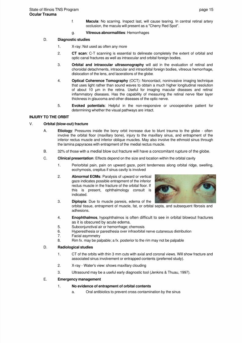
C. Clinical presentation: Effects depend on the size and location within the orbital cavity
1. Periorbital pain, pain on upward gaze, point tenderness along orbital ridge, swelling,
ecchymosis, crepitus if sinus cavity is involved
2. Abnormal EOMs: Paralysis of upward or vertical
gaze indicates possible entrapment of the inferiorrectus muscle in the fracture of the orbital floor. Ifthis is present, ophthalmology consult is
indicated.
3. Diplopia: Due to muscle paresis, edema of the
orbital tissue, entrapment of muscle, fat, or orbital septa, and subsequent fibrosis and
adhesions.
4. Enophthalmos, hypophthalmos is often difficult to see in orbital blowout fracturesas it is obscured by acute edema.
5. Subconjunctival air or hemorrhage; chemosis
6. Hyperesthesia or paresthesia over infraorbital nerve cutaneous distribution7. Facial asymmetry
8. Rim fx. may be palpable; a fx. posterior to the rim may not be palpable
D. Radiological studies
1. CT of the orbits with thin 3 mm cuts with axial and coronal views. Will show fracture and
associated sinus involvement or entrapped contents (preferred study).
2. X-ray - Water's view: shows maxillary clouding
3. Ultrasound may be a useful early diagnostic tool (Jenkins & Thuau, 1997).
E. Emergency management
1. No evidence of entrapment of orbital contents
a. Oral antibiotics to prevent cross contamination by the sinus

8/3/2019 Ocular Trauma 09
http://slidepdf.com/reader/full/ocular-trauma-09 17/38
State of Illinois TNS Program page 16
Ocular Trauma
b. Ice/cold pack if no globe rupturec. Analgesics as prescribed
d. Decongestants as prescribed
e. Caution patients not to blow their nose or perform other Valsalva maneuvers.
Precautions need to be taken to prevent vomiting. Potentially infectiousmaterial may be forced into the orbit from the sinuses leading to orbital cellulitis
and loss of vision. Patients should be told to immediately see their physician ifthey notice any deterioration in vision, diplopia with the lid lifted an increase in
pain or swelling or if they feel unwell (Shuttleworth et al, 1999).f. Ophthalmology follow-up
2. Fractures involving muscle entrapment or evidence of enophthalmos
a. Analgesics as prescribed
b. Immediate ophthalmology consult
c. Repair is controversial. Some do immediate repair, others wait 10 to14 daysand re-evaluate after edema resolves. Indications for surgery within the first
two weeks include persistent symptomatic diplopia with a positive result on aforced duction test and evidence of herniated orbital contents on CT in the
coronal plane.
VI. INJURY TO THE EYELIDS
A. Etiology: Blunt or sharp trauma to the orbital region may lead to significant functional andstructural abnormalities including a possible penetrating globe injury. Before any attempt is made
at lid repair, a thorough ocular exam must be performed and ocular injury excluded.
B. Types of lid injuries
1. Simple partial thickness
2. Lid margins3. Canalicular system involvement
4. Lateral vs. medial to puncta involvement5. Tissue loss
6. Violation of the orbital septum
C. Clinical significance: These may be associated with serious ocular injuries not apparent at first
examination. Injuries to the lids can be serious because these structures protect the eyes and
keep them moist, acting like windshield wipers to wash away foreign matter. An injured eyelidcan lose its ability to cover the eye adequately, resulting in drying of the eye, infection or clouding
of the normally clear cornea.
D. Clinical presentation/assessment
1. Note any abnormality of eyelid integrity, symmetry, contour. Avulsion injuries may result
in loss of lid skin or deeper tissue. Measure and document the size and extent of thelesions.
2. Inspect for exposed eyeball
3. Describe the amount of edema, ecchymosis, erythema, and bleeding
4. Inspect for possible injury to the margin, canthal tendons, lacrimal drainage system,
deeper lid structures or the globe
a. Medial: Damage to the lacrimal outflow system and medial canthal tendon
b. Lateral: Lacrimal gland or lateral canthal tendonc. Fatty tissue within an eyelid laceration suggests violation of the orbital septum
(Shingleton & Meade, 1998).
5. Measure the palpebral height (distance between the upper and lower lids), documentptosis
6. Note levator function in extreme down gaze and up gaze when the eyebrow isimmobilized
8/3/2019 Ocular Trauma 09
http://slidepdf.com/reader/full/ocular-trauma-09 18/38
State of Illinois TNS Program page 17
Ocular Trauma
E. Emergency management
1. Avoid vertical traction near the lid margin to prevent late ectropion (eversion or outwarddisplacement of the margin of an eyelid)
2. Save and preserve all avulsed tissue, even if the viability is questioned3. Cover wound with saline-moistened sterile dressing
4. Apply cold pack or ice compress to minimize swelling and decrease pain5. Assist physician with irrigation and debridement
6. Do not shave the eye brow when it is lacerated. If shaved, the hair may not regrow or it
may come back irregularly.7. Tetanus immunization
8. May require surgical repair by experienced ophthalmologist. Prepare for OR if the
patient sustained one or more of the following:
a. Wounds associated with ocular injury (ruptured globe)b. Wounds involving the lacrimal drainage system (punctum, canaliculus,
common duct, or lacrimal sac)c. Involvement of the levator aponeurosis causing ptosis or if orbital fat is
exposedd. Lacerations with avulsion of the medial or lateral canthal tendon
e. Wounds with intraorbital F/B that may be removedf. Those with loss of tissue (> 1/3 of the lid)
g. Lacerations of the eyelid margin. Some may be repaired in the ED by anophthalmologist. Improper repair may lead to notching of the lid margin andsevere cosmetic defects.
9. Delayed repair is usually indicated for
a. wounds at significant risk for infection;
b. human bites;c. evaluations done 8-12 hours after injury; or
d. marked lid swelling or facial edema obscuring wound details.
10. All other injuries may be primarily repaired in the ED. If suspicion of ocular F/B or
bony fracture send for US, CT, X-ray or MRI. Physician will do the following:
a. Clean area with Betadineb. Inject local Sub-q anesthetic (2% Lidocaine with Epi)
c. Irrigate with salined. Search for and remove F/B
e. Apply sterile eye drape to isolate fieldf. Instill topical anesthetic into the eye and apply a protective shell over the eye to
avoid inadvertent ocular penetrationg. Repair the laceration. Suture materials to have available: 6-0 Vicryl; 6-0 or 7-0
nylonh. Apply antibiotic ointmenti. Physician will likely prescribe systemic antibiotics if infection or contamination is
suspected
INJURIES TO THE GLOBE
VII. Chemical exposure: An ocular chemical burn is a true eye emergency. Apart from history, the
diagnosis of chemical burn is usually based on the presence of swollen eyelids with marked conjunctivalhyperemia.
A. Incidence: 10% of all ocular complaints and 80% of all ocular burns (Cuculino & DiMarco, 2002)
B. Etiology: Degree of injury depends on the pH of the material, length of exposure, and
permeability of the agent.
1. Acids: When acids come in contact with the eye, tissue proteins are released
coagulating on contact to form a protective barrier preventing further penetration. Theseinjuries are usually limited to the external surface of the globe. An exception is
hydrofluoric acid. It does not cause coagulation and is absorbed into the globe.
8/3/2019 Ocular Trauma 09
http://slidepdf.com/reader/full/ocular-trauma-09 19/38
State of Illinois TNS Program page 18
Ocular Trauma
Acid burns are generally less serious than alkali burns because they do not causeprogressive destruction of ocular tissues or collagen swelling. However, hydrofluoric
acid is extremely dangerous and can cause fatal systemic complications such ashypocalcemia and cardiac dysrhythmias (Cuculino & DiMarco, 2002). All hydrofluoric
acid exposures need immediate ophthalmologic consult and irrigation with 1% calciumgluconate. Significant exposures require systemic treatment and decontamination.
2. Alkalis: Lipophilic and cause saponification of the fatty acids in the epithelial cell
membranes. They bind to mucoproteins and collagen causing a liquefaction necrosis
and collagen denaturation. They destroy the surface tissues, affecting the eyelid,conjunctiva and mucous secreting goblet cells. Once the epithelium is damaged, thechemical rapidly penetrates the cornea and anterior chamber. When the pH > 11.5there is intraocular penetration. The higher the pH the worse the injury. They cause a
severe inflammatory response
Alkali Acid
- Ammonia
- Sodium hydroxide
- Potassium hydroxide
- Calcium hydroxide
- Magnesium hydroxide
- Sulfuric acid
- Sulfurous acid
- Hydrofluoric acid
- Acetic acid
- Chromic acid
- Hydrochloric acid
Sources of alkali substances
- Fertilizers
- Drain cleaners, lye
- Lime, mortar, cement, whitewash
- Fireworks, sparklers, flares
Sources of acidic substances
- Battery acid (leading cause)
- Glass polish (hydrofluoric acid)
- Bleach
- Vinegar
(Cuculino & DiMarco, 2002)
C. Morbidity and prognosis for the affected eye
1. About 20% result in significant visual or cosmetic disability (Cuculino & DiMarco, 2002).
2. Even after apparent removal of the agent, lodgment of tiny particles within the cul-de-sac may continue and cause progressive damage to the eye.
3. It may take 48-72 hours to make an accurate assessment of burn damage. For alkaliburns, the degree of corneal opacification and peri-limbal blanching correlate withprognosis.
4. Corneal opacification occurs due to disruption of the regularly spaced collagen network
by direct chemical damage, reactive edema, and damage from lysosomal enzymesreleased by the neutrophils. Burns at the limbal region are the most unfavorable, as they
produce vascular damage with extensive thrombosis and eventually ischemic necrosis.
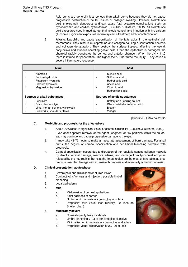
D. Clinical presentation: acute phase
1. Severe pain and diminished or blurred vision2. Conjunctival chemosis and injection; possible limbal
blanching
3. Localized edema
4. Mild
a. Mild erosion of corneal epithelium
b. Faint haziness of corneac. No ischemic necrosis of conjunctiva or sclera
d. Prognosis: mild visual loss (usually 0-2 lines onSnellen chart)
5. Moderately severe
a. Corneal opacity blurs iris detailsb. Limbal blanching < 1/3 of peri-limbal conjunctiva
c. Minimal ischemic necrosis of conjunctiva and sclerad. Prognosis: visual preservation of 20/100 or less

8/3/2019 Ocular Trauma 09
http://slidepdf.com/reader/full/ocular-trauma-09 20/38
State of Illinois TNS Program page 19
Ocular Trauma
6. Very severe
a. Blurring of pupillary outlineb. Marbleized (opacified) cornea: corneal grafts may not take
c. Ischemia of 1/3-2/3 of peri-limbal conjunctivad. Blanching of conjunctiva and scleral vessels
e. Cataract formationf. Glaucoma
g. Necrosis of intraocular contents
h. Prognosis: Poor with high incidence of corneal ulceration, perforation,vascularization and opacification. Vision - often count fingers or less.
E. Emergency management: Do not delay irrigation while trying to determine nature of the
chemical.
1. Immediate decontamination with any neutral fluid available at injury site. Removeparticulate matter immediately.
2. Rapid removal of contact lenses3. Rapid visual acuity for light perception only while preparing to irrigate
4. Instill a topical anesthetic and/or cycloplegic/mydriatic if severe pain/ciliary spasm (willdilate pupil)
5. Immediate and profuse eye irrigation with 1 - 2 liters closest neutral solution. Aimstream from inner to outer canthus making sure that superior and inferior fornices are
well flushed. Don't attempt to neutralize the chemical agent with a liquid of the oppositepH - will cause a heat reaction. Evert the eyelids, if necessary, to flush the cul-de-sacs.
Normal tear pH ranges from 7.3 - 7.7. The eye must be allowed to equilibrate for 5-10minutes after initial lavage before determining the pH. Check pH after each subsequent
liter and discontinue lavage when pH of 7.4 to 7.6 is achieved.
6. Mild chemical burns can be treated as a corneal abrasion and followed as outpatients.
Moderate to severe burns require immediate ophthalmologic consult. Theophthalmologist may prescribe treatment to reduce inflammation, promote epithelial
healing, decrease intraocular pressure, or debride the injured area. Goal is to preventscarring, cataract formation, perforation, and glaucoma (Cuculino & DiMarco, 2002).
7. Assess intraocular pressure in alkali burns after irrigation. If elevated, the physician
may order any of the following:
a. Acetazolamide (Diamox) 500 mg IM/IV or 125-250 mg QID PO
b. IV Mannitolc. Topical beta blockers: timolol maleate (Timoptic) drops
8. Ammonium titrate 5% dissolves lime9. Narcotic analgesia and sedatives as prescribed10. Topical antibiotic ointment as prescribed while epithelial defect exists
VIII. Thermal burn
A. Etiology: Flash burn, fiery explosion, cigarette ash, etc. Almost always associated with facial
burns. Even severely burned patients may avoid direct injury to the cornea and conjunctiva due
to protection by the eyelids. Burns due to toxic chemicals, hot liquids, and molten metal behavemuch like alkaline burns. Iron has a higher melting point (1200° C) and causes more damage
than other metals (lead, tin, zinc, melting point below 1000° C). Arc welding injuries commonwithout protective eyewear.
B. Clinical exam: May require local anesthetic, Desmarres lid retractors
1. Visual acuity; pupillary exam2. External exam: Spastic entropion, lid edema, orbital cellulitis
3. Slit-lamp exam: Conjunctival chemosis, blanching, corneal clarity, anterior chamberinflammation, fluorescein testing
4. Intraocular pressure testing5. In explosions: Must R/O perforating injuries, F/B6. Cornea will slough and regenerate in 4-5 days in children; 3-4 weeks in adults
C. Treatment
1. Physician will debride devitalized lid epithelium and remove F/B

8/3/2019 Ocular Trauma 09
http://slidepdf.com/reader/full/ocular-trauma-09 21/38
State of Illinois TNS Program page 20
Ocular Trauma
2. Spastic entropion: Bandage with contact lens in place. Note: the contact lens will not
remain moist if lids are not functioning properly, and frequent artificial tears andointments are often necessary.
3. Antibiotic ointments/drops as prescribed4. Cycloplegics as prescribed
5. Increased intraocular pressure: Diamox, Timoptic as prescribed6. Eyelid burn: Cool burn and then apply sterile dressings
D. Complications: Infections such as Pseudomonas keratitis, endogenous enophthalmitis from
septicemia, mucomycosis. Eyelid burns with skin loss can lead to scarring with cosmeticdisfigurement, chronic tearing, exposure keratitis, and trichiasis.
IX. U.V. radiation burns
A. Etiology: Uncommon, yet may be seen in head and neck tumors treated with radiation or those
who use tanning beds where the corneas were left unprotected, arc welders not usingappropriate eye protection, mountain climbers, or skiers outdoors in the snow on a sunny day.
B. Clinical presentation
1. Superficial punctate keratitis on the exposed eye surface, corneal epithelium sloughs2. Pain, 3-12 hours after exposure, is often delayed but severe
3. Increased tearing, burning, eyes feel full of sand
C. Emergency management
1. Treat like a corneal abrasion with pain control, antibiotic prophylaxis as prescribed, coldcompresses, lubrication
2. 12-36 hour recovery period3. Permanent scarring is rare
X. Corneal abrasion
A. Definition: Defect in the corneal epithelium
that does not penetrate Bowman's layer
B. Etiology: Very common. Results from a
direct or tangential impact to the eye, contact
lenses, and/or exposure to UV light. F/Bscan scratch the cornea at various depths.
The sharp edges of hard contacts can causesmall abrasions during normal use.
Extended wear contacts cause epithelialhypoxia, which impairs the attachment of the epithelial cells to Bowman's membrane.
This weakened attachment makes the eye more susceptible to damage from minimal trauma,such as rubbing the eyes (Cuculino & DiMarco, 2002).
C. Clinical presentation: A corneal abrasion may be associated with serious ocular injury and
therefore a complete ocular exam should be performed. Assess and record visual acuity. Topicalanesthesia aids in exam.
1. Pain: Mild to severe, and foreign body sensation that is worsened by blinking2. Copious tearing (epiphora)
3. Possible decrease in visual acuity if over the pupil4. Irregular corneal light reflex
5. Diffuse conjunctival injection6. Photophobia
7. Mild anterior chamber reaction8. Lid spasm (blepharospasm)
D. Slit lamp examination
1. Remove contact lenses
2. Stain the cornea with fluorescein
3. Inspect with a slit lamp exam (cobalt blue light) or penlight with blue filter. Abrasion willstain bright green. Other conditions, such as herpes, also cause the cornea to stain with

8/3/2019 Ocular Trauma 09
http://slidepdf.com/reader/full/ocular-trauma-09 22/38
State of Illinois TNS Program page 21
Ocular Trauma
fluorescein.
4. Note and diagram the size of the abrasion and its depth. Do not be fooled by a self-
sealing full thickness laceration and treat it merely as an abrasion. A full thicknesslaceration often shows a positive Seidel's test.
5. Examine the anterior chamber for the presence of a hyphema which may result fromblunt trauma and may require different treatment.
6. If the cornea has multiple vertical linear abrasions, a F/B adhered to the inner surface ofthe conjunctiva is likely. Evert the eyelids to examine for F/B.
E. Emergency management
1. Non-contact lens wearer
a. Pain control
(1) Topical anesthetics: A topical anesthetic may make examination
easier and alleviate pain, but DO NOT give the patient topical
anesthetic to take home. They slow epithelial repair and maypredispose the patient to further trauma as the protective reflexes of
the eye are lost (Cuculino & DiMarco, 2002).
(2) Cycloplegics (Cyclopentolate 2%) as prescribed to dilate pupil and
decrease ciliary spasm and reduce photophobia
(3) Topical NSAID drops or oral analgesics as needed
b. All objects that could cause an abrasion may be microbially contaminated andthe possibility of infection should always be considered. Topical broad
spectrum antibiotic ointment or drops per physician preference to help
prevent infection. Instruct patients that ointments may blur vision for a short
period of time. Instruct patients in the correct use of eye drops and ointments.
c. Eye patching decisions are made on a case by case basis (Cuculino &
DiMarco, 2002). Patching may slow healing and may increase the rate of
infection. It is rarely done today. Do not patch if an abrasion was caused byvegetable matter or artificial fingernails.
If prescribed, double-patch eye for 12-24 hours to help alleviate pain and limiteyelid movement. Use two pads with tape running from bone to bone in a
diagonal fashion. A pressure patch is not usually indicated when microbial
infection is suspected or a significant risk exists (contact lens wearers).
d. Tetanus prophylaxis as necessary
e. Follow-up with an ophthalmologist within 24-48 hours. The corneal epithelium
heals rapidly and most small abrasions resolve during this time. Antibioticdrops may be continued for 5-7 days.
2. Contact lens wearer: Patients with contact lenses are at particular risk for gram
negative infections such as Pseudomonas and their treatment differs from a routineabrasion.
a. Cycloplegicsb. Antibiotic drops with pseudomonas coverage (Gentamicin or Tobramycin 4-6
times/day and antibiotic ointment for H.S.c. DO NOT PATCH
d. Abstain from lens wearing for at least one weeke. Oral analgesics
f. Follow-up in 24 hours in mandatory
3. Complications
a. Corneal ulcer: Can be viral, bacterial, fungal, orchlamydial. Should be considered a true
emergency. Bacterial ulceration can rapidlyprogress to corneal perforation. Most bacterial
cases result from trauma or contact lens use orabuse. They present with pain, photophobia,
decreased visual acuity, a white corneal infiltrate

8/3/2019 Ocular Trauma 09
http://slidepdf.com/reader/full/ocular-trauma-09 23/38
State of Illinois TNS Program page 22
Ocular Trauma
(hypopyon) with ill-defined borders and/or an epithelial defect on fluoresceinstaining. They require culture, gram stain, and ophthalmology consultation.
Fortified antibiotic drops are required for treatment. Steroids are not routinelyindicated (Med-Challenger FM 06).
b. Decreased visual acuity if they involve the visual axis
c. Traumatic iritis may develop 24-72 hours after injury
d. Recurrent erosion syndrome: Recurrent abrasion days to years after a
previous injury. Patient will present with sudden severe pain when they opentheir eyelids upon waking. At risk patients: large abrasions or those caused byfingernails or hard contact lenses.
XI. Cyanoacrylate (Krazy) Glue: Direct exposure can cause a chemical keratitis. Immediately irrigate with
warm water for at least 15 minutes. Acetone or ethanol solutions should not be used in the eye. If eyelidsare bonded together, gently rub with mineral oil or a petroleum-based ointment such as bacitracin or
erythromycin. Forceful retraction should be avoided.
XII. Corneal laceration
A. Definition: Full-thickness injury to the cornea
B. Patient presentation - much the same as globe rupture
1. Teardrop pupil
2. Flat anterior chamber3. Hyphema
C. Emergency management
1. Cover eye with a protective shield2. Instruct patient not to move the eye3. Do not assess intraocular pressure
4. Position patient head up at a 30° to 45° angle5. Immediate ophthalmology consult
6. Prophylactic antibiotics and tetanus as prescribed7. Antiemetics as prescribed to prevent increase in intraocular pressure from vomiting
XIII. Corneal/conjunctival foreign bodies
A. Etiology: Fragments fly into the eye from working withoutprotective goggles. Industrial air pollution, BB pellets etc. alsocommon.
B. History: Question patient about the use of safety goggles, the
nature of the F/B, and if there was metal striking metal. If metal,identify the type of metal if possible; iron and copper are
common. High-velocity mechanisms may cause corneal perforation.
C. Clinical assessment/presentation: Most F/Bs don't penetrate the globe. However, view themas a possible indicator of concurrent intraocular or intraorbital F/B. This is especially important in
the setting of grinding or hammering metal upon metal in the absence of protective eyewear.Inspect conjunctiva, fornices, and evert the lids to search for additional F/B. If vertical linear
abrasions are seen suspect occult F/B under the lid.
1. Chief complaint: Pain, foreign body sensation, photophobia, blurred vision2. Obtain visual acuity before and after treatment3. Observe unaffected eye first for comparison
4. Inspect for red eye, tearing5. Always suspect corneal penetration if patient presents with a diffuse subconjunctival
hemorrhage, diffuse corneal damage, abnormal pupillary shape, an hyphema, or achange in anterior chamber depth vs. the noninvolved eye (Cuculino & DiMarco, 2002).
D. Diagnosis: Key feature is the finding of the F/B, a rust ring, or both. Other findings include
conjunctival injection, mild anterior chamber reaction, superficial punctate keratitis, and eyelidedema. There may be a small white infiltrate around the F/B if it has been in the cornea for
longer than 24 hours.
8/3/2019 Ocular Trauma 09
http://slidepdf.com/reader/full/ocular-trauma-09 24/38
State of Illinois TNS Program page 23
Ocular Trauma
E. Emergency management
1. Topical anesthetic as prescribed
2. Evaluate the depth of the F/B with biomicroscopy. Those deeply imbedded or in the
visual axis should be removed by ophthalmology.
3. Physician may dilate pupil with Cyclogyl 1% or Mydriacyl 1% or Homatropine 5% to
relieve iritis unless history of glaucoma.
4. Antibiotic drops pre and post-removal as prescribed
5. Conjunctival F/B: Easily removed by physician using gentle irrigation with NS,moistened cotton-tipped applicator, a foreign body spud or the bevel of a 25-27 gauge
needle.
6. Corneal F/B: Requires removal under the slit lamp
7. Iron-containing objects may leave a rust ring after removal of the F/B. The rust ring can
be removed 24-48 hours later by a spud, a 25 g needle or an ophthalmic rust ring drill. Itis sometimes safer to leave the rust ring, especially if it is central or deep. This will allow
time for it to migrate to the surface and be easily removed at a later time. Debride aslittle tissue as possible.
8. Check for corneal abrasion and perforation. The physician should document a negativeSeidel test to ensure that corneal perforation did not occur.
9. Once removed, treat the same as a corneal abrasion
a. Analgesics; may need narcotics if iritis is severeb. Antibiotic ointments (erythromycin) as prescribed
F. Complications
1. Infection - disastrous; (pseudomonas in fluorescein)2. Scarring - use slit lamp when available
XIV. Subconjunctival hemorrhage
A. Definition: Bleeding of the delicate conjunctival or episcleral vessels between conjunctiva and
sclera into the subconjunctival space.
B. Etiology: Very common; can be initiated by Valsalva maneuvers such as sneezing, coughing,
straining, or lifting heavy objects; trauma, hypertension, coagulopathy, febrile illness, or idiopathic
causes.
C. Clinical presentation: Painless, blood-red eye; may be accompanied by some irritation. Looksterrible but is usually a non-emergency if non-traumatic origin. If trauma, physician must rule out
globe rupture or retrobulbar hemorrhage.
D. Emergency management
1. Supportive and aimed to prevent further bleeding. Discontinue use of NSAIDS andaspirin for 24-48 hours.
2. Instruct patient to avoid Valsalva maneuvers
3. Instruct patient in use of artificial tears for irritation4. Usually self-limiting, spontaneously resolves in 1 - 2 weeks. Instruct patient that the
healing process will break the blood down and change color from red to green to yellowand then to brown.
5. Any globe tenderness should be evaluated for presence of infection
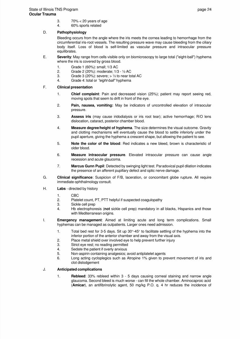
XV. Hyphema
A. Definition: Accumulation of blood within the anterior chamberdue to rupture of a vessel in the iris.
B. Etiology/MOI: Usually caused by severe blunt, and less often
perforating, trauma to the eye. In rare instances, hyphema mayoccur spontaneously as a complication of an ocular or systemic
disorder or the use of anticoagulants.
C. Demographics
1. Incidence: 17-20/1000 population/yr
2. M:F 3:1
8/3/2019 Ocular Trauma 09
http://slidepdf.com/reader/full/ocular-trauma-09 25/38
State of Illinois TNS Program page 24
Ocular Trauma
3. 70% < 20 years of age4. 60% sports related
D. Pathophysiology
Bleeding occurs from the angle where the iris meets the cornea leading to hemorrhage from thecircumferential iris-root vessels. The resulting pressure wave may cause bleeding from the ciliary
body itself. Loss of blood is self-limited as vascular pressure and intraocular pressureequilibrates.
E. Severity: May range from cells visible only on biomicroscopy to large total ("eight-ball") hyphema
where the iris is covered by gross blood.
1. Grade 1 (60%): small; 1/3 AC
2. Grade 2 (20%): moderate; 1/3 - ½ AC3. Grade 3 (20%): severe; > ½ to near total AC
4. Grade 4: total or "eight-ball" hyphema
F. Clinical presentation
1. Chief complaint: Pain and decreased vision (25%); patient may report seeing red,
moving spots that seem to drift in front of the eye.
2. Pain, nausea, vomiting: May be indicators of uncontrolled elevation of intraocular
pressure.
3. Assess iris (may cause iridodialysis or iris root tear); active hemorrhage; R/O lensdislocation, cataract, posterior chamber blood.
4. Measure degree/height of hyphema. The size determines the visual outcome. Gravity
and clotting mechanisms will eventually cause the blood to settle inferiorly under thepupil aperture, giving the hyphema a crescent shape, but allowing the patient to see.
5. Note the color of the blood: Red indicates a new bleed, brown is characteristic of
older blood.
6. Measure intraocular pressure. Elevated intraocular pressure can cause angle
recession and acute glaucoma.
7. Marcus Gunn Pupil: Detected by swinging light test. Paradoxical pupil dilation indicates
the presence of an afferent pupillary defect and optic nerve damage.
G. Clinical significance: Suspicion of F/B, laceration, or concomitant globe rupture. All requireimmediate ophthalmology consult.
H. Labs - directed by history
1. CBC2. Platelet count, PT, PTT helpful if suspected coagulopathy3. Sickle cell prep
4. Hb electrophoresis (not sickle cell prep) mandatory in all blacks, Hispanics and those
with Mediterranean origins.
I. Emergency management: Aimed at limiting acute and long term complications. Small
hyphemas can be managed as outpatients. Larger ones need admission.
1. Total bed rest for 3-5 days. Sit up 30°-45° to facilitate settling of the hyphema into the
inferior portion of the anterior chamber and away from the visual axis.2. Place metal shield over involved eye to help prevent further injury3. Strict eye rest; no reading permitted
4. Sedate the patient if overly anxious5. Non-aspirin containing analgesics; avoid antiplatelet agents
6. Long acting cycloplegics such as Atropine 1% given to prevent movement of iris andclot dislodgement
J. Anticipated complications
1. Rebleed: 33% rebleed within 3 - 5 days causing corneal staining and narrow angle
glaucoma. Second bleed is much worse - can fill the whole chamber. Aminocaproic acid(Amicar), an antifibrinolytic agent, 50 mg/kg P.O. q. 4 hr reduces the incidence of

8/3/2019 Ocular Trauma 09
http://slidepdf.com/reader/full/ocular-trauma-09 26/38
State of Illinois TNS Program page 25
Ocular Trauma
rebleed from 30% to 3%. Physicians may also prescribe oral prednisone to inhibitrebleeding through its stabilizing action on fragile vessels.
2. Increased ocular pressure: The physician may order topical and/or systemic
medications.
a. Beta blockers: Timolol to decrease production of aqueous humor
b. Adrenergicsc. Carbonic anhydrase inhibitors: Diamox
d. Avoid miotics
3. Chronic: Angle damage leading to glaucoma, corneal blood staining, optic atrophy
K. Associated injuries: Corneal laceration, occult scleral rupture, lens damage and/or dislocation,
optic atrophy, angle recession, chronic glaucoma, vitreous hemorrhage, retial detachment andCN II damage.
XVI. Penetrating/perforating globe injuries: This is a major cause of traumatic visual loss due to damage of
intraocular structures caused by the force required to rupture the globe. They require immediate carefulassessment and prompt surgical repair to prevent loss of the eye.
A. Penetrating injuries are those that cause partial disruption of the outer coats of the eye withoutinterrupting the anatomic continuity of the scleral or corneal layer,
thus preventing prolapse of ocular contents.
B. Perforating injuries are those that result in complete anatomicdisruption of the sclera or cornea. They may or may not beassociated with prolapse of uveal structures.
C. Etiology
1. Direct blunt or penetrating trauma to the eye2. As the intraocular pressure suddenly increases, the
sclera ruptures where it is the thinnest, often at the limbus, the site of extraocularmuscle insertion, and/or the site of optic nerve insertion.
D. Clinical assessment/presentation
1. Change in pupil shape (tear-drop pupil pulls to site of rupture)2. Unequal pupils
3. Black or brown defect (choroid/uveal prolapse into defect)4. Injected sclera, subconjunctival hemorrhage
5. Leakage of vitreous humor6. Pain
7. Decreased visual acuity to hand movements or light perception8. Enophthalmos
9. Lowered intraocular pressure; soft eye10. Flat or shallow anterior chamber
11. Hyphema12. Iris prolapse13. Dislocated lens, traumatic cataract
14. If lids are swollen, use great caution in pulling them apart to prevent extrusion of theeye. If you cannot retract lids, get an ophthalmologist consult ASAP.
E. Radiological evaluation: for metalF. Emergency management: Same as for an intraocular foreign body or corneal laceration
1. Never remove an impaled F/B
2. DO NOT manipulate the eye, instill eye drops, measure intraocular pressures, or apply
antibiotic ointments3. Shielding: A metal shield should rest on the bone of the brow and the cheek; it should
not exert pressure on the eye. The patient must not be able to touch or rub the eye.4. Do not remove any blood clots, foreign bodies or tissue from the eye5. Tetanus prophylaxis
6. Analgesics and sedatives may be needed7. Don't let patient bend, stoop, strain or move his or her head suddenly

8/3/2019 Ocular Trauma 09
http://slidepdf.com/reader/full/ocular-trauma-09 27/38
State of Illinois TNS Program page 26
Ocular Trauma
8. Keep patient NPO9. IV antibiotics as prescribed
10. Surgical intervention by ophthalmologist
G. Prognosis
1. Related primarily to the location and extent of damage. The more posterior the
penetration and the larger the laceration or rupture, the worse the prognosis. Throughand through injuries (BBs or pellets) have a bad prognosis. Superficial injuries to the
anterior segment structures can often be repaired by microsurgical closure, contact lensor intraocular lens optical correction, and corneal transplantation, if necessary. They
have a high rate of visual rehabilitation (Broocker, Parke & Hamill, 1996). Posteriorinjuries involving the retina are more severe. Damage may cause proliferation of scar
tissue throughout the vitreous that can result in retinal tears or detachment.
2. Also at risk for microbial endophthalmitis that can lead to blindness. This risk is
increased with an intraocular foreign body.
3. Sympathetic ophthalmia: Bilateral inflammation of the uveal tract (iris, ciliary body, and
choroid) associated with penetrating injury to one eye. Untreated, this often results inloss of the noninjured eye. This risk is reduced if the injured eye is enucleated within
10-14 days of the injury. However, the risk of this complication is very small and doesnot justify the primary removal of a severely traumatized eye unless the eye is
completely blind and there is significant exposure or extrusion of posterior ocular
contents (Broocker, Parke & Hamill, 1996).
XVII. Intraocular foreign body
A. Etiology: Metallic particles most common (80%)
B. History: Metal striking metal or mechanism indicating a high velocity projectile
C. Clinical presentation
1. Pain, irritation2. Injection
3. Entry site may be visible on biomicroscopy as a disruption of the corneal surface withsurrounding edema
4. Fluorescein stain may demonstrate a positive Seidel sign (streaking of the fluoresceinas aqueous humor leaks out of the globe)
D. Diagnosis
1. No intraocular pressure measurements2. Plain films of the orbits if suspected material is radio-opaque
3. CT of orbits is usually preferred4. Ultrasound can also identify retinal detachments
5. MRI may visualize F/B not seen on CT but must be avoided if material is metallic
E. Emergency management
1. Cover eye with a protective shield2. Apply no pressure to the globe3. Tetanus prophylaxis as prescribed
4. Immediate ophthalmology consult for removal5. IV antibiotics as prescribed - about 10% become infected. Beside usual gram positive
coverage, add pseudomonas coverage if material is dirt or vegetable matter such aswood from tree branches.
6. Antiemetics as prescribed to prevent vomiting and increased intraocular pressure
XVIII. Lens dislocation/subluxation
A. Definition: Disruption of the zonular fibers that maintain the lens in its correct position. Partialdisruption results in subluxation.
B. Etiology: Trauma to the eye. Minimal trauma can cause dislocation in patients with Marfan's
syndrome, homocystinuria, or syphilis.
C. Clinical presentation
1. Pain

8/3/2019 Ocular Trauma 09
http://slidepdf.com/reader/full/ocular-trauma-09 28/38
State of Illinois TNS Program page 27
Ocular Trauma
2. Monocular diplopia3. Edge of lens visible through the pupillary aperture. Total disruption may allow lens to
displace into the posterior chamber leading to an acute rise in intraocular pressure.
D. Emergency management: Immediate ophthalmology consult
XIX. Iridodialysis
A. Definition: Iris is torn from the ciliary body
B. Patient presentation
1. Appears as if patient has a second pupil2. May develop a secondary hyphema and monocular
diplopia if the defect is large
C. Emergency management
1. Treat hyphema
2. Immediate ophthalmology consult
XX. Retinal detachment: Separation of the neurosensory layer of the retinal pigment epithelium from its
source of blood supply via tear, fold or rent. Subretinal fluid accumulates under the neurosensory layer.
A. Etiology: Minimal to moderate trauma or proliferative retinopathy
B. Clinical presentation
1. Painless visual field deficit or decrease in vision. May be described as a
darkening/haziness or a curtain or shadow being drawn over the visual field.
2. New onset of photopsia (flashes of light or sparks); spider webs; "Floaters" or black
dots
3. If it involves the periphery, patient will have a visual field deficit
4. If it involves the macula, visual acuity will be diminished
5. Fundoscopic exam reveals billowing of the retina
6. A significant hyphema may obscure the fundus
C. Diagnosis: Beta scan ultrasound
D. Emergency management
1. Keep patient quiet and supine with both eyes patched
2. Immediate ophthalmologist intervention is required. Patients at increased risk include
those with severe myopia, trauma, prior surgery and a significant family history.
3. Retinal attachment can only be accomplished by surgery which is successful in about
80% of the cases. The eye is filled with a perfluoropropane (C3F8) and/or sulfurhexafluoride (SF8) gas bubble to reattach the retina. Patients need to be warned that it
takes six to eight weeks for these gasses to diffuse from the eye. If nitrous oxide is givento the patient while the C3F8 or SF8 are still present, it can diffuse rapidly into the gas
bubble and raise intraocular pressure causing a central retinal artery occlusion andblindness. Patients should wear medic-alert bracelets postoperatively and warn
healthcare providers that the gas bubbles may remain in their eye and they are at riskfor a C3F8 or SF8 - nitrous oxide reaction. These patients should also be cautioned about
avoiding high altitude travel until the surgeon gives them permission (Woo, 2002).
XXI. Optic nerve injury
A. Etiology: Transection or avulsion of optic nerve, hemorrhage, decreased blood flow to the optic
nerve, or pressure from bony fracture.
B. Clinical significance: Resulting blindness is often irreversible
C. Clinical presentation: Visual deficit and afferent pupillary defect on affected side unrelated to
any other associated eye injury. Physician should carefully rule out other causes of visual deficitsuch as hyphema or retinal tear. Initial fundoscopic exam may be normal.
D. Emergency management: Ophthalmology consult; surgical intervention often attempted
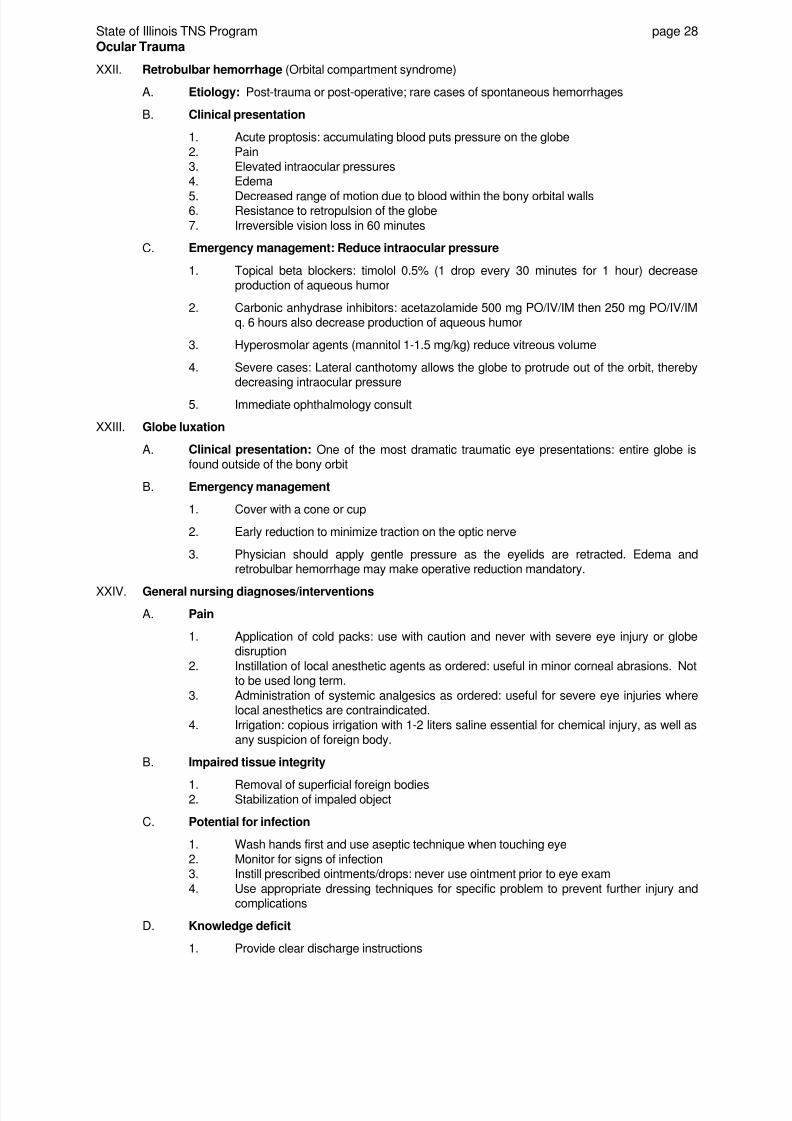
8/3/2019 Ocular Trauma 09
http://slidepdf.com/reader/full/ocular-trauma-09 29/38
State of Illinois TNS Program page 28
Ocular Trauma
XXII. Retrobulbar hemorrhage (Orbital compartment syndrome)
A. Etiology: Post-trauma or post-operative; rare cases of spontaneous hemorrhages
B. Clinical presentation
1. Acute proptosis: accumulating blood puts pressure on the globe
2. Pain3. Elevated intraocular pressures4. Edema
5. Decreased range of motion due to blood within the bony orbital walls6. Resistance to retropulsion of the globe
7. Irreversible vision loss in 60 minutes
C. Emergency management: Reduce intraocular pressure
1. Topical beta blockers: timolol 0.5% (1 drop every 30 minutes for 1 hour) decreaseproduction of aqueous humor
2. Carbonic anhydrase inhibitors: acetazolamide 500 mg PO/IV/IM then 250 mg PO/IV/IMq. 6 hours also decrease production of aqueous humor
3. Hyperosmolar agents (mannitol 1-1.5 mg/kg) reduce vitreous volume
4. Severe cases: Lateral canthotomy allows the globe to protrude out of the orbit, therebydecreasing intraocular pressure
5. Immediate ophthalmology consult
XXIII. Globe luxation
A. Clinical presentation: One of the most dramatic traumatic eye presentations: entire globe isfound outside of the bony orbit
B. Emergency management
1. Cover with a cone or cup
2. Early reduction to minimize traction on the optic nerve
3. Physician should apply gentle pressure as the eyelids are retracted. Edema andretrobulbar hemorrhage may make operative reduction mandatory.
XXIV. General nursing diagnoses/interventions
A. Pain
1. Application of cold packs: use with caution and never with severe eye injury or globedisruption
2. Instillation of local anesthetic agents as ordered: useful in minor corneal abrasions. Notto be used long term.
3. Administration of systemic analgesics as ordered: useful for severe eye injuries wherelocal anesthetics are contraindicated.
4. Irrigation: copious irrigation with 1-2 liters saline essential for chemical injury, as well asany suspicion of foreign body.
B. Impaired tissue integrity
1. Removal of superficial foreign bodies2. Stabilization of impaled object
C. Potential for infection
1. Wash hands first and use aseptic technique when touching eye
2. Monitor for signs of infection3. Instill prescribed ointments/drops: never use ointment prior to eye exam
4. Use appropriate dressing techniques for specific problem to prevent further injury andcomplications
D. Knowledge deficit
1. Provide clear discharge instructions

8/3/2019 Ocular Trauma 09
http://slidepdf.com/reader/full/ocular-trauma-09 30/38
State of Illinois TNS Program page 29
Ocular Trauma
2. Instruct patient on administration technique of prescribed medication(s)
E. Potential for visual impairment
1. Caution patient regarding loss of depth perception. Instruct re: no driving, sports, andoperating heavy machinery while patched.
2. If severely impaired, request social service assistance for help with activities of daily
living (ADLs).3. Instruct patient on maintaining a safe environment
F. Fear or anxiety related to potential or actual loss of vision
1. Maintain a quiet environment
2. Provide psychological and emotional support3. Provide frequent orientation to date, time and place, situation
4. Describe self and the environment5. Optimize use of other senses

8/3/2019 Ocular Trauma 09
http://slidepdf.com/reader/full/ocular-trauma-09 31/38
State of Illinois TNS Program page 30
Ocular Trauma
Selected Ophthalmic Medications
Name Dose Comments
Antibiotics
Sodium sulfacetamide solution Instill 1-3 drops q. 2-3 hrs
for 7 days
Sodium sulfacetamide ointment Apply 1" ribbon to the eye QID for 7 days This lacks pseudomonal coverage
Gentamicin ophthalmic solution
(0.3%)
Instill 1-2 drops QID for 7 days
Gentamicin ophthalmic ointment(0.3%)
Apply 1" ribbon to eye QID for 7 days This has anti-pseudomonal coverage
Erythromycin ophthalmic ointment Apply 1" ribbon to eye QID for 7 days
Bacitracin ophthalmic ointment Apply 1" ribbon to eye QID for 7 days This has no pseudomonal coverage
Ciprofloxacin drops Instill 1-2 drops QID for 7 days This has pseudomonal activity, but there
may be increasing strep pneumoniaresistance. This may be used in children.
Trimethoprim/Polymyxin solution Instill 1-2 drops QID for 7 days
Trimethoprim/Polymyxin ointment Apply 1" ribbon to eye QID for 7 days This has pseudomonal coverage
Tobramycin ophthalmic solution Instill 1-2 drops QID for 7 daysTobramycin ophthalmic ointment Apply 1" ribbon to eye BID/QID for 7 days
Ofloxacin ophthalmic solution Instill 1-2 drops QID for 7 days This has anti-pseudomonal propertiesand may be used in children.
Beta blockers: Reduce production of aqueous humor. Indicated for glaucoma, ocular hypertension.
timolol maleate 0.25%, 0.5%, 2.5mg/ml, 5 mg/ml (Timoptic, Timoptic-
XE)
1 drop q. 30 minutes for 1 hourthen 1 drop BID
Contraindications: Hypersensitivity to timolol
Asthma
Bradycardia; 2nd or 3rd degree AVB
Overt cardiac failure
Cardiogenic shock
levobunolol hydrochloride (Betagan) 1-2 drops 0.5% solution QD
1-2 drops 0.25% solution BID
Adverse effects:
Ocular stinging, burningBlepharitis; ocular irritation
Carbonic anhydrase inhibitors: Reduce production of aqueous humor. Indicated for chronic simple glaucoma, open-angle
glaucoma, secondary glaucoma, and preoperatively for acute angle-closure glaucoma; and ocular hypertension
acetazolamide (Diamox) 500 mg PO/IV/IM then 250 mg q. 4-6 hrsDuration 14 hrs.
Contraindications:
Hyponatremia/hypokalemia
Severe hepatic, renal, or adrenal disease
Hyperchloremic acidosis
Chronic noncongestive angle-closure
glaucoma
Hypersensitivity to acetazolamide
SE:
Taste disturbances
ParesthesiasMetabolic acidosis
N/V; headache, malaise
Depression
Thrombocytopenia
Risk of severe, potentially fatal skin
reactions including Stevens-Johnson
syndrome and Toxic Epidermal Necrosis,
especially in those of Japanese or Korean
descent.
dichlorphenamide (Daranide) Loading dose of 100-200 mg followed by 100
mg q. 12 hr until desired response
Onset: 1 hour
SE: Same as above plus anorexia, N/V
Drowsiness
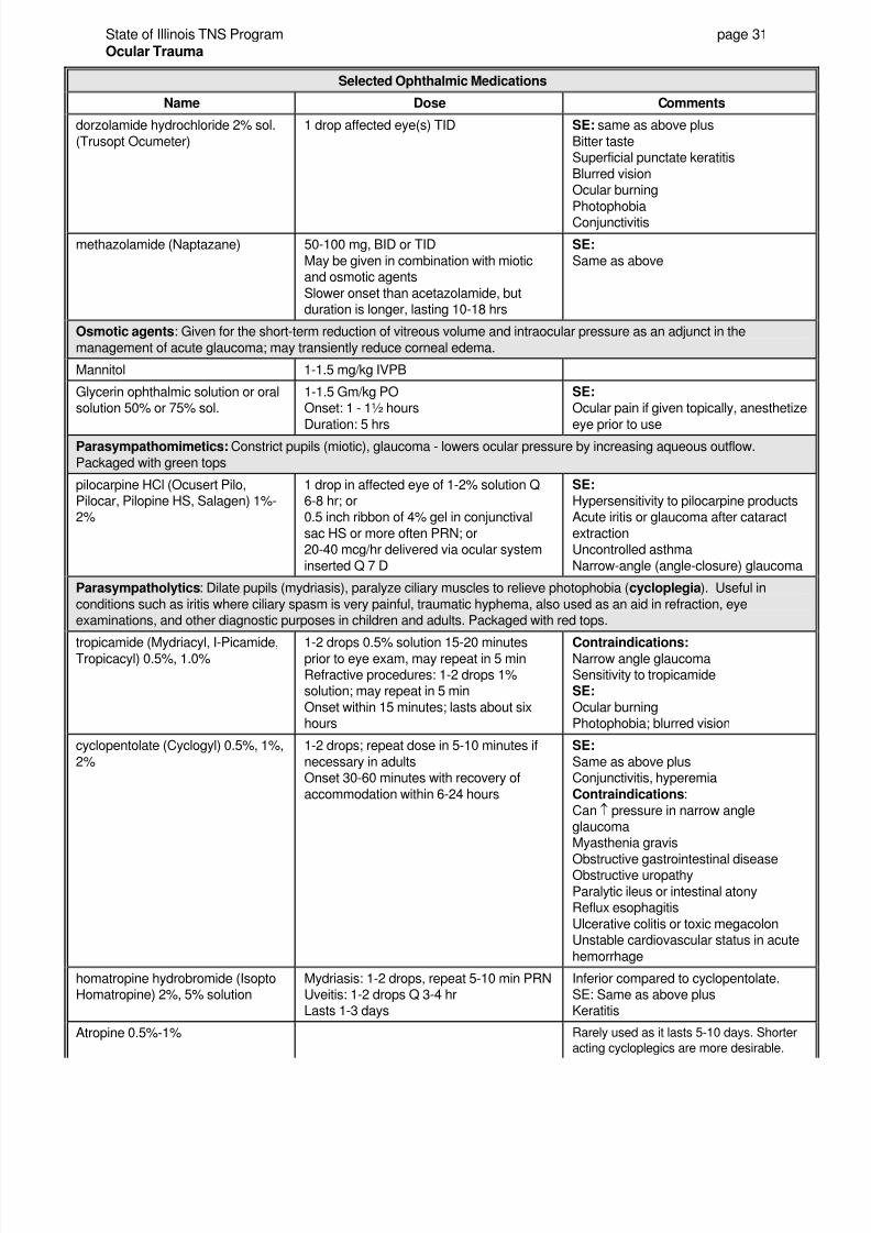
8/3/2019 Ocular Trauma 09
http://slidepdf.com/reader/full/ocular-trauma-09 32/38
State of Illinois TNS Program page 31
Ocular Trauma
Selected Ophthalmic Medications
Name Dose Comments
dorzolamide hydrochloride 2% sol.
(Trusopt Ocumeter)
1 drop affected eye(s) TID SE: same as above plus
Bitter tasteSuperficial punctate keratitis
Blurred visionOcular burning
PhotophobiaConjunctivitis
methazolamide (Naptazane) 50-100 mg, BID or TID
May be given in combination with mioticand osmotic agents
Slower onset than acetazolamide, butduration is longer, lasting 10-18 hrs
SE:
Same as above
Osmotic agents: Given for the short-term reduction of vitreous volume and intraocular pressure as an adjunct in the
management of acute glaucoma; may transiently reduce corneal edema.
Mannitol 1-1.5 mg/kg IVPB
Glycerin ophthalmic solution or oralsolution 50% or 75% sol.
1-1.5 Gm/kg POOnset: 1 - 1½ hours
Duration: 5 hrs
SE:
Ocular pain if given topically, anesthetize
eye prior to use
Parasympathomimetics: Constrict pupils (miotic), glaucoma - lowers ocular pressure by increasing aqueous outflow.
Packaged with green tops
pilocarpine HCl (Ocusert Pilo,Pilocar, Pilopine HS, Salagen) 1%-2%
1 drop in affected eye of 1-2% solution Q6-8 hr; or0.5 inch ribbon of 4% gel in conjunctival
sac HS or more often PRN; or20-40 mcg/hr delivered via ocular system
inserted Q 7 D
SE:
Hypersensitivity to pilocarpine productsAcute iritis or glaucoma after cataract
extractionUncontrolled asthma
Narrow-angle (angle-closure) glaucoma
Parasympatholytics: Dilate pupils (mydriasis), paralyze ciliary muscles to relieve photophobia (cycloplegia). Useful in
conditions such as iritis where ciliary spasm is very painful, traumatic hyphema, also used as an aid in refraction, eyeexaminations, and other diagnostic purposes in children and adults. Packaged with red tops.
tropicamide (Mydriacyl, I-Picamide,Tropicacyl) 0.5%, 1.0% 1-2 drops 0.5% solution 15-20 minutesprior to eye exam, may repeat in 5 minRefractive procedures: 1-2 drops 1%solution; may repeat in 5 min
Onset within 15 minutes; lasts about sixhours
Contraindications: Narrow angle glaucomaSensitivity to tropicamideSE:
Ocular burningPhotophobia; blurred vision
cyclopentolate (Cyclogyl) 0.5%, 1%,
2%
1-2 drops; repeat dose in 5-10 minutes if
necessary in adultsOnset 30-60 minutes with recovery of
accommodation within 6-24 hours
SE:
Same as above plusConjunctivitis, hyperemia
Contraindications:
Can ↑ pressure in narrow angleglaucomaMyasthenia gravis
Obstructive gastrointestinal diseaseObstructive uropathy
Paralytic ileus or intestinal atonyReflux esophagitis
Ulcerative colitis or toxic megacolonUnstable cardiovascular status in acute
hemorrhage
homatropine hydrobromide (IsoptoHomatropine) 2%, 5% solution
Mydriasis: 1-2 drops, repeat 5-10 min PRNUveitis: 1-2 drops Q 3-4 hrLasts 1-3 days
Inferior compared to cyclopentolate.SE: Same as above plusKeratitis
Atropine 0.5%-1% Rarely used as it lasts 5-10 days. Shorter
acting cycloplegics are more desirable.
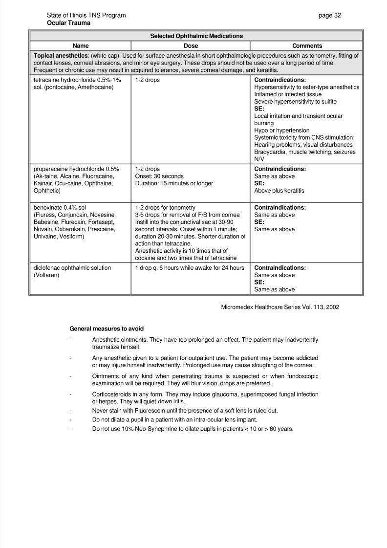
8/3/2019 Ocular Trauma 09
http://slidepdf.com/reader/full/ocular-trauma-09 33/38
State of Illinois TNS Program page 32
Ocular Trauma
Selected Ophthalmic Medications
Name Dose Comments
Topical anesthetics: (white cap). Used for surface anesthesia in short ophthalmologic procedures such as tonometry, fitting of
contact lenses, corneal abrasions, and minor eye surgery. These drops should not be used over a long period of time.Frequent or chronic use may result in acquired tolerance, severe corneal damage, and keratitis.
tetracaine hydrochloride 0.5%-1%
sol. (pontocaine, Amethocaine)
1-2 drops Contraindications:
Hypersensitivity to ester-type anesthetics
Inflamed or infected tissueSevere hypersensitivity to sulfiteSE:
Local irritation and transient ocularburning
Hypo or hypertensionSystemic toxicity from CNS stimulation:Hearing problems, visual disturbances
Bradycardia, muscle twitching, seizuresN/V
proparacaine hydrochloride 0.5%
(Ak-taine, Alcaine, Fluoracaine,Kainair, Ocu-caine, Ophthaine,
Ophthetic)
1-2 drops
Onset: 30 secondsDuration: 15 minutes or longer
Contraindications:
Same as above SE:
Above plus keratitis
benoxinate 0.4% sol
(Fluress, Conjuncain, Novesine,Babesine, Flurecain, Fortasept,Novain, Oxbarukain, Prescaine,
Univaine, Vesiform)
1-2 drops for tonometry
3-6 drops for removal of F/B from corneaInstill into the conjunctival sac at 30-90
second intervals. Onset within 1 minute;duration 20-30 minutes. Shorter duration ofaction than tetracaine.
Anesthetic activity is 10 times that ofcocaine and two times that of tetracaine
Contraindications:
Same as aboveSE:
Same as above
diclofenac ophthalmic solution
(Voltaren)
1 drop q. 6 hours while awake for 24 hours Contraindications:
Same as above SE:
Same as above
Micromedex Healthcare Series Vol. 113, 2002
General measures to avoid
- Anesthetic ointments. They have too prolonged an effect. The patient may inadvertently
traumatize himself.
- Any anesthetic given to a patient for outpatient use. The patient may become addictedor may injure himself inadvertently. Prolonged use may cause sloughing of the cornea.
- Ointments of any kind when penetrating trauma is suspected or when fundoscopic
examination will be required. They will blur vision, drops are preferred.
- Corticosteroids in any form. They may induce glaucoma, superimposed fungal infectionor herpes. They will quiet down iritis.
- Never stain with Fluorescein until the presence of a soft lens is ruled out.
- Do not dilate a pupil in a patient with an intra-ocular lens implant.
- Do not use 10% Neo-Synephrine to dilate pupils in patients < 10 or > 60 years.
8/3/2019 Ocular Trauma 09
http://slidepdf.com/reader/full/ocular-trauma-09 34/38
State of Illinois TNS Program page 33
Ocular Trauma
References
Cuculino, G.P. & DiMarco, C.J. (2002). Common ophthalmologic emergencies: A systematic approach to
evaluation and management. Emergency Medicine Reports , 23(13), 163-170.
Jenkins, C.N. & Thuau, H. (1997). Ultrasound imaging in assessment of fractures of the orbital floor. Clin Radiol ,
52(9), 708-711.
Mieler, W.F. (2001). Ocular injuries: Is it possible to further limit the occurrence rate? Arch Ophthalmol 119, 1712-1713.
Shingleton, B. & Meade, J. (1998). New England Eye Center Handbook of Eye Emergencies (pp. 45-49).
Thorofare: Slack inc.
Shuttleworth, G.N., David, D.B., Potts, M.J., Bell, C.N., & Guest, P.G. (1999). Orbital trauma: do not blow yournose. BMJ 318, 1054-1055.
Woo, E. (2002). Retinal repair risks. Nursing Spectrum , 15(16)IL, 21.

8/3/2019 Ocular Trauma 09
http://slidepdf.com/reader/full/ocular-trauma-09 35/38
State of Illinois TNS Program page 34
Ocular Trauma
GLOSSARY
Bell's Phenomenon: Reflex movement upward of eyes with eyelid closure.
Chemosis: Swelling of the conjunctiva around the cornea.
Cycloplegia: Pharmacologic paralysis of ciliary body musculature, reducing reactive spasm.
Entropion: Inturning of eyelid and lid margin.
Epiphora: Tearing.
Keratitis: Inflammation of the cornea.
Lagophthalmos: Incomplete lid closure.
Limbus: Transition zone between clear cornea and white sclera.
Mydriasis: Pupil dilation (opposite of miosis).
Phthisis bulbi: End stage non-seeing eye, with associated shrinking and disorganization of intraocular
contents.
Seidel test: Use of fluorescein to detect perforating injury; leakage of aqueous humor will create
green rivulets when observed with cobalt blue or Wood's light.
Trichiasis: Inversion of hairs about an orifice, as eyelashes that turn in and cause irritation of the
cornea.
Uvea: The middle, pigmented, vascular layer of the eye consisting of the choroid, the ciliarybody, and the iris. Also called the uveal tract.
Uveitis: Intraocular inflammation; may be anterior or posterior in location.
Eye Fun Facts:
You blink every 2-10 seconds. As you focus on each word in this sentence, your eyes swing back and
forth 100 times a second; every second the retina performs 10 billion computer-like calculations.
The human eye can perceive more than 1 million simultaneous visual impressions and is able todiscriminate among nearly 8 million gradations of color.
Having two eyes instead of just one provides us with stereo vision and depth perception. Each of our eyes
views an object from a slightly different angle. Our brains put the different images together into one three-dimensional image.
The iris of the eye provides better identification than a fingerprint. A scan of the iris has 256 different
unique characteristics. A fingerprint has only 40 (LWW.comInsider - December 2001).

8/3/2019 Ocular Trauma 09
http://slidepdf.com/reader/full/ocular-trauma-09 36/38
State of Illinois TNS Program page 35
Ocular Trauma
STUDY QUESTIONS
1. Matching
______ Cornea A. CN that closes the eyelid
______ Sclera B. CN that lifts the lid and pulls the eye to the nose
______ Limbus C. Structure that covers the pupil and iris forming a transparent, concave disc
______ Lens D. Area of the retina responsible for all vision better than 20:200
______ CN III E. Fluid contained in the anterior chamber
______ Conjunctiva F. Tough, fibrous coat that serves as the structural skeleton of the globe
______ Macula G. Region of the retina that becomes the origin of the optic nerve and is a
provides point of entry for the retinal vessels ______ Acqueous humor H. Gelatinous substance contained in the posterior cavity
______ Vitreous body I. Mucous membrane that lines the posterior surface of the eyelids and the
anterior surface of the eye ______ Optic disk J. Circular margin where the conjunctiva meets the cornea
K. Disc-shaped structure changes its thickness and shape, therebypermitting images from varied distances to be focused on the retina
2. List four things you should ask about the patient's past ocular history.
4. List three things that can be detected by eye examination using a slit lamp.
5. List three things to observe when inspecting the orbit and periocular structures.
6. What color does a corneal defect stain after instillation of fluorescein?
What is a positive Seidel test?
7. What is the range for normal intraocular pressures?
8. Describe four methods used to assess visual acuity.
9. What pathology is suggested by an afferent defect?
8/3/2019 Ocular Trauma 09
http://slidepdf.com/reader/full/ocular-trauma-09 37/38
State of Illinois TNS Program page 36
Ocular Trauma
10. What classic deficit in eye movement suggests a blow-out fracture?
A patient with a blow-out fracture is more likely to present with
A. exophthalmos.B. enophthalmos.
Why is it important to instruct a patient with a blow-out fracture not to blow their nose or perform
other Valsalva maneuvers?
11. What suture materials should you prepare if an eyelid will be primarily repaired in the ED?
12. What can be instilled into the eye prior to irrigating a chemical burn to relieve the severe pain?
13. What is the appropriate technique to irrigate an alkaline chemical burn to the eye?
What ocular pH should be achieved before irrigation is discontinued?
14. What is the appropriate management of a corneal abrasion?
Why should patients with corneal abrasions not be sent home with a bottle of a topical anesthetic?
15. What tools should you prepare for a physician to remove a superficial conjunctival foreign body?
What type of conjunctival foreign body may leave a rust ring?
16. What is the appropriate management of a patient with a hyphema?
17. What is the major complication of a hyphema?
What drug should the physician prescribe to reduce the risk of this complication?
18. What pupillary change suggests a ruptured globe?
True or false
T F Intraocular pressure must be measured in a patient with a suspected ruptured globe.
T F The nurse should remove blood clots, superficial foreign bodies, and avulsed tissue fromthe eye in the presence of a suspected perforated globe in order to facilitate an accurateeye exam.

8/3/2019 Ocular Trauma 09
http://slidepdf.com/reader/full/ocular-trauma-09 38/38
State of Illinois TNS Program page 37
Ocular Trauma
19. What should be placed over the eye if an intraocular foreign body is suspected?
If an intraocular F/B is dirt or vegetable material, what additional bacterial coverage must be presentin the prescribed antibiotics?
20. What eye injury presents with painless visual field deficit where the patient complains of flashes of
light or sparks, floaters or black dots in his line of sight?