Embed Size (px)
Citation preview

OCT e GlaucomaOCT e Glaucoma
www.amedeolucente.it No commercial interests

2
Glaucoma 2 �causa di cecità al Glaucoma 2 �causa di cecità al mondo, 1 �causa di cecità irreversibilemondo, 1 �causa di cecità irreversibile
800.000 Italia2,5% over 40 Caucasici *
Dati OMS 2010Dati OMS 2010
* Bonomi L, et al. The Egna-Neumarkt Study, 1998
2,5 milioni in USA3,36 milioni 2020

The Glaucoma ContinuumThe Glaucoma Continuum
3
Modificato da Weinreb R. et al A. J. Ophthalmol 2004; 138;458-467
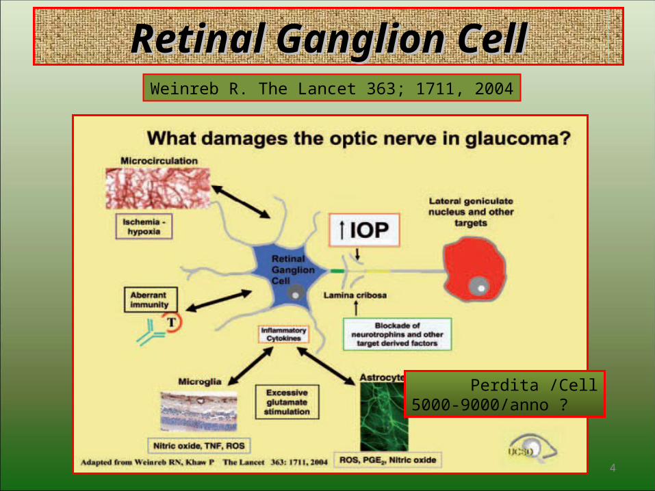
Retinal Ganglion CellRetinal Ganglion Cell
4
Weinreb R. The Lancet 363; 1711, 2004
Perdita /Cell5000-9000/anno ?
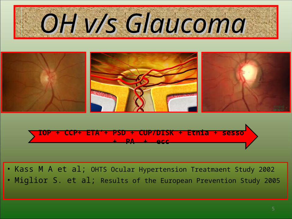
OH v/s Glaucoma OH v/s Glaucoma
• Kass M A et al; OHTS Ocular Hypertension Treatmaent Study 2002
• Miglior S. et al; Results of the European Prevention Study 2005
5
IOP + CCP+ ETA’+ PSD + CUP/DISK + Etnia + sesso + PA + ecc
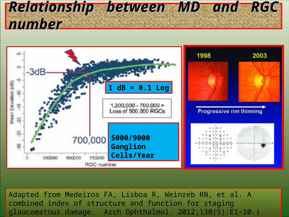
Relationship between MD and RGC numberRelationship between MD and RGC number
6
Adapted from Medeiros FA, Lisboa R, Weinreb RN, et al. A combined index of structure and function for staging glaucomatous damage. Arch Ophthalmol. 2012;130(5):E1-10.)
1 dB = 0.1 Log
5000/9000 Ganglion Cells/Year
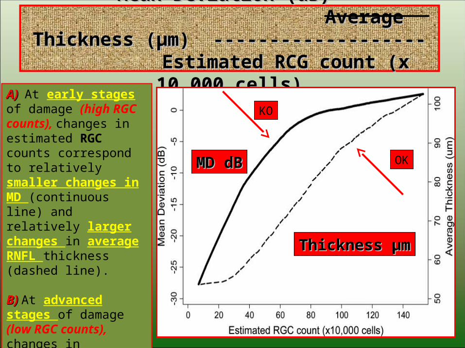
Mean Deviation (dB) Mean Deviation (dB) Average Thickness (µm) ------------------- Average Thickness (µm) -------------------
Estimated RCG count (x 10.000 cells) Estimated RCG count (x 10.000 cells)
7
MD dBMD dB
Thickness µmThickness µm
A) A) At early stages of damage (high RGC counts), changes in estimated RGC counts correspond to relatively smaller changes in MD (continuous line) and relatively larger changes in average RNFL thickness (dashed line).
B) B) At advanced stages of damage (low RGC counts), changes in estimated RGC counts correspond to relatively large changes in MD, but only small changes in average RNFL thickness.
OK
KO
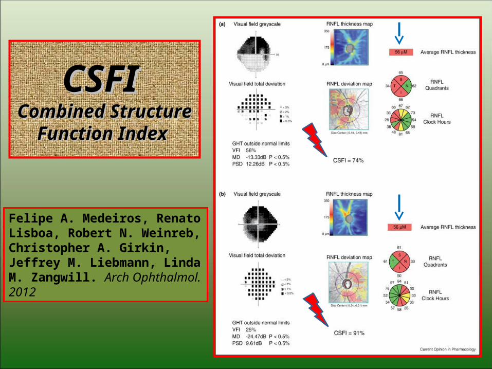
CSFI CSFI Combined Structure Combined Structure
Function Index Function Index
8
Felipe A. Medeiros, Renato Lisboa, Robert N. Weinreb, Christopher A. Girkin, Jeffrey M. Liebmann, Linda M. Zangwill. Arch Ophthalmol. 2012

Biblio CSFIBiblio CSFI 1. Sibota R, Sony P, Viney G, et al. Diagnostic capability of optical coherence tomography in evaluating the degree of glaucomatous retinal nerve fiber damage. Invest Ophthalmol Vis Sci 2006;47(5):2006-10.
2. Medeiros FA, Lisboa R, Weinreb RN, et al. A combined index of structure and function for staging glaucomatous damage. Arch Ophthalmol. 2012;130 (5):E1-10.
3. Harwerth RS, Wheat JL, Fredette MJ, Anderson DR. Linking Structure and function in glaucoma. Prog Retin Eye Res. 2010;29(4):249-71.
4. Medeiros FA, Zangwill LM, Anderson DR, et al. Estimating the rate of retinal ganglion cell loss in glaucoma. Am J Ophthalmol. 2012; Jul 26. [Epub ahead of print].
9
Specificità 95%Specificità 95%

SD-OCT & Glaucoma SD-OCT & Glaucoma
• RNFL Retinal Nerve Fiber LayerRNFL Retinal Nerve Fiber Layer• ONH Optical Nerve HeadONH Optical Nerve Head GCC Ganglion Cell ComplexGCC Ganglion Cell Complex• AS-OCT Anterior Segment OCTAS-OCT Anterior Segment OCT• SD-OCT & CVSD-OCT & CV
10
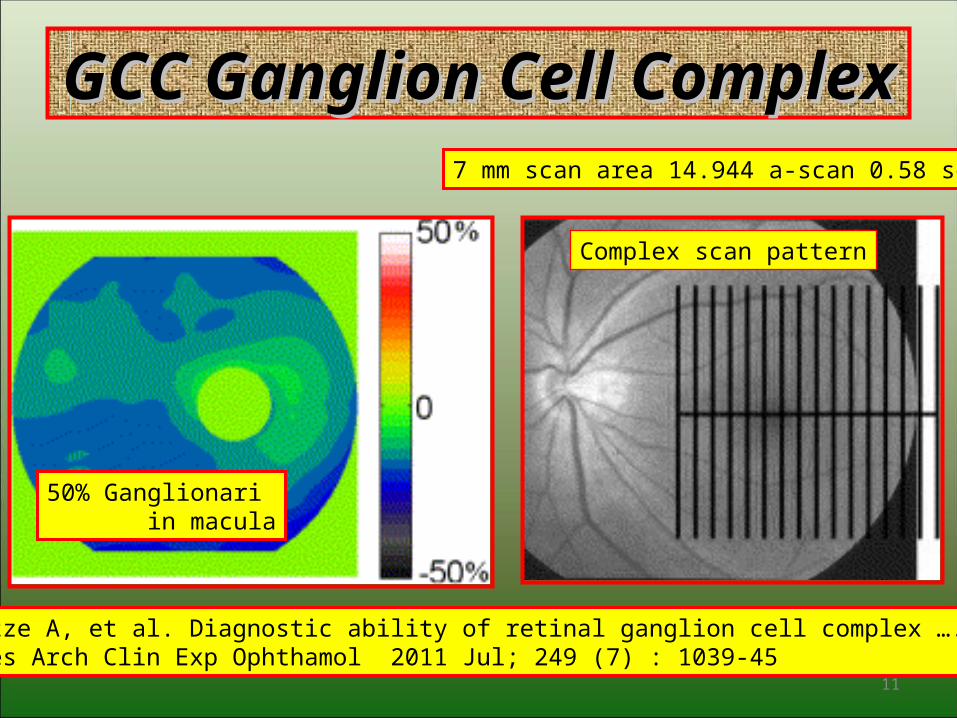
GCC Ganglion Cell ComplexGCC Ganglion Cell Complex
11
Schultze A, et al. Diagnostic ability of retinal ganglion cell complex …. Graefes Arch Clin Exp Ophthamol 2011 Jul; 249 (7) : 1039-45
Complex scan pattern
50% Ganglionari in macula
7 mm scan area 14.944 a-scan 0.58 sec

12

13

14
15
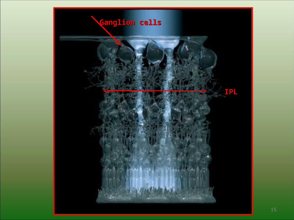
Ganglion cellsGanglion cells
IPLIPL

Abnormal GCC in spite of normal RNFL ThicknessAbnormal GCC in spite of normal RNFL Thickness
16
Garas A et al Reproducibility of retinal nerve layer and macular thickness ecc Ophthalmology 2010 Elsevier

Normal RNFL Thickness v/s abnormal GCC Normal RNFL Thickness v/s abnormal GCC
17

Report Ganglion Cell OU AnalysisReport Ganglion Cell OU Analysis
18
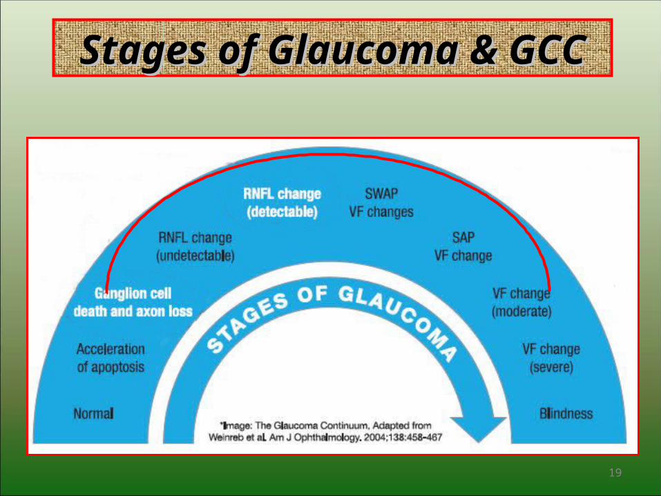
Stages of Glaucoma & GCCStages of Glaucoma & GCC
19

20

SD-OCT & GlaucomaSD-OCT & Glaucoma
• RNFL Retinal Nerve Fiber LayerRNFL Retinal Nerve Fiber Layer• ONH Optical Nerve HeadONH Optical Nerve Head• GCC Ganglion Cell ComplexGCC Ganglion Cell Complex• AS-OCT Anterior Segment OCTAS-OCT Anterior Segment OCT SD-OCT & CVSD-OCT & CV
21

SD-OCT & CVSD-OCT & CV
• Zeiss Cirrus & Humphrey con FORUMFORUM
• Heidelberg Spectralis & HEP con HEYEXHEYEX
• Optovue & Octopus Bundle Haag-Streit * Bundle Haag-Streit *
22
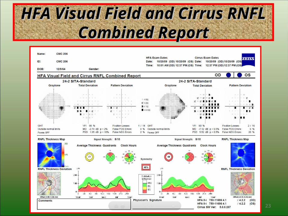
HFA Visual Field and Cirrus RNFL HFA Visual Field and Cirrus RNFL Combined ReportCombined Report
23

RNFL & Visual Field Combined OU ReportRNFL & Visual Field Combined OU Report
24

25
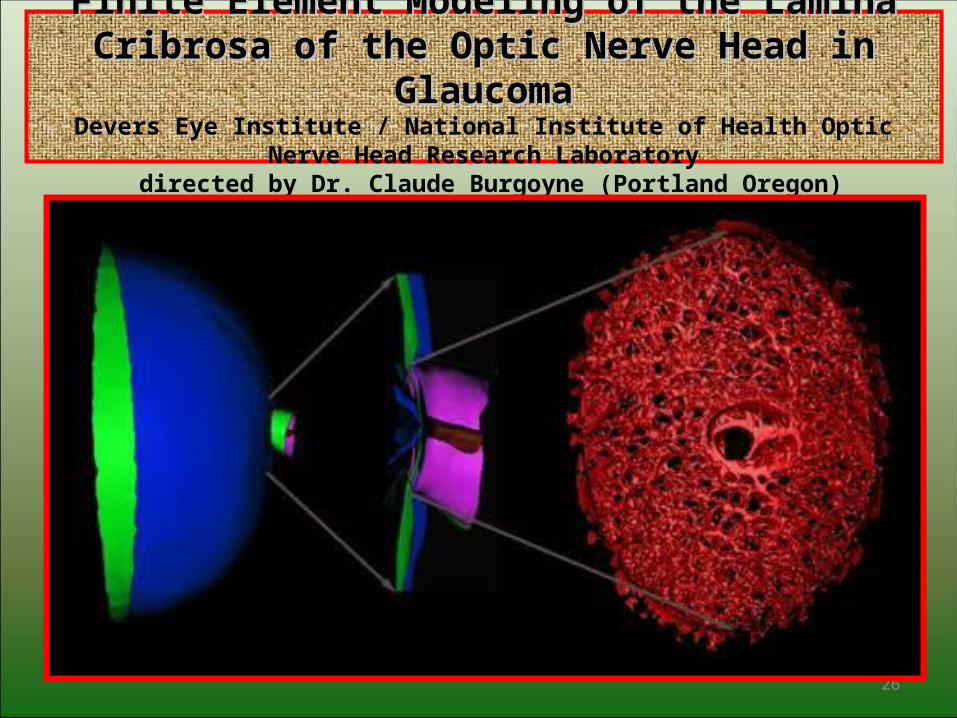
Finite Element Modeling of the Lamina Cribrosa of the Finite Element Modeling of the Lamina Cribrosa of the Optic Nerve Head in GlaucomaOptic Nerve Head in Glaucoma
Devers Eye Institute / National Institute of Health Optic Nerve Head Research Laboratory directed by Dr. Claude Burgoyne (Portland Oregon)
26

Finite Element Modeling of the Lamina Cribrosa of the Finite Element Modeling of the Lamina Cribrosa of the Optic Nerve Head in GlaucomaOptic Nerve Head in Glaucoma
Devers Eye Institute / National Institute of Health Optic Nerve Head Research Laboratory directed by Dr. Claude Burgoyne (Portland Oregon)
27
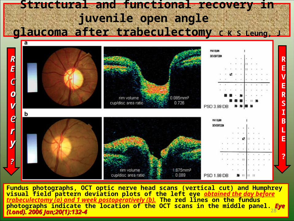
Structural and functional recovery in juvenile open angle glaucoma after trabeculectomy C K S Leung, J Woo, M K Tsang and K K Tse
Fundus photographs, OCT optic nerve head scans (vertical cut) and Humphrey visual field pattern deviation plots of the left eye obtained the day before trabeculectomy (a) and 1 week postoperatively (b). The red lines on the fundus photographs indicate the location of the OCT scans in the middle panel. Eye (Lond). 2006 Jan;20(1):132-4Eye (Lond). 2006 Jan;20(1):132-4 28
RREE
ccoovveerryy
??
REVERSIBLE
?
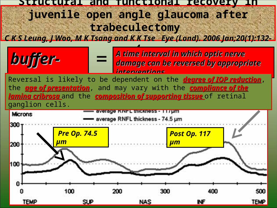
Structural and functional recovery in juvenile open angle glaucoma after trabeculectomy
C K S Leung, J Woo, M K Tsang and K K Tse Eye (Lond). 2006 Jan;20(1):132-4
29
Post Op. 117 µm Pre Op. 74.5 µm
buffer-zonebuffer-zone A time interval in which optic nerve damage can A time interval in which optic nerve damage can be reversed by appropriate interventions.be reversed by appropriate interventions.=
Reversal is likely to be dependent on the degree of IOP reductiondegree of IOP reduction, the age of presentationage of presentation, and may vary with the compliance of the lamina cribrosa compliance of the lamina cribrosa and the composition of composition of supporting tissue supporting tissue of retinal ganglion cells.

Reversibility of glaucomatous Reversibility of glaucomatous demangedemange
1.Kotecha A, Siriwardena D, Fitzke FW, Hitchings RA, Khaw PT. Optic disc changes following trabeculectomy: longitudinal and localisation of change.
Br J Ophthalmol 2001; 85: 956–961. | Article | PubMed | ISI | ChemPort |
2.Aydin A, Wollstein G, Price LL, Fujimoto JG, Schuman JS.
Optical coherence tomography assessment of retinal nerve fiber layer thickness changes after glaucoma surgery. Ophthalmology 2003; 110: 1506–1511. Article PubMed ISI
3.Tsai CS, Shin DH, Wan JY, Zeiter JH. Visual field global indices in patients with reversal of glaucomatous cupping after intraocular pressure reduction. Ophthalmology 1991; 98: 1412–1419. | PubMed | ISI | ChemPort |
30
BiblioBiblio
Grazie per l’attenzioneGrazie per l’attenzione
31


![Glaucoma Detection Using Retinal Nerve Fiber Layer Texture ......glaucoma. The best way todetect glaucoma is optic nerve head (ONH), and the imaging modalities used (optical coherencetomography[OCT],](https://img.pdfslide.us/doc/110x75/604c2d786805e8157444d61f/glaucoma-detection-using-retinal-nerve-fiber-layer-texture-glaucoma-the.jpg)


![Comparison of glaucoma diagnostic ability of retinal nerve ... · important OCT finding associated with glaucoma [15] the ganglion cell complex (GCC) scan of the RTVue system, which](https://img.pdfslide.us/doc/110x75/5e6777a5bd65a9535b60b5fa/comparison-of-glaucoma-diagnostic-ability-of-retinal-nerve-important-oct-finding.jpg)