Embed Size (px)
Citation preview

Occupational Allergies
Journal of Allergy
Guest Editors: Gordon L. Sussman, Donald H. Beezhold, and Arthur Sussman

Occupational Allergies

Journal of Allergy
Occupational Allergies
Guest Editors: Gordon L. Sussman, Donald H. Beezhold,and Arthur Sussman

Copyright © 2011 Hindawi Publishing Corporation. All rights reserved.
This is a special issue published in volume 2011 of “Journal of Allergy.” All articles are open access articles distributed under the CreativeCommons Attribution License, which permits unrestricted use, distribution, and reproduction in any medium, provided the originalwork is properly cited.

Journal of Allergy
Editorial Board
P. J. Barnes, UKWilliam E. Berger, USAK. Blaser, SwitzerlandEugene R. Bleecker, USAJan de Monchy, The NetherlandsF. JP Hoebers, The NetherlandsStephen T. Holgate, UKHenk Jansen, The NetherlandsS. L. Johnston, UKYoung J. Juhn, USA
Alan P. Knutsen, USAMarek L. Kowalski, PolandTing Fan Leung, Hong KongClare M Lloyd, UKRedwan Moqbel, CanadaD. Passali, ItalyStephen P. Peters, USADavid G. Proud, CanadaFabienne Rance, FranceAnuradha Ray, USA
H. Renz, GermanyNima Rezaei, IranRobert P. Schleimer, USARonald Simon, USAMassimo Triggiani, ItalyHugo Van Bever, SingaporeA. J. M. van Oosterhout, The NetherlandsGarry Walsh, UKRobert A. Wood, USA

Contents
Occupational Allergies, Donald H. Beezhold and Gordon L. SussmanVolume 2011, Article ID 519329, 2 pages
Comparison between Airway Responses to High versus Low Molecular Weight Compounds inOccupational Asthma, D. Talini, F. Novelli, E. Bacci, F. L. Dente, M. De Santis, A. Di Franco, L. Melosini,B. Vagaggini, and P. L. PaggiaroVolume 2011, Article ID 781470, 5 pages
Industrial Fungal Enzymes: An Occupational Allergen Perspective, Brett J. Green and Donald H. BeezholdVolume 2011, Article ID 682574, 11 pages
Vicia faba Hypersensitivity and ASA Intolerance in a Farmer: A Case Report, Elisabetta Damiani,Anna Maria Aloia, Maria Giovanna Priore, Angela Pastore, Stefania Nardulli, Cristina Lippolis,Luigi Macchia, and Antonio FerranniniVolume 2011, Article ID 191787, 4 pages
Gum Arabic as a Cause of Occupational Allergy, Arja Viinanen, Maija Salokannel, and Kaija LammintaustaVolume 2011, Article ID 841508, 5 pages
Genetic Variability in Susceptibility to Occupational Respiratory Sensitization, Berran Yucesoy andVictor J. JohnsonVolume 2011, Article ID 346719, 7 pages
Haptenation: Chemical Reactivity and Protein Binding, Itai Chipinda, Justin M. Hettick, and Paul D. SiegelVolume 2011, Article ID 839682, 11 pages
The LLNA: A Brief Review of Recent Advances and Limitations, Stacey E. Anderson, Paul D. Siegel,and B. J. MeadeVolume 2011, Article ID 424203, 10 pages
Inhalation of Ortho-Phthalaldehyde Vapor Causes Respiratory Sensitization in Mice, Victor J. Johnson,Jeffrey S. Reynolds, Wei Wang, Kara Fluharty, and Berran YucesoyVolume 2011, Article ID 751052, 12 pages
Allergic Potential and Immunotoxicity Induced by Topical Application of1-Chloro-4-(Trifluoromethyl)Benzene (PCBTF) in a Murine Model, Jennifer Franko, Laurel G. Jackson,B. Jean Meade, and Stacey E. AndersonVolume 2011, Article ID 238513, 8 pages
Occupational Asthma in Antibiotic Manufacturing Workers: Case Reports and Systematic Review,Sara Dıaz Angulo, Joanna Szram, Jenny Welch, Julie Cannon, and Paul CullinanVolume 2011, Article ID 365683, 9 pages
Exercise-Induced Bronchoconstriction and Exercise-Induced Respiratory Symptoms in Nurses,Jordan Minov, Jovanka Karadzinska-Bislimovska, Kristin Vasilevska, Snezana Risteska-Kuc, Saso Stoleski,and Dragan MijakoskiVolume 2011, Article ID 267542, 7 pages
Hindawi Publishing CorporationJournal of AllergyVolume 2011, Article ID 519329, 2 pagesdoi:10.1155/2011/519329
Editorial
Occupational Allergies
Donald H. Beezhold1 and Gordon L. Sussman2
1 Allergy and Clinical Immunology Branch, The National Institute for Occupational Safety and Health,1095 Willowdale Road, Morgantown, WV 26505, USA
2 Division of Allergy and Clinical Immunology, University of Toronto, Toronto, ON, Canada M5S 1A1
Correspondence should be addressed to Donald H. Beezhold, [email protected]
Received 23 May 2011; Accepted 23 May 2011
Copyright © 2011 D. H. Beezhold and G. L. Sussman. This is an open access article distributed under the Creative CommonsAttribution License, which permits unrestricted use, distribution, and reproduction in any medium, provided the original work isproperly cited.
Occupational immune diseases are new emerging illnessesthat affect workers in industrialized societies. Occupationalexposures to substances in the workplace environment cancause inflammation, allergy, or other potentially detrimen-tal immune responses. Personal exposure to a variety ofchemicals can exacerbate immune diseases such as contactdermatitis as well as respiratory diseases including rhinitis,asthma, and hypersensitivity pneumonitis.
Next to illnesses due to repeated traumatic injury,contact dermatitis is the second most commonly reportedoccupational illness. It can prevent individuals from per-forming job-related tasks or preclude working altogether.Occupationally related contact dermatitis is a significantpublic health burden with combined direct annual costestimates of up to $1 billion in the USA for medical costs,workers compensation, and lost time from work.
Respiratory morbidity is also a significant burden topublic health leading to lost productivity. Prevalence ratesfor occupational rhinitis are significant, varying by occu-pation between 5% and 65% and costing an estimated$593/year/employee due to productivity losses. Conservativeestimates made by the American Thoracic Society in 2003estimate that 15% of chronic obstructive pulmonary diseaseand asthma cases were work related and cost approximately$7 billion in lost productivity in the USA With the changingwork environment, new occupational hazards continue toemerge which require immunologic characterization. Inorder to reduce the morbidity and mortality associated withthese illnesses, it is critical that we identify the allergensand understand the immunological mechanism by whichthey exacerbate immune-mediated respiratory and dermaldiseases. Specific understanding of mechanism has direct
implications in developing appropriate intervention andprevention strategies.
Occupational allergy can be stratified into high-molec-ular-weight-allergen and low-molecular-weight-allergen me-diated responses. Different immunologic mechanisms medi-ate allergic reactivity to these occupational allergens as high-lighted in this issue by Talini et al. High-molecular-weight(HMW) allergens (typically proteins) induce type I hyper-sensitivity responses or typical allergies by inducing IgE anti-bodies which lead to a continuum of symptoms includingrhinitis (rhinosinusitis, conjunctivitis), hives, asthma, andlife-threatening anaphylaxis. Patients with HMW-allergen-induced asthma show a greater frequency and severity ofthe early-phase response but are less likely to demonstratea late-phase response. Occupational outbreaks of reactionsto HMW allergens can occur episodically and can besevere and life altering for those affected. These allergiescan affect large numbers of easily identified workers inspecific industries which can reach epidemic proportionssuch as latex allergy and Baker’s asthma. It can presentin a less-well-defined population or as local occurrencessuch as agricultural or food processors exposed to soy, seafoods, pollens, molds, and so forth. Research areas includeidentification and characterization of high-molecular-weightoccupational allergens. Using fungal enzymes as a prototypicHMW occupational allergen, Green et al. describe some ofthe characterized fungal enzyme allergens and discuss mon-itoring and avoidance strategies. Characterization of HMWallergens includes using proteomics, molecular techniquesand generating recombinant allergens, and producing mono-clonal antibodies for the development of immunoassays andimproved detection of the allergens in the workplace.
2 Journal of Allergy
Low-molecular-weight allergens (typically chemicals)induce type 4 hypersensitivity reactions by inducing allergen-specific T lymphocytes which can mediate contact dermatitisreactions as well as sensitizations that can lead to severeasthma such as isocyanates (auto painters) and trimelliticacid. Patients with LMW-allergen-induced asthma are morelikely to demonstrate a late-phase airway response. Thereview by Anderson et al. describes the identification of low-molecular-weight allergens in the laboratory using the locallymph node assay to determine whether new chemicals beingintroduced can cause workplace sensitizations as well astesting various components to identify the specific sensitizerand potentially nonsensitizing replacements. They examinedthe effects of chemical exposure on immune function usingselected assays from a comprehensive tiered approach. Thiscan be used in detecting toxic effects following chemicalexposure (in rodents) as adopted by the National ToxicologyProgram. The utility of analyzing potential replacementchemicals is highlighted by the study of Johnson et al.where the chemical ortho-phthalaldehyde (OPA) has beenrecommended as a substitute for glutaraldehyde as a ster-ilant in the healthcare industry. Their laboratory evidencesuggested that the replacement of the chemical OPA is alsoa strong sensitizer. Characterization of the biochemical andimmune mechanisms by which chemicals become allergens(haptenization) is described in a comprehensive review byChipinda et al. Developing new methods for screening chem-icals for potential sensitizers helps to build better models bywhich we predict whether chemicals are allergens. Yucesoy etal. describe new studies aimed at identifying occupationallysensitized individuals and understanding the genetic profileassociated with sensitizing/anaphylactic agents.
It is important to improve our basic science knowledgeand understanding of occupational allergies and their patho-genesis. If we are able to identify potential allergens, beforeclinical symptoms are observed, employers can take neces-sary precautions to minimize or eliminate their employee’sexposure.
Acknowledgments
We dedicate this issue to one of our coeditors: Dr. ArthurSussman, who passed away April 6 2011, just prior to ourspecial issues’ publication. Arthur Sussman was a pioneerin the field of allergy and immunology and witnessed itsemergence first hand during his sixty years of medicalpractice. He contributed to this issue of the journal and itis fitting that this issue would highlight emerging issues inallergy and Immunology—occupational diseases. He will besincerely missed.
Donald H. BeezholdGordon L. Sussman
Hindawi Publishing CorporationJournal of AllergyVolume 2011, Article ID 781470, 5 pagesdoi:10.1155/2011/781470
Clinical Study
Comparison between Airway Responses to High versus LowMolecular Weight Compounds in Occupational Asthma
D. Talini,1 F. Novelli,2 E. Bacci,2 F. L. Dente,2 M. De Santis,2 A. Di Franco,2 L. Melosini,2
B. Vagaggini,2 and P. L. Paggiaro2
1 Occupational Health Unit, Prevention Department, Galleria Gerace 14, 56126 Pisa, Italy2 Cardio-Thoracic and Vascular Department, University of Pisa, 56126 Pisa, Italy
Correspondence should be addressed to D. Talini, [email protected]
Received 14 February 2011; Accepted 29 March 2011
Academic Editor: Donald H. Beezhold
Copyright © 2011 D. Talini et al. This is an open access article distributed under the Creative Commons Attribution License, whichpermits unrestricted use, distribution, and reproduction in any medium, provided the original work is properly cited.
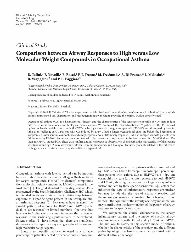
Occupational asthma (OA) is a heterogeneous disease, and the characteristics of the sensitizer responsible for OA may inducedifferent clinical, functional, and biological manifestations. We examined the characteristics of 74 patients with OA inducedby low molecular weight compounds (LMWC) or by high molecular weight compounds (HMWC) and diagnosed by specificinhalation challenge (SIC). Patients with OA induced by LMWC had a longer occupational exposure before the beginning ofsymptoms, a lower sputum eosinophilia, and a higher prevalence of late airway response (LAR), in comparison with patients withOA induced by HMWC. Pulmonary function tended to be poorer and atopy tended to be less frequent in LMWC-induced OAthan in HMWC-induced OA. These data confirm and extend previous observations showing that the characteristics of the specificsensitizer inducing OA may determine different clinical, functional, and biological features, probably related to the differencepathogenetic mechanisms underlying these different types of OA.
1. Introduction
Occupational asthma with latency period can be inducedby sensitization to either a specific allergen (high molecu-lar weight compounds, HMWC) or chemical compounds(low molecular weight compounds, LMWC) present in theworkplace [1]. The gold standard for the diagnosis of OA isrepresented by the Specific Inhalation Challenge (SIC) whichis intended to demonstrate a direct relationship betweenexposure to a specific agent present in the workplace andan asthmatic response [2]. Few studies have analysed thevariable patterns of response to HMWC and LMWC (early,dual, or late response) in limited numbers of patients, buthow worker’s characteristics may influence the pattern ofresponse to the sensitising agents remains to be explored.Recent studies [3] have shown that there are significantdifferences in the type of airway changes induced by low andhigh molecular weight agents.
Sputum eosinophilia has been reported in a variablepercentage of patients affected by occupational asthma, and
some studies suggested that patients with asthma inducedby LMWC may have a lower sputum eosinophil percentagethan patients with asthma due to HMWC [4, 5]. Sputumeosinophils increase further after exposure to both HMWCand LMWC, showing the increase in allergic airway inflam-mation induced by these specific sensitizers [6]. Factors thatinfluence the type of inflammatory responses are unclearbut may include also the type of asthmatic reaction andthe intensity of airway inflammation. In particular, it is notknown if the type and/or the severity of airway inflammationmay contribute to the determination of the pattern of airwayresponse to the specific sensitizer.
We compared the clinical characteristics, the airwayinflammatory pattern, and the model of specific airwayresponse in patients with OA induced by HMWC or LMWC.The aim was to assess, in this specific model of asthma,whether the characteristics of the sensitizer and the differentpathophysiologic mechanisms may be associated with adifferent asthma phenotype.
2 Journal of Allergy
2. Materials and Methods
We studied 74 subjects with occupational asthma due to dif-ferent sensitizers (diisocyanates, latex, hairdresser’s products,wood, and flour dusts) observed consecutively in our asthmaclinic: 48 were exposed to LMWC (isocyanates, persulfatesalts, aziridine, and phenolic resins) agents, and 26 wereexposed to HMWC (flour dusts, wood dusts, latex, deter-gents, and tobacco dusts). We selected only subjects in whomthe diagnosis of occupational asthma had been performed bymeans of positive response to specific inhalation challenge(SIC). According to the international recommendations [2,7], patients were all exposed to a known occupationalsensitizer (Table 1) showed asthma deterioration at work andnonspecific bronchial hyperresponsiveness during a workingperiod.
Bronchial hyperresponsiveness was determined by meth-acholine challenge test performed as previously reported [8];a provocative dose of a 20% decrease in FEV1 from baseline(PD20FEV1) of less than 1000 mcg was considered as positivefor bronchial hyperresponsiveness.
SIC was performed using different methods (Table 1):(a) for diisocyanates, subjects were exposed to vapoursgenerated by blowing air on the surface of a small amountof toluenediisocyanate (TDI) or warming a small amount ofmethylenediphenyl diisocyanate (MDI) at 40◦C, in a chal-lenge chamber and monitoring isocyanate concentrationswith a specific TDI/MDI detector (MDA model 7005 iso-cyanate detection equipment, MDA Scientific Inc., Glenview,IL); diluent was used as control exposure; the duration ofthe exposure was 30 min in a first test and 120 min in asecond test (if the first resulted negative) [9]; (b) for dusts(flour, wood, persulfate, latex, and tobacco), subjects inhaleddusts by a mouthpiece connected to a small box where asuspension of the dust was obtained by blowing compressedair at 5 L/min through a bottle containing the dust; lactosepowder was used as control test; the concentration of thedusts was measured by blowing air from the box througha cellulose nitrate filter of 0.8 µm porosity by means ofa vacuum pump [10]; (c) in two cases (one exposed tophenolic resins and the other to detergents), a realisticway was employed in order to simulate in laboratory theexposure of the workplace (spreading the substance on asmall surface); diluent was used as control test, and theduration of exposure was still 30 minutes. In all SIC, FEV1was measured immediately before and 5, 15, 30, and 60minutes after the exposure to the sensitizer, then hourlyfor 8 hours. A positive response was defined as a decreasein FEV1 from baseline of more than 15% during the firsthour (immediate response) or between the second and the8th hour (late response), and in absence of a more than10% decrease in FEV1 during a control test performed in adifferent day with diluent (for diisocyanates or other simplechemicals) or with lactose dust (for other dust sensitizers).
One or two weeks before challenge, other measurementsat diagnosis included skin prick tests to common allergens(to check for atopy), and collection of sputum induced bythe inhalation of saline solution. The method for inductionand processing has been previously described [11]. Total
EARDUALLAR
LMWC
∗P < .05
HMWC
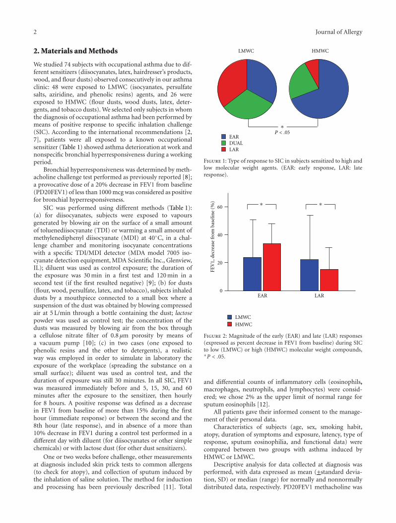
Figure 1: Type of response to SIC in subjects sensitized to high andlow molecular weight agents. (EAR: early response, LAR: lateresponse).
LAREAR
60
40
20
0
LMWC
HMWC
∗ ∗FE
V1,
decr
ease
from
base
line
(%)
Figure 2: Magnitude of the early (EAR) and late (LAR) responses(expressed as percent decrease in FEV1 from baseline) during SICto low (LMWC) or high (HMWC) molecular weight compounds,∗P < .05.
and differential counts of inflammatory cells (eosinophils,macrophages, neutrophils, and lymphocytes) were consid-ered; we chose 2% as the upper limit of normal range forsputum eosinophils [12].
All patients gave their informed consent to the manage-ment of their personal data.
Characteristics of subjects (age, sex, smoking habit,atopy, duration of symptoms and exposure, latency, type ofresponse, sputum eosinophilia, and functional data) werecompared between two groups with asthma induced byHMWC or LMWC.
Descriptive analysis for data collected at diagnosis wasperformed, with data expressed as mean (+standard devia-tion, SD) or median (range) for normally and nonnormallydistributed data, respectively. PD20FEV1 methacholine was

Journal of Allergy 3
Table 1: Characteristics of the compounds used for performing specific inhalation tests (SIC), concentrations used during SIC and durationof the exposure.
Agent Control Number of subjects Challenge concentration Time exposure
TDI vapours Diluent 37 0.002–0.003 ppm 30–120′
Flour dust Lactose dust 20 0.3–0.5 mg/m3 30′
Wood dusts Lactose dust 3 0.3–0.5 mg/m3 30′
Persulfate salts Lactose dust 6 0.05–0.1 mg/m3 30′
Aziridine Lactose dust 2 0.03–0.05 mg/m3 30′
Latex solution Normal saline 3 <0.0001 mg/m3 30′
Tobacco dusts Lactose dust 1 0.3–0.5 mg/m3 30′
Phenolic resins and Detergents vapours Diluent 2 Not measured∗ 30′∗
Subjects simulated the job activity in laboratory.
Table 2: Characteristics of the patients examined, according to thetype of the occupational sensitizer.
LMWC HMWC
Patients 48 26
Age, years 43.8± 12.0 38.9± 10.8
Gender,
Male 34 (70.8) 20 (76.9)
Female 14 (29.2) 6 (23.0)
Atopy 12 (25) 12 (46.1)
Smoking habit
Nonsmokers, N (%) 23 (47.9) 14 (53.8)
Smokers, N (%) 4 (8.3) 2 (7.7)
Ex-smokers, N (%) 21 (43.8) 10 (38.4)
Therapy N/Y 29/19 14/12
Duration of symptoms, yrs 6.1± 6.9 6.4± 5.7
Duration of exposure, yrs 20.1± 13.1 15.2± 7.7∗
Latency period, yrs 13.9± 12.7 8.7± 5.7∗
Baseline FEV1
L 3.01± 0.67 3.39± 0.68∗
% pred 89.2± 16.0 93.5± 14.8
Baseline PD20FEV1 (mg) 0.22 (0.01–5.00) 0.18 (0.02–5.00)
Sputum eosinophilia
Eosinophils, % 0.95 (0–32) 6.8 (0–43.1)∗
Data are presented as n (%), M±SD or GM (range; PD20FEV1) or median(range; eosinophils, %).∗P < .05.LMWC: low molecular weight compound; HMWC: high molecular weightcompound.
reported as geometric mean and log-transformed for sta-tistical analysis. Comparison among groups was performedby appropriate parametric (analysis of variance, Chi-squaretest, and unpaired t-test) and nonparametric tests (Mann-Whitney U test). A P value lower than 5% was considered assignificant, and a P value between 0.1 and .05 was consideredas expression of a trend.
3. Results
Patients’ characteristics are summarized in Table 2. Thesecharacteristics were similar in both groups with asthma
induced by HMWC and LMWC, except for duration ofexposure, latency period, and sputum eosinophilia. Durationof exposure and latency periods were higher in subjectswith asthma due to LMWC, who had also a lower sputumeosinophilia. FEV1 was lower in absolute value, but notin percentage of predicted, in patients with LMWC- thanin patients with HMWC-induced asthma. Atopy was morefrequently observed in patients with HMWC- than inpatients with LMWC-induced asthma, but the differencewas not statistically significant. Atopic subjects had a higherFEV1 (P = .02) and a higher percentage of eosinophils(P = .005).
The comparison among groups of subjects with asthmadue to different sensitizers was strongly affected by the lownumber of subjects included in the different groups, exceptfor patients sensitized to diisocyanates (N = 37) and to flourdust (N = 20) who were different for age, atopy, durationof exposure, and sputum eosinophil percentage, in the sameway as the difference between LMWC and HMWC. Therewas also a difference in the gender, related to the specific jobs(e.g., female in the subjects exposed to persulfate, or male insubjects exposed to diisocyanates).
Patterns of response following SIC were different forHMWC and LMWC. Subdividing subjects by type ofresponse, immediate responses (early + dual response) werecommon in subjects exposed to HMWC (Figure 1). Also,in subjects with higher sputum eosinophilia, immediateresponses were higher (61.1% versus 94.1%, P = .02).Considering all types of responses, magnitude of the early(EAR) responses was higher during SIC to LMWC while themagnitude of the late (LAR) responses was higher during SICto HMWC (Figure 2).
4. Discussion
The present study shows that few clinical characteristics maydifferentiate patients with occupational asthma induced byLMWC from those with asthma induced by HMWC. Inparticular, duration of exposure before the beginning ofasthma symptoms and the severity of the eosinophilic airwayinflammation were the only findings which differentiatedtwo groups of patients. In the same way, the pattern orairway response was consistently different, with patients withLMWC-induced asthma showing a higher frequency and
4 Journal of Allergy
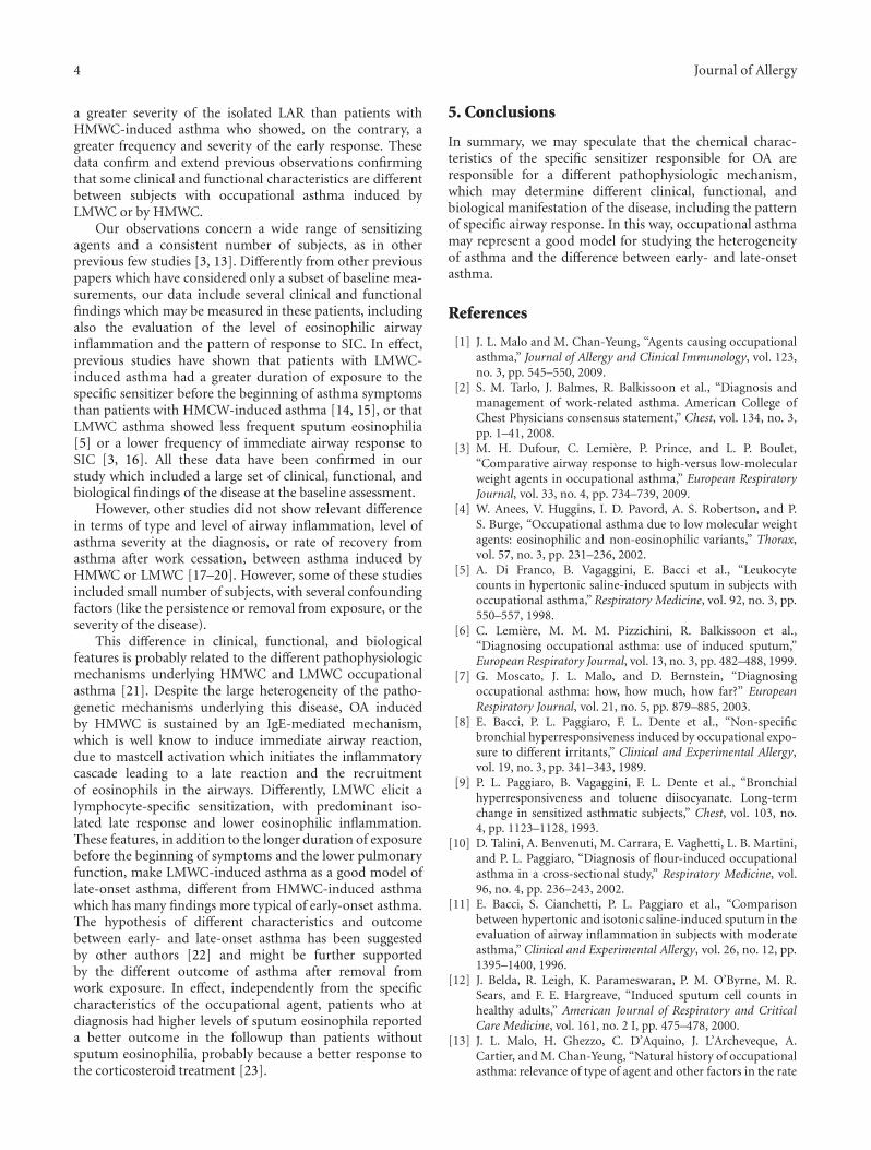
a greater severity of the isolated LAR than patients withHMWC-induced asthma who showed, on the contrary, agreater frequency and severity of the early response. Thesedata confirm and extend previous observations confirmingthat some clinical and functional characteristics are differentbetween subjects with occupational asthma induced byLMWC or by HMWC.
Our observations concern a wide range of sensitizingagents and a consistent number of subjects, as in otherprevious few studies [3, 13]. Differently from other previouspapers which have considered only a subset of baseline mea-surements, our data include several clinical and functionalfindings which may be measured in these patients, includingalso the evaluation of the level of eosinophilic airwayinflammation and the pattern of response to SIC. In effect,previous studies have shown that patients with LMWC-induced asthma had a greater duration of exposure to thespecific sensitizer before the beginning of asthma symptomsthan patients with HMCW-induced asthma [14, 15], or thatLMWC asthma showed less frequent sputum eosinophilia[5] or a lower frequency of immediate airway response toSIC [3, 16]. All these data have been confirmed in ourstudy which included a large set of clinical, functional, andbiological findings of the disease at the baseline assessment.
However, other studies did not show relevant differencein terms of type and level of airway inflammation, level ofasthma severity at the diagnosis, or rate of recovery fromasthma after work cessation, between asthma induced byHMWC or LMWC [17–20]. However, some of these studiesincluded small number of subjects, with several confoundingfactors (like the persistence or removal from exposure, or theseverity of the disease).
This difference in clinical, functional, and biologicalfeatures is probably related to the different pathophysiologicmechanisms underlying HMWC and LMWC occupationalasthma [21]. Despite the large heterogeneity of the patho-genetic mechanisms underlying this disease, OA inducedby HMWC is sustained by an IgE-mediated mechanism,which is well know to induce immediate airway reaction,due to mastcell activation which initiates the inflammatorycascade leading to a late reaction and the recruitmentof eosinophils in the airways. Differently, LMWC elicit alymphocyte-specific sensitization, with predominant iso-lated late response and lower eosinophilic inflammation.These features, in addition to the longer duration of exposurebefore the beginning of symptoms and the lower pulmonaryfunction, make LMWC-induced asthma as a good model oflate-onset asthma, different from HMWC-induced asthmawhich has many findings more typical of early-onset asthma.The hypothesis of different characteristics and outcomebetween early- and late-onset asthma has been suggestedby other authors [22] and might be further supportedby the different outcome of asthma after removal fromwork exposure. In effect, independently from the specificcharacteristics of the occupational agent, patients who atdiagnosis had higher levels of sputum eosinophila reporteda better outcome in the followup than patients withoutsputum eosinophilia, probably because a better response tothe corticosteroid treatment [23].
5. Conclusions
In summary, we may speculate that the chemical charac-teristics of the specific sensitizer responsible for OA areresponsible for a different pathophysiologic mechanism,which may determine different clinical, functional, andbiological manifestation of the disease, including the patternof specific airway response. In this way, occupational asthmamay represent a good model for studying the heterogeneityof asthma and the difference between early- and late-onsetasthma.
References
[1] J. L. Malo and M. Chan-Yeung, “Agents causing occupationalasthma,” Journal of Allergy and Clinical Immunology, vol. 123,no. 3, pp. 545–550, 2009.
[2] S. M. Tarlo, J. Balmes, R. Balkissoon et al., “Diagnosis andmanagement of work-related asthma. American College ofChest Physicians consensus statement,” Chest, vol. 134, no. 3,pp. 1–41, 2008.
[3] M. H. Dufour, C. Lemiere, P. Prince, and L. P. Boulet,“Comparative airway response to high-versus low-molecularweight agents in occupational asthma,” European RespiratoryJournal, vol. 33, no. 4, pp. 734–739, 2009.
[4] W. Anees, V. Huggins, I. D. Pavord, A. S. Robertson, and P.S. Burge, “Occupational asthma due to low molecular weightagents: eosinophilic and non-eosinophilic variants,” Thorax,vol. 57, no. 3, pp. 231–236, 2002.
[5] A. Di Franco, B. Vagaggini, E. Bacci et al., “Leukocytecounts in hypertonic saline-induced sputum in subjects withoccupational asthma,” Respiratory Medicine, vol. 92, no. 3, pp.550–557, 1998.
[6] C. Lemiere, M. M. M. Pizzichini, R. Balkissoon et al.,“Diagnosing occupational asthma: use of induced sputum,”European Respiratory Journal, vol. 13, no. 3, pp. 482–488, 1999.
[7] G. Moscato, J. L. Malo, and D. Bernstein, “Diagnosingoccupational asthma: how, how much, how far?” EuropeanRespiratory Journal, vol. 21, no. 5, pp. 879–885, 2003.
[8] E. Bacci, P. L. Paggiaro, F. L. Dente et al., “Non-specificbronchial hyperresponsiveness induced by occupational expo-sure to different irritants,” Clinical and Experimental Allergy,vol. 19, no. 3, pp. 341–343, 1989.
[9] P. L. Paggiaro, B. Vagaggini, F. L. Dente et al., “Bronchialhyperresponsiveness and toluene diisocyanate. Long-termchange in sensitized asthmatic subjects,” Chest, vol. 103, no.4, pp. 1123–1128, 1993.
[10] D. Talini, A. Benvenuti, M. Carrara, E. Vaghetti, L. B. Martini,and P. L. Paggiaro, “Diagnosis of flour-induced occupationalasthma in a cross-sectional study,” Respiratory Medicine, vol.96, no. 4, pp. 236–243, 2002.
[11] E. Bacci, S. Cianchetti, P. L. Paggiaro et al., “Comparisonbetween hypertonic and isotonic saline-induced sputum in theevaluation of airway inflammation in subjects with moderateasthma,” Clinical and Experimental Allergy, vol. 26, no. 12, pp.1395–1400, 1996.
[12] J. Belda, R. Leigh, K. Parameswaran, P. M. O’Byrne, M. R.Sears, and F. E. Hargreave, “Induced sputum cell counts inhealthy adults,” American Journal of Respiratory and CriticalCare Medicine, vol. 161, no. 2 I, pp. 475–478, 2000.
[13] J. L. Malo, H. Ghezzo, C. D’Aquino, J. L’Archeveque, A.Cartier, and M. Chan-Yeung, “Natural history of occupationalasthma: relevance of type of agent and other factors in the rate

Journal of Allergy 5
of development of symptoms in affected subjects,” Journal ofAllergy and Clinical Immunology, vol. 90, no. 6 I, pp. 937–944,1992.
[14] J. L. Malo and M. Chan-Yeung, “Current reviews of allergy andclinical immunology: occupational asthma,” Journal of Allergyand Clinical Immunology, vol. 108, no. 3, pp. 317–328, 2001.
[15] H. Allmers, B. Kirchner, H. Huber, Z. Chen, J. W. Walther,and X. Baur, “The latency period between exposure andthe symptoms in allergy to natural latex: suggestion forprevention,” Deutsche Medizinische Wochenschrift, vol. 121, pp.823–828, 1996.
[16] S. C. Stenton, “Determinants of whether occupational agentscause early, late, or dual asthmatic responses,” OccupationalMedicine, vol. 15, no. 2, pp. 431–444, 2000.
[17] C. Lemiere, A. Cartier, J. Dolovich et al., “Outcome of specificbronchial responsiveness to occupational agents after removalfrom exposure,” American Journal of Respiratory and CriticalCare Medicine, vol. 154, no. 2, pp. 329–333, 1996.
[18] L. P. Boulet, M. Boutet, M. Laviolette et al., “Airway inflam-mation after removal from the causal agent in occupationalasthma due to high and low molecular weight agents,”European Respiratory Journal, vol. 7, no. 9, pp. 1567–1575,1994.
[19] W. Anees, V. C. Moore, and P. S. Burge, “FEV decline inoccupational asthma,” Thorax, vol. 61, no. 9, pp. 751–755,2006.
[20] A. Descatha, H. Leproust, D. Choudat, R. Garnier, J. C.Pairon, and J. Ameille, “Factors associated with severity ofoccupational asthma with a latency period at diagnosis,”Allergy, vol. 62, no. 7, pp. 795–801, 2007.
[21] P. Maestrelli, P. Boschetto, L. M. Fabbri, and C. E. Mapp,“Mechanisms of occupational asthma,” Journal of Allergy andClinical Immunology, vol. 123, no. 3, pp. 531–542, 2009.
[22] C. Miranda, A. Busacker, S. Balzar, J. Trudeau, and S. E.Wenzel, “Distinguishing severe asthma phenotypes: role of ageat onset and eosinophilic inflammation,” Journal of Allergy andClinical Immunology, vol. 113, no. 1, pp. 101–108, 2004.
[23] C. Lemiere, S. Chaboillez, M. Welman, and K. Maghni, “Out-come of occupational asthma after removal from exposure: afollow-up study,” Canadian Respiratory Journal, vol. 17, no. 2,pp. 61–66, 2010.

Hindawi Publishing CorporationJournal of AllergyVolume 2011, Article ID 682574, 11 pagesdoi:10.1155/2011/682574
Review Article
Industrial Fungal Enzymes: An Occupational Allergen Perspective
Brett J. Green and Donald H. Beezhold
Allergy and Clinical Immunology Branch, Health Effects Laboratory Division, National Institute for Occupational Safety and Health,Centers for Disease Control and Prevention, Morgantown, WV 26505-2888, USA
Correspondence should be addressed to Brett J. Green, [email protected]
Received 25 February 2011; Accepted 30 March 2011
Academic Editor: Gordon L. Sussman
Copyright © 2011 B. J. Green and D. H. Beezhold. This is an open access article distributed under the Creative CommonsAttribution License, which permits unrestricted use, distribution, and reproduction in any medium, provided the original work isproperly cited.
Occupational exposure to high-molecular-weight allergens is a risk factor for the development and pathogenesis of IgE-mediatedrespiratory disease. In some occupational environments, workers are at an increased risk of exposure to fungal enzymes used inindustrial production. Fungal enzymes have been associated with adverse health effects in the work place, in particular in bakingoccupations. Exposure-response relationships have been demonstrated, and atopic workers directly handling fungal enzymes areat an increased risk for IgE-mediated disease and occupational asthma. The utilization of new and emerging fungal enzymes inindustrial production will present new occupational exposures. The production of antibody-based immunoassays is necessary forthe assessment of occupational exposure and the development of threshold limit values. Allergen avoidance strategies includingpersonal protective equipment, engineering controls, protein encapsulation, and reduction of airborne enzyme concentrations arerequired to mitigate occupational exposure to fungal enzymes.
1. Introduction
In the United States, the 2010 civilian workforce accountedfor 139 million people [1] who spend up to a quarterof their lifetime and half of their waking lives at work[2]. With changes in the global market, particularly withthe rise of biotechnology, new occupational hazards haveemerged. Approximately, 200 biotic (organisms or particlesof viral, prokaryote, or eukaryote origin) and an even greaternumber of abiotic (physical and chemical) agents havebeen associated with adverse health outcomes. In certainoccupational settings, particularly those engaged in handlingpurified microbial proteins in baking and manufacturingsectors, workers are at increased risk of becoming sensitizedand developing respiratory disease.
Occupational asthma (OA) is the most common respi-ratory disease reported in the workplace [3–8]. OA has beendefined as either irritant induced or immune mediated [6, 7].Immunologically mediated OA accounts for approximately90% of cases [9], but the severity of disease is dependenton the concentration, route, agent of exposure, and thelatency period [6, 7]. Both high- and low-molecular-weightantigens can induce OA, but the immunological mechanisms
are distinctly different. High-molecular-weight allergens aregenerally proteins that are greater than 5 kDa, and produc-tion of immunoglobulin E (IgE) results in the release ofmediators from mast cells and eosinophils [6, 7].
More than 250 high-molecular-weight allergens thatinduce OA have been identified [4, 6, 7]. Many are derivedfrom animals or plants, and exposure usually involvesmixtures of many proteins [4, 6]. Occupations wherehigh-molecular-weight allergens have been characterizedinclude seafood processing (tropomysin), dairy, poultry,citrus, greenhouse, baking, healthcare (latex), pharmaceuti-cal (drugs), and detergent manufacturing (fungal enzymes)[6]. Some of the best examples of high-molecular-weightoccupational allergens are the fungal enzymes. They areparticularly suited for study because they are often usedas purified preparations in baking, food, detergent, textile,and pharmaceutical industries [6, 10]. In this paper, wewill focus on the fungal enzymes as model high-molecular-weight allergens in industrial settings and describe themain enzymes that have been associated with occupationalsensitization and asthma. Identification of emerging fungalenzymes in manufacturing and biotechnology industries isdiscussed as well as new methods to detect and quantify
2 Journal of Allergy
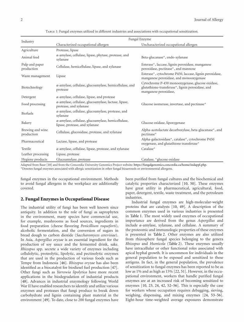
Table 1: Fungal enzymes utilized in different industries and associations with occupational sensitization.
IndustryFungal Enzyme
Characterized occupational allergen Uncharacterized occupational allergen
Agriculture Protease, lipase
Animal feedα-amylase, cellulase, lipase, phytase, protease, andxylanase
Beta-glucanase∗, endo-xylanase
Pulp and paperproduction
Cellulase, hemicellulase, lipase, and xylanaseEsterase∗, laccase, lignin peroxidase, manganeseperoxidase, pectinase∗ , and mannose
Waste management LipaseEsterase∗, cytochrome P450, laccase, lignin peroxidase,manganese peroxidase, and monooxygenase
Biotechnologyα-amylase, cellulase, glucoamylase, hemicellulase, andprotease
Cytochrome P-450 monooxygenase, glucose oxidase,glutathione-transferase∗, lignin peroxidase, andmanganese peroxidase,
Detergent α-amylase, cellulase, lipase, and protease
Food processingα-amylase, cellulase, glucoamylase, lactase, lipase,protease, and xylanase
Glucose isomerase, invertase, and pectinase∗
Biofuelsα-amylase, cellulase, glucoamylase, protease, andxylanase
Bakeryα-amylase, cellulase, glucoamylase, hemicellulase,lipase, protease, and xylanase
Glucose oxidase, lipoxygenase
Brewing and wineproduction
Cellulase, glucosidase, protease, and xylanaseAlpha-acetolactate decarboxylase, beta-glucanase∗, andpectinase∗
Pharmaceutical Lactase, lipase, and proteaseAlpha-galactosidase∗, catalase∗, cytochrome P450oxygenase, and glutathione transferase∗
Textile α-amylase, cellulase, lipase, protease, and xylanase Catalase∗
Leather processing Lipase, protease
Hygiene products Glucoamylase, protease Catalase, ∗glucose oxidase
Adapted from Baur [10] and from the Concordia University Genomics Project website: https://fungalgenomics.concordia.ca/home/indappl.php.∗Denotes fungal enzymes associated with allergic sensitization in other fungal bioaerosols or environmental allergens.
fungal enzymes in the occupational environment. Methodsto avoid fungal allergens in the workplace are additionallycovered.
2. Fungal Enzymes in Occupational Disease
The industrial utility of fungi has been well known sinceantiquity. In addition to the role of fungi as saprophytesin the environment, many species have commercial use,for example, mushrooms as food sources, ingredients infood preparation (cheese flavoring Penicillium roqueforti),alcoholic fermentation, and the conversion of sugars inbread dough to carbon dioxide (Saccharomyces cerevisiae).In Asia, Aspergillus oryzae is an essential ingredient for theproduction of soy sauce and the fermented drink, sake.Rhizopus spp. secrete a wide variety of enzymes includingcellulolytic, proteolytic, lipolytic, and pectinolytic enzymesthat are used in the production of various foods such asTempe from Indonesia [46]. Rhizopus oryzae has also beenidentified as a biocatalyst for biodiesel fuel production [47].Other fungi such as Yarrowia lipolytica have more recentapplications in the biodegradation of industrial products[48]. Advances in industrial enzymology following WorldWar II have enabled researchers to identify and utilize variousenzymes and proteases that fungi produce to break downcarbohydrate and lignin containing plant material in theenvironment [49]. To date, close to 200 fungal enzymes have
been purified from fungal cultures and the biochemical andcatalytic properties characterized [10, 50]. These enzymeshave great utility in pharmaceutical, agricultural, food,paper, detergent, textile, waste treatment, and the petroleumindustries.
Industrial fungal enzymes are high-molecular-weightproteins that are catalysts [10, 49]. A description of thecommon enzymes used in various industries is presentedin Table 1. The most widely used enzymes of occupationalimportance are derived from the genus Aspergillus andinclude α-amylase, xylanase, and cellulase. A summary ofthe proteomic and immunologic properties of these enzymesis presented in Table 2. Other enzymes are also utilizedfrom rhizosphere fungal species belonging to the generaRhizopus and Humicola (Table 2). These enzymes usuallyhave intracellular or other functional roles associated withapical hyphal growth. It is uncommon for individuals in thegeneral population to be exposed and sensitized to theseantigens. In fact, in the general population, the prevalenceof sensitization to fungal enzymes has been reported to be aslow as 1% and as high as 15% [22, 51]. However, in the occu-pational environment, workers that handle purified fungalenzymes are at an increased risk of becoming sensitized toenzymes [10, 23, 24, 42, 52–56]. This is especially the casefor workers whose occupation requires debagging, sieving,weighing, dispensing, and mixing enzymes [24, 53–56].Eight-hour time-weighted average exposures demonstrate

Journal of Allergy 3
Ta
ble
2:Fu
nga
len
zym
esas
soci
ated
wit
hoc
cupa
tion
alse
nsi
tiza
tion
and
asth
ma
inoc
cupa
tion
alen
viro
nm
ents
.
Fun
galO
rder
Fun
gals
peci
esA
llerg
enP
uta
tive
fun
ctio
nC
alcu
late
dsi
ze(k
Da)
Acc
essi
onn
um
ber
Pati
ent
reac
tivi
tyO
ccu
pati
onal
envi
ron
men
tR
efer
ence
Asc
omyc
ota
Dia
port
hal
esC
ryph
onec
tria
para
siti
caC
ryp
AP
Asp
arti
cpr
otea
se43
X63
351
5.7%
†Fo
odpr
oces
sin
g[1
1]
Euro
tial
esA
sper
gillu
sni
ger
Asp
n14
∗B
eta-
xylo
sida
se10
5A
F108
944
4–11
%††
Bak
ers,
text
ile,
dete
rgen
t,an
imal
feed
[12–
15]
Asp
n25
∗3-
phyt
ase
B(p
hos
phat
ase)
84L
2056
737
%††
An
imal
feed
[16,
17]
Asp
ngl
uco
amyl
ase
Glu
coam
ylas
e68
X00
548,
AM
2700
615–
19%†
63%††
Bak
ers
[10,
13,1
8]
Asp
nh
emic
ellu
lase
Hem
icel
lula
se22
.6A
1953
58–
43%†
100††
Bak
ers
[19–
21]
Asp
ergi
llus
oryz
aeA
spo
21∗
TAK
A-a
myl
ase
A53
X12
725,
X12
727,
M33
218,
D00
434
0.9–
35%†
1–32
%††
Bak
ers,
phar
mac
euti
cal
[12,
20–3
7]
Asp
ola
ctas
eL
acta
seA
P00
7164
29%
–31†
Ph
arm
aceu
tica
l[3
8–40
]A
spo
lipas
eL
ipas
eSR
Det
erge
nt
[41]
The
rmom
yces
lanu
gino
sus
(Hum
icol
ala
nugi
nosa
)
Th
ell
ipas
eL
ipas
e32
EU
3709
14,
AF0
5451
33%
††D
eter
gen
t,fo
odpr
oces
sin
g[4
2]
Hyp
ocre
ales
Tric
hode
rma
viri
deTr
ivce
llula
seC
ellu
lase
42E
F602
036
35%††
Tric
hode
rma
rees
eiTr
irs
cellu
lase
Cel
lula
se48
AY
9288
0913
%†
Bio
tech
nol
ogy
[43]
Sord
aria
les
Hum
icol
ain
sole
nsH
um
ince
llula
seC
ellu
lase
45P
5668
0SR
Det
erge
nt
[41]
Zyg
omyc
ota
Mu
cora
les
Rhi
zom
ucor
mie
hei
Rh
imA
PA
spar
tic
prot
ease
46M
1841
128
.6%†
Food
proc
essi
ng
[11]
Rhi
zopu
sor
yzae
Rh
iolip
ase
Lip
ase
42M
3835
2,A
B43
3531
,A
F229
435
SRP
har
mac
euti
cal
[44]
Ada
pted
from
the
IUIS
Alle
rgen
Nom
encl
atu
reSu
bcom
mit
tee,
Alle
rgom
e(h
ttp:
//w
ww
.alle
rgom
e.or
g/),
and
[45]
.∗ D
enot
esal
lerg
ens
depo
site
dto
the
IUIS
Alle
rgen
Nom
encl
atu
reSu
bcom
mit
tee.
SR:P
atie
nts
wit
ha
posi
tive
SPT
orsp
ecifi
cIg
E.
—† D
enot
esth
atpa
tien
tsw
ere
test
edw
ith
SPT.
—††
Den
otes
that
pati
ents
wer
ese
rolo
gica
llysc
reen
edu
sin
gP
harm
acia
Un
iCap
,Ras
t,or
Imm
un
oblo
t.

4 Journal of Allergy
that occupations weighing the enzyme preparations havethe lowest average exposure compared to those workers thatsieve [24]. These workers are often exposed to levels of dustthat exceed 4 mg m−3, the threshold limit value (TLV) forinhalable dust [57]. For other industrial environments thatuse lipase and cellulose in production, occupational exposureis highest in production areas and laboratories [42].
Adverse health effects associated with enzyme exposureare well characterized in the baking industry. In somecountries, bakery exposures to enzymes are one of the leadingcauses of occupational allergy [58]. Fungal enzymes arecommonly used as baking additives to improve the dough,increase shelf life, and decrease production time [19, 49,59]. Airborne concentrations ranging from 5.3 ng m−3 to200 ng m−3 have been reported in occupational environ-ments [12, 59, 60]. Occupational sensitization to fungalenzymes was first reported by Flindt [61]. Later, Baur etal. [62] demonstrated IgE sensitization in workers handlingthese products. Since the original study, fungal enzymeshave been identified as potent allergens in the occupa-tional environment [25, 26]. Prevalence of sensitization toAspergillus enzymes ranges from 8% for glucoamylase [13],11% for xylanase [13], 13% for cellulase [13], and up to34% for α-amylase [19, 27]. Sensitization to α-amylase inbakery workers results in decreased peak expiratory flow [63–66] and OA [20, 28, 67]. In one report, workers exposedto fungal enzymes induced an immediate bronchospasticreaction [49]. In the United States, the prevalence ofwork-related wheeze, runny nose, frequent sneezing, andspecific IgE to fungal enzymes was significantly higheramong highly exposed workers [68]. However, other irritant-induced mechanisms associated with high total dust levelshave also been reported in a cohort of British bakers [29, 56].To date, atopy has been hypothesized to be an important riskfactor for OA to fungal enzymes.
Occupational exposure to enzymes has been demon-strated in other industries including manufacturing [41, 53,69], pharmaceutical [25, 38], food processing [70], animalfeed, and biotechnology [43]. Like in baking environments,workers handling or in direct contact with fungal enzymesand with a history of atopy are at increased risk of becomingsensitized [10, 23, 24, 42, 52–56]. Sensitization to proteolyticenzymes has also been demonstrated in the manufacture ofdetergents [53, 71]. In the future, additional uses for fungalenzymes in industrial environments will be identified. Recentexamples include the use of α-amylase and glucoamylase forthe production of ethanol in the biofuel industry [72, 73].If proper methods of exposure prevention are not followedand exposure is not monitored in these industries, it ispossible that new groups of workers will suffer adversehealth outcomes and become sensitized to enzymes. In thefollowing sections we describe the major fungal enzymes,prevalence of sensitization, and occupational environmentthat they are most likely to be encountered.
3. Fungal Enzyme Allergens
3.1. α-Amylase. Fungal amylase is the most well-charac-terized fungal enzyme used in the occupational environment.
Originally discovered by Takamine in 1884 [49], bakershave used α-amylase as a supplement to cereal flour toimprove carbohydrate fermentation by yeasts and ultimatelythe quality of the bread [49]. α-amylase cleaves long-chaincarbohydrates into simpler sugars including maltose [49].Derived from A. oryzae, α-amylase is a 478 amino acid gly-coprotein with a molecular weight of 53 kDa (Table 2; [6]).Occupational sensitization to α-amylase was first reportedby Flindt [61] and has subsequently been identified as anallergen in baking [74], pharmaceutical [25], animal feed[12], and biotechnology industries [43]. The allergen wasoriginally designated Asp o 2 by the International Unionof Immunological Societies (IUIS) Allergen NomenclatureSubcommittee but now has been redesignated Asp o 21 [27].Since this preliminary work, α-amylase has been identifiedas one of the principle sensitizers in large-scale bakeries [24,56]. The prevalence of sensitization among bakers is variableand ranges from 0.9% to 34% [13, 18, 19, 21, 23, 24, 27–35, 54, 66, 67, 75]. Concentrations as high as 40 ng m−3
have been reported in baking environments [60]; however,α-amylase concentrations in the low ng m−3 range have beenassociated with an increased frequency of sensitization [58].
The most common tasks associated with α-amylaseexposure involve dispensing, sieving, weighing and mixing[55, 56, 60]. Exposures that exceed the maximum exposurelimit for flour dust in the United Kingdom were identi-fied in mixing, weighing [54], and dispensing operations[55]. The prevalence of sensitization to α-amylase is 9.9times greater among workers in high-exposure categoriescompared to those workers in low-exposure categories [55].Aerosolized particle size distribution analysis in bakingenvironments demonstrated that workers are exposed to α-amylase particles within the inhalable size fraction [60]. OAis commonly identified in workers sensitized to α-amylase.After bronchial provocation with α-amylase, between 16and 100% of sensitized workers were found to give apositive immediate response depending on the study [25,35, 49]. Nasal provocation with α-amylase in skin pricktest (SPT) positive workers also induced rhinitis [26].Furthermore, positive associations between α-amylase SPTand work-related respiratory symptoms have been identified[23]. Interestingly, heating α-amylase has been shown toreduce enzymatic and allergenic activity of the enzyme [76].Potential sensitization of bakers’ family members due to α-amylase associated with clothes, shoes, and bakery textileshas also been reported by Vissers [77].
3.2. γ-Amylase. γ-amylase or glucoamylase is primarilyobtained from A. niger, A. awamori, and R. delemar. Glu-coamylase is used as a dough additive by bakers, often inassociation with α-amylase. The enzyme is also used in theproduction of high-glucose syrups [46]. Glucoamylase has amolecular weight of 68 kDa (Table 2) and can remain func-tionally active at elevated pH. Glucoamylase exposure hasbeen primarily reported in baking occupations [10, 13, 18];however, occupational exposure has also been reported infruit and salad processing [52]. Sen et al. [52] demonstratedthat three workers with shortness of breath, chest tightness,and wheeze had specific IgE to glucoamylase. Quirce et al.

Journal of Allergy 5
[18] also demonstrated positive SPT to glucoamylase inall tested subjects (n = 4); however, only three of thefour patients elicited an early asthmatic response followingbronchial provocation. Airborne glucoamylase was shown in9% of air samples from a bakery [59], and median levels were10.3 ng m−3. Moderate allergenic cross-reactivity has alsobeen reported between glucoamylase and α-amylase [18].
3.3. Cellulase, Xylanase, and Hemicellulase. Cellulases areenzymes that hydrolize cellulose into glucose and are pri-marily used in the pharmaceutical, baking, detergent, andtextile industries [6, 78]. Cellulase has been purified fromseveral rhizosphere fungi including A. niger and Trichodermaviride [49], as well as Humicola insolens [41]. The molecularweight of cellulases ranges from 22 to 45 kDa (Table 2).Cellulases derived from these fungi are used in baking tobreak up roughage in dough and as a digestive aid in thefood industry [13, 79, 80]. The first case of OA causedby cellulase was reported in 1981 in a plant pathologist[49, 81], and later these findings were confirmed in twopharmaceutical workers [80], four laboratory workers [14],and two bakers [49]. In each of these studies, the workers hadspecific IgE to the cellulase antigens. In 171 German bakers,the prevalence of sensitization to cellulase was 13% [13].Airborne concentrations of cellulase have been quantifiedusing a modified dot blot technique and were <180 ng m−3
in a flour mill, crisp bread factory, and a bakery [82]. OA hasalso been reported to cellulase in the baking industry [83] aswell as from H. insolens used in the detergent industry [41].
Endo 1, 4-beta-D-xylanase and beta-xylosidase are majorenzymes involved in xylan hydrolysis [13]. Collectivelytermed xylanases, these enzymes are a type of hemicellulasethat breaks down hemicelluloses, a major component inplant cell walls [13]. Besides α-amylase, xylanases are thenext most frequently used enzymes in the baking industryto remove pentosans from bread and increase bread volume[13, 49]. The prevalence of IgE sensitization to hemicellulasewas reported to be 8% [19] and 11% for xylanase [13].Sander and colleagues [13] found that 7 of 8 bakers hadserum IgE to a 105 kDa protein in a xylanase ingredient. Thisprotein was identified using mass spectrometry to be beta-xylosidase derived from A. niger. The allergen was designatedAsp n 14 by the IUIS Allergen Nomenclature Subcommittee(Table 2). Airborne concentrations of xylanase have beenreported to be <40 ng m−3 in a flourmill and crisp breadfactory [82]. Concentrations as high as 200 ng m−3 were alsoreported in a bakery, but these values were associated withthe natural xylanase activity of wheat [82]. Case reportshave verified xylanase sensitization and the presence of anIgE mechanism in respiratory disease [15, 79]. OA hasalso been reported to xylanase in the baking industry [83],and in a case report, a baker had an immediate asth-matic response following inhalation challenge [15]. Cross-reactivity between cellulase and xylanase has been reportedto be in the range of 80–90% but no cross-reactivity hasbeen shown with α-amylase [13, 14]. Similarly, workers canalso be monosensitized to cellulase and xylanase withoutconcomitant sensitization to α-amylase [83].
3.4. Lactase. A. oryzae lactase is a high-molecular-weightprotein that is involved in the hydrolysis of the disaccharide,lactose. Lactase is used in the pharmaceutical industry todevelop dietary aids for patients intolerant to lactose. In across-sectional study of United States pharmaceutical work-ers, Bernstein and colleagues [38] identified 29% of lactase-exposed workers to have positive SPT response to lactase.Workers with a positive SPT were nine times more likelyto have respiratory symptoms than workers with a negativeSPT [38]. Interestingly, atopy was not associated with thedevelopment of respiratory symptoms. Occupational sensi-tization to lactase has been reported in workers formulatingand packaging gastrointestinal consumer products [39]. Ininhalational challenge studies conducted by Laukkanin andcolleagues [40], lactase was identified to induce occupationalIgE-mediated respiratory sensitization. Interestingly, lactaseexposure has also been identified to cause contact skinreactions [40].
3.5. Lipase. Lipase is an essential catalyst that digests water-insoluble lipids. A. oryzae and R. oryzae lipase are usedbecause of low extraction costs, thermal and pH stability,substrate specificity, and activity in organic solvents. Lipaseis predominantly used in the manufacture of laundrydetergents and in baking; however, other newer applicationshave been developed. For example, Candida antarctica lipasehas recently been used as a biocatalyst for the biofuel industry[84]. The incidence of occupational sensitization to lipase, inindustrial settings is understudied. In a preliminary analysisof detergent manufacturing workers, 3 workers were foundto be sensitized to lipase and bronchial provocation testsprovoked a reproducible asthmatic response [41]. A recentcase study of a pharmaceutical manufacturing worker alsodemonstrated sensitization to fungal lipase derived from R.oryzae but not A. oryzae α-amylase [44].
3.6. Phytase. A. niger and R. oligosporus produce phosphatasethat catalyzes the hydrolysis of phytate to lower-orderphosphate esters [16]. Termed phytase, this enzyme enhancesphosphate bioavailability in the digestive tract and hasbeen utilized in the animal feed industry during the lasttwo decades [17]. Phytase accounts for 0.1–1% of totalextractable protein from A. niger [17]. 3-phytase B derivedfrom A. niger is an 84 kDa protein that has been designatedAsp n 25 by the IUIS Allergen Nomenclature Subcommittee(Table 2). Allergic sensitization to phytase has been reportedin animal feed factory workers (7–90%), and sensitization ishighest at sites where phytase is handled in powdered form[16, 17, 69, 85]. In a cross-sectional study of 53 technicalcenter workers that produced A. niger phytase, 52% ofworkers in the high-exposure group and only 10% in thelow-exposure group were sensitized to phytase [16]. Personalexposure to phytase has been shown to exacerbate OA, andinhalation challenge tests produced immediate asthmaticresponse [86]. It has been proposed that phytase is highlysensitizing and that direct contact should be avoided in thisindustry [16].

6 Journal of Allergy
3.7. Enzymes Used in Health Care Settings: Biodiastase andFlaviastase. Fungal enzymes have a number of applicationsin the healthcare environment. Fungal enzymes derivedfrom A. niger are used in powdered form with otherenzyme extracts by pharmacists to prepare digestive powders.Biodiastase and Flaviastase are two examples of fungalenzymes that have been associated with sensitization inhospital workers and pharmaceutical workers handling theseproducts [10, 12–21, 23–35, 38–44, 51–88]. To date, healtheffects in workers exposed to these enzymes remain poorlycharacterized.
3.8. EPg22 Protease: Aspartic Protease. The aspartic proteasesproduced by Rhizomucor miehei and Cryphonectria parasiticaare utilized in almost half of the cheese production oper-ations throughout the world [46]. The proteases assist inmilk clotting and facilitate a change in cheese properties byhydrolyzing certain peptide bonds. Occupational exposureto these proteases has been associated with occupationalsensitization in a rennet production plant [11]. Specifically29% and 6% of workers had a positive skin prick test (SPT) toR. miehei and C. parasitica aspartic protease extracts, respec-tively [11]. Other novel enzymes with potential applicationin the food processing industry have been identified. Pg222is a novel extracellular protease produced by P. chrysogenum(Pg222). The enzyme was isolated from dry-cured hams andwas found to have a broad range of applications in industriesthat produce dry-cured meat products [89]. Although nooccupational sensitization has been reported to this enzyme,it demonstrates that the introduction of any new enzymecould potentially represent an occupational hazard.
4. Emerging Occupational FungalEnzyme Exposures
The utility of fungal enzymes to degrade xenobiotics andorganic compounds in the industrial sector continues tobe recognized [46]. Fungal enzymes are now being usedfor a variety of purposes across many different indus-tries. Improved biochemical and molecular technologieshave enabled the production of other potentially allergenicproteins [14]. According to Baur [10], more than 186commercial enzymes were produced in the European Unionin 2001, and many of these were produced by recombinanttechnology or had been genetically engineered. Table 1provides a summary of the major fungal enzymes that areutilized in industrial settings. All of the aforementionedenzymes that are listed in Table 1 have been identified tobe potent allergens in the workplace; however, the ability ofthe other listed enzymes to cause adverse health outcomesfollowing occupational exposure remains unclear.
Several of the enzymes presented in Table 1, not identi-fied as occupational allergens, have been identified as aller-gens associated with environmental bioaerosols. Catalase,a fungal enzyme utilized in hygiene products, pharmaceu-ticals, and textiles, has been identified as an allergen inthe entomopathogenic fungus, Metarhizium anisopliae [90].Pectinase is used in brewing and wine production, food
processing, and paper industries and allergy to pectinase hasbeen associated with occupational exposure [91]. Esterasehas been identified as an allergen in Hevea brasiliensis (natu-ral rubber latex) [92]. Beta-glucanase is used to improve thenutritional yield of animal feeds, and occupational exposurehas been shown in a case study to significantly reduceforced vital capacity and forced expired volume in 1 second(FEV1) [86]. The worker in this case study was also SPTpositive and had specific IgE to beta-glucanase [86]. In thebiotechnology and pharmaceutical industries, glutathione-S-transferase (GST) has a number of applications. GST is anapproximately 26 kDa protein that has been identified as amajor Alternaria alternata allergen and is highly conservedacross fungi [45, 93, 94]. The IUIS Allergen NomenclatureSubcommittee has designated this allergen Alt a 13 [93, 94].Interestingly, alpha-galactosidase has been associated withdelayed anaphylaxis, angioedema, or urticaria in sensitizedpatients following the ingestion of beef, pork, or lamb [95].Although the role of alpha-galactosidase and these otherenzymes following occupational exposure remains unclear,these studies provide preliminary insight into the possiblepotency of these allergens in industrial environments.
5. Immunodiagnostic Detection Methodologies
Occupational allergic sensitization to fungal enzymes isdiagnosed clinically using available in vivo SPT reagents, orin vitro assays such as Phadia ImmunoCap [7]. However, SPTreagents for most of the fungal enzymes used in industrialsettings are not commercially available and have to be eithercustom ordered or prepared individually by the investigator.Methods for SPT extract preparation that are used byinvestigators in the field have been previously described byQuirce et al. [49]. In vitro diagnostic tools that can quantifythe amount of specific IgE to an occupational allergen arenot readily available except in research laboratories whereinvestigators prepare their own inhibition or radioaller-gosorbent enzyme-linked immunosorbent assay (ELISA) toquantify specific IgE [36, 49]. To date, α-amylase (k87) isthe only fungal enzyme available on the Phadia ImmunoCaptesting panel. To confirm OA caused by fungal enzymes,bronchial provocation tests can be undertaken to documentimmediate or late-phase responses to fungal enzymes [36,49]. Positive immediate response criteria used in workersexposed to enzymes include a greater than 20% fall in FEV1,whereas a late-phase response has been considered positivewhen there is a 30% or greater fall in peak expiratoryflow rate [49]. However, there are several limitations withbronchial provocation tests that should be considered; theseare discussed in detail by Peden and Reed [7].
In order to better understand the relationships betweenoccupational fungal enzyme exposure and clinical symp-tomology, accurate information on the distribution andquantity of the fungal enzyme in the occupational envi-ronment will be required. Immunodiagnostic methods thatutilize antibodies could provide standardized methods forquantifying fungal enzyme biomarkers in a variety of

Journal of Allergy 7
occupational environments. Following validation and inter-laboratory comparison, the assays could be used for exposureassessment to determine the existence of exposure-responserelationships [58, 96]. This information is critical for thedevelopment of future threshold limit values (TLVs) andother occupational standards.
Several antibodies and immunodiagnostic methods havebeen produced to detect industrial fungal enzymes, inparticular α-amylase. These methods have been employed infield investigations and used to quantify the concentration ofthe enzyme from collected air samples. Bogdanovic et al. [97]used an enzyme immunoassay with a sensitivity of 25 pg/mLto quantify α-amylase in airborne and surface dust samplescollected from five bakeries. In the same study, a lateral flowimmunoassay for α-amylase was compared to the referenceenzyme immunoassay. The sensitivity of the lateral flowassay was 1–10 ng/mL, and extracts with >5 ng/mL allergenwere positive in the lateral flow assay [97]. In a study of507 personal air samples, Houba and colleagues [60] useda rabbit IgG capture immunoassay to quantify α-amylase inspecific baking job category. Concentrations of α-amylase upto 40 ng m−3 were quantified, and workers directly involvedwith dough preparation had the highest exposures [60].Using the same rabbit IgG sandwich assay, Nieuwenhuijsenet al. [55] identified dispensing and mixing areas to havethe highest α-amylase exposure in British bakeries and flourmills. Two monoclonal antibody- (mAb-) based ELISAshave been developed for the detection of α-amylase in theoccupational environment. Assay sensitivities ranged from0.2 ng/mL [98] to 0.6 ng/mL [99]. A quantitative mAb-mediated dot blot assay has also been previously describedfor cellulase and xylase; the detection limits reported were20 ng m−3 and 2 ng m−3, respectively [82]. mAbs to otherfungal enzymes, such as xylanase have been produced andreported in the literature [100]. Similarly, the detergentindustry has produced antibodies and immunoassays forseveral common fungal enzymes and these have been utilizedin industrial hygiene safety programs to mitigate workerexposures [101–103]. Unfortunately, for many other fungalenzymes presented in Table 1, there are no commerciallyavailable antibodies to enable quantification in the occu-pational environment. The development of fungal enzyme-specific mAbs in combination with immunodiagnostic tech-niques will further our knowledge of the exposure-responserelationships in occupational environments. Using thesemethods will also help enable the development of standardsand focus on the prevention of sensitization in heavilycontaminated work environments.
6. Allergen Avoidance and Directions forthe Future
Exposure to fungal enzymes, in particular α-amylase, is aconsiderable health risk in a number of industries. Cross-sectional studies have shown that processing workers inhigh-exposure categories who handled fungal enzymes areup to ten times more likely to be sensitized to fungalenzymes than workers in the low-exposure category [55].
Highest concentrations of enzymes in the inhalable fractionwere encountered among workers located in dispensing,mixing, weighing, and sieving occupations [54–56, 60].Airborne concentrations as high as 40 ng m−3 and in somecases even higher (200 ng m−3) have been reported forsensitized workers located in these handling areas [12, 55,60]. Concentrations in the low ng m−3 range have beenassociated with an increased frequency of sensitization [58].For other fungal enzymes, such as phytase, similar findingshave been reported [16].
The continued utilization of other previously overlookedenzymes as well as new genetically engineered enzymes invarious industries will continue to provide diagnostic chal-lenges, even for the most seasoned occupational medicineprofessional. It is likely that new cases of occupational allergicdisease will emerge following exposure to fungal industrialenzymes during the next decade. In response, identificationof exposure-response relationships will be critical for thedevelopment of TLVs and occupational exposure levels.However, this will depend on the development of suitablediagnostic antibodies and immunoassays. Currently, subtil-isin, a sereine endopeptidase derived from Bacillus subtilis, isthe only enzyme for which the American Conference of Gov-ernmental Industrial Hygienists (ACGIH) has established aTLV value (60 ng m−3). The European Union Directive alsoclassifies the fungal enzymes cellulase and α-amylase withthe risk phrase R42 (may cause sensitization by inhalation)[10]. There are currently no consensus standards for otherindustrially utilized fungal enzymes.
As a precautionary measure, it has been concluded thatall enzymes should be regarded as an allergen that can exac-erbate respiratory sensitization in susceptible populations[10, 59]. Baur [10] has further proposed that all enzymesshould be classified as R42 according to the European UnionDirective criteria. Although intervention in the bakery indus-try has had little to no effect [104], installation of engineeringcontrols and implementation of personal protective equip-ment programs in animal feed workers exposed to phytasewas shown to result in the immediate cessation of hyper-sensitivity symptoms [10]. Improvements in biotechnologyhave also included the encapsulation of some enzymes [105,106] and proteins [107]. These engineering controls havebeen proposed to reduce occupational exposure to enzymes;however, encapsulation alone may not completely preventenzyme-induced allergy and OA [108, 109]. To date, thedetergent industry has implemented a derived minimal effectlevel (DMEL) of 60 ng m−3 for pure enzyme proteins [110].Although this DMEL was provided as guidance by theACGIH, other manufacturers have implemented their ownoccupational exposure guidelines (OEGs) for fungal enzymessuch as α-amylase (5–15 ng m−3), lipase (5–20 ng m−3), andcellulase (8–20 ng m−3) [110]. In addition, the detergentindustry has developed a medical surveillance program toidentify and correct elevated exposures before occupationalillnesses occur [101–103, 111]. As a result, the incidence ofoccupational allergy has dropped substantially [101–103].Implementation of DMELs and OEGs will further assist inthe reduction of occupational exposure. Reducing worker ex-posure to fungal enzymes in industry by the implementation

8 Journal of Allergy
of engineering controls and other allergen avoidance strate-gies will continue to mitigate personal exposure and furtherreduce the occupational health risk.
Acknowledgments
This work was supported in part by an interagency agree-ment with NIEHS (Y1-ES-0001). The findings and conclu-sions in this paper are those of the authors and do notnecessarily represent the views of the National Institute forOccupational Safety and Health.
References
[1] U. S. Department of Labor, “Employment status of thecivilian noninstitutional population, 1940 to date,” Bureauof Labor Statistics, Washington, DC, USA, 2010, 2010,http://www.bls.gov/cps/cpsaat1.pdf.
[2] February 2011, http://www.healthypeople.gov/2020/topic-sobjectives2020/overview.aspx?topicid=30.
[3] B. T. Butcher and J. E. Salvaggio, “Occupational asthma,”Journal of Allergy and Clinical Immunology, vol. 78, no. 4 I,pp. 547–556, 1986.
[4] J. Dutkiewicz, L. Jablonski, and S. A. Olenchock, “Occupa-tional biohazards: a review,” American Journal of IndustrialMedicine, vol. 14, no. 5, pp. 605–623, 1988.
[5] J. Lacey and B. Crook, “Fungal and actinomycete sporesas pollutants of the workplace and occupational allergens,”Annals of Occupational Hygiene, vol. 32, no. 4, pp. 515–533,1988.
[6] F. Lachowsky and M. Lopez, “Occupational allergens,” Cur-rent Allergy and Asthma Reports, vol. 1, no. 6, pp. 587–593,2001.
[7] D. Peden and C. E. Reed, “Environmental and occupationalallergies,” Journal of Allergy and Clinical Immunology, vol.125, no. 2, pp. S150–S160, 2010.
[8] E. L. Petsonk, “Work-related asthma and implications for thegeneral public,” Environmental Health Perspectives, vol. 110,suuplement 4, pp. 569–572, 2002.
[9] C. E. Mapp, “Genetics and the occupational environment,”Current Opinion in Allergy and Clinical Immunology, vol. 5,no. 2, pp. 113–118, 2005.
[10] X. Baur, “Enzymes as occupational and environmentalrespiratory sensitisers,” International Archives of Occupationaland Environmental Health, vol. 78, no. 4, pp. 279–286, 2005.
[11] A. Jensen, S. Dahl, D. Sherson, and B. Sommer, “Respiratorycomplaints and high sensitization rate at a rennet-producingplant,” American Journal of Industrial Medicine, vol. 49, no.10, pp. 858–861, 2006.
[12] M. Vanhanen, T. Tuomi, U. Tiikkainen et al., “Sensitisationto enzymes in the animal feed industry,” Occupational andEnvironmental Medicine, vol. 58, no. 2, pp. 119–123, 2001.
[13] I. Sander, M. Raulf-Heimsoth, C. Siethoff, C. Lohaus, H. E.Meyer, and X. Baur, “Allergy to Aspergillus-derived enzymesin the baking industry: identification of β-xylosidase fromAspergillus niger as a new allergen (Asp n 14),” Journal ofAllergy and Clinical Immunology, vol. 102, no. 2, pp. 256–264,1998.
[14] K. Tarvainen, L. Kanerva, O. Tupasela et al., “Allergy fromcellulase and xylanase enzymes,” Clinical and ExperimentalAllergy, vol. 21, no. 5, pp. 609–615, 1991.
[15] X. Baur, I. Sander, A. Posch, and M. Raulf-Heimsoth,“Baker’s asthma due to the enzyme xylanase—a new occu-pational allergen,” Clinical and Experimental Allergy, vol. 28,no. 12, pp. 1591–1593, 1998.
[16] X. Baur, S. Melching-Kollmuss, F. Koops, K. Straßburger, andA. Zober, “IgE-mediated allergy to phytase—a new animalfeed additive,” Allergy, vol. 57, no. 10, pp. 943–945, 2002.
[17] G. Doekes, N. Kamminga, L. Helwegen, and D. Heederik,“Occupational IgE sensitisation to phytase, a phosphatasederived from Aspergillus niger,” Occupational and Environ-mental Medicine, vol. 56, no. 7, pp. 454–459, 1999.
[18] S. Quirce, M. Fernandez-Nieto, B. Bartolome, C. Bombın, M.Cuevas, and J. Sastre, “Glucoamylase: another fungal enzymeassociated with baker’s asthma,” Annals of Allergy, Asthmaand Immunology, vol. 89, no. 2, pp. 197–202, 2002.
[19] X. Baur, W. Sauer, and W. Weiss, “Baking additives as newallergens in Baker’s asthma,” Respiration, vol. 54, no. 1, pp.70–72, 1988.
[20] S. Quirce, M. Fernandez-Nieto, C. Escudero, J. Cuesta, M.De Las Heras, and J. Sastre, “Bronchial responsiveness tobakery-derived allergens is strongly dependent on specificskin sensitivity,” Allergy, vol. 61, no. 10, pp. 1202–1208, 2006.
[21] J. Elms, D. Fishwick, J. Walker et al., “Prevalence ofsensitisation to cellulase and xylanase in bakery workers,”Occupational and Environmental Medicine, vol. 60, no. 10, pp.802–804, 2003.
[22] R. E. Biagini, B. A. MacKenzie, D. L. Sammons et al.,“Evaluation of the prevalence of antiwheat-, anti-flour dust,and anti-α-amylase specific IgE antibodies in US blooddonors,” Annals of Allergy, Asthma and Immunology, vol. 92,no. 6, pp. 649–653, 2004.
[23] R. Houba, D. J. J. Heederik, G. Doekes, and P. E. M. van Run,“Exposure-sensitization relationship for α-amylase allergensin the baking industry,” American Journal of Respiratory andCritical Care Medicine, vol. 154, no. 1, pp. 130–136, 1996.
[24] T. A. Smith and P. W. Smith, “Respiratory symptomsand sensitization in bread and cake bakers,” OccupationalMedicine, vol. 48, no. 5, pp. 321–328, 1998.
[25] E. Losada, M. Hinojosa, S. Quirce, M. Sanchez-Cano,and I. Moneo, “Occupational asthma caused by α-amylaseinhalation: clinical and immunologic findings and bronchialresponse patterns,” Journal of Allergy and Clinical Immunol-ogy, vol. 89, no. 1 I, pp. 118–125, 1992.
[26] J. Brisman and L. Belin, “Clinical and immunologicalresponses to occupational exposure to α-amylase in thebaking industry,” British Journal of Industrial Medicine, vol.48, no. 9, pp. 604–608, 1991.
[27] X. Baur, Z. Chen, and I. Sander, “Isolation and denominationof an important allergen in baking additives: α-amylase fromAspergillus oryzae (Asp o II),” Clinical and ExperimentalAllergy, vol. 24, no. 5, pp. 465–470, 1994.
[28] J. Brisman, L. Lillienberg, L. Belin, M. Ahman, and B.Jarvholm, “Sensitisation to occupational allergens in bakers’asthma and rhinitis: a case-referent study,” InternationalArchives of Occupational and Environmental Health, vol. 76,no. 2, pp. 167–170, 2003.
[29] T. A. Smith, G. Parker, and T. Hussain, “Respiratory symp-toms and wheat flour exposure: a study of flour millers,”Occupational Medicine, vol. 50, no. 1, pp. 25–29, 2000.
[30] X. Baur, P. O. Degens, and I. Sander, “Baker’s asthma:still among the most frequent occupational respiratorydisorders,” Journal of Allergy and Clinical Immunology, vol.102, no. 6, pp. 984–997, 1998.
[31] A. Brant, J. Berriman, C. Sharp et al., “The changingdistribution of occupational asthma: a survey of supermarketbakery workers,” European Respiratory Journal, vol. 25, no. 2,pp. 303–308, 2005.
[32] T. A. Smith, K. P. S. Lumley, and E. H. K. Hui, “Allergy toflour and fungal amylase in bakery workers,” OccupationalMedicine, vol. 47, no. 1, pp. 21–24, 1997.

Journal of Allergy 9
[33] J. Walusiak, W. Hanke, P. Gorski, and C. Pałczynski, “Respi-ratory allergy in apprentice bakers: do occupational allergiesfollow the allergic march?” Allergy, vol. 59, no. 4, pp. 442–450, 2004.
[34] A. Bataille, M. Anton, F. Mollat et al., “Respiratory allergiesamong symptomatic bakers and pastry cooks: initial resultsof a prevalence study,” Allergy and Immunology, vol. 27, pp.7–10, 1995.
[35] J. Birnbaum, F. Latil, D. Vervloet, M. Senft, and J. Charpin,“The role of alpha-amylase in baker’s asthma,” Revue desMaladies Respiratoires, vol. 5, no. 5, pp. 519–521, 1988.
[36] W. E. Horner, M. Armstrong, J. El-Dahr et al., “Prevalenceof IgE reactivities in mold-allergic subjects to commerciallyavailable fungal enzymes,” Allergy and Asthma Proceedings,vol. 29, no. 6, pp. 629–635, 2008.
[37] T. Storaas, S. K. Steinsvag, E. Florvaag, A. Irgens, and T.B. Aasen, “Occupational rhinitis: diagnostic criteria, relationto lower airway symptoms and IgE sensitization in bakeryworkers,” Acta Oto-Laryngologica, vol. 125, no. 11, pp. 1211–1217, 2005.
[38] J. A. Bernstein, D. I. Bernstein, T. Stauder, Z. Lummus, andI. L. Bernstein, “A cross-sectional survey of sensitization toAspergillus oryzae-derived lactase in pharmaceutical work-ers,” Journal of Allergy and Clinical Immunology, vol. 103, no.6, pp. 1153–1157, 1999.
[39] D. C. F. Muir, A. B. Verrall, J. A. Julian, J. M. Millman, M.A. Beaudin, and J. Dolovich, “Occupational sensitization tolactase,” American Journal of Industrial Medicine, vol. 31, no.5, pp. 570–571, 1997.
[40] A. Laukkanen, P. Ruoppi, S. Remes, T. Koistinen, and S.Makinen-Kiljunen, “Lactase-induced occupational proteincontact dermatitis and allergic rhinoconjunctivitis,” ContactDermatitis, vol. 57, no. 2, pp. 89–93, 2007.
[41] A. Brant, A. Hole, J. Cannon et al., “Occupational asthmacaused by cellulase and lipase in the detergent industry,”Occupational and Environmental Medicine, vol. 61, no. 9, pp.793–795, 2004.
[42] C. R. Johnsen, T. B. Sorensen, A. I. Larsen et al., “Allergyrisk in an enzyme producing plant: a retrospective follow upstudy,” Occupational and Environmental Medicine, vol. 54, no.9, pp. 671–675, 1997.
[43] M. Vanhanen, T. Tuomi, H. Nordman et al., “Sensitizationto industrial enzymes in enzyme research and production,”Scandinavian Journal of Work, Environment and Health, vol.23, no. 5, pp. 385–391, 1997.
[44] G. Loureiro, B. Tavares, C. Pereira, M. Lundberg, and C.Chieira, “Occupational allergy to fungal lipase in the phar-maceutical industry,” Journal of Investigational Allergologyand Clinical Immunology, vol. 19, no. 3, pp. 242–244, 2009.
[45] B. Simon-Nobbe, U. Denk, V. Poll, R. Rid, and M. Breiten-bach, “The spectrum of fungal allergy,” International Archivesof Allergy and Immunology, vol. 145, no. 1, pp. 58–86, 2008.
[46] N. A. Gow and G. M. Gadd, The Growing Fungus, Chapmanand Hall, London, UK, 1995.
[47] K. Ban, M. Kaieda, T. Matsumoto, A. Kondo, and H. Fukuda,“Whole cell biocatalyst for biodiesel fuel production utilizingRhizopus oryzae cells immobilized within biomass supportparticles,” Biochemical Engineering Journal, vol. 8, no. 1, pp.39–43, 2001.
[48] P. Fickers, P. H. Benetti, Y. Wache et al., “Hydrophobicsubstrate utilisation by the yeast Yarrowia lipolytica, and itspotential applications,” Federation of European Microbiologi-cal Societies Yeast Research, vol. 5, no. 6-7, pp. 527–543, 2005.
[49] S. Quirce, M. Cuevas, M. L. Diez-Gomez et al., “Respiratoryallergy to Aspergillus-derived enzymes in bakers’ asthma,”
Journal of Allergy and Clinical Immunology, vol. 90, no. 6 I,pp. 970–978, 1992.
[50] P. Baldrian, “Fungal laccases-occurrence and properties,”Federation of European Microbiological Societies MicrobiologyReviews, vol. 30, no. 2, pp. 215–242, 2006.
[51] W. E. Horner, G. Reese, and S. B. Lehrer, “Identification ofthe allergen Psi c 2 from the basidiomycete Psilocybe cubensisas a fungal cyclophilin,” International Archives of Allergy andImmunology, vol. 107, no. 1-3, pp. 298–300, 1995.
[52] D. Sen, K. Wiley, and J. G. Williams, “Occupational asthmain fruit salad processing,” Clinical and Experimental Allergy,vol. 28, no. 3, pp. 363–367, 1998.
[53] F. G. B. G. J. van Rooy, R. Houba, N. Palmen et al., “Across-sectional study among detergent workers exposed toliquid detergent enzymes,” Occupational and EnvironmentalMedicine, vol. 66, no. 11, pp. 759–765, 2009.
[54] J. Elms, P. Beckett, P. Griffin et al., “Job categories and theireffect on exposure to fungal alpha-amylase and inhalable dustin the U.K. baking industry,” American Industrial HygieneAssociation Journal, vol. 64, no. 4, pp. 467–471, 2003.
[55] M. J. Nieuwenhuijsen, D. Heederik, G. Doekes, K. M.Venables, and A. J. Newman Taylor, “Exposure-responserelations of α-amylase sensitisation in British bakeries andflour mills,” Occupational and Environmental Medicine, vol.56, no. 3, pp. 197–201, 1999.
[56] T. A. Smith and K. P. S. Lumley, “Work-related asthma ina population exposed to grain, flour and other ingredientdusts,” Occupational Medicine, vol. 46, no. 1, pp. 37–40, 1996.
[57] I. Sander, C. Neuhaus-Schroder, M. Raulf-Heimsoth, C.Doekes, D. Heederik, and X. Baur, “Quantification of theinhalable exposure to α-amylase in two bakeries,” Pneumolo-gie, vol. 52, no. 8, pp. 440–443, 1998.
[58] D. Heederik, “Are we closer to developing threshold limitvalues for allergens in the workplace?” Current Opinion inAllergy and Clinical Immunology, vol. 1, no. 2, pp. 185–189,2001.
[59] J. Elms, E. Robinson, H. Mason, S. Iqbal, A. Garrod, and G.S. Evans, “Enzyme exposure in the British baking industry,”Annals of Occupational Hygiene, vol. 50, no. 4, pp. 379–384,2006.
[60] R. Houba, P. van Run, G. Doekes, D. Heederik, and J.Spithoven, “Airborne levels of α-amylase allergens in bak-eries,” Journal of Allergy and Clinical Immunology, vol. 99, no.3, pp. 286–292, 1997.
[61] M. L. H. Flindt, “Allergy to α-amylase and papain,” TheLancet, vol. 1, no. 8131, pp. 1407–1408, 1979.
[62] X. Baur, G. Fruhmann, B. Haug, B. Rasche, W. Reiher, and W.Weiss, “Role of Aspergillus amylase in baker’s asthma,” TheLancet, vol. 1, no. 8471, p. 43, 1986.
[63] T. Skjold, R. Dahl, B. Juhl, and T. Sigsgaard, “The incidenceof respiratory symptoms and sensitisation in baker appren-tices,” European Respiratory Journal, vol. 32, no. 2, pp. 452–459, 2008.
[64] V. C. Moore, P. Cullinan, S. Sadhra, and P. S. Burge, “Peakexpiratory flow analysis in workers exposed to detergentenzymes,” Occupational Medicine, vol. 59, no. 6, pp. 418–423,2009.
[65] T. Skjold, S. C. Nielsen, K. Adolf, H. J. Hoffmann, R.Dahl, and T. Sigsgaard, “Allergy in bakers’ apprentices andfactors associated to non-participation in a cohort study ofallergic sensitization,” International Archives of Occupationaland Environmental Health, vol. 80, no. 5, pp. 458–464, 2007.
[66] P. Cullinan, A. Cook, M. Jones, J. Cannon, B. Fitzgerald,and A. J. Newman Taylor, “Clinical responses to ingestedfungal α-amylase and hemicellulase in persons sensitized to

10 Journal of Allergy
Aspergillus fumigatus?” Allergy, vol. 52, no. 3, pp. 346–349,1997.
[67] R. Baatjies, A. L. Lopata, I. Sander et al., “Determinantsof asthma phenotypes in supermarket bakery workers,”European Respiratory Journal, vol. 34, no. 4, pp. 825–833,2009.
[68] E. H. Page, C. H. Dowell, C. A. Mueller, R. E. Biagini, and D.Heederik, “Exposure to flour dust and sensitization amongbakery employees,” American Journal of Industrial Medicine,vol. 53, no. 12, pp. 1225–1232, 2010.
[69] V. van Kampen, R. Merget, and T. Bruning, “Occupationalallergies to phytase,” Pneumologie, vol. 62, no. 12, pp. 707–710, 2008.
[70] R. Casper, M. C. Zacharisen, and J. N. Fink, “Occupationalasthma secondary to enzymes used in cheese production,”Allergy and Asthma Proceedings, vol. 29, no. 4, pp. 376–379,2008.
[71] O. Kizkin, G. Turker, S. S. Hacievliyagil, and H. Gunen,“Proteolytic enzyme sensitivity and decrease in respiratoryfunction (a 10-year follow-up),” International Archives ofOccupational and Environmental Health, vol. 75, no. 6, pp.441–444, 2002.
[72] H. Shigechi, J. Koh, Y. Fujita et al., “Direct productionof ethanol from raw corn starch via fermentation by useof a novel surface-engineered yeast strain codisplayingglucoamylase and α-amylase,” Applied and EnvironmentalMicrobiology, vol. 70, no. 8, pp. 5037–5040, 2004.
[73] D. M. Mousdale, Biofuels: Biotechnology, Chemistry, andSustainable Development, CRC Press, Taylor and FrancisGroup, Boca Raton, Fla, USA, 2008.
[74] C. P. Sandiford, R. D. Tee, and A. J. Newman Taylor, “The roleof cereal and fungal amylases in cereal flour hypersensitivity,”Clinical and Experimental Allergy, vol. 24, no. 6, pp. 549–557,1994.
[75] J. Brisman, “Baker’s asthma,” Occupational and Environmen-tal Medicine, vol. 59, no. 7, pp. 498–502, 2002.
[76] X. Baur, A. B. Czuppon, and I. Sander, “Heating inactivatesthe enzymatic activity and partially inactivates the allergenicactivity of Asp o 2,” Clinical and Experimental Allergy, vol. 26,no. 2, pp. 232–234, 1996.
[77] M. Vissers, G. Doekes, and D. Heederik, “Exposure to wheatallergen and fungal α-amylase in the homes of bakers,”Clinical and Experimental Allergy, vol. 31, no. 10, pp. 1577–1582, 2001.
[78] H. Y. Kim, D. H. Nahm, H. S. Park, and D. C. Choi,“Occupational asthma and IgE sensitization to cellulase ina textile industry worker,” Annals of Allergy, Asthma andImmunology, vol. 82, no. 2, pp. 174–178, 1999.
[79] V. van Kampen, R. Merget, and T. Bruning, “Occupationalallergies to xylanases,” Pneumologie, vol. 58, no. 2, pp. 103–106, 2004.
[80] E. Losada, M. Hinojosa, and I. Moneo, “Occupationalasthma caused by cellulase,” Journal of Allergy and ClinicalImmunology, vol. 77, no. 4, pp. 635–639, 1986.
[81] J. H. Ransom and M. Schuster, “Allergic reactions to enzymesused in plant cloning experiments,” Journal of Allergy andClinical Immunology, vol. 67, no. 5, pp. 412–415, 1981.
[82] M. Vanhanen, T. Tuomi, H. Hokkanen et al., “Enzymeexposure and enzyme sensitisation in the baking industry,”Occupational and Environmental Medicine, vol. 53, no. 10, pp.670–676, 1996.
[83] R. Merget, I. Sander, M. Raulf-Heimsoth, and X. Baur,“Baker’s asthma due to xylanase and cellulase without
sensitization to alpha-amylase and only weak sensitization toflour,” International Archives of Allergy and Immunology, vol.124, no. 4, pp. 502–505, 2001.
[84] Z. Guo and X. Xu, “New opportunity for enzymatic modifi-cation of fats and oils with industrial potentials,” Organic andBiomolecular Chemistry, vol. 3, no. 14, pp. 2615–2619, 2005.
[85] M. L. Caballero, M. Gomez, M. Gonzalez-Munoz et al.,“Occupational sensitization to fungal enzymes used inanimal feed industry,” International Archives of Allergy andImmunology, vol. 144, no. 3, pp. 231–239, 2007.
[86] T. M. O’Connor, J. F. Bourke, M. Jones, and N. Brennan,“Report of occupational asthma due to phytase and β-glucanase,” Occupational and Environmental Medicine, vol.58, no. 6, pp. 417–419, 2001.
[87] G. Y. Hur, S. Y. Shin, Y. M. Ye, D. H. Nahm, and H. S. Park,“Two cases of occupational rhinitis caused by biodiastase inhospital and pharmaceutical workers,” Allergy, vol. 62, no. 9,pp. 1096–1097, 2007.
[88] R. Pauwels, M. Devos, L. Callens, and M. van der Straeten,“Respiratory hazards from proteolytic enzymes,” The Lancet,vol. 1, no. 8065, p. 669, 1978.
[89] M. J. Benito, I. F. Connerton, and J. J. Cordoba, “Geneticcharacterization and expression of the novel fungal protease,EPg222 active in dry-cured meat products,” Applied Microbi-ology and Biotechnology, vol. 73, no. 2, pp. 356–365, 2006.
[90] M. D. W. Ward, M. J. Donohue, Y. J. Chung et al., “Humanserum IgE reacts with a Metarhizium anisopliae fungalcatalase,” International Archives of Allergy and Immunology,vol. 150, no. 4, pp. 343–351, 2009.
[91] L. Belleri, E. Brunelli, M. Crippa, M. Golia, O. Vanoni, andL. Alessio, “Occupational exposure to pectinase,” Allergy, vol.57, no. 8, p. 755, 2002.
[92] P. Rouge, R. Culerrier, M. Campistron et al., “Allergenicityof Hev b 13, a major esterase allergen in natural rubberlatex (Hevea brasiliensis) allergy, does not only depend onits carbohydrate moiety,” Molecular Immunology, vol. 47, no.4, pp. 871–877, 2010.
[93] J. Shankar, P. D. Gupta, S. Sridhara, B. P. Singh, S. N.Gaur, and N. Arora, “Immunobiochemical analysis of cross-reactive glutathione-S-transferase allergen from differentfungal sources,” Immunological Investigations, vol. 34, no. 1,pp. 37–51, 2005.
[94] J. Shankar, B. P. Singh, S. N. Gaur, and N. Arora,“Recombinant glutathione-S-transferase a major allergenfrom Alternaria alternata for clinical use in allergy patients,”Molecular Immunology, vol. 43, no. 12, pp. 1927–1932, 2006.
[95] S. P. Commins, S. M. Satinover, J. Hosen et al., “Delayedanaphylaxis, angioedema, or urticaria after consumptionof red meat in patients with IgE antibodies specific forgalactose-α-1,3-galactose,” Journal of Allergy and ClinicalImmunology, vol. 123, no. 2, pp. 426–433, 2009.
[96] D. Heederik, G. Doekes, and M. J. Nieuwenhuijsen, “Expo-sure assessment of high molecular weight sensitisers: con-tribution to occupational epidemiology and disease preven-tion,” Occupational and Environmental Medicine, vol. 56, no.11, pp. 735–741, 1999.
[97] J. Bogdanovic, M. Koets, I. Sander et al., “Rapid detection offungal α-amylase in the work environment with a lateral flowimmunoassay,” Journal of Allergy and Clinical Immunology,vol. 118, no. 5, pp. 1157–1163, 2006.
[98] J. Elms, S. Denniss, M. Smith et al., “Development andvalidation of a monoclonal based immunoassay for the mea-surement of fungal alpha-amylase: focus on peak exposures,”Annals of Occupational Hygiene, vol. 45, no. 2, pp. 89–95,2001.
Journal of Allergy 11
[99] I. Sander, C. Neuhaus-Schroder, G. Borowitzki, X. Baur, andM. Raulf-Heimsoth, “Development of a two-site enzyme-linked immunosorbent assay for γ-amylase from Aspergillusoryzae based on monoclonal antibodies,” Journal of Immuno-logical Methods, vol. 210, no. 1, pp. 93–101, 1997.
[100] W. Kurzatkowski, A. Torronen, J. Filipek et al., “Glucose-induced secretion of Trichoderma reesei xylanases,” Appliedand Environmental Microbiology, vol. 62, no. 8, pp. 2859–2865, 1996.
[101] K. Sarlo, “Control of occupational asthma and allergyin the detergent industry,” Annals of Allergy, Asthma andImmunology, vol. 90, no. 5, pp. 32–34, 2003.
[102] K. Sarlo and D. B. Kirchner, “Occupational asthma andallergy in the detergent industry: new developments,” Cur-rent Opinion in Allergy and Clinical Immunology, vol. 2, no.2, pp. 97–101, 2002.
[103] M. K. Schweigert, D. P. Mackenzie, and K. Sarlo, “Occupa-tional asthma and allergy associated with the use of enzymesin the detergent industry—a review of the epidemiology,toxicology and methods of prevention,” Clinical and Exper-imental Allergy, vol. 30, no. 11, pp. 1511–1518, 2000.
[104] T. Meijster, E. Tielemans, and D. Heederik, “Effect of anintervention aimed at reducing the risk of allergic respiratorydisease in bakers: change in flour dust and fungal alpha-amylase levels,” Occupational and Environmental Medicine,vol. 66, no. 8, pp. 543–549, 2009.
[105] H. S. Azevedo and R. L. Reis, “Encapsulation of α-amylaseinto starch-based biomaterials: an enzymatic approach totailor their degradation rate,” Acta Biomaterialia, vol. 5, no.8, pp. 3021–3030, 2009.
[106] T. Itoh, R. Ish II, S. I. Matsuura et al., “Enhancement inthermal stability and resistance to denaturants of lipaseencapsulated in mesoporous silica with alkyltrimethylammo-nium (CTAB),” Colloids and Surfaces B, vol. 75, no. 2, pp.478–482, 2010.
[107] B. Worsdorfer, K. J. Woycechowsky, and D. Hilvert, “Directedevolution of a protein container,” Science, vol. 331, no. 6017,pp. 589–592, 2011.
[108] P. Cullinan, J. M. Harris, A. J. Newman Taylor et al., “Anoutbreak of asthma in a modern detergent factory,” TheLancet, vol. 356, no. 9245, pp. 1899–1900, 2000.
[109] G. M. Liss, J. R. Kominsky, J. S. Gallagher, J. Melius,S. M. Brooks, and I. L. Bernstein, “Failure of enzymeencapsulation to prevent sensitization of workers in the drybleach industry,” Journal of Allergy and Clinical Immunology,vol. 73, no. 3, pp. 348–355, 1984.
[110] D. A. Basketter, C. Broekhuizen, M. Fieldsend et al., “Defin-ing occupational and consumer exposure limits for enzymeprotein respiratory allergens under REACH,” Toxicology, vol.268, no. 3, pp. 165–170, 2010.
[111] S. M. Tarlo and G. M. Liss, “Prevention of occupationalasthma—practical implications for occupational physicians,”Occupational Medicine, vol. 55, no. 8, pp. 588–594, 2005.

Hindawi Publishing CorporationJournal of AllergyVolume 2011, Article ID 191787, 4 pagesdoi:10.1155/2011/191787
Case Report
Vicia faba Hypersensitivity and ASA Intolerance in a Farmer:A Case Report
Elisabetta Damiani, Anna Maria Aloia, Maria Giovanna Priore, Angela Pastore,Stefania Nardulli, Cristina Lippolis, Luigi Macchia, and Antonio Ferrannini
Division of Allergy and Clinical Immunology, 11, 70124 Bari, Italy
Correspondence should be addressed to Elisabetta Damiani, [email protected]
Received 12 January 2011; Accepted 18 April 2011
Academic Editor: Donald H. Beezhold
Copyright © 2011 Elisabetta Damiani et al. This is an open access article distributed under the Creative Commons AttributionLicense, which permits unrestricted use, distribution, and reproduction in any medium, provided the original work is properlycited.
The IgE-mediated allergic reactions to food are caused, generally, by ingestion. However, they can be rarely induced by exposure toairborne food particles through the handling or the cooking. Vicia faba is a vegetable which belongs to Legumes or Fabaceae family,Fabales order. Allergic reactions after ingestion of legumes and cases of asthma after exposure to the cooking vapors have beenreported in the literature. A paper assessed the volatile substances (insect repellents) released by V. faba. The authors demonstratedthat this plant produces several chemical substances, such as small quantities of methyl salicylate. We describe a case of occupationalallergy, induced by handling during picking up of fresh broad beans, in a farmer with history of adverse reaction after eating thecooked and raw vegetable.
1. Introduction
The IgE-mediated allergic reactions to food are caused,generally, by ingestion. However, they can be rarely inducedby exposure to airborne food particles through the handlingor the cooking [1].
Vicia faba is a vegetable which belongs to Legumesor Fabaceae family, Fabales order. The name “legume orpod” derives from the fruit, which is composed from twosymmetrical valves enclosing the seeds [2]. Several legumeswere used as food, such as the kidney bean (Phaseolusvulgaris), pea (Pisum sativum), broad bean (Vicia faba), lupin(Lupinus albus), chick pea (Cicer arietinum), peanut (Arachishypogaea), soy bean (Glycine max), and lentil (Lens culinaris)[3]. Immunological cross-reactivity has been widely reportedin this family but the clinical cross-reactivity is rare [4, 5].
The broad bean is cultivated in Italy, especially in thesouthern regions and the islands although it is originatedin northern Africa and Asia. The plant grows erect and canreach a height of 1.8 m. The leaves are long with jagged edges,and the flowers are white with black stripes and a white andpurplish halo with a black spot. The fruit is a green pod witha velvety consistency that gets darker as it matures. Each pod
can contain 3–8 oval-shaped seeds that can be consumedboth cooked and raw [6].
Allergic reactions, sometimes severe, after ingestion oflegumes and cases of asthma after exposure to the cookingvapors have been reported in the literature [7–12].
The presence of 7S (from 10 to 70 kDa) and 11S globulin(from 20–25 to 35–50 kDa) has been demonstrated by Freitaset al. in a study on several legumes, including V. faba [13].In addition, some authors extracted from V. faba a VfSBPL(Vicia faba sucrose binding-like protein) of 52 kDa that hasa 58% homology with the 62 kDa protein contained in soybean (Glycine max) [14].
A paper assessed the volatile substances (insect repel-lents) released by V. faba. The authors demonstrated thatthis plant produces several chemical substances, such as smallquantities of methyl salicylate [15].
A IgE-mediated mechanism has been demonstrated ina series of six patients with referred adverse reactions aftereating fresh raw and cooked broad beans and specific IgEto the proteins extracted from the vegetable in two ofthe six patients’ serums. In the remaining four patients,the relationship between history of acetylsalicylic acid(ASA) intolerance and asthmogenic metabolites present in
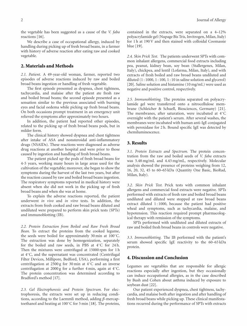
2 Journal of Allergy
the vegetable has been suggested as a cause of the V. fabareactions [16].
We describe a case of occupational allergy, induced byhandling during picking up of fresh broad beans, in a farmerwith history of adverse reaction after eating raw and cookedvegetable.
2. Materials and Methods
2.1. Patient. A 49-year-old woman, farmer, reported twoepisodes of adverse reactions induced by raw and boiledbroad beans ingestion or handling of fresh vegetable.
The first episode presented as dyspnea, chest tightness,tachycardia, and malaise after the patient ate fresh rawand boiled broad beans; the second episode presented as asensation similar to the previous associated with burningeyes and facial oedema while picking up fresh broad beans.On both occasions prompt treatment in an emergency unitrelieved the symptoms after approximately two hours.
In addition, the patient had reported other episodes,related to the picking up of fresh broad beans pods, but inmilder form.
The clinical history showed dyspnea and chest tightnessafter intake of ASA and nonsteroidal anti-inflammatorydrugs (NSAIDs). These reactions were diagnosed as adversedrug reactions at another hospital and were prior to thosecaused by ingestion and handling of fresh broad beans.
The patient picked up the pods of fresh broad beans for4-5 years, working many hours in large areas used for thecultivation of the vegetable; moreover, she began to show thesymptoms during the harvest of the last two years, but afterthe reaction caused by raw and boiled broad beans ingestion.The respiratory symptoms reported in medical history wereabsent when she did not work in the picking up of freshbroad beans and when she was at home.
To explain the adverse reactions reported, the patientunderwent in vivo and in vitro tests. In addition, theextracts from fresh cooked and raw broad beans diluted andundiluted were prepared to perform skin prick tests (SPTs)and immunoblotting (IB).
2.2. Protein Extraction from Boiled and Raw Fresh BroadBean. To extract the proteins from the cooked legume,the seeds were boiled for approximately 30 min at 100◦C.The extraction was done by homogenization, separatelyfor the boiled and raw seeds, in PBS at 4◦C for 24 h.Then the mixtures were centrifuged at 15000 rpm for 1 hat 4◦C, and the supernatant was concentrated (CentrifugalFilter Devices, Millipore, Bedford, USA), performing a firstcentrifugation at 2500 g for 30 min at 4◦C and an inversecentrifugation at 2000 g for a further 4 min, again at 4◦C.The protein concentration was determined according toBradford’s method [17].
2.3. Gel Electrophoresis and Protein Spectrum. For elec-trophoresis, the extracts were set up in reducing condi-tions, according to the Laemmli method, adding β-mercap-toethanol and heating at 100◦C for 5 min [18]. The proteins,
contained in the extracts, were separated on a 4–12%polyacrylamide gel (Nupage Bis Tris, Invitrogen, Milan, Italy)for 1 h at 190 V and then stained with colloidal Coomassieblue [19].
2.4. Skin Prick Test. The patients underwent SPTs with com-mon inhalant allergens, commercial food extracts includingpea, peanut, kidney bean, soy bean (Stallergenes, Milan,Italy), chickpea, and lentil (Lofarma, Milan, Italy), and withextracts of fresh boiled and raw broad beans undiluted anddiluted (1 : 1000, 1 : 100, 1 : 10 in saline solution and glycerol)[20]. Saline solution and histamine (10 mg/mL) were used asnegative and positive control, respectively.
2.5. Immunoblotting. The proteins separated on polyacry-lamide gel were transferred onto a nitrocellulose mem-brane (Schleicher & Schuell, Biosciences, Germany) [21].The membranes, after saturation, were incubated at 4◦Covernight with the patient’s serum. After several washes, themembranes were incubated with human anti-IgE conjugatedwith peroxidase for 2 h. Bound specific IgE was detected bychemiluminescence.
3. Results
3.1. Protein Extracts and Spectrum. The protein concen-tration from the raw and boiled seeds of V. faba extractswas 5.48 mg/mL and 4.43 mg/mL, respectively. Molecularanalysis showed the presence of proteins weighing from 12,16, 20, 32, 45 to 60–65 kDa (Quantity One Basic, BioRad,Milan, Italy).
3.2. Skin Prick Test. Prick tests with common inhalantallergens and commercial food extracts were negative. SPTsperformed with extracts of fresh raw and boiled broad beansundiluted and diluted were stopped at raw broad beansextract diluted 1 : 1000, because the patient had positivewheal and symptoms, such as tachycardia, malaise, andhypotension. This reaction required prompt pharmacolog-ical therapy with remission of the symptoms.
SPTs performed with undiluted and diluted extracts ofraw and boiled fresh broad beans in controls were negative.
3.3. Immunoblotting. The IB performed with the patient’sserum showed specific IgE reactivity to the 60–65 kDaprotein.
4. Discussion and Conclusion
Legumes are vegetables that are responsible for allergicreactions especially after ingestion, but they occasionallycan induce occupational allergies, as in the case describedby Bush and Cohen about asthma induced by exposure tosoybean dust [22].
Our patient experienced dyspnea, chest tightness, tachy-cardia, and malaise both after ingestion and after handling offresh broad beans while picking up. These clinical manifesta-tions recurred during the performance of SPTs with extracts

Journal of Allergy 3
of fresh raw broad beans diluted to a low concentration(1 : 1000) that required discontinuation testing and pharma-cological therapy. Daroca et al. reported cases of respiratorysymptoms appeared after handling of green beans or greenbeans and chards together in patients who ingested the samefood without reactions. The IB performed with patient’sserum showed several differences, although minimal, aboutthe reactivity of IgE between IB with the raw extract andthe blotting with boiled extract. This may be related to thelability of allergens of green beans to cooking, which wouldexplain the tolerance to the ingestion of cooked foods [23].Another case of asthma induced by handling and cookingvapors of green beans has been described by Igea et al. in ahousewife who tolerates ingestion of vegetable. In this case,the presence of protein heat-labile in vegetable has beensuggested [24]. In our case, the patient did not tolerate theingestion of the native (raw) and boiled food and the proteinsare well preserved also in the cooked extract.
The IB shows IgE reactive against the protein of molecu-lar weight of 60–65 kDa, present in the fresh raw and boiledbroad beans extract. This demonstrates that the reactionexperienced by the patient can be IgE mediated.
The presence of salicylates in vegetable and the role ofsalicylic acid as defense response to pathogens are knownin the literature [25–27]. In addition, after external stressstimuli, the V. faba produces small quantities of methylsalicylate and others substance that can be measured inthe surrounding air [15]. A series of patients with ASAintolerance and reactions after ingestion of V. faba cookedand raw seeds had assumed to the authors a asthmogenic roleof some metabolites of methyl salicylate present in the plant[16]. In several cases reported in the literature, the reactionscaused by the inhalation of food allergens appear, generally,in domestic environment [7, 8, 23, 24] or in industrialenvironment [22].
Our patient reported history of adverse reactions afterintake of NSAIDs and ASA, prior to those caused byingestion and handling of fresh broad beans. Moreover,the reactions that the patient experienced during working,picking up the pods of V. faba (without the concomitantingestion of seeds), were at first slight and after successiveexposures had become more serious. The evolution ofsymptoms has suspected that the patient’s ASA intolerancecould have had a significant role. In fact, the methyl salicylate,released from the vegetable in the surrounding air andinhaled by the patient during the picking up of the pods,may have induced the respiratory symptoms due to previoussensitization to the ASA. After the investigations performed,by us the patient avoided her carrying out her work where V.faba was cultivated.
Furthermore, we have demonstrated an IgE-mediatedmechanism of the reactions after ingestion of the seedsof broad beans, but we cannot exclude that the reactionsduring the picking up of broad beans could be caused by theinhalation of airborne allergens.
We concluded that the reactions after ingestion of freshbroad beans are IgE mediated, while the reactions thatoccurred during the harvest of vegetable can be explained bytwo diagnostic hypotheses:
(1) exposure to airborne allergens responsible for animmediate response,
(2) exposure to airborne salicylates responsible for aintolerance reaction.
Future studies could confirm these hypotheses and clarifythis phenomenon.
We cannot exclude, however, that reports may increaseover time, because the plants and the vegetables are increas-ingly exposed to aggressive agents and then tend to respondto these stimuli by increasing the synthesis of allergenicdefensive proteins and volatile substances. This situation,therefore, could lead to an increase of cases of occupationalallergies induced by volatile substances released from thevegetables in the surrounding air.
Conflict of Interests
The authors declare that they have no financial relationshipwith any biotechnology manufacturers that has an interestin the subject matter or materials discussed in the submittedpaper.
References
[1] J. M. James and J. F. Crespo, “Allergic reactions to foods byinhalation,” Current Allergy and Asthma Reports, vol. 7, no. 3,pp. 167–174, 2007.
[2] Wikipedia, “Fabaceae,” 2010, http://it.wikipedia.org/wiki/Fa-baceae.
[3] M. D. Ibanez, M. Martınez, J. J. Sanchez, and E. Fernandez-Caldas, “Legume cross-reactivity,” Allergologia et Immunopa-thologia, vol. 31, no. 3, pp. 151–161, 2003.
[4] J. Bernhisel-Broadbent and H. A. Sampson, “Cross-allergenic-ity in the legume botanical family in children with foodhypersensitivity,” Journal of Allergy and Clinical Immunology,vol. 83, pp. 435–440, 1989.
[5] J. Bernhisel-Broadbent, S. Taylor, and H. A. Sampson, “Cross-allergenicity in the legume botanical family in children withfood hypersensitivity. II. Laboratory correlates,” Journal ofAllergy and Clinical Immunology, vol. 84, pp. 701–709, 1989.
[6] Wikipedia, “Vicia faba,” 2010, http://en.wikipedia.org/wiki/Fava bean.
[7] J. C. Garcıa Ortiz, A. Lopez-Asunsolo, P. Cosmes, and A.M. Duran, “Bronchial asthma induced by hypersensitivity tolegumes,” Allergologia et Immunopathologia, vol. 23, no. 1, pp.38–40, 1995.
[8] J. A. Martin, J. A. Compaired, B. de la Hoz et al., “Bronchialasthma induced by chick pea and lentil,” Allergy, vol. 47, no. 2,pp. 185–187, 1992.
[9] D. Kalogeromitros, M. Armenaka, I. Galatas, O. Capellou,and A. Katsarou, “Anaphylaxis induced by lentils,” Annals ofAllergy, Asthma and Immunology, vol. 77, no. 6, pp. 480–482,1996.
[10] H. A. Sampson, “Peanut anaphylaxis,” Journal of Allergy andClinical Immunology, vol. 86, pp. 1–3, 1992.
[11] V. Matheu, M. de Barrio, Z. Sierra, M. T. Gracia-Bara, P.Tornero, and M. L. Baeza, “Lupine-induced anaphylaxis,”Annals of Allergy, Asthma and Immunology, vol. 83, no. 5, pp.406–408, 1999.

4 Journal of Allergy
[12] P. V. Niphadkar, S. P. Patil, and M. M. Bapat, “Chickpea-induced anaphylaxis,” Allergy, vol. 52, no. 1, pp. 115–116,1997.
[13] R. L. Freitas, R. B. Ferreira, and A. R. Teixeira, “Use of a singlemethod in the extraction of the seed storage globulins fromseveral legume species. Application to analyse structural com-parisons within the major classes of globulins,” InternationalJournal of Food Sciences and Nutrition, vol. 51, no. 5, pp. 341–352, 2000.
[14] U. Heim, Q. Wang, T. Kurz et al., “Expression patterns andsubcellular localization of a 52 kDa sucrose-binding proteinhomologue of Vicia faba (VfSBPL) suggest different functionsduring development,” Plant Molecular Biology, vol. 47, no. 4,pp. 461–474, 2001.
[15] B. Webster, T. Bruce, S. Dufour et al., “Identification of volatilecompounds used in host location by the black bean aphid,Aphis fabae,” Journal of Chemical Ecology, vol. 34, no. 9, pp.1153–1161, 2008.
[16] E. Damiani, A. M. Aloia, M. G. Priore et al., “Reazioneavversa dopo ingestione di Vicia faba cruda e cotta: allergiao ASA intolleranza?” Italian Journal of Allergy and ClinicalImmunology, vol. 19, no. 4, pp. 153–157, 2009.
[17] M. M. Bradford, “A rapid and sensitive method for thequantitation of microgram quantities of protein utilizing theprinciple of protein dye binding,” Analytical Biochemistry, vol.72, no. 1–2, pp. 248–254, 1976.
[18] U. K. Laemmli, “Cleavage of structural proteins during theassembly of the head of bacteriophage T4,” Nature, vol. 227,no. 5259, pp. 680–685, 1970.
[19] Q. Shi and G. Jackowski, “Gel electrophoresis of proteins,”in One Dimensional Polyacrylamide Gel Electrophoresis, B. D.Hames, Ed., pp. 1–52, Oxford University Press, New York, NY,USA, 2002.
[20] K. Skamstrup Hansen, C. Bindslev-Jensen, P. Stahl Skov et al.,“Standardization of food allergen extracts for skin prick test,”Journal of Chromatography B, vol. 756, no. 1–2, pp. 57–69,2001.
[21] H. Towbin, T. Staehelin, and J. Gordon, “Electrophoretictransfer of proteins from polyacrylamide gels to nitrocellulosesheets: procedure and some applications,” Proceedings of theNational Academy of Sciences of the United States of America,vol. 76, no. 9, pp. 4350–4354, 1979.
[22] R. K. Bush and M. Cohen, “Immediate and late onset asthmafrom occupational exposure to soybean dust,” Clinical Allergy,vol. 7, no. 4, pp. 369–373, 1977.
[23] P. Daroca, J. F. Crespo, M. Reano, J. M. James, A. Lopez-Rubio,and J. Rodriguez, “Asthma and rhinitis induced by exposureto raw green beans and chards,” Annals of Allergy, Asthma andImmunology, vol. 85, no. 3, pp. 215–218, 2000.
[24] J. M. Igea, M. Fernandez, S. Quirce, B. de la Hoz, and M. L. D.Gomez, “Green bean hypersensitivity: an occupational allergyin a homemaker,” Journal of Allergy and Clinical Immunology,vol. 94, no. 1, pp. 33–35, 1994.
[25] A. R. Swain, S. P. Dutton, and A. S. Truswell, “Salicylates infoods,” Journal of the American Dietetic Association, vol. 85, no.8, pp. 950–960, 1985.
[26] G. J. Baxter, A. B. Graham, J. R. Lawrence, D. Wiles, and J.R. Paterson, “Salicylic acid in soups prepared from organicallyand non-organically grown vegetables,” European Journal ofNutrition, vol. 40, no. 6, pp. 289–292, 2001.
[27] V. A. Halim, A. Vess, D. Scheel, and S. Rosahl, “The role ofsalicylic acid and jasmonic acid in pathogen defence,” PlantBiology, vol. 8, no. 3, pp. 307–313, 2006.

Hindawi Publishing CorporationJournal of AllergyVolume 2011, Article ID 841508, 5 pagesdoi:10.1155/2011/841508
Case Report
Gum Arabic as a Cause of Occupational Allergy
Arja Viinanen,1 Maija Salokannel,2 and Kaija Lammintausta3
1 Department of Pulmonary Diseases, Turku University Hospital, P.O. Box 52, 20521 Turku, Finland2 Occupational Health Service Unit, Mehilainen, 20520 Turku, Finland3 Department of Dermatology, Turku University Hospital, P.O. Box 52, 20521 Turku, Finland
Correspondence should be addressed to Arja Viinanen, [email protected]
Received 12 January 2011; Revised 14 March 2011; Accepted 16 March 2011
Academic Editor: Donald H. Beezhold
Copyright © 2011 Arja Viinanen et al. This is an open access article distributed under the Creative Commons Attribution License,which permits unrestricted use, distribution, and reproduction in any medium, provided the original work is properly cited.
Background. Gum arabic is a potential sensitizer in food industry. Methods. We examined 11 candy factory workers referred toexaminations due to respiratory and skin symptoms paying attention to exposure and sensitization to gum arabic. Skin tests,pulmonary function tests, and respiratory provocation tests were carried out as indicated by the symptoms and findings. Results.Occupational asthma, caused by gum arabic was diagnosed in 4/11 candy factory workers and two of them had also occupationalcontact urticaria and one had occupational rhinitis. One of them had oral symptoms associated with ingestion of productscontaining gum arabic. Conclusions. Airborne exposure to gum arabic may cause sensitization leading to allergic rhinitis, asthma,and urticaria.
1. Introduction
Gum arabic, or gum acacia is mainly derived from Acaciasenegal tree. As a nontoxic material it is used as an emulsifier,a thickening agent, and as a stabilizer in foods, with E-codeE414 [1]. It is useful in many kinds of foodstuffs because of itsvery low viscosity, complete solubility in water, and absenceof any taste or odour. Due to technical properties gum arabiccan be used in multiple applications like in pharmaceuticalindustry, lithography, and cosmetics.
Gum arabic is comprised of sugars and glucuronic acidresidues in a long chain of galactosyl units with branchedoligosaccharides attached to a polypeptide backbone. Proteincontent of gum arabic varies from 1 to 2%. IgE antibod-ies against polypeptide chains in gum arabic have beendescribed to elicit asthma in occupational exposure [2].Occasional cases of occupational asthma in printers [3],candy factory [4, 5], and pharmaceutical industry workers[2] have been described. Although gum arabic is extensivelyused in food industry ingestion of it is a rare cause ofimmediate allergy symptoms [1].
We describe several cases of occupational asthma causedby gum arabic among candy factory workers.
2. Methods
2.1. Patients. Eleven candy factory workers with respiratoryand/or skin symptoms were referred to the Allergy Unit ofTurku University Central Hospital (Table 1).
2.2. Workplace Description in the Candy Factory. This Finnishcandy factory was a major producer of multiple goodies inFinland since 1910. Gum arabic was an important ingredientin many candies. In making soft pastilles, gum arabic wasdissolved in water as kibbles without making dust in theair. Hard boiled candies, instead, were covered with spraydried gum arabic inside a rotating drum. Dry, powdered gumarabic, packed in 25 kg bags, was poured by workers into thedrums. Cornstarch, used to cover liquorice, also made dustin the air.
Working clothes were a short-sleeve jacket and/or T-shirtand pants. The workers, who had to do cleaning used rubbergloves. Other protective equipment was not required butrespiratory masks were available.
These patients were referred during the last operationyear of the factory. In the preceding year the production of
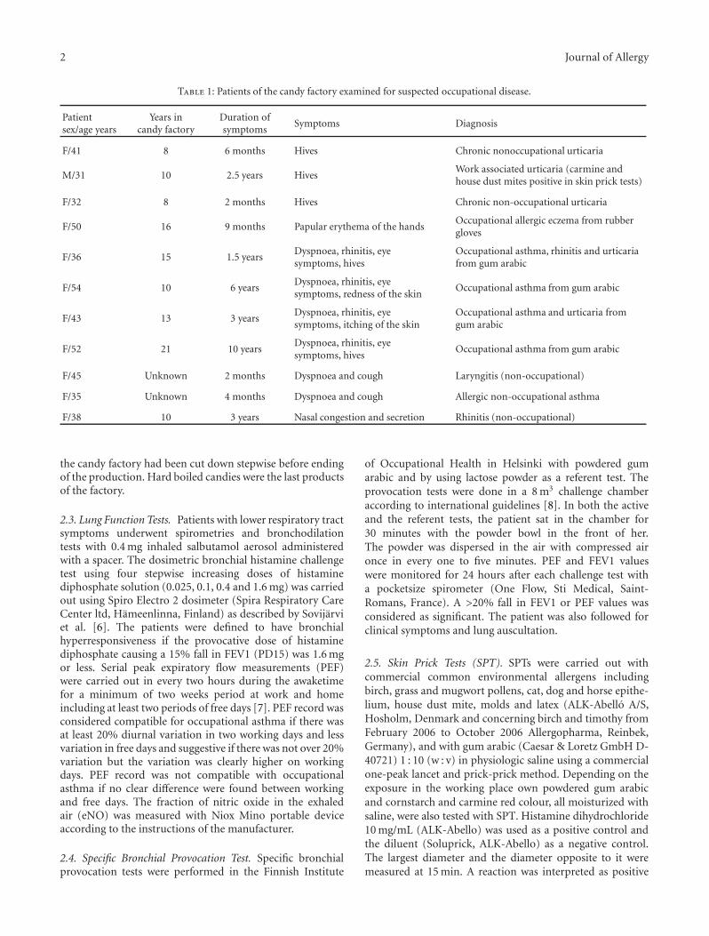
2 Journal of Allergy
Table 1: Patients of the candy factory examined for suspected occupational disease.
Patientsex/age years
Years incandy factory
Duration ofsymptoms
Symptoms Diagnosis
F/41 8 6 months Hives Chronic nonoccupational urticaria
M/31 10 2.5 years HivesWork associated urticaria (carmine andhouse dust mites positive in skin prick tests)
F/32 8 2 months Hives Chronic non-occupational urticaria
F/50 16 9 months Papular erythema of the handsOccupational allergic eczema from rubbergloves
F/36 15 1.5 yearsDyspnoea, rhinitis, eyesymptoms, hives
Occupational asthma, rhinitis and urticariafrom gum arabic
F/54 10 6 yearsDyspnoea, rhinitis, eyesymptoms, redness of the skin
Occupational asthma from gum arabic
F/43 13 3 yearsDyspnoea, rhinitis, eyesymptoms, itching of the skin
Occupational asthma and urticaria fromgum arabic
F/52 21 10 yearsDyspnoea, rhinitis, eyesymptoms, hives
Occupational asthma from gum arabic
F/45 Unknown 2 months Dyspnoea and cough Laryngitis (non-occupational)
F/35 Unknown 4 months Dyspnoea and cough Allergic non-occupational asthma
F/38 10 3 years Nasal congestion and secretion Rhinitis (non-occupational)
the candy factory had been cut down stepwise before endingof the production. Hard boiled candies were the last productsof the factory.
2.3. Lung Function Tests. Patients with lower respiratory tractsymptoms underwent spirometries and bronchodilationtests with 0.4 mg inhaled salbutamol aerosol administeredwith a spacer. The dosimetric bronchial histamine challengetest using four stepwise increasing doses of histaminediphosphate solution (0.025, 0.1, 0.4 and 1.6 mg) was carriedout using Spiro Electro 2 dosimeter (Spira Respiratory CareCenter ltd, Hameenlinna, Finland) as described by Sovijarviet al. [6]. The patients were defined to have bronchialhyperresponsiveness if the provocative dose of histaminediphosphate causing a 15% fall in FEV1 (PD15) was 1.6 mgor less. Serial peak expiratory flow measurements (PEF)were carried out in every two hours during the awaketimefor a minimum of two weeks period at work and homeincluding at least two periods of free days [7]. PEF record wasconsidered compatible for occupational asthma if there wasat least 20% diurnal variation in two working days and lessvariation in free days and suggestive if there was not over 20%variation but the variation was clearly higher on workingdays. PEF record was not compatible with occupationalasthma if no clear difference were found between workingand free days. The fraction of nitric oxide in the exhaledair (eNO) was measured with Niox Mino portable deviceaccording to the instructions of the manufacturer.
2.4. Specific Bronchial Provocation Test. Specific bronchialprovocation tests were performed in the Finnish Institute
of Occupational Health in Helsinki with powdered gumarabic and by using lactose powder as a referent test. Theprovocation tests were done in a 8 m3 challenge chamberaccording to international guidelines [8]. In both the activeand the referent tests, the patient sat in the chamber for30 minutes with the powder bowl in the front of her.The powder was dispersed in the air with compressed aironce in every one to five minutes. PEF and FEV1 valueswere monitored for 24 hours after each challenge test witha pocketsize spirometer (One Flow, Sti Medical, Saint-Romans, France). A >20% fall in FEV1 or PEF values wasconsidered as significant. The patient was also followed forclinical symptoms and lung auscultation.
2.5. Skin Prick Tests (SPT). SPTs were carried out withcommercial common environmental allergens includingbirch, grass and mugwort pollens, cat, dog and horse epithe-lium, house dust mite, molds and latex (ALK-Abello A/S,Hosholm, Denmark and concerning birch and timothy fromFebruary 2006 to October 2006 Allergopharma, Reinbek,Germany), and with gum arabic (Caesar & Loretz GmbH D-40721) 1 : 10 (w : v) in physiologic saline using a commercialone-peak lancet and prick-prick method. Depending on theexposure in the working place own powdered gum arabicand cornstarch and carmine red colour, all moisturized withsaline, were also tested with SPT. Histamine dihydrochloride10 mg/mL (ALK-Abello) was used as a positive control andthe diluent (Soluprick, ALK-Abello) as a negative control.The largest diameter and the diameter opposite to it weremeasured at 15 min. A reaction was interpreted as positive
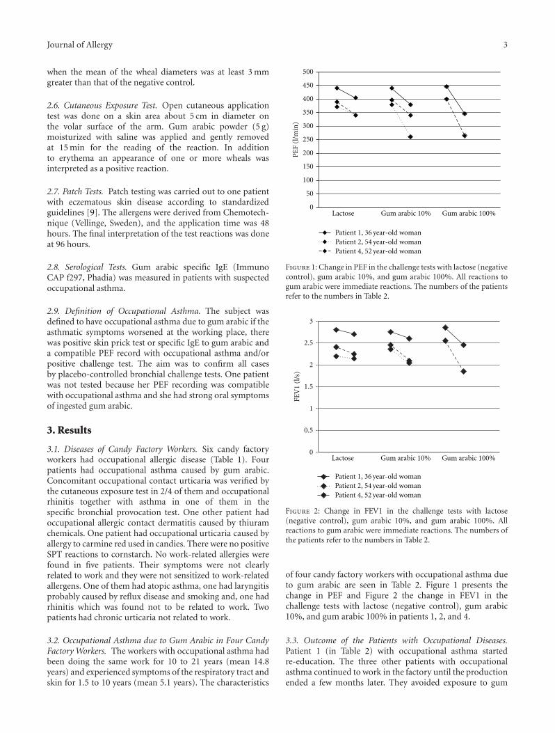
Journal of Allergy 3
when the mean of the wheal diameters was at least 3 mmgreater than that of the negative control.
2.6. Cutaneous Exposure Test. Open cutaneous applicationtest was done on a skin area about 5 cm in diameter onthe volar surface of the arm. Gum arabic powder (5 g)moisturized with saline was applied and gently removedat 15 min for the reading of the reaction. In additionto erythema an appearance of one or more wheals wasinterpreted as a positive reaction.
2.7. Patch Tests. Patch testing was carried out to one patientwith eczematous skin disease according to standardizedguidelines [9]. The allergens were derived from Chemotech-nique (Vellinge, Sweden), and the application time was 48hours. The final interpretation of the test reactions was doneat 96 hours.
2.8. Serological Tests. Gum arabic specific IgE (ImmunoCAP f297, Phadia) was measured in patients with suspectedoccupational asthma.
2.9. Definition of Occupational Asthma. The subject wasdefined to have occupational asthma due to gum arabic if theasthmatic symptoms worsened at the working place, therewas positive skin prick test or specific IgE to gum arabic anda compatible PEF record with occupational asthma and/orpositive challenge test. The aim was to confirm all casesby placebo-controlled bronchial challenge tests. One patientwas not tested because her PEF recording was compatiblewith occupational asthma and she had strong oral symptomsof ingested gum arabic.
3. Results
3.1. Diseases of Candy Factory Workers. Six candy factoryworkers had occupational allergic disease (Table 1). Fourpatients had occupational asthma caused by gum arabic.Concomitant occupational contact urticaria was verified bythe cutaneous exposure test in 2/4 of them and occupationalrhinitis together with asthma in one of them in thespecific bronchial provocation test. One other patient hadoccupational allergic contact dermatitis caused by thiuramchemicals. One patient had occupational urticaria caused byallergy to carmine red used in candies. There were no positiveSPT reactions to cornstarch. No work-related allergies werefound in five patients. Their symptoms were not clearlyrelated to work and they were not sensitized to work-relatedallergens. One of them had atopic asthma, one had laryngitisprobably caused by reflux disease and smoking and, one hadrhinitis which was found not to be related to work. Twopatients had chronic urticaria not related to work.
3.2. Occupational Asthma due to Gum Arabic in Four CandyFactory Workers. The workers with occupational asthma hadbeen doing the same work for 10 to 21 years (mean 14.8years) and experienced symptoms of the respiratory tract andskin for 1.5 to 10 years (mean 5.1 years). The characteristics
Gum arabic 100%Gum arabic 10%Lactose0
50
100
150
200
250
300
350
400
450
500
PE
F(l
/min
)
Patient 1, 36 year-old womanPatient 2, 54 year-old womanPatient 4, 52 year-old woman
Figure 1: Change in PEF in the challenge tests with lactose (negativecontrol), gum arabic 10%, and gum arabic 100%. All reactions togum arabic were immediate reactions. The numbers of the patientsrefer to the numbers in Table 2.
Gum arabic 100%Gum arabic 10%Lactose
Patient 1, 36 year-old womanPatient 2, 54 year-old womanPatient 4, 52 year-old woman
0
0.5
1
1.5
2
2.5
3
FEV
1(l
/s)
Figure 2: Change in FEV1 in the challenge tests with lactose(negative control), gum arabic 10%, and gum arabic 100%. Allreactions to gum arabic were immediate reactions. The numbers ofthe patients refer to the numbers in Table 2.
of four candy factory workers with occupational asthma dueto gum arabic are seen in Table 2. Figure 1 presents thechange in PEF and Figure 2 the change in FEV1 in thechallenge tests with lactose (negative control), gum arabic10%, and gum arabic 100% in patients 1, 2, and 4.
3.3. Outcome of the Patients with Occupational Diseases.Patient 1 (in Table 2) with occupational asthma startedre-education. The three other patients with occupationalasthma continued to work in the factory until the productionended a few months later. They avoided exposure to gum
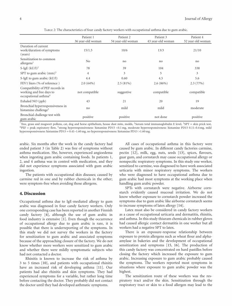
4 Journal of Allergy
Table 2: The characteristics of four candy factory workers with occupational asthma due to gum arabic.
Patient 136 year-old woman
Patient 254 year-old woman
Patient 343 year-old woman
Patient 452 year old woman
Duration of currentwork/duration of symptoms(years)
15/1.5 10/6 13/3 21/10
Sensitization to commonallergens1 No no no no
S-IgE (kU/l)2 78 39 104 338
SPT to gum arabic (mm)3 4 3 5 3
S-IgE to gum arabic (kU/l) 4.4 0.60 4.5 5.6
FEV1 liters (% of reference ) 2.0 (64%) 2.5 (81%) 2.6 (86%) 2.3 (77%)
Compatibility of PEF records inworking and free days tooccupational asthma4
not compatible suggestive compatible compatible
Exhaled NO (ppb) 43 21 20 19
Bronchial hyperresponsiveness inhistamine challenge5 no mild mild moderate
Bronchial challenge test withgum arabic
positive positive not done positive
1Tree, grass and mugwort pollens, cat, dog and horse epithelium, house dust mite, molds, 2Serum total immunoglobulin E level, 3SPT = skin prick test,
4PEF = peak expiratory flow, 5strong hyperresponsiveness: histamine PD15 <0.1 mg, moderate hyperresponsiveness: histamine PD15 0.11–0.4 mg, mildhyperresponsiveness: histamine PD15 = 0.41–1.60 mg, no hyperresponsiveness: histamine PD15 >1.60 mg.
arabic. Six months after the work in the candy factory hadended patient 3 (in Table 2) was free of symptoms withoutasthma medication. She, however, experienced angioedemawhen ingesting gum arabic containing foods. In patients 1,2, and 4 asthma was in control with medication, and theydid not experience symptoms associated with gum arabicingestion.
The patients with occupational skin diseases; caused bycarmine red in one and by rubber chemicals in the other;were symptom-free when avoiding those allergens.
4. Discussion
Occupational asthma due to IgE-mediated allergy to gumarabic was diagnosed in four candy factory workers. Onlyone corresponding case has been reported in another Finnishcandy factory [4], although the use of gum arabic infood industry is extensive [1]. Even though the occurrenceof occupational allergy due to gum arabic is rare, it ispossible that there is underreporting of the symptoms. Inthis study we did not survey the workers in the factoryfor sensitization to gum arabic and associated symptomsbecause of the approaching closure of the factory. We do notknow whether more workers were sensitized to gum arabicand whether there were mildly symptomatic subjects whohad not contacted a doctor.
Rhinitis is known to increase the risk of asthma by3 to 5 times [10], and patients with occupational rhinitishave an increased risk of developing asthma [11]. Ourpatients had also rhinitis and skin symptoms. They hadexperienced symptoms for a variable, but rather long timebefore contacting the doctor. They probably did not contactthe doctor until they had developed asthmatic symptoms.
All cases of occupational asthma in this factory werecaused by gum arabic. In different candy factories carmine,pectin [12], milk, egg, nuts, seeds [13], spices, flavours,guar gum, and cornstarch may cause occupational allergy ornonspecific respiratory symptoms. In this study one worker,sensitized to carmine, was diagnosed to have work associatedurticaria with minor respiratory symptoms. The workerswho were diagnosed to have occupational asthma due togum arabic had most symptoms at the working place whenhandling gum arabic powder.
SPTs with cornstarch were negative. Airborne corn-starch evidently caused mucosal irritation. We do notknow whether exposure to cornstarch powder increased thesymptoms due to gum arabic like airborne cornstarch seemsto increase symptoms of latex allergy [14].
Latex must also be considered in candy factory workersas a cause of occupational urticaria and dermatitis, rhinitis,and asthma. In this study thiuram chemicals in rubber gloveshad caused allergic contact dermatitis in one worker but allworkers had a negative SPT to latex.
There is an exposure-response relationship betweenexposure to protein allergens such as wheat flour and alpha-amylase in bakeries and the development of occupationalsensitization and symptoms [15, 16]. The production ofthis candy factory was concentrated on hard pastilles beforeclosing the factory which increased the exposure to gumarabic. Increasing exposure to gum arabic probably causedthe symptoms. The workers reported most symptoms insituations where exposure to gum arabic powder was thehighest.
The sensitization route of these workers was the res-piratory tract and/or the skin. Sensitization through therespiratory tract or skin to a food allergen may lead to the

Journal of Allergy 5
subsequent development of symptoms during oral exposureas has been reported for sensitization to egg in bakeryand confectionery workers [17], for lupine seeds in legumelaboratory workers [18], for carmine in a worker engaged indye manufacturing [19] and for fish [20]. Only one of thesecandy factory workers with occupational asthma reportedoral symptoms associated with ingested gum arabic. Despiteher asthma relieved after discontinuation of the exposure, theoral symptoms remained.
We have shown that gum arabic may cause occupationalallergic rhinitis and asthma with urticaria symptoms. In thisstudy the cases of occupational asthma in the candy factoryappeared when there was an increase in the exposure. Noneof the patients had any previous atopic disease. Workingmethods which produce less powder and respiratory andskin protection are recommended. Early diagnosis of occu-pational allergy due to gum arabic is important in order toprevent the development of asthma.
Acknowledgment
The authors thank Paula Kauppi, MD, for describing themethod of the provocation test with gum arabic extract.
References
[1] “Final report on the safety assessment of Acacia catechugum, Acacia concinna fruit extract, Acacia dealbata leafextract, Acacia dealbata leaf wax, Acacia decurrens extract,Acacia farnesiana extract, Acacia farnesiana flower wax, Acaciafarnesiana gum, Acacia senegal extract, Acacia senegal gumand Acacia senegal gum extract,” International Journal ofToxicology, vol. 24, no. 3, supplement, pp. 75–118, 2005.
[2] I. Sander, M. Raulf-Heimsoth, K. Wiemer, S. Kespohl, T.Bruning, and R. Merget, “Sensitization due to gum arabic(Acacia senegal): the cause of occupational allergic asthmaor crossreaction to carbohydrates,” International Archives ofAllergy and Immunology, vol. 141, no. 1, pp. 51–56, 2006.
[3] P. B. S. Fowler, “Printers’ asthma,” The Lancet, vol. 260, no.6738, pp. 755–757, 1952.
[4] K. Kilpio, T. Kallas, T. Hupli, and K. Malanin, “Allergic rhinitis,asthma and eczema caused by gum arabic in a candy factoryworker,” Duodecim, vol. 116, no. 22, pp. 2507–2509, 2000.
[5] K. Fotisch, J. Fah, B. Wuthrich, F. Altmann, D. Haustein,and S. Vieths, “IgE-antibodies specific for carbohydrates in apatient allergic to gum arabic (Acacia senegal),” Allergy, vol.53, no. 11, pp. 1043–1051, 1998.
[6] A. R. A. Sovijarvi, L. P. Malmberg, K. Reinikainen, P. Rytila,and H. Poppius, “A rapid dosimetric method with controlledtidal breathing for histamine challenge: repeatability anddistribution of bronchial reactivity in a clinical material,”Chest, vol. 104, no. 1, pp. 164–170, 1993.
[7] P. F. Gannon and P. S. Burge, “Serial peak expiratoryflow measurement in the diagnosis of occupational asthma,”European Respiratory Journal, vol. 24, pp. 57S–63S, 1997.
[8] Allergy Practice Forum, “Guidelines for the diagnosis ofoccupational asthma,” Clinical and Experimental Allergy, vol.22, no. 1, pp. 103–108, 1992.
[9] J. Wahlberg and M. Lindberg, “Patch testing ,” in ContactDermatitis, P. J. Frosch, T. Menne, and J. P. Lepoittevin, Eds.,pp. 366–382, Springer, New York, NY, USA, 4th edition, 2006.
[10] S. Guerra, D. L. Sherrill, F. D. Martinez, and R. A. Bar-bee, “Rhinitis as an independent risk factor for adult-onsetasthma,” Journal of Allergy and Clinical Immunology, vol. 109,no. 3, pp. 419–425, 2002.
[11] A. Karjalainen, R. Martikainen, T. Klaukka, K. Saarinen,and J. Uitti, “Risk of asthma among finnish patients withoccupational rhinitis,” Chest, vol. 123, no. 1, pp. 283–288,2003.
[12] A. Kraut, Z. Peng, A. B. Becker, and C. P. W. Warren, “Christ-mas candy maker’s asthma. IgG4-mediated pectin allergy,”Chest, vol. 102, no. 5, pp. 1605–1607, 1992.
[13] J. M. James and J. F. Crespo, “Allergic reactions to foods byinhalation,” Current Allergy and Asthma Reports, vol. 7, no. 3,pp. 167–174, 2007.
[14] M. Lundberg, K. Wrangsjo, and S. G. Johansson, “Latex allergyfrom glove powder—an unintended risk with the switch fromtalc to cornstarch?” Allergy, vol. 52, no. 12, pp. 1222–1228,1997.
[15] R. Houba, D. Heederik, and G. Doekes, “Wheat sensitizationand work-related symptoms in the baking industry arepreventable,” American Journal of Respiratory and Critical CareMedicine, vol. 158, no. 5, pp. 1499–1503, 1998.
[16] J. Brisman, M. J. Nieuwenhuijsen, K. M. Venables, V. Putcha,S. Gordon, and A. J. N. Taylor, “Exposure-response relationsfor work related respiratory symptoms and sensitisation in acohort exposed to α-amylase,” Occupational and Environmen-tal Medicine, vol. 61, no. 6, pp. 551–553, 2004.
[17] C. Leser, A. L. Hartmann, G. Praml, and B. Wuthrich, “The”egg-egg” syndrome: occupational respiratory allergy to air-borne egg proteins with consecutive ingestive egg allergy in thebakery and confectionery industry,” Journal of InvestigationalAllergology and Clinical Immunology, vol. 11, no. 2, pp. 89–93,2001.
[18] J. F. Crespo, J. Rodrıguez, R. Vives et al., “Occupational IgE-mediated allergy after exposure to lupine seed flour,” Journalof Allergy and Clinical Immunology, vol. 108, no. 2, pp. 295–297, 2001.
[19] S. Acero, A. I. Tabar, M. J. Alvarez, B. E. Garcia, J. M. Olaguibel,and I. Moneo, “Occupational asthma and food allergy due tocarmine,” Allergy, vol. 53, no. 9, pp. 897–901, 1998.
[20] J. Rodrıguez, M. Reano, R. Vives et al., “Occupational asthmacaused by fish inhalation,” Allergy, vol. 52, no. 8, pp. 866–869,1997.
Hindawi Publishing CorporationJournal of AllergyVolume 2011, Article ID 346719, 7 pagesdoi:10.1155/2011/346719
Review Article
Genetic Variability in Susceptibility to OccupationalRespiratory Sensitization
Berran Yucesoy and Victor J. Johnson
Toxicology and Molecular Biology Branch, Health Effects Laboratory Division, National Institute for Occupational Safety and Health,Centers for Disease Control and Prevention, Morgantown, WV 26505, USA
Correspondence should be addressed to Berran Yucesoy, [email protected]
Received 22 February 2011; Accepted 18 April 2011
Academic Editor: Gordon L. Sussman
Copyright © 2011 B. Yucesoy and V. J. Johnson. This is an open access article distributed under the Creative Commons AttributionLicense, which permits unrestricted use, distribution, and reproduction in any medium, provided the original work is properlycited.
Respiratory sensitization can be caused by a variety of substances at workplaces, and the health and economic burden linked toallergic respiratory diseases continues to increase. Although the main factors that affect the onset of the symptoms are the types andintensity of allergen exposure, there is a wide range of interindividual variation in susceptibility to occupational/environmentalsensitizers. A number of gene variants have been reported to be associated with various occupational allergic respiratory diseases.Examples of genes include, but are not limited to, genes involved in immune/inflammatory regulation, antioxidant defenses,and fibrotic processes. Most of these variants act in combination with other genes and environmental factors to modify diseaseprogression, severity, or resolution after exposure to allergens. Therefore, understanding the role of genetic variability and theinteraction between genetic and environmental/occupational factors provides new insights into disease etiology and may lead tothe development of novel preventive and therapeutic strategies. This paper will focus on the current state of knowledge regardinggenetic influences on allergic respiratory diseases, with specific emphasis on diisocyanate-induced asthma and chronic berylliumdisease.
1. Introduction
Workplace allergens can be categorized as either high or lowmolecular weight allergens. Low molecular weight (LMW)allergens such as diisocyanates, acid anhydrides, and metallicsalts are reactive chemicals with molecular weight less than1000 kD. They act as haptens and can cause sensitization thatmay or may not be associated with specific immunoglobulinE (IgE). While some LMW agents such as acid anhydrides,platinum salts, and persulfates stimulate IgE antibodies,many others including isocyanates and glutaraldehyde rarelycause IgE-mediated sensitization [1]. On the other hand,high molecular weight (HMW) protein-derived agents (e.g.,proteases, flour, and laboratory animal allergens) causeallergic sensitization through mechanisms mediated by IgE[2]. Early detection of sensitization is very important sincesensitized individuals can have life-threatening reactions tofuture exposures even years after the cessation of exposure.Although the risk of sensitization for individuals with un-
derlying atopy is higher for some exposures (particularly IgE-mediated responses), high prevalence (around 20%) of atopyin the general population indicates that atopy alone is notthe determining factor [3]. Although the main factors thataffect the onset of the symptoms are the types, duration,and intensity of allergen exposure, host genetic factors canmodulate how individuals interact with these agents andinduce a shift in the dose-response relationship [4]. Recentgenetic epidemiology research focused on common genevariants and identified a number of genetic associationsand gene-environment interactions for allergic respiratorydiseases. Understanding gene-environment interactions isespecially important to improve occupational and publichealth since environmental/occupational factors that influ-ence genetic risk are modifiable. In this respect, the resultsof molecular epidemiology studies have the potential to beused in risk evaluation and to help determine more accuratesafe occupational exposure levels, thereby contributing toimproved protection of workers at high risk. This paper will

2 Journal of Allergy
summarize the contribution of genetic variability to twoimportant occupational respiratory diseases, diisocyanate-induced asthma (DA) and chronic beryllium disease (CBD).
2. Occupational Asthma Causedby Diisocyanates
Among LMW substances, the diisocyanates are the mostfrequently reported cause of respiratory sensitization in theworkplace. These agents are widely used in polymerizationreactions for manufacturing surface coatings, varnishes,paints, urethane foams, insulation, and adhesives. Workersin these industries and workers that use these end productsmay be influenced by potential adverse health effects ofsuch chemicals. National Occupational Exposure Survey da-tabase, National Institute for Occupational Safety and Health(NIOSH), showed that at least 280,000 workers were po-tentially exposed to some form of isocyanates in the UnitedStates alone [5]. Isocyanates are the leading cause of occu-pational asthma (OA), estimated to cause asthma in 5–10% of chronically exposed workers [6–8]. Despite improvedindustrial hygiene efforts, new cases of OA continue to occur[9, 10]. The most common isomers used in industry are: thealiphatic agent 1,6-hexamethylene diisocyanate (HDI), usedprincipally as a hardener in spray paints, 4,4-di-phenylmeth-ane diisocyanate (MDI), and toluene diisocyanate (TDI).Early diagnosis of OA leads to favorable clinical outcomes(i.e., less risk of chronic and severe asthma) if affectedworkers are promptly recognized and removed from harmfulexposure [9, 11]. In addition to early case detection, it isalso important to more closely monitor the most susceptibleworkers at a preclinical stage.
Genetic epidemiologic studies have identified a numberof susceptibility markers for a variety of asthma phenotypesincluding OA. Most of these genetic studies were hinderedby difficulty in defining asthma, a complex phenotype repre-senting allergic and nonallergic types. This has led to selec-tion of intermediate or quantifiable phenotypes (e.g., airwayhyperresponsiveness, lung function, and serum IgE levels) insome association studies. On the other hand, OA is a uniquemodel in that the phenotype can be defined accurately byspecific inhalation challenge (SIC) testing often consideredthe gold standard for diagnosing OA [12]. For this reason,OA is an excellent model for studying gene-environmentinteractions since the causal agent can be identified with SICand the lag period between initial exposure and onset ofsensitization and clinical symptoms can be followed [13].
Given its immune-inflammatory nature, OA phenotypesare likely associated with specific variants of immune-/in-flammatory-related genes. Linkage studies have suggested avariety of candidate genes for asthma and related phenotypesin chromosomal regions 2q14-q32, 5q31-q33, 6p24-p21,7p15-p14, 11q13-q21, 12q21-q24, 13q12-q14, and 20p13[14–21]. In particular, variants of interleukin (IL)-4, IL-4RA,IL-13, β-adrenergic receptor (β-AR), tumor necrosis factor(TNF)-α, human leukocyte antigen (HLA)-DRB1, DQB1,the β-subunit of the high-affinity IgE receptor (FceRI),CD14, a disintegrin and a metalloproteinase 33 (ADAM33)
genes have been consistently associated with asthma-relatedphenotypes in independent studies [22, 23]. As in otherforms of asthma, inflammatory changes and allergen-specificT-lymphocytes are found in the airways of many patientswith OA, along with eosinophils, cytokines, and serum IgEantibodies [24–26]. Thus, similar genetic associations as inimmune-mediated asthma might be expected to occur inOA. Although a number of genetic association studies havebeen conducted on individuals with allergic asthma fromenvironmental causes, there are only limited studies on OA.
The Human Leukocyte Antigen (HLA) class II moleculesplay a role in the presentation of intracellularly processedpeptides to CD4+ T-helper cells. HLA class II moleculesare highly polymorphic and the variations in their proteinstructure may determine the specific epitopes presented toT cells. Therefore, HLA class II molecules are also plausiblecandidates for controlling specific immunological responsesto allergens. Genetic studies investigating the immunopatho-genesis of OA have focused on HLA Class II alleles. Bignonet al. demonstrated that HLA DQB1∗0503 and the alleliccombination DQB1∗0201/0301 were associated with sus-ceptibility to DA [27] whereas the DQB1∗0501 allele andthe DQA1∗0101-DQB1∗0501-DR1 haplotype were con-sidered protective. Subsequently, Mapp et al. confirmed theassociation with HLA-DQB1∗0503 and reported that theDQA1∗0104 allele was increased in DA compared withasymptomatic exposed workers. They also showed that“protective” alleles, HLA-DQB1∗0501 and DQA1∗0101,were increased in asymptomatic exposed workers versusthose with DA [19]. In another study, a significantly higherproportion of subjects with DA were found to expressthe HLA-DQB1∗0503-associated aspartic acid at residue 57[28]. HLA associations with DA were also investigated in apopulation of Asian workers exposed to diisocyanates butthe associations found in European workers were not entirelyreplicated. The HLA haplotypes DRB1∗15-DPB1∗05 andHLA DRB1∗1501-DQB1∗0602-DPB1∗0501 were reportedas a susceptibility marker for the development of TDI-induced asthma in Koreans [29, 30]. Bernstein et al., in-vestigated association between known SNPs in immuneresponse genes (IL-4Rα, IL-13, and CD14) and DA in agroup of exposed workers undergoing SIC testing. The re-sults demonstrated increased frequencies of IL-4RA I50Vallele and combinatorial genotypes of IL4RA (I50V), IL-13 (R110Q), and CD14 (C159T) in HDI-exposed workerssuggesting an exposure-specific interaction [31]. These find-ing supported the notion that immune mechanisms play animportant role in the pathogenesis of DA.
Since isocyanates are known to cause oxidative injuryto respiratory epithelial cells, variations within antioxidantdefense genes have been examined in workers with DA [32].Glutathione, a major antioxidant protein found in the bron-chial lining fluid and in respiratory epithelial cells, is likely toserve a protective function by binding with free isocyanatemolecules and, thereby, preventing damage to respiratoryepithelial cells or intracellular binding to respiratory epithe-lial proteins or proteins in the bronchial lumen [33]. Piirilaet al. examined polymorphisms of the glutathione S-transfer-ase (GST) genes (GSTM1, GSTM3, GSTP1, and GSTT1) in

Journal of Allergy 3
workers with DA. GSTM1 null genotype was associatedwith a 1.89-fold risk of DA. Subjects with GSTM1 null andGSTM3 AA genotypes developed late reaction in the specificbronchial provocation test with diisocyanates, individually orin combination [34]. Later, Mapp et al. assessed the GSTP1gene in TDI-exposed asymptomatic and asthmatic workers[18]. The frequency of the GSTP1 Ile105Val Val/Val genotypewas lower in subjects with DA, and was significantly loweramong subjects with airway hyperresponsiveness. In anotherstudy, the N-acetyltransferase (NAT1) slow acetylator gen-otype was associated with a 2.5-fold risk of OA among di-isocyanate-exposed workers. Interestingly, a far greater 7.77-fold risk of OA was reported among workers exposedto TDI, suggesting an exposure-specific association. Inaddition, a gene-gene interactive effect was identified in di-isocyanate-exposed workers with the combined NAT1 orNAT2 slow acetylator genotypes and GSTM1 null genotype[35]. Broberg et al. investigated the influence of variants inTDI-metabolizing genes on the associations between TDIin air (2,4-TDI and 2,6-TDI) and its metabolites toluenedi-amine (2,4-TDA and 2,6-TDA) in plasma and urine.Their results showed that the GSTP1 Ile105Val variantmodifies the association between 2,4-TDA in plasma andin urine, supporting the importance of GST system for themetabolism of TDI [36]. Based on the role of neurogenicinflammation in TDI-induced airway hyperresponsiveness,the association between neurokinin-2 receptor (NK2R) genepolymorphisms and TDI-induced asthma was investigated ina Korean population. An association was found between theNK2R 7853GG genotype and increased serum VEGF levels,suggesting that NK2R variants may modulate the airwayinflammation conferred by VEGF [37]. Another Koreanstudy investigated the possible role of β2-adrenergic receptorgene (ADRB2) polymorphisms in TDI-induced asthma. TheArg16Gly A>G, Leu134Leu G>A, and Arg175Arg C>A SNPsand haplotype [TTACGC] were found to be associated withspecific IgE sensitization in TDI-exposed workers [38]. Inanother study, genome-wide association was performed toidentify susceptibility alleles associated with asthma inducedby TDI. The results showed significant association betweengenetic polymorphisms (rs10762058, rs7088181, rs4378283,and rs1786929) of catenin α3 (CTNNA3) and susceptibilityto TDI-induced asthma [39]. The CTNNA3 variants havebeen suggested to influence TDI-induced asthma risk byincreasing epithelial damage and airway inflammation.
3. Chronic Beryllium Disease
Chronic beryllium disease (CBD) is a serious granulomatouslung disease caused by beryllium (Be) exposure in the work-place. CBD continues to occur in industries where Be is man-ufactured and processed such as aerospace, nuclear, automo-tive, and electronics. NIOSH estimated that up to 134,000workers in the United States were exposed to beryllium[40]. Be exposure leads to a cell-mediated hypersensitivity(delayed, type IV) reaction in which Be haptenates nativeproteins leading to the production of the specific allergen[41]. It is known that accumulation of Be-specific CD4(+)
T cells and persistent lung inflammation play a key role inthe immunopathogenesis of CBD. Prior to the developmentof CBD, many exposed workers become sensitized and manyof those eventually develop CBD. Approximately 50% ofsensitized individuals have CBD at initial clinical evaluation[42]. Be-specific T-cell proliferative responses are detectedin the blood of exposed workers using the Be lymphocyteproliferation test (BeLPT) [43, 44]. The BeLPT has beenshown to identify approximately 70 to 94% of cases of BeSand CBD and widely used in screening and surveillanceof Be-exposed workers [45–47]. Epidemiological studiesshowed the prevalence rates of BeS and CBD to be between5–21% and 3–21% among beryllium workers, respectively[48, 49]. The pathologic progression from BeS to CBD isnot well understood warranting further research into thepathophysiological mechanisms and susceptibility markersof BeS and CBD. Such efforts will be important for earlydetection and disease prevention in Be-exposed workers.
A number of molecular epidemiology studies showedthat the presence of glutamic acid in position 69 of the B1chain of the HLA-DPB1 molecule confers an increased riskfor both BeS and CBD [41, 50–54]. The HLA-DPB1Glu69frequency was reported to be between 39–90% in sensitizedworkers and 53–97% in workers with CBD as comparedto 19–48% in nonsensitized workers [41, 50–52, 55–61].Importantly, studies have demonstrated a dose-dependenteffect of HLA-DPB1Glu69 alleles suggesting that Glu69is a potential marker of disease severity in addition tooverall disease risk [41]. Although HLA-DPB1Glu69 is morefrequent in individuals with BeS and CBD (73–95%), 30–40% of exposed workers carrying HLA-DPB1Glu69 do notdevelop CBD or BeS [41, 50, 62]. This suggests that otherhost and environmental factors likely play key roles in thepathogenesis of CBD. Studies investigating the interactionbetween the HLA-DPB1 Glu69 and Be exposure showedindependent and additive effects of Glu69 carriage and Beexposure in the development of BeS and CBD [58, 63].
Rossman et al. reported that HLA-DQB1Gly86 and HLA-DRB1Ser11 alleles occurred more often in individuals withCBD [55]. Maier et al. found that HLA-DRB1∗01 andDQB1∗05 alleles were less frequent in workers with CBD.They also reported that DRB1∗13 and DQB1∗06 wereassociated with CBD in the absence of Glu69 [41]. A recentstudy showed that the DRβE71 allele is a risk factor forboth CBD and BeS in the absence of Glu69 and highlightedthe importance of interactions between peptides and T cellsin the development of CBD [61]. Chemically specific Be-protein interactions were also investigated using a com-putational approach. Glu69 alleloforms with the greatestnegative surface charge were found to confer the highest riskfor CBD and irrespective of allele, equal risk for BeS [64].Current HLA research includes investigating whether the riskis associated with any or only certain Glu69 alleles or alleliccombinations.
Non-HLA genetic studies also identified some significantassociations. Sato et al. investigated eight SNPs within CCR5gene that is implicated in the chemotaxis and activation ofleukocyte subsets. The results showed that CCR5-5663 and-3458 variants were associated with worsening pulmonary

4 Journal of Allergy
Table 1: Examples of genetic associations for DA and CBD.
Disease Gene Variation RR; P value; OR (95% CI) Reference
DA
HLA-DQB1 ∗0503 RR = 9.8,P < .04 [27]
HLA-DQB1 ∗0501 RR = 0.14,P < .03 [27]
HLA-DQA1 ∗0104 P = .008 [19]
HLA-DRB1-DPB1 ∗15∗05 P = .001 [29]
GSTM1 Null 1.89 (1.01–3.52) [34]
NAT1 Slow acetylator 7.77 (1.18–51.6) [35]
IL4RA, IL-13 R, CD14 I50V-R110Q-C159T 6.4 (1.57–26.12) [31]
CTNNA3 rs1786929 P = .015 [39]
CBD
HLA-DPB1 Glu69 9.4 (5.4–16.6) [50]
HLA-DQB1 G86 P < .04 [55]
HLA-DRB1 S11 P < .03 [55]
CCR5 -3458 P < .0001 [65]
TGFβ1 -509 P = .01 [66]
GCLC TNR 7/7 0.28 (0.08–0.95) [67]
GCLM -588 C/C 3.07 (1.00–9.37) [67]
IL-1A -1142 3.02 (1.36–6.70) [69]
-3769 2.51 (1.21–5.19) [69]
-4697 2.56 (1.24–5.29) [69]
RR: relative risk; OR: odds ratio.
function over time in CBD [65]. The -509C and codon 10Tvariants of the transforming growth factor-β1 (TGFβ) gene,a cytokine with a major role in the fibrotic/Th1 response,were found to be associated with more severe granulomatousdisease in CBD [66]. Since glutathione has been reportedto be increased in CBD, genetic variants of the glutamatecysteine ligase (GCL), a rate-limiting enzyme in GSH syn-thesis, were investigated. GCL consists of a catalytic subunit(GCLC) and modifier subunit (GCLM). GCLC trinucleotiderepeat polymorphism (7/7 genotype) and the GCLM-588SNP were found to be associated with altered susceptibilityto CBD [67]. While Saltini et al. reported an associationbetween the TNFα-308∗02 variant and BeS and CBD, thisresult was not confirmed in a large population-based study[57, 68]. A recent study showed that IL-1α-1142, -3769,and -4697 variants were significantly associated with CBDcompared to individuals with BeS or nonsensitized workersafter adjusting for Glu69 status [69]. These results suggestedthat the formation of granulomas in CBD may requirean independent inflammatory response controlled by genesunrelated to beryllium recognition. Table 1 lists some exam-ples of associations found for both DA and CBD.
4. Conclusions
Genetic association studies can provide more accurate in-formation on the interindividual variability, thereby con-tributing to establishment of more accurate exposure limitsin the workplace. These efforts, in a larger perspective, pro-vide opportunities to effectively target engineering controls,personal protective equipment, and intervention strategies to
protect the health of high-risk workers. With the advances inhigh-throughput technologies and computational method-ologies, this information could be used in designing betterpredictive models to incorporate genetic variability into riskevaluation and improving the regulation and redefinition ofacceptable exposure levels in the workplace. Success of suchapproaches depends on how molecular epidemiology studiesovercome some of the current challenges. Despite the rapidgrowth of published associations, some of the genetic associ-ations lack consistency across different studies. The inconsis-tency in results might be explained by the differences in studypopulations, phenotype characterization, exposure assess-ment, characterization of other environmental exposure(e.g., air pollution, smoking), intermediate phenotypes (e.g.,airway hyperresponsiveness), statistical inconsistencies andother potentially modifiable risk factors such as lifestyle. Forexample, allele/carrier frequencies of the HLA-DPB1Glu69ranged between 0.21/0.38 to 0.33/0.59 across differentethnic populations [70]. This emphasizes the importanceof replication studies in independent populations witha different genetic background. Although the genetics ofallergic respiratory diseases including DA and CBD haveyet to be fully characterized, summarized discoveries holdpromise for the identification of susceptibility profiles andcharacterization of gene-environment interactions. It is tobe hoped that future genetic association studies with large,well-characterized populations through national and inter-national collaborations will increase the understanding of thepathogenesis of these diseases and help identify novel thera-peutic targets and preventative/educational strategies for bet-ter identification and management of occupational diseases.

Journal of Allergy 5
References
[1] P. Maestrelli, P. Boschetto, L. M. Fabbri, and C. E. Mapp,“Mechanisms of occupational asthma,” Journal of Allergy andClinical Immunology, vol. 123, no. 3, pp. 531–542, 2009.
[2] E. Meijer, D. E. Grobbee, and D. Heederik, “Detection ofworkers sensitised to high molecular weight allergens: adiagnostic study in laboratory animal workers,” Occupationaland Environmental Medicine, vol. 59, no. 3, pp. 189–195, 2002.
[3] M. Chan-Yeung, “Occupational asthma,” The New EnglandJournal of Medicine, vol. 333, pp. 107–112, 1995.
[4] S. N. Kelada, D. L. Eaton, S. S. Wang, N. R. Rothman, and M. J.Khoury, “The role of genetic polymorphisms in environmen-tal health,” Environmental Health Perspectives, vol. 111, no. 8,pp. 1055–1064, 2003.
[5] NIOSH, National Occupational Exposure Survey (NOES),1981–1983, 1983.
[6] C. A. Redlich and M. H. Karol, “Diisocyanate asthma: clinicalaspects and immunopathogenesis,” International Immuno-pharmacology, vol. 2, no. 2, pp. 213–224, 2002.
[7] J. A. Bernstein, “Overview of diisocyanate occupational asth-ma,” Toxicology, vol. 111, pp. 181–189, 1996.
[8] K. Rydzynski and C. Palczynski, “Occupational allergy as achallenge to developing countries,” Toxicology, vol. 198, no. 1–3, pp. 75–82, 2004.
[9] D. I. Bernstein, L. Korbee, T. Stauder et al., “The lowprevalence of occupational asthma and antibody-dependentsensitization to diphenylmethane diisocyanate in a plantengineered for minimal exposure to diisocyanates,” Journal ofAllergy and Clinical Immunology, vol. 92, no. 3, pp. 387–396,1993.
[10] M. L. Wang and E. L. Petsonk, “Symptom onset in thefirst 2 years of employment at a wood products plant usingdiisocyanates: some observations relevant to occupationalmedical screening,” American Journal of Industrial Medicine,vol. 46, no. 3, pp. 226–233, 2004.
[11] S. M. Tarlo, D. Banks, G. Liss, and I. Broder, “Outcome deter-minants for isocyanate induced occupational asthma amongcompensation claimants,” Occupational and EnvironmentalMedicine, vol. 54, no. 10, pp. 756–761, 1997.
[12] H. G. Ortega, D. N. Weissman, D. L. Carter, and D. Banks, “Useof specific inhalation challenge in the evaluation of workers atrisk for occupational asthma: a survey of pulmonary, allergy,and occupational medicine residency training programs in theUnited States and Canada,” Chest, vol. 121, no. 4, pp. 1323–1328, 2002.
[13] A. J. Frew, “What can we learn about asthma from study-ing occupational asthma?” Annals of Allergy, Asthma andImmunology, vol. 90, no. 5, pp. 7–10, 2003.
[14] Y. Zhang, N. I. Leaves, G. G. Anderson et al., “Positionalcloning of a quantitative trait locus on chromosome 13q14that influences immunoglobulin E levels and asthma,” NatureGenetics, vol. 34, no. 2, pp. 181–186, 2003.
[15] M. Allen, A. Heinzmann, E. Noguchi et al., “Positional cloningof a novel gene influencing asthma from Chromosome 2q14,”Nature Genetics, vol. 35, no. 3, pp. 258–263, 2003.
[16] P. Van Eerdewegh, R. D. Little, J. Dupuis et al., “Association ofthe ADAM33 gene with asthma and bronchial hyperrespon-siveness,” Nature, vol. 418, no. 6896, pp. 426–430, 2002.
[17] T. Laitinen, A. Polvi, P. Rydman et al., “Characterizationof a common susceptibility locus for asthma-related traits,”Science, vol. 304, no. 5668, pp. 300–304, 2004.
[18] C. E. Mapp, A. A. Fryer, N. D. Marzo et al., “GlutathioneS-transferase GSTP1 is a susceptibility gene for occupationalasthma induced by isocyanates,” Journal of Allergy and ClinicalImmunology, vol. 109, no. 5, pp. 867–872, 2002.
[19] C. E. Mapp, B. Beghe, A. Balboni et al., “Association betweenHLA genes and susceptibility to toluene diisocyanate-inducedasthma,” Clinical and Experimental Allergy, vol. 30, no. 5,pp. 651–656, 2000.
[20] Z. Wang, C. Chen, T. Niu et al., “Association of asthmawith β-adrenergic receptor gene polymorphism and cigarettesmoking,” American Journal of Respiratory and Critical CareMedicine, vol. 163, no. 6, pp. 1404–1409, 2001.
[21] C. Brasch-Andersen, Q. Tan, A. D. Børglum et al., “Significantlinkage to chromosome 12q24.32-q24.33 and identificationof SFRS8 as a possible asthma susceptibility gene,” Thorax,vol. 61, no. 10, pp. 874–879, 2006.
[22] C. Ober and S. Hoffjan, “Asthma genetics 2006: the long andwinding road to gene discovery,” Genes and Immunity, vol. 7,no. 2, pp. 95–100, 2006.
[23] C. E. Mapp, “The role of genetic factors in occupationalasthma,” European Respiratory Journal, vol. 22, no. 1, pp. 173–178, 2003.
[24] A. M. Bentley, P. Maestrelli, M. Saetta et al., “Activated T-lymphocytes and eosinophils in the bronchial mucosa inisocyanate-induced asthma,” Journal of Allergy and ClinicalImmunology, vol. 89, no. 4, pp. 821–829, 1992.
[25] H. S. Park, H. Y. Kim, D. H. Nahm, J. W. Son, and Y. Y.Kim, “Specific IgG, but not specific IgE, antibodies to toluenediisocyanate-human serum albumin conjugate are associatedwith toluene diisocyanate bronchoprovocation test results,”Journal of Allergy and Clinical Immunology, vol. 104, no. 4 I,pp. 847–851, 1999.
[26] J. L. Malo and M. Chan-Yeung, “Occupational asthma,”Journal of Allergy and Clinical Immunology, vol. 108, no. 3,pp. 317–328, 2001.
[27] J. S. Bignon, Y. Aron, . Li Ya Ju et al., “HLA Class II alleles inisocyanate-induced asthma,” American Journal of Respiratoryand Critical Care Medicine, vol. 149, no. 1, pp. 71–75, 1994.
[28] A. Balboni, O. P. Baricordi, L. M. Fabbri, E. Gandini,A. Ciaccia, and C. E. Mapp, “Association between toluenediisocyanate-induced asthma and DQB1 markers: a possiblerole for aspartic acid at position 57,” European RespiratoryJournal, vol. 9, no. 2, pp. 207–210, 1996.
[29] S. H. Kim, H. B. Oh, K. W. Lee et al., “HLA DRB1∗15-DPB1∗05 haplotype: a susceptible gene marker for isocyanate-induced occupational asthma?” Allergy, vol. 61, no. 7, pp. 891–894, 2006.
[30] J. H. Choi, K. W. Lee, C. W. Kim et al., “The HLADRB1∗1501-DQB1∗0602-DPB1∗0501 haplotype is a risk fac-tor for toluene diisocyanate-induced occupational asthma,”International Archives of Allergy and Immunology, vol. 150,no. 2, pp. 156–163, 2009.
[31] D. I. Bernstein, N. Wang, P. Campo et al., “Diisocyanateasthma and gene-environment interactions with IL4RA, CD-14 and IL-13 genes,” Annals of Allergy, Asthma and Immunol-ogy, vol. 97, no. 6, pp. 800–806, 2006.
[32] A. V. Wisnewski, Q. Liu, J. Liu, and C. A. Redlich, “Glutathioneprotects human airway proteins and epithelial cells fromisocyanates,” Clinical and Experimental Allergy, vol. 35, no. 3,pp. 352–357, 2005.
[33] R. C. Lantz, R. Lemus, R. W. Lange, and M. H. Karol, “Rapidreduction of intracellular glutathione in human bronchialepithelial cells exposed to occupational levels of Toluene
6 Journal of Allergy
diisocyanate,” Toxicological Sciences, vol. 60, no. 2, pp. 348–355, 2001.
[34] P. Piirila, H. Wikman, R. Luukkonen et al., “Glutathione S-transferase genotypes and allergic responses to diisocyanateexposure,” Pharmacogenetics, vol. 11, no. 5, pp. 437–445, 2001.
[35] H. Wikman, P. Piirila, C. Rosenberg et al., “N-acetyltransferasegenotypes as modifiers of diisocyanate exposure-associatedasthma risk,” Pharmacogenetics, vol. 12, no. 3, pp. 227–233,2002.
[36] K. E. Broberg, M. Warholm, H. Tinnerberg et al., “The GSTP1Ile105 Val polymorphism modifies the metabolism of toluenedi-isocyanate,” Pharmacogenetics and Genomics, vol. 20, no. 2,pp. 104–111, 2010.
[37] Y. M. Ye, Y. M. Kang, S. H. Kim et al., “Relationshipbetween neurokinin 2 receptor gene polymorphisms andserum vascular endothelial growth factor levels in patientswith toluene diisocyanate-induced asthma,” Clinical andExperimental Allergy, vol. 36, no. 9, pp. 1153–1160, 2006.
[38] Y. M. Ye, Y. M. Kang, S. H. Kim et al., “Probable role of beta2-adrenergic receptor gene haplotype in toluene diisocyanate-induced asthma,” Allergy, Asthma and Immunology Research,vol. 2, no. 4, pp. 260–266, 2010.
[39] S. H. Kim, B. Y. Cho, C. S. Park et al., “Alpha-T-catenin(CTNNA3) gene was identified as a risk variant for toluenediisocyanate-induced asthma by genome-wide associationanalysis,” Clinical and Experimental Allergy, vol. 39, no. 2,pp. 203–212, 2009.
[40] P. K. Henneberger, S. K. Goe, W. E. Miller, B. Doney, andD. W. Groce, “Industries in the United States with airborneberyllium exposure and estimates of the number of currentworkers potentially exposed,” Journal of Occupational andEnvironmental Hygiene, vol. 1, no. 10, pp. 648–659, 2004.
[41] L. A. Maier, D. S. McGrath, H. Sato et al., “Influence ofMHC CLASS II in susceptibility to beryllium sensitization andchronic beryllium disease,” Journal of Immunology, vol. 171,no. 12, pp. 6910–6918, 2003.
[42] L. S. Newman, M. M. Mroz, R. Balkissoon, and L. A. Maier,“Beryllium sensitization progresses to chronic beryllium dis-ease: a longitudinal study of disease risk,” American Journal ofRespiratory and Critical Care Medicine, vol. 171, no. 1, pp. 54–60, 2005.
[43] K. Kreiss, S. Wasserman, M. M. Mroz, and L. S. Newman,“Beryllium disease screening in the ceramics industry: bloodlymphocyte test performance and exposure-disease relations,”Journal of Occupational Medicine, vol. 35, no. 3, pp. 267–274,1993.
[44] L. S. Newman, “Significance of the blood beryllium lym-phocyte proliferation test,” Environmental Health Perspectives,vol. 104, no. 5, pp. 953–956, 1996.
[45] K. Kreiss, M. M. Mroz, B. Zhen, J. W. Martyny, and L.S. Newman, “Epidemiology of beryllium sensitization anddisease in nuclear workers,” American Review of RespiratoryDisease, vol. 148, no. 4, pp. 985–991, 1993.
[46] M. M. Mroz, K. Kreiss, D. C. Lezotte, P. A. Campbell, andL. S. Newman, “Reexamination of the blood lymphocytetransformation test in the diagnosis of chronic berylliumdisease,” Journal of Allergy and Clinical Immunology, vol. 88,no. 1, pp. 54–60, 1991.
[47] K. Kreiss, F. Miller, L. S. Newman, E. A. Ojo-Amaize, M. D.Rossman, and C. Saltini, “Chronic beryllium disease—fromthe workplace to cellular immunology, molecular immuno-genetics, and back,” Clinical Immunology and Immunopathol-ogy, vol. 71, no. 2, pp. 123–129, 1994.
[48] K. Kreiss, G. A. Day, and C. R. Schuler, “Beryllium: a modernindustrial hazard,” Annual Review of Public Health, vol. 28,pp. 259–277, 2007.
[49] D. Middleton and P. Kowalski, “Advances in identifyingberyllium sensitization and disease,” International Journal ofEnvironmental Research and Public Health, vol. 7, no. 1,pp. 115–124, 2010.
[50] E. C. McCanlies, J. S. Ensey, C. R. Schuler, K. Kreiss, and A.Weston, “The association between HLA-DPB1 and chronicberyllium disease and beryllium sensitization,” AmericanJournal of Industrial Medicine, vol. 46, no. 2, pp. 95–103, 2004.
[51] L. Richeldi, R. Sorrentino, and C. Saltini, “HLA-DPB1 glu-tamate 69: a genetic marker of beryllium disease,” Science,vol. 262, no. 5131, pp. 242–244, 1993.
[52] Z. Wang, G. M. Farris, L. S. Newman et al., “Berylliumsensitivity is linked to HLA-DP genotype,” Toxicology, vol. 165,no. 1, pp. 27–38, 2001.
[53] A. P. Fontenot, M. Torres, W. H. Marshall, L. S. Newman,and B. L. Kotzin, “Beryllium presentation to CD4+ T cellsunderlies disease-susceptibility HLA-DP alleles in chronicberyllium disease,” Proceedings of the National Academy ofSciences of the United States of America, vol. 97, no. 23,pp. 12717–12722, 2000.
[54] G. Lombardi, C. Germain, J. Uren et al., “HLA-DP allele-specific T cell responses to beryllium account for DP-associated susceptibility to chronic beryllium disease,” Journalof Immunology, vol. 166, no. 5, pp. 3549–3555, 2001.
[55] M. D. Rossman, J. Stubbs, W. H. A. Chung Lee, E. Argyris,E. Magira, and D. Monos, “Human leukocyte antigen Class IIamino acid epitopes: susceptibility and progression markersfor beryllium hypersensitivity,” American Journal of Respira-tory and Critical Care Medicine, vol. 165, no. 6, pp. 788–794,2002.
[56] Z. Wang, P. S. White, M. Petrovic et al., “Differential suscep-tibilities to chronic beryllium disease contributed by differentGlu69 HLA-DPB1 and -DPA1 alleles,” Journal of Immunology,vol. 163, no. 3, pp. 1647–1653, 1999.
[57] C. Saltini, L. Richeldi, M. Losi et al., “Major histocompatibilitylocus genetic markers of beryllium sensitization and disease,”European Respiratory Journal, vol. 18, no. 4, pp. 677–684, 2001.
[58] L. Richeldi, K. Kreiss, M. M. Mroz, B. Zhen, P. Tartoni, andC. Saltini, “Interaction of genetic and exposure factors inthe prevalence of berylliosis,” American Journal of IndustrialMedicine, vol. 32, no. 4, pp. 337–340, 1997.
[59] M. Amicosante, F. Berretta, M. Rossman et al., “Identificationof HLA-DRPheβ47 as the susceptibility marker of hypersen-sitivity to beryllium in individuals lacking the berylliosis-associated supratypic marker HLA-DPGluβ69,” RespiratoryResearch, vol. 6, article 94, 2005.
[60] K. I. Gaede, M. Amicosante, M. Schurmann, E. Fireman,C. Saltini, and J. Muller-Quernheim, “Function associatedtransforming growth factor-β gene polymorphism in chronicberyllium disease,” Journal of Molecular Medicine, vol. 83,no. 5, pp. 397–405, 2005.
[61] K. D. Rosenman, M. Rossman, V. Hertzberg et al., “HLAclass II DPB1 and DRB1 polymorphisms associated withgenetic susceptibility to beryllium toxicity,” Occupational andEnvironmental Medicine. In press.
[62] G. Samuel and L. A. Maier, “Immunology of chronic berylliumdisease,” Current Opinion in Allergy and Clinical Immunology,vol. 8, no. 2, pp. 126–134, 2008.
[63] M. V. Van Dyke, M. M. Mroz, and L. J. Silveira, “Exposureand genetics increase risk of beryllium sensitisation and
Journal of Allergy 7
chronic beryllium disease in the nuclear weapons industry,”Occupational and Environmental Medicine. In press.
[64] J. A. Snyder, E. Demchuk, E. C. McCanlies et al., “Impactof negatively charged patches on the surface of MHC classII antigen-presenting proteins on risk of chronic berylliumdisease,” Journal of the Royal Society Interface, vol. 5, no. 24,pp. 749–758, 2008.
[65] H. Sato, L. Silveira, P. Spagnolo et al., “CC chemokine receptor5 gene polymorphisms in beryllium disease,” European Respi-ratory Journal, vol. 36, no. 2, pp. 331–338, 2010.
[66] A. C. Jonth, L. Silveira, T. E. Fingerlin et al., “TGF-β 1variants in chronic beryllium disease and sarcoidosis,” Journalof Immunology, vol. 179, no. 6, pp. 4255–4262, 2007.
[67] L. M. Bekris, H. M. A. Viernes, F. M. Farin, L. A. Maier, T. J.Kavanagh, and T. K. Takaro, “Chronic beryllium disease andglutathione biosynthesis genes,” Journal of Occupational andEnvironmental Medicine, vol. 48, no. 6, pp. 599–606, 2006.
[68] E. C. McCanlies, C. R. Schuler, K. Kreiss, B. L. Frye, J. S. Ensey,and A. Weston, “TNF-α polymorphisms in chronic berylliumdisease and beryllium sensitization,” Journal of Occupationaland Environmental Medicine, vol. 49, no. 4, pp. 446–452, 2007.
[69] E. C. McCanlies, B. Yucesoy, A. Mnatsakanova et al.,“Association between IL-1A single nucleotide polymorphismsand chronic beryllium disease and beryllium sensitization,”Journal of Occupational and Environmental Medicine, vol. 52,pp. 680–684, 2010.
[70] A. Weston, J. Ensey, K. Kreiss, C. Keshava, and E. McCan-lies, “Racial differences in prevalence of a supratypic HLA-genetic marker immaterial to pre-employment testing forsusceptibility to chronic beryllium disease,” American Journalof Industrial Medicine, vol. 41, no. 6, pp. 457–465, 2002.

Hindawi Publishing CorporationJournal of AllergyVolume 2011, Article ID 839682, 11 pagesdoi:10.1155/2011/839682
Review Article
Haptenation: Chemical Reactivity and Protein Binding
Itai Chipinda, Justin M. Hettick, and Paul D. Siegel
Health Effects Laboratory Division, National Institute for Occupational Safety and Health, Centers for Disease Control and Prevention,MS L-2040, 1095 Willowdale Road, Morgantown, WV 26505, USA
Correspondence should be addressed to Paul D. Siegel, [email protected]
Received 23 February 2011; Accepted 27 April 2011
Academic Editor: Gordon L. Sussman
Copyright © 2011 Itai Chipinda et al. This is an open access article distributed under the Creative Commons Attribution License,which permits unrestricted use, distribution, and reproduction in any medium, provided the original work is properly cited.
Low molecular weight chemical (LMW) allergens are commonly referred to as haptens. Haptens must complex with proteins tobe recognized by the immune system. The majority of occupationally related haptens are reactive, electrophilic chemicals, or aremetabolized to reactive metabolites that form covalent bonds with nucleophilic centers on proteins. Nonelectrophilic proteinbinding may occur through disulfide exchange, coordinate covalent binding onto metal ions on metalloproteins or of metalallergens, themselves, to the major histocompatibility complex. Recent chemical reactivity kinetic studies suggest that the rateof protein binding is a major determinant of allergenic potency; however, electrophilic strength does not seem to predict theability of a hapten to skew the response between Th1 and Th2. Modern proteomic mass spectrometry methods that allow detaileddelineation of potential differences in protein binding sites may be valuable in predicting if a chemical will stimulate an immediateor delayed hypersensitivity. Chemical aspects related to both reactivity and protein-specific binding are discussed.
1. Introduction
The term, “hapten,” was coined by Landsteiner and Jacobs[1] and is derived from the Greek “hapten”, meaning “tofasten.” Haptens are low molecular weight (LMW; <1000daltons) chemicals that must bind to a carrier molecule to beantigenic. The carrier is usually an endogenous or exogenousprotein to which the LMW chemical is covalently bound. Thehapten hypothesis was originally proposed to explain bothhumoral and cellular immune responses to LMW chemicalsobserved by Landsteiner and Jacobs [1] in their research.The absolute requirement for covalent binding of a haptento a protein for immune recognition in the developmentof all drug/LMW chemical allergies has been challenged inrecent years [2], but substantial evidence exists for this to bea prominent mechanism through which chemicals and drugsor their metabolites become antigenic.
The role of chemical reactivity has been proposed to beone of the major determinants in allergic contact dermatitis(ACD). Over the years, extensive databases containing rep-resentative chemicals that are skin sensitizers have been pub-lished [3–5]. In the context of occupational health, predictive
toxicology, and ensuring overall safety of manufacturedproducts, it is important that skin sensitization potentialof new and existing chemicals be assessed. The use ofguinea pigs has been the experimental model of choicein evaluating the skin sensitization potential of chemicals[6, 7] until about a decade ago when the local lymph nodeassay (LLNA) was adopted after extensive interlaboratoryvalidation [8].
Although ACD is a Type IV hypersensitivity response,the ability of a chemical to bind to macromolecules is alsothought to be important for immediate (Type I) hyper-sensitivity sensitization and reactions in both the skin andlung. Presently, why a specific LMW chemical predominatelyskews the immune system toward Type I versus TypeIV hypersensitivity is not known. Electrophilic reactivityalone does not seem to distinguish respiratory and dermalsensitizers such as toluene diisocyanate and dichlorobenzene,respectively. Selective protein targets or sites on a proteinmay be important and recent advances in protein massspectrometric analysis now provide the capability to betterexplore how and where such chemicals bind. The presentpaper discusses these physical chemical aspects related toformation of the hapten-carrier protein complex.
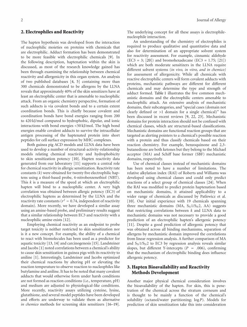
2 Journal of Allergy
2. Electrophiles and Reactivity
The hapten hypothesis was developed from the interactionof nucleophilic moieties on proteins with chemicals thatare electrophilic. Adduct formation has been demonstratedto be more feasible with electrophilic chemicals [9]. Inthe following description, haptenation within the skin isdiscussed, as most of the research knowledge gained hasbeen through examining the relationship between chemicalreactivity and allergenicity in this organ system. An analysisof two published databases [4, 5] containing more than300 chemicals demonstrated to be allergens by the LLNAreveals that approximately 40% of the skin sensitizers have atleast an electrophilic center that is amenable to nucleophilicattack. From an organic chemistry perspective, formation ofsuch adducts is via covalent bonds and to a certain extentcoordination bonds. This is chiefly because covalent andcoordination bonds have bond energies ranging from 200to 420 kJ/mol compared to hydrophobic, dipolar, and ionicinteractions with bond energies <50 kJ/mol. The high bondenergies enable covalent adducts to survive the intracellularantigen processing of the haptenated protein into shortpeptides for cell surface expression by MHC complexes.
Both guinea pig ACD models and LLNA data have beenused to develop a number of structural-activity-relationshipmodels relating chemical reactivity and hydrophobicityto skin sensitization potency [10]. Hapten reactivity datagenerated from our laboratory [11] supports a central rolefor chemical reactivity in allergic sensitization. Reactivity rateconstants (k) were obtained for twenty five electrophilic hap-tens using a thiol-based probe, 4-nitrobenzenethiol (NBT).This k is a measure of the speed at which an electrophilichapten will bind to a nucleophilic center. A very highcorrelation was obtained between allergic potency (EC3) ofelectrophilic haptens as determined by the LLNA and thereactivity rate constants (r2 = 0.74, independent of reactivitydomain). More recently, we have developed a similar assayusing an amine-based probe, and preliminary results suggestthat a similar relationship between EC3 and reactivity with anucleophilic amine exists [12].
Employing chemical reactivity as an endpoint to probetarget toxicity is neither restricted to skin sensitization noris it a new concept. For example, the ability of a chemicalto react with biomolecules has been used as a predictor foraquatic toxicity [13, 14] and carcinogenesis [15]. Landsteinerand Jacobs [1] noted correlations between a chemical’s abilityto cause skin sensitization in guinea pigs with its reactivity toaniline [1]. Interestingly, Landsteiner and Jacobs optimizedtheir chemical reactions by altering pH or elevating thereaction temperature to observe reaction to nucleophiles likebutylamine and aniline. It has to be noted that many covalentadducts that would otherwise form under harsh conditionsare not formed as reaction conditions (i.e., temperature, pH)and medium are adjusted to physiological-like conditions.More recently, reactivity assays utilizing cysteine, lysine,glutathione, and several model peptides have been developedand efforts are underway to validate them as alternativein chemico methods for screening skin sensitizers [16–19].
The underlying concept for all these assays is electrophile-nucleophile interaction.
An understanding of the chemistry of electrophiles isrequired to produce qualitative and quantitative data andalso for determination of an appropriate solvent systemfor reactivity assessment. For example, cinnamic aldehyde(EC3 = 3; [20]) and bromohexadecane (EC3 = 1.75; [21])which are both moderate sensitizers in the LLNA requiredifferent solvent systems (in vivo, in vitro, and in chemico)for assessment of allergenicity. While all chemicals withreactive electrophilic centers will form covalent adducts withproteins, mechanistic pathways are different for differentchemicals and may determine the type and strength ofadduct formed. Table 1 illustrates the five common mech-anistic domains and the electrophilic centers amenable tonucleophilic attack. An extensive analysis of mechanisticdomains, their subcategories, and “special cases (domain notclearly defined or >1 domain for a single chemical)” hasbeen discussed in recent reviews [9, 22, 23]. Mechanisticdomains for protein interaction should not be confused withchemical classes, which are structural-based classifications.Mechanistic domains are functional reaction groups that aretargeted as alerting pointers to a chemical’s possible reactionwith a protein and thus classify chemicals based on theirreaction chemistry. For example, benzoquinone and 2,3-butanedione are both ketones but they belong to the Michaelacceptor (MA) and Schiff base former (SBF) mechanisticdomains, respectively.
Use of chemical classes instead of mechanistic domainshas been noted to have a number of limitations. Therelative alkylation index (RAI) of Roberts and Williams wasdeveloped using chemical classes and could only predictreactions of a select group of chemical classes [24]. Whenthe RAI was modified to predict protein haptenation basedon mechanistic domains, it attained applicability to awider range of chemicals as discussed by Patlewicz et al.[10]. Our initial experience with 19 chemicals spanningthree mechanistic domains (MA, SN1/SN2, AA) suggeststhat restricting correlations between k and LLNA data tomechanistic domains was not necessary to provide a goodprediction of an electrophilic hapten’s allergenic potency[11]. Despite a good prediction of allergenic potency thatwas obtained across all binding mechanisms, separation ofallergens by mechanistic domain improved the correlationsfrom linear regression analysis. A further comparison of MAand SN1/SN2 to EC3 by regression analysis reveals similarslopes, but different Y-intercepts (P = .006), confirmingthat the mechanism of electrophilic binding does influenceallergenic potency.
3. Hapten Bioavailability and ReactivityMethods Development
Another major physical chemical consideration involvesthe bioavailability of the hapten. For skin, this is pene-tration of the chemical across the stratum corneum andis thought to be mainly a function of the chemical’ssolubility (octanol/water partitioning; logP). Models forprediction of skin sensitization take this into consideration
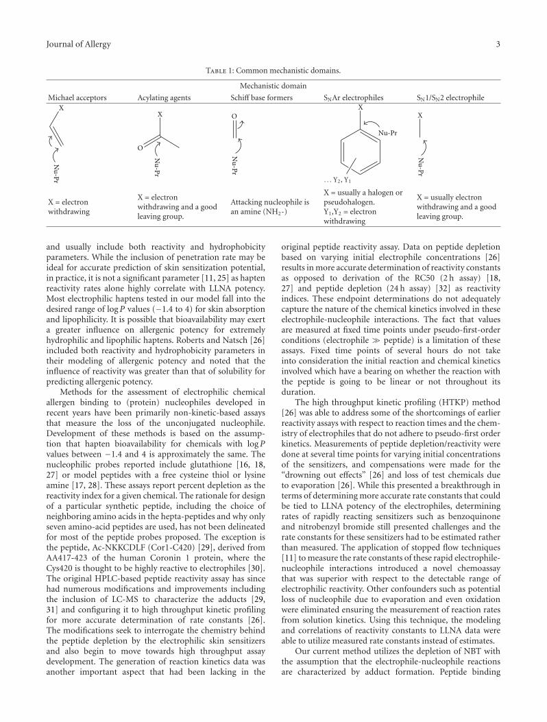
Journal of Allergy 3
Table 1: Common mechanistic domains.
Mechanistic domain
Michael acceptors Acylating agents Schiff base formers SNAr electrophiles SN1/SN2 electrophileX
Nu
-Pr
Nu
-Pr
O
X
Nu
-Pr
O
Nu-Pr
X
. . . Y2, Y1
Nu
-Pr
X
X = electronwithdrawing
X = electronwithdrawing and a goodleaving group.
Attacking nucleophile isan amine (NH2-)
X = usually a halogen orpseudohalogen.Y1,Y2 = electronwithdrawing
X = usually electronwithdrawing and a goodleaving group.
and usually include both reactivity and hydrophobicityparameters. While the inclusion of penetration rate may beideal for accurate prediction of skin sensitization potential,in practice, it is not a significant parameter [11, 25] as haptenreactivity rates alone highly correlate with LLNA potency.Most electrophilic haptens tested in our model fall into thedesired range of logP values (−1.4 to 4) for skin absorptionand lipophilicity. It is possible that bioavailability may exerta greater influence on allergenic potency for extremelyhydrophilic and lipophilic haptens. Roberts and Natsch [26]included both reactivity and hydrophobicity parameters intheir modeling of allergenic potency and noted that theinfluence of reactivity was greater than that of solubility forpredicting allergenic potency.
Methods for the assessment of electrophilic chemicalallergen binding to (protein) nucleophiles developed inrecent years have been primarily non-kinetic-based assaysthat measure the loss of the unconjugated nucleophile.Development of these methods is based on the assump-tion that hapten bioavailability for chemicals with logPvalues between −1.4 and 4 is approximately the same. Thenucleophilic probes reported include glutathione [16, 18,27] or model peptides with a free cysteine thiol or lysineamine [17, 28]. These assays report percent depletion as thereactivity index for a given chemical. The rationale for designof a particular synthetic peptide, including the choice ofneighboring amino acids in the hepta-peptides and why onlyseven amino-acid peptides are used, has not been delineatedfor most of the peptide probes proposed. The exception isthe peptide, Ac-NKKCDLF (Cor1-C420) [29], derived fromAA417-423 of the human Coronin 1 protein, where theCys420 is thought to be highly reactive to electrophiles [30].The original HPLC-based peptide reactivity assay has sincehad numerous modifications and improvements includingthe inclusion of LC-MS to characterize the adducts [29,31] and configuring it to high throughput kinetic profilingfor more accurate determination of rate constants [26].The modifications seek to interrogate the chemistry behindthe peptide depletion by the electrophilic skin sensitizersand also begin to move towards high throughput assaydevelopment. The generation of reaction kinetics data wasanother important aspect that had been lacking in the
original peptide reactivity assay. Data on peptide depletionbased on varying initial electrophile concentrations [26]results in more accurate determination of reactivity constantsas opposed to derivation of the RC50 (2 h assay) [18,27] and peptide depletion (24 h assay) [32] as reactivityindices. These endpoint determinations do not adequatelycapture the nature of the chemical kinetics involved in theseelectrophile-nucleophile interactions. The fact that valuesare measured at fixed time points under pseudo-first-orderconditions (electrophile � peptide) is a limitation of theseassays. Fixed time points of several hours do not takeinto consideration the initial reaction and chemical kineticsinvolved which have a bearing on whether the reaction withthe peptide is going to be linear or not throughout itsduration.
The high throughput kinetic profiling (HTKP) method[26] was able to address some of the shortcomings of earlierreactivity assays with respect to reaction times and the chem-istry of electrophiles that do not adhere to pseudo-first orderkinetics. Measurements of peptide depletion/reactivity weredone at several time points for varying initial concentrationsof the sensitizers, and compensations were made for the“drowning out effects” [26] and loss of test chemicals dueto evaporation [26]. While this presented a breakthrough interms of determining more accurate rate constants that couldbe tied to LLNA potency of the electrophiles, determiningrates of rapidly reacting sensitizers such as benzoquinoneand nitrobenzyl bromide still presented challenges and therate constants for these sensitizers had to be estimated ratherthan measured. The application of stopped flow techniques[11] to measure the rate constants of these rapid electrophile-nucleophile interactions introduced a novel chemoassaythat was superior with respect to the detectable range ofelectrophilic reactivity. Other confounders such as potentialloss of nucleophile due to evaporation and even oxidationwere eliminated ensuring the measurement of reaction ratesfrom solution kinetics. Using this technique, the modelingand correlations of reactivity constants to LLNA data wereable to utilize measured rate constants instead of estimates.
Our current method utilizes the depletion of NBT withthe assumption that the electrophile-nucleophile reactionsare characterized by adduct formation. Peptide binding
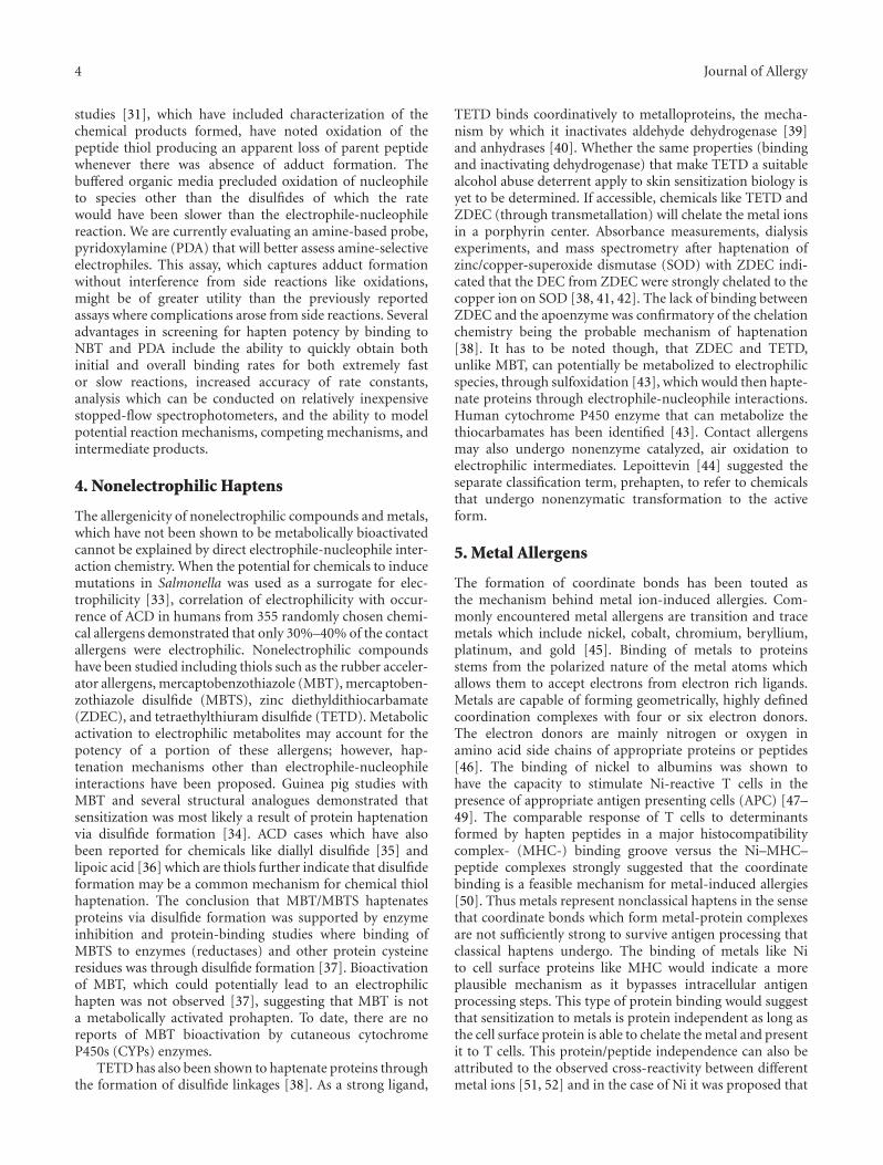
4 Journal of Allergy
studies [31], which have included characterization of thechemical products formed, have noted oxidation of thepeptide thiol producing an apparent loss of parent peptidewhenever there was absence of adduct formation. Thebuffered organic media precluded oxidation of nucleophileto species other than the disulfides of which the ratewould have been slower than the electrophile-nucleophilereaction. We are currently evaluating an amine-based probe,pyridoxylamine (PDA) that will better assess amine-selectiveelectrophiles. This assay, which captures adduct formationwithout interference from side reactions like oxidations,might be of greater utility than the previously reportedassays where complications arose from side reactions. Severaladvantages in screening for hapten potency by binding toNBT and PDA include the ability to quickly obtain bothinitial and overall binding rates for both extremely fastor slow reactions, increased accuracy of rate constants,analysis which can be conducted on relatively inexpensivestopped-flow spectrophotometers, and the ability to modelpotential reaction mechanisms, competing mechanisms, andintermediate products.
4. Nonelectrophilic Haptens
The allergenicity of nonelectrophilic compounds and metals,which have not been shown to be metabolically bioactivatedcannot be explained by direct electrophile-nucleophile inter-action chemistry. When the potential for chemicals to inducemutations in Salmonella was used as a surrogate for elec-trophilicity [33], correlation of electrophilicity with occur-rence of ACD in humans from 355 randomly chosen chemi-cal allergens demonstrated that only 30%–40% of the contactallergens were electrophilic. Nonelectrophilic compoundshave been studied including thiols such as the rubber acceler-ator allergens, mercaptobenzothiazole (MBT), mercaptoben-zothiazole disulfide (MBTS), zinc diethyldithiocarbamate(ZDEC), and tetraethylthiuram disulfide (TETD). Metabolicactivation to electrophilic metabolites may account for thepotency of a portion of these allergens; however, hap-tenation mechanisms other than electrophile-nucleophileinteractions have been proposed. Guinea pig studies withMBT and several structural analogues demonstrated thatsensitization was most likely a result of protein haptenationvia disulfide formation [34]. ACD cases which have alsobeen reported for chemicals like diallyl disulfide [35] andlipoic acid [36] which are thiols further indicate that disulfideformation may be a common mechanism for chemical thiolhaptenation. The conclusion that MBT/MBTS haptenatesproteins via disulfide formation was supported by enzymeinhibition and protein-binding studies where binding ofMBTS to enzymes (reductases) and other protein cysteineresidues was through disulfide formation [37]. Bioactivationof MBT, which could potentially lead to an electrophilichapten was not observed [37], suggesting that MBT is nota metabolically activated prohapten. To date, there are noreports of MBT bioactivation by cutaneous cytochromeP450s (CYPs) enzymes.
TETD has also been shown to haptenate proteins throughthe formation of disulfide linkages [38]. As a strong ligand,
TETD binds coordinatively to metalloproteins, the mecha-nism by which it inactivates aldehyde dehydrogenase [39]and anhydrases [40]. Whether the same properties (bindingand inactivating dehydrogenase) that make TETD a suitablealcohol abuse deterrent apply to skin sensitization biology isyet to be determined. If accessible, chemicals like TETD andZDEC (through transmetallation) will chelate the metal ionsin a porphyrin center. Absorbance measurements, dialysisexperiments, and mass spectrometry after haptenation ofzinc/copper-superoxide dismutase (SOD) with ZDEC indi-cated that the DEC from ZDEC were strongly chelated to thecopper ion on SOD [38, 41, 42]. The lack of binding betweenZDEC and the apoenzyme was confirmatory of the chelationchemistry being the probable mechanism of haptenation[38]. It has to be noted though, that ZDEC and TETD,unlike MBT, can potentially be metabolized to electrophilicspecies, through sulfoxidation [43], which would then hapte-nate proteins through electrophile-nucleophile interactions.Human cytochrome P450 enzyme that can metabolize thethiocarbamates has been identified [43]. Contact allergensmay also undergo nonenzyme catalyzed, air oxidation toelectrophilic intermediates. Lepoittevin [44] suggested theseparate classification term, prehapten, to refer to chemicalsthat undergo nonenzymatic transformation to the activeform.
5. Metal Allergens
The formation of coordinate bonds has been touted asthe mechanism behind metal ion-induced allergies. Com-monly encountered metal allergens are transition and tracemetals which include nickel, cobalt, chromium, beryllium,platinum, and gold [45]. Binding of metals to proteinsstems from the polarized nature of the metal atoms whichallows them to accept electrons from electron rich ligands.Metals are capable of forming geometrically, highly definedcoordination complexes with four or six electron donors.The electron donors are mainly nitrogen or oxygen inamino acid side chains of appropriate proteins or peptides[46]. The binding of nickel to albumins was shown tohave the capacity to stimulate Ni-reactive T cells in thepresence of appropriate antigen presenting cells (APC) [47–49]. The comparable response of T cells to determinantsformed by hapten peptides in a major histocompatibilitycomplex- (MHC-) binding groove versus the Ni–MHC–peptide complexes strongly suggested that the coordinatebinding is a feasible mechanism for metal-induced allergies[50]. Thus metals represent nonclassical haptens in the sensethat coordinate bonds which form metal-protein complexesare not sufficiently strong to survive antigen processing thatclassical haptens undergo. The binding of metals like Nito cell surface proteins like MHC would indicate a moreplausible mechanism as it bypasses intracellular antigenprocessing steps. This type of protein binding would suggestthat sensitization to metals is protein independent as long asthe cell surface protein is able to chelate the metal and presentit to T cells. This protein/peptide independence can also beattributed to the observed cross-reactivity between differentmetal ions [51, 52] and in the case of Ni it was proposed that
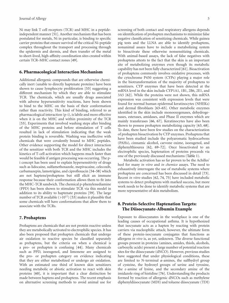
Journal of Allergy 5
Ni may link T cell receptors (TCR) and MHC in a peptide-independent manner [51]. Another mechanism that has beenpostulated for metals, Ni in particular, is binding to specificcarrier proteins that ensure survival of the critical Ni-peptidecomplex throughout the transport and processing throughthe epidermis and dermis, and then transfer of the metalto short-lived, high-affinity coordination sites created withincertain TCR–MHC contact zones [49].
6. Pharmacological Interaction Mechanism
Additional allergenic compounds that are otherwise chemi-cally inert (unable to directly haptenate proteins) have beenshown to cause lymphocyte proliferation [53] suggesting adifferent mechanism by which they are able to stimulateTCR. The chemicals, which are usually drugs associatedwith adverse hypersensitivity reactions, have been shownto bind to the MHC on the basis of their conformationrather than reactivity. This kind of binding, referred to aspharmacological interaction (p-i), is labile and more effectivewhen it is on the MHC and within proximity of the TCR[53]. Experiments that involved washing steps after bindingof the MHC proteins and before stimulation of T cellsresulted in lack of stimulation indicating that the weakprotein binding is reversible. Washing was ineffective withchemicals that were covalently bound to MHC peptides.Other evidence supporting the model for direct interactionof the sensitizer with both TCR and the MHC includes thekinetics of T-cell activation which happens much faster thanwould be feasible if antigen processing was occurring. The p-i concept has been used to explain hypersensitivity of drugssuch as lidocaine, sulfamethoxazole, mepivacaine, celecoxib,carbamazepin, lamotrigine, and ciprofloxacin [54–58] whichare not haptens/prohaptens but still elicit an immuneresponse because their conformation allows them to fit intothe MHC-TCR sandwich. The chemical p-phenylenediamine(PPD) has been shown to stimulate TCR via this model inaddition to its ability to haptenate proteins [59]. The largenumber of TCR available (>1012) [53] makes it plausible thatsome chemicals will have conformations that allow them toassociate with the TCRs.
7. Prohaptens
Prohaptens are chemicals that are not protein reactive unlessthey are metabolically activated to electrophilic species. It hasalso been proposed that prohapten chemicals that undergoair oxidation to reactive species be classified separatelyas prehaptens, but the criteria on when a chemical isa pro- or prehapten is confusing [44]. Many chemicalssuch as PPD, isoeugenol, and limonene are assigned tothe pro- or prehapten category on evidence indicatingthat they are either metabolized or undergo air oxidation.With an estimated one third of known skin sensitizersneeding metabolic or abiotic activation to react with skinproteins [60], it is important that a clear distinction bemade between haptens and prohaptens. The recent emphasison alternative screening methods to avoid animal use for
screening of both contact and respiratory allergens dependson identification of prohapten mechanisms to minimize falsenegative classification of sensitizing chemicals. While guineapig tests and the LLNA are able to identify prohaptens,nonanimal assays have to include a metabolizing systemto bioactivate these otherwise nonsensitizing chemicals.With animal-based assays, the lack of false negatives withprohaptens attests to the fact that the skin is an importantsite of metabolizing enzymes even though its metaboliccapability has not been fully characterized [61]. Bioactivationof prohaptens commonly involves oxidative processes, withthe cytochrome P450 system (CYPs) playing a major rolein the biotransformation of the majority of prohaptens tosensitizers. CYP enzymes that have been detected at themRNA level in the skin include CYP1A1, 1B1, 2B6, 2E1, and3A5 [62]. While the proteins were not shown, this mRNAexpression was consistent with expression levels that werefound for normal human epidermal keratinocytes (NHEKs)and dermal fibroblasts [63–65]. Other metabolic enzymesidentified in the skin include monooxygenases, dehydroge-nases, esterases, amidases, and Phase II enzymes which aremainly transferases [66, 67]. Keratinocytes have also beenshown to possess prohapten metabolizing capacity [63, 68].To date, there have been few studies on the characterizationof prohapten bioactivation by CYP enzymes. Prohaptens thathave been studied include the polyaromatic hydrocarbons(PAHs), cinnamic alcohol, carvone oxime, isoeugenol, anddiphenylthiourea [62, 69–72]. Once bioactivated to anelectrophilic species, haptenation of proteins proceeds viaone of the previously discussed mechanisms (Table 1).
Metabolic activation has so far proven to be the Achilles’heel for many in vitro and in chemico assays. The need toexhaustively interrogate the use of metabolic systems whereprohaptens are concerned has been discussed in detail [73].Recent in vitro studies [62, 74, 75] have included metabolicsystems to detect prohaptens with marked success, but morework needs to be done to identify metabolic systems that aremore representative of skin metabolism.
8. Protein-Selective Haptenation Targets:The Diisocyanate-Albumin Example
Exposure to diisocyanates in the workplace is one of theleading causes of occupational asthma. It is hypothesizedthat isocyanate acts as a hapten by reacting with proteincarriers via nucleophilic attack; however, the ultimate formof these protein-isocyanate conjugates that functions asallergens in vivo is, as yet, unknown. The diverse functionalgroups present in proteins (amines, amides, thiols, alcohols,carboxylic acids) present a large number of potential reactionsites for the diisocyanate (dNCO). However, previous studieshave suggested that under physiological conditions, theseare limited to N-terminal α-amines, the sulfhydryl groupof cysteine, the hydroxyl groups of serine and tyrosine,the ε-amine of lysine, and the secondary amine of theimidazole ring of histidine [76]. Understanding the productsformed by reaction of allergenic dNCOs such as methylenediphenyldiisocyanate (MDI) and toluene diisocyanate (TDI)

6 Journal of Allergy
with biological molecules is critical to understanding themechanisms by which these chemicals affect living systems.Tandem mass spectrometry performed on a quadrupoletime-of-flight (qTOF) mass spectrometer [77] is particularlywell suited for the characterization of chemically modi-fied proteins. Proteins of interest may be digested witha proteolytic enzyme (such as trypsin) and the resultantpeptides analyzed with high sensitivity and mass accuracy.Because covalent modification of an amino acid residueresults in a change in that residue’s mass, accurate massdetermination of the fragment ions produced by collision-induced dissociation [78] allows unambiguous assignmentof the site of modification. Such experiments have becomeroutine for the analysis of posttranslational modificationssuch as acetylation, glycosylation, and phosphorylation,among many others [79].
Recent efforts in our laboratory [81, 90] and others[80] have begun to focus on harnessing the power oftandem mass spectrometry to determine how and wheredNCOs modify model peptides and proteins. Hydrolysisof the isocyanate functional group to a primary amineis a competing reaction under aqueous conditions. Thesehydrolyzed isocyanate amines may then undergo nucle-ophilic addition to another dNCO molecule. Therefore,conjugation products observed upon reaction of dNCOswith model peptides and proteins in vitro results in a complexvariety of different reaction products, including intra- andintermolecular crosslinking, dNCO self-polymerization, anddNCO hydrolysis. Our initial study focused on determiningthe site of dNCO modification on model bioactive peptidesconjugated under aqueous conditions [81]. Analysis of theseconjugates by tandem mass spectrometry revealed that thedNCO was bound preferentially to the N-terminal amine ofeach of the peptides examined. Furthermore, when a peptidewith an N-terminal residue containing a side chain amine(lysine, arginine) was reacted with dNCO, intramolecularcrosslinking with the side chain amine becomes competitivewith hydrolysis, however, the reactivity decreases as theresidue is displaced further from the N-terminus. The resultsof this peptide study suggested agreement with a long-heldhypothesis that the N-terminal amine of protein chains is alikely target for isocyanate conjugation [76, 82]. Studies ofthe kinetics of isocyanate binding with protein functionalgroups [83] determined that at pH 7, reaction with an N-terminal amine should proceed approximately 100 timesfaster than the ε-amine of the lysine side chain. The differenceis due to the relative pKa of the two functional groups(α-NH3
+ pKa ∼ 9 versus Lys ε-NH3+ pKa ∼10.5), as dNCO
conjugation proceeds through the neutral –NH2 rather thanthe charged –NH3
+ species.
9. Conclusion
Although tandem MS studies of dNCO-conjugated peptidesare useful for determining the diverse chemical speciesproduced when dNCO binds amino acids, they do notproduce the unique chemical microenvironments presentedby the complex three-dimensional structure of proteins. Inorder to understand how dNCOs react in these complex
Table 2
Residue MDI TDI
Asp1∗ X X
Lys4∗ X X
Lys12 X X
Lys73 X X
Gln104 X
Lys106 X
Lys136 X X
Lys137∗ X X
Lys159 X
Lys190 X X
Gln196 X
Lys199∗ X X
Lys205 X
Lys212 X
Lys262∗ X
Lys274 X
Lys276 X
Lys281 X
Lys351∗ X X
Lys378 X
Lys402 X
Lys413∗ X X
Lys414∗ X X
Lys432∗ X X
Lys436∗ X X
Lys439∗ X X
Lys444∗ X X
Lys524 X X
Lys525∗ X X
Lys534 X
Lys536 X X
Lys541∗ X X
Lys545 X
Lys557 X
Lys560 X
Lys573 X
Lys574 X∗
MDI binding sites indentified in [80].
environments, studies on model proteins are essential. Serumalbumin is an appropriate model protein, as it is monomericand its sequence and three dimensional structure have beenwell defined. It is naturally abundant (35–50 mg/mL inserum), found in most tissues, and has been identified asa target of dNCO binding in vivo [84–89]. Recently, ourlaboratory completed an extensive analysis of the bindingsites of TDI on human serum albumin [90]. At high(40 : 1 dNCO : protein) ratios, near-stoichiometric bindingwas observed; TDI binds at thirty-seven sites on the protein,including the N-terminal amine on aspartic acid at positionone and the side chain of thirty-four lysine residues. At lowerconjugation ratios (1 : 2 dNCO : protein), a small subset of
Journal of Allergy 7
(a) (b)
Figure 1: (a) Lysine residues observed bound to MDI and TDI on human serum albumin. (b) All lysine residues on human serum albumin.
Figure 2: Lysine residues reactive to TDI but not MDI.
these thirty-seven sites is conserved, with binding observedat the N-terminus and four lysine residues, suggesting thesesites are preferred binding sites. Kristiansson and coworkers[91] determined that at a tenfold molar excess, HHPA boundto thirty-seven sites on human serum albumin, including theN-terminal aspartic acid and thirty-six lysine residues.
Interestingly, all 59 lysine residues of human serumalbumin have been determined to be solvent accessible, butonly 37 are reactive toward TDI, while 19 are reactive towardMDI (Hettick and Siegel, unpublished data). Human serumalbumin is a highly charged molecule, in part accountingfor its high solubility. For example, Figure 1 provides twoviews of human serum albumin based on its crystal structure[92]. In Figure 1(a), the lysine residues that bind MDI andTDI are highlighted, whereas in Figure 1(b), all 59 lysineresidues are highlighted. Steric effects are insufficient toexplain the difference between accessibility and observedbinding. It is therefore likely that the microenvironmentof the binding site(s) determines whether or not a certainlysine residue is reactive toward isocyanate. Lysine 199, whichis known to bind hydrophobic anions such as aspirin andbenzyl penicillin, was also determined to be a predominantbinding site for both TDI and MDI. Gerig and Reinheimer[93] determined that the pKa of the aspirin binding site
(later determined to be Lys199) of albumin was 7.9. Theseauthors hypothesized based on the reactivity of humanserum albumin with dinitrofluorobenzene that there existtwo lysines on HSA that have a pKa as low as 7.9. In addition,Lys199 has been shown by molecular dynamics calculationsto be predominantly uncharged, undergoing proton transferwith the nearby Lys195 [94]. This study elegantly suggeststhe reason we observed dNCO bound to Lys199, and not thenearby the Lys195. Other sites noted to be abundant bindersof dNCO, such as Lys439 and Lys525, have been observed toundergo nonenzymatic glycosylation in vivo. Glycosylationis generally observed to occur at lysine residues located nearanother amino group, presumed to be charged [95]. Lys439is located in a region with two other nearby lysine residues(Lys432 and Lys436) and Lys525 is part of a dilysine motif.
More recently, we have begun an investigation tocompare the binding sites of TDI and MDI on serumalbumin under identical conditions (40 : 1 dNCO : HSA;50 mM NH4HCO3, pH 7.9). Under these conditions, MDI isobserved to bind to a subset of 19 of the 37 sites observed forTDI (see Table 2). Although it would be tempting to attributedifferences in binding between TDI and MDI to steric effects,as seen in Figure 2, many of the lysine residues that arereactive toward TDI but not MDI are open and highlyaccessible. We therefore hypothesize that the difference inobserved binding between TDI and MDI is attributable toa combination of steric effects and the increased reactivityof TDI. The electron withdrawing character of the secondN=C=O group on the aromatic ring of TDI significantlyincreases the reactivity of the first isocyanate. In contrast,the reactivity of the isocyanate functional group(s) on MDIis lower because the p-ethyl phenylisocyanate substituentis less electron-withdrawing. Wisnewski and coworkers alsoexamined the reaction products between MDI and humanserum albumin by HPLC-MS/MS. Their data indicated14 binding sites on albumin, including 12 lysine and 2asparagine residues, in relatively good agreement with theresults presented in Table 2. In addition, these authorssuggested that the four “dilysine” (KK) motifs in humanserum albumin are important binding sites, and that MDIshows reactive specificity for the second lysine. As discussedpreviously, the ability of a lysine residue to transfer its proton
8 Journal of Allergy
to a nearby lysine or histidine residue may, in fact, leadto increased reactivity toward dNCOs. However, as dilysinemotifs account for 8 of 36 TDI binding sites and 6 of 19 MDIbinding sites, it is clear that two lysine residues in a “KK”arrangement are not essential to binding.
It has been recognized for over 80 years that the abilityof a hapten to react to a protein was central to its abilityto produce allergic sensitization. Haptenation of a proteincan occur by multiple mechanisms (primarily electrophilicattack) and is dependent on many factors such as chemicalproperties, bioavailability, and site of exposure. Recentstudies have greatly expanded the knowledge in this area bydemonstrating that it is the rate at which an electrophilichapten reacts with a nucleophile center that is a centraldeterminant in its dermal sensitization potency. In addi-tion, the chemical mechanism of binding (i.e., mechanisticdomain) also influences allergenic potency. These physicalchemical factors, however, have not been shown to be relatedto skewing of the immunological response toward Th1 versusTh2. Studies are currently utilizing modern proteomic massspectrometry to identify hapten binding sites on proteins andto identify specific hapten target proteins. It is possible thatthe protein-specific factors may play a role in the ultimatenature of the immune response.
Abbreviations
AA: Acylating agentsACD: Allergic contact dermatitisCYPs: Cytochrome P450sdNCO: DiisocyanateEC3: Concentration of allergen producing a
3-fold lymph node cell proliferation =LLNA threshold
HPLC-MS/MS: High performance liquidchromatography-tandem massspectrometry
HSA: Human serum albuminLLNA: Murine local lymph node assayLMW: Low molecular weightlogP: Octanol/water partitioningMA: Michael acceptorMHC: Major histocompatibility complexMBT: MercaptobenzothiazoleMBTS: Mercaptobenzothiazole disulfideNBT: 4-nitrobenzenethiolMDI: Methylene diphenyldiisocyanatePPD: p-phenylenediamineqTOF: Quadrupole time-of-flightRAI: Relative alkylation indexSBF: Schiff base formerSN1/SN2: Nucleophilic substitution unimolecular or
bimolecularSOD: Superoxide dismutaseTCR: T-cell receptorsTETD: Tetraethylthiuram disulfideTDI: Toluene diisocyanateZDEC: Zinc diethyldithiocarbamate.
Acknowledgment
This work was supported in part by an interagency agree-ment with NIEHS (Y1-ES-0001). The findings and conclu-sions in this paper are those of the authors and do notnecessarily represent the views of the National Institute forOccupational Safety and Health.
References
[1] K. Landsteiner and J. Jacobs, “Studies on the sensitizationof animals with simple chemical compounds,” Journal ofExperimental Medicine, vol. 64, pp. 625–639, 1936.
[2] W. J. Pichler, “Pharmacological interaction of drugs withantigen-specific immune receptors: the p-i concept,” CurrentOpinion in Allergy and Clinical Immunology, vol. 2, no. 4, pp.301–305, 2002.
[3] M. T. Cronin and D. A. Basketter, “Multivariate QSARanalysis of a skin sensitization database,” SAR & QSAR inEnvironmental Research, vol. 2, no. 3, pp. 159–179, 1994.
[4] G. F. Gerberick, C. A. Ryan, P. S. Kern et al., “Compilationof historical local lymph node data for evaluation of skinsensitization alternative methods,” Dermatitis, vol. 16, no. 4,pp. 157–202, 2005.
[5] P. S. Kern, G. F. Gerberick, C. A. Ryan, I. Kimber, A.Aptula, and D. A. Basketter, “Local lymph node data forthe evaluation of skin sensitization alternatives: a secondcompilation,” Dermatitis, vol. 21, no. 1, pp. 8–32, 2010.
[6] E. V. Buehler, “Delayed contact hypersensitivity in the GuineaPig,” Archives of Dermatology, vol. 91, pp. 171–179, 1965.
[7] B. Magnusson and A. M. Kligman, “The identification of con-tact allergens by animal assay. The guinea pig maximizationtest,” Journal of Investigative Dermatology, vol. 52, no. 3, pp.268–276, 1969.
[8] J. H. Dean, L. E. Twerdok, R. R. Tice, D. M. Sailstad, D. G.Hattan, and W. S. Stokes, “ICCVAM evaluation of the murinelocal lymph node assay. Conclusions and recommendationsof an independent scientific peer review panel,” RegulatoryToxicology and Pharmacology, vol. 34, no. 3, pp. 258–273, 2001.
[9] M. Divkovic, C. K. Pease, G. F. Gerberick, and D. A. Basketter,“Hapten-protein binding: from theory to practical applicationin the in vitro prediction of skin sensitization,” ContactDermatitis, vol. 53, no. 4, pp. 189–200, 2005.
[10] G. Patlewicz, D. W. Roberts, and E. Uriarte, “A comparisonof reactivity schemes for the prediction skin sensitizationpotential,” Chemical Research in Toxicology, vol. 21, no. 2, pp.521–541, 2008.
[11] I. Chipinda, R. O. Ajibola, M. K. Morakinyo, T. B. Ruwona,R. H. Simoyi, and P. D. Siegel, “Rapid and simple kineticsscreening assay for electrophilic dermal sensitizers usingnitrobenzenethiol,” Chemical Research in Toxicology, vol. 23,no. 5, pp. 918–925, 2010.
[12] I. Chipinda, W. Mbiya, R. Ajibola, M. K. Morakinyo, R. H.Simoyi, and P. D. Siegel, “Pyridoxylamine reactivity kineticsas an amine based probe for screening electrophilic dermalsensitizers,” in Proceedings of the ICCVAM Meeting, 2011,Abstract 2011.
[13] D. W. Roberts, T. W. Schultz, E. M. Wolf, and A. O. Aptula,“Experimental reactivity parameters for toxicity modeling:application to the acute aquatic toxicity of S2 electrophilesto Tetrahymena pyriformis,” Chemical Research in Toxicology,vol. 23, no. 1, pp. 228–234, 2010.

Journal of Allergy 9
[14] T. W. Schultz, C. L. Sparfkin, and A. O. Aptula, “Reactivity-based toxicity modelling of five-membered heterocyclic com-pounds: application to Tetrahymena pyriformis,” SAR andQSAR in Environmental Research, vol. 21, no. 7-8, pp. 681–691,2010.
[15] D. C. Liebler, “Protein damage by reactive electrophiles: targetsand consequences,” Chemical Research in Toxicology, vol. 21,no. 1, pp. 117–128, 2008.
[16] A. O. Aptula, G. Patlewicz, D. W. Roberts, and T. W. Schultz,“Non-enzymatic glutathione reactivity and in vitro toxicity:a non-animal approach to skin sensitization,” Toxicology, vol.20, no. 2, pp. 239–247, 2006.
[17] G. F. Gerberick, J. D. Vassallo, R. E. Bailey, J. G. Chaney, S.W. Morrall, and J. P. Lepoittevin, “Development of a peptidereactivity assay for screening contact allergens,” ToxicologicalSciences, vol. 81, no. 2, pp. 332–343, 2004.
[18] T. W. Schultz, J. W. Yarbrough, and E. L. Johnson, “Structure-activity relationships for reactivity of carbonyl-containingcompounds with glutathione,” SAR and QSAR in Environmen-tal Research, vol. 16, no. 4, pp. 313–322, 2005.
[19] T. W. Schultz, R. E. Carlson, M. T. Cronin et al., “Aconceptual framework for predicting the toxicity of reactivechemicals: modeling soft electrophilicity,” SAR and QSAR inEnvironmental Research, vol. 17, no. 4, pp. 413–428, 2006.
[20] D. A. Basketter, Z. M. Wright, E. V. Warbrick et al., “Humanpotency predictions for aldehydes using the local lymph nodeassay,” Contact Dermatitis, vol. 45, no. 2, pp. 89–94, 2001.
[21] P. D. Siegel, A. Fedorowicz, L. Butterworth et al., “Physical-chemical and solvent considerations in evaluating the influ-ence of carbon chain length on the skin sensitization activityof 1-bromoalkanes,” Toxicological Sciences, vol. 107, no. 1, pp.78–84, 2009.
[22] A. O. Aptula and D. W. Roberts, “Mechanistic applicabilitydomains for nonanimal-based prediction of toxicological endpoints: general principles and application to reactive toxicity,”Chemical Research in Toxicology, vol. 19, no. 8, pp. 1097–1105,2006.
[23] D. W. Roberts, G. Patlewicz, P. S. Kern et al., “Mechanisticapplicability domain classification of a local lymph node assaydataset for skin sensitization,” Chemical Research in Toxicology,vol. 20, no. 7, pp. 1019–1030, 2007.
[24] D. W. Roberts and D. L. Williams, “The derivation of quan-titative correlations between skin sensitisation and physio-chemical parameters for alkylating agents, and their applica-tion to experimental data for sultones,” Journal of TheoreticalBiology, vol. 99, no. 4, pp. 807–825, 1982.
[25] D. W. Roberts and D. A. Basketter, “A quantitative structure-activity/dose relationship for contact allergenic potential ofaklkyl group transfer agents,” Toxicology, vol. 4, no. 4-5, pp.686–687, 1990.
[26] D. W. Roberts and A. Natsch, “High throughput kinetic profil-ing approach for covalent binding to peptides: application toskin sensitization potency of Michael acceptor electrophiles,”Chemical Research in Toxicology, vol. 22, no. 3, pp. 592–603,2009.
[27] J. W. Yarbrough and T. W. Schultz, “Abiotic sulfhydryl reac-tivity: a predictor of aquatic toxicity for carbonyl-containingalpha,beta-unsaturated compounds,” Chemical Research inToxicology, vol. 20, no. 3, pp. 558–562, 2007.
[28] G. F. Gerberick, J. D. Vassallo, L. M. Foertsch, B. B. Price, J.G. Chaney, and J. P. Lepoittevin, “Quantification of chemical
peptide reactivity for screening contact allergens: a classifica-tion tree model approach,” Toxicological Sciences, vol. 97, no.2, pp. 417–427, 2007.
[29] A. Natsch, H. Gfeller, M. Rothaupt, and G. Ellis, “Utility andlimitations of a peptide reactivity assay to predict fragranceallergens in vitro,” Toxicology, vol. 21, no. 7, pp. 1220–1226,2007.
[30] M. K. Dennehy, K. A. Richards, G. R. Wernke, Y. Shyr, andD. C. Liebler, “Cytosolic and nuclear protein targets of thiol-reactive electrophiles,” Chemical Research in Toxicology, vol.19, no. 1, pp. 20–29, 2006.
[31] A. Natsch and H. Gfeller, “LC-MS-based characterization ofthe peptide reactivity of chemicals to improve the in vitroprediction of the skin sensitization potential,” ToxicologicalSciences, vol. 106, no. 2, pp. 464–478, 2008.
[32] G. F. Gerberick, J. D. Vassallo, R. E. Bailey, J. G. Chaney, S.W. Morrall, and J. P. Lepoittevin, “Development of a peptidereactivity assay for screening contact allergens,” ToxicologicalSciences, vol. 81, no. 2, pp. 332–343, 2004.
[33] H. S. Rosenkranz, G. Klopman, Y. P. Zhang, C. Graham, andM. H. Karol, “Relationship between allergic contact dermatitisand electrophilicity,” Environmental Health Perspectives, vol.107, no. 2, pp. 129–132, 1999.
[34] I. Chipinda, X. D. Zhang, R. H. Simoyi, and P. D. Siegel, “Mer-captobenzothiazole allergenicity-role of the thiol group,”Cutaneous and Ocular Toxicology, vol. 27, no. 2, pp. 103–116,2008.
[35] U. Jappe, B. Bonnekoh, B. M. Hausen, and H. Gollnick,“Garlic-related dermatoses: case report and review of theliterature,” American Journal of Contact Dermatitis, vol. 10, no.1, pp. 37–39, 1999.
[36] A. Bergqvist-Karlsson, I. Thelin, and O. Bergendorff, “Contactdermatitis to alpha-lipoic acid in an anti-wrinkle cream,”Contact Dermatitis, vol. 55, no. 1, pp. 56–57, 2006.
[37] I. Chipinda, J. M. Hettick, R. H. Simoyi, and P. D. Siegel,“Oxidation of 2-mercaptobenzothiazole in latex gloves and itspossible haptenation pathway,” Chemical Research in Toxicol-ogy, vol. 20, no. 8, pp. 1084–1092, 2007.
[38] I. Chipinda, J. M. Hettick, R. H. Simoyi, and P. D. Siegel, “Zincdiethyldithiocarbamate allergenicity: potential haptenationmechanisms,” Contact Dermatitis, vol. 59, no. 2, pp. 79–89,2008.
[39] J. F. Towell, J. K. Cho, B. L. Roh, and R. I. Wang, “Disulfiramand erythrocyte aldehyde dehydrogenase inhibition,” ClinicalPharmacology and Therapeutics, vol. 33, no. 4, pp. 517–521,1983.
[40] L. Morpurgo, A. Desideri, A. Rigo, P. Viglino, and G. Rotilio,“Reaction of N,N-diethyldithiocarbamate and other bidentateligands with Zn, Co and Cu bovine carbonic anhydrases.Inhibition of the enzyme activity and evidence for stableternary enzyme-metal-ligand complexes,” Biochimica et Bio-physica Acta, vol. 746, no. 3, pp. 168–175, 1983.
[41] R. E. Heikkila, F. S. Cabbat, and G. Cohen, “In vivo inhibitionof superoxide dismutase in mice by diethyldithiocarbamate,”Journal of Biological Chemistry, vol. 251, no. 7, pp. 2182–2185,1976.
[42] H. P. Misra, “Reaction of copper-zinc superoxide dismutasewith diethyldithiocarbamate,” Journal of Biological Chemistry,vol. 254, no. 22, pp. 11623–11628, 1979.
[43] A. Madan, A. Parkinson, and M. D. Faiman, “Identificationof the human P-450 enzymes responsible for the sulfoxidationand thiono-oxidation of diethyldithiocarbamate methyl ester:

10 Journal of Allergy
role of P-450 enzymes in disulfiram bioactivation,” Alcoholis-mml, vol. 22, no. 6, pp. 1212–1219, 1998.
[44] J. P. Lepoittevin, “Metabolism versus chemical transformationor pro- versus prehaptens?” Contact Dermatitis, vol. 54, no. 2,pp. 73–74, 2006.
[45] L. A. Garner, “Contact dermatitis to metals,” DermatologicTherapy, vol. 17, no. 4, pp. 321–327, 2004.
[46] Y. Zhang and D. E. Wilcox, “Thermodynamic and spectro-scopic study of Cu(II) and Ni(II) binding to bovine serumalbumin,” Journal of Biological Inorganic Chemistry, vol. 7, no.3, pp. 327–337, 2002.
[47] C. Moulon, Y. Choleva, H. J. Thierse, D. Wild, and H.U. Weltzien, “T cell receptor transfection shows non-HLA-restricted recognition of nickel by CD8+ human T cells to bemediated by alphabeta T cell receptors,” Journal of InvestigativeDermatology, vol. 121, no. 3, pp. 496–501, 2003.
[48] H. J. Thierse, C. Moulon, Y. Allespach et al., “Metal-pro-tein complex-mediated transport and delivery of Ni2+ toTCR/MHC contact sites in nickel-specific human T cellactivation,” Journal of Immunology, vol. 172, no. 3, pp. 1926–1934, 2004.
[49] H. J. Thierse, K. Gamerdinger, C. Junkes, N. Guerreiro, and H.U. Weltzien, “T cell receptor (TCR) interaction with haptens:metal ions as non-classical haptens,” Toxicology, vol. 209, no.2, pp. 101–107, 2005.
[50] L. Lu, J. Vollmer, C. Moulon, H. U. Weltzien, P. Marrack, andJ. Kappler, “Components of the ligand for a Ni++ reactivehuman T cell clone,” Journal of Experimental Medicine, vol.197, no. 5, pp. 567–574, 2003.
[51] K. Gamerdinger, C. Moulon, D. R. Karp et al., “A new typeof metal recognition by human T cells: contact residues forpeptide-independent bridging of T cell receptor and majorhistocompatibility complex by nickel,” Journal of ExperimentalMedicine, vol. 197, no. 10, pp. 1345–1353, 2003.
[52] P. Griem, V. C. Von, K. Panthel, S. L. Best, P. J. Sadler, and C. F.Shaw, “T cell cross-reactivity to heavy metals: identical crypticpeptides may be presented from protein exposed to differentmetals,” European Journal of Immunology, vol. 28, no. 6, pp.1941–1947, 1998.
[53] S. J. Posadas and W. J. Pichler, “Delayed drug hypersensitivityreactions—new concepts,” Clinical and Experimental Allergy,vol. 37, no. 7, pp. 989–999, 2007.
[54] D. J. Naisbitt, J. Farrell, G. Wong et al., “Characterization ofdrug-specific T cells in lamotrigine hypersensitivity,” Journalof Allergy and Clinical Immunology, vol. 111, no. 6, pp. 1393–1403, 2003.
[55] D. J. Naisbitt, M. Britschgi, G. Wong et al., “Hypersensitivityreactions to carbamazepine: characterization of the specificity,phenotype, and cytokine profile of drug-specific T cell clones,”Molecular Pharmacology, vol. 63, no. 3, pp. 732–741, 2003.
[56] B. Schnyder, D. Mauri-Hellweg, M. Zanni, F. Bettens, and W.J. Pichler, “Direct, MHC-dependent presentation of the drugsulfamethoxazole to human alphabeta T cell clones,” Journalof Clinical Investigation, vol. 100, no. 1, pp. 136–141, 1997.
[57] M. P. Zanni, D. Mauri-Hellweg, C. Brander et al., “Character-ization of lidocaine-specific T cells,” Journal of Immunology,vol. 158, no. 3, pp. 1139–1148, 1997.
[58] M. P. Zanni, G. S. Von, B. Schnyder et al., “HLA-restricted,processing- and metabolism-independent pathway of drugrecognition by human alpha beta T lymphocytes,” Journal ofClinical Investigation, vol. 102, no. 8, pp. 1591–1598, 1998.
[59] S. Sieben, Y. Kawakubo, M. A. Al, H. F. Merk, and B.Blomeke, “Delayed-type hypersensitivity reaction to para-phenylenediamine is mediated by 2 different pathways ofantigen recognition by specific alphabeta human t-cell clones,”Journal of Allergy and Clinical Immunology, vol. 109, no. 6, pp.1005–1011, 2002.
[60] C. M. Smith and S. A. M. Hotchkiss, Allergic ContactDermatitis: Chemical and Metabolic Mechanisms, Taylor andFrancis, London, UK, 2001.
[61] S. Gibbs, J. J. van de Sandt, H. F. Merk, D. J. Lockley, R.U. Pendlington, and C. K. Pease, “Xenobiotic metabolismin human skin and 3D human skin reconstructs: a review,”Current Drug Metabolism, vol. 8, no. 8, pp. 758–772, 2007.
[62] M. A. Bergstrom, H. Ott, A. Carlsson et al., “A skin-likecytochrome P450 cocktail activates prohaptens to contactallergenic metabolites,” Journal of Investigative Dermatology,vol. 127, no. 5, pp. 1145–1153, 2007.
[63] J. M. Baron, D. Holler, R. Schiffer et al., “Expression ofmultiple cytochrome P450 enzymes and multidrug resistance-associated transport proteins in human skin keratinocytes,”Journal of Investigative Dermatology, vol. 116, no. 4, pp. 541–548, 2001.
[64] A. Janmohamed, C. T. Dolphin, I. R. Phillips, and E. A. Shep-hard, “Quantification and cellular localization of expression inhuman skin of genes encoding flavin-containing monooxyge-nases and cytochromes P450,” Biochemical Pharmacology, vol.62, no. 6, pp. 777–786, 2001.
[65] M. Saeki, Y. Saito, M. Nagano, R. Teshima, S. Ozawa, andJ. Sawada, “mRNA expression of multiple cytochrome P450isozymes in four types of cultured skin cells,” InternationalArchives of Allergy and Immunology, vol. 127, no. 4, pp. 333–336, 2002.
[66] C. Cheung, N. G. Davies, J. O. Hoog, S. A. Hotchkiss, andC. K. Smith Pease, “Species variations in cutaneous alcoholdehydrogenases and aldehyde dehydrogenases may impacton toxicological assessments of alcohols and aldehydes,”Toxicology, vol. 184, no. 2-3, pp. 97–112, 2003.
[67] C. K. Smith Pease, D. A. Basketter, and G. Y. Patlewicz,“Contact allergy: the role of skin chemistry and metabolism,”Clinical and Experimental Dermatology, vol. 28, no. 2, pp. 177–183, 2003.
[68] J. M. Baron and H. F. Merk, “Drug metabolism in the skin,”Current Opinion in Allergy and Clinical Immunology, vol. 1, no.4, pp. 287–291, 2001.
[69] J. Ashby, J. Hilton, R. J. Dearman, R. D. Callander, and I.Kimber, “Mechanistic relationship among mutagenicity, skinsensitization, and skin carcinogenicity,” Environmental HealthPerspectives, vol. 101, no. 1, pp. 62–67, 1993.
[70] C. Cheung, S. A. Hotchkiss, and C. K. Pease, “Cinnamic com-pound metabolism in human skin and the role metabolismmay play in determining relative sensitisation potency,” Jour-nal of Dermatological Science, vol. 31, no. 1, pp. 9–19, 2003.
[71] K. Samuelsson, M. A. Bergstrom, C. A. Jonsson, G. Westman,and A. T. Karlberg, “Diphenylthiourea, a common rubberchemical, is bioactivated to potent skin sensitizers,” ChemicalResearch in Toxicology, vol. 24, no. 1, pp. 35–44, 2011.
[72] C. K. Smith, C. A. Moore, E. N. Elahi, A. T. Smart, and S. A.Hotchkiss, “Human skin absorption and metabolism of thecontact allergens, cinnamic aldehyde, and cinnamic alcohol,”Toxicology and Applied Pharmacology, vol. 168, no. 3, pp. 189–199, 2000.
[73] S. Coecke, H. Ahr, B. J. Blaauboer et al., “Metabolismml:a bottleneck in in vitro toxicological test development. The
Journal of Allergy 11
report and recommendations of ECVAM workshop 54,”Alternatives to Laboratory Animals, vol. 34, no. 1, pp. 49–84,2006.
[74] I. Chipinda, T. B. Ruwona, S. P. Templeton, and P. D. Siegel,“Use of the human monocytic leukemia THP-1 cell line andco-incubation with microsomes to identify and differentiatehapten and prohapten sensitizers,” Toxicology, vol. 280, no. 3,pp. 135–143, 2011.
[75] G. F. Gerberick, J. A. Troutman, L. M. Foertsch et al., “Inves-tigation of peptide reactivity of pro-hapten skin sensitizersusing a peroxidase-peroxide oxidation system,” ToxicologicalSciences, vol. 112, no. 1, pp. 164–174, 2009.
[76] G. Sabbioni, R. Hartley, and S. Schneider, “Synthesis ofadducts with amino acids as potential dosimeters for thebiomonitoring of humans exposed to toluenediisocyanate,”Chemical Research in Toxicology, vol. 14, no. 12, pp. 1573–1583, 2001.
[77] I. V. Chernushevich, A. V. Loboda, and B. A. Thompson, “Anintroduction to quadrupole-time-of-flight mass spectrome-try,” Journal of Mass Spectrometry, vol. 36, no. 8, pp. 849–856,2001.
[78] L. Sleno and D. A. Volmer, “Ion activation methods fortandem mass spectrometry,” Journal of Mass Spectrometry, vol.39, no. 10, pp. 1091–1112, 2004.
[79] A. Pandey and M. Mann, “Proteomics to study genes andgenomes,” Nature, vol. 405, no. 6788, pp. 837–846, 2000.
[80] A. V. Wisnewski, J. Liu, and C. A. Redlich, “Antigenic changesin human albumin caused by reactivity with the occupationalallergen diphenylmethane diisocyanate,” Analytical Biochem-istry, vol. 400, no. 2, pp. 251–258, 2010.
[81] J. M. Hettick, T. B. Ruwona, and P. D. Siegel, “Structuralelucidation of isocyanate-peptide adducts using tandem massspectrometry,” Journal of the American Society for MassSpectrometry, vol. 20, no. 8, pp. 1567–1575, 2009.
[82] G. Sabbioni, R. Hartley, D. Henschler, A. Hollrigl-Rosta, R.Koeber, and S. Schneider, “Isocyanate-specific hemoglobinadduct in rats exposed to 4,4’- methylenediphenyl diiso-cyanate,” Chemical Research in Toxicology, vol. 13, no. 2, pp.82–89, 2000.
[83] G. R. Stark, “Reactions of cyanate with functional groupsof proteins. 3. Reactions with amino and carboxyl groups,”Biochemistry, vol. 4, no. 6, pp. 1030–1036, 1965.
[84] P. Campo, A. V. Wisnewski, Z. Lummus et al., “Diisocyanateconjugate and immunoassay characteristics influence detec-tion of specific antibodies in HDI-exposed workers,” Clinical& Experimental Allergy, vol. 37, no. 7, pp. 1095–1102, 2007.
[85] R. Jin, B. W. Day, and M. H. Karol, “Toluene diisocyanateprotein adducts in the bronchoalveolar lavage of guinea pigsexposed to vapors of the chemical,” Chemical Research inToxicology, vol. 6, no. 6, pp. 906–912, 1993.
[86] G. Johannesson, C. J. Sennbro, P. Willix, C. H. Lindh,and B. A. Jonsson, “Identification and characterisation ofadducts between serum albumin and 4,4′-methylenediphenyldiisocyanate (MDI) in human plasma,” Archives of Toxicology,vol. 78, no. 7, pp. 378–383, 2004.
[87] A. V. Wisnewski, R. Srivastava, C. Herick et al., “Identificationof human lung and skin proteins conjugated with hexamethy-lene diisocyanate in vitro and in vivo,” American Journal ofRespiratory and Critical Care Medicine, vol. 162, no. 6, pp.2330–2336, 2000.
[88] A. V. Wisnewski, M. H. Stowe, A. Cartier et al., “Isocyanatevapor-induced antigenicity of human albumin,” Journal of
Allergy and Clinical Immunology, vol. 113, no. 6, pp. 1178–1184, 2004.
[89] Y. M. Ye, C. W. Kim, H. R. Kim et al., “Biophysicaldeterminants of toluene diisocyanate antigenicity associatedwith exposure and asthma,” Journal of Allergy and ClinicalImmunology, vol. 118, no. 4, pp. 885–891, 2006.
[90] J. M. Hettick and P. D. Siegel, “Determination of the toluenediisocyanate binding sites on human serum albumin bytandem mass spectrometry,” Analytical Biochemistry, vol. 414,no. 2, pp. 232–238, 2011.
[91] M. H. Kristiansson, C. H. Lindh, and B. A. Jonsson, “Deter-mination of hexahydrophthalic anhydride adducts to humanserum albumin,” Biomarkers, vol. 8, no. 5, pp. 343–359, 2003.
[92] X. M. He and D. C. Carter, “Atomic structure and chemistry ofhuman serum albumin,” Nature, vol. 358, no. 6383, pp. 209–215, 1992.
[93] J. T. Gerig and J. D. Reinheimer, “Modification of humanserum albumin with trifluoromethyl-substituted aryl halidesand sulfonates,” Journal of the American Chemical Society, vol.97, no. 1, pp. 168–173, 1975.
[94] N. Dıaz, D. Suarez, T. L. Sordo, and K. M. Merz, “Moleculardynamics study of the IIA binding site in human serumalbumin: influence of the protonation state of Lys195 andLys199,” Journal of Medicinal Chemistry, vol. 44, no. 2, pp.250–260, 2001.
[95] N. Iberg and R. Fluckiger, “Nonenzymatic glycosylation ofalbumin in vivo. Identification of multiple glycosylated sites,”Journal of Biological Chemistry, vol. 261, no. 29, pp. 13542–13545, 1986.

Hindawi Publishing CorporationJournal of AllergyVolume 2011, Article ID 424203, 10 pagesdoi:10.1155/2011/424203
Review Article
The LLNA: A Brief Review of Recent Advances and Limitations
Stacey E. Anderson, Paul D. Siegel, and B. J. Meade
Health Effects Laboratory Division, National Institute for Occupational Safety and Health, 1095 Willowdale Drive, Morgantown,WV 26505, USA
Correspondence should be addressed to Stacey E. Anderson, [email protected]
Received 16 February 2011; Accepted 31 March 2011
Academic Editor: Gordon L. Sussman
Copyright © 2011 Stacey E. Anderson et al. This is an open access article distributed under the Creative Commons AttributionLicense, which permits unrestricted use, distribution, and reproduction in any medium, provided the original work is properlycited.
Allergic contact dermatitis is the second most commonly reported occupational illness, accounting for 10% to 15% of alloccupational diseases. This highlights the importance of developing rapid and sensitive methods for hazard identification ofchemical sensitizers. The murine local lymph node assay (LLNA) was developed and validated for the identification of lowmolecular weight sensitizing chemicals. It provides several benefits over other tests for sensitization because it provides aquantitative endpoint, dose-responsive data, and allows for prediction of potency. However, there are also several concerns withthis assay including: levels of false positive responses, variability due to vehicle, and predictivity. This report serves as a concisereview which briefly summarizes the progress, advances and limitations of the assay over the last decade.
1. The Murine Local Lymph Node Assay
Allergic disease continues to be an important environmentaland occupational health concern. Allergic contact dermatitis(ACD) is the second most commonly reported occupationalillness, accounting for 10% to 15% of all occupational diseas-es. This poses a significant public health burden with com-bined annual costs of up to $1 billion for medical costs, work-ers compensation, and lost time from work. This highlightsthe importance of developing rapid and sensitive methodsfor hazard identification of chemical sensitizers. Historically,guinea pig tests (GPT; i.e., the guinea pig maximization(GPMT) and the Buehler assay (BA)) were used for this pur-pose. The human repeated insult patch test (HIRPT) is stillused in many countries as a confirmatory test for skin aller-gens; however, ethical concerns and the existence of reliablealternative testing procedures have largely eliminated the jus-tification for the HIRPT [1]. The murine local lymph nodeassay (LLNA) was developed in 1989 [2] and continues toundergo refinement as an alternative for the evaluation ofsensitizing potential of low molecular weight (LMW) chem-icals. The LLNA has been evaluated extensively in the contextof both national and international interlaboratory trials. Thisdata has been reviewed and validated in the USA by the In-
teragency Coordinating Committee on the Validation ofAlternative Methods (ICCVAM) [3, 4] and in Europe by theEuropean Center for the Validation of Alternative Methods(ECVAM) [5] resulting in the LLNA becoming the preferredmethod for assessing skin sensitization potential by variousregulatory authorities [6, 7]. In 2002, the LLNA was adoptedby the Organization for Economic Co-operation and De-velopment (OECD) as a standalone method (OECD 429).Recently the LLNA has been designated as the initial re-quirement for sensitization testing with the new registration,evaluation, authorization and restriction of chemical sub-stances (REACH) regulation in the European Union.
The basic principle underlying the traditional LLNA isthat chemical sensitizers induce a primary proliferation (in-duction phase) of lymphocytes in the lymph nodes drainingthe site of chemical application which can be quantified usingmeasurement of radiolabeled thymidine incorporation intothe lymph node cellular DNA. Low molecular weight (LMW)chemical sensitizers, referred to as haptens (or prohaptens),are themselves too small to be allergenic and must bind toa protein to be allergenic. Three major cells types, kerati-nocytes, Langerhans cells, and T-lymphocytes, have beenidentified as central in the induction phase of ACD. Therole of keratinocytes in both the induction and elicitation

2 Journal of Allergy
phases has been recently reviewed [8]. Haptens can directlystimulate keratinocytes present in the epidermis of the skinto release inflammatory mediators such as interleukins 1, 6and 18, granulocyte-macrophage colony-stimulating factor,and tumor necrosis factor-α. The chemokine CCL2, whichcan recruit dendritic cells into the site of inflammation, isalso upregulated in keratinocytes following hapten exposure.Langerhans cells (LCs), immature dendritic cells (DCs)present within the epidermis, take up and process haptenatedprotein within the major histocompatibility complex (MHCII). In the presence of the proper cytokine-signaling milieu,LCs migrate from the skin through the afferent lymphatics tolymph nodes draining the site of contact and become matureDCs during that process. DCs then present the haptenatedpeptide to responsive T-lymphocytes [9, 10]. Activated T-lymphocytes divide and differentiate into both T-effectorand T-memory cells which starts the central phase of sen-sitization [11], and it is this allergen-driven proliferation re-sponse that is quantified in the LLNA.
In the performance of the LLNA (based on the originalguidelines), mice (female CBA/Ca or CBA/J preferred strain;minimum of four per group) are topically exposed to accept-ed vehicle, increasing concentrations (minimum of three) ofLMW chemical, or accepted positive control on the dorsalsurface of each ear once a day for three consecutive days.On day 6, mice are injected, intravenously, via the lateral tailvein with 3H-thymidine (3H-T). Five hours later, followingsacrifice of the left and right draining auricular lymph nodeslocated at the bifurcation of the jugular vein are excised andpooled for each animal. Lymphocyte proliferation, deter-mined by quantification of radioactive (3H-T) incorporationin the draining lymph nodes, is evaluated using a liquidscintillation analyzer. A chemical is classified as a sensitizerif at one or more test concentration it induces a three-foldor greater increase in draining lymph node cell proliferationcompared with concurrent vehicle-treated control mice anddata follows dose-response kinetics. The data generatedfrom the LLNA has been demonstrated to provide a simplemeans of obtaining an objective, quantitative evaluation ofsensitization. From the analysis generated during the reviewprocess, the LLNA was determined to be 86% accurate (N =97), 82% specific (N = 33), 87% sensitive (N = 93) withpositive predictivity of 93% (N = 87) when comparedto GPT [4]. More detailed descriptions of the LLNA arereported elsewhere [4, 12].
2. Benefits over Previously Used Assays forSkin Sensitization
There are many advantages to the LLNA in comparison toGPT (OECD test guideline 406, [13]. The LLNA provides aquantitative endpoint, dose-responsive data, allows for pre-diction of potency (EC3; effective concentration for a SI of 3in proliferation of lymph node cells) and does not require theuse of an adjuvant. GPT, which evaluate the elicitation phaseof skin sensitization provide a qualitative endpoint whichtends to be highly variable in part due to its subjective nature,and these tests are not typically used for estimations of po-
tency. Evaluation of the sensitization phase as an endpointresults in a reduced time for animals to be on study andeliminates the discomfort associated with the development ofinflammation in the elicitation phase of the response. Com-pared to GPT, the LLNA reduces animal numbers needed,improves animal welfare, and decreases experimental timeand costs. This report is not intended as an exhaustive updateon the progress of the LLNA, but rather a concise reviewwhich will briefly summarize the progress, advances, andlimitations of the assay over the last decade.
3. The Potential Use of the LLNA inRisk Assessment
Although the LLNA was originally validated for the purposeof hazard identification, much attention and debate havebeen recently focused on the potential use of the LLNA inhuman quantitative risk assessment [14]. Risk assessment isgenerally viewed as a four-step process: hazard identification,dose-response assessment, exposure assessment, and riskcharacterization. The LLNA provides information for use inthe first two steps. Lymphocyte proliferation has been provento be related both causally and quantitatively to chemicalsensitization. Potency estimation can therefore be made bycomparing the concentration of chemicals necessary for theacquisition of sensitization. The EC3, effective chemical con-centration required for a SI = 3, can be mathematically de-rived by linear interpolation of dose-response data [15]. TheEC3 value has been shown to be highly reproducible, con-sistent among laboratories and stable over time [16–18].While the EC3 value, is not a measure of absolute po-ten-cy which can be directly extrapolated to humans, it is anobjective measure of relative allergenic potency of one poten-tial sensitizer with that of another [19]. In 2003, after exten-sive laboratory investigations, four categorizes of chemicalsensitization potency, with 10-fold difference in EC3 value(extreme (<0.1%), strong (≥0.1–<1.0%), moderate (≥1.0–<10%), and weak (≥10%)), were determined by the ECE-TOC (European Center for Ecotoxicology and Toxicology ofChemicals) [20]. Recently, these classification schemes forregulatory purposes have been reevaluated by ICCVAM andthe European Chemical Bureau, and their findings will bereleased later this year [14].
The results from numerous studies support the use ofthe LLNA in quantitative risk assessment by demonstratingan overall association between EC3 and relative potency ofchemical allergens in humans [21–25]. EC3 values of 26chemicals were found to have a linear relationship with thethreshold for the induction of sensitization derived fromhuman repeated insult patch test [21]. From the analysis gen-erated during its review process, the accuracy of the LLNAversus human tests (human maximization test and humanpatch test allergen) was 72% (N = 74) while the positivepredictivity was 96% (N = 51) [4].
There are concerns about extrapolating data generatedfrom the LLNA for potential use in risk assessment. TheLLNA is based on the induction phase of the hypersensitivityresponse following acute (3 consecutive days) exposure.

Journal of Allergy 3
The complexity of the induction-elicitation response andthe degree to which skin sensitization influences the dose ofchemical necessary to elicit a reaction are important factorsto consider. Theoretically, elicitation thresholds are lowerthan those required for induction. Furthermore, the dose re-quired for induction may be dependent on duration frequen-cy and site of exposure. Human chemical exposure may bethe result of an incidental single contact, repeated exposure,or continual exposure. These types of scenarios can presentdifficulties when trying to classify weak (high EC3) versusstrong (low EC3) sensitizers. In general, chemicals withhigh EC3 are considered to be of low risk to human whilechemicals with a low EC3 value present a much higher risk.While this scenario usually holds true, there are other factorsthat need to be considered such as: is there a greater risk forallergy when there is frequent exposure to a weak sensitizerversus infrequent exposure to a strong or extreme sensitizer?For example, studies have found that although methyl meth-acrylate is a weak sensitizer (EC3 value of 60–90%), numer-ous cases of skin sensitization have been reported in individ-uals exposed to plastic materials [26]. This finding could berelated to factors such as duration of exposure, exposure con-centration, and route of exposure. In occupational settings,workers have a greater potential for exposure to pure, undi-luted chemicals than the general public which would mostlikely be exposed to a diluted version in a consumer product.While attempts are being made to use data generated fromLLNA studies toward utilization in risk assessment, all ofthese factors need to be carefully considered.
4. Limitations of the LLNA
From the analysis generated during its review process, theaccuracy of the LLNA versus GPMT/BA was 89% (N = 97),LLNA versus all GPT was 86% (N = 126), the LLNA versushuman data was 72% (N = 74), GPMT/BA versus humanwas 72% (N = 57), and all guinea pig tests (GPT) versushuman’s was 73% (N = 62) [4]. In terms of accuracy,sensitivity, specificity, and positive and negative predictivity,the performance of the LLNA was found to be similar tothat of the GPMT/BA. Equally important, the performanceof the LLNA and the GPMT/BA was similar when eachassay was compared to human data. No predictive toxicologytests will ever be 100% accurate, and because of this, it isimportant to understand the limitations of each assay [27].Inconsistencies between LLNA and human patch test datahave been documented [28]. While the mouse has beenidentified as the optimal experimental model for the LLNA,rodents have been shown to have increased skin penetrationof chemicals compared to humans [29, 30]. These types ofinterspecies differences may contribute to some of the incon-sistencies between animal and human skin sensitization tests,therefore, confounding interpretation of the results especiallywith respect to potency determination. This section willbriefly describe some of the limitations that have beenidentified for the LLNA.
4.1. Level of False Positives. Irritants and sensitizers can bothinduce lymphocyte proliferation. While sensitizers generate
antigen-specific lymphocyte proliferation, this response isnonspecific for irritants. The use of 3H-T incorporation formeasurements of lymphocyte proliferation in the LLNA doesnot allow for differentiation of the two. For this reason, it haslong been debated that the LLNA may give an unacceptablenumber of false positives when nonsensitizing irritatingchemicals are tested [17, 31]. The determination of an SIvalue of 3 as indicative of skin sensitization potential wasmade after extensive evaluations of chemical datasets. It is athreshold set as a precautionary measure to try and accountfor background fluctuations in lymphocyte proliferation[15]. For example, topical application of the well-studiedsurfactant sodium lauryl sulfate (SLS) has been shown to testpositive in the LLNA with SI values above the threshold limit(3-fold increase) [9, 17, 31–34]. In contrast to the scenariopresented for SLS, when numerous nonsensitizing skinirritants were evaluated using the LLNA, the majority testednegative [35]. However, positive responses, occasionallyconflicting with data generated from other studies, to othernonsensitizing irritants have been reported and include:chloroform/methanol, Triton X-100, oxalic acid, methylsalicylate, and nonanoic acid [34, 36]. Similar to SLS, the SIvalues obtained for these compounds in the LLNA were mostoften low and close to the threshold level. More recently,7/9 nonsensitizing irritating compounds (oleic acid, linoleicacid, linolenic acid, undecylenic acid, maleic acid, squalene,and octinol) tested positive in the LLNA with the highest SIvalue of each substance between the range of 4.4–16.1 [37].It is important to point out that these types of limitationsare not unique to the LLNA and have also been associatedwith GPT for skin sensitization [38] as well as with humanpatch test studies [39].
Numerous methods have been developed based on themechanisms underlying the induction of sensitization totry and distinguish between sensitizing and irritating com-pounds. These include but are not limited to: measurementsof antigen expression on Langerhans cells [40], cytokineproduction [41–43], DC activation [44], and lymph node cellphenotyping [45–48]. At this time, these modifications of thestandard LLNA are intended for use as research tools and arenot validated for the purpose of hazard identification.
4.2. Variability due to Vehicle. Lymphocyte proliferationhas also been shown to be influenced by several factorsincluding vehicle selection [49]. OECD recommended vehi-cles include: acetone/olive oil (AOO: 4 : 1 v/v), dimethylfor-mamide, methyl ethyl ketone, propylene glycol, dimethylsulphoxide, and dimethyl sulfoxide (DMSO). Several of thesevehicles including AOO, DMSO, and propylene glycol havebeen shown to augment the LLNA response of certainchemicals. For example, AOO has been shown to give highlyvariable results when used as a vehicle in the LLNA [50, 51].In addition, research suggests that olive oil itself may causecontact allergy [52, 53]. DMSO is a polar solvent that isknown as a penetration enhancer and may augment bioa-vailability of the allergen across the stratum cornea. Anothercommonly used LLNA vehicle propylene glycol has beenshown to suppress the proliferative effects of certain chem-icals such as 2,4-dinitrochlorobenzene (DNCB) [54]. Select

4 Journal of Allergy
vehicles with the ability to enhance or suppress proliferativeresponses may be an important consideration for weaksensitizers with high EC3 values. Jowsey et al. [55] reportedthat solvent selection was very important when conductingthe LLNA. They tested 15 different solvents with multipleallergens and found that when propylene glycol was used asthe vehicle, the EC3 obtained for the chemicals varied by >10fold compared to the other vehicles used. This is consistentwith our study of bromoalkanes [56]. An approximate 3-folddifference in lymph node cell stimulation for C18 and C19bromoalkanes was observed when dissolved in AOO versusbutanol/tetrahydrofuran (1/1). In addition, the importanceof other physical/chemical considerations for solvent selec-tion and the potential influences on test results were noted.While allergens may be soluble in AOO, the acetone quicklyvolatilizes away during application of allergen to the skin.Test compounds such as bromohexane may be lost due tovolatility, while the longer chain bromoalkanes result in largeparticulate-olive oil slurries that are poorly retained on theskin with application in AOO. However, while these areimportant factors to considerer, the degree of augmentationdue to vehicle selection has not typically been shown to affectthe category of sensitization because the SI value is based onincrease in lymphocyte proliferation over vehicle control.
4.3. Inability to Distinguish Specific Type of HypersensitivityResponse. There are two types of chemical allergy whichare of greatest relevance for occupational and consumerexposures: skin sensitization causing allergic contact der-matitis (Th1-type immune response) and sensitization of therespiratory tract associated with allergic rhinitis and asthma(Th2-immune response). In addition to identifying contactsensitizers, it is generally accepted that LMW respiratorysensitizers also test positive in the LLNA because the initialsensitization or induction phase of allergy is similar for bothtypes of allergic responses [57]. Based on this concept, aLMW chemical testing negative in the LLNA can be classifiedas nonsensitizing for urticarial, contact, and respiratoryallergies. However, there are currently no validated methodsto distinguish between these two types. Modifications of theLLNA have been developed to try and classify the type ofchemical sensitizer. These include but are not limited tomethods that evaluate serum IgE levels (representative of aTh2-type immune response) [57], cytokine fingerprinting(analysis of Th1 versus Th2 cytokines) [58–60], the mouseear swelling test (MEST) [61], and immune cell phenotyping[48, 62].
Although there is mounting evidence that lymphocyteproliferation can be used to identify contact and respiratoryallergens, dermal application is the only route of exposurevalidated for the LLNA. While there is significant evidencesuggesting that dermal exposure to sensitizers such as theisocyanates and acid anhydrides can induce respiratory tractsensitization [63–65], there are currently no validated testmethods to identify compounds, including high molecularweight protein allergens that cannot pass through the skin.Attempts are currently being made to address these issuesand will be discussed in greater detail later in this paper.
5. What Is in Store for the Future?
5.1. Updated OECD Guidelines. The OECD Guidelines forthe testing of chemicals are periodically reviewed as a re-sult of nominations of new methods highlighting scientificprogress, changing regulatory needs, and animal welfare con-siderations. Many modifications to the OECD guidelineshave been recently published [66] and will be briefly de-scribed in this section.
5.2. Nonradioactive Alternatives. Two modifications of theLLNA to utilize nonradioactive endpoints have been devel-oped. Advantages of these assays include the elimination ofoccupational exposure to radioactivity and issues related toradioactive waste. The LLNA: DA and LLNA: BrdU haverecently been reviewed, validated, and recommended by aninternational peer review panel as useful for identifyingskin sensitizing and non-sensitizing substances, with certainlimitations [66]. These methods are considered to be ofequal merit to the standard LLNA and may be employedas an alternative to GPT or the standard LLNA. Positive ornegative results no longer require additional confirmation.As with all the validated tests discussed, both positive andnegative (solvent) controls must be run in parallel to thetest substance. The concept for the LLNA: BrdU method(OECD 429B) is similar to that of the standard LLNA and isbased on the incorporation and quantification of BrdU intoproliferating cells in the auricular lymph nodes followingtopical chemical exposure [67]. BrdU incorporation ismeasured by peroxidase-labeled BrdU-specific antibody. Fol-lowing addition of a substrate, reaction with the peroxidaseproduces a colored product that is quantified at a specificabsorbance using a microtiter plate reader. A chemical isconsidered a sensitizer if an SI value ≥1.6 is obtained.
The second approved nonradioactive method is theLLNA: DA (OECD 442A). The LLNA: DA (developed byDaicel Chemical Industries, Ltd.) uses quantification ofadenosine triphosphate (ATP) content (known to correlatewith living cell number) measured using bioluminescence asan indicator of increased lymphocyte proliferation [68]. Themethod utilizes the luciferase enzyme to catalyze the forma-tion of light from ATP and luciferin, which is measured usinga luminometer and linearly related to the ATP concentration[66]. A chemical is considered a sensitizer if an SI value≥1.8is obtained. Although both of these assays provide quanti-tative data suitable for dose-response assessment, the resultsmay not be directly compared to the EC3 values obtained forthe LLNA. The thresholds to determine sensitization (SI) forthese assays are lower than that established for the standardLLNA, and SI values for equivalent doses of allergen also tendto be lower. It has not been established, to our knowledge,if this shift in basal and allergen-induced SIs will providecomparable EC3 determinations to the traditional LLNA. Aswith any assay, there are limitations to these modifications.Certain chemicals, such as ones that affect ATP levels, havebeen determined to be inappropriate for use with these typesof assay. In addition, ATP is very labile and the assay, aspresently validated, requires immediate analysis after samplerecovery.

Journal of Allergy 5
5.3. Reduced LLNA. The reduced LLNA (rLLNA) is a validat-ed and accepted modification to the standard LLNA whichwas purposed in an effort to reduce experimental animal usefor the assessment of skin sensitization potential of chemicalsas well as to address the demand for increases in sensitizationtesting of chemicals required by REACH [66]. This alterationof the original protocol requires only a single, high-concen-tration test group (the highest dose that does not producesignificant irritation) and a positive control group [69, 70].When used to test a substance for the potential to cause aller-gic contact dermatitis, the rLLNA uses fewer animals thanthe LLNA to provide a “yes-no” result. ICCVAM has recom-mended that the rLLNA be used routinely to determine theallergic contact dermatitis hazard potential of chemicals andproducts before conducting the multidose LLNA in cases thatdo not require dose-response information. Since the rLLNAuses only a negative control group and a high-dose group,use of the rLLNA can reduce the number of animals neededfor each test by 40% compared to the multidose LLNA.Clear justification and scientific rationale must be providedbefore utilization of this method because it cannot generatedose-response or potency data that could be used for riskassessment.
5.4. Testing of Formulations. The standard LLNA was notoriginally evaluated for the testing of formulations. However,ICCVAM recently recommended, due to a nomination by theUS Consumer Product Safety Commission, to reevaluate theLLNA applicability domain. This would allow the LLNA tobe used to test any chemical or product, including pesticideformulations, metals, substances in aqueous solutions, andother products such as natural complex substances and dyesunless the chemical or product to be tested has propertiesthat may interfere with the ability of the LLNA to detect skin-sensitizing substances [71]. This conclusion was based onthe compilation of data from previously described researchas well as newly generated data obtained for the purposeof this evaluation. For immunotoxicological evaluation ofinvestigational new drugs, the FDA requires that “when amurine LLNA is conducted to support the safety of clinicaltrials, the sensitizing potential of the drug substance, clinicalexcipient, and clinical formulation should be evaluated”[72]. This modification expanded the use and application ofthe LLNA to test formulations found in occupational settingsand consumer products as well as individual chemicals.
5.5. Nonanimal Alternatives to the Standard LLNA. Muchfocus has been placed on the use of in vitro, in chemico, andin silico alternatives due to the increasing public and politicalconcerns regarding the use of animals in research. Thesuccessful development, evaluation, and validation of thesenonanimal alternatives for evaluation of skin sensitizationwill depend heavily on the precision and accuracy with whichthey can predict the in vivo classification of sensitizing chem-ical. They must be able to predict the complex interactionof the chemical with all aspects of the immune response.Numerous methods are being developed for this purpose and
are based on specific mechanistic steps that occur during skinsensitization including but not limited to: protein/peptidebinding and haptenization, activation of keratinocytes andDC, and T-cell proliferation [73].
A number of in silico methods currently exist and aimto predict a novel chemical reactivity based on the knownin vivo reactivity of existing structurally similar chemicals.This kind of theoretical computer modeling is referred to asstructural activity relationship (SAR) or quantitative struc-tural activity relationship (QSAR). Derek for Windows andTOPKAT are examples that have been used for several years.This type of predictive tool allows the user to input a chemi-cal structure and obtain a readout of the chemical constructsthat could potentially lead to sensitization [74]. Challengeswith this type of modeling include the analysis of chemicalsrequiring metabolic activation. Although these models maybe helpful as an initial screen, inconsistent results have beenobserved when compared to animal models of sensitization[75, 76].
Computer and skin models are also being developed toevaluate chemical epidermal bioavailability based on physi-cal/chemical data [77]. A chemical must react with host pro-teins to produce an altered or haptenated selfprotein beforesensitization can occur [78–80]. Additionally, some chemi-cals termed prohaptens require metabolic or chemical con-version before they can react with a protein. Efforts havealso focused on investigating whether the intrinsic sensitizingpotential of a chemical can be predicted from its electrophilicreactivity [81]. The direct peptide reactivity assay (DPRA)investigates peptide reactivity kinetics to evaluate sensitiza-tion and is currently undergoing validation by ECVAM. Itaims to model protein haptenation in chemico, by measuringthe depletion of two synthetic peptides which are typicalreaction targets [82].
There are numerous in vitro tools being developed forprediction of skin sensitizers. Several are based on criticalsteps in chemical sensitization including keratinocyte andDC activation [73, 77]. These pivotal points of sensitizationresult in numerous cellular and molecular processes relatedto antigen processing and presentation which can be evalu-ated and measured. These include upregulation of costimula-tory molecules (CD83, CD86, CD40, and CD80) and variouscytokines (IL-1β, IL-8, TNF-α, and IL-10) which can be mea-sured by methods such as flow cytometry and quantitativereal-time PT-PCR. Models are being developed using LC-like dendritic cells derived from human bone marrow, cordblood, or peripheral blood precursors [83, 84] as well asDC-like cell lines including THP1 [84, 85], U937 [86], KG-1 [87, 88] and MUTZ-3 [89]. Limitations with these typesof protocols include: donor-to-donor variability (blood-derived DC-), complexity, expense, and lab-to-lab variability.
Two in vitro test methods, myeloid U973 skin sensiti-zation test (MUSST) and human cell line activation test(h-CLAT) have been evaluated and accepted for prevali-dation as alternatives for evaluating skin sensitization [77]by ECVAM. Limited success has also been obtained whenantigen-proliferative responses by naıve T-lymphocytes havebeen evaluated in vitro following coincubation with chemi-cal sensitizer-treated DCs or LCs [90–92]. These types of

6 Journal of Allergy
assays have been able to identify strong but not weak sen-sitizers.
It is anticipated that in the next several years a multipa-rameter system will be available as a nonanimal alternativefor the prediction of skin sensitization. Increased confidencefor a method would be based on inclusion of analysis of mul-tiple phases of chemical sensitization.
5.6. Modifications of the LLNA to Identify Respiratory Aller-gens. Given that positive results can be obtained in the LLNAfor contact and respiratory allergens [57], numerous in vitroand animal models have been investigated to differentiatethese responses. However, none are widely applied or fullyaccepted, most probably due to the complexity of the systemand lack of validation efforts [93, 94]. Respiratory allergensare defined by their ability to provoke a Th2-type immuneresponse. While a negative result in the LLNA typically ex-cludes a LMW chemical as a respiratory sensitizer, thereis currently no validated screening for the identification ofchemicals or proteins that result in allergic sensitization ofthe respiratory tract. Numerous methods and endpoints havebeen purposed for the identification of respiratory sensitizersand will briefly be described here.
It has long been debated which is the best route of expo-sure when investigating respiratory sensitizers. Althoughtopical application of select strong respiratory sensitizershas been shown to result in sensitization of the respiratorytract, [63–65] this does not hold true for all LMW chemicalsensitizers or high molecular weight protein allergens thatcannot pass through the skin. Alternative routes of sensi-tization including intranasal, intratracheal, oropharyngeal,and intradermal have been examined to try and address thisissue [95–97]. However, there are several disadvantages withthese routes of exposure including chemical solubility (mostare not water-soluble), vehicle selection, species variability,requirements for sophisticated equipment and expense havemade this a difficult task [57]. These complexities haveprevented a general consensus on the best exposure route forevaluation of respiratory sensitizers.
Several endpoints have been examined to try and dis-tinguish respiratory sensitizers from contact sensitizers andare typically based on differences between Th1-(allergic al-veolitis/hypersensitivity pneumonitis) and Th2-immune re-sponse (allergic asthma and/or rhinitis) [57]. These includethe analysis of total serum IgE levels and cytokine fingerprinting [98–100]. While IgE levels tend to be a hallmarkof respiratory sensitization and allergic asthma, they do notalways correlate to clinical manifestation of asthma [101–103]. This may be due to antigen specificity or failure ofthe detection of antigen-specific IgE. Identification of respir-atory allergen has also been attempted through cytokineprofiling. Respiratory allergens generally induce increases inthe Th2 cytokines, IL-4, IL-5, IL-10, and IL-13, while contactallergens have been associated with increases in the Th1cytokines, INF-γ and TNF-α [58, 104]. This differentiationis not absolute as some respiratory sensitizers have alsobeen shown to increase INF-γ [105]. Other asthma mod-els examine lung function along with histopathology, fol-
lowing dermal sensitization and respiratory challenge, forthe evaluation of respiratory sensitization [106, 107]. Theabove-mentioned factors along with method variability havecomplicated the development of a standardized assay for theidentification of respiratory sensitizers. Similar to the LLNA,limitations with these types of models have also been iden-tified and include false positives associated with exposure torespiratory irritants and difficulties with potency measure-ments [57].
6. Conclusions
In conclusion, it should be restated that no toxicologypre-dictive test is perfect, and each will always require a balancebetween sensitivity and specificity. The LLNA has beenrecognized as a gold standard for hazard identification ofLMW sensitizer for the last decade, and most of the identifiedlimitations are not unique to the LLNA itself but rather to theuse of an animal model. Many modifications to the originalLLNA OECD guidelines have been published, and othersare currently being developed. Among the biggest challengesahead are maintaining predictive value while moving fromwhole animal to in vitro systems.
Acknowledgment
The findings and the conclusions in this paper are those ofthe authors and do not necessarily represent the views of theNational Institute for Occupational Safety and Health.
References
[1] D. A. Basketter, “The human repeated insult patch test inthe 21st century: a commentary,” Cutaneous and OcularToxicology, vol. 28, no. 2, pp. 49–53, 2009.
[2] I. Kimber, J. Hilton, and C. Weisenberger, “The murine locallymph node assay for identification of contact allergens: apreliminary evaluation of in situ measurement of lympho-cyte proliferation,” Contact Dermatitis, vol. 21, no. 4, pp. 215–220, 1989.
[3] J. H. Dean, L. E. Twerdok, R. R. Tice, D. M. Sailstad,D. G. Hattan, and W. S. Stokes, “ICCVAM evaluation ofthe murine local lymph node assay: II. Conclusions andrecommendations of an independent scientific peer reviewpanel,” Regulatory Toxicology and Pharmacology, vol. 34,no. 3, pp. 258–273, 2001.
[4] ICCVAM, “Interagency Coordinating Committee on theValidation of Alternative Methods. The Murine Local LymphNode Assay: a test method for assessing the allergic contactdermatitis potential of chemicals/compounds. The resultsof an independent peer review evaluation coordinated bythe ICCVAM an the NICEATM,” National Institute of En-vironmental Health Sciences, NIH Publication no. 99-4494,http://www.iccvam.niehs.nih.gov/.
[5] H. Spielmann, L. Muller, D. Averbeck et al., “The secondECVAM workshop on phototoxicity testing: the report andrecommendations of ECVAM workshop 42,” Alternatives toLaboratory Animals, vol. 28, no. 6, pp. 777–814, 2000.
[6] A. Cockshott, P. Evans, C. A. Ryan et al., “The local lymphnode assay in practice: a current regulatory perspective,”

Journal of Allergy 7
Human and Experimental Toxicology, vol. 25, no. 7, pp. 387–394, 2006.
[7] U. EPA, “Health effects Test Guidelines,” OPPTS 870.2600Skin Sensitization, 2003.
[8] C. Albanesi, “Keratinocytes in allergic skin diseases,” CurrentOpinion in Allergy and Clinical Immunology, vol. 10, no. 5,pp. 452–456, 2010.
[9] D. A. Basketter, G. F. Gerberick, I. Kimber, and S. E. Loveless,“The local lymph node assay: a viable alternative to currentlyaccepted skin sensitization tests,” Food and Chemical Toxicol-ogy, vol. 34, no. 10, pp. 985–997, 1996.
[10] I. Kimber and M. Cumberbatch, “Dendritic cells and cuta-neous immune responses to chemical allergens,” Toxicologyand Applied Pharmacology, vol. 117, no. 2, pp. 137–146, 1992.
[11] J. Oort and J. L. Turk, “A histological and autoradiographicstudy of lymph nodes during the development of contactsensitivity in the guinea-pig,” British Journal of ExperimentalPathology, vol. 46, pp. 147–154, 1965.
[12] OECD, Testing Guideline 429. Skin Sensitization: Local LymphNode Assay. Updated Guideline, Adopted 24th April 2002,Organisation for Economic Co-operation and Development,Paris, France, 2002.
[13] OECD, Testing Guideline 406. Skin Sensitization: UpdatedGuideline, Adopted 17th July 1992, Organisation for Eco-nomic Co-operation and Development, Paris, France, 1992.
[14] S. E. Loveless, A. M. Api, R. W. Crevel et al., “Potency valuesfrom the local lymph node assay: application to classification,labelling and risk assessment,” Regulatory Toxicology andPharmacology, vol. 56, no. 1, pp. 54–66, 2010.
[15] D. A. Basketter, L. J. Lea, K. Cooper et al., “Threshold forclassification as a skin sensitizer in the local lymph nodeassay: a statistical evaluation,” Food and Chemical Toxicology,vol. 37, no. 12, pp. 1167–1174, 1999.
[16] R. J. Dearman, J. Hilton, P. Evans, P. Harvey, D. A. Basketter,and I. Kimber, “Temporal stability of local lymph node assayresponses to hexyl cinnamic aldehyde,” Journal of AppliedToxicology, vol. 18, no. 4, pp. 281–284, 1998.
[17] S. E. Loveless, G. S. Ladics, G. F. Gerberick et al., “Furtherevaluation of the local lymph node assay in the final phaseof an international collaborative trial,” Toxicology, vol. 108,no. 1-2, pp. 141–152, 1996.
[18] E. V. Warbrick, R. J. Dearman, L. J. Lea, D. A. Basketter,and I. Kimber, “Local lymph node assay responses to para-phenylenediamine: intra- and inter-laboratory evaluations,”Journal of Applied Toxicology, vol. 19, no. 4, pp. 255–260,1999.
[19] I. Kimber, R. J. Dearman, D. A. Basketter, C. A. Ryan, and G.F. Gerberick, “The local lymph node assay: past, present andfuture,” Contact Dermatitis, vol. 47, no. 6, pp. 315–328, 2002.
[20] ECETOC, “Contact sensitisation: classification according topotency,” Technical Report 87, European Centre for Ecotoxi-cology and Toxicology of Chemicals, Brussels, Belgium, 2003.
[21] D. A. Basketter, C. Clapp, D. Jefferies et al., “Predictive iden-tification of human skin sensitization thresholds,” ContactDermatitis, vol. 53, no. 5, pp. 260–267, 2005.
[22] D. A. Basketter, G. F. Gerberick, and I. Kimber, “Measure-ment of allergenic potency using the local lymph node assay,”Trends in Pharmacological Sciences, vol. 22, no. 6, pp. 264–265, 2001.
[23] G. F. Gerberick, M. K. Robinson, C. A. Ryan et al., “Contactallergenic potency: correlation of human and local lymphnode assay data,” American Journal of Contact Dermatitis,vol. 12, no. 3, pp. 156–161, 2001.
[24] P. Griem, C. Goebel, and H. Scheffler, “Proposal for arisk assessment methodology for skin sensitization basedon sensitization potency data,” Regulatory Toxicology andPharmacology, vol. 38, no. 3, pp. 269–290, 2003.
[25] K. Schneider and Z. Akkan, “Quantitative relationshipbetween the local lymph node assay and human skin sen-sitization assays,” Regulatory Toxicology and Pharmacology,vol. 39, no. 3, pp. 245–255, 2004.
[26] C. J. Betts, R. J. Dearman, J. R. Heylings, I. Kimber, and D. A.Basketter, “Skin sensitization potency of methyl methacrylatein the local lymph node assay: comparisons with guinea-pig data and human experience,” Contact Dermatitis, vol. 55,no. 3, pp. 140–147, 2006.
[27] D. A. Basketter, J. F. McFadden, F. Gerberick, A. Cockshott,and I. Kimber, “Nothing is perfect, not even the local lymphnode assay: a commentary and the implications for REACH,”Contact Dermatitis, vol. 60, no. 2, pp. 65–69, 2009.
[28] D. Zaghi and H. I. Maibach, “The local lymph nodeassay compared with the human maximization test as anindicator of allergic potency in humans using patch test clinicpopulations,” Cutaneous and Ocular Toxicology, vol. 28, no. 2,pp. 61–64, 2009.
[29] A. Garnett, S. A. Hotchkiss, and J. Caldwell, “Percutaneousabsorption of benzyl acetate through rat skin in vitro.3. A comparison with human skin,” Food and ChemicalToxicology, vol. 32, no. 11, pp. 1061–1065, 1994.
[30] A. Mint, S. A. Hotchkiss, and J. Caldwell, “Percutaneousabsorption of diethyl phthalate through rat and human skinin vitro,” Toxicology in Vitro, vol. 8, no. 2, pp. 251–256, 1994.
[31] D. A. Basketter, E. W. Scholes, and I. Kimber, “The per-formance of the local lymph node assay with chemicalsidentified as contact allergens in the human maximizationtest,” Food and Chemical Toxicology, vol. 32, no. 6, pp. 543–547, 1994.
[32] D. A. Basketter, J. Miettinen, and A. Lahti, “Acute irritantreactivity to sodium lauryl sulfate in atopics and non-atopics,” Contact Dermatitis, vol. 38, no. 5, pp. 253–257, 1998.
[33] M. Cumberbatch, R. C. Scott, D. A. Basketter et al., “Influ-ence of sodium lauryl sulphate on 2,4-dinitrochlorobenzene-induced lymph node activation,” Toxicology, vol. 77, no. 1-2,pp. 181–191, 1993.
[34] J. Montelius, H. Wahlkvist, A. Boman, P. Fernstrom, L.Grabergs, and J. E. Wahlberg, “Experience with the murinelocal lymph node assay: inability to discriminate betweenallergens and irritants,” Acta Dermato-Venereologica, vol. 74,no. 1, pp. 22–27, 1994.
[35] R. J. Dearman, D. A. Basketter, and I. Kimber, “Local lymphnode assay: use in hazard and risk assessment,” Journal ofApplied Toxicology, vol. 19, no. 5, pp. 299–306, 1999.
[36] J. Montelius, H. Wahlkvist, A. Boman, and J. E. Wahlberg,“Murine local lymph node assay for predictive testing ofallergenicity: two irritants caused significant proliferation,”Acta Dermato-Venereologica, vol. 78, no. 6, pp. 433–437,1998.
[37] R. Kreiling, H. M. Hollnagel, L. Hareng et al., “Comparisonof the skin sensitizing potential of unsaturated compoundsas assessed by the murine local lymph node assay (LLNA)and the guinea pig maximization test (GPMT),” Food andChemical Toxicology, vol. 46, no. 6, pp. 1896–1904, 2008.
[38] D. A. Basketter and I. Kimber, “Skin sensitization, falsepositives and false negatives: experience with guinea pigassays,” Journal of Applied Toxicology, vol. 30, no. 5, pp. 381–386, 2010.
8 Journal of Allergy
[39] J. E. Wahlberg and A. Boman, “Prevention of contactdermatitis from solvents,” Current Problems in Dermatology,vol. 25, pp. 57–66, 1996.
[40] S. Aiba and S. I. Katz, “Phenotypic and functional char-acteristics of in vivo-activated Langerhans cells,” Journal ofImmunology, vol. 145, no. 9, pp. 2791–2796, 1990.
[41] A. Coquette, N. Berna, A. Vandenbosch, M. Rosdy, B. DeWever, and Y. Poumay, “Analysis of interleukin-1aplha (IL-1alpha) and interleukin-8 (IL-8) expression and release in invitro reconstructed human epidermis for the prediction of invivo skin irritation and/or sensitization,” Toxicology in Vitro,vol. 17, no. 3, pp. 311–321, 2003.
[42] E. Corsini, M. Mitjans, V. Galbiati, L. Lucchi, C. L. Galli,and M. Marinovich, “Use of IL-18 production in a humankeratinocyte cell line to discriminate contact sensitizers fromirritants and low molecular weight respiratory allergens,”Toxicology in Vitro, vol. 23, no. 5, pp. 789–796, 2009.
[43] R. J. Vandebriel, J. L. Pennings, K. A. Baken et al., “Ker-atinocyte gene expression profiles discriminate sensitizingand irritating compounds,” Toxicological Sciences, vol. 117,no. 1, pp. 81–89, 2010.
[44] F. Straube, O. Grenet, P. Bruegger, and P. Ulrich, “Con-tact allergens and irritants show discrete differences inthe activation of human monocyte-derived dendritic cells:consequences for in vitro detection of contact allergens,”Archives of Toxicology, vol. 79, no. 1, pp. 37–46, 2005.
[45] M. Cumberbatch, S. J. Gould, S. W. Peters, D. A. Basketter,R. J. Dearman, and I. Kimber, “Langerhans cells, antigenpresentation, and the diversity of responses to chemicalallergens,” Journal of Investigative Dermatology, vol. 99, no. 5,pp. 107S–108S, 1992.
[46] O. de Silva, M. J. Perez, N. Pineau, A. Rougier, and K. G.Dossou, “Local lymph node assay: study of the in vitroproliferation and control of the specificity of the response byFACScan analysis,” Toxicology in Vitro, vol. 7, no. 4, pp. 299–303, 1993.
[47] G. F. Gerberick, L. W. Cruse, C. A. Ryan et al., “Use of aB cell marker (B220) to discriminate between allergens andirritants in the local lymph node assay,” Toxicological Sciences,vol. 68, no. 2, pp. 420–428, 2002.
[48] T. S. Manetz and B. J. Meade, “Development of a combinedirritancy/phenotypic analysis assay for the identificationand differentiation of chemicals with the potential to elicitirritation, IgE-mediated, or T cell mediated hypersensitivityresponses,” American Journal of Industrial Medicine, vol. 36,supplement 1, pp. 136–138, 1999.
[49] H. F. McGarry, “The murine local lymph node assay: regula-tory and potency considerations under REACH,” Toxicology,vol. 238, no. 2-3, pp. 71–89, 2007.
[50] J. Montelius, A. Boman, H. Wahlkvist, and J. E. Wahlberg,“The murine local lymph node assay: search for an alter-native, more adequate, vehicle than acetone/olive oil (4:1),”Contact Dermatitis, vol. 34, no. 6, pp. 428–430, 1996.
[51] D. A. Basketter and I. Kimber, “Olive oil: suitability for use asa vehicle in the local lymph node assay,” Contact Dermatitis,vol. 35, no. 3, pp. 190–191, 1996.
[52] E. Adisen and M. Onder, “Allergic contact dermatitis fromLaurus nobilis oil induced by massage,” Contact Dermatitis,vol. 56, no. 6, pp. 360–361, 2007.
[53] M. Isaksson and M. Bruze, “Occupational allergic contactdermatitis from olive oil in a masseur,” Journal of theAmerican Academy of Dermatology, vol. 41, no. 2, pp. 312–315, 1999.
[54] J. R. Heylings, H. M. Clowes, M. Cumberbatch et al., “Sensi-tization to 2,4-dinitrochlorobenzene: influence of vehicle onabsorption and lymph node activation,” Toxicology, vol. 109,no. 1, pp. 57–65, 1996.
[55] I. R. Jowsey, C. J. Clapp, B. Safford, B. T. Gibbons, and D.A. Basketter, “The impact of vehicle on the relative potencyof skin-sensitizing chemicals in the local lymph node assay,”Cutaneous and Ocular Toxicology, vol. 27, no. 2, pp. 67–75,2008.
[56] P. D. Siegel, A. Fedorowicz, L. Butterworth et al., “Physical-chemical and solvent considerations in evaluating the influ-ence of carbon chain length on the skin sensitization activityof 1-bromoalkanes,” Toxicological Sciences, vol. 107, no. 1,pp. 78–84, 2009.
[57] J. H. Arts and C. F. Kuper, “Animal models to test respiratoryallergy of low molecular weight chemicals: a guidance,”Methods, vol. 41, no. 1, pp. 61–71, 2007.
[58] R. J. Dearman, D. A. Basketter, and I. Kimber, “Characteriza-tion of chemical allergens as a function of divergent cytokinesecretion profiles induced in mice,” Toxicology and AppliedPharmacology, vol. 138, no. 2, pp. 308–316, 1996.
[59] R. J. Dearman, E. V. Warbrick, R. Skinner, and I. Kim-ber, “Cytokine fingerprinting of chemical allergens: speciescomparisons and statistical analyses,” Food and ChemicalToxicology, vol. 40, no. 12, pp. 1881–1892, 2002.
[60] S. Kondo, S. Pastore, G. M. Shivji, R. C. McKenzie, and D.N. Sauder, “Characterization of epidermal cytokine profilesin sensitization and elicitation phases of allergic contactdermatitis as well as irritant contact dermatitis in mouseskin,” Lymphokine and Cytokine Research, vol. 13, no. 6,pp. 367–375, 1994.
[61] S. C. Gad, B. J. Dunn, D. W. Dobbs, C. Reilly, and R. D.Walsh, “Development and validation of an alternative dermalsensitization test: the mouse ear swelling test (MEST),”Toxicology and Applied Pharmacology, vol. 84, no. 1, pp. 93–114, 1986.
[62] T. S. Manetz, D. A. Pettit, and B. J. Meade, “The determi-nation of draining lymph node cell cytokine mRNA levelsin BALB/c mice following dermal sodium lauryl sulfate,dinitrofluorobenzene, and toluene diisocyanate exposure,”Toxicology and Applied Pharmacology, vol. 171, no. 3,pp. 174–183, 2001.
[63] C. F. Kuper, W. H. Heijne, M. Dansen et al., “Molecularcharacterization of trimellitic anhydride-induced respiratoryallergy in Brown Norway rats,” Toxicologic Pathology, vol. 36,no. 7, pp. 985–998, 2008.
[64] J. Pauluhn, “Brown Norway rat asthma model of diphenyl-methane-4,4′-diisocyanate (MDI): analysis of the elicitationdose-response relationship,” Toxicological Sciences, vol. 104,no. 2, pp. 320–331, 2008.
[65] X. D. Zhang, A. F. Hubbs, and P. D. Siegel, “Changes inasthma-like responses after extended removal from exposureto trimellitic anhydride in the Brown Norway rat model,”Clinical and Experimental Allergy, vol. 39, no. 11, pp. 1746–1753, 2009.
[66] OECD, Testing Guideline 429, 429A, 429B. Skin Sensitization:Local Lymph Node Assay. Updated Guideline, Adopted 22thJuly 2010, Organisation for Economic Co-operation andDevelopment, Paris, France, 2010.
[67] M. Takeyoshi, K. Iida, K. Shiraishi, and S. Hoshuyama,“Novel approach for classifying chemicals according to skinsensitizing potency by non-radioisotopic modification of thelocal lymph node assay,” Journal of Applied Toxicology, vol. 25,no. 2, pp. 129–134, 2005.

Journal of Allergy 9
[68] K. Idehara, G. Yamagishi, K. Yamashita, and M. Ito, “Char-acterization and evaluation of a modified local lymph nodeassay using ATP content as a non-radio isotopic endpoint,”Journal of Pharmacological and Toxicological Methods, vol. 58,no. 1, pp. 1–10, 2008.
[69] I. Kimber, R. J. Dearman, C. J. Betts et al., “The local lymphnode assay and skin sensitization: a cut-down screen toreduce animal requirements?” Contact Dermatitis, vol. 54,no. 4, pp. 181–185, 2006.
[70] C. A. Ryan, J. G. Chaney, P. S. Kern et al., “The reducedlocal lymph node assay: the impact of group size,” Journalof Applied Toxicology, vol. 28, no. 4, pp. 518–523, 2008.
[71] ICCVAM, “Interagency Coordinating Committee on the Val-idation of Alternative Methods. ICCVAM Test Method Evu-lation Report on Using the Local Lymph Node Assay forTesting Pesticide Formulations, Metals, Substances in Aque-ous Solutions, and Other Products. The results of an inde-pendent peer review evaluation coordinated by the ICCVAMand the NICEATM,” National Institute of Environmen-tal Health Sciences, NIH Publication no. 10-7512, http://www.iccvam.niehs.nih.gov/.
[72] FDA, “Guidance for Industry: Immunotoxicology Evaluationof Investigational New Drugs,” Food and Drug Adminis-tration, Center for Drug Evaluation and Research (CDER),2002, http://www.fda.gov/downloads/Drugs/GuidanceCom-plianceRegulatoryInformation/Guidances/UCM079239.pdf.
[73] D. Basketter and G. Maxwell, “In vitro approaches tothe identification and characterization of skin sensitizers,”Cutaneous and Ocular Toxicology, vol. 26, no. 4, pp. 359–373,2007.
[74] G. Patlewicz, A. O. Aptula, E. Uriarte et al., “An evaluation ofselected global (Q)SARs/expert systems for the prediction ofskin sensitisation potential,” SAR and QSAR in Environmen-tal Research, vol. 18, no. 5-6, pp. 515–541, 2007.
[75] S. E. Anderson, K. K. Brown, L. F. Butterworth et al., “Evalua-tion of irritancy and sensitization potential of metalworkingfluid mixtures and components,” Journal of Immunotoxicol-ogy, vol. 6, no. 1, pp. 19–29, 2009.
[76] L. P. Myers, B. F. Law, A. Fedorowicz et al., “Identification ofphenolic dermal sensitizers in a wound closure tape,” Journalof Immunotoxicology, vol. 4, no. 4, pp. 303–310, 2007.
[77] P. Aeby, T. Ashikaga, S. Bessou-Touya et al. et al., “Identifyingand characterizing chemical skin sensitizers without animaltesting: colipa’s research and method development program,”Toxicology in Vitro, vol. 24, no. 6, pp. 1465–1473, 2010.
[78] K. Landsteiner and J. Jacobs, “Studies on the sensitizationof animals with simple chemical compounds,” Journal ofExperimental Medicine, vol. 61, pp. 643–656, 1935.
[79] K. Landsteiner and J. Jacobs, “Studies on the sensitization ofanimals with simple chemical compounds: III. Anaphylaxisinduced by arsphenamine,” Journal of Experimental Medicine,vol. 64, pp. 717–721, 1936.
[80] K. Landsteiner and J. Jacobs, “Studies on the sensitizationof animals with simple chemical compounds: II.,” Journal ofExperimental Medicine, vol. 64, pp. 625–639, 1936.
[81] I. Chipinda, R. O. Ajibola, M. K. Morakinyo, T. B. Ruwona,R. H. Simoyi, and P. D. Siegel, “Rapid and simple kineticsscreening assay for electrophilic dermal sensitizers usingnitrobenzenethiol,” Chemical Research in Toxicology, vol. 23,no. 5, pp. 918–925, 2010.
[82] G. F. Gerberick, J. D. Vassallo, L. M. Foertsch, B. B. Price,J. G. Chaney, and J. P. Lepoittevin, “Quantification ofchemical peptide reactivity for screening contact allergens:
a classification tree model approach,” Toxicological Sciences,vol. 97, no. 2, pp. 417–427, 2007.
[83] S. Casati, P. Aeby, D. A. Basketter et al., “Dendritic cells asa tool for the predictive identification of skin sensitisationhazard,” Alternatives to Laboratory Animals, vol. 33, no. 1,pp. 47–62, 2005.
[84] H. Sakaguchi, T. Ashikaga, M. Miyazawa et al., “Developmentof an in vitro skin sensitization test using human cell lines;human Cell Line Activation Test (h-CLAT). II. An inter-laboratory study of the h-CLAT,” Toxicology in Vitro, vol. 20,no. 5, pp. 774–784, 2006.
[85] Y. Yoshida, H. Sakaguchi, Y. Ito, M. Okuda, and H. Suzuki,“Evaluation of the skin sensitization potential of chemicalsusing expression of co-stimulatory molecules, CD54 andCD86, on the naive THP-1 cell line,” Toxicology in Vitro,vol. 17, no. 2, pp. 221–228, 2003.
[86] F. Python, C. Goebel, and P. Aeby, “Assessment of the U937cell line for the detection of contact allergens,” Toxicology andApplied Pharmacology, vol. 220, no. 2, pp. 113–124, 2007.
[87] T. Ashikaga, Y. Yoshida, M. Hirota et al., “Development ofan in vitro skin sensitization test using human cell lines: thehuman Cell Line Activation Test (h-CLAT). I. Optimizationof the h-CLAT protocol,” Toxicology in Vitro, vol. 20, no. 5,pp. 767–773, 2006.
[88] B. A. Hulette, C. A. Ryan, and G. F. Gerberick, “Elucidatingchanges in surface marker expression of dendritic cells fol-lowing chemical allergen treatment,” Toxicology and AppliedPharmacology, vol. 182, no. 3, pp. 226–233, 2002.
[89] P. Azam, J. L. Peiffer, D. Chamousset et al., “The cytokine-dependent MUTZ-3 cell line as an in vitro model forthe screening of contact sensitizers,” Toxicology and AppliedPharmacology, vol. 212, no. 1, pp. 14–23, 2006.
[90] M. Krasteva, C. Moulon, J. Peguet-Navarro, P. Courtelle-mont, G. Redziniak, and D. Schmitt, “In vitro sensitizationof human T cells with hapten-treated Langerhans cells: ascreening test for the identification of contact allergens,”Current Problems in Dermatology, vol. 25, pp. 28–36, 1996.
[91] N. Rougier, G. Redziniak, D. Mougin, D. Schmitt, and C.Vincent, “In vitro evaluation of the sensitization potential ofweak contact allergens using langerhans-like dendritic cellsand autologous T cells,” Toxicology, vol. 145, no. 1, pp. 73–82, 2000.
[92] T. Rustemeyer, S. De Ligter, B. M. Von Blomberg, P. J.Frosch, and R. J. Scheper, “Human T lymphocyte priming invitro by haptenated autologous dendritic cells,” Clinical andExperimental Immunology, vol. 117, no. 2, pp. 209–216, 1999.
[93] J. H. Arts, C. Mommers, and C. de Heer, “Dose-responserelationships and threshold levels in skin and respiratoryallergy,” Critical Reviews in Toxicology, vol. 36, no. 3, pp. 219–251, 2006.
[94] J. Pauluhn, R. Dearman, J. Doe, P. Hext, and T. D.Landry, “Respiratory hypersensitivity to diphenylmethane-4,4′-diisocyanate in guinea pigs: comparison with trimelliticanhydride,” Inhalation Toxicology, vol. 11, no. 3, pp. 187–214,1999.
[95] D. R. Boverhof, B. B. Gollapudi, J. A. Hotchkiss, M. Osterloh-Quiroz, and M. R. Woolhiser, “A draining lymph node assay(DLNA) for assessing the sensitizing potential of proteins,”Toxicology Letters, vol. 193, no. 2, pp. 144–151, 2010.
[96] R. J. Dearman, R. A. Skinner, N. E. Humphreys, andI. Kimber, “Methods for the identification of chemicalrespiratory allergens in rodents: comparisons of cytokineprofiling with induced changes in serum IgE,” Journal ofApplied Toxicology, vol. 23, no. 4, pp. 199–207, 2003.

10 Journal of Allergy
[97] I. Kimber, J. Hilton, D. A. Basketter, and R. J. Dearman,“Predictive testing for respiratory sensitization in the mouse,”Toxicology Letters, vol. 86, no. 2-3, pp. 193–198, 1996.
[98] S. Azadi, K. J. Klink, and B. J. Meade, “Divergent immunolog-ical responses following glutaraldehyde exposure,” Toxicologyand Applied Pharmacology, vol. 197, no. 1, pp. 1–8, 2004.
[99] R. J. Dearman, D. A. Basketter, and I. Kimber, “Variableeffects of chemical allergens on serum IgE concentrationin mice. Preliminary evaluation of a novel approach to theidentification of respiratory sensitizers,” Journal of AppliedToxicology, vol. 12, no. 5, pp. 317–323, 1992.
[100] S. E. Anderson, C. Umbright, R. Sellamuthu et al., “Irritancyand allergic responses induced by topical application ofortho-phthalaldehyde,” Toxicological Sciences, vol. 115, no. 2,pp. 435–443, 2010.
[101] W. S. Beckett, “Occupational respiratory diseases,” The NewEngland Journal of Medicine, vol. 342, no. 6, pp. 406–413,2000.
[102] M. H. Karol and Y. Alarie, “IgE antibodies in TDI workers,”The Journal of Allergy and Clinical Immunology, vol. 65, no. 2,p. 162, 1980.
[103] M. H. Karol, H. H. Ioset, and Y. C. Alarie, “TolylspecificIgE antibodies in workers with hypersensitivity to toluenediisocyanate,” American Industrial Hygiene Association Jour-nal, vol. 39, no. 6, pp. 454–458, 1978.
[104] L. M. Plitnick, S. E. Loveless, G. S. Ladics et al., “Identifyingairway sensitizers: cytokine mRNA profiles induced byvarious anhydrides,” Toxicology, vol. 193, no. 3, pp. 191–201,2003.
[105] H. O. Ku, S. H. Jeong, H. G. Kang et al., “Intracellularexpression of cytokines and granzyme B in auricular lymphnodes draining skin exposed to irritants and sensitizers,”Toxicology, vol. 250, no. 2-3, pp. 116–123, 2008.
[106] D. R. Boverhof, R. Billington, B. B. Gollapudi et al., “Respi-ratory sensitization and allergy: current research approachesand needs,” Toxicology and Applied Pharmacology, vol. 226,no. 1, pp. 1–13, 2008.
[107] K. J. Klink and B. J. Meade, “Dermal exposure to 3-amino-5-mercapto-1,2,4-triazole (AMT) induces sensitization andairway hyperreactivity in BALB/c mice,” Toxicological Sci-ences, vol. 75, no. 1, pp. 89–98, 2003.
Hindawi Publishing CorporationJournal of AllergyVolume 2011, Article ID 751052, 12 pagesdoi:10.1155/2011/751052
Research Article
Inhalation of Ortho-Phthalaldehyde Vapor Causes RespiratorySensitization in Mice
Victor J. Johnson,1 Jeffrey S. Reynolds,2 Wei Wang,1 Kara Fluharty,1 and Berran Yucesoy1
1 Toxicology and Molecular Biology Branch, Health Effects Laboratory Division, National Institute for Occupational Safety and Health,Centers for Disease Control and Prevention, Morgantown, WV 26505-2888, USA
2 Pathology and Physiology Research Branch, Health Effects Laboratory Division, National Institute for Occupational Safety and Health,Centers for Disease Control and Prevention, Morgantown, WV 26505-2888, USA
Correspondence should be addressed to Victor J. Johnson, [email protected]
Received 22 February 2011; Accepted 27 April 2011
Academic Editor: Gordon L. Sussman
Copyright © 2011 Victor J. Johnson et al. This is an open access article distributed under the Creative Commons AttributionLicense, which permits unrestricted use, distribution, and reproduction in any medium, provided the original work is properlycited.
Ortho-Phthalaldehyde (OPA) has been approved for high-level sterilization of heat-sensitive medical instruments and isincreasingly being used as a replacement in the healthcare industry for glutaraldehyde, a known sensitizer. Numerous case reportshave been published indicating workers and patients experiencing respiratory problems, anaphylaxis, skin reactivity, and systemicantibody production. Our laboratory previously demonstrated that OPA is a dermal sensitizer in mice. The goal of the presentstudy was to determine if OPA is a respiratory sensitizer following inhalation exposure. Mice were exposed to OPA vapor and airwayand lymph nodes were examined for cytokine gene expression and alterations in lymphocyte populations. Inhalation of OPA for3 days resulted in a concentration-dependent increase in lymphocyte proliferation, mainly B lymphocytes, in the draining lymphnodes. A secondary challenge of mice with OPA resulted in a dramatic increase in the population of B lymphocytes expressing IgE.Expression of Th2 (IL-4, IL-5, and IL-13) and anti/proinflammatory (IL-10, TNFα, and IL-1β) cytokine genes was upregulated inthe lymph nodes and the nasal mucosa. Mice exposed to the higher concentrations of OPA-produced OPA-specific IgG1 antibodiesindicating systemic sensitization. These findings provide evidence that OPA has the potential to cause respiratory sensitization inmice.
1. Introduction
Ortho-Phthalaldehyde (OPA) is an aromatic dialdehyde, usedas a high-level antimicrobial disinfectant for medical equip-ment which is sensitive to normal heat or steam sterilizationprocesses, including endoscope, cystoscopes, and certaindental instruments. For 40 years, glutaraldehyde, anotherdialdehyde, has been the primary choice for disinfectingheat-sensitive medical devices; however, it has been reportedto be a chemical sensitizer. Glutaraldehyde is known to havehigh affinity for biological amines, and its use as a tissuefixative capitalizes on this property. As such, glutaraldehydeand dialdehydes as a chemical class can bind to nativeproteins, thus, altering their presentation to the immunesystem. Haptenization of native proteins can lead to an aber-rant immune response and the development of allergy.Several human studies have demonstrated the presence of IgE
antibodies specific for glutaraldehyde adducts in the serum ofexposed workers with respiratory disease [1, 2]. Importantly,workplace exposure to glutaraldehyde is known to induceoccupational asthma [2–4] and allergic contact dermatitis[5] suggesting the need for safer alternatives. OPA has shownsuperior antimycobactericidal activity as compared to glu-taraldehyde [6], allowing for its use at lower concentrations.In addition, low volatility and no need for activation haveincreased the use of OPA as a more practical alternative toglutaraldehyde.
It is estimated that 3253 workers were potentially exposedto OPA compared to 376,330 for glutaraldehyde from 1981–1983 [7]. If OPA was fully adopted as an alternative forglutaraldehyde, it is a reasonable assumption that more than300,000 US workers could now be exposed. The estimateduse of OPA in 2002 was between 10,000 and 500,000 pounds[8]. OPA is commonly considered as a “safe” alternative to

2 Journal of Allergy
glutaraldehyde despite a paucity of information regardingthe toxicity of this chemical and the potential health effectsassociated with exposure. Very few toxicology studies areavailable in the published literature to establish the safetyof OPA. The few toxicity studies that have been performedsuggest that OPA may be a chemical irritant and sensitizerand may act as an adjuvant for other allergens [9–12].Currently there are no regulations regarding proper useand safe exposure levels of OPA in spite of the potentialof exposure for a large number of healthcare workers andtheir patients. Concentrations of OPA ranging from 1.0 to13.5 ppb have been detected in air samples collected from anendoscope cleaning unit of a hospital that used OPA as itsprimary disinfectant [13–15].
Several case reports have been presented in the litera-ture questioning the safe substitution of OPA as a high-level sterilant in the healthcare industry. Fujita et al. [15]investigated a case involving a female nurse who exhibitedslight dyspnea and dry cough that began a few months afterswitching to OPA for high-level sterilization in the endoscopyunit. The patient was subsequently diagnosed with bronchialasthma and experienced episodic attacks when workingin the endoscopy unit. Another report identified fourpatients who experienced nine episodes of anaphylaxis withassociated respiratory symptoms after a urology practiceswitched from using glutaraldehyde to OPA for cystoscopedisinfection [16]. In a separate report, anaphylactic reactionswith respiratory involvement occurred in two bladder cancerpatients following repeated cystoscopic examination of theirtumors [17] and a woman receiving repeated checkups bylaryngoscopy [18, 19]. Two potential cases of occupationalasthma in healthcare workers disinfecting endoscopes andsimilar devices with OPA have also been reported [20]. Thesecase reports demonstrate that occupational and medicalexposure to OPA can induce systemic anaphylaxis as well aspose a risk of respiratory sensitization.
Toxicity data derived from animal studies will be impor-tant for regulating and setting occupational exposure limitsfor OPA. Our laboratory recently demonstrated that micedermally exposed to OPA tested positive in the local lymphnode assay (LLNA) with associated increases in total andOPA-specific IgE levels, suggesting an IgE-mediated allergicmechanism [9]. The EC3 value for OPA was 0.051%, ten-fold lower than the working concentration for disinfection,establishing this chemical as a strong dermal sensitizer. Thegoal of the present studies was to determine the respiratorysensitization potential of inhalation exposure to OPA vapor.
2. Methods
2.1. OPA Exposure System. In order to study the potentialfor the inhalation of OPA to cause respiratory sensitization,a nose-only exposure system was developed to minimizeskin contact. The OPA exposure system consists of twomajor components, the vapor generator and the nose-onlyinhalation tower. Briefly, OPA (0–1000 ppb) was dissolved inddH2O as a vehicle for delivery to the generator. The OPAgenerator consisted of a stainless steel septum T that was
heated with a variable resistance heat rope to an internalair temperature of 105◦C, a temperature that would ensurecomplete vaporization of the water vehicle. Air flow throughthe generator and exposure system was set at 10 LPM, andtemperature of the system was controlled at 74 ± 2◦C. Thisflow rate equated to 1 LPM/active exposure port on the nose-only tower and is sufficient to ensure adequate removal ofexhalation gases to prevent dilution of the OPA atmosphereand rebreathing of respiratory gases, namely, CO2 [21]. AKDS100 syringe pump (KD Scientific Inc., Holliston, MA)was used to inject the OPA/vehicle (water) into the generatorat a flow rate of 117 μL/min conditioning the system airto 50 ± 3% relative humidity. Temperature and humiditywere monitored using an HMP243 humidity/temperaturetransmitter (Vaisala Inc., Woburn, MA) placed in a nose-onlyrestrainer to position the sensor in the breathing zone. Theconcentration of the OPA injection solution was adjustedto provide the desired OPA vapor concentration given thefixed injection rate of 117 μL/min. The OPA/air mixturethen entered a mixing chamber prior to the nose-onlyinhalation tower. The exposure atmosphere was pumpedinto the directed-flow nose-only exposure tower (InToxProducts, Moriarty, MN). A vacuum was applied to theexhaust plenum to maintain a constant negative pressure of−0.1 inches water across the exposure tower controlled bya real-time electronic pressure controller (Alicat Scientific,Tucson, AZ) that monitored and dynamically adjusted thepressure within the exhaust plenum. This minimized leaksto atmosphere as well as ensured sufficient removal ofrespiratory gases from the breathing zone. Mice were exposedto OPA using nose-only restrainers. The calculated OPAvapor concentration was confirmed empirically using alaboratory assay based upon the fluorescence of OPA-proteinconjugates. Air samples from the inhalation tower weresampled from the breathing zone of a free exposure portusing a midget impinger. The sample was used to detect afixed concentration of L-alanine as the amine acceptor, andthe OPA concentration was determined relative to an OPAstandard curve monitored at 442 nm.
2.2. Experimental Animals. Female specific-pathogen-freeinbred C57BL/6 mice were purchased from Jackson Labora-tories (Bar Harbor, ME) at 6 to 7 weeks of age. Upon arrival,the mice were quarantined for 2 weeks and acclimated to a12-hour light/dark cycle. Animals were housed in ventilatedmicroisolator cages in environmentally controlled conditionsat NIOSH animal facilities in compliance with AAALAC-approved guidelines and an approved IACUC protocol.The animal rooms were monitored for specific pathogensthrough disease surveillance and a sentinel animal program.Food and water were provided ad libitum. Mice were ran-domized across two exposure paradigms (Figure 1) each witha control (filtered and conditioned air) and four concen-trations of OPA (125–1000 ppb). The sensitization exposureparadigm involved 4-hour inhalation exposures (4 hours/exposure) on day 1–3 followed by euthanasia 48 hoursafter final exposure. The sensitization/challenge exposureparadigm consisted of inhalation exposure on days 1–3 andagain on days 16–18 followed by sacrifice 48 hours later.
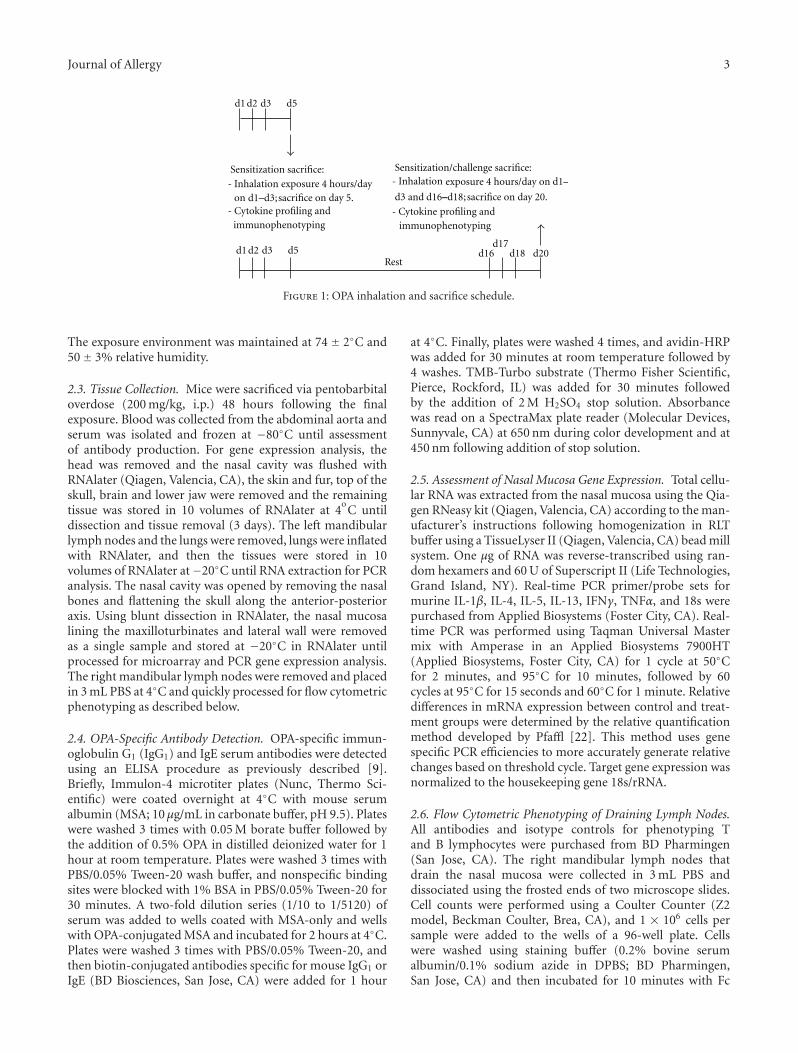
Journal of Allergy 3
d1 d2 d3 d5
d16d17
d18 d20
Sensitization/challenge sacrifice:exposure 4 hours/day on
d3 and sacrifice on day 20.
profiling andimmunophenotyping
Sensitization sacrifice:
on sacrifice on day 5.
Restd1 d2 d3 d5
exposure 4 hours/day- Inhalation
- Cytokine profiling andimmunophenotyping
- Inhalation
- Cytokine
d16–d18– ;
d1–
d1–d3;
Figure 1: OPA inhalation and sacrifice schedule.
The exposure environment was maintained at 74 ± 2◦C and50± 3% relative humidity.
2.3. Tissue Collection. Mice were sacrificed via pentobarbitaloverdose (200 mg/kg, i.p.) 48 hours following the finalexposure. Blood was collected from the abdominal aorta andserum was isolated and frozen at −80◦C until assessmentof antibody production. For gene expression analysis, thehead was removed and the nasal cavity was flushed withRNAlater (Qiagen, Valencia, CA), the skin and fur, top of theskull, brain and lower jaw were removed and the remainingtissue was stored in 10 volumes of RNAlater at 4
oC until
dissection and tissue removal (3 days). The left mandibularlymph nodes and the lungs were removed, lungs were inflatedwith RNAlater, and then the tissues were stored in 10volumes of RNAlater at−20◦C until RNA extraction for PCRanalysis. The nasal cavity was opened by removing the nasalbones and flattening the skull along the anterior-posterioraxis. Using blunt dissection in RNAlater, the nasal mucosalining the maxilloturbinates and lateral wall were removedas a single sample and stored at −20◦C in RNAlater untilprocessed for microarray and PCR gene expression analysis.The right mandibular lymph nodes were removed and placedin 3 mL PBS at 4◦C and quickly processed for flow cytometricphenotyping as described below.
2.4. OPA-Specific Antibody Detection. OPA-specific immun-oglobulin G1 (IgG1) and IgE serum antibodies were detectedusing an ELISA procedure as previously described [9].Briefly, Immulon-4 microtiter plates (Nunc, Thermo Sci-entific) were coated overnight at 4◦C with mouse serumalbumin (MSA; 10 μg/mL in carbonate buffer, pH 9.5). Plateswere washed 3 times with 0.05 M borate buffer followed bythe addition of 0.5% OPA in distilled deionized water for 1hour at room temperature. Plates were washed 3 times withPBS/0.05% Tween-20 wash buffer, and nonspecific bindingsites were blocked with 1% BSA in PBS/0.05% Tween-20 for30 minutes. A two-fold dilution series (1/10 to 1/5120) ofserum was added to wells coated with MSA-only and wellswith OPA-conjugated MSA and incubated for 2 hours at 4◦C.Plates were washed 3 times with PBS/0.05% Tween-20, andthen biotin-conjugated antibodies specific for mouse IgG1 orIgE (BD Biosciences, San Jose, CA) were added for 1 hour
at 4◦C. Finally, plates were washed 4 times, and avidin-HRPwas added for 30 minutes at room temperature followed by4 washes. TMB-Turbo substrate (Thermo Fisher Scientific,Pierce, Rockford, IL) was added for 30 minutes followedby the addition of 2 M H2SO4 stop solution. Absorbancewas read on a SpectraMax plate reader (Molecular Devices,Sunnyvale, CA) at 650 nm during color development and at450 nm following addition of stop solution.
2.5. Assessment of Nasal Mucosa Gene Expression. Total cellu-lar RNA was extracted from the nasal mucosa using the Qia-gen RNeasy kit (Qiagen, Valencia, CA) according to the man-ufacturer’s instructions following homogenization in RLTbuffer using a TissueLyser II (Qiagen, Valencia, CA) bead millsystem. One μg of RNA was reverse-transcribed using ran-dom hexamers and 60 U of Superscript II (Life Technologies,Grand Island, NY). Real-time PCR primer/probe sets formurine IL-1β, IL-4, IL-5, IL-13, IFNγ, TNFα, and 18s werepurchased from Applied Biosystems (Foster City, CA). Real-time PCR was performed using Taqman Universal Mastermix with Amperase in an Applied Biosystems 7900HT(Applied Biosystems, Foster City, CA) for 1 cycle at 50◦Cfor 2 minutes, and 95◦C for 10 minutes, followed by 60cycles at 95◦C for 15 seconds and 60◦C for 1 minute. Relativedifferences in mRNA expression between control and treat-ment groups were determined by the relative quantificationmethod developed by Pfaffl [22]. This method uses genespecific PCR efficiencies to more accurately generate relativechanges based on threshold cycle. Target gene expression wasnormalized to the housekeeping gene 18s/rRNA.
2.6. Flow Cytometric Phenotyping of Draining Lymph Nodes.All antibodies and isotype controls for phenotyping Tand B lymphocytes were purchased from BD Pharmingen(San Jose, CA). The right mandibular lymph nodes thatdrain the nasal mucosa were collected in 3 mL PBS anddissociated using the frosted ends of two microscope slides.Cell counts were performed using a Coulter Counter (Z2model, Beckman Coulter, Brea, CA), and 1 × 10⁶ cells persample were added to the wells of a 96-well plate. Cellswere washed using staining buffer (0.2% bovine serumalbumin/0.1% sodium azide in DPBS; BD Pharmingen,San Jose, CA) and then incubated for 10 minutes with Fc

4 Journal of Allergy
block (clone 2.4G2). The cells were then incubated withanti-CD3 (APC, clone 145-2C11)/anti-CD4 (FITC, cloneRM4-5)/anti-CD8 (PE, clone 53-6.7) or anti-CD45RA/B220(PE, clone RA3-6B2)/anti-IgE antibodies (FITC, clone R-35-72) or the appropriate isotype controls diluted in stainingbuffer for 30 minutes. Cells were washed, incubated withpropidium iodide (PI) for 5 minutes to stain dead cells.After a final wash, cells were resuspended in staining bufferand analyzed with a FACSCaliber flow cytometer (BectonDickinson, San Jose, CA) using a PI viability gate. Data fora total of 10,000 cells were collected based on the forward-side scatter lymphocyte gate.
2.7. Statistical Analysis. The OPA inhalation exposure systemis capable of exposing mice to a single concentration with anequivalent inhalation tower used for the concurrent controlgroup. Therefore, all concentrations for each exposureparadigm (Figure 1) required its own concurrent control,and all data analysis was performed relative to the concurrentcontrol. Treatment effects were determined using a Student’st-test comparing a single OPA concentration to the concur-rent control. Differences were considered significant at P <.05. For clarity of presentation, data have been normalizedto the concurrent control group and shown as fold change,unless otherwise stated.
3. Results
3.1. OPA Inhalation Did Not Induce Overt Signs of SystemicToxicity and Respiratory Distress. Inhalation exposure toOPA did not cause observable clinical signs of systemictoxicity or respiratory distress throughout the exposure doseresponse. All mice actively groomed following each exposureperiod and were observed to eat and drink shortly afterexposure. However, mice treated with 1000 ppb OPA for3 days followed by sacrifice on the 5th day (sensitizationexposure; Figure 1) had lost approximately 10% of theirinitial body weight versus 1.9% in the controls. Over therest period, mice regained weight at a similar rate as thecontrol group. Subsequent inhalation challenge with thesame OPA concentration (sensitization/challenge exposure;Figure 1) resulted in a smaller body weight loss (5% loss frominitial body weight). Similar body weight loss was observedin mice exposed to 500 ppb OPA but concentrations below500 ppb showed similar body weight changes as observed inthe control mice (data not shown). No mice died from OPAinhalation prior to the scheduled study termination.
3.2. Inhalation Exposure to OPA Vapor Induced the Produc-tion of OPA-Specific Antibodies. Serum was collected andevaluated to determine if mice exposed to OPA developedsystemic sensitization resulting in the production of anti-bodies specific for OPA-conjugated protein. No OPA-specificantibodies were observed in any of the control mice or miceexposed to OPA using the sensitization exposure. In contrast,inhalation challenge of mice to≥500 ppb OPA vapor resultedin OPA-specific IgG1 production. The OD450 values were1.76 ± 0.02 and 1.35 ± 0.35 for a 1/20 dilution of serum
from mice treated with 500 or 1000 ppb OPA, respectively,versus a background OD450 in the control sera of 0.086±0.02.Serum from mice treated with 500 and 1000 ppb OPA alsoshowed mild antibody specificity for native MSA as observedin our previous studies following dermal exposure to OPA[9]. OPA-specific IgE was not detected in the serum of anymice (data not shown).
3.3. OPA Inhalation Induces Cytokine Gene Expression inthe Airways and Draining Lymph Nodes. Gene expressionanalysis of key cytokine genes can provide insight intothe immunotoxicity of inhaled chemicals especially whenexamined in the target mucosal tissue lining the airways andthe associated draining lymph nodes. Pro/anti-inflammatorycytokines (IL-10, TNFα, IL-1β) are important in the initialstages of an immune response to a chemical sensitizer andcan shape the developing immune response towards Th1immunity (IFNγ), Th2 immunity (IL-4, IIL-5, IL-13), or acombination. Sensitization and allergy are supported mainlyby Th2 cytokine responses although Th1 cytokines havebeen shown to be important for chemical sensitizers [23].Lymph nodes in the cervical region drain the skin of thehead and the nasal mucosa. Since OPA is a highly reactivechemical, the mandibular lymph nodes draining the nasalmucosa, a target site, were examined for gene expressionchanges. The inhalation of OPA during sensitization resultedin concentration-dependent upregulation of IL-4 and IL-5expression (significant increases ≥500 ppb OPA; Table S1)in the mandibular lymph nodes (Figure 2). The expressionof TNFα, IL-1β, and IL-10 were also increased in the lymphnodes of mice treated with 1000 ppb OPA. Subsequent OPAchallenge for an additional 3 days resulted in increased IL-4gene expression throughout the concentration range tested(Figure 2; Table S1). The expressions of IL-10 and IL-1β werealso increased in the lymph nodes following challenge withOPA although to a lesser extent than following sensitizationalone (Figure 2; Table S1).
The inhalation of OPA also increased cytokine geneexpression in the mucosal tissue lining the upper airways(Figure 3; Table S1). Increased IL-4 expression in miceexposed to 1000 ppb OPA and a concentration-dependentstimulation of IL-1β in the nasal mucosa occurred in miceexposed to OPA during sensitization only suggesting acuteinflammation. Challenging mice after an 11-day rest periodto the same concentration of OPA vapor for an additional3 days caused marked concentration-dependent increases inall of the cytokine genes interrogated. IL-4, IL-5, and IL-1βwere upregulated at all concentrations of OPA while IL-13,IFNγ, TNFα, and IL-10 were increased at ≥500 ppb. Micethat inhaled the higher concentrations of OPA also showedincreased expression of IL-4, IL-5, IL-13, and IL-1β in thelung tissues although not statistically significant (Figure 4;Table S1).
3.4. Respiratory Exposure to OPA Induces Lymphocyte Prolif-eration and an Allergic Phenotype in the Mandibular LymphNodes. A hallmark characteristic of respiratory allergy isproliferation of lymphocytes, namely, B lymphocytes in thelymph nodes draining the target tissue. OPA inhalation

Journal of Allergy 5
0
10
20
0
20
0
5
0
5
0
5
0
5
0
5
0
5
0
5
0
5
0
5
0
5
0
10
0
Sensitizationexposure
Sensitization/challenge exposure
Fold
chan
ge(r
elat
ive
toA
IRco
ntr
ol)
IL-4
IL-5
IL-13
IFNγ
IL-10
TNFα
IL-1β
∗
∗
∗
∗
∗ ∗
∗
∗
OPA vapor concentration
Figure 2: Inhalation of OPA vapor induces the expression of Th2 and pro/anti-inflammatory cytokines in the draining lymph nodes of mice.Mice were exposed to OPA (125, 250, 500, 1000 ppb) or filtered air according to the schedule shown in Figure 1. Two days followingthe final exposure, the mandibular lymph nodes from the left side of the neck were removed and processed for gene expression analysis.Data are presented as mean (n = 5) and represent fold change relative to the concurrent control group. ∗Indicates that the cytokine wassignificantly increased at one or more of the OPA concentrations. Refer to Table S1 of the supplementary material available online at doi:10.1155/2011/751052 for the empirical gene expression data.
resulted in a concentration-dependent increase in total lym-phocytes in the mandibular lymph nodes with a maximumincrease of approximately 4 fold over the control mice(Figure 5). Both T lymphocytes and B lymphocytes wereincreased in the lymph nodes of OPA-exposed mice, how-ever, the increase in B lymphocytes was much greater. Theratio of T : B lymphocytes was reduced in a concentration-dependent manor in mice treated with OPA reflecting
the greater proliferation of B lymphocytes relative to Tlymphocytes (Table 1). The ratio of CD4:CD8 T lymphocytesin the draining lymph nodes did not change in any of thetreatment groups relative to the controls (data not shown).
3.5. OPA Inhalation Induces Isotype Switching to IgE+ B Lym-phocytes in the Draining Lymph Nodes. An important char-acteristic of an allergic response is the isotype switch to

6 Journal of Allergy
50
40
0
50
0
50
0
0
0
0
0
0
40
0
50
0
15
0
15
100
0
100
10
0
10
5
0
5
Sensitizationexposure
Fold
chan
ge(r
elat
ive
toA
IRco
ntr
ol)
IL-4
IL-5
IL-13
IFNγ
IL-10
TNFα
IL-1β
∗
∗ ∗
∗
∗
∗
∗
∗
Sensitization/challenge exposure
OPA vapor concentration
Figure 3: Inhalation of OPA vapor induces the expression of Th2, Th1, and pro/anti-inflammatory cytokines in the nasal mucosa of mice. Micewere exposed to OPA (125, 250, 500, 1000 ppb) or filtered air according to the schedule shown in Figure 1. Two days following the finalexposure, the mucosal tissue lining the maxilloturbinates and lateral wall of the nasal cavity were removed and processed for gene expressionanalysis. Data are presented as mean (n = 5) and represent fold change relative to the concurrent control group. ∗Indicates that the cytokinewas significantly increased at one or more of the OPA concentrations. Refer to Table S1 for the empirical gene expression data.
IgE producing B lymphocytes. We utilized multicolor flowcytometry to identify the B lymphocyte population in thedraining lymph nodes and enumerated the IgE+ and IgE−
populations using an anti-IgE antibody. Figure 6 shows thatinhalation exposure to OPA during sensitization only didnot induce isotype switching to IgE+ B lymphocytes at anyconcentration tested. In contrast, inhalation challenge 11days later to the same OPA concentration resulted in amarked increase in the number of B lymphocytes expressingIgE indicating isotype switching to IgE (Figure 6). Thepercentage of total B-lymphocytes producing IgE in the
mandibular lymph nodes increased from 7.32±1.08% in thecontrol group to 11.38 ± 0.99, 34.01 ± 5.06, 41.62 ± 8.72,and 22.67 ± 2.09% in mice exposed to 125, 250, 500, and1000 ppb OPA, respectively. These effects were significant atall concentrations of OPA tested.
4. Discussion
Ortho-Phthalaldehyde has been approved by the FDA forthe high-level disinfection of reusable heat sensitive medical/dental instruments [24] and is increasingly being substituted
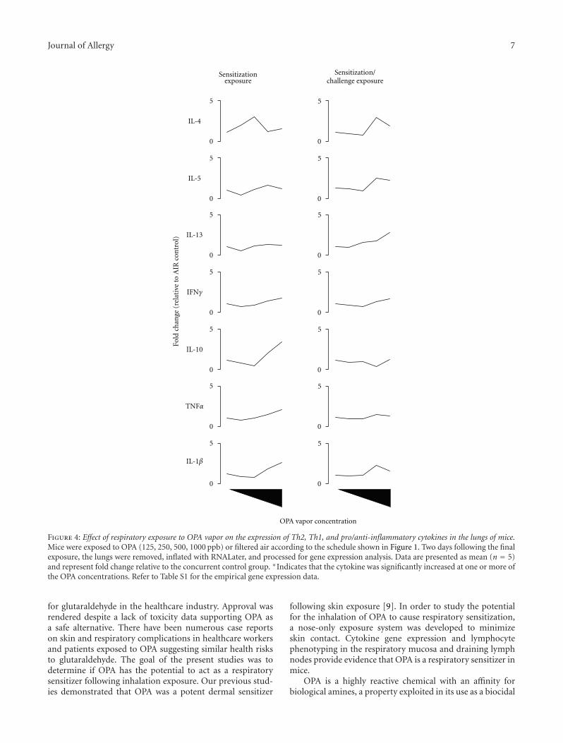
Journal of Allergy 7
Fold
chan
ge(r
elat
ive
toA
IRco
ntr
ol)
0
5
0
5
0
5
0
5
0
5
0
5
0
5
0
5
0
5
0
5
0
5
0
5
0
5
0
5
IL-4
IL-5
IL-13
IFNγ
IL-10
TNFα
IL-1β
Sensitization/challenge exposure
Sensitizationexposure
OPA vapor concentration
Figure 4: Effect of respiratory exposure to OPA vapor on the expression of Th2, Th1, and pro/anti-inflammatory cytokines in the lungs of mice.Mice were exposed to OPA (125, 250, 500, 1000 ppb) or filtered air according to the schedule shown in Figure 1. Two days following the finalexposure, the lungs were removed, inflated with RNALater, and processed for gene expression analysis. Data are presented as mean (n = 5)and represent fold change relative to the concurrent control group. ∗Indicates that the cytokine was significantly increased at one or more ofthe OPA concentrations. Refer to Table S1 for the empirical gene expression data.
for glutaraldehyde in the healthcare industry. Approval wasrendered despite a lack of toxicity data supporting OPA asa safe alternative. There have been numerous case reportson skin and respiratory complications in healthcare workersand patients exposed to OPA suggesting similar health risksto glutaraldehyde. The goal of the present studies was todetermine if OPA has the potential to act as a respiratorysensitizer following inhalation exposure. Our previous stud-ies demonstrated that OPA was a potent dermal sensitizer
following skin exposure [9]. In order to study the potentialfor the inhalation of OPA to cause respiratory sensitization,a nose-only exposure system was developed to minimizeskin contact. Cytokine gene expression and lymphocytephenotyping in the respiratory mucosa and draining lymphnodes provide evidence that OPA is a respiratory sensitizer inmice.
OPA is a highly reactive chemical with an affinity forbiological amines, a property exploited in its use as a biocidal

8 Journal of Allergy
0
1
2
3
4
5 Total lymphocytes
∗
∗
∗
∗ ∗
∗
125 250 500 1000 125 250 500 1000
Fold
chan
gefr
omco
ncu
rren
tA
IRco
ntr
ol
OPA (ppb)
Sensitizationexposure
Sensitization/challengeexpousre
(a)
0
2
4
6
8
10
125 250 500 1000 125 250 500 1000
∗
∗
∗
∗∗ ∗
∗
Fold
chan
gefr
omco
ncu
rren
tA
IRco
ntr
ol
OPA (ppb)
Sensitizationexposure
Sensitization/challengeexpousre
B lymphocytes ( CD45/B220)
(b)
0
0.5
1
1.5
2
2.5
3
125 250 500 1000 125 250 500 1000
∗
∗
∗ ∗ ∗
Fold
chan
gefr
omco
ncu
rren
tA
IRco
ntr
ol
OPA (ppb)
Sensitizationexposure
Sensitization/challengeexpousre
T lymphocytes (CD3)
(c)
CD4+ T-cells
0
0.5
1
1.5
2
2.5
3
125 250 500 1000 125 250 500 1000
Fold
chan
gefr
omco
ncu
rren
tA
IRco
ntr
ol
OPA (ppb)
∗
∗
∗∗
Sensitizationexposure
Sensitization/challengeexpousre
(d)
125 250 500 1000 125 250 500 10000
0.5
1
1.5
2
2.5
3
Fold
chan
gefr
omco
ncu
rren
tA
IRco
ntr
ol
OPA (ppb)
CD8+ T-cells
∗
∗
∗ ∗ ∗
Sensitizationexposure
Sensitization/challengeexpousre
(e)
Figure 5: OPA inhalation stimulates B- and T-lymphocyte proliferation in the draining lymph nodes of mice. Mice were exposed to OPA orfiltered air according to the schedule shown in Figure 1. Two days following the final exposure, the mandibular lymph nodes from the rightside of the neck were removed and immediately processed into single-cell suspensions. Scatter properties were used to identify lymphocytesfollowed by fluorescent antibodies labeling to identify B lymphocytes, T lymphocytes (total, CD4+ and CD8+) using flow cytometry. Data arepresented as fold change relative to the concurrent control. Statistical analysis was performed on the absolute cell counts for each lymphocytepopulation. Hatched bars represent mice that were exposed for 3 days followed by sacrifice on day 5 (sensitization exposure, Figure 1). Solidbars represent mice that were exposed on d1–d3, d16–d18 and sacrificed on d20 (sensitization/challenge exposure, Figure 1). Mean ± SEM(n = 5). ∗Significantly different from concurrent control group at P < .05. Absolute cell counts are presented in Table S2.
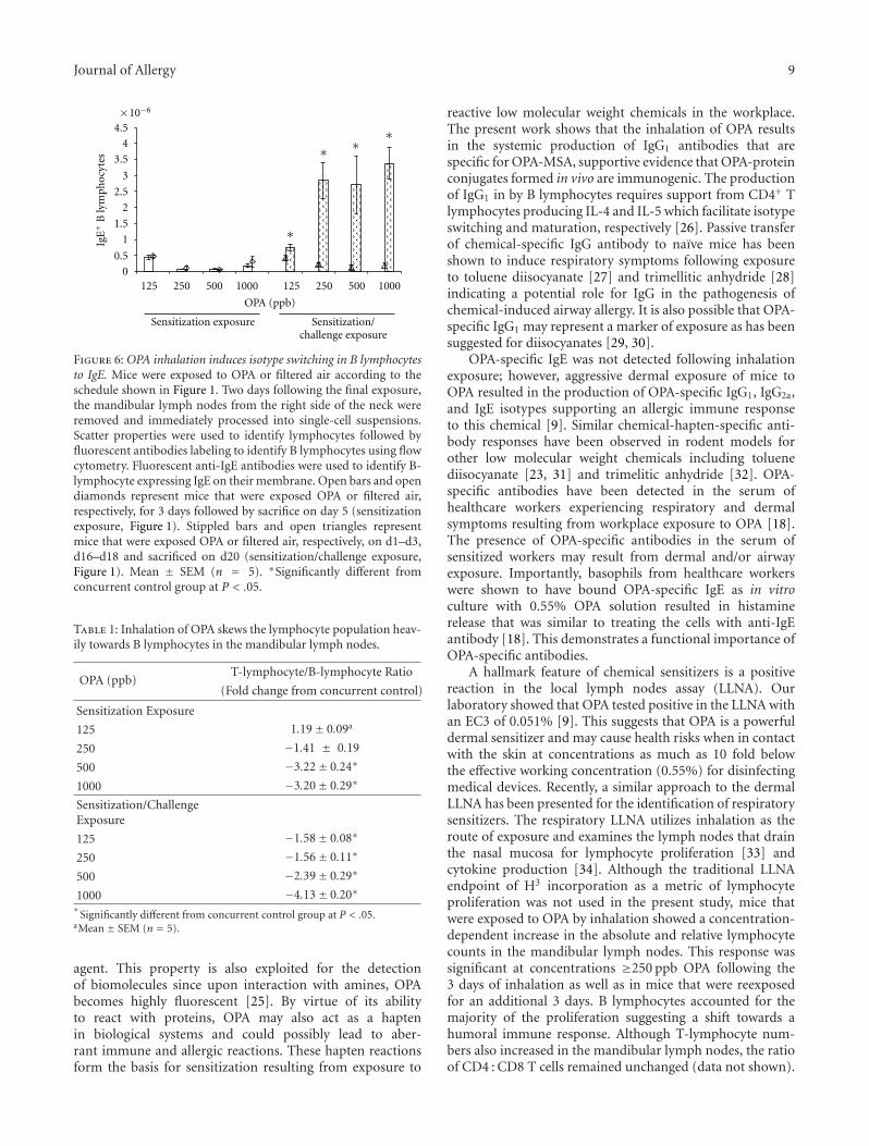
Journal of Allergy 9
0
0.5
1.5
2
2.5
3
3.5
4
4.5 ∗∗∗
∗
125 250 500 1000 125 250 500 1000
Sensitization exposure Sensitization/challenge exposure
OPA (ppb)
IgE
+
×10−6
1
Bly
mph
ocyt
es
Figure 6: OPA inhalation induces isotype switching in B lymphocytesto IgE. Mice were exposed to OPA or filtered air according to theschedule shown in Figure 1. Two days following the final exposure,the mandibular lymph nodes from the right side of the neck wereremoved and immediately processed into single-cell suspensions.Scatter properties were used to identify lymphocytes followed byfluorescent antibodies labeling to identify B lymphocytes using flowcytometry. Fluorescent anti-IgE antibodies were used to identify B-lymphocyte expressing IgE on their membrane. Open bars and opendiamonds represent mice that were exposed OPA or filtered air,respectively, for 3 days followed by sacrifice on day 5 (sensitizationexposure, Figure 1). Stippled bars and open triangles representmice that were exposed OPA or filtered air, respectively, on d1–d3,d16–d18 and sacrificed on d20 (sensitization/challenge exposure,Figure 1). Mean ± SEM (n = 5). ∗Significantly different fromconcurrent control group at P < .05.
Table 1: Inhalation of OPA skews the lymphocyte population heav-ily towards B lymphocytes in the mandibular lymph nodes.
OPA (ppb)T-lymphocyte/B-lymphocyte Ratio
(Fold change from concurrent control)
Sensitization Exposure
125 1.19± 0.09a
250 −1.41 ± 0.19
500 −3.22± 0.24∗
1000 −3.20± 0.29∗
Sensitization/ChallengeExposure
125 −1.58± 0.08∗
250 −1.56± 0.11∗
500 −2.39± 0.29∗
1000 −4.13± 0.20∗∗
Significantly different from concurrent control group at P < .05.aMean ± SEM (n = 5).
agent. This property is also exploited for the detectionof biomolecules since upon interaction with amines, OPAbecomes highly fluorescent [25]. By virtue of its abilityto react with proteins, OPA may also act as a haptenin biological systems and could possibly lead to aber-rant immune and allergic reactions. These hapten reactionsform the basis for sensitization resulting from exposure to
reactive low molecular weight chemicals in the workplace.The present work shows that the inhalation of OPA resultsin the systemic production of IgG1 antibodies that arespecific for OPA-MSA, supportive evidence that OPA-proteinconjugates formed in vivo are immunogenic. The productionof IgG1 in by B lymphocytes requires support from CD4+ Tlymphocytes producing IL-4 and IL-5 which facilitate isotypeswitching and maturation, respectively [26]. Passive transferof chemical-specific IgG antibody to naıve mice has beenshown to induce respiratory symptoms following exposureto toluene diisocyanate [27] and trimellitic anhydride [28]indicating a potential role for IgG in the pathogenesis ofchemical-induced airway allergy. It is also possible that OPA-specific IgG1 may represent a marker of exposure as has beensuggested for diisocyanates [29, 30].
OPA-specific IgE was not detected following inhalationexposure; however, aggressive dermal exposure of mice toOPA resulted in the production of OPA-specific IgG1, IgG2a,and IgE isotypes supporting an allergic immune responseto this chemical [9]. Similar chemical-hapten-specific anti-body responses have been observed in rodent models forother low molecular weight chemicals including toluenediisocyanate [23, 31] and trimelitic anhydride [32]. OPA-specific antibodies have been detected in the serum ofhealthcare workers experiencing respiratory and dermalsymptoms resulting from workplace exposure to OPA [18].The presence of OPA-specific antibodies in the serum ofsensitized workers may result from dermal and/or airwayexposure. Importantly, basophils from healthcare workerswere shown to have bound OPA-specific IgE as in vitroculture with 0.55% OPA solution resulted in histaminerelease that was similar to treating the cells with anti-IgEantibody [18]. This demonstrates a functional importance ofOPA-specific antibodies.
A hallmark feature of chemical sensitizers is a positivereaction in the local lymph nodes assay (LLNA). Ourlaboratory showed that OPA tested positive in the LLNA withan EC3 of 0.051% [9]. This suggests that OPA is a powerfuldermal sensitizer and may cause health risks when in contactwith the skin at concentrations as much as 10 fold belowthe effective working concentration (0.55%) for disinfectingmedical devices. Recently, a similar approach to the dermalLLNA has been presented for the identification of respiratorysensitizers. The respiratory LLNA utilizes inhalation as theroute of exposure and examines the lymph nodes that drainthe nasal mucosa for lymphocyte proliferation [33] andcytokine production [34]. Although the traditional LLNAendpoint of H3 incorporation as a metric of lymphocyteproliferation was not used in the present study, mice thatwere exposed to OPA by inhalation showed a concentration-dependent increase in the absolute and relative lymphocytecounts in the mandibular lymph nodes. This response wassignificant at concentrations ≥250 ppb OPA following the3 days of inhalation as well as in mice that were reexposedfor an additional 3 days. B lymphocytes accounted for themajority of the proliferation suggesting a shift towards ahumoral immune response. Although T-lymphocyte num-bers also increased in the mandibular lymph nodes, the ratioof CD4 : CD8 T cells remained unchanged (data not shown).
10 Journal of Allergy
Isotype switching to IgE is a cardinal feature of a typeI hypersensitivity response. The serum of mice exposed toOPA via inhalation was negative for IgE antibodies specificfor OPA-MSA although our previous work showed thatdermal exposure to OPA did induce OPA-specific IgE [9].The lack of systemic IgE may be due to the sensitivity of theELISA assay or may result from a less aggressive sensitizationresponse following inhalation exposure relative to dermal.Phenotyping the B-lymphocyte population in the draininglymph nodes can provide clues to the isotype specificityof the proliferating population. It has been shown that Blymphocytes from the lymph nodes of mice exposed to Th2respiratory sensitizers are positive for IgE on their plasmamembrane [35–38]. The inhalation of OPA vapor duringsensitization did not increase the population of IgE+ Blymphocytes at any concentration tested despite the dramaticproliferation observed for the B-lymphocyte population. Incontrast, there was a significant increase in the numberof IgE+ B-lymphocytes in the mandibular lymph nodes atall concentrations of OPA following OPA challenge. Nearly50% of the B lymphocytes from mice exposed to 500 ppbOPA were positive for IgE indicating isotype switching toan allergic phenotype, evidence that OPA may cause type Ihypersensitivity.
Isotype switching to IgE is regulated by Th2 cytokines,namely, IL-4 [39] which is produced primarily by CD4+
T lymphocytes [40]. There was increased proliferation ofT lymphocytes following the inhalation of OPA, and thesecells likely play an important role in shaping the humoralimmune response to this chemical. The inhalation of OPAresulted in a concentration-dependent increase in IL-4 andIL-5 gene expression in the mandibular lymph nodes evenfollowing sensitization exposure regimen. Mice that werechallenged with OPA showed significant upregulation of IL-4gene expression at all concentrations of OPA supporting classswitching to IgE in the B-lymphocyte population. In con-trast, Th2 cytokine gene expression was unaltered in the nasalmucosa and lung following sensitization exposure. Inhala-tion challenge with OPA caused a marked concentration-dependent increase in the expression of the Th2 cytokine IL-4, IL-5, and IL-13 and to a lesser degree, the Th1 cytokineIFNγ. This strong shift towards Th2 cytokine productionin the target mucosal tissue further supports an allergicresponse to OPA inhalation. In addition to these cytokines,the proinflammatory cytokine IL-1β was upregulated and isknown to play an important role in priming antigen present-ing cells. Th2 cytokine gene expression in the lungs was alsoupregulated in mice that received the challenge exposure toOPA although not statistically significant. The ability of OPAto stimulate Th2 and proinflammatory cytokines may beone mechanism responsible for its adjuvant-like propertiesobserved in a murine OVA allergy model [11].
Overall, the findings of this study demonstrate that OPAhas the potential to induce respiratory sensitization followinginhalation exposure. Characteristics of the immune responseto OPA suggest that exposure to this chemical may cause atype I hypersensitivity response as there was a large increasein the IgE+ B-lymphocyte population. The immune responseto OPA is similar to other low and high molecular weight
chemicals that are known to cause occupational rhinitis andasthma, indicating the need for the regulation of OPA inthe workplace. The concentration range tested in the presentstudy is at least 10 fold higher than air concentrations thatcan be found in the workplace [14]. Therefore, further stud-ies with lower concentrations and longer exposure periodsare important to better characterize the immunotoxicityof OPA and to facilitate risk assessment. Additionally, theunderstanding the interaction between skin and respiratoryexposure will be important for risk assessment for OPA. Pre-vious work with other low molecular weight sensitizers haveshown that skin exposure can prime the immune system torespond to future airway exposures [41–44]. There is a highpotential for skin exposure during manual reprocessing ofmedical devices with OPA. The potent sensitizing response todermal exposure [9] may significantly lower the threshold airconcentration of OPA required to elicit an immune responsein the airways. Our laboratory is currently examining theinteraction between skin and airways in the developmentof OPA-induced allergic respiratory disease using the mousemodel.
Disclosure
The findings and conclusions in this report are those of theauthors and do not necessarily represent the views of theNational Institute for Occupational Safety and Health.
Acknowledgment
These studies were funded by a Grant from the NationalOccupational Research Agenda (NORA), NIOSH. V. J.Johnson’s current address is BRT-Burleson Research Tech-nologies, Inc., Morrisville, NC 27560, USA.
References
[1] A. D. Curran, P. S. Burge, and K. Wiley, “Clinical and im-munologic evaluation of workers exposed to glutaraldehyde,”Allergy, vol. 51, no. 11, pp. 826–832, 1996.
[2] F. Di Stefano, S. Siriruttanapruk, J. McCoach, and P. S. Burge,“Glutaraldehyde: an occupational hazard in the hospitalsetting,” Allergy, vol. 54, no. 10, pp. 1105–1109, 1999.
[3] F. Di Stefano, S. Siriruttanapruk, J. S. McCoach, and P. S.Burge, “Occupational asthma due to glutaraldehyde,” MonaldiArchives for Chest Disease, vol. 53, pp. 50–55, 1998.
[4] P. F. Gannon, P. Bright, M. Campbell, S. P. O’Hickey, and P.S. Burge, “Occupational asthma due to glutaraldehyde andformaldehyde in endoscopy and X ray departments,” Thorax,vol. 50, no. 2, pp. 156–159, 1995.
[5] L. E. Maier, H. P. Lampel, T. Bhutani, and S. E. Jacob, “Handdermatitis: a focus on allergic contact dermatitis to biocides,”Dermatologic Clinics, vol. 27, no. 3, pp. 251–264, 2009.
[6] C. Lerones, A. Mariscal, M. Carnero, A. Garcıa-Rodrıguez,and J. Fernandez-Crehuet, “Assessing the residual antibacterialactivity of clinical materials disinfected with glutaraldehyde,o-phthalaldehyde, hydrogen peroxide or 2-bromo-2-nitro-1,3-propanediol by means of a bacterial toxicity assay,” ClinicalMicrobiology and Infection, vol. 10, no. 11, pp. 984–989, 2004.

Journal of Allergy 11
[7] National Institute for Occupational Safety and Health(NIOSH), “National Occupational Exposure Survey (NOES)1981–1983. Estimated Number of Employees PotentiallyExposed to Specific Agents by Occupation with 2-DigitStandard industrial Classification (SIC),” 1990.
[8] United States Environmental Protection Agency (USEPA),“Toxic Substances Control Act (TSCA) Inventory Update Re-porting (IUR), Non-confidential IUR Production VolumeInformation,” 2006, [Search used CAS No. 643798], http://www.epa.gov/oppt/iur/tools/data/2002-vol.html.
[9] S. E. Anderson, C. Umbright, R. Sellamuthu et al., “Irritancyand allergic responses induced by topical application of ortho-phthalaldehyde,” Toxicological Sciences, vol. 115, no. 2, pp.435–443, 2010.
[10] T. Morinaga, G. Hasegawa, S. Koyama, Y. Ishihara, and T.Nishikawa, “Acute inflammation and immunoresponsesinduced by ortho-phthalaldehyde in mice,” Archives of Toxicol-ogy, vol. 84, no. 5, pp. 397–404, 2010.
[11] G. Hasegawa, T. Morinaga, and Y. Ishihara, “Ortho-phthal-aldehyde enhances allergen-specific IgE production withoutallergen-specific IgG in ovalbumin-sensitized mice,” Toxicol-ogy Letters, vol. 185, no. 1, pp. 45–50, 2009.
[12] M. Ayaki, K. Shimada, S. Yaguchi, R. Koide, and A. Iwasawa,“Corneal and conjunctival toxicity of disinfectants—assessingsafety for use with ophthalmic surgical instruments,” Regula-tory Toxicology and Pharmacology, vol. 48, no. 3, pp. 292–295,2007.
[13] C. Marena, L. Lodola, A. Marone Bianco et al., “Monitoringair dispersed concentrations of aldehydes during the use ofortho-phthalaldehyde and glutaraldehyde for high disinfectionof endoscopes,” Giornale Italiano di Medicina del Lavoro edErgonomia, vol. 25, pp. 131–136, 2003.
[14] S. P. Tucker, “Determination of ortho-phthalaldehyde in airand on surfaces,” Journal of Environmental Monitoring, vol. 10,no. 11, pp. 1337–1349, 2008.
[15] H. Fujita, M. Ogawa, and Y. Endo, “A case of occupationalbronchial asthma and contact dermatitis caused by ortho-phthalaldehyde exposure in a medical worker,” Journal of Oc-cupational Health, vol. 48, pp. 413–416, 2006.
[16] W. N. Sokol, “Nine episodes of anaphylaxis following cys-toscopy caused by Cidex OPA (ortho-phthalaldehyde) high-level disinfectant in 4 patients after cytoscopy,” Journal ofAllergy and Clinical Immunology, vol. 114, pp. 392–397, 2004.
[17] D. E. Cooper, A. A. White, A. N. Werkema, and B. K. Auge,“Anaphylaxis following cystoscopy with equipment sterilizedwith Cidex OPA (ortho-phthalaldehyde): a review of twocases,” Journal of Endourology, vol. 22, no. 9, pp. 2181–2184,2008.
[18] M. Suzukawa, A. Komiya, R. Koketsu et al., “Three casesof ortho-phthalaldehyde-induced anaphylaxis after laryn-goscopy: detection of specific IgE in serum,” Allergology Inter-national, vol. 56, no. 3, pp. 313–316, 2007.
[19] M. Suzukawa, M. Yamaguchi, A. Komiya, M. Kimura, T. Nito,and K. Yamamoto, “Ortho-phthalaldehyde-induced anaphy-laxis after laryngoscopy,” Journal of Allergy and Clinical Im-munology, vol. 117, no. 6, pp. 1500–1501, 2006.
[20] A. Franchi and G. Franco, “Evidence-based decision making inan endoscopy nurse with respiratory symptoms exposed to thenew ortho-phthalaldehyde (OPA) disinfectant,” OccupationalMedicine, vol. 55, no. 7, pp. 575–578, 2005.
[21] J. Pauluhn and A. Thiel, “A simple approach to validationof directed-flow nose-only inhalation chambers,” Journal ofApplied Toxicology, vol. 27, no. 2, pp. 160–167, 2007.
[22] M. W. Pfaffl, “A new mathematical model for relative quantifi-cation in real-time RT-PCR,” Nucleic Acids Research, vol. 29,no. 9, p. e45, 2001.
[23] V. J. Johnson, B. Yucesoy, J. S. Reynolds et al., “Inhalation oftoluene diisocyanate vapor induces allergic rhinitis in mice,”Journal of Immunology, vol. 179, no. 3, pp. 1864–1871, 2007.
[24] United States Food and Drug Administration (USFDA),“FDA-Cleared Sterilants and High Level Disinfectants withGeneral Claims for Processing Reusable Medical and DentalDevices—March 2009,” 2009, http://www.fda.gov/MedicalDe-vices/DeviceRegulationandGuidance/ReprocessingofSingle-UseDevices/ucm133514.htm.
[25] J. R. Benson and P. E. Hare, “O-phthalaldehyde: fluorogenicdetection of primary amines in the picomole range. Com-parison with fluorescamine and ninhydrin,” Proceedings of theNational Academy of Sciences of the United States of America,vol. 72, no. 2, pp. 619–622, 1975.
[26] R. L. Coffman, H. F. Savelkoul, and D. A. Lebman, “Cytokineregulation of immunoglobulin isotype switching and expres-sion,” Seminars in Immunology, vol. 1, no. 1, pp. 55–63, 1989.
[27] J. M. Matheson, V. J. Johnson, V. Vallyathan, and M. I. Luster,“Exposure and immunological determinants in a murinemodel for toluene diisocyanate (TDI) asthma,” ToxicologicalSciences, vol. 84, no. 1, pp. 88–98, 2005.
[28] D. G. Fraser, F. M. Graziano, C. P. Larsen, and J. F. Regal, “Therole of IgG1 and IgG2 in trimellitic anhydride-induced allergicresponse in the guinea pig lung,” Toxicology and AppliedPharmacology, vol. 150, no. 2, pp. 218–227, 1998.
[29] A. Cartier, L. Grammer, J. L. Malo et al., “Specific serumantibodies against isocyanates: association with occupationalasthma,” Journal of Allergy and Clinical Immunology, vol. 84,no. 4, pp. 507–514, 1989.
[30] M. Dragos, M. Jones, J. L. Malo, H. Ghezzo, and D. Gautrin,“Specific antibodies to diisocyanate and work-related respira-tory symptoms in apprentice car-painters,” Occupational andEnvironmental Medicine, vol. 66, no. 4, pp. 227–234, 2009.
[31] J. M. Matheson, R. W. Lange, R. Lemus, M. H. Karol, andM. I. Luster, “Importance of inflammatory and immunecomponents in a mouse model of airway reactivity to toluenediisocyanate (TDI),” Clinical and Experimental Allergy, vol. 31,no. 7, pp. 1067–1076, 2001.
[32] X. D. Zhang, D. K. Murray, D. M. Lewis, and P. D. Siegel,“Dose-response and time course of specific IgE and IgG aftersingle and repeated topical skin exposure to dry trimelliticanhydride powder in a Brown Norway rat model,” Allergy, vol.57, no. 7, pp. 620–626, 2002.
[33] J. H. Arts, W. H. de Jong, J. J. van Triel et al., “The respiratorylocal lymph node assay as a tool to study respiratory sensitiz-ers,” Toxicological Sciences, vol. 106, no. 2, pp. 423–434, 2008.
[34] W. H. De Jong, J. H. Arts, A. De Klerk et al., “Contact andrespiratory sensitizers can be identified by cytokine profilesfollowing inhalation exposure,” Toxicology, vol. 261, no. 3, pp.103–111, 2009.
[35] T. S. Manetz and B. J. Meade, “Development of a flowcytometry assay for the identification and differentiation ofchemicals with the potential to elicit irritation, IgE- mediated,or T cell-mediated hypersensitivity responses,” ToxicologicalSciences, vol. 48, no. 2, pp. 206–217, 1999.
[36] T. S. Manetz, D. A. Pettit, and B. J. Meade, “The determinationof draining lymph node cell cytokine mRNA levels in BALB/cmice following dermal sodium lauryl sulfate, dinitrofluo-robenzene, and toluene diisocyanate exposure,” Toxicology andApplied Pharmacology, vol. 171, no. 3, pp. 174–183, 2001.

12 Journal of Allergy
[37] K. J. Klink and B. J. Meade, “Dermal exposure to 3-amino-5-mercapto-1,2,4-triazole (AMT) induces sensitization andairway hyperreactivity in BALB/c mice,” Toxicological Sciences,vol. 75, no. 1, pp. 89–98, 2003.
[38] S. Azadi, K. J. Klink, and B. J. Meade, “Divergent immunolog-ical responses following glutaraldehyde exposure,” Toxicologyand Applied Pharmacology, vol. 197, no. 1, pp. 1–8, 2004.
[39] R. S. Geha, H. H. Jabara, and S. R. Brodeur, “The regulationof immunoglobulin E class-switch recombination,” NatureReviews Immunology, vol. 3, no. 9, pp. 721–732, 2003.
[40] C. Ozdemir, M. Akdis, and C. A. Akdis, “T-cell response toallergens,” Chemical Immunology and Allergy, vol. 95, pp. 22–44, 2010.
[41] M. Kodama, K. Asano, T. Oguma et al., “Strain-specific phe-notypes of airway inflammation and bronchial hyperrespon-siveness induced by epicutaneous allergen sensitization inBALB/c and C57BL/6 mice,” International Archives of Allergyand Immunology, vol. 152, supplement 1, pp. 67–74, 2010.
[42] J. A. Vanoirbeek, V. De Vooght, H. M. Vanhooren, T. S.Nawrot, B. Nemery, and P. H. Hoet, “How long do the systemicand ventilatory responses to toluene diisocyanate persist indermally sensitized mice?” Journal of Allergy and ClinicalImmunology, vol. 121, no. 2, pp. 456–463.e5, 2008.
[43] H. S. Akei, E. B. Brandt, A. Mishra et al., “Epicutaneousaeroallergen exposure induces systemic TH2 immunity thatpredisposes to allergic nasal responses,” Journal of Allergy andClinical Immunology, vol. 118, no. 1, pp. 62–69, 2006.
[44] E. L. Petsonk, M. L. Wang, D. M. Lewis, P. D. Siegel, and B.J. Husberg, “Asthma-like symptoms in wood product plantworkers exposed to methylene diphenyl diisocyanate,” Chest,vol. 118, no. 4, pp. 1183–1193, 2000.

Hindawi Publishing CorporationJournal of AllergyVolume 2011, Article ID 238513, 8 pagesdoi:10.1155/2011/238513
Research Article
Allergic Potential and Immunotoxicity Induced by TopicalApplication of 1-Chloro-4-(Trifluoromethyl)Benzene (PCBTF)in a Murine Model
Jennifer Franko, Laurel G. Jackson, B. Jean Meade, and Stacey E. Anderson
Health Effects Laboratory Division, National Institute for Occupational Safety and Health (NIOSH), 1095 Willowdale Drive,Morgantown, WV 26505-2888, USA
Correspondence should be addressed to Jennifer Franko, [email protected]
Received 11 January 2011; Accepted 11 March 2011
Academic Editor: Gordon L. Sussman
Copyright © 2011 Jennifer Franko et al. This is an open access article distributed under the Creative Commons AttributionLicense, which permits unrestricted use, distribution, and reproduction in any medium, provided the original work is properlycited.
The purpose of the studies in this paper was to evaluate the allergic potential, immunotoxicity, and irritancy of the occupationallyrelevant chemical, 1-chloro-4-(trifluoromethyl)benzene, also known as parachlorobenzotrifluoride (PCBTF), following dermalexposure in a murine model. Evaluation of the sensitization potential, conducted using the local lymph node assay (LLNA) atconcentrations ranging from 50% to 100%, identified a dose-dependent increase in lymphocyte proliferation with a calculatedEC3 value of 53.1%. While no elevations in total or specific IgE were observed after exposure to any concentration of thechemical, significant increases in IFN-γ protein production by stimulated draining lymphoid cells were observed, indicating aT-cell-mediated response. Dermal exposure to PCBTF was not found to alter the immune response to a T-cell-dependant antigen.These results demonstrate that PCBTF has the potential to induce allergic sensitization following dermal exposure and based onLLNA results would be classified as a weak sensitizer.
1. Introduction
1-Chloro-4-(trifluoromethyl)benzene, a fluorinated toluene,also known as parachlorobenzotrifluoride (PCBTF), is achemical used as an intermediate in a wide range of organicreactions for the synthesis of dyes, pharmaceuticals, pesti-cides, insecticides and herbicides [1]. It is primarily used asa solvent in commercial surface finishes, such as vapor de-greasing, precision wipe cleaning, cold cleaning and electron-ics cleaning, and is manufactured in both pure and blendedformulations based upon specific cleaning requirements[2, 3]. It is also used as an ink solvent in the printingindustry and is a component (5–12%) of low volatile organiccompound (VOC) compliant polyurethane finishes. PCBTFis not considered to be an air toxin or ozone depleter. Ithas therefore recently been considered exempt from VOCregulations [4], which has led to an increase in its use as areplacement for other solvents previously used in the man-ufacture of a variety of commercially-available paints, inks,
and other products and finishes (Oxsol 100, OccidentalChemical Co.) [1].
PCBTF was recently nominated by the National Toxicol-ogy Program (NTP) for toxicological characterization due toits unknown chronic toxicity profile and changes in its indus-trial and consumer use [1]. In addition, its improper use ordisposal may lead to an increase in public exposure outsideof the occupational context. There are currently no Occu-pational Safety & Health Administration (OSHA), NationalInstitute for Occupational Safety and Health (NIOSH), orAmerican Conference of Governmental Industrial Hygienists(ACGIH) limits regulating PCBTF exposure [1].
Although the health effects of PCBTF have not been thor-oughly tested, epidemiological studies in workers have re-ported increases in respiratory and stomach cancers [5].Animal studies investigating the health effects of PCBTFexposure are limited. In a 13-week inhalational study in rats,no changes were identified in any measured clinical chem-istry parameter, at doses up to 252 ppm, and no adverse
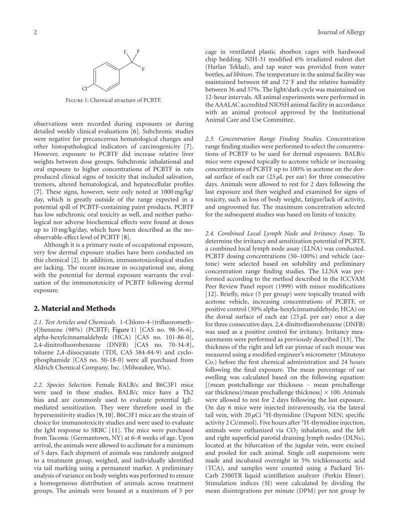
2 Journal of Allergy
Cl
F
FF
Figure 1: Chemical structure of PCBTF.
observations were recorded during exposures or duringdetailed weekly clinical evaluations [6]. Subchronic studieswere negative for precancerous hematological changes andother histopathological indicators of carcinogenicity [7].However, exposure to PCBTF did increase relative liverweights between dose groups. Subchronic inhalational andoral exposure to higher concentrations of PCBTF in ratsproduced clinical signs of toxicity that included salivation,tremors, altered hematological, and hepatocellular profiles[7]. These signs, however, were only noted at 1000 mg/kg/day, which is greatly outside of the range expected in apotential spill of PCBTF-containing paint products. PCBTFhas low subchronic oral toxicity as well, and neither patho-logical nor adverse biochemical effects were found at dosesup to 10 mg/kg/day, which have been described as the no-observable-effect level of PCBTF [8].
Although it is a primary route of occupational exposure,very few dermal exposure studies have been conducted onthis chemical [2]. In addition, immunotoxicological studiesare lacking. The recent increase in occupational use, alongwith the potential for dermal exposure warrants the eval-uation of the immunotoxicity of PCBTF following dermalexposure.
2. Material and Methods
2.1. Test Articles and Chemicals. 1-Chloro-4-(trifluorometh-yl)benzene (98%) (PCBTF; Figure 1) [CAS no. 98-56-6],alpha-hexylcinnamaldehyde (HCA) [CAS no. 101-86-0],2,4-dinitrofluorobenzene (DNFB) [CAS no. 70-34-8],toluene 2,4-diisocyanate (TDI, CAS 584-84-9) and cyclo-phosphamide [CAS no. 50-18-0] were all purchased fromAldrich Chemical Company, Inc. (Milwaukee, Wis).
2.2. Species Selection. Female BALB/c and B6C3F1 micewere used in these studies. BALB/c mice have a Th2bias and are commonly used to evaluate potential IgE-mediated sensitization. They were therefore used in thehypersensitivity studies [9, 10]. B6C3F1 mice are the strain ofchoice for immunotoxicity studies and were used to evaluatethe IgM response to SRBC [11]. The mice were purchasedfrom Taconic (Germantown, NY) at 6–8 weeks of age. Uponarrival, the animals were allowed to acclimate for a minimumof 5 days. Each shipment of animals was randomly assignedto a treatment group, weighed, and individually identifiedvia tail marking using a permanent marker. A preliminaryanalysis of variance on body weights was performed to ensurea homogeneous distribution of animals across treatmentgroups. The animals were housed at a maximum of 5 per
cage in ventilated plastic shoebox cages with hardwoodchip bedding. NIH-31 modified 6% irradiated rodent diet(Harlan Teklad), and tap water was provided from waterbottles, ad libitum. The temperature in the animal facility wasmaintained between 68 and 72◦F and the relative humiditybetween 36 and 57%. The light/dark cycle was maintained on12-hour intervals. All animal experiments were performed inthe AAALAC accredited NIOSH animal facility in accordancewith an animal protocol approved by the InstitutionalAnimal Care and Use Committee.
2.3. Concentration Range Finding Studies. Concentrationrange finding studies were performed to select the concentra-tions of PCBTF to be used for dermal exposures. BALB/cmice were exposed topically to acetone vehicle or increasingconcentrations of PCBTF up to 100% in acetone on the dor-sal surface of each ear (25 μL per ear) for three consecutivedays. Animals were allowed to rest for 2 days following thelast exposure and then weighed and examined for signs oftoxicity, such as loss of body weight, fatigue/lack of activity,and ungroomed fur. The maximum concentration selectedfor the subsequent studies was based on limits of toxicity.
2.4. Combined Local Lymph Node and Irritancy Assay. Todetermine the irritancy and sensitization potential of PCBTF,a combined local lymph node assay (LLNA) was conducted.PCBTF dosing concentrations (50–100%) and vehicle (ace-tone) were selected based on solubility and preliminaryconcentration range finding studies. The LLNA was per-formed according to the method described in the ICCVAMPeer Review Panel report (1999) with minor modifications[12]. Briefly, mice (5 per group) were topically treated withacetone vehicle, increasing concentrations of PCBTF, orpositive control (30% alpha-hexylcinnamaldehyde; HCA) onthe dorsal surface of each ear (25 μL per ear) once a dayfor three consecutive days. 2,4-dinitrofluorobenzene (DNFB)was used as a positive control for irritancy. Irritancy mea-surements were performed as previously described [13]. Thethickness of the right and left ear pinnae of each mouse wasmeasured using a modified engineer’s micrometer (MitutoyoCo.) before the first chemical administration and 24 hoursfollowing the final exposure. The mean percentage of earswelling was calculated based on the following equation:[(mean postchallenge ear thickness − mean prechallengeear thickness)/mean prechallenge thickness] × 100. Animalswere allowed to rest for 2 days following the last exposure.On day 6 mice were injected intravenously, via the lateraltail vein, with 20 μCi 3H-thymidine (Dupont NEN; specificactivity 2 Ci/mmol). Five hours after 3H-thymidine injection,animals were euthanized via CO2 inhalation, and the leftand right superficial parotid draining lymph nodes (DLNs),located at the bifurcation of the jugular vein, were excisedand pooled for each animal. Single cell suspensions weremade and incubated overnight in 5% trichloroacetic acid(TCA), and samples were counted using a Packard Tri-Carb 2500TR liquid scintillation analyzer (Perkin Elmer).Stimulation indices (SI) were calculated by dividing themean disintegrations per minute (DPM) per test group by
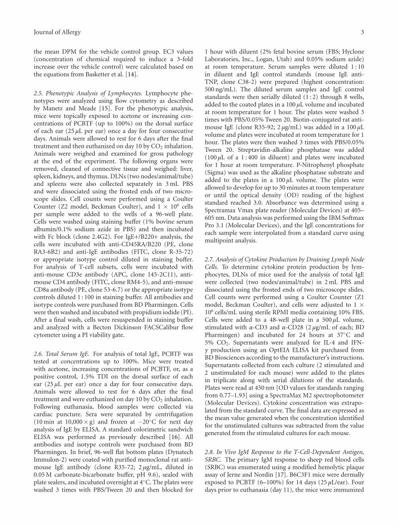
Journal of Allergy 3
the mean DPM for the vehicle control group. EC3 values(concentration of chemical required to induce a 3-foldincrease over the vehicle control) were calculated based onthe equations from Basketter et al. [14].
2.5. Phenotypic Analysis of Lymphocytes. Lymphocyte phe-notypes were analyzed using flow cytometry as describedby Manetz and Meade [15]. For the phenotypic analysis,mice were topically exposed to acetone or increasing con-centrations of PCBTF (up to 100%) on the dorsal surfaceof each ear (25 μL per ear) once a day for four consecutivedays. Animals were allowed to rest for 6 days after the finaltreatment and then euthanized on day 10 by CO2 inhalation.Animals were weighed and examined for gross pathologyat the end of the experiment. The following organs wereremoved, cleaned of connective tissue and weighed: liver,spleen, kidneys, and thymus. DLNs (two nodes/animal/tube)and spleens were also collected separately in 3 mL PBSand were dissociated using the frosted ends of two micro-scope slides. Cell counts were performed using a CoulterCounter (Z2 model, Beckman Coulter), and 1 × 10⁶ cellsper sample were added to the wells of a 96-well plate.Cells were washed using staining buffer (1% bovine serumalbumin/0.1% sodium azide in PBS) and then incubatedwith Fc block (clone 2.4G2). For IgE+/B220+ analysis, thecells were incubated with anti-CD45RA/B220 (PE, cloneRA3-6B2) and anti-IgE antibodies (FITC, clone R-35-72)or appropriate isotype control diluted in staining buffer.For analysis of T-cell subsets, cells were incubated withanti-mouse CD3e antibody (APC, clone 145-2C11), anti-mouse CD4 antibody (FITC, clone RM4-5), and anti-mouseCD8a antibody (PE, clone 53-6.7) or the appropriate isotypecontrols diluted 1 : 100 in staining buffer. All antibodies andisotype controls were purchased from BD Pharmingen. Cellswere then washed and incubated with propidium iodide (PI).After a final wash, cells were resuspended in staining bufferand analyzed with a Becton Dickinson FACSCalibur flowcytometer using a PI viability gate.
2.6. Total Serum IgE. For analysis of total IgE, PCBTF wastested at concentrations up to 100%. Mice were treatedwith acetone, increasing concentrations of PCBTF, or, as apositive control, 1.5% TDI on the dorsal surface of eachear (25 μL per ear) once a day for four consecutive days.Animals were allowed to rest for 6 days after the finaltreatment and were euthanized on day 10 by CO2 inhalation.Following euthanasia, blood samples were collected viacardiac puncture. Sera were separated by centrifugation(10 min at 10,000× g) and frozen at −20◦C for next dayanalysis of IgE by ELISA. A standard colorimetric sandwichELISA was performed as previously described [16]. Allantibodies and isotype controls were purchased from BDPharmingen. In brief, 96-well flat bottom plates (DynatechImmulon-2) were coated with purified monoclonal rat anti-mouse IgE antibody (clone R35-72; 2 μg/mL, diluted in0.05 M carbonate-bicarbonate buffer, pH 9.6), sealed withplate sealers, and incubated overnight at 4◦C. The plates werewashed 3 times with PBS/Tween 20 and then blocked for
1 hour with diluent (2% fetal bovine serum (FBS; HycloneLaboratories, Inc., Logan, Utah) and 0.05% sodium azide)at room temperature. Serum samples were diluted 1 : 10in diluent and IgE control standards (mouse IgE anti-TNP, clone C38-2) were prepared (highest concentration:500 ng/mL). The diluted serum samples and IgE controlstandards were then serially diluted (1 : 2) through 8 wells,added to the coated plates in a 100 μL volume and incubatedat room temperature for 1 hour. The plates were washed 3times with PBS/0.05% Tween 20. Biotin-conjugated rat anti-mouse IgE (clone R35-92; 2 μg/mL) was added in a 100 μLvolume and plates were incubated at room temperature for 1hour. The plates were then washed 3 times with PBS/0.05%Tween 20. Streptavidin-alkaline phosphatase was added(100 μL of a 1 : 400 in diluent) and plates were incubatedfor 1 hour at room temperature. P-Nitrophenyl phosphate(Sigma) was used as the alkaline phosphatase substrate andadded to the plates in a 100 μL volume. The plates wereallowed to develop for up to 30 minutes at room temperatureor until the optical density (OD) reading of the higheststandard reached 3.0. Absorbance was determined using aSpectramax Vmax plate reader (Molecular Devices) at 405–605 nm. Data analysis was performed using the IBM SoftmaxPro 3.1 (Molecular Devices), and the IgE concentrations foreach sample were interpolated from a standard curve usingmultipoint analysis.
2.7. Analysis of Cytokine Production by Draining Lymph NodeCells. To determine cytokine protein production by lym-phocytes, DLNs of mice used for the analysis of total IgEwere collected (two nodes/animal/tube) in 2 mL PBS anddissociated using the frosted ends of two microscope slides.Cell counts were performed using a Coulter Counter (Z1model, Beckman Coulter), and cells were adjusted to 1 ×10⁶ cells/mL using sterile RPMI media containing 10% FBS.Cells were added to a 48-well plate in a 500 μL volume,stimulated with α-CD3 and α-CD28 (2 μg/mL of each; BDPharmingen) and incubated for 24 hours at 37◦C and5% CO2. Supernatants were analyzed for IL-4 and IFN-γ production using an OptEIA ELISA kit purchased fromBD Biosciences according to the manufacturer’s instructions.Supernatants collected from each culture (2 stimulated and2 unstimulated for each mouse) were added to the platesin triplicate along with serial dilutions of the standards.Plates were read at 450 nm [OD values for standards rangingfrom 0.77–1.93] using a SpectraMax M2 spectrophotometer(Molecular Devices). Cytokine concentration was extrapo-lated from the standard curve. The final data are expressed asthe mean value generated when the concentration identifiedfor the unstimulated cultures was subtracted from the valuegenerated from the stimulated cultures for each mouse.
2.8. In Vivo IgM Response to the T-Cell-Dependent Antigen,SRBC. The primary IgM response to sheep red blood cells(SRBC) was enumerated using a modified hemolytic plaqueassay of Jerne and Nordin [17]. B6C3F1 mice were dermallyexposed to PCBTF (6–100%) for 14 days (25 μL/ear). Fourdays prior to euthanasia (day 11), the mice were immunized

4 Journal of Allergy
with 7.5 × 10⁷ SRBC by intravenous injection in a 200 μLvolume. All SRBC for these studies were drawn froma single donor animal (Lampire Laboratories, Pipersville,Penn). On the day of sacrifice, mice were euthanized byCO2 asphyxiation, body and organ weights were recordedand spleens were collected in 3 mL of Hanks Balanced SaltSolution (HBSS). Single cell suspensions of the spleens fromindividual animals were prepared in HBSS by disruptingthe spleen between the frosted ends of microscopic slides.To identify the total number of spleen cells, 20 μL of cellswere added to 10 mL of isoton buffer (1 : 500) and two dropsof Zap-o-globin were added to lyse red blood cells. Cellswere then counted using a Coulter counter. 1 : 30 and 1 : 120dilutions of spleen cells were made. One hundred μL ofthe dilutions were added to a test tube containing a 0.5 mLwarm agar/dextran mixture (0.5% Bacto-Agar, DIFCO; and0.05% DEAE dextran, Sigma), 25 μL of 1 : 1 ratio of SRBCsuspension, and 25 μL of 1 : 4 dilution (1 mL lyophilized)guinea pig complement (Cedarlane Labs). Each sample wasvortexed, poured into a petri dish, covered with a microscopecoverslip, and incubated for 3 hours at 37◦C. The plaques(representing antibody forming B-lymphocytes) were viewedand quantified after this incubation. Results were expressedas specific activity (IgM PFC per 106 spleen cells) and totalactivity (IgM PFC per spleen).
2.9. Statistical Analysis. For analysis of the data generatedfrom the described animal studies, the data were first testedfor homogeneity using the Bartlett’s Chi Square test. Ifhomogeneous, a one-way analysis of variance (ANOVA) wasconducted. If the ANOVA showed significance at P < .05or less, the Dunnett’s Multiple Range t test was used tocompare treatment groups with the control group. Lineartrend analysis was performed to determine if PCBTF hadexposure concentration-related effects for the specified end-points. Statistical analysis was performed using Graph PadPrism version 5.0 (San Diego, CA). Statistical significance isdesignated by ∗(P ≤ .05) and ∗∗(P ≤ .01).
3. Results
3.1. In Vivo Studies Identified PCBTF to Be an Allergic Sensi-tizer. Dermal exposure to PCBTF was not found to be toxicat any concentration tested (data not shown). For this reason,concentrations of PCBTF up to 100% were tested in thesubsequent studies. No ear swelling was observed in miceafter dermal exposure to PCBTF (Figure 2), suggesting thatPCBTF is nonirritating. DNFB (0.03%) was used as a positivecontrol for irritancy studies and resulted in an averagesignificant increase of 84% in ear swelling after application(data not shown). In the LLNA, dose-dependent (LinearTrend test; P < .01) increases in DLN proliferation wereobserved after treatment with PCBTF, with counts from the75% and 100% PCBTF exposed animals being significantlyelevated over the vehicle control animals (Figure 3). SI valueswere 2.6, 5.3, and 5.3 for the 50%, 75%, and 100% exposuregroups, respectively. An EC3 value of 53.1% (Figure 3) wascalculated. HCA (30%) was used as a positive control for
10075500
PCBTF (v/v) (%)
0
5
10
15
20
Incr
ease
inea
rsw
ellin
g(%
)
Figure 2: Ear swelling as a result of topical application of PCBTF.Analysis of irritation after topical application of PCBTF. Barsrepresent mean ± SE of 5 mice (10 ears) per group.
10075500
PCBTF (v/v) (%)
0
1000
2000
3000
dpm
/2n
odes
EC3 = 53.1%
2.6
5.35.3
∗∗∗∗
Figure 3: Allergic sensitization potential after dermal exposure toPCBTF. Analysis of the allergic sensitization potential of PCBTFusing the LLNA. 3H-thymidine incorporation into draining lymphnode cells of BALB/c mice following exposure to vehicle or con-centration of PCBTF. Numbers in boxes appearing above the barsrepresent the stimulation indices for each concentration tested. Barsrepresent mean ± SE of 5 mice per group. Levels of statisticalsignificance are denoted ∗∗(P < .01) as compared to acetonevehicle.
these experiments and resulted in an average SI value of 24.5(data not shown). Consistent with the LLNA results, PCBTFexposure also significantly elevated the cellularity of the DLN(Figure 4) following exposure to all concentrations.
3.2. Exposure to PCBTF Did Not Induce an Increase in Localor Systemic IgE Levels. No changes in body or organ weightswere observed after exposure to PCBTF for these studies(data not shown). The mechanisms of PCBTF sensitizationwere further investigated using phenotypic analysis of B220+and IgE+B220+ expressing cells in the DLNs and spleen. Noincreases were observed in the B220+ cell or IgE+B220+ cellpopulations (Table 1) in the DLN or spleen. No changes inthe percentage of CD4+ or CD8+ T cells in the DLN or

Journal of Allergy 5
10075500
PCBTF (v/v) (%)
0
0.2
0.4
0.6
0.8
1
Cel
ls/D
LN
(×10
6)
∗∗ ∗∗∗
Figure 4: Increased cellularity of DLN after PCBTF exposure.Analysis of lymphocyte cellularity (cells/DLN) following exposureto PCBTF. Bars represent means± SE of 5 mice per group. Levels ofstatistical significance are denoted as ∗(P ≤ .05) and ∗∗(P ≤ .01) ascompared to acetone vehicle.
10075500
PCBTF (v/v) (%)
0
200
400
600
800
1000
Tota
lIgE
(ng/
mL
)
Figure 5: Lack of increase in serum IgE following PCBTF exposure.Analysis of total serum IgE after exposure to PCBTF. Bars representmeans ± SE of 5 mice per group.
spleen were observed after exposure to any concentration ofPCBTF (data not shown). Consistent with the IgE+B220+results, exposure to PCBTF did not elevate total serum IgElevels after exposure to any treatment groups (Figure 5).Dermal exposure to the respiratory sensitizer TDI (1.5%)significantly elevated the B220+ (19.8 ± 2.3%) cell popula-tion, IgE+B220+ (13.3 ± 2.8%) cell populations, and totalIgE (1587 ± 109 ng/mL) levels (data not shown).
3.3. Exposure to PCBTF Increased Production of IFN-γ, butNot IL-4 by Stimulated DLN Cells. Levels of IL-4 and IFN-γcytokine production by stimulated draining lymphoid cellswere analyzed to evaluate the effect of PCBTF exposureon Th1/Th2 balance. A dose-responsive (Linear Trend test;P < .01) increase in IFN-γ protein production by the DLNwas observed after dermal exposure to PCBTF. Significantelevations in cytokine production were observed at PCBTF
concentrations 50% and greater (Figure 6(a)). The maxi-mum increase in INF-γ protein expression was calculatedto be 1,174 ± 169 pg/mL. No significant alterations inIL-4 production were detected at any dose concentration(Figure 6(b)). To confirm the lack of Th2 cytokine inductionfollowing PCBTF exposure, cytokine mRNA levels werealso analyzed in the DLNs of dermally exposed animals.Consistent with the protein data, no increase in IL-4 orIL-13 expression was observed (data not shown). Dermaltreatment with 1.5% TDI elevated IL-4 and IL-13 mRNAexpression in the DLN and significantly enhanced IL-4protein production by stimulated draining lymphoid cells(496 ± 20 pg/mL) (data not shown). The T-cell-mediatedsensitizer HCA (30%) significantly elevated IFN-γ expres-sion (2,122 ± 67 pg/mL) (data not shown).
3.4. Dermal Exposure to PCBTF Did Not Alter the IgM Re-sponse to SRBC. To evaluate immunosuppressive potential,the murine splenic IgM response to SRBC was examined fol-lowing a 14-day exposure to PCBTF. No changes in total(PFC/spleen) or specific (PFC/106 cells) IgM antibodyactivity to SRBC were observed after exposure to any con-centration of PCBTF (Figure 7). Animals exposed to the pos-itive control, cyclophosphamide, had a significantly reducedspecific spleen IgM response (67%) and total IgM response(54%) compared to the acetone control animals. No changesin body or organ weights were observed for these animals(data not shown).
4. Discussion
More than 13 million workers in the United States arepotentially exposed to chemicals that can be absorbedthrough the skin. Dermal exposure to chemicals in theworkplace has a great potential to affect immune function.Contact dermatitis is the second most commonly reportedoccupational illness responsible for up to 30% of all casesof occupational disease in industrialized nations [18]. Thismay result in considerable social and economic implications,including time off from work, loss of workplace productivity,reduced quality of life, and medical and worker’s compensa-tion costs, accounting for the loss of billions of dollars [19].
Results from these studies suggest that PCBTF is a weakT-cell-mediated sensitizer. This was evidenced by an increasein lymphocyte proliferation and IFN-γ protein productionby stimulated draining lymphoid cells, in the absence ofelevations in markers of IgE-mediated sensitization, suchas total IgE and IL-4 (mRNA and protein) production,following dermal exposure in mice. The EC3 value forPCBTF, 53.1%, falls within the working concentration forthis chemical, as it is used at concentrations up to 100% asa solvent and as a chemical intermediate in the productionof other chemicals. This suggests a greater predicted riskfor worker sensitization. Immune suppression and otherimmunotoxic effects were not observed after exposure to thischemical.
Although exposure can occur through inhalation anddermal contact during its production and use [2], there
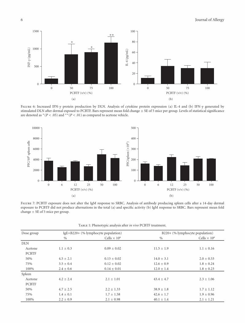
6 Journal of Allergy
10075500
PCBTF (v/v) (%)
0
500
1000
1500IN
F-γ
(pg/
mL
)
∗∗
∗∗
(a)
10075500
PCBTF (v/v) (%)
0
20
40
60
80
100
IL-4
(pg/
mL
)
(b)
Figure 6: Increased IFN-γ protein production by DLN. Analysis of cytokine protein expression (a) IL-4 and (b) IFN-γ generated bystimulated DLN after dermal exposed to PCBTF. Bars represent mean fold change ± SE of 5 mice per group. Levels of statistical significanceare denoted as ∗(P < .05) and ∗∗(P < .01) as compared to acetone vehicle.
10050251260
PCBTF (v/v) (%)
0
2000
4000
6000
8000
10000
PFC
/106
sple
ence
lls
(a)
10050251260
PCBTF (v/v) (%)
0
100
200
300
400
500P
FC/s
plee
n(×
103)
(b)
Figure 7: PCBTF exposure does not alter the IgM response to SRBC. Analysis of antibody producing spleen cells after a 14-day dermalexposure to PCBTF did not produce alternations in the total (a) and specific activity (b) IgM response to SRBC. Bars represent mean foldchange ± SE of 5 mice per group.
Table 1: Phenotypic analysis after in vivo PCBTF treatment.
Dose group IgE+B220+ (% lymphocyte population) B220+ (% lymphocyte population)
% Cells × 106 % Cells × 106
DLN
Acetone 1.1 ± 0.3 0.09 ± 0.02 11.5 ± 1.9 1.1 ± 0.16
PCBTF
50% 4.5 ± 2.1 0.13 ± 0.02 14.0 ± 3.1 2.0 ± 0.53
75% 3.5 ± 0.4 0.12 ± 0.02 12.6 ± 0.9 1.8 ± 0.24
100% 2.4 ± 0.6 0.14 ± 0.01 12.0 ± 1.4 1.8 ± 0.23
Spleen
Acetone 4.2 ± 2.4 2.1 ± 1.01 43.4 ± 4.7 2.3 ± 1.06
PCBTF
50% 4.7 ± 2.5 2.2 ± 1.33 38.9 ± 1.8 1.7 ± 1.12
75% 1.4 ± 0.1 1.7 ± 1.58 42.6 ± 1.7 1.9 ± 0.96
100% 2.2 ± 0.9 2.1 ± 0.98 40.1 ± 1.4 2.1 ± 1.21
Journal of Allergy 7
are no standards in place to limit occupational exposure toPCBTF [1]. For occupational exposures, an 8-hour time-weighted average (TWA) permissible exposure level of20 ppm has been suggested by the Kowa American Corpora-tion, the United States chemical importation company thathas renominated PCBTF for toxicological evaluation [1, 6].In additional to occupational exposure, there is also thepotential for exposure of the general public through PCBTF-containing products and PCBTF in and around ground water[20]. Detectable levels of PCBTF have been identified inworkers (<1 ppm) as well as wildlife (0.17–2.0 ppm) [21, 22].
PCBTF is a halogenated solvent. These types of solventsare usually considered more toxic to humans and usuallycapable of causing greater environmental damage. Examplesof other halogenated solvents include trichloroethylene,tetrachloroethylene, 1,1,1-trichloroethane, carbon tetrachlo-ride, and dichloromethane. The majority of these solventsare classified as carcinogens and central nervous systemdepressants and can enter the body through respiratoryor dermal exposure [23]. In addition to the effects to thecentral nervous system, workplace exposure to many of thesesolvents have been associated with toxic effects in the liverand kidney [24], as well as immune cell activation [25–31].The effects of PCBTF exposure on immune parameters areconsistent with those for other halogenated solvents, such astrichloroethylene, which has been shown to enhance activa-tion markers on and IFN-γ secretion by splenic CD4+ T-cells[25–27]. Enhanced T-cell activation due to trichloroethyleneexposure has been demonstrated to enhance autoimmune-related responses in both humans and mouse models [28,29]. Due to the wide range of health effects associated withhalogenated solvent exposure, occupational exposure limitshave been set for these compounds, which have resulted inmore stringent ventilation controls and personal protectiveequipment use by workers.
5. Conclusion
These are the first studies to indentify the sensitization poten-tial of PCBTF following dermal exposure. Results from thesestudies indicate the importance of avoiding dermal contactwith PCBT, due to its potential to function as a T-cell-mediated sensitizer and encourages the need to determineappropriate PPE to prevent worker exposure.
Disclosure
The findings and conclusions in this report are those of theauthors and do not necessarily represent the views ofthe National Institute for Occupational Safety and Health,Centers for Disease Control and Prevention.
References
[1] NTP, National Toxicology Program, Chemical InformationProfile for 1-Chloro-4-(trifluoromethyl)benzene. NationalInstitute of Environmental Health Sciences, Research TrianglePark, NC, USA, 2009.
[2] HSDB, “Hazardous Substances Data Bank. 1-Chloro-4-(trifluoromethyl)benzene,” HSDB Record No. 4251, February2003, http://toxnet.nlm.nih.gov/cgi-bin/sis/search/f?./temp/∼CkwdFP:4.
[3] J. J. Maul, P. J. Ostrowski, G. A. Ublacker, B. Linclau, and D.P. Curran, “Benzotrifluoride and derivatives: useful solventsfor organic synthesis and fluorosynthesis,” Topics in CurrentChemistry, vol. 206, pp. 79–105, 1999.
[4] EPA, U.S., “National volatile organic comound emissin stan-dards for aerosol coatings,” Federal Register, vol. 74, no. 62, pp.14941–14949, 2009.
[5] OCC, Occidental Chemical Corporation. Initial submission:mortality patterns of workers int he Niagara Plant (finalreport on mixtures of chemical substances) with attachmentsand cover letter dated 022192. TSCATS [Unpublished Healthand Safety Studies submitted to EPA]. U.S. EPA/OPTS PublicFiles. Microfiche No. 0537001. Document No. 88-920001103.Record from Biblioline Basic, ECIS Record ID: TS-045214;abstract from TSCATS Low Detail Report link available atU.S. EPA, 1983, http://yosemite.epa.gov/oppts/epatscat8.nsf/ReportSearchView/915F0B8E49E020F185257252006EAB2A.
[6] P. E. Newton, H. F. Bolte, W. R. Richter, M. B. Akinsanya, J.B. Knaak, and L. W. Smith, “Inhalation toxicity, neurotoxicity,and toxicokinetic studies of p-chlorobenzotrifluoride,” Inhala-tion Toxicology, vol. 10, no. 1, pp. 33–48, 1998.
[7] Hooker Chemical Company, “Acute oral toxicity (LD50) inalbine rats,” TSCATS [Unpublished Health and Safety Studiessubmitted to EPA]. U.S. EPA/OPTS Public Files. MicroficheNo. 0508138. Document No. 40-7952012, 1979.
[8] A. Macri, C. Ricciardi, A. V. Stazi et al., “Subchronic oraltoxicity of 4-chloro-alpha, alpha, alpha-trifluorotoluene inSprague-Dawley rats,” Food and Chemical Toxicology, vol. 25,no. 10, pp. 781–786, 1987.
[9] K. J. Klink and B. J. Meade, “Dermal exposure to 3-amino-5-mercapto-1,2,4-triazole (AMT) induces sensitization andairway hyperreactivity in BALB/c mice,” Toxicological Sciences,vol. 75, no. 1, pp. 89–98, 2003.
[10] M. R. Woolhiser, A. E. Munson, and B. J. Meade, “Comparisonof mouse strains using the local lymph node assay,” Toxicology,vol. 146, no. 2-3, pp. 221–227, 2000.
[11] M. I. Luster, C. Portier, D. G. Pait et al., “Risk assessmentin immunotoxicology. I. Sensitivity and predictability ofimmune tests,” Fundamental and Applied Toxicology, vol. 18,no. 2, pp. 200–210, 1992.
[12] NIEHS, “National Institute of Environmental Health Sciences;the murine local lymph node assay: a test method forassessing the allergic contact dermatitis potential of chemicals/compounds,” Federal Register, vol. 64, pp. 14006–14007, 1999.
[13] M. R. Woolhiser, A. E. Munson, and B. J. Meade, “Role of sen-sitization routes in the development of type I hypersensitivityto natural rubber latex in mice,” American Journal of IndustrialMedicine, vol. 36, no. 1, pp. 139–141, 1999.
[14] D. A. Basketter, L. J. Lea, A. Dickens et al., “A comparisonof statistical approaches to the derivation of EC3 values fromlocal lymph node assay dose responses,” Journal of AppliedToxicology, vol. 19, no. 4, pp. 261–266, 1999.
[15] T. S. Manetz and B. J. Meade, “Development of a flow cytome-try assay for the identification and differentiation of chemicalswith the potential to elicit irritation, IgE- mediated, or T cell-mediated hypersensitivity responses,” Toxicological Sciences,vol. 48, no. 2, pp. 206–217, 1999.
[16] J. E. Butler, “Enzyme-linked immunosorbent assay,” Journal ofImmunoassay, vol. 21, no. 2-3, pp. 165–209, 2000.

8 Journal of Allergy
[17] N. K. Jerne and A. A. Nordin, “Plaque formation in agar bysingle antibody-producing cells,” Science, vol. 140, no. 3565, p.405, 1963.
[18] B. D. Lushniak, “The importance of occupational skin diseasesin the United States,” International Archives of Occupationaland Environmental Health, vol. 76, no. 5, pp. 325–330, 2003.
[19] C. A. Burnett, B. D. Lushniak, W. McCarthy, and J. Kaufman,“Occupational dermatitis causing days away from work inU.S. private industry, 1993,” American Journal of IndustrialMedicine, vol. 34, no. 6, pp. 568–573, 1998.
[20] D. Botta and E. Mantica, “Micropollutants migration fromion-exchange resins into water,” Water Research, vol. 33, no.4, pp. 1054–1064, 1999.
[21] EPA, U.S., “4-chlorobenzotrifluoride; decision not to test,”Federal Register, vol. 50, no. 292, pp. 42216–42221, 1985.
[22] M. P. Yurawecz, “Gas-liquid chromatographic and massspectrometric indentification of chlorinated trifluorotolueneresidues in Niagara River fish,” Journal of the Association ofOfficial Analytical Chemists, vol. 62, no. 1, pp. 36–40, 1979.
[23] NIOSH, “Control of Exposure to Perchloroethylene in Com-mercial Drycleaning,” Hazard Controls: Publication 97-157.National Institute for Occupational Safety and Health, 1997,http://www.cdc.gov/niosh/hc19.html.
[24] R. O. Recknagel, “Carbon tetrachloride hepatotoxicity,” Phar-macological Reviews, vol. 19, no. 2, pp. 145–208, 1967.
[25] K. M. Gilbert, J. M. Griffin, and N. R. Pumford, “Trichloroeth-ylene activates CD4+ T cells: potential role in an autoimmuneresponse,” Drug Metabolism Reviews, vol. 31, no. 4, pp. 901–916, 1999.
[26] J. M. Griffen, S. J. Blossom, S. K. Jackson, K. M. Gilbert, and N.R. Pumford, “Triethylen accelerates an autoimmune responseby Th1 T cell actiation in MRL+/+ mice,” Immunopharmacol-ogy, vol. 46, pp. 123–137, 2000.
[27] J. M. Griffin, K. M. Gilbert, L. W. Lamps, and N. R. Pumford,“CD4 T-cell activation and induction of autoimmune hepatitisfollowing trichloroethylene treatment in MRL+/+ mice,”Toxicological Sciences, vol. 57, no. 2, pp. 345–352, 2000.
[28] I. Iavicoli, A. Marinaccio, and G. Carelli, “Effects of occupa-tional trichloroethylene exposure on cytokine levels in work-ers,” Journal of Occupational and Environmental Medicine, vol.47, no. 5, pp. 453–457, 2005.
[29] G. S. Cooper, S. L. Makris, P. J. Nietert, and J. Jinot, “Evidenceof autoimmune-related effects of trichloroethylene exposurefrom studies in mice and humans,” Environmental HealthPerspectives, vol. 117, no. 5, pp. 696–702, 2009.
[30] M. Seo, T. Yamagiwa, R. Kobayashi et al., “A small amount oftetrachloroethylene ingestion from drinking water acceleratesantigen-stimulated allergic responses,” Immunobiology, vol.213, no. 8, pp. 663–669, 2008.
[31] D. Jırova, I. Sperlingova, M. Halaskova, H. Bendova, and L.Dabrowska, “Immunotoxic effects of carbon tetrachloride—the effect on morphology and function of the immune systemin mice,” Central European Journal of Public Health, vol. 4, no.1, pp. 16–20, 1996.
Hindawi Publishing CorporationJournal of AllergyVolume 2011, Article ID 365683, 9 pagesdoi:10.1155/2011/365683
Review Article
Occupational Asthma in Antibiotic Manufacturing Workers: CaseReports and Systematic Review
Sara Dıaz Angulo,1 Joanna Szram,2 Jenny Welch,2 Julie Cannon,2 and Paul Cullinan2
1 Centro Medico de Asturias, Avendia Jose Marıa Richard, Oviedo, 33193 Asturias, Spain2 Department of Occupational & Environmental Medicine, National Heart & Lung Institute (Imperial College) and Royal BromptonHospital, Sydney Street, London SW3 6NP, UK
Correspondence should be addressed to Sara Dıaz Angulo, [email protected]
Received 27 November 2010; Accepted 20 January 2011
Academic Editor: Gordon L. Sussman
Copyright © 2011 Sara Dıaz Angulo et al. This is an open access article distributed under the Creative Commons AttributionLicense, which permits unrestricted use, distribution, and reproduction in any medium, provided the original work is properlycited.
Background. The risks of occupational asthma (OA) from antibiotics are uncertain. We report 4 new cases and a systematic reviewof the literature. Methods. Cases were identified through a specialist clinic, each underwent specific provocation testing (SPT). Wesubsequently reviewed the published literature. Results. The patients were employed in the manufacture of antibiotics; penicillinswere implicated in three cases, in the fourth erythromycin, not previously reported to cause OA. In two, there was evidence ofspecific IgE sensitisation. At SPT each developed a late asthmatic reaction and increased bronchial hyperresponsiveness. 36 casereports have been previously published, 26 (citing penicillins or cephalosporins). Seven cross-sectional workplace-based surveysfound prevalences of 5–8%. Conclusions. OA in antibiotic manufacturers may be more common than is generally recognised.Its pathogenesis remains unclear; immunological tests are of uncertain value and potential cases require confirmation with SPT.Further study of its frequency, mechanisms, and diagnosis is required.
1. Introduction
Among the several hundred workplace agents implicated ascauses of occupational asthma (OA), several are encounteredin the pharmaceutical manufacturing industry [1]. Theseinclude antibiotics which, through inhalation, may induceasthma in exposed employees. The role of dermal exposureand the detailed pathogenesis of the condition—as withmany other low molecular mass causes of OA—remainunclear. Immunological tests are of uncertain value and mostdiagnoses require confirmation with specific provocationtesting.
The first case of occupational antibiotic allergy wasdescribed in 1953, but a relatively small number of individualcase reports have been published subsequently. Very largequantities of antibiotics are produced in almost everycountry but the frequency of OA in those who manufacturethem remains unclear. While prevalence estimates of around10% have been reported [2–4], clinical experience suggestsa far less common problem. Here we report three new casesof occupationally-induced penicillin allergy and, for the first
time, a case of asthma induced by work in erythromycinmanufacture. The reports are supplemented by a systematicliterature review of antibiotic occupational asthma.
2. Methods
2.1. Case Reports. New cases were seen and diagnosed atRoyal Brompton Hospital in London, UK between 1995 and2009. Specific IgE measurements, where attempted, werecarried out by either the radioallergosorbent test (RAST)method or the commercial ImmunoCAP assay. Each patientunderwent controlled, single-blind, specific provocationtesting using a dust-tipping method, with small quantities ofthe relevant antibiotic powder mixed with a larger amountof dried lactose; active and control exposures were carriedout on sequential days with the patients having stoppedany asthma medications prior to the tests. Responses wereassessed using serial FEV1 measurements following challengeand by changes in bronchial responsiveness to inhaledhistamine using the Yan technique [5].

2 Journal of Allergy
2.2. Systematic Review of Literature. We were not able toidentify any previous systematic review of this subject. Wesearched the published literature using the Medline databasebetween 1953 and February 2010. Both key word- and textword-based searches were performed with combinationsof the terms “occupational asthma”, “asthma”, “respiratorysensitisation”, and “antibiotics”. In addition, we examined thereference lists of relevant articles. Both case reports (n =21 papers) and workforce studies (n = 7) were retrieved(one case report in Russian was not included.) and theirinformation extracted on to a standard form independentlyby two reviewers.
We used a quantitative structure-activity relationship(qSAR) model to examine the potential for each publishedantibiotic to act as a respiratory sensitising agent. “Hazardindices” for each were calculated, where possible, using theChemical Asthma Hazard Assessment Program [6] whichgenerates a risk prediction for organic compounds with amolecular mass of less than 1 kDa.
3. Findings
3.1. Case Reports (Table 1, Figure 1). Each of the four caseswas involved in the primary manufacture or formulationof antibiotics in the United Kingdom and had presentedwith new onset, work-related asthmatic symptoms. Threeworked with penicillins and their derivatives, the fourth witha variety of medicines including erythromycin. This last, awoman of 52, developed asthma 22 years previously, twoyears after starting work on the packaging lines. She reportedthat her symptoms worsened with exposure to granulatederythromycin and on two occasions in the year prior toreferral had required treatment with oral corticosteroids. Atreferral, she had normal spirometry, was demonstrated to beatopic on skin prick testing, and completed a series of peakflow measurements which showed significant variability onboth work and rest days, with no clear work-related pattern.
All four patients underwent single-blind, controlledspecific provocation testing using a dust-tipping method.Each of the four developed a (predominantly) late asthmaticreaction (Figure 1) accompanied by increased bronchialhyperresponsiveness as demonstrated by a fall in histaminePC20 concentration following challenge; these findings wererepeatable on further testing but not seen after identicalexposure to lactose powder alone. Following diagnosis, eachpatient avoided further exposure at work to the causativeantibiotic with improvement or resolution of their symp-toms. They were advised to avoid therapeutic use of therelevant antibiotic.
3.2. Literature Review (Table 2). Previous published casereports, including a total of 37 patients, are summarisedin Table 2. All but one of the cases was employed in themanufacture of antibiotics. In 25 individuals, the diagnosiswas supported by specific inhalation testing which promptedearly, late, and dual asthmatic responses in 14 (56%), six(24%), and three (12%) patients, respectively, with theresults of the remaining two positive tests being unspecified.
A hazard index was calculated for all bar two of theimplicated antibiotics (the exceptions being vancomycin andcolomycin, both of which have a molecular mass greater than1 kDa) and was in all cases between 0.99 and 1.00.
Three reports describe patients sensitised to penicillins.In the first, published in 1953, Eaton Roberts describedtwo employees in a US factory with clinical evidence ofOA ascribed to penicillin; apart from skin prick testingwith procaine penicillin (negative), he did not perform anyobjective investigations [7]. Similarly, Tara in 1957 [8] andGaultier et al. in 1960 [9] reported a total of six Frenchworkers with clinical evidence of new-onset asthma afterexposure to penicillin dust.
Synthetic penicillins have also been reported as a causeof work-related asthma. Davies et al. [10] used specificprovocation testing to confirm OA from inhaled ampicillinin three UK employees; in each case skin prick testswith ampicillin and a variety of penicillin antigens werenegative. Oral challenge with ampicillin induced asthmaticand other allergic symptoms in two of the three cases. Losadaet al. described two cases of dyspnoea after inhalation ofsemisynthetic penicillins in workers in the manufacture ofantibiotics [11]; in neither case was there objective evidenceof immune sensitisation to these antibiotics. In Germany,Wuthrich and Hartmann [12] described a single case of OAfrom ampicillin; specific IgE antibodies to benzylpenicilloylwere detected and cumulative inhalation testing with lac-tose, tetracycline, ampicillin, and chloramphenicol causeda delayed asthmatic reaction. A factory worker in Belgiumdeveloped OA from amoxicillin [13] and subsequently, whileemployed as a nurse, to latex. Piperacillin, a semisyntheticpenicillin, has also been reported to cause OA, the diagnosissupported by both skin prick testing and an immediateasthmatic response to specific inhalational testing [26].
There are several reports of cephalosporins and asso-ciated precursors and derivatives as causative agents forOA; 11 cases are summarised in Table 2 with supportingevidence from specific provocation testing in ten, albeit mostfrequently reported as immediate asthmatic responses only.Serum and skin prick tests of immunological sensitisationwere only occasionally positive [15, 19, 20], and oralchallenges, both positive, were reported in just two patients[11, 18].
The remaining eight reported cases developed OAduring the manufacture of antibiotics other than penicillinsor cephalosporins. Most have been in workers exposed tothe macrolide spiramycin, an antibiotic widely used in thelivestock industry and particularly with poultry so thattraces may be found in some chicken eggs. Davies andPepys [23] described the case of a pharmaceutical worker inwhom the diagnosis of OA from spiramycin was confirmedby specific inhalation challenge; he reported symptoms oneating eggs but had negative skin prick tests to egg extracts.Six other cases, in Canadian and Italian manufacturers[4, 25], have been reported, in each case following specificinhalation testing. In one case [25], an immediate asthmaticresponse to provocation testing was also elicited by adipicacid, an additive used to bind spiramycin. In the only caseof reported antibiotic OA outside the manufacturing sector,

Journal of Allergy 3
Table 1: Cases of occupational asthma from antibiotics identified at Royal Brompton Hospital in the period 1995–2009.
caseYear of
diagnosisWorkplaceexposure
Allergicsymptoms
Latency Specific IgE Bronchial provocation test
Agent FEV1 responseIncrease inhistaminereactivity
a 1995 penicillin wheeze 19 years not done penicillin late Yes
b 1996 amoxicillin wheeze, cough 27 yearspenicilloyl G (+)penicilloyl V (+)
amoxicillin late yes
c 2000 amoxicillin wheeze, cough 27 years amoxicilloyl (+) amoxicillin late yes
d 2009 erythromycinwheeze,rhinitis
2 yearserythromycinethylsuccinate
(−)
erythromycinethylsuccinate
late Yes
3.1
0.35
0.17
−45
−30
−15
0
15
30
0 5 10 15 30 60 2 3 4 5 6 7 8 9 10 11 12 13 14 15 16
Control6 APA 1% 20 mins6 APA 2% 20 mins
Ch
ange
inba
selin
eFE
V1
(%)
(mins) Time after challenge (hours)
(a)
−45
−30
−15
0
15
30
Ch
ange
inba
selin
eFE
V1
(%)
0 5 10 15 30 60 2 3 4 5 6 7 8 9 10 11 12
(mins) Time after challenge (hours)
ControlAmoxicillin 1%Amoxicillin 1%
16
8.8
5.2
(b)
−45
−30
−15
0
15
30
Ch
ange
inba
selin
eFE
V1
(%)
0 5 10 15 30 60 2 3 4 5 6 7 8 9 10 11
(mins) Time after challenge (hours)
Control
0.5% amoxicillin 20 mins1% amoxicillin 15 mins
12.2
3.3
3.3
(c)
−45
−30
−15
0
15
30
Ch
ange
inba
selin
eFE
V1
(%)
0 5 10 15 30 60 2 3 4 5 6 7 8 9 10 11
(mins) Time after challenge (hours)
∗ ∗
∗∗
∗Salbutamol
5.73.1
3.5
Control1 g erythromycin1 g erythromycin
(d)
Figure 1: Changes in FEV1 and histamine reactivity following bronchial provocation testing in four antibiotic manufacturing workers.Serial FEV1 measurements (y-axis) are plotted against time after provocation (x-axis); a fall of greater than 15% from baseline indicated asignificant response. A late reaction (greater than two hours after challenge) is seen in all cases; an early reaction was also seen in case c. Thenumerical value associated with each challenge plot is the postchallenge histamine PC20 result (mg/ml histamine): the lower the PC20 thegreater the degree of bronchial hyperresponsiveness.
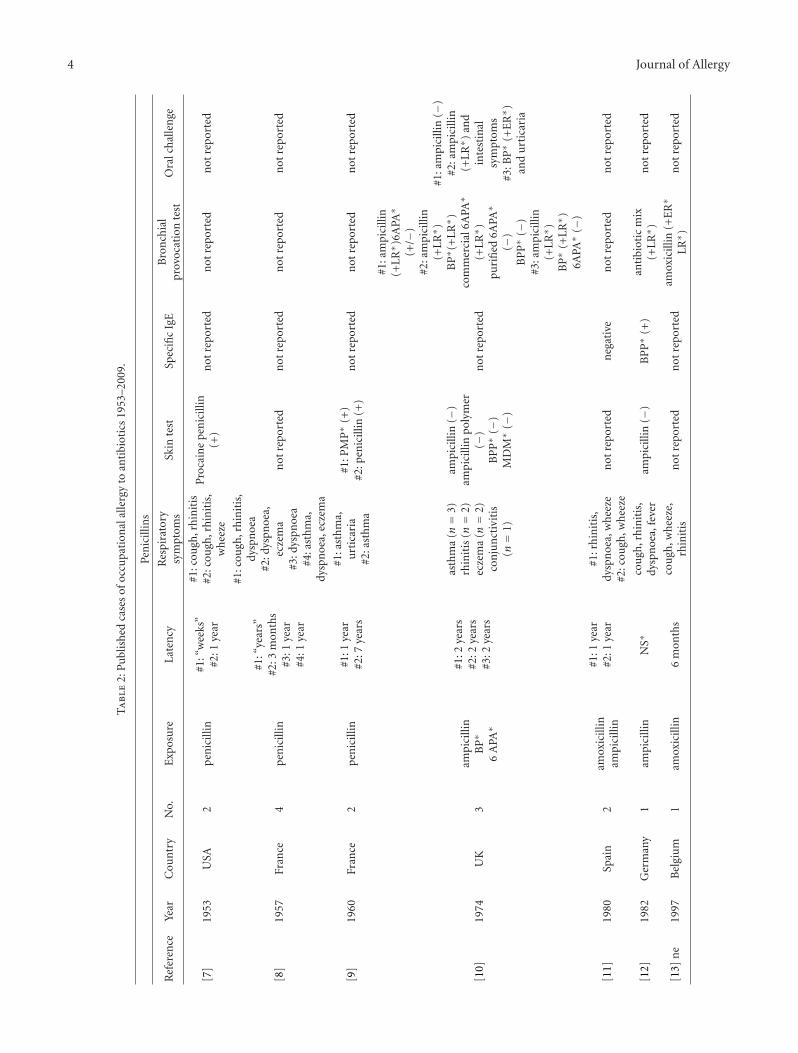
4 Journal of Allergy
Ta
ble
2:P
ubl
ish
edca
ses
ofoc
cupa
tion
alal
lerg
yto
anti
biot
ics
1953
–200
9.
Pen
icill
ins
Ref
eren
ceYe
arC
oun
try
No.
Exp
osu
reLa
ten
cyR
espi
rato
rysy
mpt
oms
Skin
test
Spec
ific
IgE
Bro
nch
ial
prov
ocat
ion
test
Ora
lch
alle
nge
[7]
1953
USA
2p
enic
illin
#1:“
wee
ks”
#2:1
year
#1:c
ough
,rh
init
is#2
:cou
gh,r
hin
itis
,w
hee
ze
Pro
cain
epe
nic
illin
(+)
not
repo
rted
not
repo
rted
not
repo
rted
[8]
1957
Fran
ce4
pen
icill
in
#1:“
year
s”#2
:3m
onth
s#3
:1ye
ar#4
:1ye
ar
#1:c
ough
,rh
init
is,
dysp
noe
a#2
:dys
pnoe
a,ec
zem
a#3
:dys
pnoe
a#4
:ast
hm
a,dy
spn
oea,
ecze
ma
not
repo
rted
not
repo
rted
not
repo
rted
not
repo
rted
[9]
1960
Fran
ce2
pen
icill
in#1
:1ye
ar#2
:7ye
ars
#1:a
sth
ma,
urt
icar
ia#2
:ast
hm
a
#1:P
MP∗
(+)
#2:p
enic
illin
(+)
not
repo
rted
not
repo
rted
not
repo
rted
[10]
1974
UK
3am
pici
llin
BP∗
6A
PA∗
#1:2
year
s#2
:2ye
ars
#3:2
year
s
asth
ma
(n=
3)rh
init
is(n=
2)ec
zem
a(n=
2)co
nju
nct
ivit
is(n=
1)
ampi
cilli
n(−
)am
pici
llin
poly
mer
(−)
BP
P∗
(−)
MD
M∗
(−)
not
repo
rted
#1:a
mpi
cilli
n(+
LR∗ )
6APA
∗
(+/−
)#2
:am
pici
llin
(+L
R∗ )
BP∗ (
+L
R∗ )
com
mer
cial
6APA
∗
(+L
R∗ )
puri
fied
6APA
∗
(−)
BP
P∗
(−)
#3:a
mpi
cilli
n(+
LR∗ )
BP∗
(+L
R∗ )
6APA
∗(−
)
#1:a
mpi
cilli
n( −
)#2
:am
pici
llin
(+L
R∗ )
and
inte
stin
alsy
mpt
oms
#3:B
P∗
(+E
R∗ )
and
urt
icar
ia
[11]
1980
Spai
n2
amox
icill
inam
pici
llin
#1:1
year
#2:1
year
#1:r
hin
itis
,dy
spn
oea,
wh
eeze
#2:c
ough
,wh
eeze
not
repo
rted
neg
ativ
en
otre
port
edn
otre
port
ed
[12]
1982
Ger
man
y1
ampi
cilli
nN
S∗co
ugh
,rh
init
is,
dysp
noe
a,fe
ver
ampi
cilli
n(−
)B
PP∗
(+)
anti
biot
icm
ix(+
LR∗ )
not
repo
rted
[13]
ne
1997
Bel
giu
m1
amox
icill
in6
mon
ths
cou
gh,w
hee
ze,
rhin
itis
not
repo
rted
not
repo
rted
amox
icill
in(+
ER∗
LR∗ )
not
repo
rted
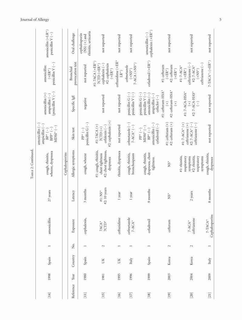
Journal of Allergy 5
Ta
ble
2:C
onti
nu
ed.
[14]
1998
Spai
n1
amox
icill
in27
year
sco
ugh
,rh
init
is,
wh
eeze
,dys
pnoe
a
amox
icill
in( −
)am
pici
llin
(−)
BP∗
(−)
BP
P∗
(−)
MD
M∗
(−)
amox
icill
in(+
)am
pici
llin
(−)
(pen
icill
inV
(−)
amox
icill
in:
(+E
R∗ )
pen
icill
inV
(−)
amox
icill
in(+
LR∗ )
pen
icill
inV
(−)
Cep
hal
ospo
rin
s
Ref
eren
ceYe
arC
oun
try
No.
Exp
osu
reLa
ten
cyA
llerg
icsy
mpt
oms
Skin
test
Spec
ific
IgE
Bro
nch
ial
prov
ocat
ion
test
Ora
lch
alle
nge
[11]
1980
Spai
n1
ceph
alex
in,
3m
onth
sco
ugh
,wh
eeze
PP∗
(−)
pen
icill
inG
(−)
neg
ativ
en
otre
port
edce
phal
ospo
rin
(NS)
(+)
and
rhin
itis
,urt
icar
ia
[15]
1981
UK
27A
CA∗
7CT
D∗
#1:N
S∗
#2:1
0ye
ars
#1:c
ough
,rh
init
is,
ches
tti
ghtn
ess
#2:c
hes
tti
ghtn
ess,
dysp
noe
a
#1:7
AC
A(+
)7C
TD
(+)
cefa
lexi
n(−
)#2
:cep
hal
exin
(+)
not
repo
rted
#1:7
AC
A(+
ER∗ )
7CT
D(+
ER∗ )
ceph
alex
in(−
)#2
:cep
hal
exin
(+E
R∗ )
not
repo
rted
[16]
1995
UK
1ce
ftaz
idim
e1
year
rhin
itis
,dys
pnoe
an
otre
port
edn
otre
port
edce
ftaz
idim
e(+
ER∗
LR∗ )
not
repo
rted
[17]
1996
Ital
y1
cefm
etaz
ole
7-A
CA∗
1ye
arco
ugh
,rh
init
is,
bron
chos
pasm
cefm
etaz
ole
( −)
7-A
CA∗
(−)
pen
icill
inG
( −)
pen
icill
inV
(−)
cefm
etaz
ole
(+N
S∗)
7-A
CA
(+N
S∗)
not
repo
rted
[18]
1999
Spai
n1
cefa
drox
il9
mon
ths
cou
gh,r
hin
itis
,dy
spn
oea,
ches
tti
ghtn
ess
PP∗
(−)
MD
M∗
(−)
BP∗
(−)
amox
icill
in(−
)ce
fadr
oxil
(−)
pen
icill
inG
(−)
pen
icill
inV
(−)
amox
icill
in(−
)am
pici
llin
(−)
cefa
clor
(−)
cefa
drox
il(+
ER∗ )
amox
icill
in( −
)ce
phal
exin
(+E
R∗ )
[19]
2003
Kor
ea2
ceft
eram
NS∗
NS∗
#1:c
efte
ram
(+)
#2:c
efte
ram
(+)
#1:c
efte
ram
-HSA
∗
(+)
#2:c
efte
ram
-HSA
∗
(+)
#1:c
efte
ram
(+E
R∗ )
#2:c
efte
ram
(+E
R∗ )
not
repo
rted
[20]
2004
Kor
ea2
7-A
CA∗
ceft
riax
one
2ye
ars
#1:r
hin
itis
,re
spir
ator
ysy
mpt
oms
#2:r
hin
itis
,re
spir
ator
ysy
mpt
oms
#1:7
-AC
A∗
(+)
ceft
riax
one
(+)
#2:7
-AC
A∗
(−)
ceft
riax
one
(−)
#1:7
-AC
A-H
SA∗
(+)
#2:7
-AC
A-H
AS∗
(−)
#1:7
-AC
A∗
(+E
R∗ )
ceft
riax
one
(−)
#2:7
-AC
A∗
(+N
S∗)
ceft
riax
one
(−)
not
repo
rted
[21]
2009
Ital
y1
7-TA
CA∗
Cep
hal
ospo
rin
s8
mon
ths
cou
gh,r
hin
itis
,dy
spn
oea
not
repo
rted
not
repo
rted
7-TA
CA∗ :
(+E
R∗ )
not
repo
rted

6 Journal of Allergy
Ta
ble
2:C
onti
nu
ed.
Mis
cella
neo
us
Ref
eren
ceYe
arC
oun
try
No.
Exp
osu
reLa
ten
cyA
llerg
icsy
mpt
oms
Skin
test
Spec
ific
IgE
Bro
nch
ial
prov
ocat
ion
test
Ora
lch
alle
nge
[22]
1977
Indi
a1
tetr
acyc
line
1ye
arco
ugh
,wh
eeze
,dy
spn
oea
not
repo
rted
not
repo
rted
tetr
acyc
line
(+E
R∗ )
tetr
acyc
line
(+E
R∗ )
and
urt
icar
ia
[23]
1975
UK
1sp
iram
ycin
1ye
arco
ugh
,rh
init
is,
dysp
noe
a,de
rmat
itis
spir
amyc
in(+
)n
otre
port
edsp
iram
ycin
(+L
R∗ )
not
rep
orte
d
[24]
1979
Ital
y1
spir
amyc
in1
year
cou
gh,a
sth
ma,
derm
atit
issp
iram
ycin
(+)
not
repo
rted
chic
kfe
edw
ith
spir
amyc
in(+
LR∗ )
not
repo
rted
[25]
1984
Ital
y2
spir
amyc
inad
ipid
acid
#1:1
4ye
ars
#2:7
mon
ths
#1:d
yspn
oea
#2:c
ough
,dy
spn
oea
not
repo
rted
not
repo
rted
#1:s
pira
myc
inad
ipat
e(+
ER∗
LR∗ )
#2:s
pira
myc
inad
ipat
e(+
ER∗ )
spir
amyc
inba
se(+
ER∗
LR∗ )
not
repo
rted
[26]
1995
Ital
y1
pip
erac
illin
22m
onth
srh
init
is,d
yspn
oea,
wh
eeze
,ras
hpi
pera
cilli
n(+
)n
otre
port
edpi
pera
cilli
n(+
ER∗ )
not
repo
rted
[27]
2006
Kor
ea2
thia
mph
enic
olN
S∗#1
:rh
init
is,a
sth
ma
#2:r
hin
itis
,ast
hm
a
#1:t
hia
mph
enic
ol(+
)#2
:th
iam
phen
icol
(+)
#1:t
hia
mph
enic
ol(+
)#2
thia
mph
enic
ol(+
)
#1:t
hia
mph
enic
ol(+
ER∗ )
#2:t
hia
mph
enic
ol(+
ER∗ )
not
repo
rted
[28]
2009
Kor
ea1
van
com
ycin
5m
onth
srh
init
is,c
hes
tti
ghtn
ess
van
com
ycin
(−)
van
com
ycin
-HSA
∗
(−)
not
repo
rted
not
repo
rted
[29]
2010
Spai
n1
colo
myc
in3
mon
ths
rhin
itis
,cou
gh,
wh
eeze
,dys
pnoe
an
otre
port
edn
egat
ive
colo
myc
in(+
ER∗ )
not
repo
rted
∗N
S:n
otsp
ecifi
ed,∗
ER
:ear
ly(a
sth
mat
ic)
resp
onse
,∗L
R:l
ate
(ast
hm
atic
)re
spon
se.
∗ HSA
:hu
man
seru
mal
bum
in,∗
MD
M:m
inor
dete
rmin
ant
(pen
icill
in)
mix
,∗B
P:b
enzy
lpen
icill
in.
∗ (B
)PP
:(be
nzy
l)pe
nic
illoy
lpol
ylys
ine,∗ P
MP
:ph
enox
ymet
hylp
enic
illin
,∗6A
PA:6
amin
ope
nic
illan
icac
id,∗
7-A
CA
:7am
inoc
eph
alos
pora
nic
acid
,∗7C
TD
:tos
ylat
edi
hydr
ate
deri
vati
veof
7AC
A.
∗ 7-T
AC
A:7
-am
ino-
3th
iom
etih
yl-3
-cep
hal
ospo
ran
icac
id.

Journal of Allergy 7
Paggiaro et al. [24] described dermatitis and asthma due tospiramycin in a chick breeder; a skin prick test to spiramycinwas positive as was specific inhalation testing with chick feedcontaining the antibiotic.
Menon and Das [22] documented in an Indian workerimmediate asthmatic reactions to intradermal, inhalationand oral testing with tetracycline but no response toinhalation testing with two antibiotics (nystatin and chloro-mycetin) to which he was not exposed at work. Three casesof OA from thiamphenicol—a methyl-sulfonyl analogueof chloramphenicol—were reported by Ye et al. [27].Vancomycin has been described as a cause of OA in a singlepatient [28], the diagnosis confirmed by serial peak flowmeasurements. The most recently reported case was of OAdue to the polymyxin antibiotic colomycin in an antibiotictransport and storage worker, who demonstrated an earlyasthmatic reaction following specific inhalation challenge[29]; no evidence of specific IgE was found despite extensivein vitro immunological testing.
3.3. Workforce Studies. We identified seven reports of epi-demiological studies carried out in antibiotic manufacturingsites; all but one was of cross-sectional design. In severalcases, the absence of detailed information on the size ofthe exposed populations precludes any estimate of diseaseprevalence.
Briatico-Vangosta et al. [2] surveyed 91 Italian work-ers exposed to cephalosporins with a combination of asymptoms questionnaire, skin prick and intradermal testing,and “on-off” testing of asthma symptoms and pulmonaryfunction. On this basis, OA was reported for seven (8%)employees. Skin testing produced immediate-type responsesin five workers with OA (in three cases with prick testing)suggesting to the authors that the asthma had, in some casesat least, arisen through a specific IgE-related mechanism.
Chida and Uehata [30] surveyed by interview 24 em-ployees of a pharmaceutical factory which produced twoantibiotics (ampicillin and cephalexin) as well as an anti-spasmodic and three anti-inflammatory drugs. Those withrespiratory symptoms (n = 18) underwent skin prick,serum, and pulmonary function testing. Probable antibiotic-related asthma was claimed for four employees, two of whomhad immediate responses to skin testing with ampicillin. Inthe absence of any clear information on the population atrisk, a prevalence estimate is not available.
Phenylglycine acid chloride (PG-AC) is a highly reactivecompound used chiefly in the manufacture of ampicillin andother antibiotic side chains. A survey of 24 workers involvedin the production of PG-AC [3] included a symptomsquestionnaire, examination of occupational health records,skin testing, and spirometry. Seven workers were felt, onthe basis of their history alone, to have a respiratoryallergy to PG-AC; all had positive responses to prick and/orintradermal skin testing with the conjugated compound.Two were admitted to hospital for specific inhalation testingwith positive findings. A further seven employees weredeemed to have “irritant” respiratory responses to PG-AC(all skin tests negative) and in nine the history was considered“equivocal”; two of this last group had positive skin test
results. The authors commented that the amino groupsin PG-AC predispose to the formation of hapten-proteinconjugates and hence its allergenicity. Again, in the absenceof any information on the size of the exposed workforce, anestimate of prevalence in this setting is unavailable.
Carnevale et al. [31] studied 67 workers employed inmanufacturing and encapsulating antibiotics in Italy. Follow-ing questionnaire, clinical and laboratory investigations, two(3%) cases of OA due to ampicillin were recorded. In bothcases skin tests were negative. Post- and preshift urine testingconfirmed the systemic absorption of ampicillin by theseworkers. A similar survey was reported by Carlesi et al. [32].Among 26 employees in an antibiotic manufacturing plant,eight (31%) claimed respiratory symptoms which in twocases (8%) were suggestive of asthma. A further three em-ployees had positive skin prick tests to penicillin G and/oramoxicillin.
Two surveys of workers involved in spiramycin manufac-ture have been reported. Malo and Cartier [4] investigatedall 51 employees at a processing plant in Canada. Twelve,on the basis of a compatible history or evidence of (work-related) bronchial hyperreactivity, underwent specific in-halation testing with positive findings in four cases. Themeasured prevalence of 8% was considered by the authors,to be a minimum estimate. In Italy, a 12-year prospectivestudy of 305 workers in a spiramycin-manufacturing plant[33] suggested work-related asthma in 15 (5%), all of whomhad positive epicutaneous or intradermal skin tests to spira-mycin. Four had additional symptoms of rhinitis and oneof urticaria. In the full study population, 41 employees(13%) had positive skin tests, 37 (90%) of them with allergicsymptoms of some kind.
4. Discussion
Our cases add three to the previously reported eight casesof OA attributed to inhalation of synthetic penicillins duringtheir manufacture, and the first case of disease arising fromworkplace exposure to erythromycin. An additional sevencases of penicillin OA were reported to a UK nationalsurveillance scheme between 1989 and 2009 (THOR per-sonal communication).
An examination of published findings from surveillanceschemes in other parts of the world found specific referenceto antibiotics in none although the Propulse scheme inQuebec collated seven cases of OA (2.4% of the total)attributed to “medical drugs” [34]. Our systematic reviewof the remaining literature revealed a total of 37 casespublished over a period of almost 50 years, although otherswere identified during the course of seven epidemiologicalstudies, the most recent of which was published over 20years ago. It was often difficult, in reviewing the workplacesurveys, to obtain meaningful estimates of prevalence butwhat information was available suggests that about 10% ofthose surveyed had disease suggestive of OA. Thus since thefirst report in 1953 there appears to have been only sporadicattention paid to what may be a significant occupational risk.
The clinical features of the published cases are similar tothose found in other examples of allergic OA and are broadly

8 Journal of Allergy
indicative of an immediate-type respiratory hypersensitivity.A latent period of asymptomatic exposure was reported forall, usually of fewer than 24 months although occasionallyfar longer, and rhinitis was a frequent accompaniment toasthma symptoms. In those cases where specific inhalationtesting was used in diagnosis and the findings reportedin full, a late or dual asthmatic response was reported in43%. In most cases of isolated early asthmatic responses tospecific provocation, there was evidence of immunologicalsensitisation on skin or serum testing. Each of the implicatedantibiotics had a very high hazard index derived from aquantitative structure-activity relationship analysis [35]; inthe clinical context of a patient with characteristic symp-toms, index values such as these have a high positive pre-dictive value for OA [36]. Finally, Roberts [7] reported theapparently successful, subcutaneous desensitisation of twopatients with OA from penicillin.
Nonetheless, as with many other low-molecular-weightcauses of the disease, the immunological details of antibiotic-related OA remain unclear. While an IgE-associated mecha-nism is likely in many cases, the possibility of sensitisationarising from an alternative mechanism, perhaps throughcross-linking of cell surface receptors, cannot be ruled out.Even when, as is not always the case, their techniques aredescribed, the variety of skin test methods used in thepublished reports makes their interpretation and diagnosticsignificance uncertain. In any case, the results were oftennegative and where they were not there is no systematicinformation on the findings among exposed but non-asthmatic employees. Similar comments apply to the use oftests for serum-specific IgE antibodies which were less oftenperformed and more often negative although some successhas been recorded for in-house assays using cephalosporinconjugates [20, 21] or thiamphenicol [27]. Thus, and prob-ably in contrast to their use in oral antibiotic allergy [37],the value of available tests for evidence of immunologicalsensitisation in this context remains unclear.
Penicillins and cephalosporins are common causes oforal drug allergy which in most cases is attributed to an IgE-associated immune response to one or more hapten-proteinconjugates. Penicillins are composed of betalactam and thi-azolidine rings with one or more differentiating side chains;the instability of the betalactam causes its carbonyl group toform amide-bonds with the amino groups of lysine residuesfrom nearby proteins. “Minor” antigenic determinants, someformed from side chain-protein conjugates, may also induceIgE immune responses. 7 aminocephalosporanic acid, theactive nucleus of cephalosporins, is structurally similar tothe active nucleus of the penicillins, consisting of betalactamand 6 dyhydrothiazolidine rings. Hapten-protein conjugatesof the cephalosporin betalactam are relatively unstable. IgEantibodies that react to cephalosporins detect a large numberof specific antigens derived from protein conjugates formedfrom the side chain(s), the side chain plus a portion of thebetalactam ring, or the complete cephalosporin. On the basisof ELISA inhibition assays in two patients with OA fromcefteram, Suh et al. [19] suggested that specific IgE responsesto the cephalosporin may involve haptens, new antigenicdeterminants, or both and are likely to vary between patients.
Seven of the identified case reports describe oral chal-lenges to the causative antibiotic (in six cases a penicillin orcephalosporin) in patients with OA. All but one producedan allergic response. The three cases reported by Davieset al. [10] had subtly different responses to inhaled andoral (negative in one patient) provocation tests suggestingthe possibility that individually they were responsive todifferent hapten-protein antigens. The patient described byJimenez et al. [14] had allergic responses to both inhaledand oral amoxicillin, but not to oral penicillin V suggestingto the authors that the responsible antigenic determinantlay in the aminohydroxyphenyl side-chain of amoxicillin.In contrast, Sastre et al. [18] reported the case of a patientwith OA attributed to cefadroxil, a cephalosporin that sharesa side chain with amoxicillin. Oral challenge with cefadroxil,but not with amoxicillin, produced an asthmatic responsesuggesting that her sensitisation was to the dihydrothiazinecephalosporin ring rather than to its side chain. Losada et al.[11] reported a further patient with asthmatic responses toboth inhaled and oral cephalosporin but not to penicillin G.Oral allergy may also develop in patients with OA attributedto other antibiotics [22]. While the evidence base is weak,and we cannot exclude the possibility of bias in those selectedfor oral challenge or in publication, we suggest that there issufficient experience to advise that patients with OA froman inhaled antibiotic encountered at work should avoidtaking the same—or closely related—drug by mouth unlessits safety has been established by a carefully supervisedprovocation test.
We have been unable to find valid information on thenumbers of exposed employees in any parts of the world butwe expect these to number many thousands. Much man-ufacture is carried out in conditions where exposures toemployees are very low but this is not always the case, andwe note the general movement of pharmaceutical manu-facturing away from its traditional base in western Europeand North America. We note also that most publishedevidence relates to older types of antibiotic. This is likely toreflect the number and exposures of those involved in theirmanufacture rather than any intrinsic hazard; the hazardindices for newer antibiotics such as flucloxacillin (0.909),clarithromycin (0.997), tobramycin (1.0), and azithromycin(1.0) are no lower. While cases of OA arising from antibioticmanufacture are rarely published, the available epidemio-logical evidence, admittedly scanty, sometimes deficient andall of it dated, suggests that the risks may be higher thanmany appreciate. We suggest that further workplace-basedsurveys, of careful design and supported by improvementsin immunological diagnosis, are required.
References
[1] J.-L. Malo, H.-S. Park, and I. L. Bernsein, “Other chemicalsubstances causing occupational asthma,” in Asthma in theWorkplace, I. L. Bernstein, M. Chan-Yeung, J.-L. Malo, and D.I. Bernstein, Eds., pp. 555–580, Marcel Dekker, London, UK,3rd edition, 2006.
[2] G. Briatico-Vangosa, F. Beretta, S. Bianchi, A. Cardani,M. Marchisio, and C. Nava, “Bronchial asthma caused by

Journal of Allergy 9
7-aminocephalosporanic acid in workers engaged in theproduction of cephalosporins,” Medicina del Lavoro, vol. 72,no. 6, pp. 488–493, 1981.
[3] J. K. Kammermeyer and K. P. Mathews, “Hypersensitivity tophenylglycine acid chloride,” Journal of Allergy and ClinicalImmunology, vol. 52, no. 2, pp. 73–84, 1973.
[4] J. L. Malo and A. Cartier, “Occupational asthma in workers ofa pharmaceutical company processing spiramycin,” Thorax,vol. 43, no. 5, pp. 371–377, 1988.
[5] A. J. Woolcock, K. Yan, and C. Salome, “Methods for assessingbronchial reactivity,” European Journal of Respiratory Diseases,vol. 128, supplement, p. 1, 1983.
[6] 2010, http://www.medicine.manchester.ac.uk/oeh/research/workrelatedillhealth/asthma/.
[7] A. E. Roberts, “Occupational allergic reactions among workersin a penicillin manufacturing plant; simple and inexpensivemethod of diagnosis and treatment,” A. M. A. Archives ofIndustrial Hygiene and Occupational Medicine, vol. 8, no. 4,pp. 340–346, 1953.
[8] S. Tara, “Asthma caused by penicillin,” Archives des MaladiesProfessionnelles de Medecine du Travail et de Securite Sociale,vol. 18, no. 3, pp. 274–277, 1957.
[9] M. Gaultier, E. Fournier, and P. Gervais, “Occupationalasthma caused by allergy to penicillin,” Archives des MaladiesProfessionnelles de Medecine du Travail et de Securite Sociale,vol. 21, pp. 13–23, 1960.
[10] R. J. Davies, D. J. Hendrick, and J. Pepys, “Asthma due toinhaled chemical agents: ampicillin, benzyl penicillin, 6 aminopenicillanic acid and related substances,” Clinical Allergy, vol.4, no. 3, pp. 227–247, 1974.
[11] E. Losada Cosmes, M. Hinojosa Macias, R. Acover Sanchezet al., “Asma por inhalacion por penicilina ambiental,”Allergologia et Immunopathologia, vol. 7, supplement, pp.288–293, 1980.
[12] B. Wuthrich and A. L. Hartmann, “Occupation-relatedbronchial asthma caused by ampicillin. Diagnostic signi-ficance of occupation-specific inhalation provocation tests,”schweizerische Medizinische Wochenschrift, vol. 112, no. 29, pp.1046–1048, 1982.
[13] O. Vandenplas, J. P. Delwiche, and M. De Jonghe, “Asthma tolatex and amoxicillin,” Allergy, vol. 52, no. 11, pp. 1147–1149,1997.
[14] I. Jimenez, E. Anton, I. Picans, I. Sanchez, M. D. Quinones,and J. Jerez, “Occupational asthma specific to amoxicillin,”Allergy, vol. 53, no. 1, pp. 104–105, 1998.
[15] I. I. Coutts, M. B. Dally, and A. J. N. Taylor, “Asthma inworkers manufacturing cephalosporins,” British MedicalJournal, vol. 283, no. 6297, p. 950, 1981.
[16] S. C. Stenton, J. H. Dennis, and D. J. Hendrick, “Occupationalasthma due to ceftazidime,” European Respiratory Journal, vol.8, no. 8, pp. 1421–1423, 1995.
[17] G. Fracchia, L. Paita, R. Maglio, and T. Malamani, “Occupa-tional asthma caused by cefmetazole and 7-aminocepha-losporanic acid: description of a clinical case,” GiornaleItaliano di Medicina del Lavoro, vol. 18, no. 1–3, pp. 3–5, 1996.
[18] J. Sastre, S. Quirce, A. Novalbos, M. Lluch-Bernal, C. Bombın,and A. Umpierrez, “Occupational asthma induced by cephalo-sporins,” European Respiratory Journal, vol. 13, no. 5, pp.1189–1191, 1999.
[19] Y. J. Suh, Y. M. Lee, J. H. Choi, C. H. Suh, D. H. Nahm, andH. S. Park, “Heterogeneity of IgE response to cefteram pivoxilwas noted in 2 patients with cefteram-induced occupationalasthma,” Journal of Allergy and Clinical Immunology, vol. 112,no. 1, pp. 209–210, 2003.
[20] H. S. Park, K. U. Kim, Y. M. Lee et al., “Occupational asthmaand IgE sensitization to 7-aminocephalosporanic acid,”Journal of Allergy and Clinical Immunology, vol. 113, no. 4, pp.785–787, 2004.
[21] G. Pala, P. Pignatti, L. Perfetti, R. Cosentino, and G. Mos-cato,“Occupational asthma and rhinitis induced by a cephalo-sporin intermediate product: description of a case,” Allergy,vol. 64, no. 9, pp. 1390–1391, 2009.
[22] M. P. S. Menon and A. K. Das, “Tetracycline asthma: a casereport,” Clinical Allergy, vol. 7, no. 3, pp. 285–290, 1977.
[23] R. J. Davies and J. Pepys, “Asthma due to inhaled chemicalagents: the macrolide antibiotic spiramycin,” Clinical Allergy,vol. 5, no. 1, pp. 99–107, 1975.
[24] P. L. Paggiaro, A. M. Loi, and G. Toma, “Bronchial asthmaand dermatitis due to spiramycin in a chick breeder,” ClinicalAllergy, vol. 9, no. 6, pp. 571–574, 1979.
[25] G. Moscato, L. Naldi, and F. Candura, “Bronchial asthma dueto spiramycin and adipic acid,” Clinical Allergy, vol. 14, no. 4,pp. 355–361, 1984.
[26] G. Moscato, E. Galdi, J. Scibilia et al., “Occupational asthma,rhinitis and urticaria due to piperacillin sodium in apharmaceutical worker,” European Respiratory Journal, vol. 8,no. 3, pp. 467–469, 1995.
[27] Y. M. Ye, H. M. Kim, C. H. Suh, D. H. Nahm, and H. S.Park, “Three cases of occupational asthma induced by thiam-phenicol: detection of serum-specific IgE,” Allergy, vol. 61, no.3, pp. 394–395, 2006.
[28] G. S. Choi, J. M. Sung, J. W. Lee, Y. M. Ye, and H. S. Park,“A case of occupational asthma caused by inhalation of van-comycin powder,” Allergy, vol. 64, no. 9, pp. 1391–1392, 2009.
[29] S. Gomez-Olles, M. F. Madrid-San, M. J. Cruz, and X. Muoz,“Occupational asthma due to colistin in a pharmaceuticalworker,” Chest, vol. 137, no. 5, pp. 1200–1202, 2010.
[30] T. Chida and T. Uehata, “Some experiments on the allergicreaction among workers in a pharmaceutical factory (author’stranslation),” Sangyo Igaku, vol. 21, no. 5, pp. 422–432, 1979.
[31] F. Carnevale, M. Valsecchi, and D. Grazioli, “Health aspectsof antibiotic manufacture with particular emphasis onrespiratory and genital disturbances (author’s translation),”Lavoro Umano, vol. 29, no. 1, pp. 1–14, 1977.
[32] G. Carlesi, E. Ferrea, C. Melino, A. Messineo, and E. Pacelli,“Aspects of environmental health and of pathology causedby pollution with amoxicillin in a pharmaceutical industry,”Nuovi Annali d”Igiene e Microbiologia, vol. 30, no. 2, pp.185–196, 1979.
[33] C. Nava, “Rischi lavorativiti da antibiotici: contributo allostudio della patologia professionale da spiramicina,” Securitas,vol. 61, pp. 275–280, 1976.
[34] S. Provencher, F. P. Labreche, and L. De Guire, “Physicianbased surveillance system for occupational respiratorydiseases: the experience of PROPULSE, Quebec, Canada,”Occupational and Environmental Medicine, vol. 54, no. 4, pp.272–276, 1997.
[35] J. Jarvis, M. J. Seed, R. A. Elton, L. Sawyer, and R. M.Agius, “Relationship between chemical structure and theoccupational asthma hazard of low molecular weight organiccompounds,” Occupational and Environmental Medicine, vol.62, no. 4, pp. 243–250, 2005.
[36] M. Seed and R. Agius, “Further validation of computer-based prediction of chemical asthma hazard,” OccupationalMedicine, vol. 60, no. 2, pp. 115–120, 2009.
[37] M. J. Torres, M. Blanca, J. Fernandez et al., “Diagnosis ofimmediate allergic reactions to beta-lactam antibiotics,”Allergy, vol. 58, no. 10, pp. 961–972, 2003.

Hindawi Publishing CorporationJournal of AllergyVolume 2011, Article ID 267542, 7 pagesdoi:10.1155/2011/267542
Clinical Study
Exercise-Induced Bronchoconstriction and Exercise-InducedRespiratory Symptoms in Nurses
Jordan Minov,1 Jovanka Karadzinska-Bislimovska,1 Kristin Vasilevska,2
Snezana Risteska-Kuc,1 Saso Stoleski,1 and Dragan Mijakoski1
1 Department of Cardiorespiratory Functional Diagnostics, Institute for Occupational Health of R. Macedonia—WHO CollaboratingCenter and GA2LEN Collaborating Center, II Makedonska Brigada 43, 1000 Skopje, Macedonia
2 Institute for Epidemiology and Biostatistics, 1000 Skopje, Macedonia
Correspondence should be addressed to Jordan Minov, [email protected]
Received 20 December 2010; Accepted 28 February 2011
Academic Editor: Donald H. Beezhold
Copyright © 2011 Jordan Minov et al. This is an open access article distributed under the Creative Commons Attribution License,which permits unrestricted use, distribution, and reproduction in any medium, provided the original work is properly cited.
In order to assess prevalence and characteristics of exercise-induced respiratory symptoms (EIRSs) and exercise-inducedbronchoconstriction (EIB) in health care workers, we performed a cross-sectional study including 48 female nurses from primarycare settings and an equal number of female office workers studied as a control group. The evaluation of examined groups includedcompletion of a questionnaire, skin prick tests to common inhalant allergens, spirometry, and exercise and histamine challenge.We found a similar prevalence of EIRSs and EIB in both groups. EIB was closely related to asthma, atopy, family history ofasthma, and positive histamine challenge in either group, while the association between EIB and daily smoking in nurses wasof borderline statistical significance. Bronchial reaction to exercise was significantly higher in nurses than in controls with EIB.With the exception of exercise induced wheezing, EIRSs were weakly associated with EIB in both groups with a large proportionof false positive results.
1. Introduction
Exercise-induced bronchoconstriction (EIB), also referredto as exercise-induced asthma (EIA), is a manifestation ofbronchial hyperresponsiveness (BHR) that occurs in themajority of patients with current symptomatic asthma, espe-cially in the patients with moderately to severely increasedresponsiveness [1–3]. The current thinking about mecha-nisms by which EIB develops emphasizes the loss of heatand/or water from the airways during exercise that leads torelease of proinflammatory mediators [4]. Airborne particlesand pollutants, as well as airborne allergens, are considered asstimulants that contribute to EIB [5]. A fish oil supplemen-tation may have a protective effect on EIB, which is probablyattributed to its anti-inflammatory properties [6].
Results from several studies indicated that BHR preva-lence is higher in females than in males [7, 8]. The mecha-nisms responsible for a higher susceptibility of the airwaysin females to nonspecific stimuli include lower airwaycalibre, lower body weight, greater cholinergic irritability,and hormonal factors [9].
On the other side, data from the studies carried out in thelast two decades suggest an increased risk for asthma amonghealth care workers, yet only a few specific determinantshave been elucidated [10–13]. As Delclos et al. [14] havesuggested, the contribution of occupational exposures to res-piratory impairment and asthma in health care professionalsis not trivial, meriting both implementation of appropriatecontrols and further studies.
To our knowledge, so far, there is no study assessingexercise-induced respiratory symptoms (EIRSs) and EIB inhealth care professionals. In the present study, we assessedeffects of occupational exposure on EIRSs and EIB amonghealth care workers by comparison of their prevalence andcharacteristics between females working as nurses in primarycare settings and female office workers.
2. Materials and Methods
2.1. Study Design and Setting. A cross-sectional survey wascarried out in a university research laboratory, that is,
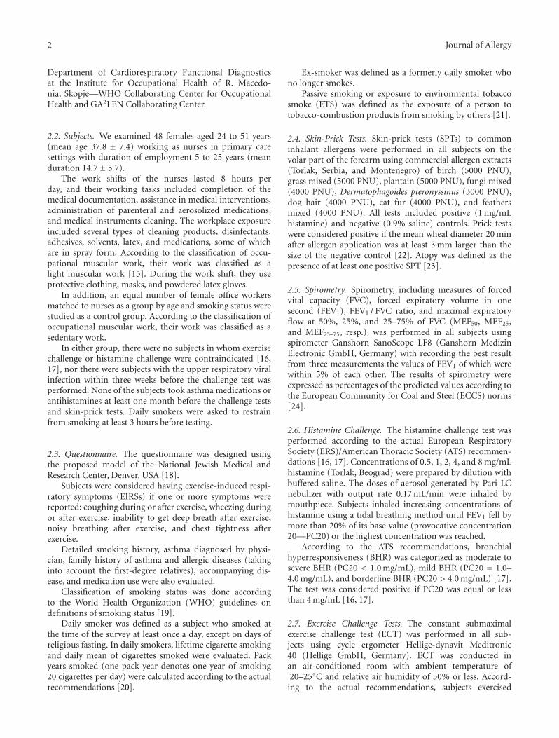
2 Journal of Allergy
Department of Cardiorespiratory Functional Diagnosticsat the Institute for Occupational Health of R. Macedo-nia, Skopje—WHO Collaborating Center for OccupationalHealth and GA2LEN Collaborating Center.
2.2. Subjects. We examined 48 females aged 24 to 51 years(mean age 37.8 ± 7.4) working as nurses in primary caresettings with duration of employment 5 to 25 years (meanduration 14.7± 5.7).
The work shifts of the nurses lasted 8 hours perday, and their working tasks included completion of themedical documentation, assistance in medical interventions,administration of parenteral and aerosolized medications,and medical instruments cleaning. The workplace exposureincluded several types of cleaning products, disinfectants,adhesives, solvents, latex, and medications, some of whichare in spray form. According to the classification of occu-pational muscular work, their work was classified as alight muscular work [15]. During the work shift, they useprotective clothing, masks, and powdered latex gloves.
In addition, an equal number of female office workersmatched to nurses as a group by age and smoking status werestudied as a control group. According to the classification ofoccupational muscular work, their work was classified as asedentary work.
In either group, there were no subjects in whom exercisechallenge or histamine challenge were contraindicated [16,17], nor there were subjects with the upper respiratory viralinfection within three weeks before the challenge test wasperformed. None of the subjects took asthma medications orantihistamines at least one month before the challenge testsand skin-prick tests. Daily smokers were asked to restrainfrom smoking at least 3 hours before testing.
2.3. Questionnaire. The questionnaire was designed usingthe proposed model of the National Jewish Medical andResearch Center, Denver, USA [18].
Subjects were considered having exercise-induced respi-ratory symptoms (EIRSs) if one or more symptoms werereported: coughing during or after exercise, wheezing duringor after exercise, inability to get deep breath after exercise,noisy breathing after exercise, and chest tightness afterexercise.
Detailed smoking history, asthma diagnosed by physi-cian, family history of asthma and allergic diseases (takinginto account the first-degree relatives), accompanying dis-ease, and medication use were also evaluated.
Classification of smoking status was done accordingto the World Health Organization (WHO) guidelines ondefinitions of smoking status [19].
Daily smoker was defined as a subject who smoked atthe time of the survey at least once a day, except on days ofreligious fasting. In daily smokers, lifetime cigarette smokingand daily mean of cigarettes smoked were evaluated. Packyears smoked (one pack year denotes one year of smoking20 cigarettes per day) were calculated according to the actualrecommendations [20].
Ex-smoker was defined as a formerly daily smoker whono longer smokes.
Passive smoking or exposure to environmental tobaccosmoke (ETS) was defined as the exposure of a person totobacco-combustion products from smoking by others [21].
2.4. Skin-Prick Tests. Skin-prick tests (SPTs) to commoninhalant allergens were performed in all subjects on thevolar part of the forearm using commercial allergen extracts(Torlak, Serbia, and Montenegro) of birch (5000 PNU),grass mixed (5000 PNU), plantain (5000 PNU), fungi mixed(4000 PNU), Dermatophagoides pteronyssinus (3000 PNU),dog hair (4000 PNU), cat fur (4000 PNU), and feathersmixed (4000 PNU). All tests included positive (1 mg/mLhistamine) and negative (0.9% saline) controls. Prick testswere considered positive if the mean wheal diameter 20 minafter allergen application was at least 3 mm larger than thesize of the negative control [22]. Atopy was defined as thepresence of at least one positive SPT [23].
2.5. Spirometry. Spirometry, including measures of forcedvital capacity (FVC), forced expiratory volume in onesecond (FEV1), FEV1 / FVC ratio, and maximal expiratoryflow at 50%, 25%, and 25–75% of FVC (MEF50, MEF25,and MEF25–75, resp.), was performed in all subjects usingspirometer Ganshorn SanoScope LF8 (Ganshorn MedizinElectronic GmbH, Germany) with recording the best resultfrom three measurements the values of FEV1 of which werewithin 5% of each other. The results of spirometry wereexpressed as percentages of the predicted values according tothe European Community for Coal and Steel (ECCS) norms[24].
2.6. Histamine Challenge. The histamine challenge test wasperformed according to the actual European RespiratorySociety (ERS)/American Thoracic Society (ATS) recommen-dations [16, 17]. Concentrations of 0.5, 1, 2, 4, and 8 mg/mLhistamine (Torlak, Beograd) were prepared by dilution withbuffered saline. The doses of aerosol generated by Pari LCnebulizer with output rate 0.17 mL/min were inhaled bymouthpiece. Subjects inhaled increasing concentrations ofhistamine using a tidal breathing method until FEV1 fell bymore than 20% of its base value (provocative concentration20—PC20) or the highest concentration was reached.
According to the ATS recommendations, bronchialhyperresponsiveness (BHR) was categorized as moderate tosevere BHR (PC20 < 1.0 mg/mL), mild BHR (PC20 = 1.0–4.0 mg/mL), and borderline BHR (PC20 > 4.0 mg/mL) [17].The test was considered positive if PC20 was equal or lessthan 4 mg/mL [16, 17].
2.7. Exercise Challenge Tests. The constant submaximalexercise challenge test (ECT) was performed in all sub-jects using cycle ergometer Hellige-dynavit Meditronic40 (Hellige GmbH, Germany). ECT was conducted inan air-conditioned room with ambient temperature of20–25◦C and relative air humidity of 50% or less. Accord-ing to the actual recommendations, subjects exercised
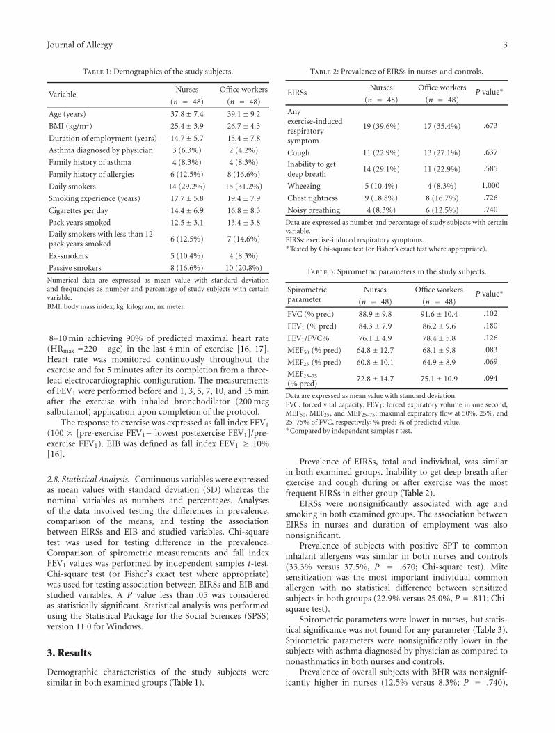
Journal of Allergy 3
Table 1: Demographics of the study subjects.
VariableNurses Office workers
(n = 48) (n = 48)
Age (years) 37.8± 7.4 39.1± 9.2
BMI (kg/m2) 25.4± 3.9 26.7± 4.3
Duration of employment (years) 14.7± 5.7 15.4± 7.8
Asthma diagnosed by physician 3 (6.3%) 2 (4.2%)
Family history of asthma 4 (8.3%) 4 (8.3%)
Family history of allergies 6 (12.5%) 8 (16.6%)
Daily smokers 14 (29.2%) 15 (31.2%)
Smoking experience (years) 17.7± 5.8 19.4± 7.9
Cigarettes per day 14.4± 6.9 16.8± 8.3
Pack years smoked 12.5± 3.1 13.4± 3.8
Daily smokers with less than 12pack years smoked
6 (12.5%) 7 (14.6%)
Ex-smokers 5 (10.4%) 4 (8.3%)
Passive smokers 8 (16.6%) 10 (20.8%)
Numerical data are expressed as mean value with standard deviationand frequencies as number and percentage of study subjects with certainvariable.BMI: body mass index; kg: kilogram; m: meter.
8–10 min achieving 90% of predicted maximal heart rate(HRmax =220 − age) in the last 4 min of exercise [16, 17].Heart rate was monitored continuously throughout theexercise and for 5 minutes after its completion from a three-lead electrocardiographic configuration. The measurementsof FEV1 were performed before and 1, 3, 5, 7, 10, and 15 minafter the exercise with inhaled bronchodilator (200 mcgsalbutamol) application upon completion of the protocol.
The response to exercise was expressed as fall index FEV1
(100 × [pre-exercise FEV1− lowest postexercise FEV1]/pre-exercise FEV1). EIB was defined as fall index FEV1 ≥ 10%[16].
2.8. Statistical Analysis. Continuous variables were expressedas mean values with standard deviation (SD) whereas thenominal variables as numbers and percentages. Analysesof the data involved testing the differences in prevalence,comparison of the means, and testing the associationbetween EIRSs and EIB and studied variables. Chi-squaretest was used for testing difference in the prevalence.Comparison of spirometric measurements and fall indexFEV1 values was performed by independent samples t-test.Chi-square test (or Fisher’s exact test where appropriate)was used for testing association between EIRSs and EIB andstudied variables. A P value less than .05 was consideredas statistically significant. Statistical analysis was performedusing the Statistical Package for the Social Sciences (SPSS)version 11.0 for Windows.
3. Results
Demographic characteristics of the study subjects weresimilar in both examined groups (Table 1).
Table 2: Prevalence of EIRSs in nurses and controls.
EIRSsNurses Office workers
P value∗(n = 48) (n = 48)
Anyexercise-inducedrespiratorysymptom
19 (39.6%) 17 (35.4%) .673
Cough 11 (22.9%) 13 (27.1%) .637
Inability to getdeep breath
14 (29.1%) 11 (22.9%) .585
Wheezing 5 (10.4%) 4 (8.3%) 1.000
Chest tightness 9 (18.8%) 8 (16.7%) .726
Noisy breathing 4 (8.3%) 6 (12.5%) .740
Data are expressed as number and percentage of study subjects with certainvariable.EIRSs: exercise-induced respiratory symptoms.∗Tested by Chi-square test (or Fisher’s exact test where appropriate).
Table 3: Spirometric parameters in the study subjects.
Spirometricparameter
Nurses Office workers P value∗(n = 48) (n = 48)
FVC (% pred) 88.9± 9.8 91.6± 10.4 .102
FEV1 (% pred) 84.3± 7.9 86.2± 9.6 .180
FEV1/FVC% 76.1± 4.9 78.4± 5.8 .126
MEF50 (% pred) 64.8± 12.7 68.1± 9.8 .083
MEF25 (% pred) 60.8± 10.1 64.9± 8.9 .069
MEF25–75
(% pred)72.8± 14.7 75.1± 10.9 .094
Data are expressed as mean value with standard deviation.FVC: forced vital capacity; FEV1: forced expiratory volume in one second;MEF50, MEF25, and MEF25–75: maximal expiratory flow at 50%, 25%, and25–75% of FVC, respectively; % pred: % of predicted value.∗Compared by independent samples t test.
Prevalence of EIRSs, total and individual, was similarin both examined groups. Inability to get deep breath afterexercise and cough during or after exercise was the mostfrequent EIRSs in either group (Table 2).
EIRSs were nonsignificantly associated with age andsmoking in both examined groups. The association betweenEIRSs in nurses and duration of employment was alsononsignificant.
Prevalence of subjects with positive SPT to commoninhalant allergens was similar in both nurses and controls(33.3% versus 37.5%, P = .670; Chi-square test). Mitesensitization was the most important individual commonallergen with no statistical difference between sensitizedsubjects in both groups (22.9% versus 25.0%, P = .811; Chi-square test).
Spirometric parameters were lower in nurses, but statis-tical significance was not found for any parameter (Table 3).Spirometric parameters were nonsignificantly lower in thesubjects with asthma diagnosed by physician as compared tononasthmatics in both nurses and controls.
Prevalence of overall subjects with BHR was nonsignif-icantly higher in nurses (12.5% versus 8.3%; P = .740),
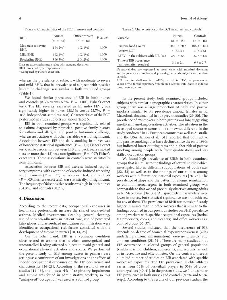
4 Journal of Allergy
Table 4: Characteristics of the ECT in nurses and controls.
BHRNurses Office workers
P value∗(n = 48) (n = 48)
Moderate to severeBHR
2 (4.2%) 1 (2.1%) 1.000
Mild BHR 1 (2.1%) 1 (2.1%) 1.000
Borderline BHR 3 (6.3%) 2 (4.2%) 1.000
Data are expressed as mean value with standard deviation.BHR: bronchial hyperresponsiveness.∗Compared by Fisher’s exact test.
whereas the prevalence of subjects with moderate to severeand mild BHR, that is, prevalence of subjects with positivehistamine challenge, was similar in both examined groups(Table 4).
We found similar prevalence of EIB in both nursesand controls (8.3% versus 6.3%, P = 1.000; Fisher’s exacttest). The EIB severity, expressed as fall index FEV1, wassignificantly higher in nurses (28.1% versus 22.7%, P =.033; independent-samples t-test). Characteristics of the ECTperformed in study subjects are shown Table 5.
EIB in both examined groups was significantly relatedto asthma diagnosed by physician, positive family historyfor asthma and allergies, and positive histamine challenge,whereas association with other variables was nonsignificant.Association between EIB and daily smoking in nurses wasof borderline statistical significance (P = .062; Fisher’s exacttest), while association between EIB and pack years smoked(less or more than 12) was nonsignificant (P = .097; Fisher’sexact test). These associations in controls were statisticallynonsignificant.
Association between EIB and exercise-induced respira-tory symptoms, with exception of exercise-induced wheezingin both nurses (P = .037; Fisher’s exact test) and controls(P = .034; Fisher’s exact test), was statistically nonsignificant.The frequency of false positive results was high in both nurses(84.3%) and controls (88.2%).
4. Discussion
According to the recent data, occupational exposures inhealth care professionals increase the risk of work-relatedasthma. Medical instruments cleaning, general cleaning,use of solvents/adhesives in patient care, use of powderedlatex gloves, and aerosolized medication administration wereidentified as occupational risk factors associated with thedevelopment of asthma in nurses [10, 14, 25].
On the other hand, EIB is a common conditionclose related to asthma that is often unrecognized anduncontrolled leading affected subjects to avoid general andoccupational physical activities and sports. We performedthe present study on EIB among nurses in primary caresettings as a continuum of our investigations on the effects ofspecific occupational exposures on the EIB occurrence andcharacteristics [26–28]. According to the results of severalstudies [11–13], the lowest risk of respiratory impairmentand asthma was found in administrative workers, so this“unexposed” occupation was used as a control group.
Table 5: Characteristics of the ECT in nurses and controls.
VariableNurses Controls
(n = 48) (n = 48)
Exercise load (Watt) 102.1± 20.3 106.3± 16.1
Positive ECT 4 (8.3%) 3 (6.3%)
ΔFEV1 in the subjects with EIB (%) 28.1± 3.4 22.7± 1.5
Time of EIB occurrence(minutes after exercise)
6.1± 2.1 6.9± 2.7
Numerical data are expressed as mean value with standard deviationand frequencies as number and percentage of study subjects with certainvariable.ECT: exercise challenge test; ΔFEV1: a fall in FEV1 of pre-exercisevalue; FEV1: forced expiratory volume in 1 second; EIB: exercise-inducedbronchoconstriction.
In the present study, both examined groups includedsubjects with similar demographic characteristics. In eithergroup, there was a large proportion of daily and passivesmokers similar to its prevalence among females in R.Macedonia documented in our previous studies [29, 30]. Theprevalence of ex-smokers in both groups was low, suggestinginsufficient smoking cessation activities. The situation in thedeveloped countries seems to be somewhat different. In thestudy conducted in 12 European countries as well as Australiaand the USA, Janson et al. [31] reported that both activeand passive smoking rates have declined since the early 1990sbut indicated lower quitting rates and higher risk of passivesmoking among people with fewer qualifications and lessskilled occupation groups.
We found high prevalence of EIRSs in both examinedgroups that is similar to the findings of several studies whichinvestigated EIB in different subpopulations of both sexes[32, 33] as well as to the findings of our studies amongworkers with different occupational exposures [26–28]. Theprevalence of atopy and the pattern of allergic sensitizationto common aeroallergens in both examined groups wascomparable to that we had previously observed among adultsin R. Macedonia [34, 35]. All spirometric parameters werelower in nurses, but statistical significance was not achievedfor any of them. The prevalence of BHR was nonsignificantlyhigher in nurses than in office workers that is similar to thefindings obtained in our previous studies on BHR prevalenceamong workers with specific occupational exposures (herbaltea processors, cooks, and cleaners) and office workers as acontrol group [36, 37].
Several studies indicated that the occurrence of EIBdepends on degree of bronchial hyperresponsiveness (aliasunderlying chronic inflammation), exercise intensity, andambient conditions [38, 39]. There are many studies aboutEIB occurrence in selected groups of general population(children, school children, adolescents, and recruits) as wellas in recreative and elite athletes. On the contrary, there isa limited number of studies on EIB associated with specificworkplace exposures. The EIB prevalence in elite athletesvaries from 12% of basketball players to 55% of cross-country skiers [40, 41]. In the present study, we found similarEIB prevalence in both nurses and controls (8.3% and 6.3%,resp.). According to the results of our previous studies, the
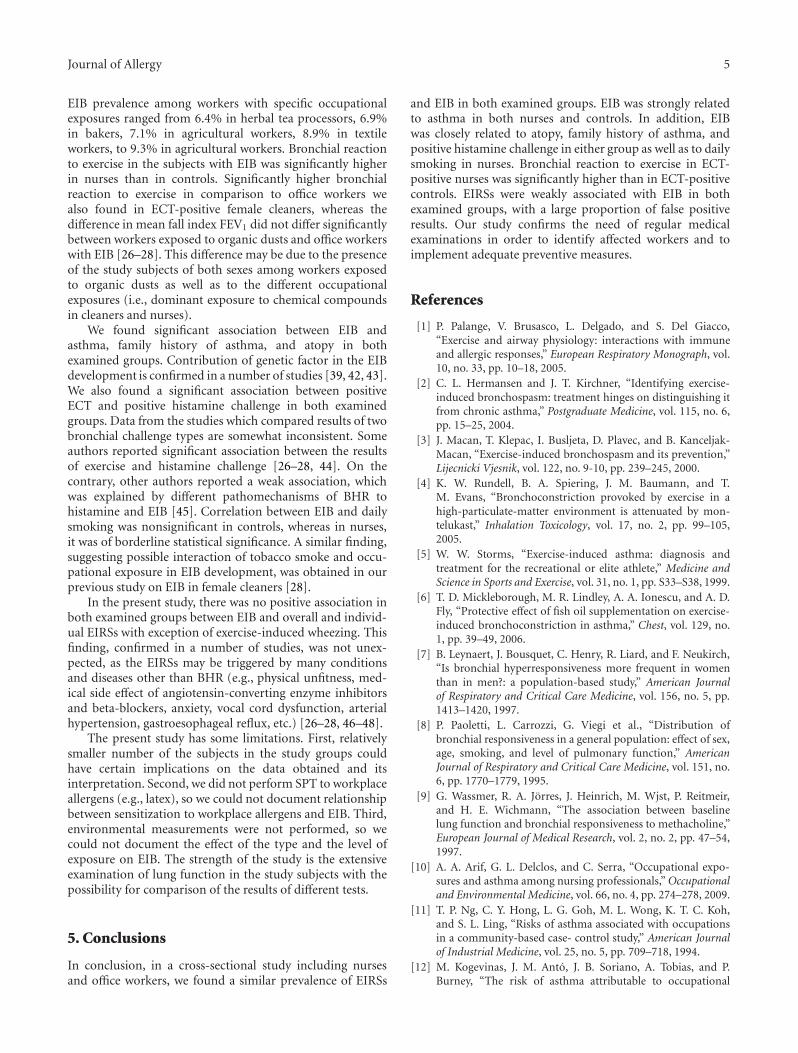
Journal of Allergy 5
EIB prevalence among workers with specific occupationalexposures ranged from 6.4% in herbal tea processors, 6.9%in bakers, 7.1% in agricultural workers, 8.9% in textileworkers, to 9.3% in agricultural workers. Bronchial reactionto exercise in the subjects with EIB was significantly higherin nurses than in controls. Significantly higher bronchialreaction to exercise in comparison to office workers wealso found in ECT-positive female cleaners, whereas thedifference in mean fall index FEV1 did not differ significantlybetween workers exposed to organic dusts and office workerswith EIB [26–28]. This difference may be due to the presenceof the study subjects of both sexes among workers exposedto organic dusts as well as to the different occupationalexposures (i.e., dominant exposure to chemical compoundsin cleaners and nurses).
We found significant association between EIB andasthma, family history of asthma, and atopy in bothexamined groups. Contribution of genetic factor in the EIBdevelopment is confirmed in a number of studies [39, 42, 43].We also found a significant association between positiveECT and positive histamine challenge in both examinedgroups. Data from the studies which compared results of twobronchial challenge types are somewhat inconsistent. Someauthors reported significant association between the resultsof exercise and histamine challenge [26–28, 44]. On thecontrary, other authors reported a weak association, whichwas explained by different pathomechanisms of BHR tohistamine and EIB [45]. Correlation between EIB and dailysmoking was nonsignificant in controls, whereas in nurses,it was of borderline statistical significance. A similar finding,suggesting possible interaction of tobacco smoke and occu-pational exposure in EIB development, was obtained in ourprevious study on EIB in female cleaners [28].
In the present study, there was no positive association inboth examined groups between EIB and overall and individ-ual EIRSs with exception of exercise-induced wheezing. Thisfinding, confirmed in a number of studies, was not unex-pected, as the EIRSs may be triggered by many conditionsand diseases other than BHR (e.g., physical unfitness, med-ical side effect of angiotensin-converting enzyme inhibitorsand beta-blockers, anxiety, vocal cord dysfunction, arterialhypertension, gastroesophageal reflux, etc.) [26–28, 46–48].
The present study has some limitations. First, relativelysmaller number of the subjects in the study groups couldhave certain implications on the data obtained and itsinterpretation. Second, we did not perform SPT to workplaceallergens (e.g., latex), so we could not document relationshipbetween sensitization to workplace allergens and EIB. Third,environmental measurements were not performed, so wecould not document the effect of the type and the level ofexposure on EIB. The strength of the study is the extensiveexamination of lung function in the study subjects with thepossibility for comparison of the results of different tests.
5. Conclusions
In conclusion, in a cross-sectional study including nursesand office workers, we found a similar prevalence of EIRSs
and EIB in both examined groups. EIB was strongly relatedto asthma in both nurses and controls. In addition, EIBwas closely related to atopy, family history of asthma, andpositive histamine challenge in either group as well as to dailysmoking in nurses. Bronchial reaction to exercise in ECT-positive nurses was significantly higher than in ECT-positivecontrols. EIRSs were weakly associated with EIB in bothexamined groups, with a large proportion of false positiveresults. Our study confirms the need of regular medicalexaminations in order to identify affected workers and toimplement adequate preventive measures.
References
[1] P. Palange, V. Brusasco, L. Delgado, and S. Del Giacco,“Exercise and airway physiology: interactions with immuneand allergic responses,” European Respiratory Monograph, vol.10, no. 33, pp. 10–18, 2005.
[2] C. L. Hermansen and J. T. Kirchner, “Identifying exercise-induced bronchospasm: treatment hinges on distinguishing itfrom chronic asthma,” Postgraduate Medicine, vol. 115, no. 6,pp. 15–25, 2004.
[3] J. Macan, T. Klepac, I. Busljeta, D. Plavec, and B. Kanceljak-Macan, “Exercise-induced bronchospasm and its prevention,”Lijecnicki Vjesnik, vol. 122, no. 9-10, pp. 239–245, 2000.
[4] K. W. Rundell, B. A. Spiering, J. M. Baumann, and T.M. Evans, “Bronchoconstriction provoked by exercise in ahigh-particulate-matter environment is attenuated by mon-telukast,” Inhalation Toxicology, vol. 17, no. 2, pp. 99–105,2005.
[5] W. W. Storms, “Exercise-induced asthma: diagnosis andtreatment for the recreational or elite athlete,” Medicine andScience in Sports and Exercise, vol. 31, no. 1, pp. S33–S38, 1999.
[6] T. D. Mickleborough, M. R. Lindley, A. A. Ionescu, and A. D.Fly, “Protective effect of fish oil supplementation on exercise-induced bronchoconstriction in asthma,” Chest, vol. 129, no.1, pp. 39–49, 2006.
[7] B. Leynaert, J. Bousquet, C. Henry, R. Liard, and F. Neukirch,“Is bronchial hyperresponsiveness more frequent in womenthan in men?: a population-based study,” American Journalof Respiratory and Critical Care Medicine, vol. 156, no. 5, pp.1413–1420, 1997.
[8] P. Paoletti, L. Carrozzi, G. Viegi et al., “Distribution ofbronchial responsiveness in a general population: effect of sex,age, smoking, and level of pulmonary function,” AmericanJournal of Respiratory and Critical Care Medicine, vol. 151, no.6, pp. 1770–1779, 1995.
[9] G. Wassmer, R. A. Jorres, J. Heinrich, M. Wjst, P. Reitmeir,and H. E. Wichmann, “The association between baselinelung function and bronchial responsiveness to methacholine,”European Journal of Medical Research, vol. 2, no. 2, pp. 47–54,1997.
[10] A. A. Arif, G. L. Delclos, and C. Serra, “Occupational expo-sures and asthma among nursing professionals,” Occupationaland Environmental Medicine, vol. 66, no. 4, pp. 274–278, 2009.
[11] T. P. Ng, C. Y. Hong, L. G. Goh, M. L. Wong, K. T. C. Koh,and S. L. Ling, “Risks of asthma associated with occupationsin a community-based case- control study,” American Journalof Industrial Medicine, vol. 25, no. 5, pp. 709–718, 1994.
[12] M. Kogevinas, J. M. Anto, J. B. Soriano, A. Tobias, and P.Burney, “The risk of asthma attributable to occupational
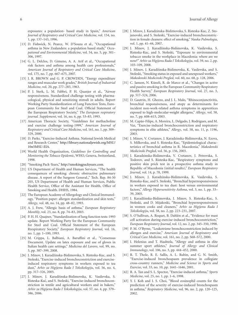
6 Journal of Allergy
exposures: a population- based study in Spain,” AmericanJournal of Respiratory and Critical Care Medicine, vol. 154, no.1, pp. 137–143, 1996.
[13] D. Fishwick, N. Pearce, W. D’Souza et al., “Occupationalasthma in New Zealanders: a population based study,” Occu-pational and Environmental Medicine, vol. 54, no. 5, pp. 301–306, 1997.
[14] G. L. Delclos, D. Gimeno, A. A. Arif et al., “Occupationalrisk factors and asthma among health care professionals,”American Journal of Respiratory and Critical Care Medicine,vol. 175, no. 7, pp. 667–675, 2007.
[15] J. R. BROWN and G. P. CROWDEN, “Energy expenditureranges and muscular work grades,” British Journal of IndustrialMedicine, vol. 20, pp. 277–283, 1963.
[16] P. J. Sterk, L. M. Fabbri, P. H. Quanjer et al., “Airwayresponsiveness. Standardized challenge testing with pharma-cological, physical and sensitizing stimuli in adults. ReportWorking Party Standardization of Lung Function Tests, Euro-pean Community for Steel and Coal. Official Statement ofthe European Respiratory Society,” The European respiratoryjournal. Supplement, vol. 16, no. 6, pp. 53–83, 1993.
[17] American Thoracic Society, “Guidelines for methacholineand exercise challenge testing-1999,” American Journal ofRespiratory and Critical Care Medicine, vol. 161, no. 1, pp. 309–329, 2000.
[18] D. Parks, “Exercise-Induced Asthma. National Jewish Medicaland Research Center,” http://library.nationaljewish.org/MSU/10n5MSU-EIA.
[19] World Health Organization, Guidelines for Controlling andMonitoring the Tobacco Epidemic, WHO, Geneva, Switzerland,1998.
[20] “Smoking Pack-Years,” http://smokingpackyears.com.[21] US Department of Health and Human Services, “The health
consequences of smoking: chronic obstructive pulmonarydisease. A report of the Surgeon General.,” Tech. Rep. 84-50205, US Department of Health and Human Services, PublicHealth Service, Office of the Assistant for Health, Office ofSmoking and Health. DHHS, 1984.
[22] The European Academy of Allergology and Clinical Immunol-ogy, “Position paper: allergen standardization and skin tests,”Allergy, vol. 48, no. 14, pp. 48–82, 1993.
[23] A. J. Frew, “Allergic basis of asthma,” European RespiratoryMonthly, vol. 23, no. 8, pp. 74–83, 2003.
[24] P. H. H. Quajner, “Standardization of lung function tests-1993update. Report Working Party for the European Communityfor Steel and Coal. Official Statement of the EuropeanRrespiratory Society,” European Respiratory Journal, vol. 16,no. 1, pp. 1–100, 1993.
[25] M. Crippa, L. Balbiani, A. Baruffini et al., “ConsensusDocument. Update on latex exposure and use of gloves inItalian health care settings,” Medicina del Lavoro, vol. 99, no.5, pp. 387–399, 2008.
[26] J. Minov, J. Karadzinska-Bislimovska, S. Risteska-Kuc, and S.Stoleski, “Exercise-induced bronchoconstriction and exercise-induced respiratory symptoms in workers exposed to teadust,” Arhiv za Higijenu Rada I Toksikologiju, vol. 56, no. 4,pp. 317–326, 2005.
[27] J. Minov, J. Karadzinska-Bislimovska, K. Vasilevska, S.Risteska-Kuc, and S. Stoleski, “Exercise-induced bronchocon-striction in textile and agricultural workers and in bakers,”Arhiv za Higijenu Rada I Toksikologiju, vol. 57, no. 4, pp. 379–386, 2006.
[28] J. Minov, J. Karadzinska-Bislimovska, S. Risteska-Kuc, Z. Sto-janovski, and S. Stoleski, “Exercise-induced bronchoconstric-tion in female cleaners: effect of smoking,” Nauka Pulmologia,vol. 1, pp. 41–44, 2007.
[29] J. Minov, J. Karadzinska-Bislimovska, K. Vasilevska, S.Risteska-Kuc, and S. Stoleski, “Exposure to environmentaltobacco smoke in the workplace in Macedonia: where are wenow?” Arhiv za Higijenu Rada I Toksikologiju, vol. 59, no. 2, pp.103–109, 2008.
[30] J. Minov, J. Karadzinska-Bislimovska, K. Vasilevska, and S.Stoleski, “Smoking status in exposed and unexposed workers,”Makedonski Medicinski Pregled, vol. 60, no. 68, p. 128, 2006.
[31] C. Janson, N. Kunzli, R. de Marco et al., “Changes in activeand passive smoking in the European Community RespiratoryHealth Survey,” European Respiratory Journal, vol. 27, no. 3,pp. 517–524, 2006.
[32] D. Gautrin, H. Ghezzo, and J. L. Malo, “Rhinoconjunctivitis,bronchial responsiveness, and atopy as determinants forincident non-work-related asthma symptoms in apprenticesexposed to high-molecular-weight allergens,” Allergy, vol. 58,no. 7, pp. 608–615, 2003.
[33] M. Capao-Filipe, A. Moreira, L. Delgado, J. Rodrigues, and M.Vaz, “Exercise-induced bronchoconstriction and respiratorysymptoms in elite athletes,” Allergy, vol. 58, no. 11, p. 1196,2003.
[34] J. Minov, V. Cvetanov, J. Karadzinska-Bislimovska, N. Ezova,S. Milkovska, and S. Risteska-Kuc, “Epidemiological charac-teristics of bronchial asthma in R. Macedonia,” MakedonskiMedicinski Pregled, vol. 56, p. 156, 2003.
[35] J. Karadzinska-Bislimovska, V. Cvetanov, J. Petrovska, S.Todorov, and S. Risteska-Kuc, “Respiratory symptoms andpositive skin prick test in a prospective asthma study inRepublic of Macedonia (initial results),” European RespiratoryJournal, vol. 14, p. 78, 1999.
[36] J. Minov, J. Karadzinska-Bislimovska, K. Vasilevska, S.Risteska-Kuc, and S. Stoleski, “Bronchial hyperresponsivenessin workers exposed to tea dust: host versus environmentalfactors,” Allergy Hypersensitivity Asthma, vol. 3, no. 1, pp. 33–39, 2005.
[37] J. Karadzinska-Bislimovska, J. Minov, S. Risteska-Kuc, S.Stoleski, and D. Mijakoski, “Bronchial hyperresponsivenessin women cooks and cleaners,” Arhiv za Higijenu Rada IToksikologiju, vol. 58, no. 2, pp. 223–231, 2007.
[38] S. O’Sullivan, A. Roquet, B. Dahlen et al., “Evidence for mastcell activation during exercise-induced bronchoconstriction,”European Respiratory Journal, vol. 12, no. 2, pp. 345–350, 1998.
[39] P. M. O’Byrne, “Leukotriene bronchoconstriction induced byallergen and exercise,” American Journal of Respiratory andCritical Care Medicine, vol. 161, no. 2, pp. S68–S72, 2000.
[40] I. Helenius and T. Haahtela, “Allergy and asthma in elitesummer sport athletes,” Journal of Allergy and ClinicalImmunology, vol. 106, no. 3, pp. 444–452, 2000.
[41] R. T. Thole, R. E. Sallis, A. L. Rubin, and G. N. Smith,“Exercise-induced bronchospasm prevalence in collegiatecross-country runners,” Medicine and Science in Sports andExercise, vol. 33, no. 10, pp. 1641–1646, 2001.
[42] R. A. Tan and S. L. Spector, “Exercise-induced asthma,” SportsMedicine, vol. 25, no. 1, pp. 1–6, 1998.
[43] Y. I. Koh and I. S. Choi, “Blood eosinophil counts for theprediction of the severity of exercise-induced bronchospasmin asthma,” Respiratory Medicine, vol. 96, no. 2, pp. 120–125,2002.

Journal of Allergy 7
[44] T. Klepac-Pulanic, J. Macan, D. Plavec, and B. Kanceljak-Macan, “Exercise and allergic diseases,” Arhiv za Higijenu RadaI Toksikologiju, vol. 55, no. 2-3, pp. 197–204, 2004.
[45] G. F. Joos, B. O’Connor, S. D. Anderson et al., “Indirect airwaychallenges,” European Respiratory Journal, vol. 21, no. 6, pp.1050–1068, 2003.
[46] K. C. Ringsberg, H. Wetterqvist, O. Lowhagen, and T. Sivik,“Physical capacity and dyspnea in patients with asthma-likesymptoms but negative asthma tests,” Allergy, vol. 52, no. 5,pp. 532–540, 1997.
[47] P. Weiner, N. Konson, A. Sternberg, D. Zamir, and Z. Fireman,“Is gastro-oesophageal reflux a factor in exercise-inducedasthma?” Respiratory Medicine, vol. 92, no. 8, pp. 1071–1075,1998.
[48] O. Lowhagen, M. Arvidsson, P. Bjarneman, and N. Jorgensen,“Exercise-induced respiratory symptoms are not alwaysasthma,” Respiratory Medicine, vol. 93, no. 10, pp. 734–738,1999.