Embed Size (px)
Citation preview

OCCASIONAL PAPER NO. 141
RECORDS OF THE
ZOOLOGICAL SURVEY OF INDIA
OCCASIONAL PAPER No. 141
STUDIES ON THE ENDOCOMMENSAL CILIATES OF ANURANS OF ANDHRA PRADESH
By
C. KALAVATI, C. C. NARASIHAMMURTI
AND
y. USHARANI
Department of Zoology, Andhra University, Waltair-530 003
----Edited by the Director, Zoological Survey of India
1991

@ Copyright Government of India, 1991
Published in June, 1991
PRICE: Inland : Rs. 45-00 Foreign: £ 3·00 $ 4'00
PRINTED IN INDIA BY THE BANI 'PB.1!S!~ 16, H1!MBNDRA SEN STREET,
CALCUTTA-700 006, PUBLISHED BY THB DIRECTOR, AND PRODUCED
BY THE PUBLICATION DIVISION ZOOLOGICAL SURVEY 01' INDIA,
CALCUTTA-700 072

RECORDS OF THE
ZOOLOGICAL SURVEY OF INDIA Occasional Paper
No. 141
INTRODUCTION
MATERIAL AND METHODS
ABBREVIATIONS USED
CLASSIFIED LIST
TAXONOMIC ACCOUNT
SUMMARY
ACKNOWLEDGEMENTS
REfERENCES
1991
CONTENTS
...
...
.. ,
... •••
'" ,.,
Pages 1-65
PAGE
1 2 2 4 5
53 54 54

INTRODUCTION
Anurans harbour a rich endocommensal fauna from Protozoa to Acanthocephala, each having a different grade of host-parasite relationships. Among protozoans, the rectal ciliates outnumber all others and constitute a very interesting heterogenous group of organisms that coexist in one and the same environment to the extent that literature on these forms CCis as confusing and confused as is the taxonomy of the ciliates themselves" (Corliss, 1978).
There is much information on different species of endocommensal rectal ciliates of anurans from other parts of the world namely, Brazil (Amaro and Sena, 1967, 1968 ; Albaret, 1967-1975) ; Ohio, U. S. A. (Earl Paul, 1970, 1972, and England (Cox, 1971). In the Indian context our knowledge on these forms remains comparatively scanty. Ghosh (1921) was the first to report Nyctotherus cordi/ormis in Bufo Inelano8tictus from Calcutta. Bhatia and Gulati (1927) studied the parasitic ciliates from frogs and toads of Punjab. de Mello (1932) worked on the protozoan endocommensals of Amphibia of Malabar and Nova Goa. Ray (1932) gave a description of Dilleria (= Balantidium) sushlii from Rana tigrina from Calcutta. Uttangi (1948-1958) carried out extensive investigations of Anura of Dharwar (Karnataka). Shete (1982, 1984) reported 10 species of Nyctotheroides and 8 species of Balantidium from amphibians of Aurangabad (Maharashtra).
The present study which was undertaken for a period of two years (1981-1983) was aimed at studying the endocomlnensal ciliates of anurans of Andhra Pradesh. During the study, besides 6 known species, 18 new species, 2 each of genera nemely, Balantidium, Trichodina, Metasicuophora and Prosicuophora; 4 Nyctotheroides and 6 Sicuophora were encountered, described and their systematic position discussed. The paper contains key to the identification of all Indian species and individual descriptions of those forms found in Andhra Pradesh,

REe. ZOOL. SURV. INDIA, Oce. PAPER No. 141
MATERIAL AND METHODS
For examining parasites, frogs of varying sizes ocllected from different habitats in Andhra Pradesh (India) were brought to the laboratory and maintained in 2' X l' X 1 1/2' size aquaria tanks. During this perion the animals were fed with odonate and other insect larvae. In some cases, frogs were dissected immediately after collection and examined for parasitic ciliates since it was felt that delay in examination could sometimes result in defaunation.
Autopsied individuals showed a large number of ciliates living endocommensally at the junction of the large intestine and upper part of the rectum. Smears were prepared from rectal contents, wet-fixed in Schaudinn's, alcoholic Bouin's or Carnoy's fluid and stained .suitably with different histological stains suc~ as Heidenhain's, Delafield's, Ehrlich's or phosphotungstic acid haematoxylin, Azocarmine, Aniline blue, Mallory's triple stain and Feulgen. Histochemical techniques such as Periodic Acid/Schiff (PAS), mercuric bromophenol blue, alcian blue, phloxine and Steedman's alcian blue were used to demonstrate adhesive organelle and dry silver method after Klein (1958) and Silver impregnation technique with ammonical silver nitrate for studying ciliary rows and infraciliature. All measurements were given 1n microns (urn) and diagrams drawn with the aid of a camera lucida.
ABBREVIATIONS USED
AD.-Adhesive disc without border; AO.-Adhesive organelle; AZM.-Adoral zone of membranelle; CV.Contractile vacuole; Cy. Ph.-Cytopharynx; Cy. Py.Cytopyge; Cy. Py. c.-Cytopygeal cilia; D.-Denticle; DR.-Denticulate ring; E.--Ectoplasm; Endo. -Endoplasm; Es.-Episarc ; Fi.-Fibrils; Fi. K.-' Fibrail of karyophore; Fi. S.-Fibrillar structures; GB.-Glycogen bodies; 1.Infundibulum; K.-Karyophore; Ma. N.-Macronucleus; P.-Peristome; RB.-Refringent bodies; S.-Suture i V.Vestibule; Ve.-Velum.

KALAVATI et al.; Studies on the Endocommensal ciliates 3
Classified list of endocommensal ciliates of amphibians described so far from India (Classification according to Levine et a!., 1980 ; Species arranged chronologically).
Phylum: CILIOPHORA Class: KINETOFRAGMINOPHOREA
Subclass: VESTIBULIFERIA Order: TRICHOSTOMATIDA
Suborder: TRICHOSTOMATINA
Family: BALANTlDIlDAE
Genus: Balantidium Claparede and Lachmann, 1858
* 1. B. eiongatllln Stein, 1867 *2. B. duodeni Stein, 1867 3. B. rotundunl Bezzenberger, 1904
*4. B. gracile Bezzenberger, 1904 S. B. helenae Bezzenberger, 1904 6. B. alnygdelli Bhatia and Gulati, 1927 7. B. bicavala Bhatia and Gulati, 1927 8. B. tigrinae Shete and Krishnamurthy, 1984 9. B. aurangabadensis Shete and Krishnamurthy, 1984
10. B. rallae Shete and Krishnamurthy, 1984 11. B. nzegaslomae Shete and Krishnamurthy, 1984 12. B. cyanophlycti Shete and Krishnamurthy, 1984 13. B. corlissi Shete and Krishnamurthy, 1984 14. B. mininucleatum Shete and Krishnamurthy, 1984 15. B. gallapatii Shete and Krishnamurthy, 1984
* 16. B. rhacophori n. sp. *17. B. wallairensis n. sp.
Class: OLIGOHYMENOPHOREA Subclass: PERITRICHIA
Order: PERITRICHIDA Suborder: MOBILINA
Fan1i1y: TRICHODINIDAE
Genus: Trichodina Ehrenberg, 1830
1. T. cyallophylcli D. sp. 2. T. wa/lairellsis n. sp.

4 REe. ZOOL. SURV. INDIA, Oce. PAPER No. 141
Class: Subclass:
Order: Suborder:
POL YHYMENOPHOREA SPIROTRICHIA HETEROTRICHIDA CLEVELANDELLINA
Family: SICUOPHORIDAB
Gellus: Sicuopbora de puytorac and Grain, 1969
1. S. kalii (Uttangi, 1950) 2. S. InacropharYllgea (Bezzenberger, 1903) 3. S. nzagna (Bezzenberger, 1904) 4. S. malabarica n. sp. S. S. wallairensis n. sp. 6. S. limnochari D. sp. 7. S. fragilis D. sp. 8. S. puytoraci D. sp. 9. S. levinei D. sp.
10. S. ranae D. sp.
Genus: Metasicuopbora Albaret, 1973 1. M. cyanophylycti D. sp. 2. M. melanosticti D. sp.
Genus: Prosicuopbora de Puytorac and Ok tern, 1967 1. P. andhrae D. sp. 2. P. hexadactyli D. sp.
Family: NYCTOTHEROIDAE
Genus: Nyctotheroides Grasse, 1928
1. N. cordi/ormis Stein (Ehren berg, 1938) 2. N. reniformis (Bhatia and Gluati, 1927) 3. N. cochlearis (Uttangi, 19~8) 4. N. caucopusi (Uttangi, 1951) 5. N. systoma (UUangi, 1958) 6. N. limnocharis (Uttangi, 1958) 7. N. curtipes (Uttangi, 1958) 8. N. breviceps (Uttangi, 1958) 9. N. bufonis (Uttangi, 1958)
10. N. (Aduncoperistomatus) cyanophlycli (Shete, 1982) 11. N. (A.) melanoslicli (Shete, 1982) 12. N. (A.) aurangabadensis (Shete, 1982) 13. N. (A.) nlarathwadensis (Shete, 1982)

KALAVATI et at. ; Studies on the Endocommensal ciliates 5
14. N. anlaroi (Shete, 1982) 1 S. N. ratnagirensis (Shete, 1982) 16. N. indicll1ll (Sbete, 1982) 16. N. rhacophori n. sp. 18. N. hha/iae n. sp. 19. N. foliatus D. sp. 20. N. hexad3cty/i n. sp.
TAXONOMIC ACCOUNT
Key to the identification of families described during the study (after Corliss, 1978).
Family: BALANTlDllDAE Reichnow in DofJ.ein and Reichnow, 1929
Cytostome at the base of vestibulam, anterior in position, somatic ciliation uniform. Parasitic in diverse hosts.
Family: TRICHODINIDAE Claus, 1874
Body cylindrical or barrel-shaped, adoral spiral 1800, 2-3
spirals, buccal ciliature conspicuous, denticles complex, macronucleus sausage to horse-shoe shaped, widely distributed in a variety of hosts.
Family: SICUOPHORIDAE Amaro, 1972
Body ovoid to ellipsoidal, sucker often extensive, present on concave surface of body, reinforced with polysaccharide skeletal elements. Macronucleus compact, kinetal sutures variable. Endocommensal in vertebrate hosts only.
Family: NYCTOTHERIDAE Amaro, 1972
Body ovoid to reniform, buccal ciliature running from near apical to sub-equatorial region forming a'S' shaped curve, nlacronucleus compact, karyophore present. Parasites of different hosts.
Genus: Balantidium Claparede and Lachmann, 1858
Genus Balantidium is istablished by Claparede and Lachmann in 1858 for a parasite which they observed in the
6 REC. ZOOl. SURV. INDIA, Oce. PAPER No. 141
rectum of frogs. The genus is characterised by the presence of a cytostome situated at the base of an anteriorly located vestibule, a body of uniform ciliation and an oval macronucleus.
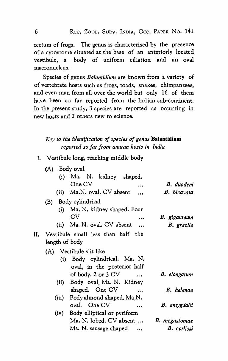
Species of genus Balantidium are known from a variety of of vertebrate hosts such as frogs, toads, snakes, chimpanzees, and even man from allover the world but only 16 of them have been so far reported from the Indian sub-continent. In the present study, 3 species are reported as occurring in new hosts and 2 others new to science.
Key to the identification of species of genus Balantidium reported so far from anuran hosts in India
I. Vestibule long, reaching middle body
(A) Body oval (i) Ma. N. kidney shaped.
OneCV (ii) Ma.N. oval. CV absent •••
(B) Body cylindrical (i) Ma. N. kidney shaped. Four
CV •.. (ii) Ma. N. oval. CV absent ...
II. Vestibule small less than half the length of body
(A) Vestibule slit like (i) Body cylindrical. Ma. N.
oval, in the posterior half of body. 2 or 3 CV ...
(ii) Body oval, Ma. N. Kidney shaped. One CV ...
(iii) Body almond shaped. Ma,N. oval. One CV ..•
(iv) Body elliptical or pyriform Ma. N. lobed. CV absent .. c
Ma. N. sausage shaped •••
B. duodenl B. bicavata
B. giganteum B. gracile
B. elongatum
B. helenae
B. amygdalli
B. megastomae B. corlissi
KALAVATI et al. : Studies on the Endocommensal ciliates 7
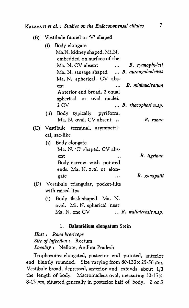
(B) Vestibule funnel or 'V' shaped
(i) Body elongare Ma.N. kidney shaped. Mi.N. embedded on surface of the Ma. N. CV absent B. cyanophylcti Ma. N. sausage shaped ... B. aurangabadensis Ma. N. spherical. CV abs-ent ... B. mininuclealum Anterior end broad. 2 equal spherical or oval nuclei. 2 CV ..• B. rhacophori n.sp.
(ii) Body typically pyriform. Ma. N. oval. CV absent ...
(C) Vestibule terminal, asymmetrical, sac-like
(i) Bod y elongate Ma. N. 'C' shaped. CV absent Body narrow with pointed ends. Ma. N. oval or elon-gate ...
(D) Vestibule triangular, pocket-like with raised lips
(i) Body flask-shaped. Ma. N. oval. Mi. N. spherical near
B. ranae
B. tigrinae
B. ganapatii
Ma. N. one CV ... B. waltairensis n.sp.
1. Balantidium elongatom Stein
Host: Rana breviceps Site of infection: Rectum Locality: Nellore, Andhra Pradesh
Trophozoites elongated, posterior end pointed, anterior end bluntly rounded. Size varying from 80-120 X 25-56 Pm. Vestibule broad, depressed, anterior and extends about 1/3 the length of body. Macronucleus oval, measuring 10-15 X
8-12 Ilm, situated generally in posterior half of body. 2 or 3

8 REe. ZOOL. SURV. INDIA, OCc. PAPER No. 141
contractile vacuoles. Cytoplasm granular. Body ciliation uniform. Kineties parallel. 36-42. Posterior cilia longer cilia longer than body cilia.
Remarks:
This form resembled those described by Bhatia and Gulati (1927) examined from R. tigrina in Punjab (Lahore). The present host, R. breviceps, is a new host record from Andhra pradesh for this species.
2. Balantidium duodeni Stein
Ilost: R. tigrina Site of infectiyn: Rectum Locality: Vizianagaram, Andhra Pradesh
Trophozoites oval or slightly bean-shaped. Size varying from 60-80X35-68 p.m. Vestibule narrow, tubular, extending up to the middle of body. Macronucleus kidney shaped, measured 10-16 X 5-10 pm. Micronucleus small, spherical, situated near one end of macronucleus. Cytopyge small, cleft-like. Single contractile vacuole. Cytoplasm granular, clearly demarcated into deeply stained ectoplasm and lightly stained endoplasm. Body ciliation uniform. 68-62 kineties.
Remarks:
This form resembled the one described by Bhatia and Gulati (1927) from Punjab in all other characters except that the species is much smaller. The present host is also a new host record from Andhra Pradesh.
3. Balantidium helenae Bezzenberger
Host: R. limnocharis Site of infection: Colon Locality: Visakhapatnam, Andhra Pradesh
Trophozoites oval, ends broadly rounded, size varying from 110-140x 60-70 urn. Vestibule broad, extending about 1/5 the length of body. Macronucleus kidney shaped generally situated in posterior region of body measuring 18-20 X 10-16 Itrn. Micronucleus not observed. Cytoplasm granular with large number of vacuoles.

KALAVATI et al. : Studies on the Endocommensal ciliates 9
Body cilia longer in posterior region forming a small tuft. Kineties discontinuous •
. Py
, I 20 }Ina
2
Fig. 1 Balantidium rhacophori n. sp.-morphology Fig. 2 Showing caliary rows Flg.3 Cyst
1Spm

10 REe. ZOOL. SURV. INDIA, Oce. PAPER No. 141
Remarks:
This form resembled those described by Bezzenberger (1904) in body size •. Though Bhatia and Gulati (1927) reported much smaller individuals, the authors have not encountered such small sized forms. The present species differs from the original form in the presence of a large number of vacuoles in its cytoplasm. Whereas the two earlier records were from R. tigrina, its present occurrence in R. Iimnocharl8 is the species new host record from Andhra Pradesh.
4. Balantidium rhacophori n. sp.
Host: Rhacophorus maculatus (Gray) Site of infection: Rectum and colon Locality: Araku Valley (Visakhapatnam District, Andhra
Pradesh) Incidence: 2/23
Type slides: ZSI pt. 2067 (Holotype) 2068 (Paratype)
Trophzoites elongated with broad anterior and narrow posterior ends. Size varying from 150-1300 X 30-70 Pm (n = 50). Width: length ratio generally 1 : 5.6. Vestibule funnel shaped and located at anterior tip that appears as a depression in fresh preparations. Vestibule opens into a small tubular cytopharynx through a narrow cytostome. Cytopharynx 15-48 /lm long.
Nuclei spherical or oval, rarely one and more often 2 of equal diameter, located in the anterior half of body measuring 10-15 X 10-12 /lm and stained deep. Micronucleus not observed. Two contractile vacuoles, one each in anterior and posterior halfs. Cytoplasm granular. Cytopyge small and triangular.
Ciliation uniform. Peristomial cilia longer. Kineties 24-38, parallel, converging near vestibule. Vestibular region with a number of closely arranged basal granules.
Reproduction through binary fission. Observed merely in forms collected from gravid female hosts during January

KALAVATI et al. : Studies on the Endocommensal ciliates 11
year ·1981~1982. Cysts oval measuring 25-40 X 50-90 J.lm. Cyst wall thin and transparent. Binucleate. Noticed during March-April months.
Remarks:
Altogether 16 species of Balantidium have been reported from anuran hosts in India but none from Rhacophorus. In comparison, the present species had no resemblance with any of the other known forms in its body size, nature of cytopharynx and nucleus. The species is also considered unique in the presence of 2 identical nuclei in over 90 010 of the trophzoites examined. Krascheninnkow (1959) reported 'monsters' of B. coli with 2 or 3 nuclei occasionally, which could be due to their incomplete division in cojugation.
Tn view of these distinct characters namely, presence of 2 identical nuclei, a slightly curved cytopharynx, funnel-shaped vestibule and 2 contractile vacuoles and based on its first record in Rhacophorus, the species is considered new to science and the name Balantidium rhaco phori n. sp. after the host is proposed.
5. Balantidium waltairensis n. sp. (Figs. 4-5)
Host: Rana cyanophlyctis Site of infection: Rectum Locality: Visakhapatnam (Andhra Pradesh) Incidence: 3/158 Type slides: ZSI, pt. 2069
Trophozoites typically £last-shaped. Posterior end broad and rounded, anterior end narrow, and pointed, measuring 110-140 X 90-105 /lm (n= 50). Width: length ratio 3 : 4. Vestibule broad, triangular with pocket-like raised lips with slight curvature. Left lip with a row of peristomial cilia. Cytopharynx tubular, 30-44 pm long, extending up to 1/3rd length of body. Macronucleus oval, always on the left side of body, 25-35 X 15-20 llm. Micronucleus spherical, 4-8 llm in diameter, located in close proximity to macronucleus. Only one contractile vacuole, 35-45 J.lm in diameter in diastole, always on the right side seen. Cytopyge not observed.

12 REc. ZOOL. SuaVe INDIA, Oce. PAPER No. 141
Ciliation uniform, ciliary rows parallel starting from a suture at anterior end below vestibule. Kinetosomes appearing
cv Ma.N
2O,um
Fig. 4 Balantidium waltairensis n. sp.-morpholoIY Fig. S Showing ciliary rows

KALAVA'r) et al. : Studies on the Endocommensal ciliates 13
pale when impregnated with silver. Kineties discontinuous. Stages of binary fission and cysts not observed.
Remarks:
The present form though resembled closely B. elongatum Stein, 1857 in body dimensions, 90-142 X 33-62 /lm (cf. Bhatia and Gulati, 1927), the species differed considerably in length: width ratios. Similarly while the nlacronucleus in the present form was found o~ the left side usually in the middle 1/3 of body, in B. elongatum it was seen in posterior region. It is significant that even though the other two species namely, B. gracilis and B helenae (Bezzenberger, 1904), occurred in anurans such as R. hexadactyla, R. linll10charis and R. tigrina, besides Rana cyanophlyctis the present species was encountered merely in R. cyanophlyctis. It differed from both B. gracilis and B. helenae in body size and shape.
In view of the distinct nature of the species stated above it is considered new to science for which Balantidium waltairensis n.sp. is proposed.
Genus: Tricbodina Ehrenberg, 1830
Several species of Trichodina are reported from piscean and amphibian hosts, both as ecto-and endoparasites. While the former are generally non-specific and occur on the body surface of fishes, the latter are more or less specific to frogs and toads living in their urinary bladder. So far, 5 species of Trichodina have been reported from frogs and toads and the other~ among newts all in urinary bladder. Lorn (1958) listed trichdinids of amphibians and proposed a scheme for their identification. The present account deals with 2 more new species of Trichodlna, one from the rectum of Rana cyanophlyctis and the other, in the urinarp bladder of R. breviceps and R. cyanopelyctis.
Genus: Trichodina Ehrenberg, 1930
Denticulate ring consists of denticles with two projectionsdistal blade and proximal ray. Adoral region with an angle of 350·450°, performing 1 to 1 1/2 circles.

14 REe. ZOOL. SURV. INDIA, OCc. PAPER No. 141
Key to the identification of species of the genus Tricbodina from anuran hosts in India :
I. Adoral zone with an angle of 360-420°, performs one complete turn. 28-42 denticles. Ma.N. bean-shaped. Mi.N. spherical. One CV in the median line. Velum incipient. T. cyanophyicli
II. Adoral zone with an angle of 390-4500
, performs 1 1/2 turns. 48-52 denticles. Ma.N. bent with one long and one short arm. Velum distinct with marginal cilia. . •.
n. sp,
T. waltairensis n. sp.
1. Trichodina cyanophlycti D. sp. (Figs. 6-8)
Host: R. cyanophlyctis Site of infection: Rectum Locality: Eluru (Andhra Pradesh) Iucidence; 1/120 Type slides: ZSI, pt. 2070
Trophozoites generally free in lumen but occasionally attached to rectal wall. Body oval or rectangular measuring 60-186x 28-128 P,m (n=50). Adoral zone having an angle of 360-420° and performs one complete turn at anterior end of body, parallel to adhesive disc. Adhesive disc 28-100 Pm, with an outer thin membrane having a row of cilia and a small depression in the centre on aboral side. Striated membrane reinforced with 4 to 5 radial pins in between two denticles. Denticulate ring 20-68 /-lm in diameter. Denticles varying from from 28-42 in number, 12-18 pm in size. Inner ray 5-10 /-lm, distal blade 6-8 Pm.
Macronucleus bean-shaped 35-48 X 15-20 p,m in size, located at right angles to main axis of body. Micronucleus small, spherical, located close to macronucleus near indentation. Cytoplasm hyaline. Velum not clear. InCipient folding of ectoplasm present, occastionally representing

KAIAVATI et al. : Studies on the Endocommensal ciliates 15
velum. Contractile vacuole single and in median line of body. No apparent damage to host tissue seen.
AD
AD
6
ADa
~::L.>.~~~~~- 0
Fig. 6 Trichodina cyanophlyc!i n. sp.-morphology Fig.7 Adhesive disc FiS- 8 Denticles

16 REe. ZOOL, SURV. INDIA, Oce. PAPER No. 141
Remarks:
Five species of Trichodina have so far been reported from the urinary bladder of frogs and toads. These include T. urinicola Fulton, 1923 from Bufo sp. (U.S.A.); T. xenopodis Fantham, 1924, Xenopus laevis T. bufonis Fantham, 1924, Bufo regularis (S. Africa); T. enlzii Bretschneider, 1935, Rana esculenla (Europe); T. ranae da Cunha, 1950, R. esculenta, .R lessonae and R. rudibunda Perezi (Europe). T. xenopodis Fantham, 1924 is reported from Xenopus laevis (S. Africa). The present form is the largest reported so far being as large as 186 X 128 p.m. In comparison with other species, T. entzi (60 X 130 pm) is perhaps the closest to the present form but differs from it in the presence of a cylindrical body and a 'U' shaped macronucleus. As regards the number of denticles, they are relatively more in the present form. With the exception of T. xenopodis (48-64), the maximum number of denticles in all others remained at 36. In T. urinicola forma bohemica, the number of denticles, however, is nearly the same as in the present form but it differs in. having a bellshaped conical body.
In view of the above differences and also since it is the first report from the rectum of a frog in India, it is considered new to science and the name Trichodina cyanophycti n. sp. after the host is proposed.
2. Trichodina waltairensis n. sp. (Figs. 9-11)
Host: Rana breviceps and R. cyanophlyctis
Site of infection: Urinary bladder Locality: Vizianagatam (Andhra Pradesh) Incidence: 6/42 R. hreviceps
2/56 R. cyanophlyctis Type slides: ZSI, Pt. 2071 Holotype
2072 Para type
Trophozoites cylindrical, 90-150 X 40-50 pm. Adoral zone of angle 390-450° forming approximately one and half turns around body with a row of cilia. Adhesive disc 56-80 p.m with an outer striated membrane. Denticulate ring 40-50 Pm

KAtAVATI et ale : Studies on the Endocommensal ciliates 17
in diameter, denticles 48-52, 8-10 radial pins in between two denticles arranged in pairs. Denticles 10-12 pm long, inner
i[
3
~[ 9
ADS
Fig. 9 Trichonil1a waltairensis D. sp.-morpholo gy Fig. 10 Adhesive disc Fig. 11 . Denticles
11

18 REe. ZOOL. SURV. INDIA, Oce. PAPER No. 141
ray of 4-5 /lm and a distal blade of 6-7 Itm. Distal blode has small, spine-like projtion. Macronucleus bent with one each of short and long arms, measuring 4C-50 X 4-5 pm. ~1icro
nucleus small, spherical situated near long arm of macronucleus. Velum deeply stained without marginal cilia. Cytoplasm hyaline, vacuolated. No contractlie vacuole.
Remarks:
This species resembles T. entzii in body size and T. xenopodis in the number of denticles, but in both cases, the macronucleus is either horse-shoe or 'U' shaped with equal arms, The macronucleus in the present form has one each of short and long arms. The velum, thovgh well developed, is non-ciliated and there is a shift of adoral zone. This species is different from T. cyanohlycti n. sp., described earlier, in structure, site of infection, host and locality.
In view of the above, and based on its occurrence, the first of its kind, in the urinary bladder of R. breviceps, the .species is considered new to science and the name Trichodina waltairensis n.sp. is suggested.
Genus: Sicuopora de puytorac and Grain
Genus Sicuophora was established by de puytorac and Grain (1968) for a ciliate they collected from the rectum of Xenopus fraseri. Genus Sicuophora is characterised by the presence of an adhesive organelle on concave side reinforced with polysaccharide skeletal elements and a ciliary system which c.onsists of a well developed pre-oral, right, apical and caudal parts. The macronucleus has generally two dorsal branches, one of which extends along peristome and the other being venttal in position. The oval micronucleus is situated at extremities of macronucleus and embedded on ventral surface. A karyophore is often present.
Altogether seven species namely, S. macrophaayngea found in R. cyanophlyctis (Bezzenberger, 8903), S. magna, R. hexadactyla (Bezzenberger, 1904); S. chen;, R. spinosa (Witchermann, 1934); S. kalil, R. curlipes (Uttangi, 1950); S. malabarica> R. tigrina (Amaro and Sena> 1967) ; S. xenopi, Xenopus
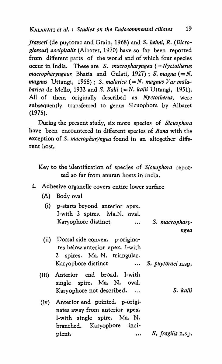
KALAVATI et ale : Studies on the Endocommensal ciliates 19
Jrasserl (de puytorac and Grain, 1968) and S. heimi, R. (Dicroglossus) occipitalis (Albaret, 1970) have so far been reported from different parts of the world and of which four species occur in India. These are S. macropharyngea (= Nyctutherus lIzacropharyngeus Bhatia and Gulati, 1927) ; S. magna (=N. magnus Uttangi, 1958); S. malarica (= N. magnus Var malabarica de Mello, 1932 and S. Kalii (== N. kalii Uttangi, 1951). All of them originally described as Nyctotherus, were subsequently transferred to genus Sicuophora by Albaret (1975).
During the present study, six more species of Sicuophora have been encountered in different species of Rana with the exception of S. macropharyngea found in an altogether different host.
Key to the identification of species of Sicuophora repor· ted so far fronl anuran hosts in India.
I. Adhesive organelle covers entire lower surface
(A) Body oval
(i) p-starts beyond anterior apex. I-with 2 spires. Ma.N. oval. Karyophore distinct ...
(ii) Dorsal side convex. p-orlglnates below anterior apex. I-with
2 spires. Ma. N. triangular.
s. macrophary-ngea
Karyopbore distinct ... S. puytoraci n.sp.
(iii) Anterior end broad. I-with single spire. Ma. N. oval. Karyophore not described. . ..
(iv) Anterior end pointed. p-originates away from anterior apex. I-with single spire. Ma. N. branched. Karyophore inci-pient. • ••
s. kalli
S. fragi/is n.sp.
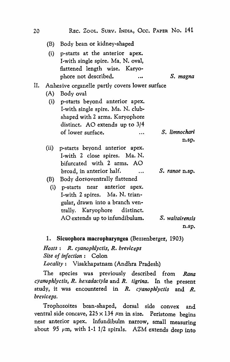
20 REe. ZOOL. SURV. INDIA, OCc. PAPER No. 141
(B) Body bean or kidney-shaped
(i) p-starts at the anterior apex. I ... with single spire. Ma. N. oval, flattened length wise. Karyophore not described. . .. S. magna
II. Anhesive organelle partly covers lower surface (A) Body oval
(i) p-starts beyond anterior apex. I-with single spire. Ma. N. clubshaped with 2 arms. Karyophore distinct. AO extends up to 3/4 of lower surface. S. limnochari
(ii) p-starts beyond anterior apex. I-with 2 close spires. Ma. N. bifurcated with 2 arms. AO broad, in anterior half.
(B) Body dorsoventrally flattened (i) p-starts near anterior apex.
I-with 2 spires. Ma. N. triangular, drawn into a branch ventrally. Karyophore distinct. AO extends up to infundibulum.
n.sp.
S. ranae n.sp.
S. waltairensis n.sp.
1. Sicuophora macropharyngea (Bezzenberger, 1903)
Hosts: R. cyanophlye/is, R. breviceps Site of infection: Colon Locality: Visakhapatnam (Andhra Pradesh)
The species was previously described from Rana cyanophlyctis, R. hexadactyla and R. tigrina. In the present study, it was encountered in R. cyanophlyetis and R. breviceps.
Trophozoites bean-shaped, dorsal side convex and ventral side concave, 225 X 134 !-lm in size. Peristome begins near anterior apex. Infundibulm narrow, small measuring about 95 I'm, with 1-1 1/2 spirals. AZM extends deep into

KALAVATI et ale : Studies on the Endocommensal ciliates 21
infundibulum. Macronucleus irregular measuring 55 .. 21 fim, karyophore thin. Micronucleus not observed. Adhesive organelle on concave side measured 200 X 120 !-lm, extending over entire body surface with distinct fibrillar structures. Body uniformly ciliated. Ciliary rows parallel. Cytopygeal canal narrow and cleft-like.
Remarks:
This form reselnbles N. macropharyngeus described by Bhatia and Gulati (1927) in all its morphological features. In 1967, de puytorac and Oktem transferred this species to genus Prosicuophora. Albaret (1973) identified the adhesive organelle and included it in genus Sicuophora. Our obserations clearly showed presence of a broad adhesive organelle on lower side, considered as a diagnostic feature of the genus. R. breviceps is also a new host record for this species.
2. Sicuophora waltairensis n. sp. (Figs. 12-15)
Hosts: R. cyanophlyctis, R. breviceps, R. limnocharis Site of infection: Rectum Locality: Waltair (Andhra Pradesh) Incidence: R. cyanophlyctis 80/158
R. breviceps 36/138 R. limnocharis 2/28
Type slides: ZSI, pt. 2073
Trophozoites oval, 250-290 X 140-175 !-lm, dorsoventrally flattened, apex rounded. Length: width 1 : 1.8. Peristome long, extending up to anterior apex, 100-125 /-tm in length. Infundibulum narrow, funnel-shaped taking a sharp bend about half the body length making an angle of about 90°. AZM extending deep into infundibulum, 290-305 !-lm in length with 2 spirals, and a short non-ciliated distal portion. Macronucleus triangular, 32-64 I'm, drawn out as a branch on ventral side. Micronucleus adherent to macronucleus at broad end. Karyophore distinct, attached to pellicle by refractile fibrillar structure.
Adhesive organelle on concave side, 196·220 X 60-124 pm,

22 REC. ZOOL. SURV. INDIA, OCC. PAPER No. 141
enveloping all organelle extending posteriorly beyoncl
.I./!;~~~,~ ~---AZM ~-p
~- Fi.~ ~-MaN ..... .....w.~-lvliN
~ .. ~~-1
·'''/''*'~~~-Cy.Ph
~~~-Cy.Py
12 50}Jm
~~-AO
l' Fig. 12 SiCUop/,Foa wa/tairensis D. sp.-morphology Fig. 13 Showing fibrillar structures Fig. 14 Showing adhesive organelle Fig. 15 Showing ciliary rows
13

KAtAVATI et al. : Studies on the Elldocommensal ciliates 23
infundibulm. PAS positive, Steedman's alcian blue positive, fibrillar structures present between adhesive organelle and pellicle. They stain yellow with phosphotungstic acid haematoxylin.
Body uniformly ciliated, ciliary rows parallel, sutures two, one at either end. Cytopygeal canal with a bulbous base extending between infundibulum and posterior end. Cysts spherical, 60-75 Jlm in diameter. Common in summer.
Remarks:
The present form is assigned to genus Sicuophora de puytorac and Grain (1968) since it has all the characters of this genus. A comparison of the present sprcies with others described so far showed that it does not resemble any one of them (Table 1), except S macropharyngea (=N. macropharyngeus Bhatia and Gulati, 1927) and S. malabarica (= N. magnus malabarica de Mello, 1932) with which it has some affinity. However, the species differs from the latter form in peristome length, presence of two spires in infundibulum and position of the nucleus. The karyophore, connected to pellicle by branched fibres in the present form, though resembles S. xenopi, the species differs from it in other characters such as size and shape of the body, length of peristome and macronucleus. In view of these features, the species is considered new to science and the name Sicuophora waltairensis n. sp. after the type locality, is suggested.
3. Sicuopbora limnochari n. sp. (Figs. 16-19)
Hosts: Rana limnocharis, R. cyanophlyctis Site of infection: Rectum Locality: Vizianagaram (Andhra Pradesh) Incidence: R. limnocharis 32/46
R. cyanophlyctis - 12/56 Type slides: ZSI, pt. 2074
Trophozoites flat, oval, ends rounded, 290-310 X 170-200 Ilm in size. Peristome starts beyond anterior apex and extends to less than middle of body, takes a broad bend nlaking an

TABLE I
Secies of Sicuophora described so far from different parts of world
Species and author
Sicuophora xenopi de puytorac and Grain, 1968
Synonyms Body Length X width
150x 100/lm
s. cheni (Wichterman, Nyctotherus chen; wich- 179 X 125 1934) de puytorac and terman, 1934 wichter-Grain, 1968 mania cheni (wichterman,
1234) Earl, 1972
Macronucleus Host and locality
Spherical 20 {lm xenopus fraseri, G~bon in diameter and France
X. mulleTi, Maboka, Central Africa
Quadritriangular Rana spinsa, China 49 X 26 Karyo-phore present
s. kaTTi (Uttangi, 1950) de puytorac and Grain, 1968
Nyctotherus kalli (Utta- 280 X 223 with a
kalli around
Body Ovoid, 101 X 30 Rana cUTtipes, Tad ngi, 1950) Nyctoth ero ides (Uttangi, 1950) Amaro and Sena 1967 Wichtermania kalTi Earl, 1972
flange p.m Poles, India
[Conld.
N o o ~
en c:: ~ •
o n n .

Species and author
s. heimi Albaret, 1970
s. macropharyngea (Bezzenberger, 1903) Albaret, 1973
s. magnr (Bezzenberger, 1904) Albaret, 1975
Synonyms
Nyetotherus macropharyngeus Bezzenberger, 1903 Nyctotheroides macropharyngeus (Bezzenberger, 1903) Amaro and Sena, 1967 Prosicuophora macropharyngeus (Bezzenberger, 1903) de puytorac and Oktem, 1967 Nyctotherus magnus Bezzenberger, 1904 Nyetotheroides magnus
Body Length X width Macronucleus Host and locality
200x 150 Ovoid
225x 135 Ovoid
290 x 230
/lm 50 /Lm long Rana (Dicroglossus) Occi-transverse patalis Republic of
Congo
/lm Irregular
Ptyehodena perreti, Republic of Central Africa
55 X 21 /lm Kar- Rana cyanophlyetis yophore present R. regularis, R. hexada
ctyla
I'm Flattened
R. tigrina, Afganisthan, Asia
R. hexadactyla, Asia [Contd.
Species and author
S •• malabarica (Amaro and Sena, 1367) Albaret. 1975
Synonyms Body Length x width
(Bezzenberger, 1904) Ovoid Amaro and Sena, 1967 Prosicuophora magnus (Bezzenherger, 1904) de puytorac and Oktem, Sicuophora mabokensis Albaret, 1970
Nyctotherus magnus Var. 250 X 140 malabarica de Mello, 1932 Nyctotheroides mag-nus malabarica (de Mello, 1932) Amaro and Sena, 1967 Prosicuophora malabarica Earl. 1972
p.m
Macronucleus Host and locality
R. (Dicrog/ossus) occipitalis, Central Africa and Republic of Gongo R. cyanophlyctis, Afganis-
than
R. malabaricus, India
N o o t""I • en c: ~ •
Z 8 > .. o o o •
§' " Z o •

KALA VATl el a/. : Studies on the Endocommensal ciliates 27
angle of 100°. AZM continues into infundibulum from
Cy.Ph Cy
17
16 f ....
SO)Jm
5----.
Fig. 16 Sicuophora limnochari n. sp.-morphology Fig. 17 Enlarged view of macronucleus-note the two arms. Fig. 18 Showing adhesive oaganelle Fig. 19 Showing ciliary rows
MiN

28 REC. ZOOL. SURV. INDIA, OCC. PAPER No. 141
peristome, measures 92-128 Jlm in length. Infundibulum starts at middle of body, runs obliquely downwards to right, forms a single spire, 120-135 Pm in length, ends at a distance of 64-90 Pm from posterior end. Macronucleus club-shaped 55-68 X 40-45 ttm, with a dorsal and venral arm. Micronucleus small, spherical 2-5 /lm in diameter, embedded at the end of club-shaped macronucleus. Nuclear complex enclosed in karyophore, attached to peristomial wall by fibrillar structures. Cytopygeal tube 48-60 I'm in length with a vacuole at distal end.
Adhesive organelle large, 225-240 X 152-270 /tm. Outer rim with a number of uniformaly arranged fibrils and greater concentration of polysaccharide material near periphery. Body ciliation uniform, kineties parallel, converge at sutures. Cilia prominent in the region of adhesive organelle.
Remarks:
Presence of a large, adhesive organelle on concave side qualified the species, inclusion in genus Sicuophora. Sizewise (290-310 X 170-200 /lm), this form resembled S. magna closely. However, it differed from it in the length of peristome and presence of a single spire in cytopharynx. The macronucleus is also characteristically club-shaped and bifurcated, the adhesive organelle extending to 3/4 length of the body. The species differed fronl all others described so far, including those found in this study, in body size, shape of cytopharynx, size of macronucleus, peristome and fibrillar structures (Tables I and I). Finally, the present record is also the first account of the species from R. limnocharis based on which it is considered new to science for which the name Sicuophora limnochari n. sp. after the host is proposed.
4. Sicuophora fragilis n. sp. (Figs. 20-23)
Hosts: Rana cyanophlyetis, R. hexadactyla Site of infection: Colon Locality: Nellore (Andhra Pradesh)

1<ALAVATI et al.: Studies on the Elldocommen,sal ciliates 29
Incidence: R. cyanophlyctis 18/33 R. hexadactyla 11/28
Type slides: iSI, pt. 2075 (Holotype) 2076 (Paratype)
MaN~~~~ AZM ~.,,. .. __
P .
20
50 pm
21
Fig. 20 Sicuophora jragUis D. sp.-morphology Fig; 21 Showing fibrillar structures
Fig. 22 Showing the adhesive organelle Fig. 23 Showing the ciliary rows

30 REC. ZOOL. SuaVe INDIA, OCC. PAPER No. 141
Trophozoites broadly oval, anterior end slightly pointed, posterior broadly rounded, dorso-ventrally flattened, glossy in appearance, transparent, thin greenish yellow in colour, measuring 180-220 X 132-144 Itm. Peristome 72-96p,m long, originating 20-25 P,m away from anterior apex, joins infundibulum appearing like a deep depression in fresh condition. Infundibulum 96-128 p.m in length, makes an acute angle of 80 0 with peristome, runs ventrally forming a small arch in middle region of body. AZM extending deep into infundibulum. Macronucleus massive, measuring 58-80 !-lm, tip bifurcated, located in the angle of peristome. Karyophore incipient. Micronucleus not distinct. Cytopygeal tube 35-45 11m long with a bulbous tip.
Adhesive organelle large, 172-186 X 96-132 JIm, occupying most of concave surface. Close knit mucopoly-saccharide fibrils, thick in posterior region than in anterior, attached to pellicle by discontinuous fibres seen. Bifid, refractile hooklike fibrillar structures, staining yellow with phosphotungstic acid haematoxylin, present. Body ciliation unifrom. Kineties obliquely arranged, 'S' shaped. Suture transverse, band-like on right side of infundibulm.
Remarks:
The present species does not resemble any other Sicuophora described so far in its features (Tables I and II). However, the form has a close affinity with S. heimi in body measurements, except for a long and spiral infundibulum, which in the present form is bent and not like a spiral. Macronucleus in this species fairly massive and appeared distinct in having an incipient karyophore. Similarly, the oblique cytopygeal tube is absent in S helmi. Further, the host of S. heimi is different being R. occipitalis.
In comparison though the present species is recorded from the same host as S. waltairensis n. sp. described earlier, it considerably differs from it in body shape, peristome, cytopharynx and adhesive organelle (Table II). The species appeared unique in having a transparent body with glossy appearance based on which features it is considered as a new

Characters
Table II
Comparise of the body measurements of Sicuophora species described (n=50)
s. wallairensis n.sp. S. limnochari n.sp. S. iraqi/is n.sp. S. puytorac; n.sp. S. levine; n.sp. S. ranae D.Sp.
Range (I'm)
Mean (I'm)
Range (I'm)
Mean (pm)
Range (porn)
Mean Range Mean Range Mean Range Mean (,urn) (pm) (#Am) (#Am) (pm) (porn) (}Am)
----------------------------------------Length of the body 250-290 270.0
157.5 290-310 170-200
300.0 185.0 1:1.6 110.0 227.5
180-220 132-144
1 : 1.4 72-96 96-128 58-80
200.0 180-250 215.0 90-108 99.0 130-184 157.0 Width of the body 140-175 138.0 124-160 142.0 64-78 71.0 112-136 124.0
1 : I.S 1:1.39 1:1.2 Width: Length 1 : 1.8 Length of p 100-125 92-128 84.0 72-104 88.0 30-40 35.0 54-90 77.0 Length of I 290-305
112.5 297.5 120-135
55.68 1 ]2.0 200-340 320.0 50-68 59.0 124-158 141.0
Length of Ma.N. 32-64 48.0 61.5 69.0 36-50 43.0 18-26 21.5 40-45 42.5 Distance of Ma.N. from the anterior apex Distance of P from the posterior end Lenglh of the cytopygeal canal Length of the adhesive organelle Width of the adhesive organelle Host and percentage of infectioD'
Site of infection .Locality
55-64
72-96
75-88
196-220
59.5
84.0
82.5
208.0
45-64
64-90
48-60
225 .. 240
58.S
77.0
54.0
232.5
32 .. 44
60-74
35-45
172-186
38.0 42-56 59.0 18-26 22.0 18-26 22.0
67.0 30-35 32.5 20-26 23.0 45-65 55.0
40.0 24-52 38.0 8-12 iO.o 20-30 25.0
179.5 150-210 180.0 68-82 75.0 72-104 88.0
80-124 102.0 152-170 161-0 96-132 114.0 80-90 85.0 36 .. 48 42.0 86-128 112.0 R. cyanophlyctis- R. limnocharis- R. cyano phlyetis-
48% 70% 54.6% R. breviceps-26% R. cyanophlyctis-24% R. hexadactyla.39% R. limnocharis-7%
Rectum Rectum Colon
R. brericeos- R. cyanophlyctis- R. /lmnocharis-70% 6% 50%
Juvenile frogs only R. hexadactyla-25% R. eyanophlyetis-6.5%
Rectum Rectum Rectum Visakhapatnam, Vizianagaram, Andhra pradesh, Andhra pradesh,
Nellore, Machilipatnam, Visakhapatnam, Eluru, Andhra pradesh, Andra prades" Andhra pradesh, Andhra pradesh,
India India India India India India
..

32 REe. ZOOL. SURV. INDIA, Oee. PAPER No. 141
species for which the name Sicuophora jragilis n. sp. is proposed.
5. SicDophora puytoraci n. sp.
Host: Rana breviceps Site of infection: Rectum Locality: Machilipatnam (Andhra Pradesh) Incidence: 29/45
Type slides: ZSI, pt. 2077
Trophozoites oval, anterior end bluntly pointed, posterior end rounded, 180-250 X 124-160 /lm in size. Convex dorsally, concave ventrally, showing characteristic wobbling ~movement anchoring to debris by adhesive organelle. Peristome 72-104 /-lm long, commences below anterior apex. Infundibulum 300-340 /lm, with 2 widely spaced spires, in posterior half of body. AZM well formed and deeply stained. Macronucleus triangular 36-50 p.m, with drawn out corners. Micronucleus adherent to the corner, 3.6-5.0 X 2.0-2.8 p.m in size. Karyophore distinct. Fibrillar structures curved radiating from karyophore towards pellicle. Cytopygeal tube 24-52 /lm, runs obliquely towards median line opening into a vacuole below infundibular spiral (Fig. 24). Adhesive organelle oval, measuring 150-210 X 80-90 tlrn, sbowing numerous irregularly distributed PAS positive rod-like polysaccharide bodies. They stained deeply with alcian blue. Ciliation uniforn. Ciliary rows parallel, converging near sutures straight.
Remarks:
The species resembles S. heimi and S. limnochari n. sp. (this study) (Tables I and II) in size but differs from both in having a longer peristome, infundibulum with 2 spirals and a triangular macronucleus. Adhesive organelle is unique being in middle of concave side unlike in other species where it is adherent to anterior apex. While cytopygeal tube opens into a contractile vacuole in the present species, it is not so in S. limnochari n. sp. The additional difference being that

KALAVATI el al. : Studies on the Endocommensal ciliates 33
s. limnochari n. sp. has a curved infundibulum absent in the present species. Hostwise, while S. heimi was reported from R. occipitalis ; S. limnochari n. sp. from both R. hexadactyla
s
~~~-AZM
~'!!:0.:::f~~- p
24
F i ----.lrto-J)..
25
~~-Ma.N
~~-~'~.'> . .:.".""., ~~- AZM
26 27
Fig. 24 Sicliophortl puytoraci D. sp.-morphology Fig. 25 Showing fibrillar structures Fig.26 Showing adhesive organelle Fig. 27 Sicuophora levine; n. sp.-morphology
~-p
-I

34 REe. ZOOL. SURV. INOlA, OCc. PAPER No. 141
and R. cyanophlyctis, the present form was encountered merely in R. breviceps.
In view of these reasons and based on the host specificity and occurrence in a geographical different locality, the species is considered new to science for which the name Sicuophora puytoraci n: sp. is proposed.
6. Sicuophora levinei n. sp.
Host: Rana cyanophlyctis Site of infection: Rectum Locality: Visakhapatnam (Andhra Pradesh) Incidence: 8/158
Type slides: ZSI, pt. 2078 Holotype 9079 Paratype
Occurs merely in juvenile frogs. Body flat, posterior end pointed, 90-108 X 64-78 pm in size. Peristome 30-40 pm originates 5-10 pm away from anterior apex. Infundibulum funnel-shaped 50-68 pm, making a prominent 'C' shaped curve in middle of body. AZM conspicuous. Macronucleus irregular, 18-26 X 12-20 pm, transversely placed, parallel to infundibulum, sometimes with drawnout ends. Cytopyge and cytopygeal tube incipient, faintly seen at posterior end. Fibrillar structures absent (Fig. 27)· Adhesive organelle oval, measuring 68-82 X 36-48 /-lm, placed 15-25 !lm away from anterior apex in posterior half of body. It has a net-work of thick and thin fibrils alternating with each other (F ig. 28), Ciliation uniform. Ciliary rows parallel. Two straight sutures.
The present form is unique in having a 'C' shaped infundibulum and differs from all other species in body size being only 99 X 71 flm (mean). The position and nature of fibres in adhesive organelle are also different from other species (Tables I and II) as also the fibrillar structures. Further, the parasite's presence merely in the metamorphosing individuals and late tadpole stages and never in adult frogs even from the same locality constituted an important criterion.
KALAVATI et al. : Studies on the Endocommensal ciliates 35
In view of the reasons mentioned above, the present form is considered new to science for which the name Sicuophora levinei n. sp. in honour of Professor N. D. Levine, Emeritus Professor, University of Illinois, U. S. A. is proposed.
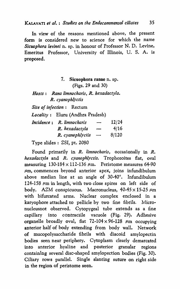
7. Sicuophora ranae n. sp. (Figs. 29 and 30)
Hosts: Rana limnocharis, R. hexadactyla. R. cyanophlyctis
Site of infection: Rectum
Locality: Eluru (Andhra Pradesh)
Incidence; R. limnocharis R. hexadactyla R. cyanophlyctis
Type slides: ZSI, pt. 2080
12/24 4/16
8/120
Found primarily in R. limnocharis, occasio.I).ally in R. hexadactyla and R. cyanophlyctis. Trophozoites flat, ov.al measuring 130-184 X 112-136 !lm. Peristome measures 64 .. 90 tIm, commences beyond anterior apex, joins infundibulum above median line at an angle of 30-40°. Infundibulum 124-158 ttm in length, with two close spires on left side of body. AZM conspicuous. Macronucleus, 40-45 X 15-25 /-lm with bifurcated arms. Nuclear complex enclosed in a karyophore attached to pellicle by two fine fibrils. Micronucleusnot observed. Cytopygeal tube extends as a fine capillary into contrac~ile vacuole (Fig. 29). Adhesive organelle broadly oval, flat 72-104 X 96-128 Pm occupying anterior half of body extending from body wall. Network of mucopolysaccharide fibrils with discoid amylopectin bodies seen near periphery. Cytoplasm clearly demarcated into anterior hyaline and posterior granular regions containIng several disc-shaped amylopection bodies (Fig. 30). Ciliary rows parallel. Single slanting suture on right side in the region of peristome seen.

36 REC. ZOOL. SURV. INDIA, OCC. PAPER No. 141
Remarks; The present form resembles S. heimi found in the rectum
of R. occipitalis in size but differs from it in the size' of
26
Fi.K-~
30
s;;;:.;c:~--AZ~A
~~--p ~~~~~~~~~---K
~~--Ma.N
"
20pn
~-cv
29
Fig. 28 Showing adhesive organelle Big.29 Sicuophora range D. sp.-morphology Fig. 30 Showing the adhesive organelle

KALAVATI et al. : Studies on the Endocommensal ciliates 37
peristome, position and size of infundibulum and the adhesive organelle. The adhesive organelle while in S. heimi extends along the entire concave surface of the body, in the present form it is flal, less than half the length of body and not depressed and cup-like as in genus Prosicuophora.
The only previous report of a species of S;cuophora from R. limnocharis is S. limnochari n. sp. (this study) whicll differs considerably fron1 the form under discussion in all characters (Table II) and hence considered as a new species for which the name Sicuophora ranae n. sp. is proposed.
Genus: Metasicuophora
Albaret (1973) estahlised a new genus Metasicuophorn for an endocommensal ciliate, M. petteri which he found in the rectum of Xenopus fraseri. The genus is characterised by the presence of terminal enlargements for marginal plaques of adhesive organelle and well developed skeletal elements of upper surface. So far, genus Metasicuophora is solely represented by M. petter; until this study when two additional species were encountered in the colon of Rana cyanophlyctis and Bufo melanostictus collected locally. For reasons discussed elsewhere in the text these species are considered new to science.
Genus: Metasicuophora Albaret, 1973
Body oval. Ao with characteristic terminal enlargements for marginal plaques. Skeletal elements well developed.
Key to the identification of species of genus Metasicuophora described so far from anuran hosts in India :
I. Body small, oval. Ectosarc thick.
p-short, commences about 10 11m from anterior apex. I-with 2 spires. Ma.N. club-shaped. Mi.N. small spherical. Ao occupies entire lower surface. Marginal plaques broad. . .. M. cyanvphl)'cti
n.sp.

38 REe. ZOOL. SuaVe INOlA, Oce. PAPER No. 141
II. Body flat, round. Ectosarc thick. p-Iong, statts beyond anterior apex. I-with one spire. Ma.N. trapezium shaped. Ao broad with double layered marginal plaques with dilated ends. . .. M. melanJsticti
n. sp.
I, Metasicuophora cyaoophlycti n.sp. (Figs. 31 and 32)
Host: Rana cyanophlyctis Site of infection: Colon Locality: Repalle, Guntur District (Andhra pradesh) Incidence: 2/126
Type slides: ZSI, pt. 2081 Holotype 2082 Paratype
Trophozoites small, oval, flattened 75-95 X 45-65 Pm. Length: width ratio 3:2. Outer ectosarc 10 lIm, peristome commences about 10 JAm from apex, 30-45 Jo'm long, makes an angle of 40-50 0 with infundibulum forming two spires in ·equatorial region in posterior half of body leading to nonciliated cytopharynx. AZM well developed. Macronucleus club-shaped, 15-30 X 10-20 Pm, placed above infundibulum • . Micronucleus small, spherical, situated near macronucleus.
Numerous thick refractile elements seen on upper surface of body. Adhesive organelle occupies entire lower surface, peripheral portion of which shows broad marginal plaques which stain deep with PAS technique and Steedman's alcian -blue. Cytopyge small and cleftlike. Contractile vacuoles not observed.
Remarks:
The present form is assigned to genus M et asicuoph ora since it has broad marginal plaques in adhesive organelle and a well developed skleton in upper surface. It differs from M. petteri, the only described species so far, in being much smaller in size (Table Ill). In M. petteri, while the macronucleus is irregular, it is club-shaped in the present form.

KALAVATI et al. : Studies on the Endocommensal ciliates 39
In view of these differences concerning its structure and based on its first occurrence in R. cyanophlyctis in the Indian
31
20 pm
Fig. 31 Fig. 32 FiB- 33
·~tf::.~~-AZM ~~~-p
:JM:~~~-Ma N
Cy.Ph
ES
AZM P
I
ES
Cy.Py-~cl
Melasicuophora cyanophlycti n. sp.-morphology An enlarged view of the margi.nal plaques AI etasicuophora melanosticli D. sp.-morphology
32
33
E ~ o m

40 REC. ZOOL. SURV. INDIA, OCC. PAPER No. 141
subcontinent, the species is described as new to for science which the name Metasicuophora cyanophlycti n.sp. afther the host is proposed.
2. Metasicoophora melanosticti n.sp. (Figs. 33 and 34)
Host: Bufo melanostictus Site of infection: Intestine and colon Locality: Machilipatnam (Andhra pradesh) Incidence: 4/32 Type slides: ZSI, pt. 2083 Holotype
2084 Paratype
Mainly encountered in toads. Trophozoites flat, round, slightly convex dorsally, concave ventrally, measuring 125-168 X 100-125 !-lm. Ectosarc 10-15 Pm thick. Peristome starts beyond anterior apex, measures 60-75 /-lm in length and extends towards median line. Infundibulum placed at right angles forming a single spire. Macronucleus trapeziumshaped 35-42 X 20-30 !-lm with drawn-out corners. Micronucleus not observed. Numerous fibrillar skeletal structures, irregularly distributed on upper surface seen. Adhesive organelle broad with double layered marginal plaques with dilated ends present in lower surface. They stain deep with PAS and Steedman's alcian blue. Cytopygeal tube at posterior end, 26-32 /-lm in length. Contractile vacuole not seen.
Remarks: This species differs from both M. petteri in body size,
length of peristome, size and shape of macronucleus (Table III) ; and M. cyanophlycti n.sp. (described earlier in the study). Albaret (1973) noted that among these forms the skeletal structures, made up of polysaccharides, constitute complementary taxonomic critera with a phylogenetic significance. The present species shows an adhesive organelle with broad marginal plaques arranged in double layers unlike the other two species. Further, the present record is also the first report of the species from Bufo melanostictus in India. Based on these findings, the species is considered new to science for which the name Metasicuophora melanosticti n.sp. after the host1 is proposed,

KAlAVATI et ale : Studies on the Endocommensal ciliates 41
Genus: Prosicuophora
Genus Prosicuophora was established by de Puytorac and Oktem in 1967, for a heterotrich ciliate which they encountered in the rectum of Bufo superciliaris and Hy/arana albolabris albolabris from Gabon, France and named its type species as P. basoglui. They identified members of the genus as commensals of batrachian anurans-'Ventose' occupying anterior region of iower surface, reinforced \vith armature, lower surface depressed to form a cup-like sucker anteriorly (c. f. Albaret, 1975). Puytorac and Oktem (1967) transferred
TABLE III
Comparison of the charcters of the species of J.Yetasicuophora (Measurements in microns)
Character M. petteri M. cyanophlycti M. melanosticti n. sp. n. sp.
Length of body 240-328 75-95 125-168 Width of body 210-290 45-65 100-125 Length of peristome 150 30-45 60-75 Distance from anterior apex 45 10 from the apex 10-15 beyond
apex Length of the infundibulum 200 120-138 65-90 Size of the macronucleus 15-30 X 10-20 35-42 X 20-30 Adhesive Broad marginal Doubled layered organelle plaques 72.5-80 marginal plaques
95-106 Host Xenopus Rana Bufo
fraseri cyanophlyctis melanostictus Site of infection Rectum Colon Intestine and
colon
Locality Latleboke Repalle Machilipatnam (Republic of (Andhra (Andhra Central Pradesh, Pradesh, India) Africa) India)
a
42 REC. ZOOL. SURV. INDIA, Occ. PAPER No. 141
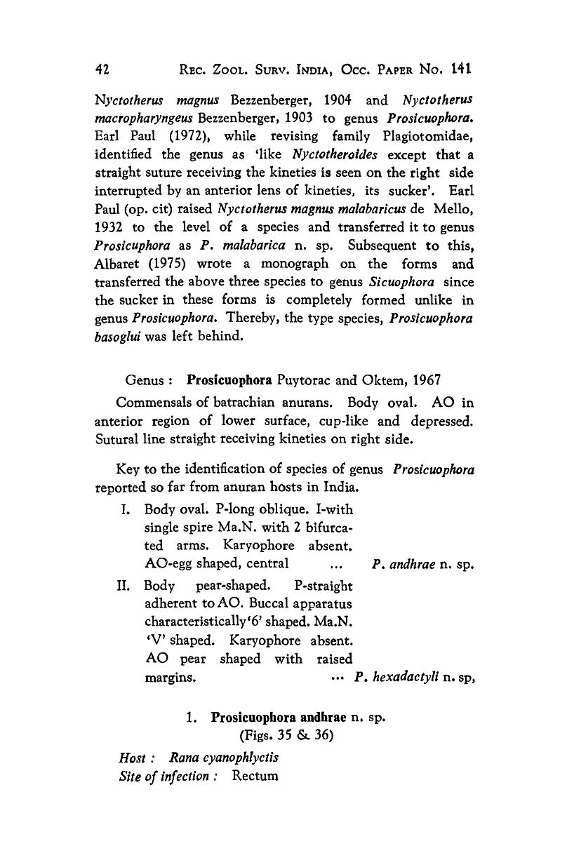
Nyctotherus magnus Bezzenberger, 1904 and Nyctotherus mac10pharyngeus Bezzenberger, 1903 to genus Prosicuophora. Earl Paul (1972), while revising family Plagiotomidae, identified the genus as 'like Nyctotheroides except that a straight suture receiving the kineties is seen on the right side interrupted by an anterior lens of kineties, its sucker'. Earl Paul (op. cit) raised Nyctotherus magnus malabaricus de Mello, 1932 to the level of a species and transferred it to genus Prosicuphora as P. malabarica n. sp. Subsequent to this, Albaret (1975) wrote a monograph on the forms and transferred the above three species to genus Sicuophora since the sucker in these forms is completely formed unlike in genus Prosicuophora. Thereby, the type species, Prosicuophora basoglui was left behind.
Genus: Prosicuophora Puytorac and Oktem, 1967
Commensals of batrachian anurans. Body oval. AO in anterior region of lower surface, cup·like and depressed. Sutural1ine straight receiving kineties on right side.
Key to the identification of species of genus Prosicuophora reported so far from anuran hosts in India.
I. Body oval. P-Iong oblique. I-with single spire Ma.N. with 2 bifurcated arms. Karyophore absent. AO-egg shaped, central P. andhrae n. sp.
II. Body pear-shaped. P-straight adherent to AO. Buccal apparatus characteri.stically'6' shaped. Ma.N. 'V' shaped. Karyophore absent. AO pear shaped with raised margins. ••• P. hexadactyli n. sp,
1. Prosicuopbora andhrae n. sp. (Figs. 35 & 36)
Host: Rana cyanophlyclis Site of infection 00 Rectum

KALABATI et al. : Studies on the Endocommensal ciliates 13
Locality: Visakhapatnam (Andhra Pradesh) Incidence: 261/58
AZM P
Cy. Ph.
CV
MaN' e ~ AO 8
r ..-
AO 100 Jjm
I E ~
8 .-
36
Cy.Py 39
Fig. 34 An enlarged view of the marginal plaques Fig. 3S Prosicuophora andhrae n. sp.-morphology Fig. 36 Adhesive organelle Fig. 37 Prosicuophora hexadactyli n. sp.-dorsal view Fig. 38 Ventral view FIg. 39 Adhesive organelle

44 REe. ZOOL. SURV. INDIA, Oce. PAPER No. 141
Type slides: ZSI, pt. 2085 Holotype 2086 Paratype
Trophozoites oval, 280-350 X 180-300 Ilm in size, anterior end pointed, posterior rounded. Peristome oblique 50-80 Itm long, originates approximately 30-40 /Lm from anterior apex, joins funnel-shaped infundibulum in middle region making an angle of 70-800
• Infundibulum has a single spire. Macronucleus 40-50 X 20-30 pm with two bifurcated arms. No karyophore. Micronucleus small, spherical, adherent to macronucleus on upper surface. Cytopyge cleft-like near posterior end. With long-tuft of cilia. 3-4 contractile vacuoles. Adhesive organelle egg-shaped, 150-180 X 90-130 pm, median, in the lower surface of anterior end, appears depressed with raised margins and irregularly distributed PAS positive, rod-like bodies. Ciliary rows slanting and discontinuous. Suture single, straight, seen on left side.
Remarks:
The present form is assigp.ed to genus Prosicuophora based on the presence of kineties, single suture and a depressed, cup-like adhesive organelle. It differs from the one form namely, P. basoglui in body size, being comparatively smaller (Table IV). In P. basoglui while the sukcer is pushed to a side, in the present form it is median in position. The transverse fibres and karyophore described in P. basoglui have not been observed in the present form.
In view of these features and based on the species first report from R. cyanophlyclis in a geographically different region, it is considered new to science for which the name Prosicuophora andJzrae n. sp. is proposed.
2. Prosicuophora hexadactyli n. sp. (Figs. 37-39)
Host: Rana hexadactyla Site of infection: Rectum Locality: Visakhapatnam and Chirala (Andhra Pradesh) Incidence: 3/138 in Visakhapatnam
2/18 in Chirala

KALABATI et al. ; Studies on the Endocommensal ciliates 45
Type slides: ZSI, pt. 2087 Holotype 2088 Paratype
Rare endocommensal. Body pear-shaped, 300-380 X 180-250 Ilm with a narrow pointed anterior end. Peristome 100-120 Pm, adherent to margin of adhesive organelle, joining funnel-shaped infundibulum in midde region making an angle of 140-1600 Infundibulunl with two spires, generally in posterior half of body with small non-ciliated distal portion. AZM well developed. Trophozoites have deep depression in this region in fresh condition. The buccal apparatus has characteristic '6' shaped appearance. Macronucleus 'V' shaped measuring 40-60 X 35-50 Pm and has two equal arms. No karyophore. Adhesive organelle pear-shaped 180-250 X
120-180 Ilm, antero-lateraI, with central cup-like depression and raised margins, bordered on right side by peristome. It is filled with a network of fibrils bordered by a number of PAS positive granules. Cytopyge small and cleft-like. No cytopygeal tuft of cilia. Contractile vacuole absent. Ciliary rows discontinuous, slanting and do not take silver impregnation uniformly. No vertical suture on right side.
Remarks:
The present form differs considerably from both P. basoglui as well as P. andhrae n. sp. (this study). It is unique in appearance with a pear-shaped anterolateral sucker (adhesive organelle) the olargin of which is bordered by peristome and AZM. Buccal apparatus has characteristic '6' like appearance as shown in figure. The difference in size measurements between the three species is given in Table IV. It may be said that the present form differs from P. basoglui and P. andhra~ n. sp. in several respects including host and locality based on which the species is considered new to science for which the name Prosicuophora hexadactyli n. sp. is proposed.
TABLE IV
Genus: Nyctotheroides Grasse, 1928 Oeder: HETEROTRICHIDA
Suborder: CLEVELANDELLINA

46- REC. ZOOL. SuaVe INOlA, OCc. PAPER No. 141
Family: NYCTOTHEROlDAE
Genus Nyctotheroides Grasse, 1928 is identified by the absence of a karyophore, and a left suture receiving slanting kinities which crosses the apex to continue as anterior right dorsum. Exclusively these are parasites of anurans.
Comparison of the characters of the species of Prosicuophora (Measurements in microns)
Character P. basoglui P. andhrae P. hexadactyli
Length of body 373 Width of body Length : Width Macronucleus with two
branches Size Karyophore Present Length of peristome Length of infundibulum Adhesive organelle 220-230
Position of adhesive organelle
Number of contractile vacuoles
Pushed to a side
n. sp.
280-350 180-300 1 : 1.5
Oval with two arms
40-50 X 20-30 Absent
50-80
230-250 Egg-shaped 150-180 X
90-130 On the lower side in the anterior end in the median line
3-4
n. sp.
300-380 180-250
1 : 2.5-3.0 'V'shaped
40-60 X 35-': 0 Absent 100-120
300-310 Pear-shaped
180-250 X
120-180 An terola teral on the lower side
Nil Host BUfo regularis Rana cyanophly- Rana hexadac-
Locality
B. superciliaris ctis tyla Hylarana albo-laris albolaris Gabon, France Visakhapat- Visakhapatnam
nam (Andhra and Chirala Pradesh, (Andhra India) Pradesh, India)
KALABATI et 01. : Studies on the Endocommensal ciliates 47
Information on Prosicuophora basoglui from Albaret (1975) As many as 86 species of Nyctotheroides have so far been
reported from anuran hosts from different parts of the world of which 16 species namely, N. cordiformis, N. reniformis, N. eoch/earls, N. caucopusi, N. systoma, N. limnocharis, N. curtipes, N. breviceps, N. bufonis, N. (Aduncoperistomatus) cyanophlycti, N. (A) melanosticti, N. (A) aurangabadensis, N. (A) marathwadensis, N. amaroi, N. ratnagirensis and N. indicum have been described from India. Shete (1982a, 1982b) included the parasites under 2 sub-genera, Aduncoperistomatus and Nyctotheroides following Grasse (1928). Since Earl Paul (1972) united the four sub-genera of Nyctotheroides into a single genus, all the species described by Shete (1982a, 1982b) were considered as different species of the genus Nyctotheroides.
In the present study, 4 new species of Nyctotheroides are described from frogs of Andhra Pradesh along with 2 previously described species namely, N. cordiformis and N. reniformis.
Key to the identification of species of Nyctotheroides reported from anurans in India.
I. Body oval (i) Cy. ph. nearly straight Ma. N.
kidney shaped p. not descri-bed. ... N. cordiformis
(ii)
(iii)
ey. ph. narrow. Ma.N. massive, oval. p-starts at anterior apex, I-curved. Cy. py. canal small .... Ends rounds. Ma.N. massive, sausage shaped. Mi.N. small in Ma.N. depression. p-Iong, nearly reaching posterior middle I-small. •••
(iv) With ventral indentation. Ma. N. massive, curved. P-small does not extend middle of the body. I-funnel shaped, short, slightly curved. Cy. py. canal angular open into large CV ..•
N. systoma
N. curtipes
N, limnocharis

48 REC. ZOOL. SURV. INDIA, OCC. PAPER No. 141
(v) Anterior end narrow. Ma. N. oval P-confined to anterior re-gion. I-narrow, tubular, oblique. Cy. py, cleft like. One CV ...
(vi) Posterior end with knob-like projection. Ma.N. sausage shaped. P-extends through the length of the body. I-narrow and curved.
(vii) Ends bluntly rounded. Ma. N. broadly rounded, narrow ven-trally and notched dorsally. Cy.
N. bufonis
N. breviceps
py. Y-shaped N. cyanophlycti
(viii) Cy. ph. J-shaped. Ma.N. elon-gated. P-near middle of the body. Cy. py. wide. . .. N. aurangabadensis
(ix) Cy. ph. uniform. Ma.N. elon-gate, ovoid, irregular. Cy. py. narrow tubular. Opens into CV. N. indicum
(x) Ma.N. triangular. Karyophore absent. P-starts below anterior end. I-J-shaped. Cy. py, opens into a CV. ... N. rhacophori n.sp.
(xi) Ma.N. conical. MLN. oval adherent to Ma.N. P-starts beyond anterior apex. I-with 2 spires. ... N. foliatus n.sp.
(xii) With deep depression on one side. Ma.N. sausage shaped. P-small. I-slightly curved, C-shaped. ..• N. hexadactyli
(xiii) Cy. ph. curved. Ma.N. massive, ovoid. Cy.py. extends up to
n.sp.
the tip of Cy. ph, ..• N. marathwadensis P.sp.

KAtABATI et ale : Studies on the Endocommensal ciliates 49
II. Body kidney or bean shaped
(i) Cy.ph. reaching beyond middle of the body. Ma.N. kidney shaped.
(ii) Cy. ph. st.aight, oblique. Ma.N. club-shaped. Mi-n. large, o~a1. Cy. py. cleft-like angular
III. Body spoon shaped
(i) Cy. ph. funnel shaped. Ma.N. long and ellipsoidal. Mi.N. not observed. . ..
(IV) Body elliptical
(i) Cy.ph. typically J-shaped. Ma.N. triangular and irregular. A loose membrane around Ma.N. present. Cy. py. tubular narrow long and delicate. . ..
(ii) P-extends to the middle of the body. Ma.N. large, elongated, sausage shaped. Cy.py. absent···
(iii) Anterior half triangular. P-extends 2/3 length of body. Ma.N. elongated, banana shaped. Cy. py. straight, delicate, narrow one or more CV
(V) Body pyramidal or conical
(i) Anterior end pointed. P .. starts from centre of anterior apex. ey. ph. with globular distal end. Buccal apparatus forms a characteristic S-shaped structure. Ma.N. triangular. Cy.py.
N. reniformis
N. caucopusi
N. eoch! earis
N. melanosticti
N. amaroi
N. ratnagirensis
opens into CV. .., N. bhatiae n. sp.
7

50 REC. ZOOL. SURV. INDIA, OCc. PAPER No. 141
1. Nyctotberoides cordiformis Ehrenberg
Host: Rana cyanophlyctis
Site of infection: Rectum
Locality: Vizianagaram (Andhra Pradesh)
Trophozoites oval measuring 80-120 X 50-85 p.m. Peristome short, starts beyond anterior apex and meets infundibulum at 90°, extending transversely up to middle of body. Macronucleus typically kidney shaped. No karyophore. Micronucleus not observed. Cytoplasm hyaline and ciliation uniform.
Remarks:
Bhatia and Gulati (1927) reported N. cardiformis from Bufo melanostictus from Punjab; Stein (1867) from Rona esculenta and R. temoporia ; de Mello (1932), R. cyanophlyctis in Goa and Uttangi (1950), R. curtipes and R. hreviceps in Mysore. The present report is the species' first record from Andhra Pradesh.
2. Nyctotheroides reniformis Bhatia and Gulati
Host: Rana hexadactyla
Site of infection: Rectum
Locality: Repalle, Guntur District, Andhra Pradesh
Trophozoites kidney-shaped measuring 80-100 X 40-60 Pm,
with a concavity on one side. Peristome starts at anterior apex and extends to middle body. Infundibulum oblique and curved. Adoral zone of membranellae well developed. Macronucleus large and oval. Micronucleus small, spherical and adherent to macronucleus. Cytoplasm hyaline.
Remarks:
Bhatia and Gulati (1927) reported N. reniformis from Bufo macro tis. Rana hexadactyla is the species new host record from Andhra Pradesh.

KALABATI et at. : Studies on the Endocommensal ciliates 51
3. Nyctotheroides rhacophori n. sp. ( Fig. 40 )
Host: Rhacoplzorus maculatus Gray Site 0/ infection: Colon Locality: Aruku Valley, Visakhapatnam District
(Andhra Pradesh)
Incidence: 2/23 Type slides: ZSI, Pt. 2089 Holotype
2090 Para type
Body oval, 80-90 X 40-50 /lm. Anterior end pointed, posterior rounded. Dorsal side concave, ventral slightly convex. Peristome 20-25 ttm starting about 10-12 /lm below anterior apex. Peristome meets infundibulum at an angle of 120°. Infundibulum narrow, 'J' shaped, 40-45 /-lm in length. Cytostome ventral in position. AZM continues from peristome into infundibulum. Cytopharynx narrow, hyaline and non-ciliated.
Macronucleus triangular 20-36 X 15-25 pm, situated above infundibulum, about 15 um from anterior apex. Karyophore absent. Micronucleus embedded ill macronucleus.
Cytopyge 3-4 tim, opens into contractile vacuole, visible only in fresh condition. Cytoplasm filled with deeply stained amylopectin granules.
Cilliation uniform. Kineties parallel, extending from anterior to posterior end, slanting, joining left suture at posterior end.
Remarks:
In general, this form resembled N. bufonis (Uttangi, 1958) Amaro and Sena (1967) parasitic in the rectum of the toad, Bufo melanostictus, but differed in size and structure being much smaller. In N. bufonis, the infundibulum takes a characteristic curvature starting from cytostome, running initially dorsally and later bending along whole length its posterior end remaining straight. However, in the present form the infundibulum takes a cJ' shaped bend with a hyaline non-ciliated cytopharynx at the distal end.
52 REC. ZOOL. SuaVe INDIA, OCC. PAPER No. 141
The massive macronucleus in N. bufonis is oval measuring 40-45 p,m while in the present form it is triangular measuring only 20-36 p.m.
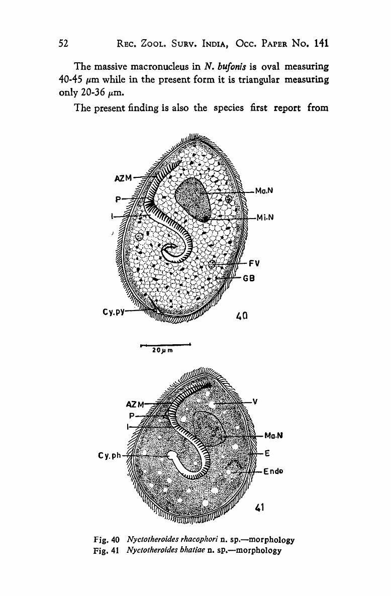
The present finding is also the species first report from
Mi.N
40
I I 20}l m
Mo.N
E
EndD
Fig. 40 Nyclolheroides rhacopJzori D. sp.-morpho!ogy Fig. 41 Nyctotheroides bhaliae. D. sp.-morphology

KALABATI el al.: Studies 011 the Endocolnmensai ciliates 53
Rhacophorus maculatus in India. In view of the distinctiveness exhibited by the form, it is considered new to science for which the name Nyctotheroides rhacophori n. sp. is proposed.
4. Nyctotheroides bhatiae n. sp.
Host: Rana limnocharis Site of infection: Rectum Locality: Visakhapatnam (Andhra Pradesh) Incidence: 6/28 Type slides: ZSI, pt. 2091 Holotype
2092 Paratype
Incidence and intensity of infection low. Occurred merely in adult frogs during Novenlber-December period (1981 and 1982). Trophozoites generally pyramidal or conical but sometimes spherical. Anterior end pointed, posterior end oval measuring 50-65 X 40-45 pm. Transparent and glossy in fresh condition. Peristome 25 p.m in length, starts from the middle of anterior apex and joins infundibulum. Infundibulum 30-40 p.m, located on dorsal side, making an angle of 90° and extending to middle of the body as nonciliated cytopharynx. Distal end of cytopharynx globular. Buccal apparatus forming a characteristic'S' shaped structure. AZM extends through the length of peristome and infundibulum.
Macrouncleus triangular, 15 x 10 Ilm, situated about 15 pm from the apex, almost adherent to infundibulum. Micronucleus not seen. Cytopyge absent. Cytoplasm hyaline with a number of small vacuoles and without any inclusions.
Kineties parallel, slanting with 2 sutures, one anterior and the other posterior. Ciliation uniform.
Remarks ,"
The present form is unique in its pyramidal or conical body shape and presence of '8' shaped buccal apparatus. It resembles N. landanae (Albaret, 1968) in having a cytopharynx with a globular distal end. However, N. landanae is typically oval in shape with an elongated macronucleus and a

54 REC. ZOOl. SURV. INDIA, Oce. PAPER No. I'll
cytopygeal canal opening into contractile vacuole while in the present form the macronucleus is triangular and the cytopyge is altogether absent.
In view of the differences is shape and structure and considering that the species is from an altogether different host it is contended that the parasite is new to science and herein described as Nyctotheroides bhatiae n. sp.
5. Nyctotheroides foliatus n. sp. (Fig. 42)
Host: Rana breviceps Site of infection: Rectum Locality: Visakhapatnam (Andhra Pradesh) Incidence: 56/138
Type slides: ZSI, Pt. 2093 Holotype 2094 Paratype
Trophozoites broadly oval, flat, with thin rounded ends, measuring 90-100 X 60-80 p.m. Generally glides on the slide rather than swim with active ciliary movements.
Peristome 55-65 Pm, originates well beyond anterior apex, and runs parallel to ventral body surface to join the funnelshaped infundibulum making an angle of 60-70°. Infundibulum narrows into a tubular part and measures 90-110 Pm. Cytopharynx 40-45 porn makes two spiral coils in the middle of the body. AZM extends through peristome and infundibulum.
Macronucleus conical 20-30 X 7-10 Pm, situated about 15-20 ttm away from anterior apex in peristomial-infundibular angle. Micronucleus snlalJ, oval, adherent to macronucleus.
Cytopyge 6-7 tlm, opens into a big contractile vacuole.
Cytoplasm granular with large number of discoid bodies. Ciliation uniform. Kineties slanting and meet near left suture at anterior apex.
Remarks:
This species resembles N. rtniformis (Bhatia and Gulati,

KALABATI et ale : Studies on the Endocommensal ciliates 55
1927), Amaro and Sena (1907) and N. cordiformis (Stein, 1867) reported from India in size but differs from them in the
cv
AZM~~..;;;;:;::J'"
p---.,l! ........ ~ ....
42
~~-MQ.N
MOIN
Mi.N
l~
43
Fig. 42 Nyctotheroides foliatus D. sp.-morphology Fi$_ 43 Nyctothero;des hexadactyli D. sp.-morpholo~y
56 RIC. ZOOL. SuaVe INDIA, Oce. PAPER No. 141
presence of a flattened, leaf-like body. The cytopharynx in the present form has two spires while it is merely curved both in N. reniformis and N. cordiformis. The species though resembles N. spirostomatus Amaro and Sena (1967) in the shape of the cytopharynx, it differs from it in size and position of the macronucleus. Similarly, while N. spirostomat us has the characteristic karyophore the same is absent in the present form. While N. breviceps, the only other species reported from Rana breviceps (Uttangi, 1958), has a unique knob-like blunt conical projection at the posterior end and a long peristomial groove extending for nearly the entire length, these characters are absent in the present form.
In view of the above and based on the morphological differences with others described so far the species is considered new to science and the name Nyctotheroides foliatus n. sp. is proposed.
6. Nyctotheroides hexadactyli n.sp. (Fig. 43)
Host: Rana hexadactyla Site of infection: Colon and rectum Locality.e Vizianagaram (Andhra Pradesh) Incidence: 3/32 Type slides: ZSI, pt. 2095 Hclotype
2096 Para type
Incidence of infection maximum during February-April, 1981 and 1982. Other than a few Opalina spp., no endocommensal ciliates were observed to coinhabit the rectum.
Trophozoites oval, small 50-68 X 30-35 /lm. Dorsal side convex. A deep depression in the region of infundibulum on ventral side seen. Peristome small, 16p.m in length and begins at anterior apex and joins the tubular infundibulum making an angle of 90°. Infundibulum curved or 'C' shaped, 30-35 p.m in length. AZM extends through the length of peristome and infundibulum. Cytopharynx not observed. Macronucleus massive, sausageshaped, 25-30 X 6.10 Pm,
generally bent, follows the course of infundibulum. Mirconucleus not observed.

KAIABATI et al. : Studies on the E"docommensal ciliates 57
Cytopyge cleft-like, 4 I'm in length with cytopygeal tuft of cilia. No contractile vacuole. Cytoplasm homogenous and granular.
Ciliation uniform. Kineties parallel and slanting. Suture in peristomial region on ventral side.
Remarks:
This species is relatively small in size and has no structural resemblance with any others described so far. The shape of the body is characreristically peculiar and resembles somewhat the kidney-shaped trophozoites of N. relliformis and N. cordi/ormis. However, it differs from both of them in all other characters such as body length, peristome and infundibulum.
A massive sausage-shaped macronucleus of the kind noiced in the present form has been reported earlier in N. breviceps (Uttangi, 1958; Amaro and Sena, 1967). However, the species differed from N. breviceps in the absence of protruberance and long double spiraJled cytopharynx. The species also differed from all others described in this study in its morphology.
In view of the above and based on the first report of the species in R. hexadactyla, it is considered new to science for which the name Nyctotheroides hexadactYli n. sp. is proposed.
SUMMARY
Endocommensal ciliates of anurans of Andhra Pradesh have been studied and 2 new species of the genus Balantidium namely, Balantidium rhacophori from rectum and colon of Rhacophorus maculatus and B. wallairensis, rectum of Rana cyanophlyctis; 2 new species of Trichodina, T. cyanophlycti rectum of R. cyanophlyctis; T. waltairensis, urinary bladder of R. breviceps and R. cyanophlyctis; 6 new species of Sicuophora, S. waltairensis, rectum of R. cyanophlyctis, R. breviceps and R. limnocharis; S. limnochari, rectum R. limnocharis and R. cyanophlyctis; S. tragilis, colon of R. cyanophlyctis and R. hexadactyla ; S. puytoraci, rectum of R.
~

58 REC. ZOOl. SURV. INDIA, Oce. PAPER No. 141
breviceps; S. levinei, rectum of R. cyanophlyctis ; S_ ranae, rectum of R. limnocharis, R. hexadactyla and R. cyanophlyctis ; 2 new species of Prosicuophora, P. andhrae, rectum of R. cyanophlyctis ; P. hexadachy/i rectum of R. hexadactyla; 2 new species of Metasicuophora, M. cyanophlycti, colon of R. tcyanophlyctis; M. melanosticti, intestine and colon of Bufo melanostictus and 4 new species of Nyctotheroides, N. rhacophori, colon of RhacophoruJ maculatus; N. bhatiae, rectum of R. limnocharis; N. foliatus, rectum of R. breviceps and N. hexadactyli, colon and rectum of R. hexadactyla have been encountered, the same described and their systematic position discussed.
Keys to the identification of species of six genera, Balantidium, Trichodina, Sicuophora, Metasicuophora, Prosicuophora and Nyctotheroides described from India also given.

Details of the endocommensal ciliates of Anura of Andhra Pradesh submitted for deposition ~ in the Zoological Survey of India, Calcutta t'"
> t::'
Slide Name of the species Name of host Site of Place of collection Date of > ~ ....
No. infection collection ~ ~
(1) (2) (3) (4) (5) (6) ~ :"""
ZSI 1 Balantid!um rhacophori n. sp. Rhacophorus maculatus Rectum & Araku Valley Dec. '83 ••
Pt. 2067 (Holotype colon (Visakhapatnam Ot.) ~ ZSI lA Balantidium rhacophori n. sp. Rizacopizoriis maculatu~ Rectum & Araku Valley Dec. '83
s::: ~
Pt. 2068 (Paratype) colon ~ tI2
ZSI 2 B. waltairensis n. sp. Rana cyanophlyctis Rectum Visakhapatnam Feb. '83 0 ;:
Pt. 2069 (Holotype and paratype) (through yaar) ..... ~
ZSI 3 Trichodina cyanophlycti Rana cyanophlyctis RecUm Visakhapatnam and Aug.-Oct. '83 ~
!i1 Pt. 2070 (Holotype and paratype) Eluru and '84 ZSI 4 T. wallvirensis n. sp. R. breviceps and Urinary Vizianagaram Feb. & March ~
~
Pt. 2071 (Holotype) R. cyanophlyctis bladder '84 0 ~
ZSI 4A T. waltairensis n. sp. R. breviceps and Urinary Vizianagaram Feb. & March ~ Pt. 2072 (Paratype) R. cyanophlyctis bladder '84
~ ~ ~
ZSI 5 Sicuophora waltairensis n. sp. R. cyanophlyctis Rectum Visakhapatnam Jan.-April ~ ...... Pt. 2073 (Holo and paratypes) R. breviceps 1983 & '84 ~ -. :::-:
ZSI 6 S. Iimnochari n. sp. R. Iimnocharis Rectum Vizianagaram June-July '84 ~ ..... Pt. 2074 (Holotype and paratype) R. cyanophlyctis ~
~
ZSI 7 S. /ragilis n. sp. R. cyanophlyctis Colon Nellore Aug. 1984 Pt. 2075 (Holotype) R. hexadac/yla
[ Con/d. CJl \0

(1) (2) (3) (4) (5) (6) 0\ 0
ZSI 7A S fi 01" . raql .. ls n. sp. R. cyanophlyctis Colon Nellore Aug. 1984 Pt. 2076 (Paratype) R. hezadactyla
ZSI 8 S. puytorac; D. sp. R. hreviceps Rectum Macbilipatnam May, 1983
Pt. 2077 (Holotype and paratype) ZSI 9 S. levinei n. sps R. cyanophlyctis Rectum Visakhapatnam July-Aug. '84 ~
tn Pt. 2078 (Holotype) () · ZSI 9A S. levine; n. sp. R. cyanophlyc/is Rectum Visakhapatnam July-Aug. '84 N Pt. 2079 (Paratype) 0
0 ZSI 10 S. ranae D. sp. R. limnoch oris Rectum Eluru Sept. 1984 t-4
• Pt. 2080 (Holotype and paratype) R. hexadactyla en
R. cyanophlyctis c ~
ZSI 11 Metasicuophora cyanophlycti R. cyanophlyclts ColoD Repalle (Guntur Dt.) Oct. 1984 · Pt. 2080
to---4 n. sp. Z
(Holotype) tj
): ZSI ItA Metasicuophora cyanophlycti R. cyanoph{vctis Colon Repalle (Gunter D~.) Oct. 1984 ...
Pt. 2082 n. sp. 0 n
(Paratype) n . ZS[ 12 M. melanosticti D. sp. Bu/o melanoslictus Intestine & Machilipatnam April, 1984 ~
Pt. 2083 (Holotype) colon > ~
ZSI 12A M. melanosticti n. sp. Bu/o melanostictus Intestine & Machilipatnam April, 1984 tz:I ~
Pt. 2084 (Paratype) colon Z ZSI 13 Prosicuophora andhrae D. sp. Rana cyanophlyclis Ructum Visakhapatnam Sept. 1983 0
• Pt.208S (Holotype) t-'"
+ [ Contd • .....

(1) (2) (3) (4) (5) (6) ~ > t'"
ZSI 13A Prosicuophora andkrae n. sp. Rana cyanophJyclis Rectum Visakhapatnam Sept. 1983 > as Pt. 2086 (Paratype) >
~ ZSI 14 P. hexadacty/i D. sp. R. hexadactyl.:z V isakhapatnam and Apr. 1984
... Rectum (b
Pt. 2087 Chirala May, 1984 ...
(Holotype) ~
ZSI 14A P. hexadactyli D. sp. R. hexadactyla Visakhapatnam and Apr. 1984 ......
Rectum • .. Pt. 2088 (Paratype) Chirala May, 1984
~ ZS[ 15 Nyctolheroides rhacophori Rhacophorus maculatus Colon Araku Valley Dec.-Jan. '83 .....
;: Pt. 2089 D. sp. (Visakhapatnam Dt.) S:
~
(Holotype) f'.a
C> ZSI 15A Nyctotheroides rhacophori Rhacophorus maculatus Colon Araku Valley Dec -Jan. '83 :s
..... Pte 2090 n. sp. (Visakhapatnam Dt.) ~
~
(Paratype) ~ ZSI 16 N. bhatiae D. sp. R. limnoc/zaris Rectum Visakhapatnam Nov.-Dec. '84 ~ Pt. 2091 (Holotype ~
C>
ZSI 16A N. bhatiae n. SPa R. Iimnocharis Rectum V isakhapatnam Nov.-Dec. '84 :i :s Pt. 2092 (Paratype) ~ ::s ZSI 17 N. foliatus R. breviceps Rectum Visakhapatnam June, 1983 f'.a
~ ...... Pt. 2093 (Holotype) ~ -. ZSI 17A N./oliatus R. breviceps Rectum Visakhapatnam June, 1983 ::::-. Q. Pt. 2094 ~ (Paratype) .....
ZSI 18 N. hexadactyli n. Spa R. hexadactyla Colon & Vizianagaarm May, 1983 ~ Pt. 2095 (Holotype) Rectum
ZSI 18A N. hexadactyli D. sp. R. hexadactyJa Colon & Vizianagaram May, 1983 Pt. 9096 (Paratype) Rectum 0\ ......

62 REC. ZOOL. SURV. INDIA, OCC. PAPFR No. 141
ACKNOWLEDGEMENTS
We are grateful to the authorities of Andhra University for the excellent facilities provided to carry out this work. One of us (YU) is thankful to the Council of Scientific and Industrial Research, New Delhi for the award of a Junior Research Fellowship during which tenure this work was carried out. Our thanks are due to Dr. Linda Sametz De Walterstein, Head Librarian, Head Librarian Rio de Janeiro, Brazil for Spanish translations.
REFERENCES
Albaret, J. L. 1967. Description de deux nouveaux nyctotheres parasites de Bufo regularis (Reuss) de Brassaville. BUll. Mus. Nat. Hists. Nat. (Ser.)., 2, 40 : 814 .. 822.
Albaret, J. L. 1968. Parasicuophora mante/tae n. gen., n. sp., cilie plagiotomidae parasite de Mantella auron/laca. Babiacian Anome de Madagascar. Protist%gica, 4 (4) : 449-451.
Albaret, J. L. 1970. Observations cytologiques sur les nyctotheres des genres Prosicuophora de Puytorac et Oktem et Sicuophora de Puytorac and Grain cilies parasites de batraciens anoures d' Afrique Noire. Description de deux especes nouvelles. Protistologica 6: 183-198.
Albaret, J. L. 1972. Description de cinq especes nouvelles du genre Nyctotheroides Grasse cilies heterotriches parasites de batraciens anoures malgaches. Bull. Mus. Nat. Hist. Nat. Zoo1., 44 : 521-531.
Albaret, J. L. 1973. Observations sur de Nyctotheres des genres Sicuophora de Puytorac and Grain, Metasicuophora gen. n. et. Parasicuophora Albaret cities parasites de Batracines Albaret cilies parasites de Batracines Anoures. J. Protozool., 20 (1) : 51·55.
KALABATI et ale : Studies on the Endocommensal ciliates 63
Albaret, J. L. 1975. Etude systematique et cytologique sur les cities Heterotriches endocommensaux. Men. Mus. Nat!' Hist. Sere A. Zoo!., 89 : 1-114.
Amaro, A. and Sena, S. 1967a. Provisional list of Nyctotherus reported in Brazil (in Spanish). Atas. Soc. BioI. Rio de Janiero., 8 (6) : 66-69.
Amaro, A. and Sena, S. 1967b. Synopsis of the species of the genus Nyctotherus Leidy, 1849 (Ciliata, Heterotrichida) reported in Brazil (in Spanish). Atas. Soc. BioI. Rio de Janeiro., 12 (1); 1·6.
Amaro, A. and Sena, S. 1968a. Sobre a sistematica de genero Nyctotheroides Grasse, 1928 (Ciliata, Heterotrichida). Atas. Soc. BioI. Rio de Janeiro., 11 (4) : 137-139.
Amaro, A. and Sena, S. 1968b. Nyctotheroides spirostomatus sp. n. an enterozoan of Bufo crucifer wied in the State of Bahia, Brazil (in Spanish). Atas. Soc. BioI. Rio de Janiero., 11 (5): 175-176.
Amaro, A. and Sena, S. 1968c. Sinopse das especies brasileiras do genero Nyctotheroides Grasse, 1928 (Ciliata, Heterotrichida): Subgeneros N. (Sigma peristomatus) Amaro and Sena, 1968 and N. (Aduncoperistomatus) Amaro and Sena, 1968. Atas. Soc. BioI. Rio de Janeiro., 11(6): 184-185.
Bezzenberger, E. 1904. Uber Infusorien aus asiatischlen Anuren. Arch. fur Protistenk., 3 : 138-174.
Bhatia, B. L. and Gulati. A. N. 1927. On some parasitic ciliates from Indian frogs, toads, earthworms and cockroaches. Arch. fur. Protistenk., 57 : 85-120.
*Bretschneider, L. H. 1935. Der Feinbau Trichodina entzii sp. nou. Parazity perehoscki. i Jadavitye Zivotrye Sbornik rabot Posvjascony 25 Letiju haucnoj deja tel nosti Prof. E. N. Parlovskogo. Izd. V sis. Inst. Eksp. Med. Moskwa-Leningrade, 363-366.
Corliss, J. O. 1978. The ciliated Protozoa: Characterisation classification and guide to the literature (2nd ed): 455 pp. Pergmon Press, London and New York.

64 REc. ZOOL. SURV. INDIA, OCC. PAPER No. 141
Cox. F. E. O. 2971. Parasites of British smphibians. J. biol~ Educ., 5 : 35-51.
Cunha, A. da. 1950. Trichodina ranae n. sp. un Urceolaire parasite de la Vessie LTrinaire de La Grcnouille. Mem. e. estud. Zool. Univ. Coimbra., 202 : 506-522.
Earl Paul, R. 1970. Some protozoan endosymbionts in Ohio frogs. Acta Protozoo!', 7(34) : 491-503.
Earl Paul, R. 1972. Synopsis of the plagiotomidae, new superfamily. Acta Protozool., 9 : 247 -261.
Earl Paul, R. 1980. Standardization of Nyctotheran ciliate descriptions. Zool. Anz., 205 (5/6) ; 371-374.
Fanthom, H. B. 1924. Some parasitic Protozoa from South Africa. S. Afr. J. Sci., 21 : 435-447.
Fulton, J. F. 1923. Trichodina pediculus and a new closely related species Trichodina urinicola. Proc. Boston. Soc. Nat. Hist., 37 : 1-29.
Ghosh, E. N. 1921. Infusoria from the environment of Calcutta. Bu11. Carmichael. Med. ColI. Cal. No. II.
Grasse, P. P. 1928. Sur quelques Nyctotherus (infusories heterotriches) nouveaux ou peu connus. Ann. Prot., 1 : 1-41.
Klein, B. M. 1958. The dry silver method and its proper use. J. protozoo!., 5 : 99.
Krascheninnikow, S. 1959. Abnormal infraciliates of Balantidium coli and B. caviae (?) and some morphological observations on these species. J. Protozoo!', 6 (1) : 61-68.
Lom, J. 1958. A contribution to the systematics and morphology of endoparasitic Trichodinids from the urinary bladder of amphibians with a proposal of uniform specific characteristics. J. Protozool., 5 (4) ; 251-263.
Mello. de, I. F. 1932. Contribution a 1 etude des infusoires parasites des Anoures de Malabar. Ree. Ind, Mus, Calcutta, 34 : 89-124,

KALABATI et al. : Studies on the Endocommensal ciliates 65
Puytorac, P. de and Grain, J. 1968. Structure et ultrastructure de Sicuophora xenopi n. gen., n. sp. cilie Heterotricha parasite du Babiecian, Xenopus fraseri Boul. Protistologica, 4 : 405-414.
Puytorac, P. de. and Oktem, N. 1967. Observations cytologiques sur les Nyctotherus Leidy et Prosicuophora n. gen., cilies parasites de batracien Anoures du Gabon. BioI. Gabon., 3 : 223-243.
Ray, H.N. 1932. On the morphology of Balantidium sushilii n. sp. from Rana tigrina Daud. J. Roy. Microscop. Soc., 52 : 374-382.
Shete, S.O. 1982a. Observations on the rectal ciliates of the genus Nyctolheroides Grasse, 1928 from Indian amphibians. 1. Subgenus Aduncup eristomat us. Arch. Protistenk., 125 : 167-112.
Shete, S.O. 1982h. Observations on the rectal ciliates of the genus Nyctotheroides Grasse, 1928 from Indian amphibians. 2. Subgenus Nyctotheroides. Arch. Protistenk., 125 : 173-180.
Shete, S.O. 1984. Observations on the rectal ciliates of the genus Balanitidium Claperede and Lachmann, 1958 from Indian amphibians Rana tigrina and R. cyanophlyctis.
Arch. Protistenk., 128 : 179-194.
·Stein, F. 1867. Der organismus der Infusionsthiere. Pt. 2:1.
Uttangi, J.C. 1948. Note on Nyctotherus cochlearis n. sp. from Rana curtipes Jordon. Curr. Sci., XVII: 325-327.
Uttangi, J.O. 1950. On a new ciliate Nyctotherus kalli n.sp. found in the tadpoles of Rana curtipes. Curr. Sci., XIX: 287-288.
Uttangi, J. C. 1951. On a new species of Nyctotherus found in Uperodon systoma Schneieer (Microhylidae). Curr. Sci., XX : 210-211.
Uttangi, J.C. 1958. On the morphology of 5 new species of Nyctotherus found in frogs and toads from Dharwar. Jour. Univ. Bombay. Sec.B-, Z6(5) : 50-64.
*Not referred to in original
![Sawyeria marylandensis (Heterolobosea) Has a … · mitochondria and hydrogenosomes (e.g., Nyctotherus ovalis [7] * Corresponding author. Mailing address: Department of Biochem-istry](https://img.pdfslide.us/doc/110x75/605fb88c3c04374bf918aee4/sawyeria-marylandensis-heterolobosea-has-a-mitochondria-and-hydrogenosomes-eg.jpg)


![[1] 1 2 5 6 15 a 15 a Q) ¥33, ¥33, 000. - 000. - 000. - 15 14 29 04 51 16 53 53 00 17 50 56 08 50 00 40 15 46 09 18 00 09 35 141 141 141 141 141 141 141 141 141 141 141 141 54 49](https://img.pdfslide.us/doc/110x75/5f09a6d27e708231d427dc4e/1-1-2-5-6-15-a-15-a-q-33-33-000-000-000-15-14-29-04-51-16-53-53.jpg)