Embed Size (px)
Citation preview
Arch. Protistenk. 124 (1981) : 405-409
Department of Zoology, Ms rathwad a Univers it y , Aurangabad,
::\Iaharashtra, India
Observations on the Rectal Flagellates of Indian Amphibians.III. Genus: Trichomitus SWEZY, 1915
By R. KRISHNAMURTHY and S. G. SHETE
With 7 Figures
Summary
The paper describes a flag ellate Trichomitus aurangabadensis n, sp , from Rana tigrina in India.
It is charact erised by the absen ce of pelt a and presence of a short undulating membrane and
endoa xos t yla r granules.
Introduction
A sur vey of rectal protozoa of frogs in Maharashtra State was carried out between1976 and 1978. Besides several ciliates and opalinates these hosts showed presenceof numerous flagellate species. Thi s communication gives an account of a flagellatebelonging to the genus Trichomitus SWEZY, 1915.
Material and Methods
The fecal conten t s of the rec t um were diluted with 0.7 % norm al saline and examined forpre sen ce of flagellates. P erman en t prep arat ion s were both by dry and wet methods. The drysm ears were fixed in methan ol an d staine d with GlElIISA'S stain . The wet smears were fix ed inS CIIAUDINN'S fluid and stained with phosphotungstic haematoxylin. The drawings were m ad ewith the help of a camera lu cida a t a magnification of 2000 x . Measurements were based on acoun t of 50 organisms.
Results
1'richomitus aurangabadensis n. sp. (Figs. 1-7)
This flagellate was found in the rectum of the frog Rana tiqrina. The infection wascommon.
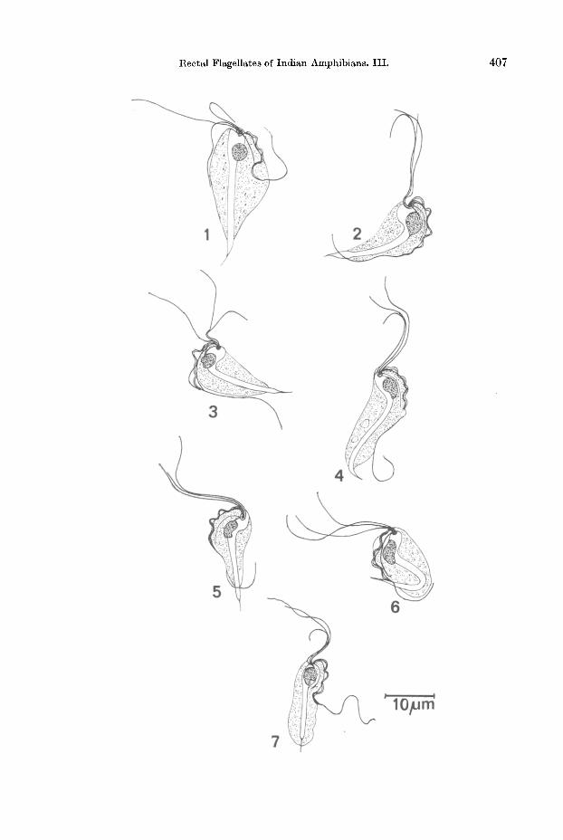
Morphology: The parasit e is elongated, fusiform in shape, with a broader anterior half and a conical and narrow posterior half (Figs. 1-3,5).
A distinct blepharoplast is seen close to the anterior end of the body (Figs. 3-6),giving rise to the various mastigont elements. There are three anterior flagella whichare unequal. The shortest flagellum is about three fourths of t he body length, whilethe longest measures one and one-four th times the length of the body. In most cases
406 R. KRISHNAMURTHY and S. G. SHETE
the three flagella run close together. The posterior flagellum runs along the borderof an undulating membrane and becomes free posteriorly. The free part of the posteriorflagellum is about as long as the body. The undulating membrane is conspicuous,extending to about one third of the body length (Figs. 1, 3, 4) or slightly more (Figs. 2,5, 6, 7). The membrane is thrown into 3-5 folds, of variable depth. Besides theposterior flagellum, the membrane is bordered by an accessory filament which isalmost as thick as the flagellum and runs the entire length of the undulating membrane. There is a costa which is uniform in its thickness and runs along the base ofthe undulating membrane, almost midway between the membrane and the axostyle.The costa is of the same thickness as the flagella and is as long as the undulatingmembrane. No paracostal granules are present.
The axostyle is extremely well developed, stout and tubular. Its anterior part isexpanded into a distinct capitulum while the trunk is almost uniform in diameter.Posteriorly it projects out of the body and tapers into a fine spike. The spike measures1.8-5.1 fJ,m in length with an average of 2.8 fJ,m. There are no endoaxostylar granules.
The nucleus lies near the capitulum and is conspicuous. It is almost spherical(Fig. 1) somewhat elongated (Figs. 4, 6) or kidney shaped (Fig. 5).
The cytoplasm is slightly vacuolated but devoid of granular inclusions.
The dimensions of the parasite are as follows: (All measurements are in microns).
Sr. No.
1.
2.
3.4.
5.6.
7.
8.
Particulars
Length of the bodyBreadth of the bodyLength of the axostylar spike
Length of the anterior flagellum ILength of the anterior flagellum IILength of the anterior flagellum IIILength of the free posterior flagellumSize of the nucleus
Minimum
10.3
5.6
1.8
8.4
12.2
14.5
13.1
2.6 X 1.8
Maximum
25.4
15.0
5.1
23.5
24.4
26.3
26.8
5.1 X 3.2
Average
19.0
9.2
2.8
16.2
18.0
22.31\).7
3.9 X 2.7
Figs. 1-7. Trichomitus auranqabadensie n. sp.
Fig. 1. Showing costa, axostylar structure, spherical nucleus and short undulating membrane.
Fig. 2. Showing longer undulating membrane and unequal anterior flagella.
Fig. 3. Showing blepharoplast and origin of mastigont elements.
Fig. 4. Showing axostylar structure, short undulating membrane and elongated nucleus.
Fig. 5. Showing reniform nucleus.
Figs. 6, 7. Showing general structure.
Rectal Flagellates of Indian Amphibians. III. 407
408 R. KRISHNAlI1URTHY and S. G. SHETE
Discussion: The genus Trichomitus contains nine species namely:
1.2.3.
4.5.6.7.8.9.
T·faecalis
T. wenyoni
T. batrachorum
T. marmotae
T. ulmeri
T. rotunda
T.lisbemyi
T. hyderabadensis
T. qromulae
CLEVELAND, 1928
WENRICH and NIE, 1949(PERTY, 1852)
HONIGBERG, 1953
GABEL,1954GABEL,1954
HIBLER et al., 1960JANAKIDEVI, 1961KRISHNAMURTHY, 1968KRISHNAMURTHY and MADRE, 1977
A comparison of present species with these organisms shows several differences.This species is distinctly larger in size than all the species described so far. Its typicallyelongated and fusiform shape and the absence of pelta besides the short undulatingmembrane distinguish this species from T. batrachorum.
In not having a pelta, this species resembles T. ulmeri, T. rotunda, T. hyderabadensis and T. granulae. However it is demarcated from all these species by other characters. It is much larger than T. ulmeri (10.3-25.4x5.6-15pm as against 4-9x 1-4pm) and has a much shorter trailing flagellum (about as long as body, as againsttwo and a half times the body length). Conspicuous difference in size also demarcatesthis species from T. rotunda which measures 6.83-11.4X4.56-7.44pm. The presentspecies is distinguished from T. hyderabadensis by its slightly larger size, by the absence of an additional accessory filament, by its shorter trailing flagellum and distinctly shorter undulating membrane. The short undulating membrane also demarcates this species from T. granulae which has an undulating membrane extending upto two thirds of the body length. Further the endoaxostylar granules, characteristicof the latter species, are absent in the form described here.
In the light of its distinctness, particularly its large size and relatively short undulating membrane, this species, is designated as Trichomitus aurangabadensis n. sp,
SpeciesHostHabitatLocality
Trichomitus aurangabadensis n. sp.Rana tigrina
RectumAurangabad, Maharashtra, India.
The slides of the type material of the species described is deposited in the Protozoology Sec
tion, Department of Zoology, Marathwada University, Aurangabad.
Acknowledgement
The authors are thankful to Dr. R. NAGABHUSHANAM for providing facilities and for his encouragement.
Rectal Flagellates of Indian Amphibians. III.
Literature
409
CL:EVELAND, L. R.: Tritrichomonas [aecalis nov. sp. of man; its ability to grow and multiply in
definitely in feces diluted with tap water and in frogs and tadpoles. Am. J. Hyg. 8 (1928):232-255.
GABEL, J. R.: A new protozoan, Poratrichomonas ulmeri (Mastigophora), from the American wood
chuck, Mormota monax LINNAEL"S. J. Tennessee Acad. Sci. 29 (1954): 200-265.
HIBLER, C. P., et al.: The morphology and incidence of the trichomonads of Swine, Tritricho
monas suis (GRUBY and D:ELAFOND), Tritrichomonas rotunda n, sp. and Trichomonas buttreyi
n. sp. J. Protozool. 7 (1900): 159 -171.
HONIGBERG, B. M.: Structure, taxonomic status and host list of Trichomonad batrachorum (PERTY).J. Parasit. 39 (1953): 191-208.
JANKIDEVI, K.: Tritrichomonas lissemyi n. sp. A parasitic protozoan from the turtle. Ann. Maq.of Nat. Rist. 13 (1901): 411-414.
KRISHNAMURTHY, R.: On a new flagellate Trichomitus hyderabadensis n. sp. from the commonfrog, Rana tigrina. Jour. Bomb. Nat. Hist. Soc. 65 (1908): 513-516.
- and MADRE, V. E.: "A new trichomonad flagellate Trichomitus granulae n. sp. from the rectumof the frog Rana cyunophlyctis in Maharashtra, India". Abst. I. Nat. Congo Paraaitol, (1977): 15.
SWEZY, 0.: On a new trichomonad flagellate, Trichomitus parous from the intestine of Amphibians. Univ. Calif. Publ. Zoo!' 1~ (1915): 89-94.
WENRICH, D. R., and NIE, D.: The morphology of Trichomonas wenyoni (Protozoa, Mastigophora). J. Morph. 85 (1949) 519-531.
Authors' address: Dr. R. KRISHNAMURTHY and S. G. SHETE, Department of Zoology, Marathwada University, Aurangabad - 431004, India.