Embed Size (px)
Citation preview
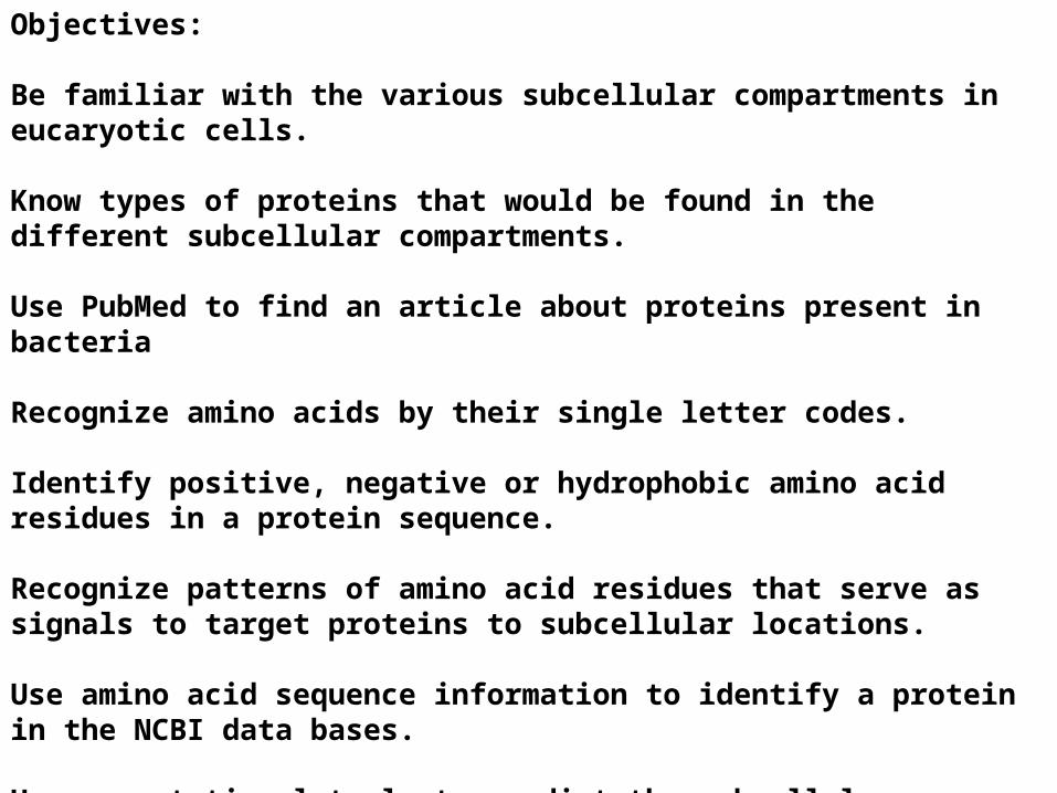
Objectives:
Be familiar with the various subcellular compartments in eucaryotic cells.
Objectives:
Be familiar with the various subcellular compartments in eucaryotic cells.
Know types of proteins that would be found in the different subcellular compartments.
Objectives:
Be familiar with the various subcellular compartments in eucaryotic cells.
Know types of proteins that would be found in the different subcellular compartments.
Use PubMed to find an article about proteins present in bacteria
Objectives:
Be familiar with the various subcellular compartments in eucaryotic cells.
Know types of proteins that would be found in the different subcellular compartments.
Use PubMed to find an article about proteins present in bacteria
Recognize amino acids by their single letter codes.
Objectives:
Be familiar with the various subcellular compartments in eucaryotic cells.
Know types of proteins that would be found in the different subcellular compartments.
Use PubMed to find an article about proteins present in bacteria
Recognize amino acids by their single letter codes.
Identify positive, negative or hydrophobic amino acid residues in a protein sequence.
Recognize patterns of amino acid residues that serve as signals to target proteins to subcellular locations.
Use amino acid sequence information to identify a protein in the NCBI data bases.
Use computational tools to predict the subcellular location for a protein of given sequence (homework)
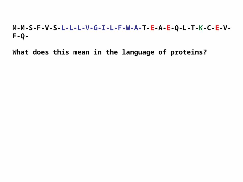
M-M-S-F-V-S-L-L-L-V-G-I-L-F-W-A-T-E-A-E-Q-L-T-K-C-E-V-F-Q-
What does this mean in the language of proteins?
M-M-S-F-V-S-L-L-L-V-G-I-L-F-W-A-T-E-A-E-Q-L-T-K-C-E-V-F-Q-
What does this mean in the language of proteins?
What would be the subcellular location of a protein with this sequence of amino acids?
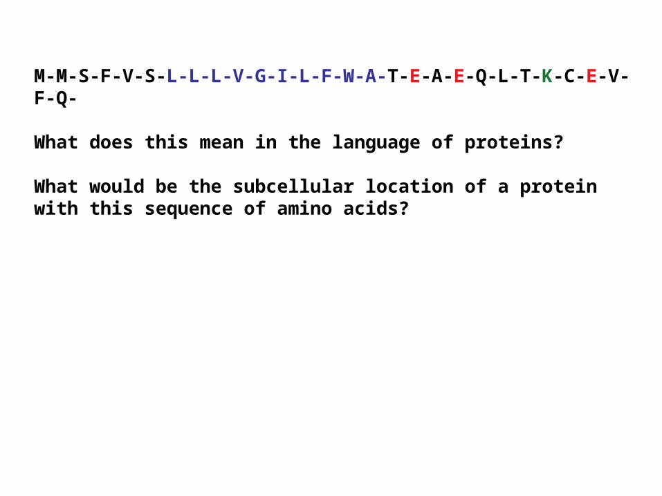
M-M-S-F-V-S-L-L-L-V-G-I-L-F-W-A-T-E-A-E-Q-L-T-K-C-E-V-F-Q-
What does this mean in the language of proteins?
What would be the subcellular location of a protein with this sequence of amino acids?
How would such a protein be delivered to its final location?
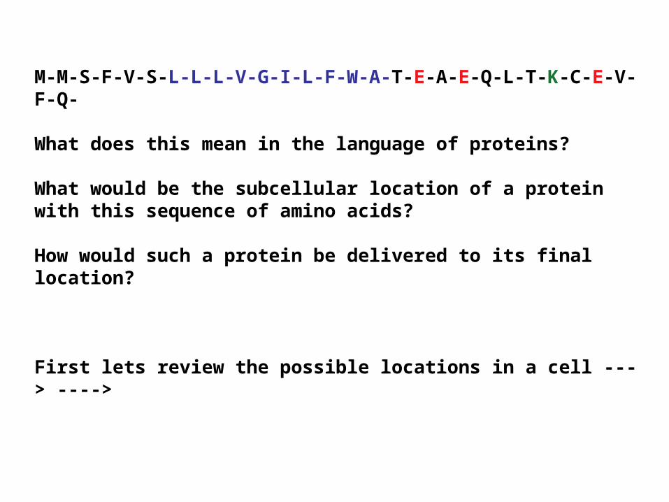
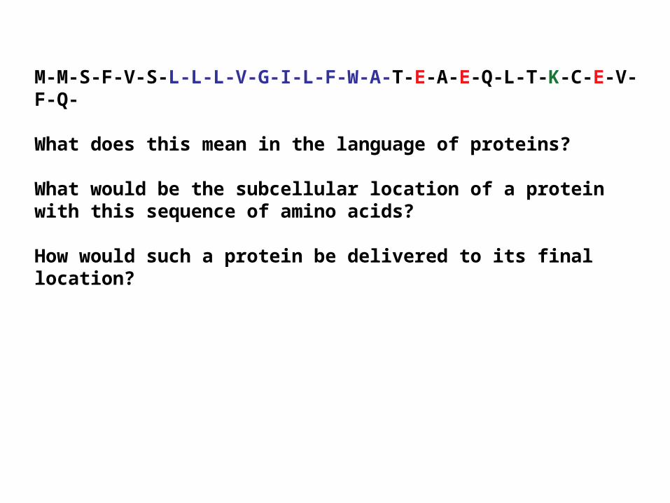
M-M-S-F-V-S-L-L-L-V-G-I-L-F-W-A-T-E-A-E-Q-L-T-K-C-E-V-F-Q-
What does this mean in the language of proteins?
What would be the subcellular location of a protein with this sequence of amino acids?
How would such a protein be delivered to its final location?
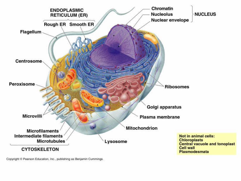
First lets review the possible locations in a cell ---> ---->
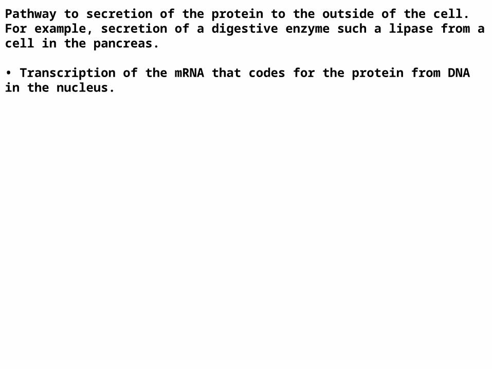
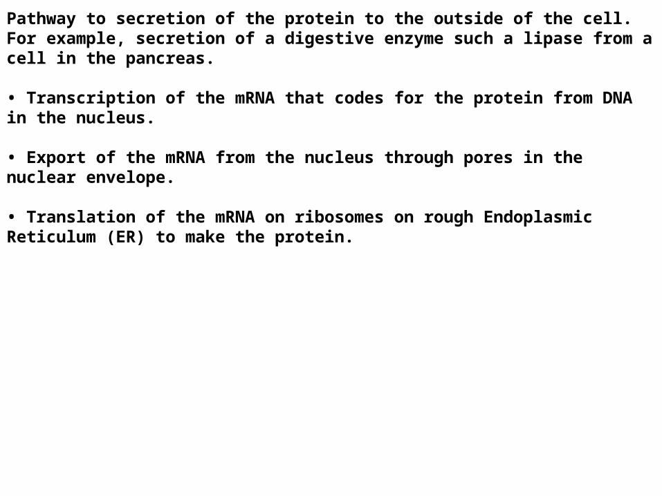
Pathway to secretion of the protein to the outside of the cell.For example, secretion of a digestive enzyme such a lipase from a cell in the pancreas.
• Transcription of the mRNA that codes for the protein from DNA in the nucleus.
Pathway to secretion of the protein to the outside of the cell.For example, secretion of a digestive enzyme such a lipase from a cell in the pancreas.
• Transcription of the mRNA that codes for the protein from DNA in the nucleus.
• Export of the mRNA from the nucleus through pores in the nuclear envelope.
• Translation of the mRNA on ribosomes on rough Endoplasmic Reticulum (ER) to make the protein.
•The protein is threaded into the lumen of the ER because of signal sequence of amino acids (blue) near amino terminus of the protein.
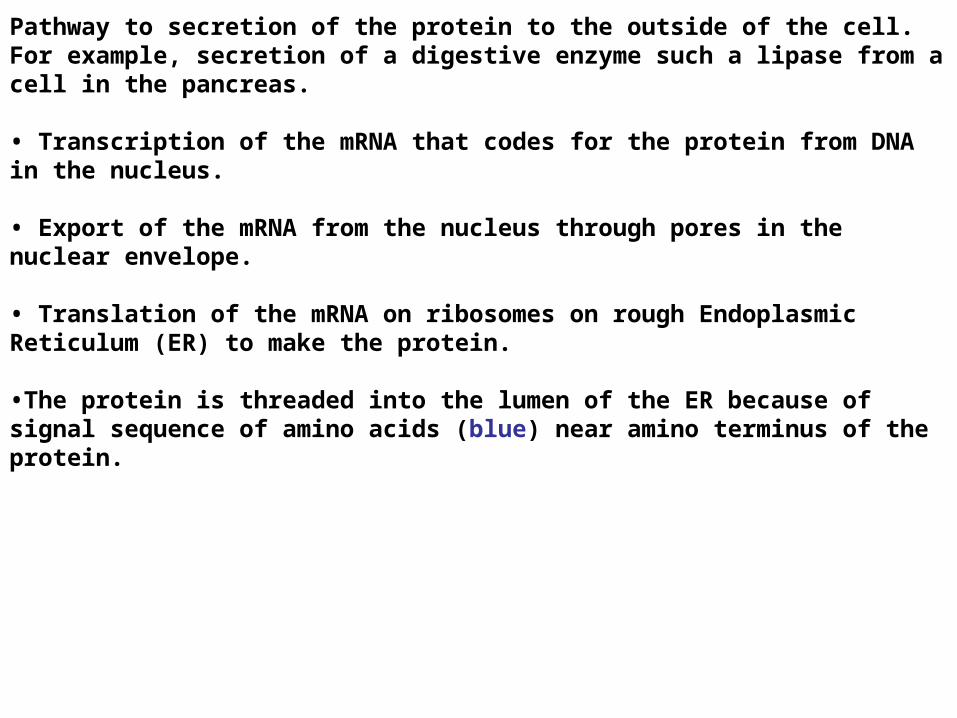
•The protein is passed on to the Golgi.
Pathway to secretion of the protein to the outside of the cell.For example, secretion of a digestive enzyme such a lipase from a cell in the pancreas.
• Transcription of the mRNA that codes for the protein from DNA in the nucleus.
• Export of the mRNA from the nucleus through pores in the nuclear envelope.
• Translation of the mRNA on ribosomes on rough Endoplasmic Reticulum (ER) to make the protein.
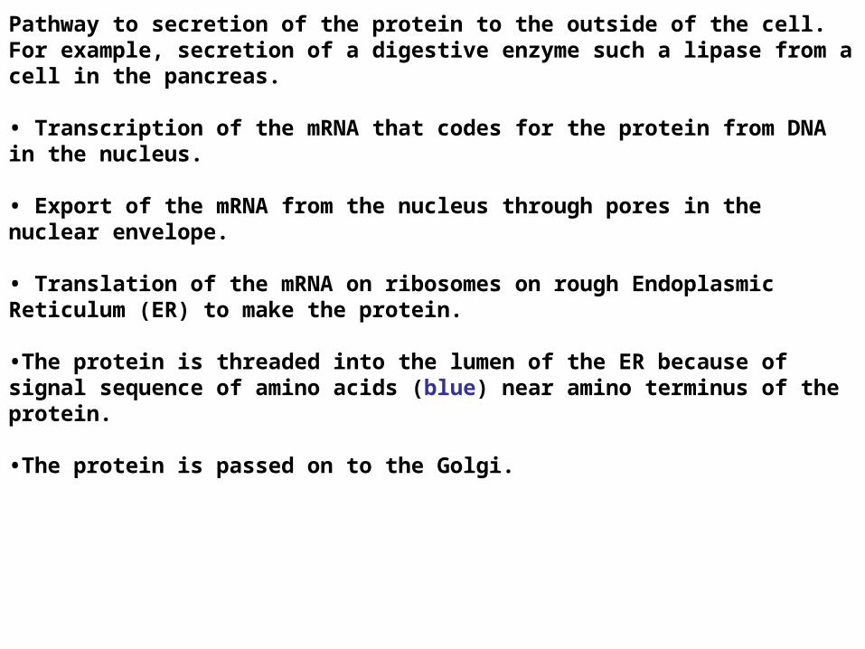
•The protein is threaded into the lumen of the ER because of signal sequence of amino acids (blue) near amino terminus of the protein.
•The protein is passed on to the Golgi.
•The protein is enclosed in a membrane vesicle which leaves the Golgi and takes it to the Plasma Membrane (PM)
Pathway to secretion of the protein to the outside of the cell.For example, secretion of a digestive enzyme such a lipase from a cell in the pancreas.
• Transcription of the mRNA that codes for the protein from DNA in the nucleus.
• Export of the mRNA from the nucleus through pores in the nuclear envelope.
• Translation of the mRNA on ribosomes on rough Endoplasmic Reticulum (ER) to make the protein.
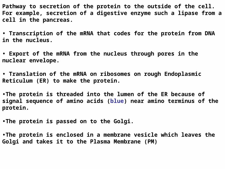
•The protein is threaded into the lumen of the ER because of signal sequence of amino acids (blue) near amino terminus of the protein.
•The protein is passed on to the Golgi.
•The protein is enclosed in a membrane vesicle which leaves the Golgi and takes it to the Plasma Membrane (PM)
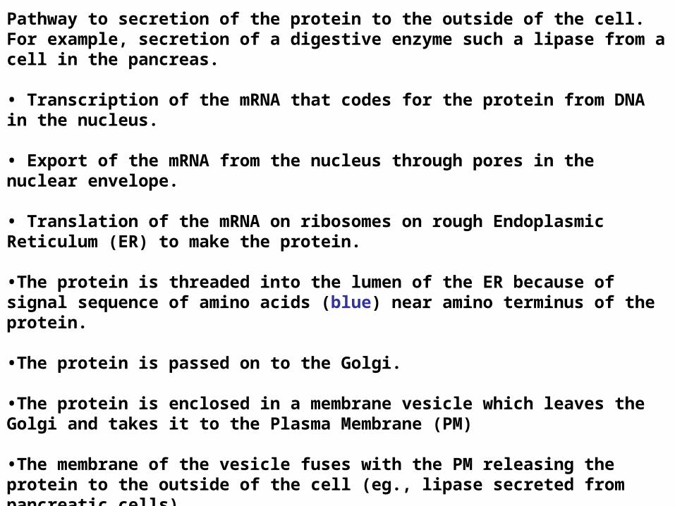
•The membrane of the vesicle fuses with the PM releasing the protein to the outside of the cell (eg., lipase secreted from pancreatic cells)
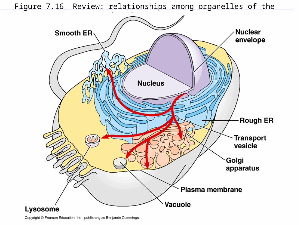
Figure 7.16 Review: relationships among organelles of the endomembrane system

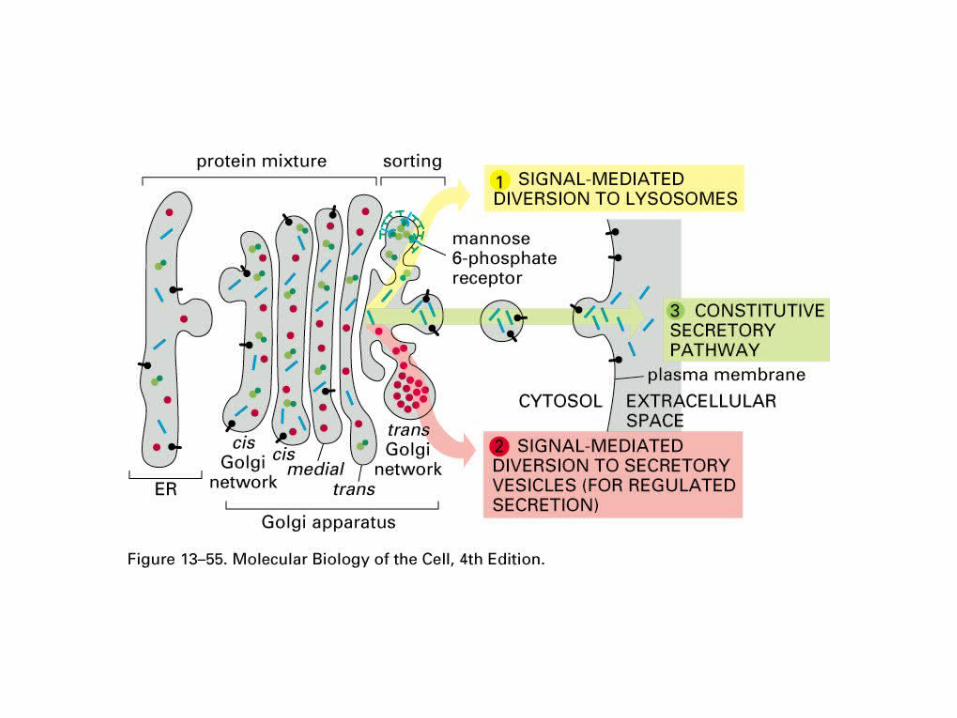
Proteins that follow this ER/Golgi pathway can also go to -
Plasma Membrane, eg. Integrins
Integrins are proteins that recognize other cells, cause cells to stick together.
Human diseases result from defects in integrin genes.
A defect in integrin beta3 causes prolonged bleeding, because
blood plateletes can’t stick together. Glanzman's Thrombasthenia.
With defects in either alpha6 or beta4 integrin skin cells cannot stick together well. Patients are born with blistering epidermis and also have blisters within the mouth and digestive tract...depending on the severity of the disease. Some die
within days and others live. Junctional epidermolysis bullosa
Proteins that follow this ER/Golgi pathway can also go to -
Plasma Membrane, eg. Integrins
Integrins are proteins that recognize other cells, cause cells to stick together.
Lysosomes, Hydrolases.
Hydrolases are digestive enzymes that use water to break apart molecules such as proteins, DNA, lipids, polysaccharides.
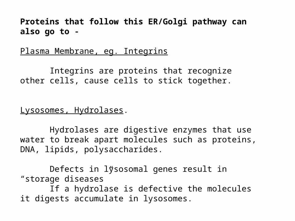
Defects in lysosomal genes result in “storage diseases”If a hydrolase is defective the molecules it digests
accumulate in lysosomes.
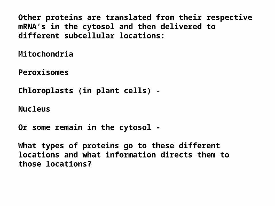
Other proteins are translated from their respective mRNA’s in the cytosol and then delivered to different subcellular locations:
Mitochondria
Peroxisomes
Chloroplasts (in plant cells) -
Nucleus
Or some remain in the cytosol -
What types of proteins go to these different locations and what information directs them to those locations?
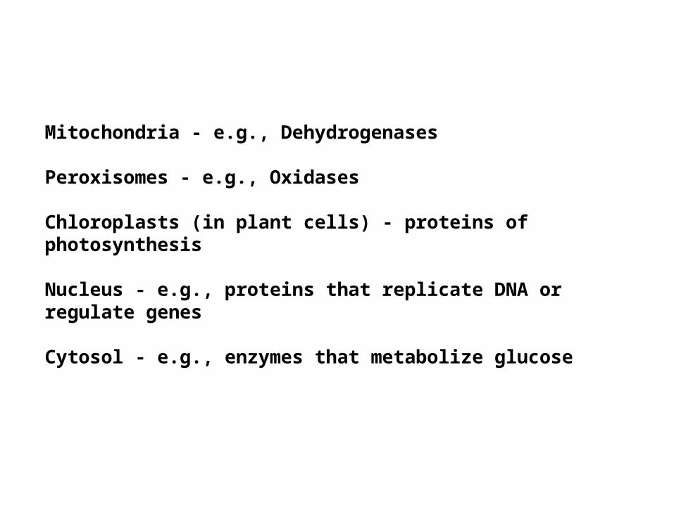
Mitochondria - e.g., Dehydrogenases
Peroxisomes - e.g., Oxidases
Chloroplasts (in plant cells) - proteins of photosynthesis
Nucleus - e.g., proteins that replicate DNA or regulate genes
Cytosol - e.g., enzymes that metabolize glucose
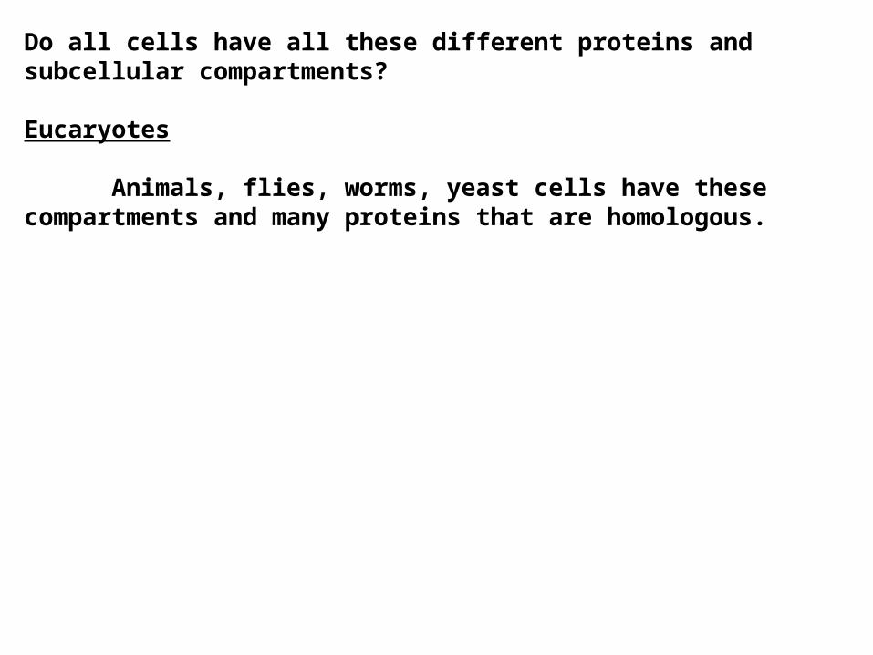
Do all cells have all these different proteins and subcellular compartments?
Eucaryotes
Animals, flies, worms, yeast cells have these compartments and many proteins that are homologous.
Do all cells have all these different proteins and subcellular compartments?
Eucaryotes
Animals, flies, worms, yeast cells have these compartments and many proteins that are homologous.
Plant cells have all the compartments plus chloroplasts and a central vacuole.
Do all cells have all these different proteins and subcellular compartments?
Eucaryotes
Animals, flies, worms, yeast cells have these compartments and many proteins that are homologous.
Plant cells have all the compartments plus chloroplasts and a central vacuole.
Procaryotes
Bacterial cells do not have the compartments and have fewer genes, fewer proteins.
Each cell of an organism has DNA that encodes all the possible genes for that organism. Are all the possible proteins present in every cell of the organism?
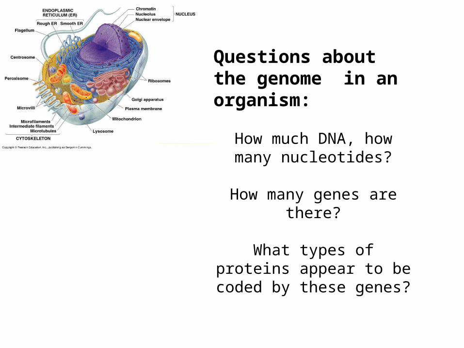
Questions about the genome in an organism:
How much DNA, how many nucleotides?
How many genes are there?
What types of proteins appear to be coded by these genes?
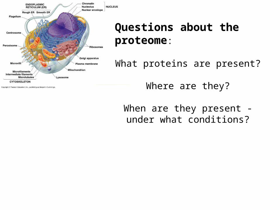
Questions about the proteome:
What proteins are present?
Where are they?
When are they present - under what conditions?
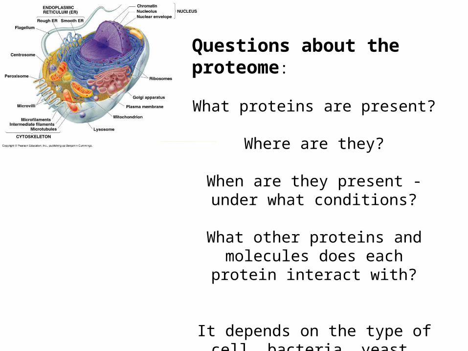
Questions about the proteome:
What proteins are present?
Where are they?
When are they present - under what conditions?
What other proteins and molecules does each protein interact with?
It depends on the type of cell, bacteria, yeast, worm, fly, plant, human
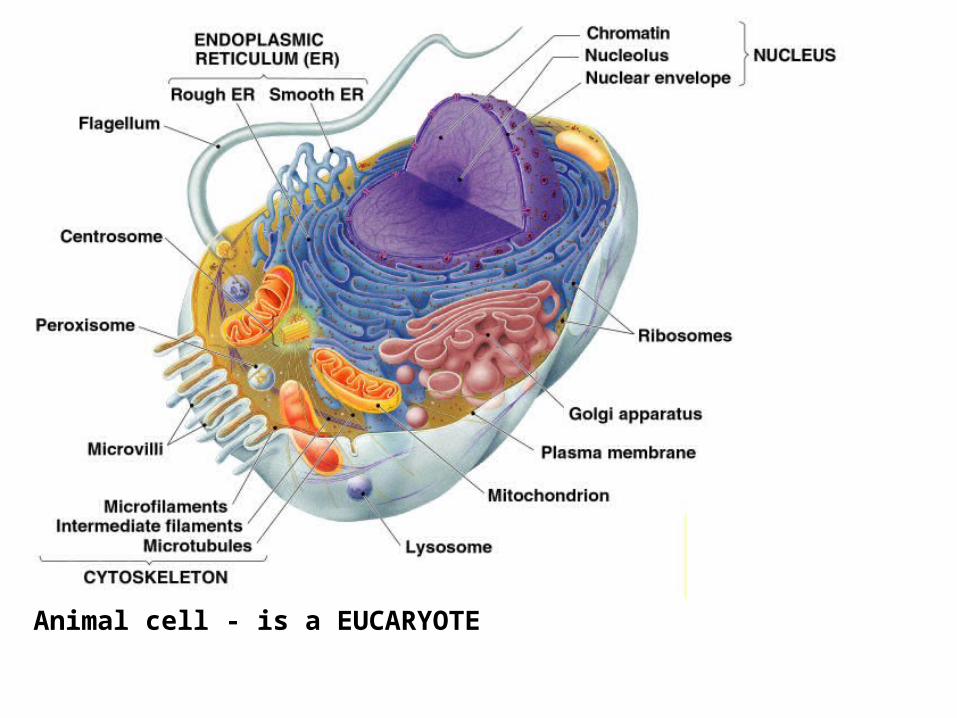
Animal cell - is a EUCARYOTE
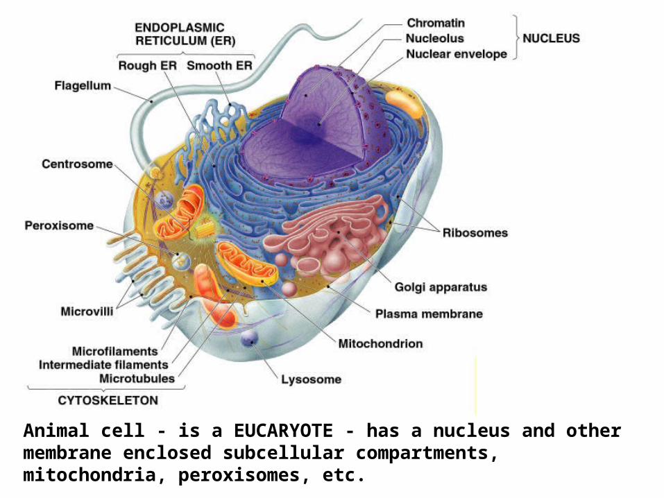
Animal cell - is a EUCARYOTE - has a nucleus and other membrane enclosed subcellular compartments, mitochondria, peroxisomes, etc.
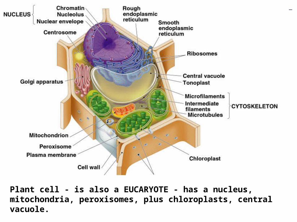
Plant cell - is also a EUCARYOTE - has a nucleus, mitochondria, peroxisomes, plus chloroplasts, central vacuole.
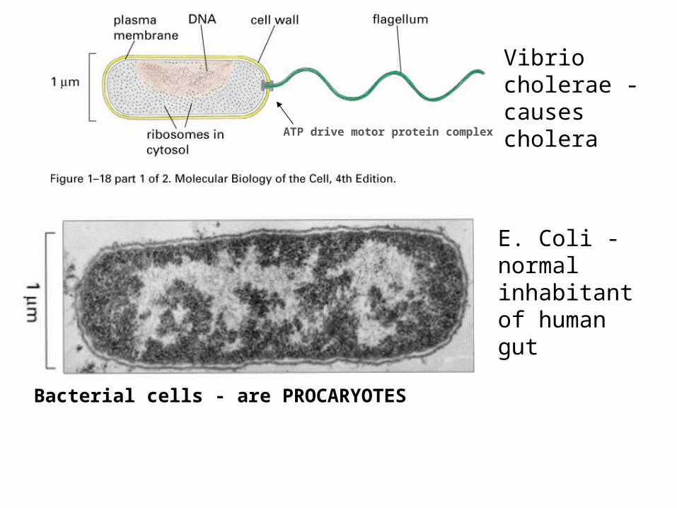
Vibrio cholerae - causes cholera
E. Coli - normal inhabitant of human gut
ATP drive motor protein complex
Bacterial cells - are PROCARYOTES
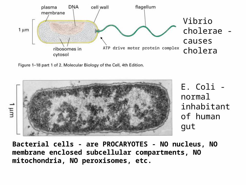
Vibrio cholerae - causes cholera
E. Coli - normal inhabitant of human gut
ATP drive motor protein complex
Bacterial cells - are PROCARYOTES - NO nucleus, NO membrane enclosed subcellular compartments, NO mitochondria, NO peroxisomes, etc.
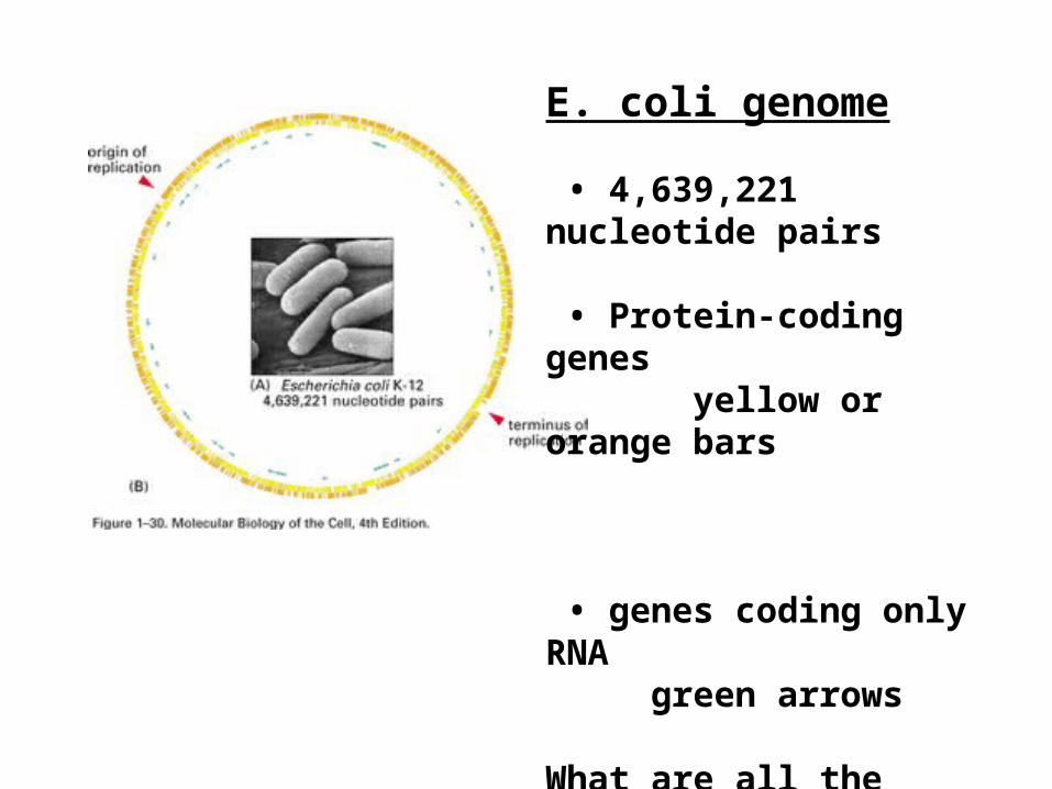
E. coli genome
• 4,639,221 nucleotide pairs
• Protein-coding genes yellow or orange bars
• genes coding only RNA green arrows
What are all the different types of RNAs?
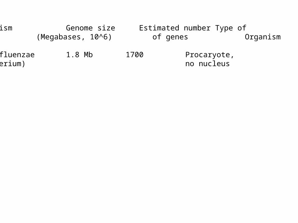
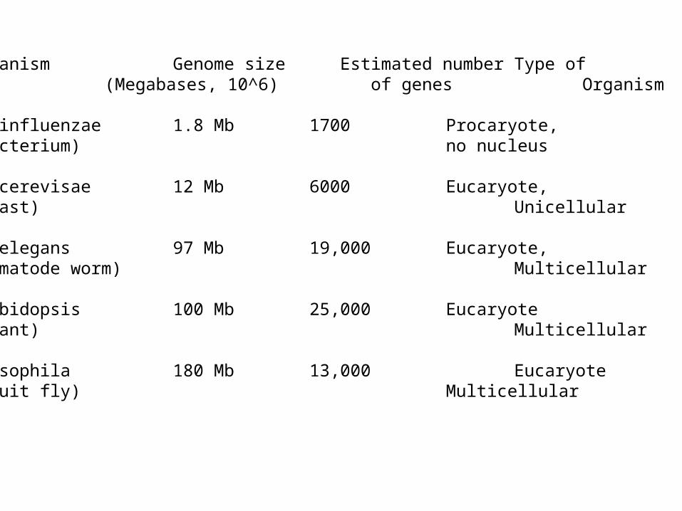
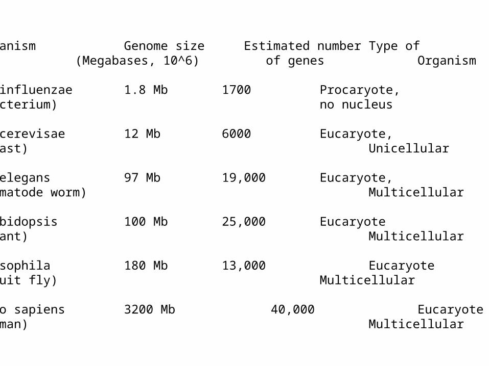
Organism Genome size Estimated number Type of. (Megabases, 10^6) of genes Organism .
H. influenzae 1.8 Mb 1700 Procaryote, (bacterium) no nucleus
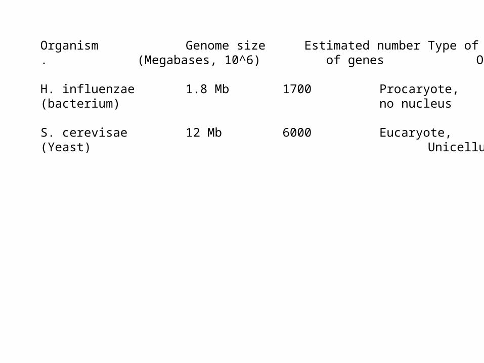
Organism Genome size Estimated number Type of. (Megabases, 10^6) of genes Organism .
H. influenzae 1.8 Mb 1700 Procaryote, (bacterium) no nucleus
S. cerevisae 12 Mb 6000 Eucaryote,(Yeast) Unicellular
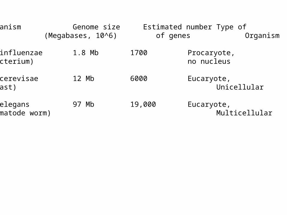
Organism Genome size Estimated number Type of. (Megabases, 10^6) of genes Organism .
H. influenzae 1.8 Mb 1700 Procaryote, (bacterium) no nucleus
S. cerevisae 12 Mb 6000 Eucaryote,(Yeast) Unicellular
C. elegans 97 Mb 19,000 Eucaryote,(nematode worm) Multicellular
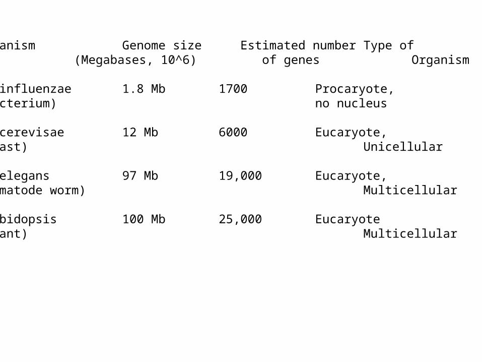
Organism Genome size Estimated number Type of. (Megabases, 10^6) of genes Organism .
H. influenzae 1.8 Mb 1700 Procaryote, (bacterium) no nucleus
S. cerevisae 12 Mb 6000 Eucaryote,(Yeast) Unicellular
C. elegans 97 Mb 19,000 Eucaryote,(nematode worm) Multicellular
Arabidopsis 100 Mb 25,000 Eucaryote(plant) Multicellular
Drosophila 180 Mb 13,000 Eucaryote(fruit fly) Multicellular
Homo sapiens 3200 Mb 40,000 Eucaryote(human) Multicellular
Are all the genes in a cell producing proteins?
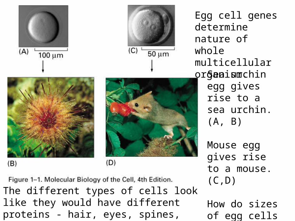
Egg cell genes determine nature of whole multicellular organism.
Sea urchin egg gives rise to a sea urchin. (A, B)
Mouse egg gives rise to a mouse. (C,D)
How do sizes of egg cells compare to E.coli?
The different types of cells look like they would have different proteins - hair, eyes, spines, etc.
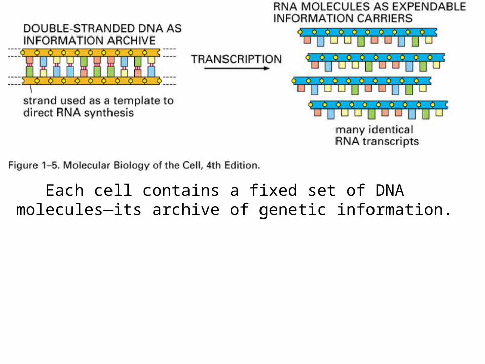
Each cell contains a fixed set of DNA molecules—its archive of genetic information.
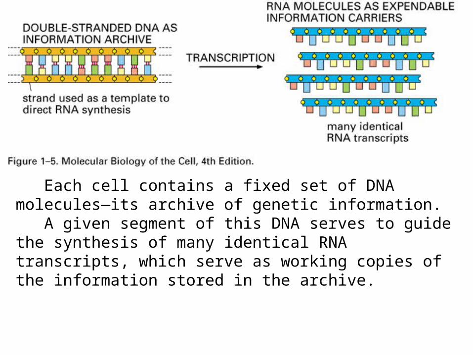
Each cell contains a fixed set of DNA molecules—its archive of genetic information. A given segment of this DNA serves to guide the synthesis of many identical RNA transcripts, which serve as working copies of the information stored in the archive.
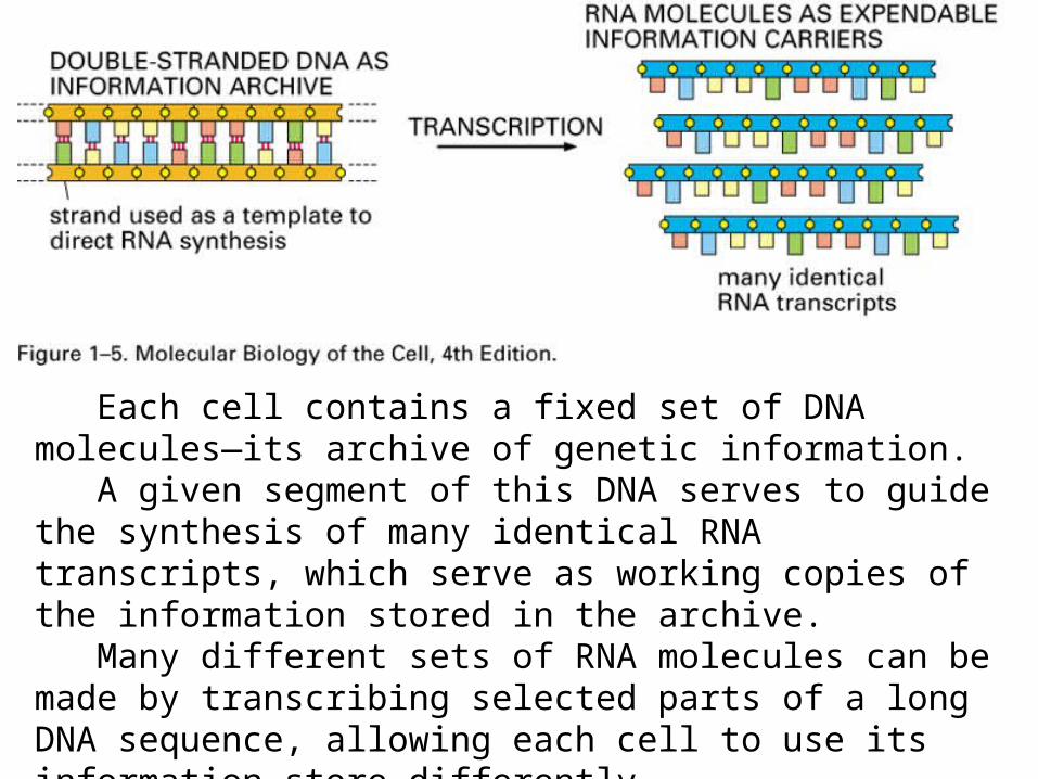
Each cell contains a fixed set of DNA molecules—its archive of genetic information. A given segment of this DNA serves to guide the synthesis of many identical RNA transcripts, which serve as working copies of the information stored in the archive. Many different sets of RNA molecules can be made by transcribing selected parts of a long DNA sequence, allowing each cell to use its information store differently.
Kirkpatrick C, Maurer LM, Oyelakin NE, Yoncheva YN, Maurer R, Slonczewski JL. Acetate and formate stress: opposite responses in the proteome of Escherichia coli.J Bacteriol. 2001 Nov;183(21):6466-77.
Blankenhorn D, Phillips J, Slonczewski JL. Acid- and base-induced proteins during aerobic and anaerobic growth of Escherichia coli revealed by two-dimensional gel electrophoresis.J Bacteriol. 1999 Apr;181(7):2209-16.
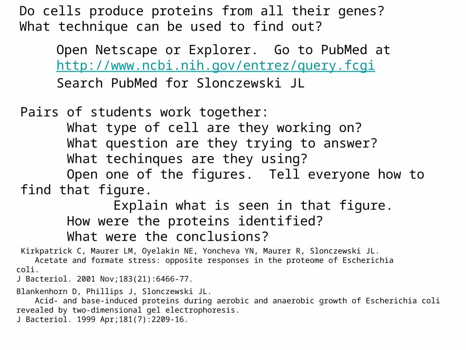
Open Netscape or Explorer. Go to PubMed at http://www.ncbi.nih.gov/entrez/query.fcgiSearch PubMed for Slonczewski JL
Pairs of students work together:What type of cell are they working on?What question are they trying to answer?What techinques are they using?Open one of the figures. Tell everyone how to find that figure.
Explain what is seen in that figure.How were the proteins identified?What were the conclusions?
Do cells produce proteins from all their genes? What technique can be used to find out?
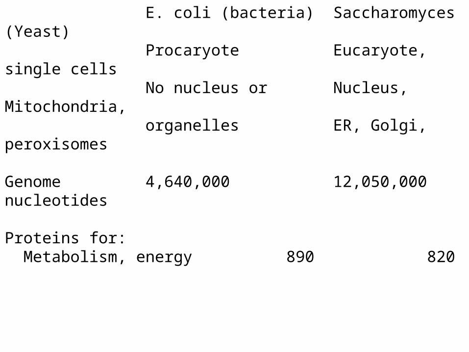
E. coli (bacteria) Saccharomyces (Yeast)Procaryote Eucaryote, single cellsNo nucleus or Nucleus, Mitochondria,organelles ER, Golgi, peroxisomes
Genome 4,640,000 12,050,000 nucleotides
Proteins for: Metabolism, energy 890 820
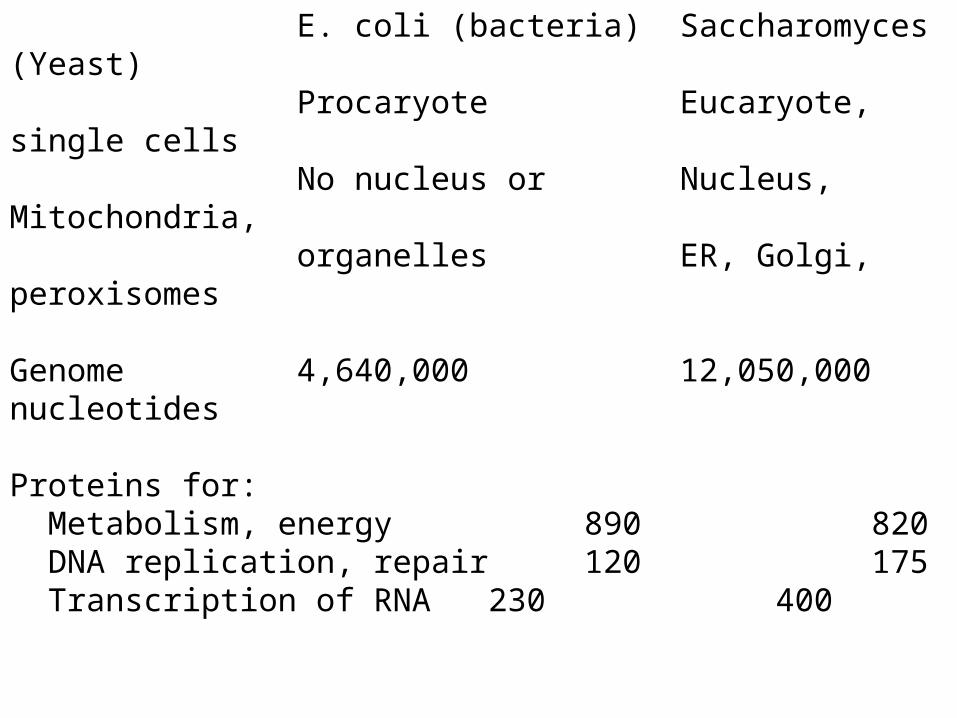
E. coli (bacteria) Saccharomyces (Yeast)Procaryote Eucaryote, single cellsNo nucleus or Nucleus, Mitochondria,organelles ER, Golgi, peroxisomes
Genome 4,640,000 12,050,000 nucleotides
Proteins for: Metabolism, energy 890 820 DNA replication, repair 120 175 Transcription of RNA 230 400
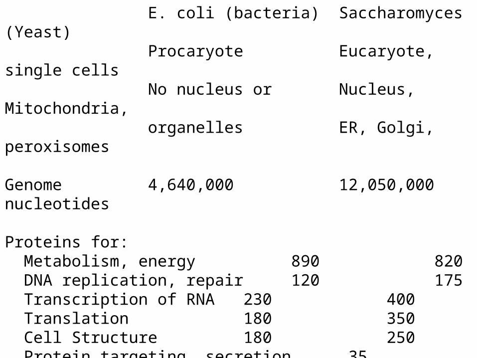
E. coli (bacteria) Saccharomyces (Yeast)Procaryote Eucaryote, single cellsNo nucleus or Nucleus, Mitochondria,organelles ER, Golgi, peroxisomes
Genome 4,640,000 12,050,000 nucleotides
Proteins for: Metabolism, energy 890 820 DNA replication, repair 120 175 Transcription of RNA 230 400 Translation 180 350 Cell Structure 180 250 Protein targeting, secretion 35 430
Does E.coli produce all proteins constantly, or selected ones?Where are the proteins located in the yeast cell?
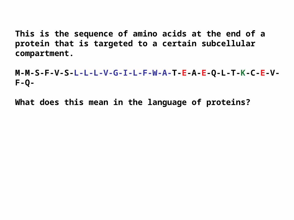
This is the sequence of amino acids at the end of a protein that is targeted to a certain subcellular compartment.
M-M-S-F-V-S-L-L-L-V-G-I-L-F-W-A-T-E-A-E-Q-L-T-K-C-E-V-F-Q-
What does this mean in the language of proteins?
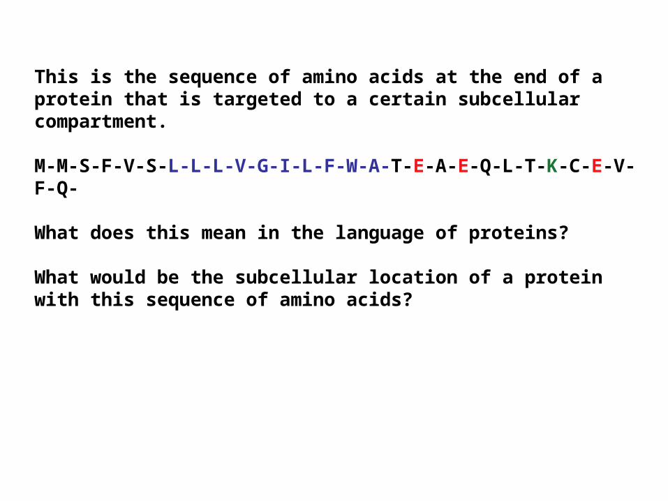
This is the sequence of amino acids at the end of a protein that is targeted to a certain subcellular compartment.
M-M-S-F-V-S-L-L-L-V-G-I-L-F-W-A-T-E-A-E-Q-L-T-K-C-E-V-F-Q-
What does this mean in the language of proteins?
What would be the subcellular location of a protein with this sequence of amino acids?
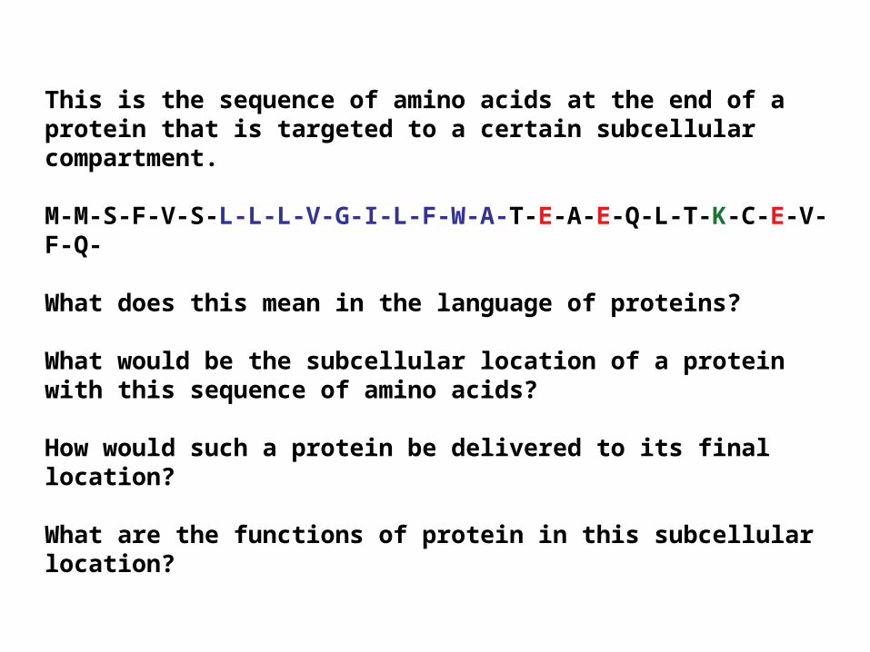
How would such a protein be delivered to its final location?
What are the functions of protein in this subcellular location?
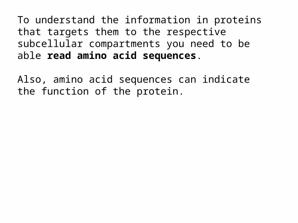
To understand the information in proteins that targets them to the respective subcellular compartments you need to be able read amino acid sequences.
Also, amino acid sequences can indicate the function of the protein.
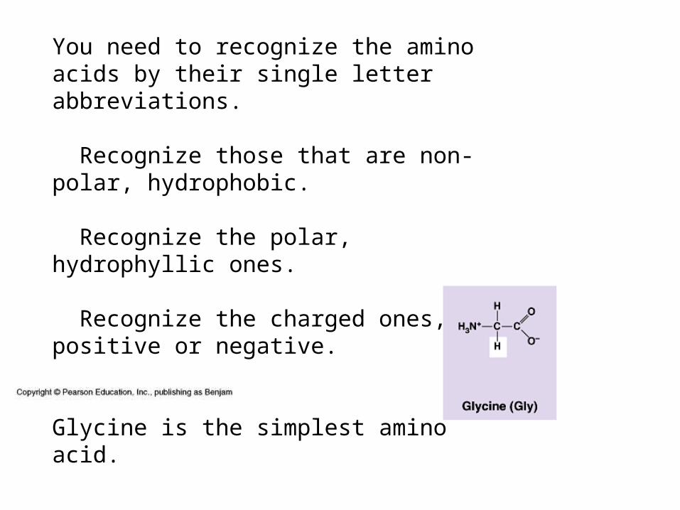
You need to recognize the amino acids by their single letter abbreviations.
Recognize those that are non-polar, hydrophobic.
Recognize the polar, hydrophyllic ones.
Recognize the charged ones, positive or negative.
You need to recognize the amino acids by their single letter abbreviations.
Recognize those that are non-polar, hydrophobic.
Recognize the polar, hydrophyllic ones.
Recognize the charged ones, positive or negative.
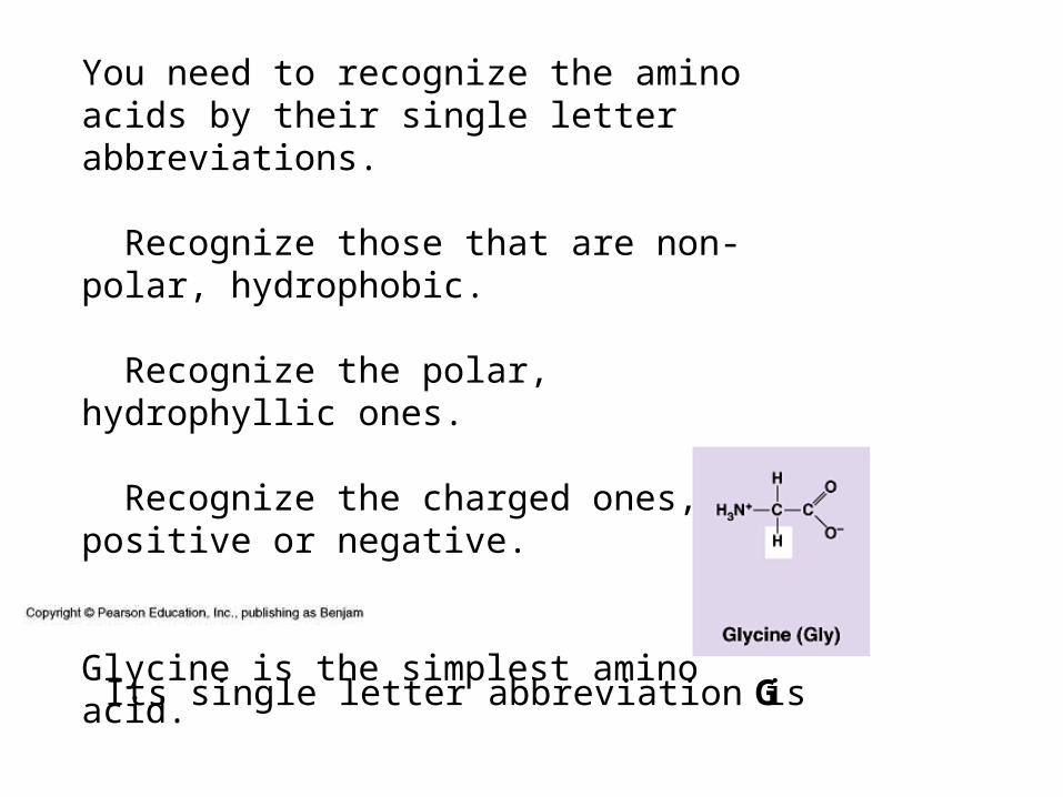
Glycine is the simplest amino acid.
G
You need to recognize the amino acids by their single letter abbreviations.
Recognize those that are non-polar, hydrophobic.
Recognize the polar, hydrophyllic ones.
Recognize the charged ones, positive or negative.
Glycine is the simplest amino acid.
Its single letter abbreviation is
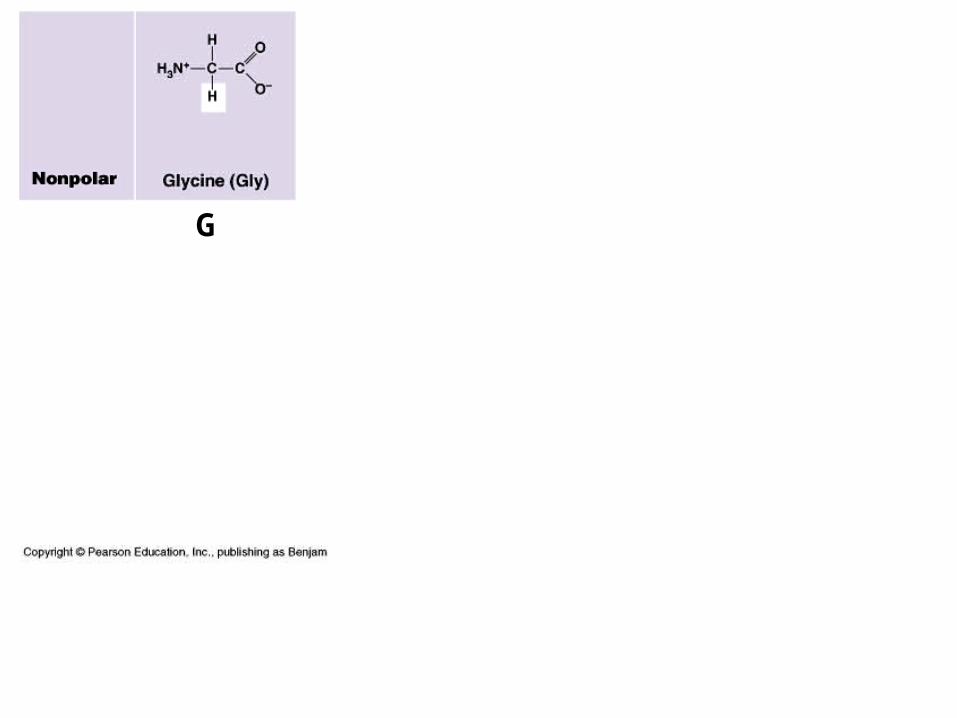
G
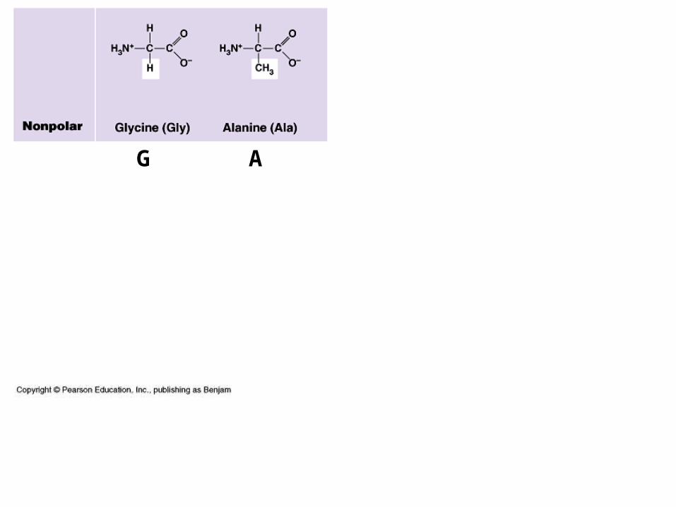
G A
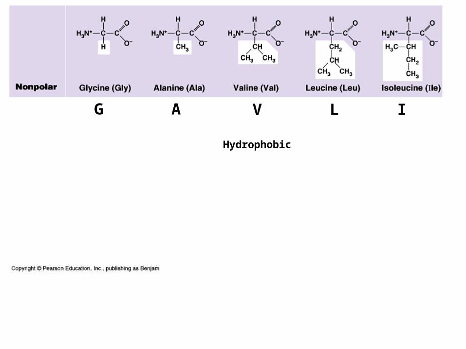
G A V L I
Hydrophobic
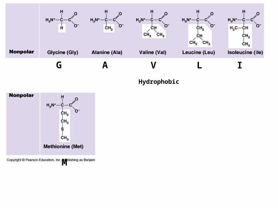
G A V L I
M
Hydrophobic
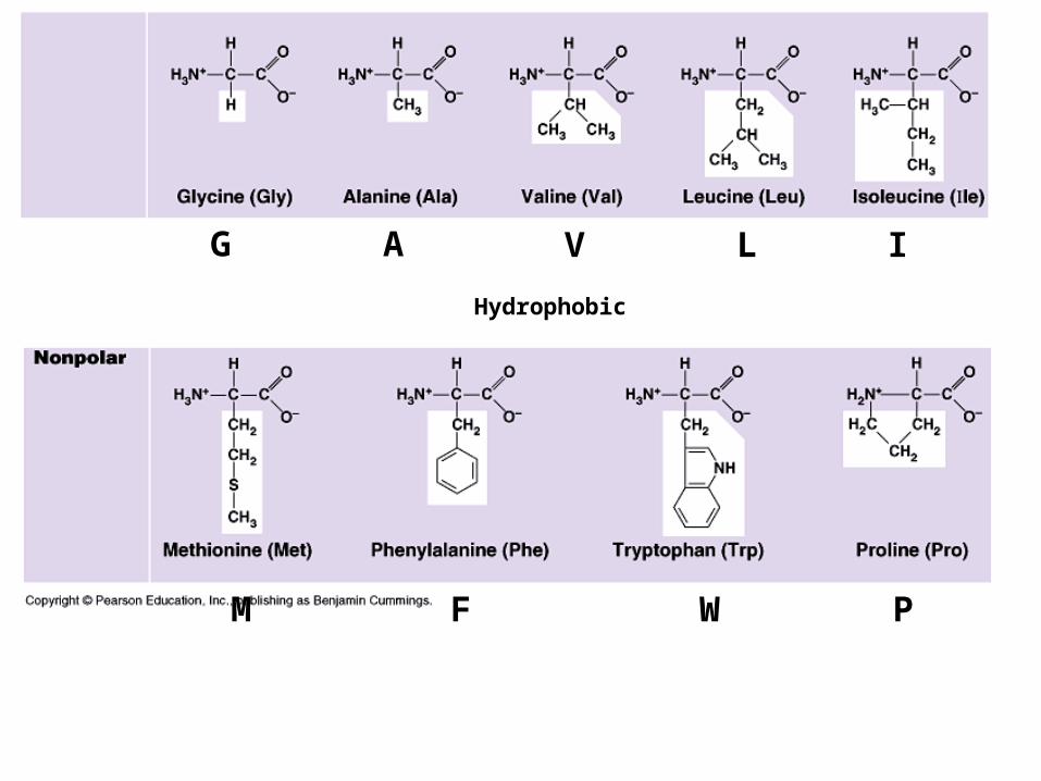
G A V L I
M F W P
Hydrophobic
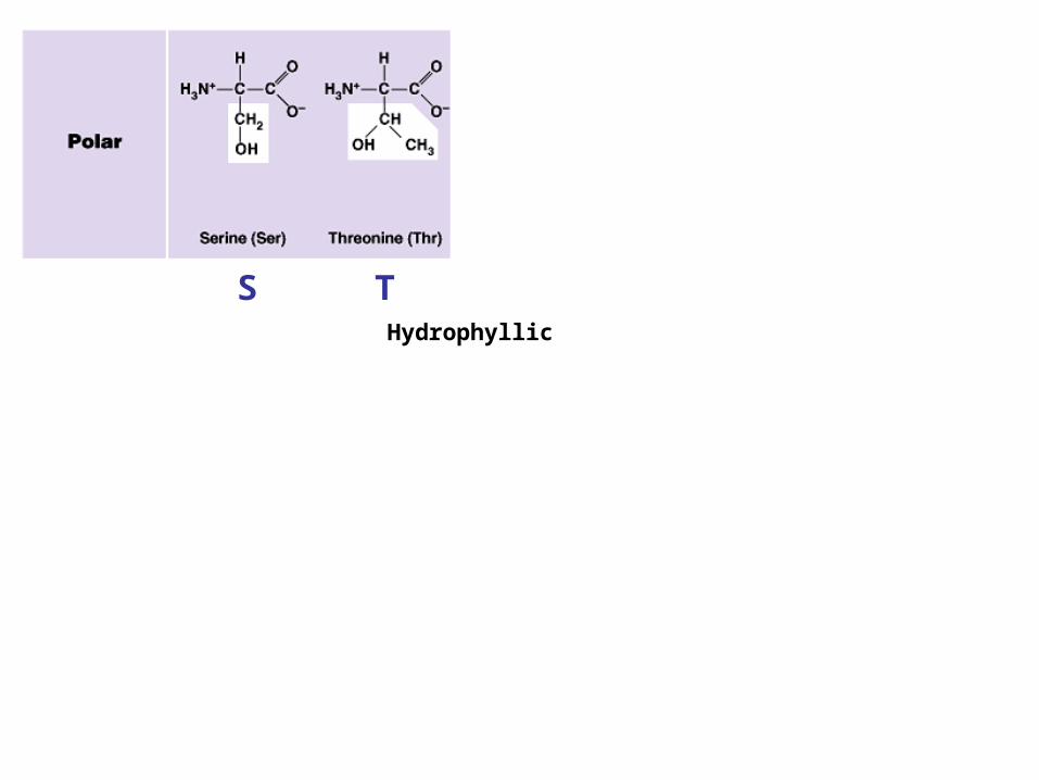
S THydrophyllic
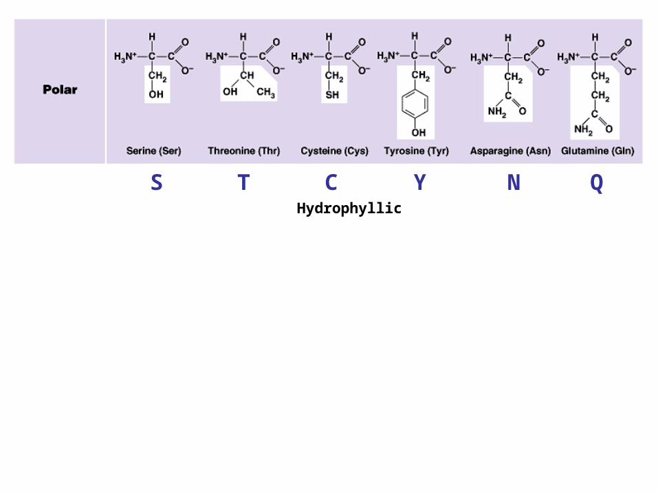
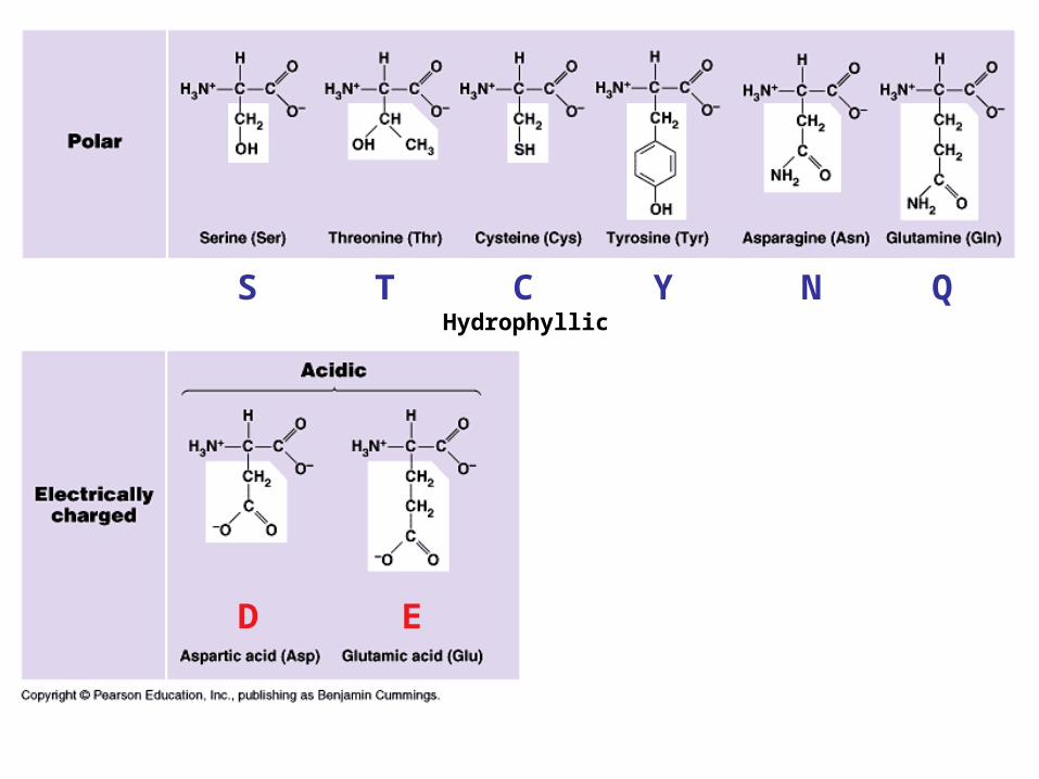
S T C Y N QHydrophyllic
S T C Y N Q
ED
Hydrophyllic
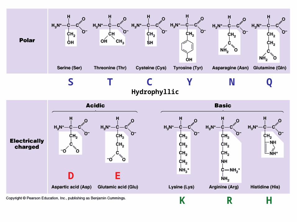
S T C Y N Q
ED
K R H
Hydrophyllic
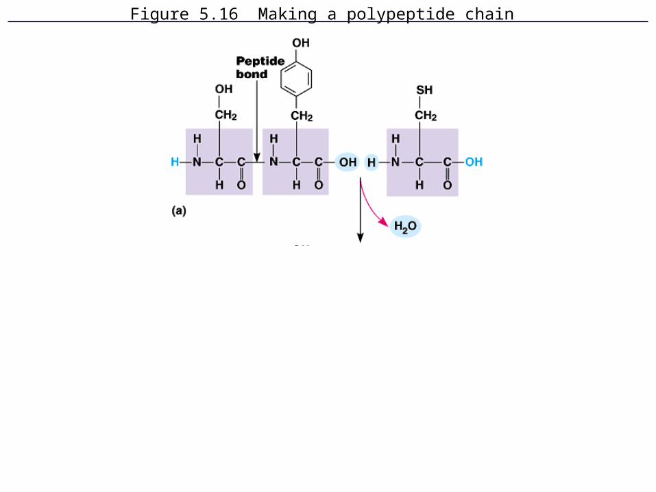
Figure 5.16 Making a polypeptide chain
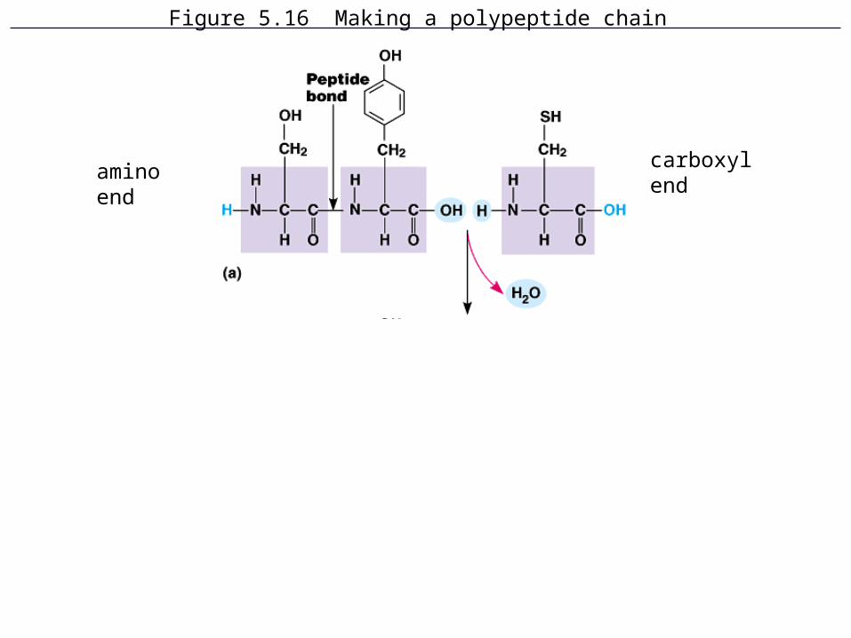
Figure 5.16 Making a polypeptide chain
aminoend
carboxylend
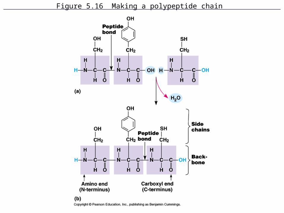
Figure 5.16 Making a polypeptide chain
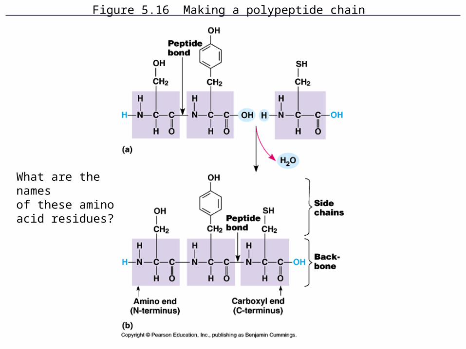
Figure 5.16 Making a polypeptide chain
What are the namesof these amino acid residues?
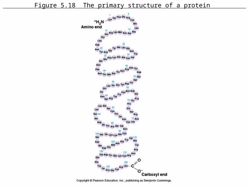
Figure 5.18 The primary structure of a protein
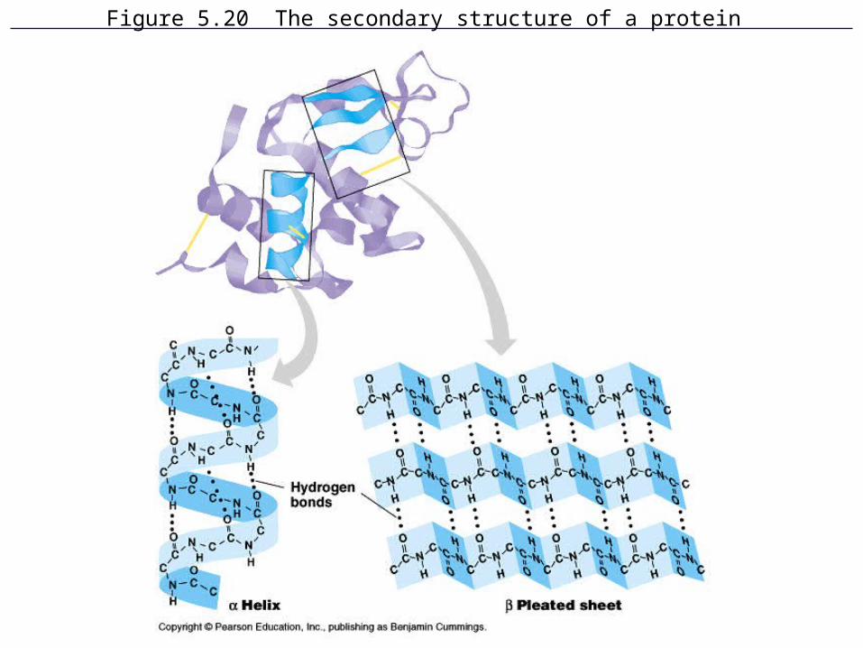
Figure 5.20 The secondary structure of a protein
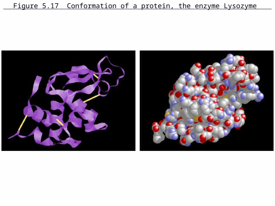
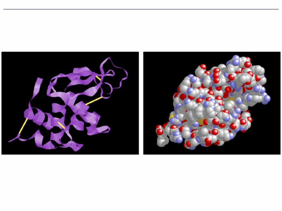
Figure 5.17 Conformation of a protein, the enzyme Lysozyme
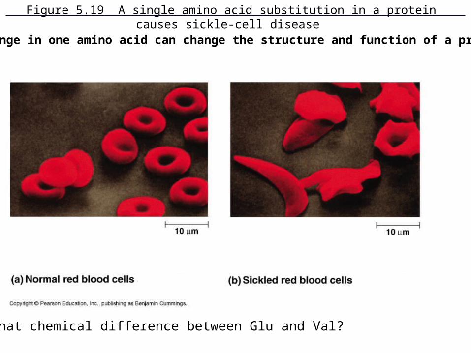
Figure 5.19 A single amino acid substitution in a protein causes sickle-cell disease
What chemical difference between Glu and Val?
A change in one amino acid can change the structure and function of a protein
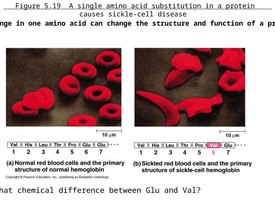
Figure 5.19 A single amino acid substitution in a protein causes sickle-cell disease
What chemical difference between Glu and Val?
A change in one amino acid can change the structure and function of a protein
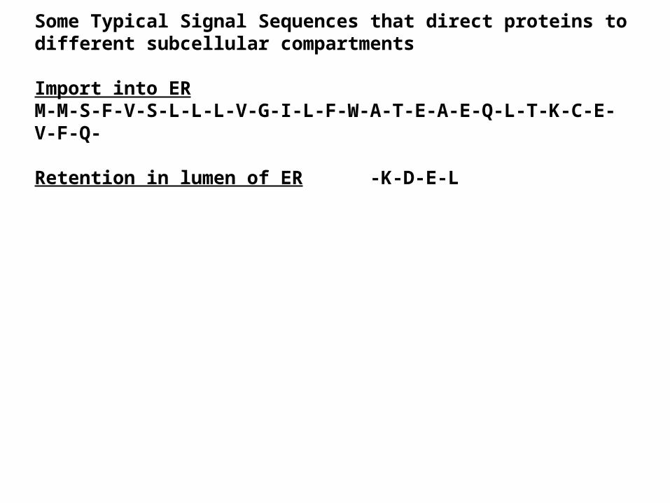
Some Typical Signal Sequences that direct proteins to different subcellular compartments
Import into ERM-M-S-F-V-S-L-L-L-V-G-I-L-F-W-A-T-E-A-E-Q-L-T-K-C-E-V-F-Q-
Retention in lumen of ER -K-D-E-L
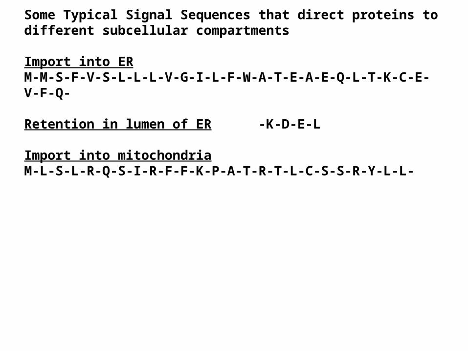
Some Typical Signal Sequences that direct proteins to different subcellular compartments
Import into ERM-M-S-F-V-S-L-L-L-V-G-I-L-F-W-A-T-E-A-E-Q-L-T-K-C-E-V-F-Q-
Retention in lumen of ER -K-D-E-L
Import into mitochondria M-L-S-L-R-Q-S-I-R-F-F-K-P-A-T-R-T-L-C-S-S-R-Y-L-L-
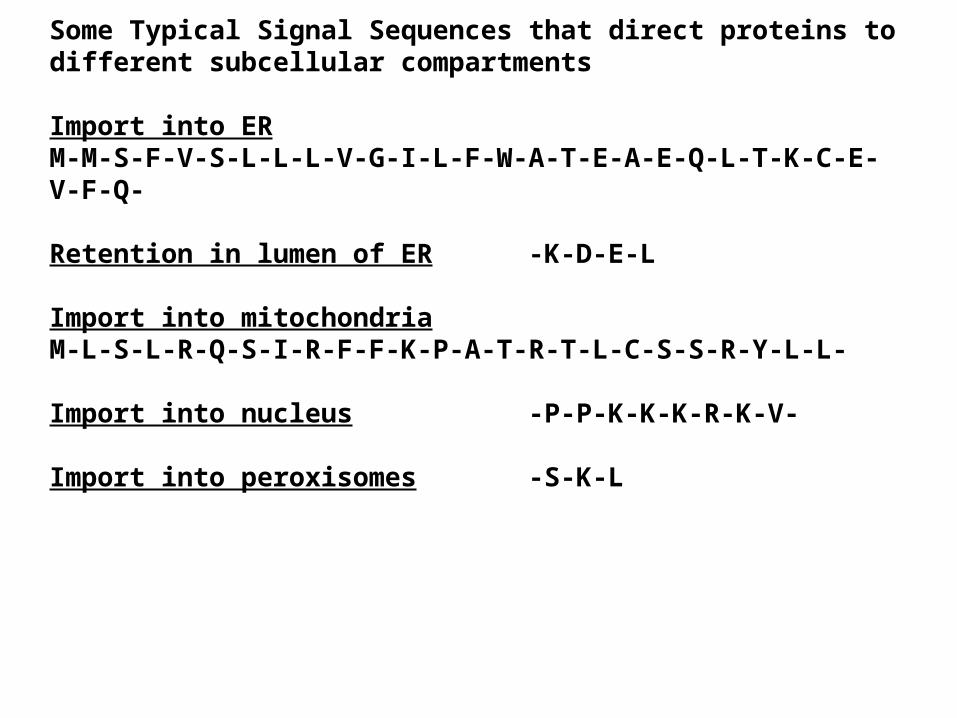
Some Typical Signal Sequences that direct proteins to different subcellular compartments
Import into ERM-M-S-F-V-S-L-L-L-V-G-I-L-F-W-A-T-E-A-E-Q-L-T-K-C-E-V-F-Q-
Retention in lumen of ER -K-D-E-L
Import into mitochondria M-L-S-L-R-Q-S-I-R-F-F-K-P-A-T-R-T-L-C-S-S-R-Y-L-L-
Import into nucleus -P-P-K-K-K-R-K-V-
Import into peroxisomes -S-K-L
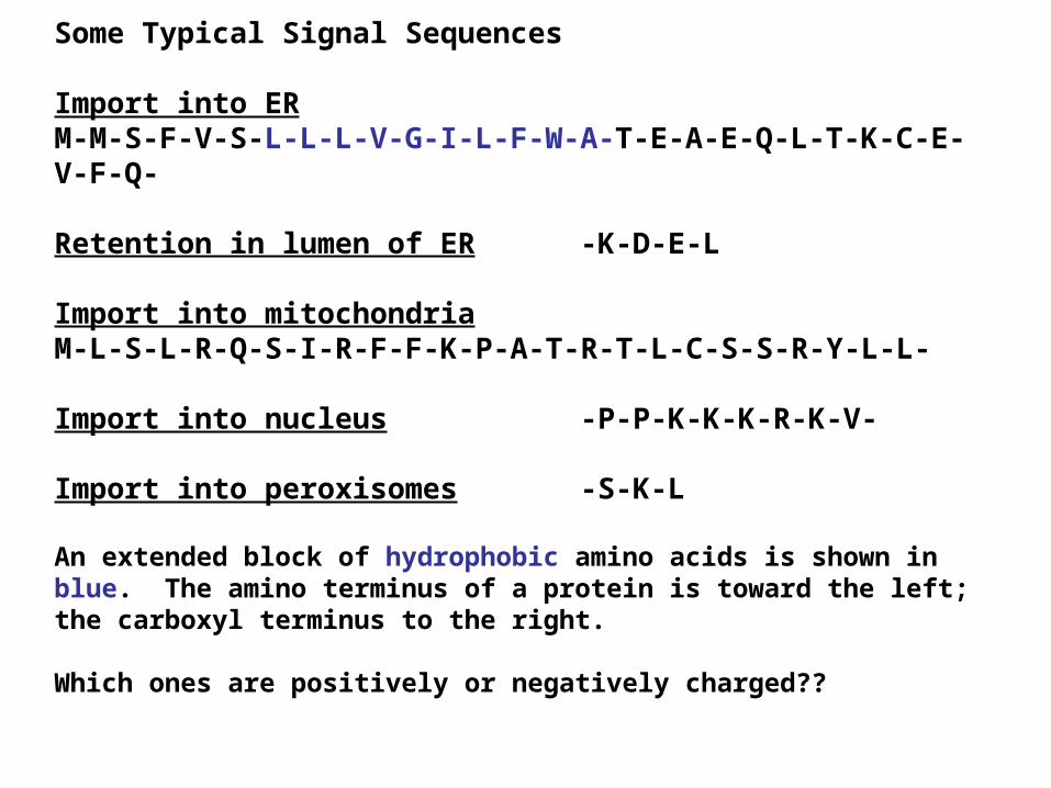
Some Typical Signal Sequences
Import into ERM-M-S-F-V-S-L-L-L-V-G-I-L-F-W-A-T-E-A-E-Q-L-T-K-C-E-V-F-Q-
Retention in lumen of ER -K-D-E-L
Import into mitochondria M-L-S-L-R-Q-S-I-R-F-F-K-P-A-T-R-T-L-C-S-S-R-Y-L-L-
Import into nucleus -P-P-K-K-K-R-K-V-
Import into peroxisomes -S-K-L
An extended block of hydrophobic amino acids is shown in blue. The amino terminus of a protein is toward the left; the carboxyl terminus to the right.
Which ones are positively or negatively charged??
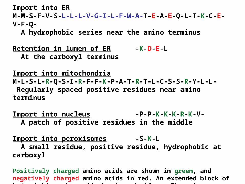
Import into ERM-M-S-F-V-S-L-L-L-V-G-I-L-F-W-A-T-E-A-E-Q-L-T-K-C-E-V-F-Q- A hydrophobic series near the amino terminus
Retention in lumen of ER -K-D-E-L At the carboxyl terminus
Import into mitochondria M-L-S-L-R-Q-S-I-R-F-F-K-P-A-T-R-T-L-C-S-S-R-Y-L-L- Regularly spaced positive residues near amino terminus
Import into nucleus -P-P-K-K-K-R-K-V- A patch of positive residues in the middle
Import into peroxisomes -S-K-L A small residue, positive residue, hydrophobic at carboxyl
Positively charged amino acids are shown in green, and negatively charged amino acids in red. An extended block of hydrophobic amino acids is shown in blue. The amino terminus of a protein is toward the left; the carboxyl terminus to the right.
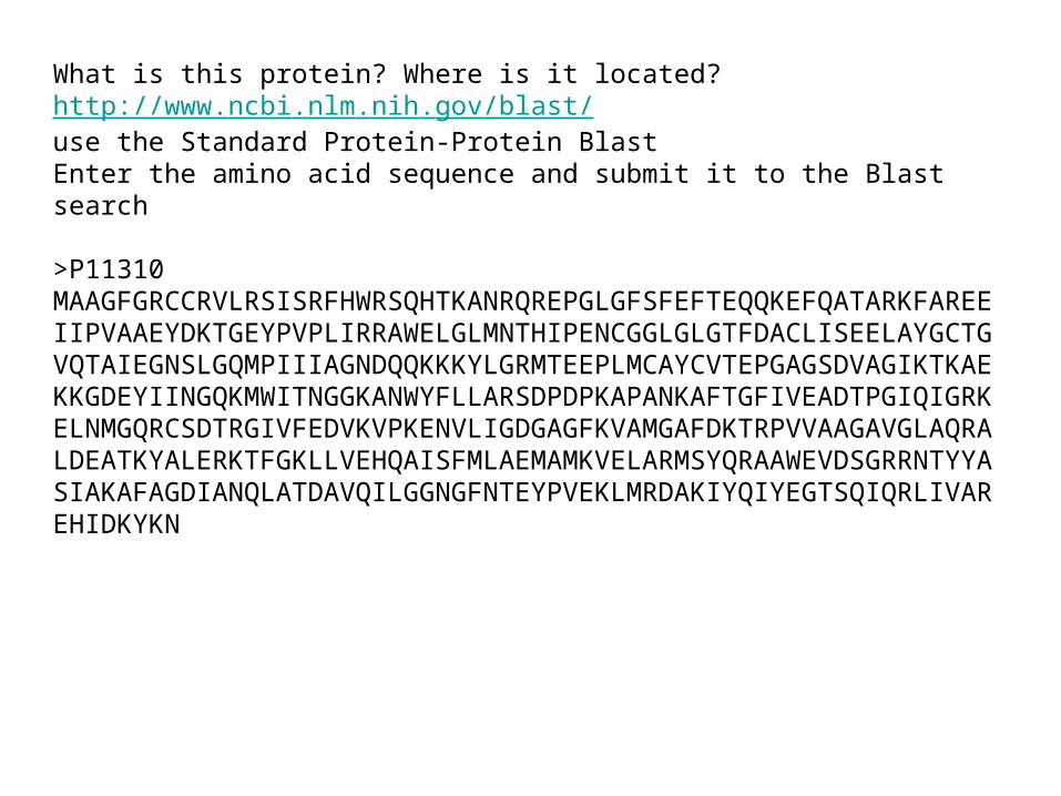
What is this protein? Where is it located?http://www.ncbi.nlm.nih.gov/blast/use the Standard Protein-Protein BlastEnter the amino acid sequence and submit it to the Blast search
>P11310MAAGFGRCCRVLRSISRFHWRSQHTKANRQREPGLGFSFEFTEQQKEFQATARKFAREEIIPVAAEYDKTGEYPVPLIRRAWELGLMNTHIPENCGGLGLGTFDACLISEELAYGCTGVQTAIEGNSLGQMPIIIAGNDQQKKKYLGRMTEEPLMCAYCVTEPGAGSDVAGIKTKAEKKGDEYIINGQKMWITNGGKANWYFLLARSDPDPKAPANKAFTGFIVEADTPGIQIGRKELNMGQRCSDTRGIVFEDVKVPKENVLIGDGAGFKVAMGAFDKTRPVVAAGAVGLAQRALDEATKYALERKTFGKLLVEHQAISFMLAEMAMKVELARMSYQRAAWEVDSGRRNTYYASIAKAFAGDIANQLATDAVQILGGNGFNTEYPVEKLMRDAKIYQIYEGTSQIQRLIVAREHIDKYKN
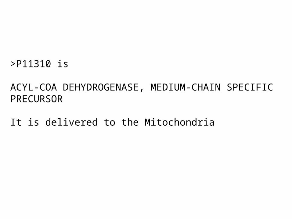
>P11310 is
ACYL-COA DEHYDROGENASE, MEDIUM-CHAIN SPECIFIC PRECURSOR
It is delivered to the Mitochondria
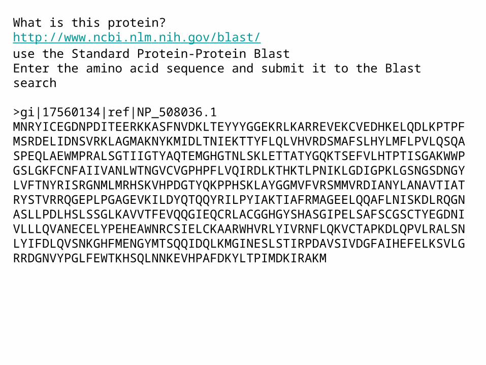
What is this protein?http://www.ncbi.nlm.nih.gov/blast/use the Standard Protein-Protein BlastEnter the amino acid sequence and submit it to the Blast search
>gi|17560134|ref|NP_508036.1MNRYICEGDNPDITEERKKASFNVDKLTEYYYGGEKRLKARREVEKCVEDHKELQDLKPTPFMSRDELIDNSVRKLAGMAKNYKMIDLTNIEKTTYFLQLVHVRDSMAFSLHYLMFLPVLQSQASPEQLAEWMPRALSGTIIGTYAQTEMGHGTNLSKLETTATYGQKTSEFVLHTPTISGAKWWPGSLGKFCNFAIIVANLWTNGVCVGPHPFLVQIRDLKTHKTLPNIKLGDIGPKLGSNGSDNGYLVFTNYRISRGNMLMRHSKVHPDGTYQKPPHSKLAYGGMVFVRSMMVRDIANYLANAVTIATRYSTVRRQGEPLPGAGEVKILDYQTQQYRILPYIAKTIAFRMAGEELQQAFLNISKDLRQGNASLLPDLHSLSSGLKAVVTFEVQQGIEQCRLACGGHGYSHASGIPELSAFSCGSCTYEGDNIVLLLQVANECELYPEHEAWNRCSIELCKAARWHVRLYIVRNFLQKVCTAPKDLQPVLRALSNLYIFDLQVSNKGHFMENGYMTSQQIDQLKMGINESLSTIRPDAVSIVDGFAIHEFELKSVLGRRDGNVYPGLFEWTKHSQLNNKEVHPAFDKYLTPIMDKIRAKM
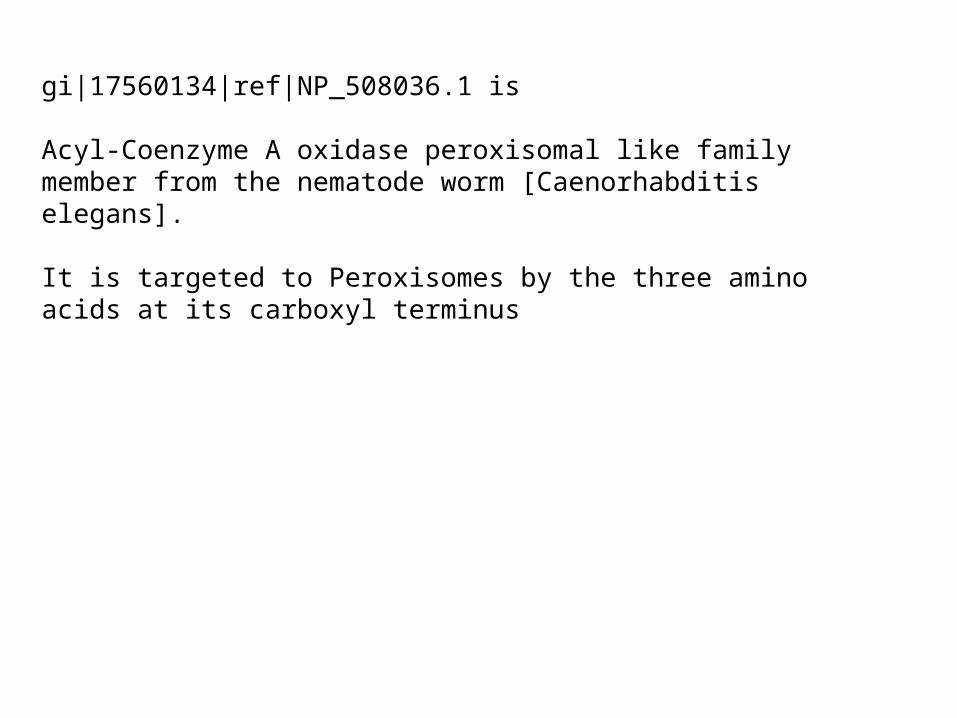
gi|17560134|ref|NP_508036.1 is
Acyl-Coenzyme A oxidase peroxisomal like family member from the nematode worm [Caenorhabditis elegans].
It is targeted to Peroxisomes by the three amino acids at its carboxyl terminus
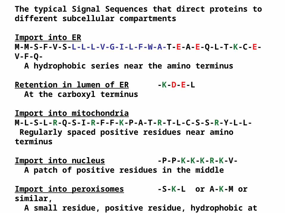
The typical Signal Sequences that direct proteins to different subcellular compartments
Import into ERM-M-S-F-V-S-L-L-L-V-G-I-L-F-W-A-T-E-A-E-Q-L-T-K-C-E-V-F-Q- A hydrophobic series near the amino terminus
Retention in lumen of ER -K-D-E-L At the carboxyl terminus
Import into mitochondria M-L-S-L-R-Q-S-I-R-F-F-K-P-A-T-R-T-L-C-S-S-R-Y-L-L- Regularly spaced positive residues near amino terminus
Import into nucleus -P-P-K-K-K-R-K-V- A patch of positive residues in the middle
Import into peroxisomes -S-K-L or A-K-M or similar, A small residue, positive residue, hydrophobic at carboxyl
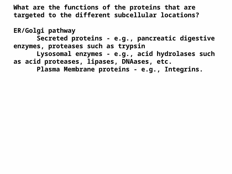
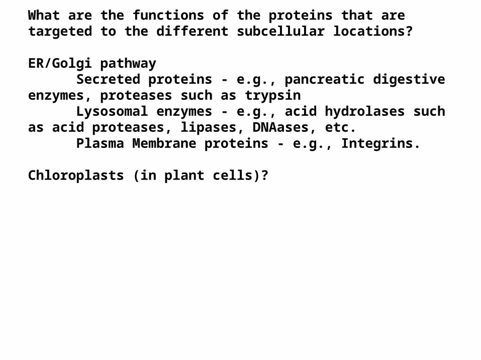
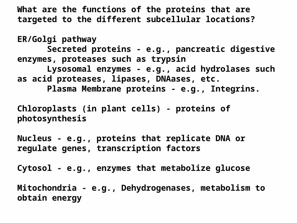
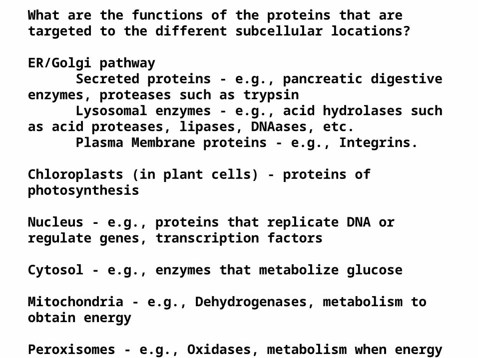
What are the functions of the proteins that are targeted to the different subcellular locations?
ER/Golgi pathwaySecreted proteins - e.g., pancreatic digestive enzymes,
proteases such as trypsinLysosomal enzymes - e.g., acid hydrolases such as acid
proteases, lipases, DNAases, etc.Plasma Membrane proteins - e.g., Integrins.
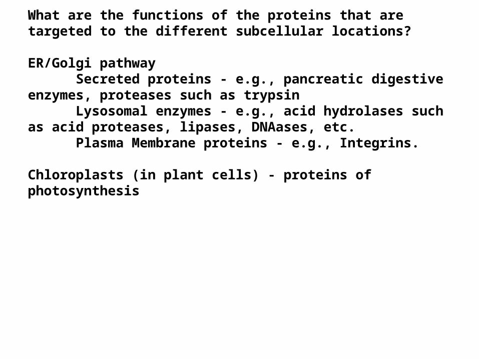
Chloroplasts (in plant cells)?
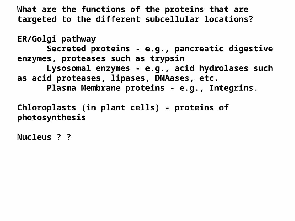
What are the functions of the proteins that are targeted to the different subcellular locations?
ER/Golgi pathwaySecreted proteins - e.g., pancreatic digestive enzymes,
proteases such as trypsinLysosomal enzymes - e.g., acid hydrolases such as acid
proteases, lipases, DNAases, etc.Plasma Membrane proteins - e.g., Integrins.
Chloroplasts (in plant cells) - proteins of photosynthesis
Nucleus ? ?
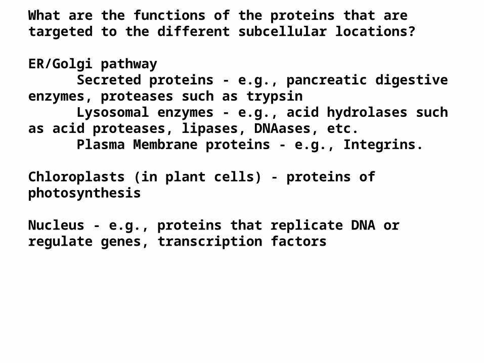
What are the functions of the proteins that are targeted to the different subcellular locations?
ER/Golgi pathwaySecreted proteins - e.g., pancreatic digestive enzymes,
proteases such as trypsinLysosomal enzymes - e.g., acid hydrolases such as acid
proteases, lipases, DNAases, etc.Plasma Membrane proteins - e.g., Integrins.
Chloroplasts (in plant cells) - proteins of photosynthesis
Nucleus - e.g., proteins that replicate DNA or regulate genes, transcription factors
Cytosol - e.g., enzymes that metabolize glucose
Mitochondria - e.g., Dehydrogenases, metabolism to obtain energy
Peroxisomes - e.g., Oxidases, metabolism when energy is not needed
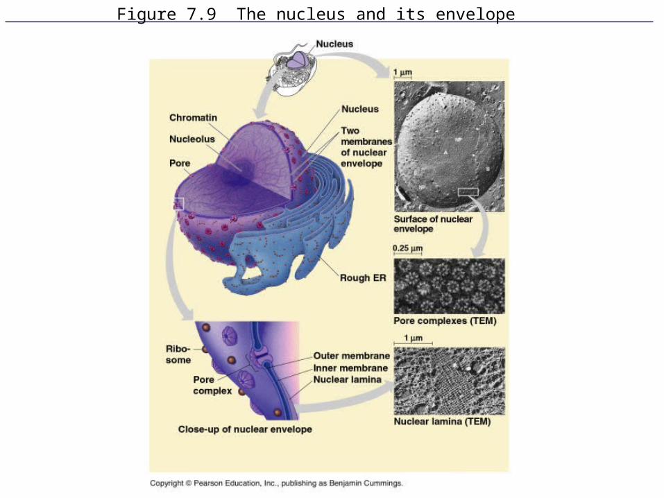
Figure 7.9 The nucleus and its envelope
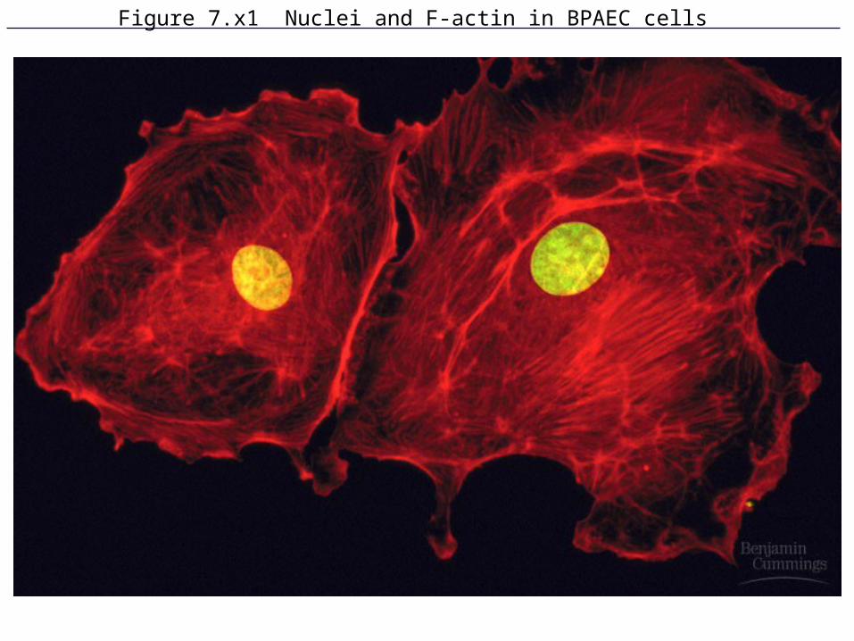
Figure 7.x1 Nuclei and F-actin in BPAEC cells
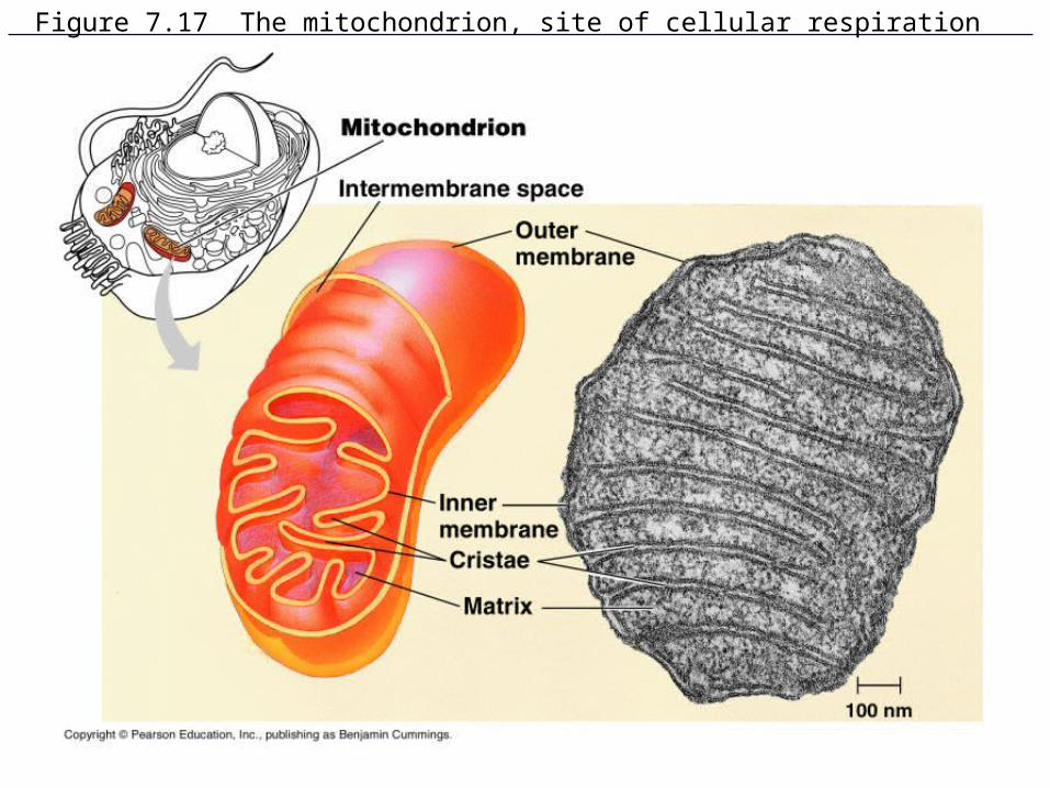
Figure 7.17 The mitochondrion, site of cellular respiration
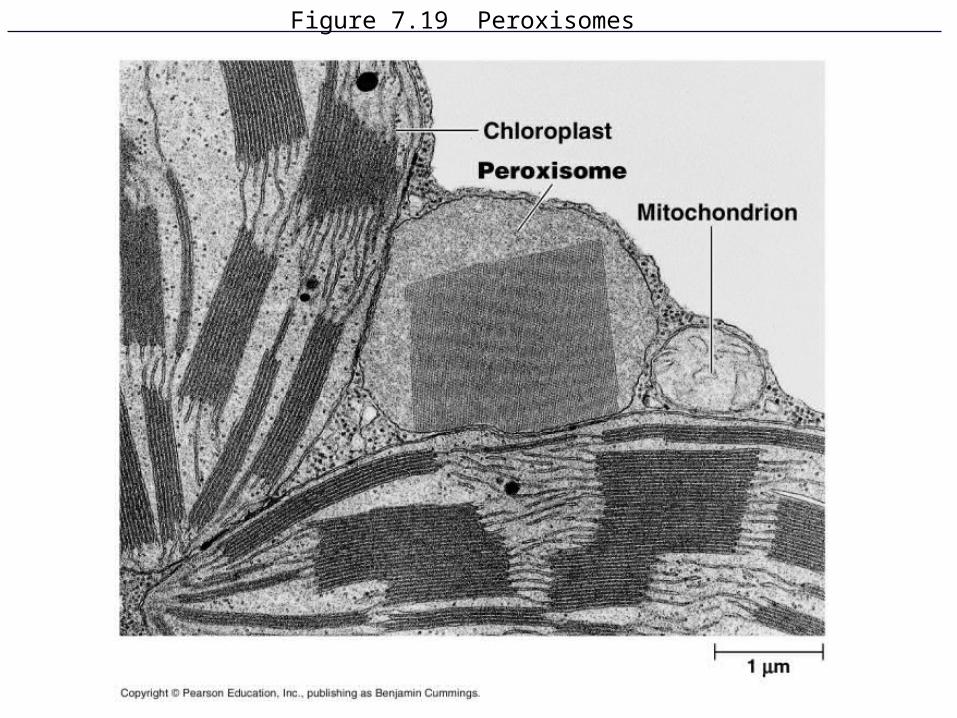
Figure 7.19 Peroxisomes

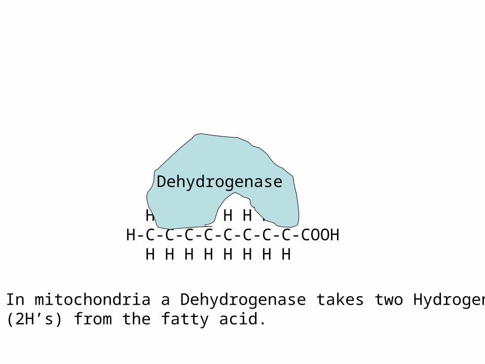
H H H H H H H HH-C-C-C-C-C-C-C-C-COOH H H H H H H H H
A Fatty acid, which can be oxidized in mitochondriaor peroxisomes
A comparison of a mitochondrial dehydrogenase to a peroxisomal oxidase, both of which metabolize fat.
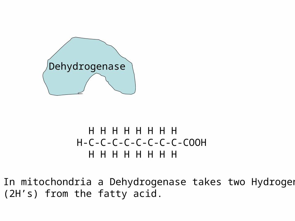
H H H H H H H HH-C-C-C-C-C-C-C-C-COOH H H H H H H H H
In mitochondria a Dehydrogenase takes two Hydrogens(2H’s) from the fatty acid.
Dehydrogenase
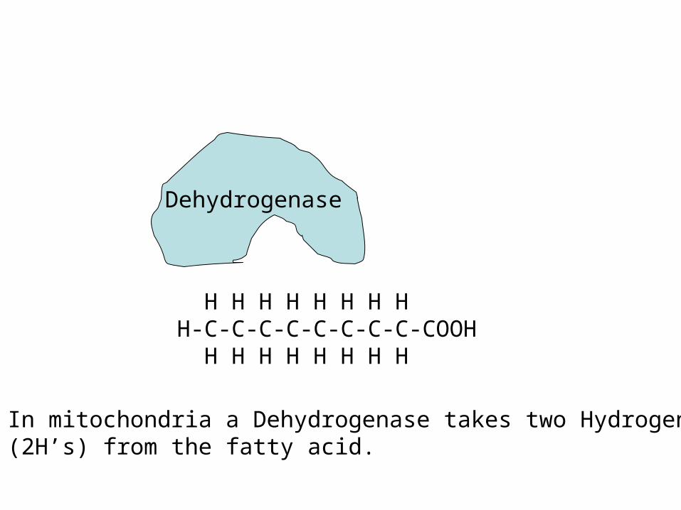
H H H H H H H HH-C-C-C-C-C-C-C-C-COOH H H H H H H H H
In mitochondria a Dehydrogenase takes two Hydrogens(2H’s) from the fatty acid.
Dehydrogenase
H H H H H H H HH-C-C-C-C-C-C-C-C-COOH H H H H H H H H
In mitochondria a Dehydrogenase takes two Hydrogens(2H’s) from the fatty acid.
Dehydrogenase
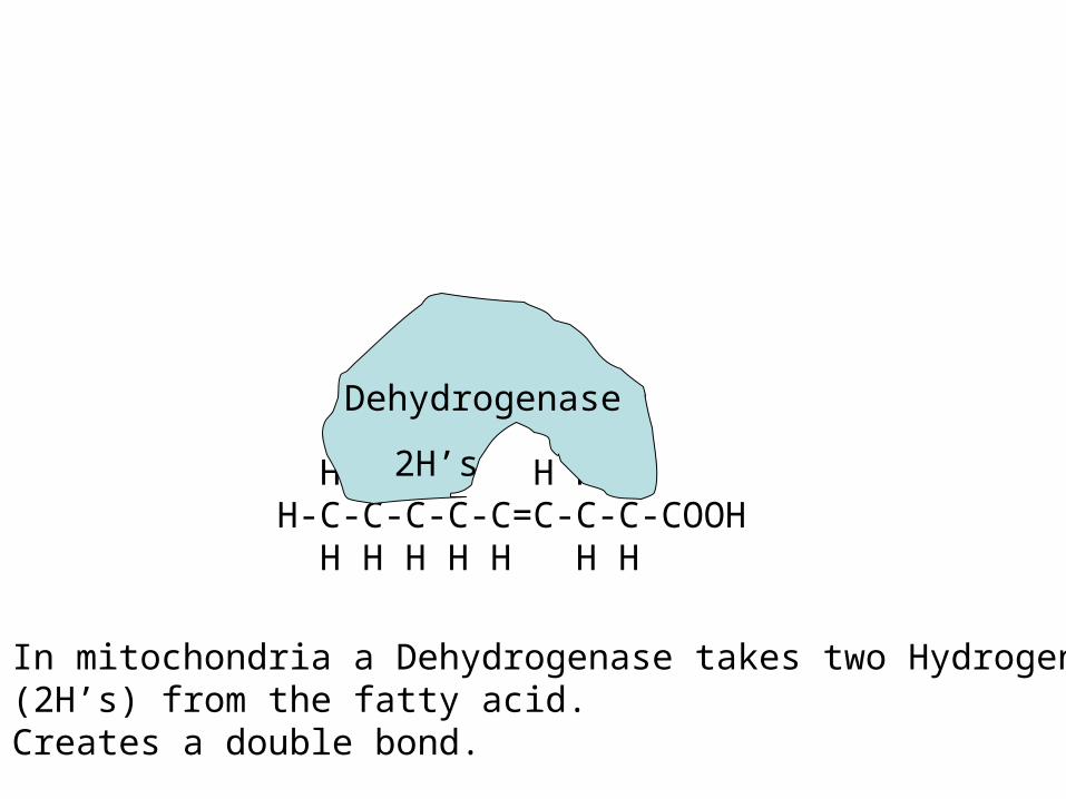
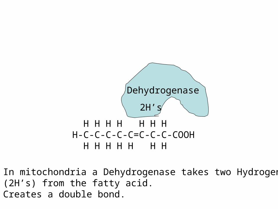
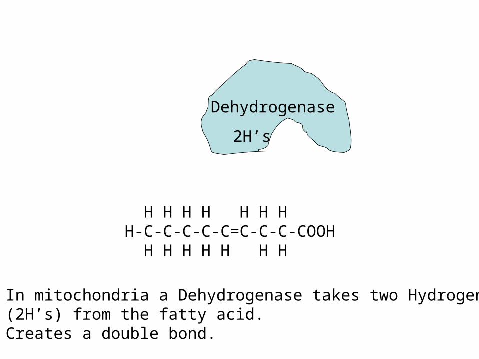
H H H H H H HH-C-C-C-C-C=C-C-C-COOH H H H H H H H
In mitochondria a Dehydrogenase takes two Hydrogens(2H’s) from the fatty acid.Creates a double bond.
Dehydrogenase
2H’s
H H H H H H HH-C-C-C-C-C=C-C-C-COOH H H H H H H H
In mitochondria a Dehydrogenase takes two Hydrogens(2H’s) from the fatty acid.Creates a double bond.
Dehydrogenase
2H’s
H H H H H H HH-C-C-C-C-C=C-C-C-COOH H H H H H H H
In mitochondria a Dehydrogenase takes two Hydrogens(2H’s) from the fatty acid.Creates a double bond.
Dehydrogenase
2H’s
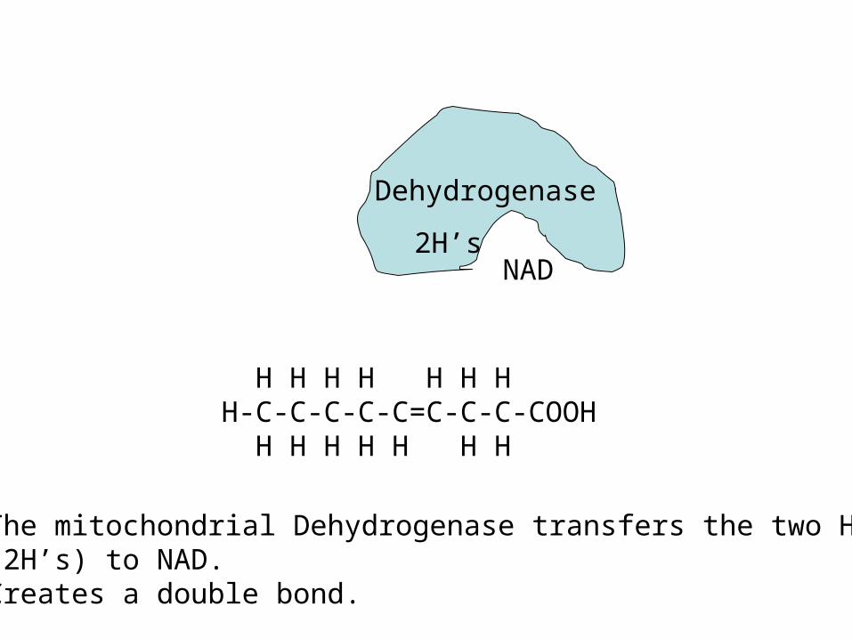
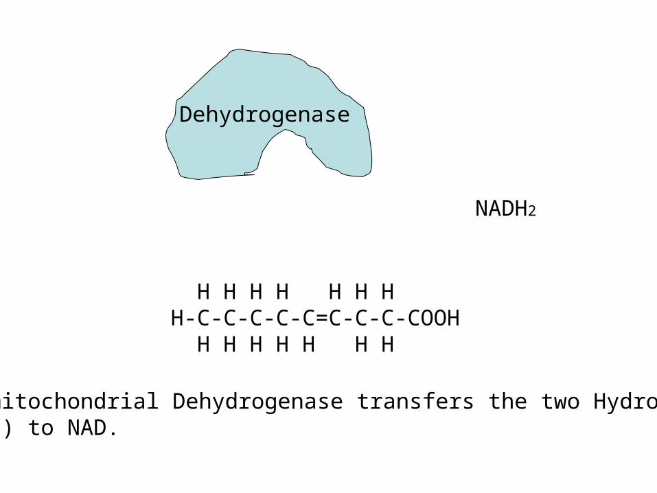
H H H H H H HH-C-C-C-C-C=C-C-C-COOH H H H H H H H
The mitochondrial Dehydrogenase transfers the two Hydrogens(2H’s) to NAD.Creates a double bond.
Dehydrogenase
2H’sNAD
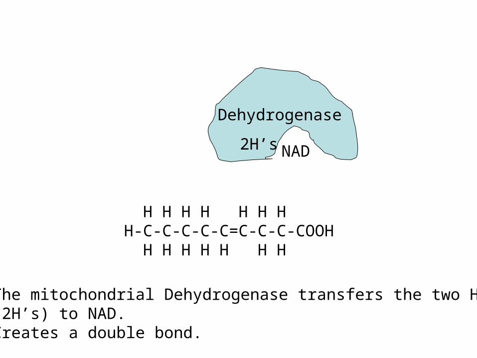
H H H H H H HH-C-C-C-C-C=C-C-C-COOH H H H H H H H
The mitochondrial Dehydrogenase transfers the two Hydrogens(2H’s) to NAD.Creates a double bond.
Dehydrogenase
2H’s NAD
H H H H H H HH-C-C-C-C-C=C-C-C-COOH H H H H H H H
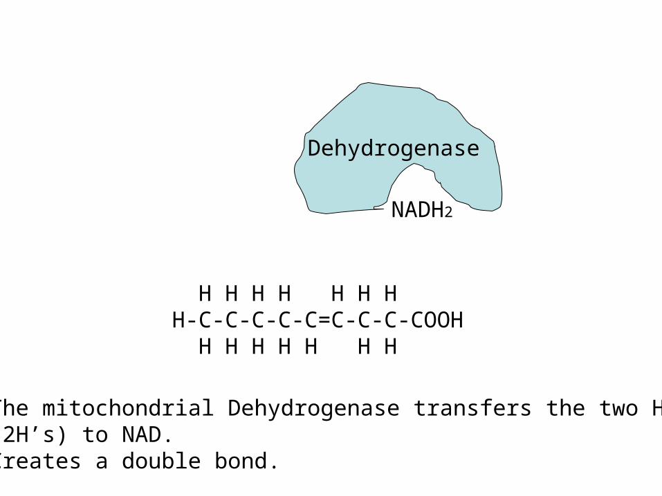
The mitochondrial Dehydrogenase transfers the two Hydrogens(2H’s) to NAD.Creates a double bond.
Dehydrogenase
NADH2
H H H H H H HH-C-C-C-C-C=C-C-C-COOH H H H H H H H
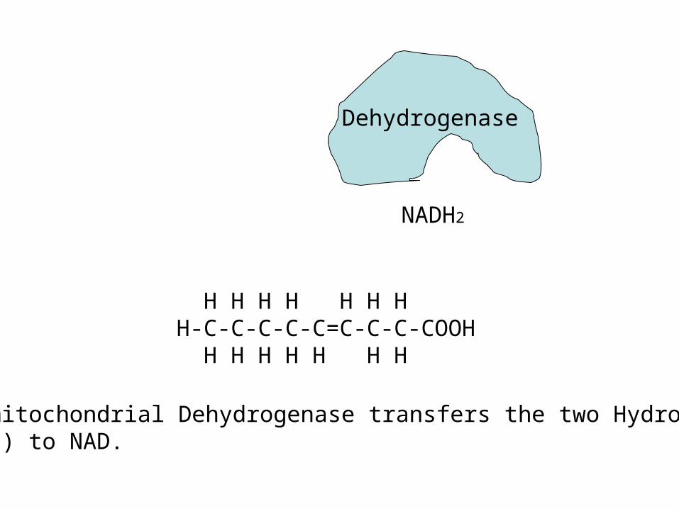
The mitochondrial Dehydrogenase transfers the two Hydrogens(2H’s) to NAD.
Dehydrogenase
NADH2
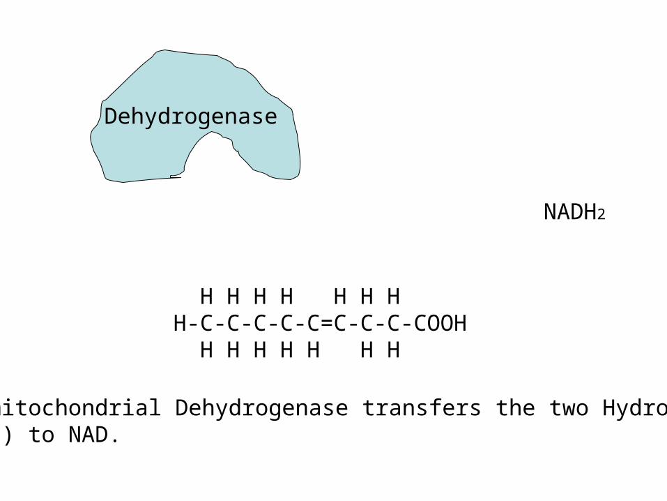
H H H H H H HH-C-C-C-C-C=C-C-C-COOH H H H H H H H
The mitochondrial Dehydrogenase transfers the two Hydrogens(2H’s) to NAD.
Dehydrogenase
NADH2
H H H H H H HH-C-C-C-C-C=C-C-C-COOH H H H H H H H
The mitochondrial Dehydrogenase transfers the two Hydrogens(2H’s) to NAD.
Dehydrogenase
NADH2
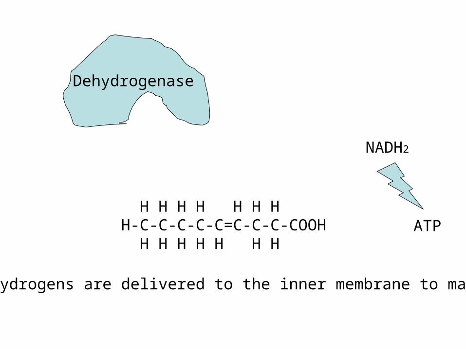
H H H H H H HH-C-C-C-C-C=C-C-C-COOH H H H H H H H
The Hydrogens are delivered to the inner membrane to make ATP
Dehydrogenase
NADH2
ATP
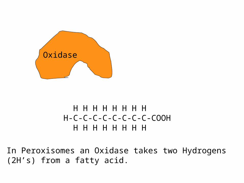
H H H H H H H HH-C-C-C-C-C-C-C-C-COOH H H H H H H H H
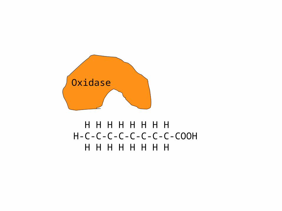
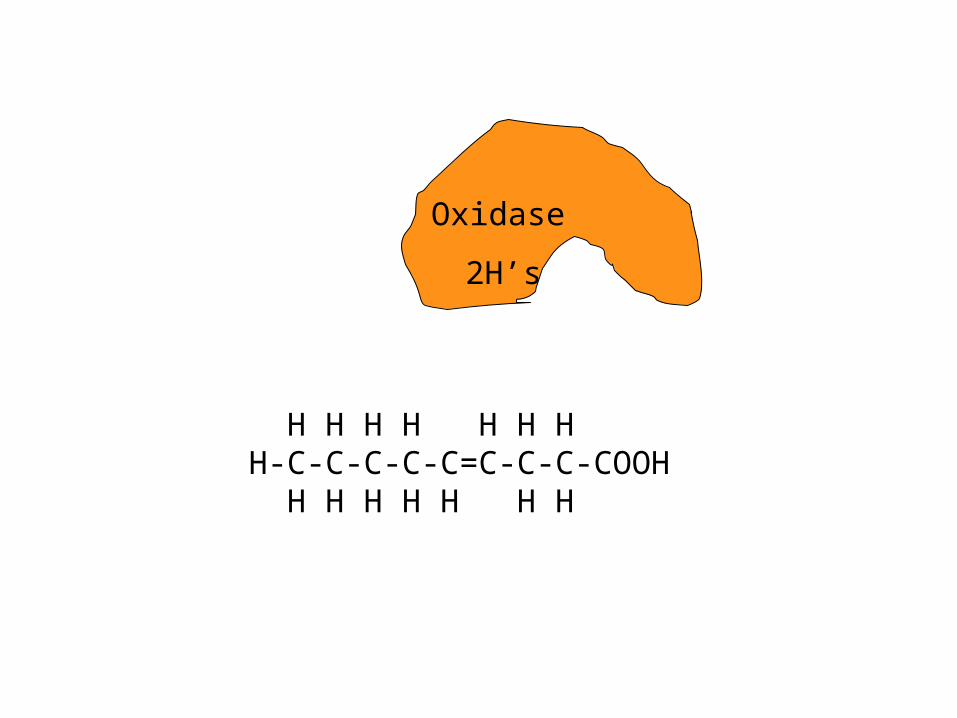
In Peroxisomes an Oxidase takes two Hydrogens(2H’s) from a fatty acid.
Oxidase
H H H H H H H HH-C-C-C-C-C-C-C-C-COOH H H H H H H H H
Oxidase
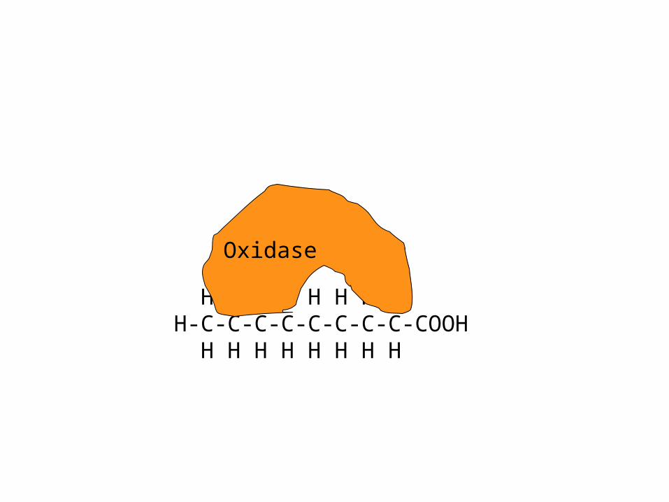
H H H H H H H HH-C-C-C-C-C-C-C-C-COOH H H H H H H H H
Oxidase
H H H H H H HH-C-C-C-C-C=C-C-C-COOH H H H H H H H
Oxidase
2H’s
H H H H H H HH-C-C-C-C-C=C-C-C-COOH H H H H H H H
Oxidase
2H’s
H H H H H H HH-C-C-C-C-C=C-C-C-COOH H H H H H H H
Oxidase
2H’s
H H H H H H HH-C-C-C-C-C=C-C-C-COOH H H H H H H H
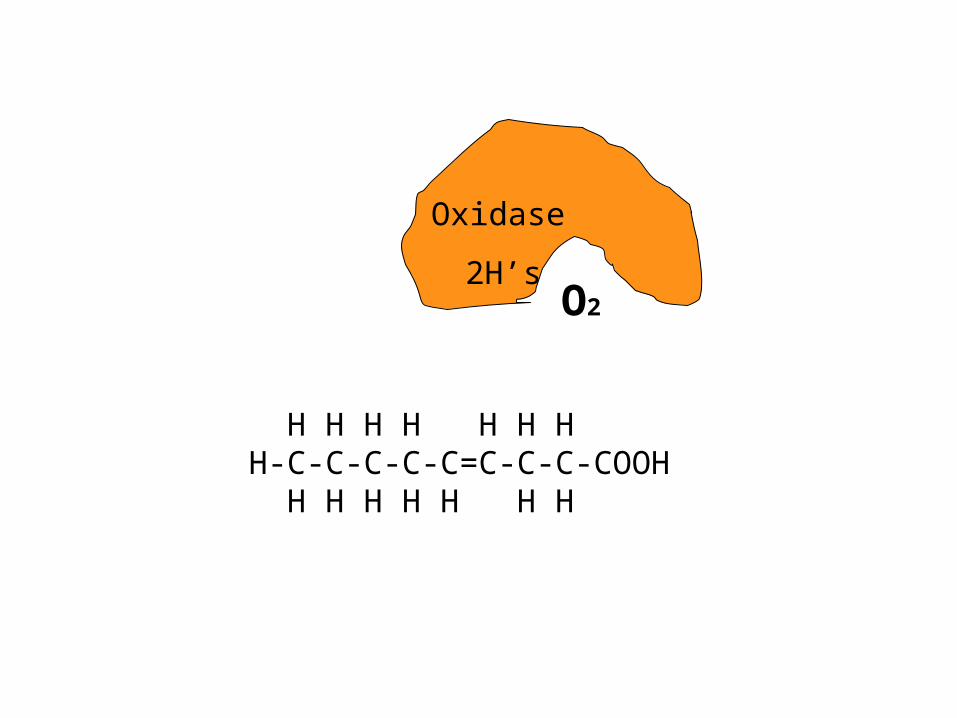
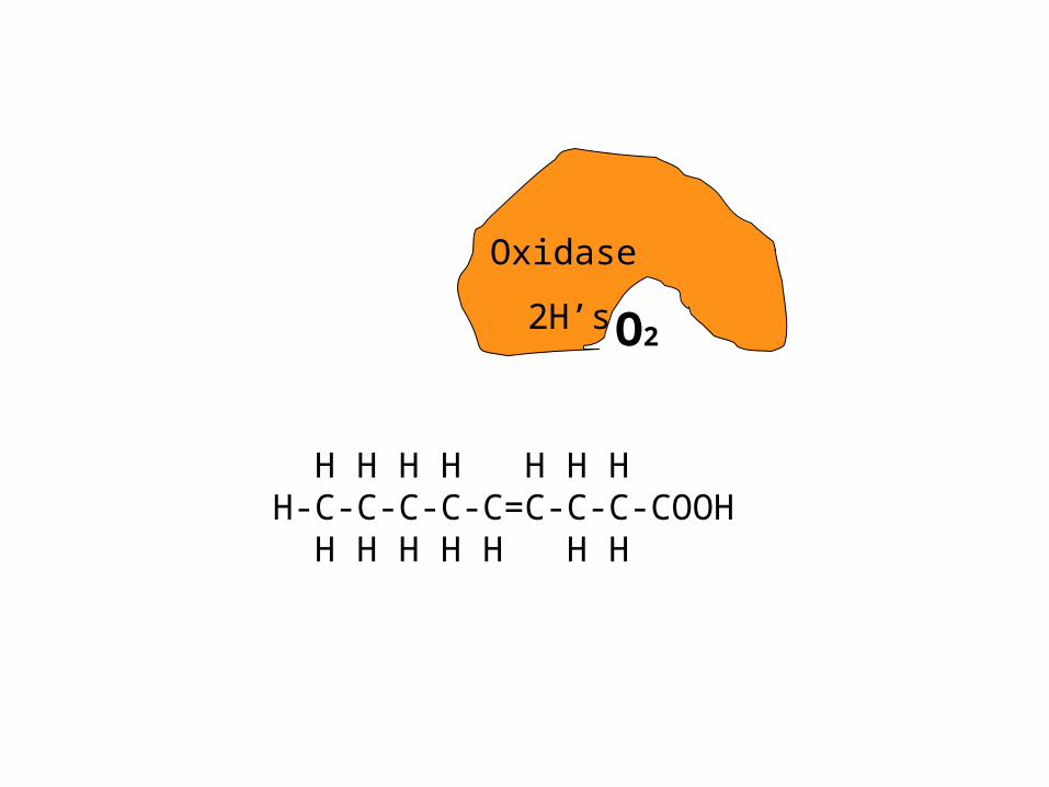
Oxidase
2H’sO2
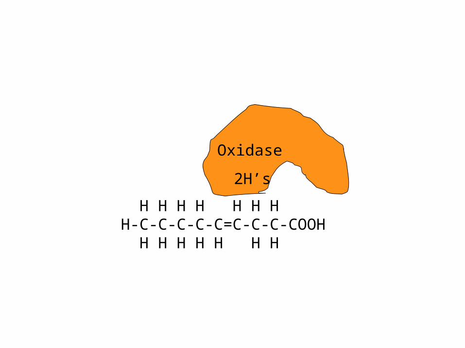
H H H H H H HH-C-C-C-C-C=C-C-C-COOH H H H H H H H
Oxidase
2H’s O2
H H H H H H HH-C-C-C-C-C=C-C-C-COOH H H H H H H H
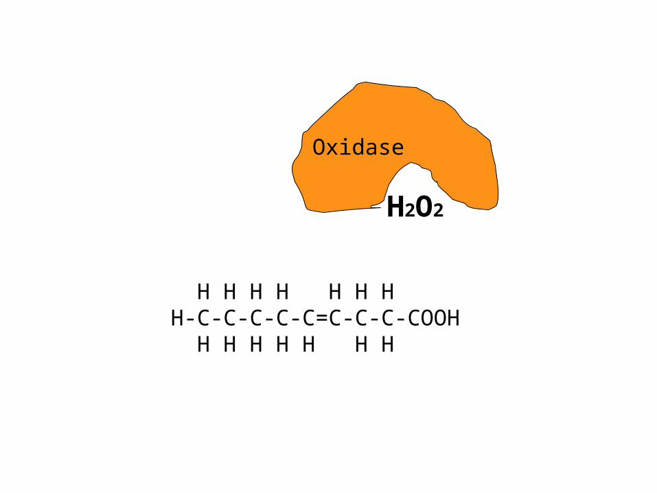
Oxidase
H2O2
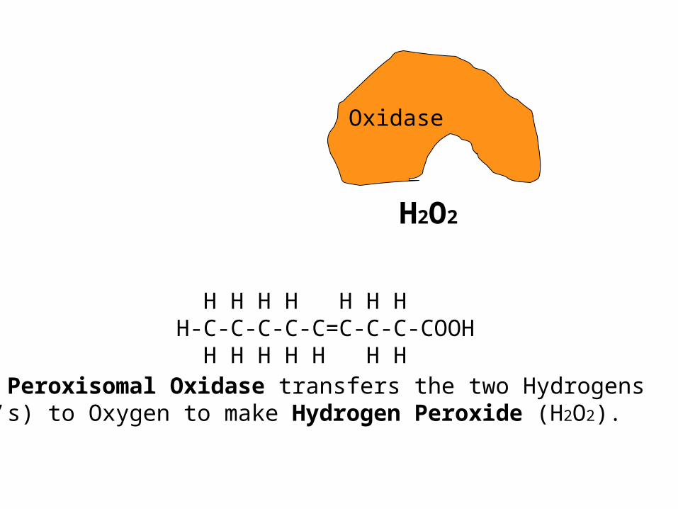
H H H H H H HH-C-C-C-C-C=C-C-C-COOH H H H H H H H
The Peroxisomal Oxidase transfers the two Hydrogens(2H’s) to Oxygen to make Hydrogen Peroxide (H2O2).
Oxidase
H2O2
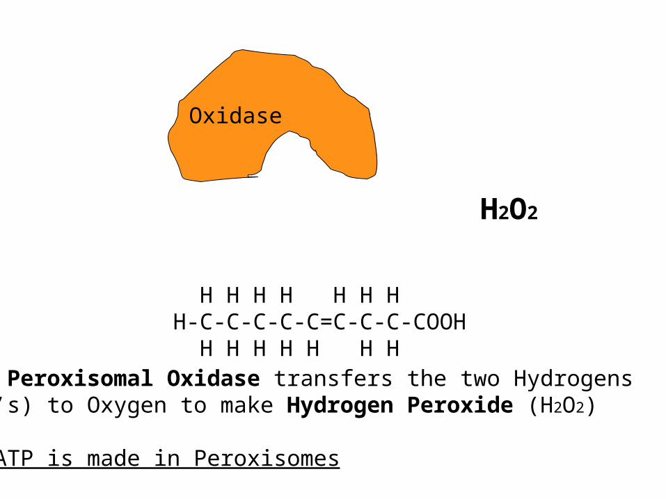
H H H H H H HH-C-C-C-C-C=C-C-C-COOH H H H H H H H
The Peroxisomal Oxidase transfers the two Hydrogens(2H’s) to Oxygen to make Hydrogen Peroxide (H2O2)
No ATP is made in Peroxisomes
Oxidase
H2O2
What happens if there is a genetic defect in a peroxisomal protein?
If one of the nucleotides is changed in the gene, in the DNA, then an amino acid may be changed and the resulting protein may no longer function.
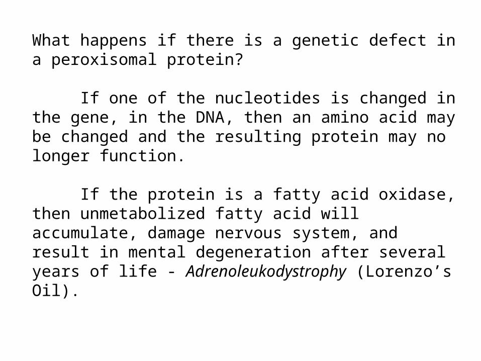
What happens if there is a genetic defect in a peroxisomal protein?
If one of the nucleotides is changed in the gene, in the DNA, then an amino acid may be changed and the resulting protein may no longer function.
If the protein is a fatty acid oxidase, then unmetabolized fatty acid will accumulate, damage nervous system, and result in mental degeneration after several years of life - Adrenoleukodystrophy (Lorenzo’s Oil).
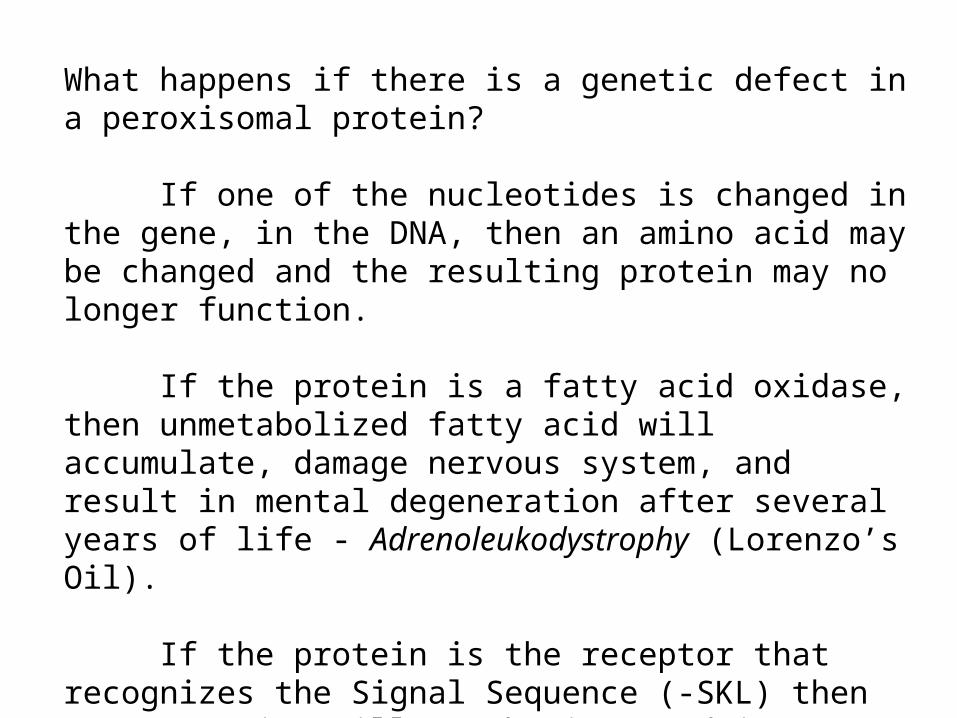
If the protein is the receptor that recognizes the Signal Sequence (-SKL) then most proteins will not be imported into peroxisomes. Infant does not survive - Zellwegers Syndrome.
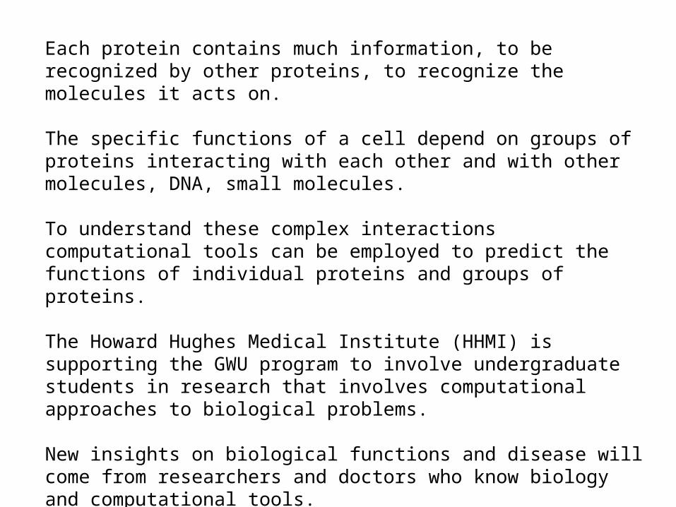
Each protein contains much information, to be recognized by other proteins, to recognize the molecules it acts on.
The specific functions of a cell depend on groups of proteins interacting with each other and with other molecules, DNA, small molecules.
To understand these complex interactions computational tools can be employed to predict the functions of individual proteins and groups of proteins.
The Howard Hughes Medical Institute (HHMI) is supporting the GWU program to involve undergraduate students in research that involves computational approaches to biological problems.
New insights on biological functions and disease will come from researchers and doctors who know biology and computational tools.
The HHMI program has funds to support summer undergraduate research internships, new computer science courses for biologists, new courses where biology and computer science students will work together to investigate biological problems.