Embed Size (px)
Citation preview
Objectification of the Qi Blood Yin Yang Deficiency Pattern by Using a Facial Color AnalysisHye Bin Park1, Junsang Yu2, Hyun Sook Lee1*
Abstract
��Objectives: This study aimed to assess a Qi Blood Yin Yang evaluation method systematically and objectively and to identify the correlation between the Qi Blood Yin Yang deficiency pattern (QBYYDP) and facial color.
Methods: Thirty-seven participants (17 males, 20 fe-males) were enrolled in this study. Twenty-four (10 males, 14 females) had ages from 40 to over 60, and 13 (7 males and 6 females) were in their twenties. After suffi-cient rest, facial images were taken with a camera. Based on the results from a questionnaire survey, we divided the participants into five groups: the normal and the Qi-, Blood-, Yin-, and Yang-deficient groups, after which the relationships between the L, ‘a’, and ‘b’ values in the Lab color system and the characteristics of the participants in each of the deficient groups were elucidated using a facial color analysis program.
Results: The color analysis for Qi-deficient (QD) par-ticipants revealed that the L value was fairly decreased in comparison with the normal participants, but the ‘a’ and ‘b’ values were almost the same. A comparison be-tween the normal and the Yang-deficient (YaD) groups revealed that the L values were somewhat lower com-pared to the normal group, but the ‘a’ and ‘b’ values were not statistically different. For the Yin-deficient (YiD) group, the L value was slightly lower compared to the
normal group, but the ‘a’ and ‘b’ values were almost the same and the R values were slightly increased. For the Blood-deficient (BD) group, the L values were slightly increased compared to the normal group, but the ‘a’ and ‘b’ values were decreased slightly.
Conclusion:� This study obtained objective, reliable data for judging the QBYYDP by using facial images and a color analysis program. However, further study with at least 10 or more subjects in each of the deficient groups is necessary to confirm our findings.
1. Introduction
Inspection, one of four diagnostic methods in tradi-tional Korean medicine, is a very important method, as are the hearing, inquiring and percussion or touch-ing methods. Therefore, doctors who can read patients’ faces and understand their diseases based on those readings were often referred to as ‘utmost doctors’ [1]. Inspection includes determining the patient’s spirit, facial color, facial and body shapes, movements, lo-cal circumstances, tongue’s shape, excretion, color, condition, and amounts of excretion. Traditional doc-tors collect much information on the basis of inspec-tion [2]. Above all, the spirit and the color of the whole body are two key points in terms of inspection, and the face is the area most frequently used. Using the face for inspection has many advantages, for the patient’s emotions and his or her physiological and pathologi-cal conditions are well expressed on his or her face [3].
In the context of traditional Korean medicine, the heart controls the blood vessels, and the richness of the heart’s energy appears on the face. The Hand and
Original article
Key Wordsdeficiency pattern questionnaires, facial color analysis,
Qi-blood, traditional Korean medicine, Yin-Yang
ISSN 2093-6966 [Print], ISSN 2234-6856 [Online]Journal of Pharmacopuncture 2017;20[2]:100-106DOI: https://doi.org/10.3831/KPI.2017.20.013
This is an Open-Access article distributed under the terms of the Creative CommonsAttribution Non-Commercial License (http://creativecommons.org/licenses/by-nc/4.0/) which permits unrestricted noncommercial use, distribution, and reproduction in anymedium, provided the original work is properly cited.
This paper meets the requirements of KS X ISO 9706, ISO 9706-1994 and ANSI/NISOZ39.48-1992 (Permanence of Paper).
*Corresponding AuthorHyun Sook Lee. Department of Oriental Biomedical Engineering, College of Health Sci-ence, Sangji University, 83 Sangjidae-gil, Wonju-si, Gangwon-do 26339, KoreaTel: +82-33-730-0416 Fax:+82-33-730-7652E-mail:[email protected]
ⓒ 2017 Korean Pharmacopuncture Institute http://www.journal.ac
1 �Department�of�Oriental�Biomedical�Engineering,�College�of�Health�Science,�Sangji�University,�Wonju,�Korea2��Department�of�Sasang�Constitutional�Medicine,�Oriental�Medicine�Hospital�of�Sangji�University,�Wonju,�Korea
Received: Feb 21, 2017 Reviewed: Mar 12, 2017 Accepted: May 04, 2017
http://www.journal.ac 101
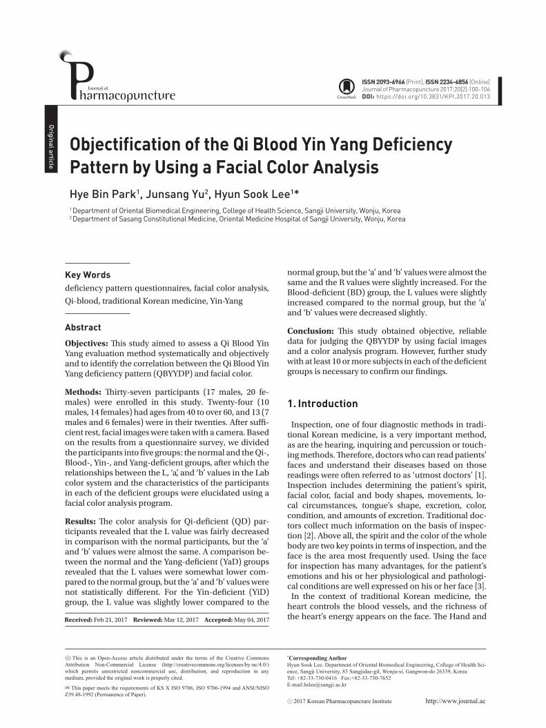
Figure� 1� Facial image camera (Dermavision) with blackout for
blocking light.
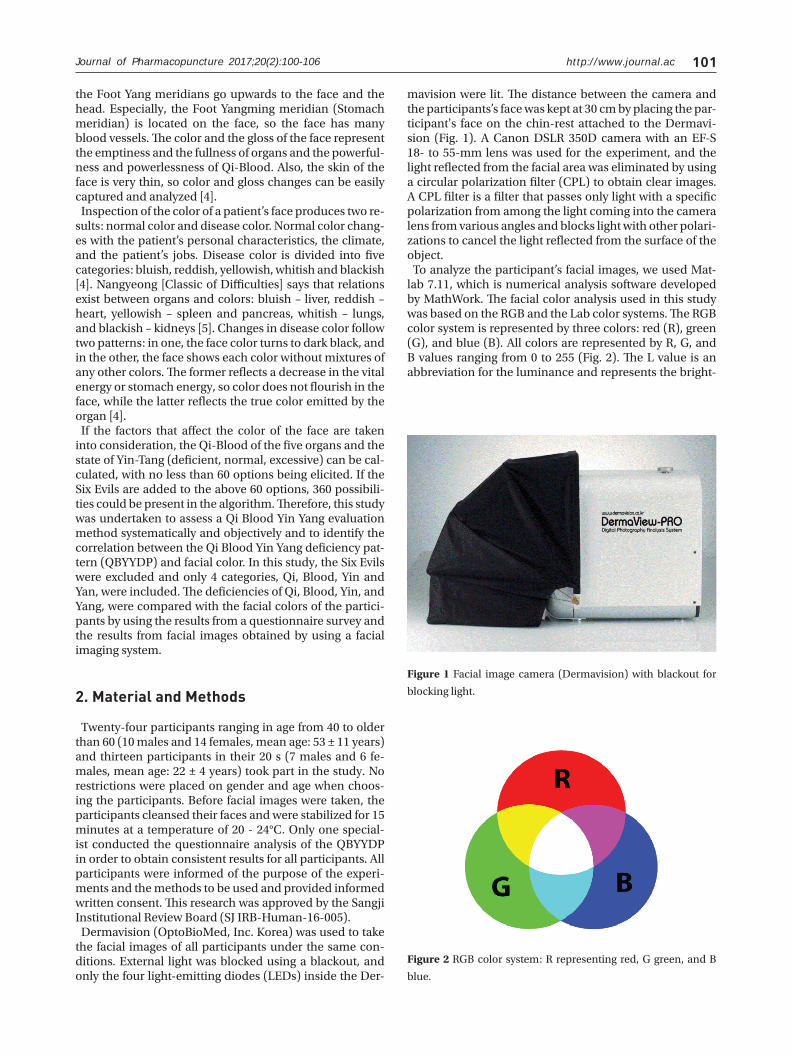
Figure�2�RGB color system: R representing red, G green, and B
blue.
the Foot Yang meridians go upwards to the face and the head. Especially, the Foot Yangming meridian (Stomach meridian) is located on the face, so the face has many blood vessels. The color and the gloss of the face represent the emptiness and the fullness of organs and the powerful-ness and powerlessness of Qi-Blood. Also, the skin of the face is very thin, so color and gloss changes can be easily captured and analyzed [4].
Inspection of the color of a patient’s face produces two re-sults: normal color and disease color. Normal color chang-es with the patient’s personal characteristics, the climate, and the patient’s jobs. Disease color is divided into five categories: bluish, reddish, yellowish, whitish and blackish [4]. Nangyeong [Classic of Difficulties] says that relations exist between organs and colors: bluish – liver, reddish – heart, yellowish – spleen and pancreas, whitish – lungs, and blackish – kidneys [5]. Changes in disease color follow two patterns: in one, the face color turns to dark black, and in the other, the face shows each color without mixtures of any other colors. The former reflects a decrease in the vital energy or stomach energy, so color does not flourish in the face, while the latter reflects the true color emitted by the organ [4].
If the factors that affect the color of the face are taken into consideration, the Qi-Blood of the five organs and the state of Yin-Tang (deficient, normal, excessive) can be cal-culated, with no less than 60 options being elicited. If the Six Evils are added to the above 60 options, 360 possibili-ties could be present in the algorithm. Therefore, this study was undertaken to assess a Qi Blood Yin Yang evaluation method systematically and objectively and to identify the correlation between the Qi Blood Yin Yang deficiency pat-tern (QBYYDP) and facial color. In this study, the Six Evils were excluded and only 4 categories, Qi, Blood, Yin and Yan, were included. The deficiencies of Qi, Blood, Yin, and Yang, were compared with the facial colors of the partici-pants by using the results from a questionnaire survey and the results from facial images obtained by using a facial imaging system.
2. Material and Methods
Twenty-four participants ranging in age from 40 to older than 60 (10 males and 14 females, mean age: 53 ± 11 years) and thirteen participants in their 20 s (7 males and 6 fe-males, mean age: 22 ± 4 years) took part in the study. No restrictions were placed on gender and age when choos-ing the participants. Before facial images were taken, the participants cleansed their faces and were stabilized for 15 minutes at a temperature of 20 - 24°C. Only one special-ist conducted the questionnaire analysis of the QBYYDP in order to obtain consistent results for all participants. All participants were informed of the purpose of the experi-ments and the methods to be used and provided informed written consent. This research was approved by the Sangji Institutional Review Board (SJ IRB-Human-16-005).
Dermavision (OptoBioMed, Inc. Korea) was used to take the facial images of all participants under the same con-ditions. External light was blocked using a blackout, and only the four light-emitting diodes (LEDs) inside the Der-
mavision were lit. The distance between the camera and the participants’s face was kept at 30 cm by placing the par-ticipant's face on the chin-rest attached to the Dermavi-sion (Fig. 1). A Canon DSLR 350D camera with an EF-S 18- to 55-mm lens was used for the experiment, and the light reflected from the facial area was eliminated by using a circular polarization filter (CPL) to obtain clear images. A CPL filter is a filter that passes only light with a specific polarization from among the light coming into the camera lens from various angles and blocks light with other polari-zations to cancel the light reflected from the surface of the object.
To analyze the participant’s facial images, we used Mat-lab 7.11, which is numerical analysis software developed by MathWork. The facial color analysis used in this study was based on the RGB and the Lab color systems. The RGB color system is represented by three colors: red (R), green (G), and blue (B). All colors are represented by R, G, and B values ranging from 0 to 255 (Fig. 2). The L value is an abbreviation for the luminance and represents the bright-
Journal of Pharmacopuncture 2017;20(2):100-106
http://www.journal.ac102 Journal of Pharmacopuncture 2017;20(2):100-106
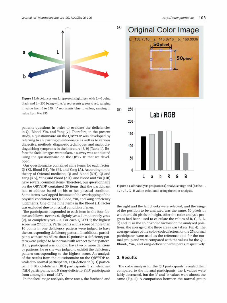
ness, while ‘a’ and ‘b’ are the chromatic parameters for green-to-red and blue-to-yellow, respectively [6]. In the color analysis program developed in this study, the values of L, ‘a’, and ‘b’ were calculated and were found to range
from 0 to 255 (Fig. 3).For a medical doctor to diagnose a patient through in-
spection, the doctor must evaluate the QBYYDP. The most objective diagnosis is known to be obtained by asking the
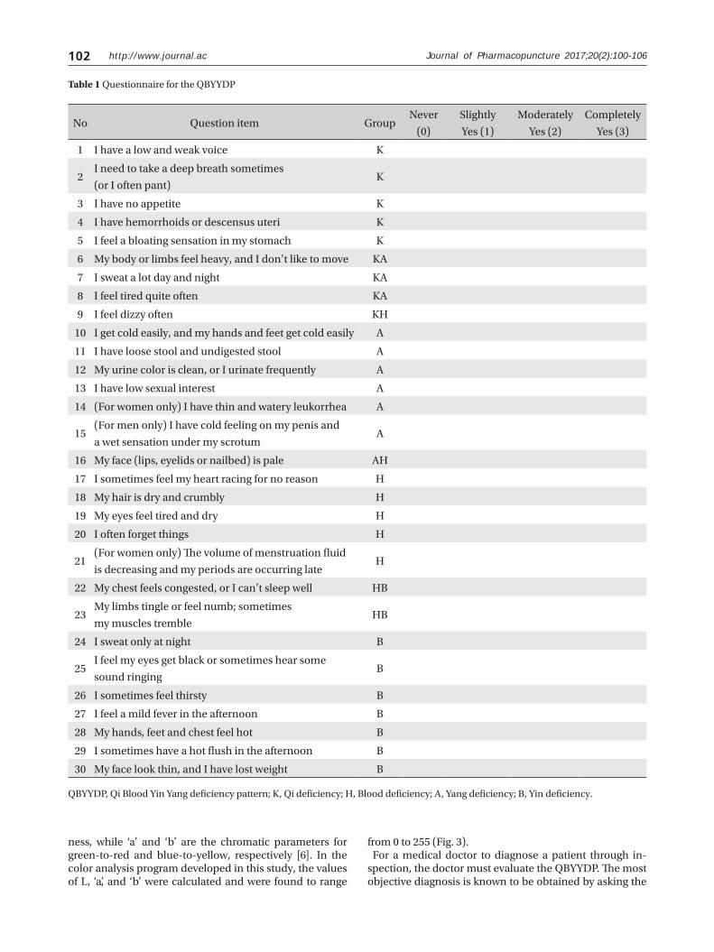
Table�1�Questionnaire for the QBYYDP
QBYYDP, Qi Blood Yin Yang deficiency pattern; K, Qi deficiency; H, Blood deficiency; A, Yang deficiency; B, Yin deficiency.
No Question item GroupNever
(0)
Slightly
Yes (1)
Moderately
Yes (2)
Completely
Yes (3)
1 I have a low and weak voice K
2I need to take a deep breath sometimes
(or I often pant)K
3 I have no appetite K
4 I have hemorrhoids or descensus uteri K
5 I feel a bloating sensation in my stomach K
6 My body or limbs feel heavy, and I don’t like to move KA
7 I sweat a lot day and night KA
8 I feel tired quite often KA
9 I feel dizzy often KH
10 I get cold easily, and my hands and feet get cold easily A
11 I have loose stool and undigested stool A
12 My urine color is clean, or I urinate frequently A
13 I have low sexual interest A
14 (For women only) I have thin and watery leukorrhea A
15(For men only) I have cold feeling on my penis and
a wet sensation under my scrotumA
16 My face (lips, eyelids or nailbed) is pale AH
17 I sometimes feel my heart racing for no reason H
18 My hair is dry and crumbly H
19 My eyes feel tired and dry H
20 I often forget things H
21(For women only) The volume of menstruation fluid
is decreasing and my periods are occurring lateH
22 My chest feels congested, or I can’t sleep well HB
23My limbs tingle or feel numb; sometimes
my muscles trembleHB
24 I sweat only at night B
25I feel my eyes get black or sometimes hear some
sound ringingB
26 I sometimes feel thirsty B
27 I feel a mild fever in the afternoon B
28 My hands, feet and chest feel hot B
29 I sometimes have a hot flush in the afternoon B
30 My face look thin, and I have lost weight B
http://www.journal.ac 103
patients questions in order to evaluate the deficiencies in Qi, Blood, Yin, and Yang [7]. Therefore, in the present study, a questionnaire on the QBYYDP was developed by referring to an existing questionnaire as well as to various dialectical methods, diagnostic techniques, and major dis-tinguishing symptoms in the literature [8, 9] (Table 1). Be-fore the facial images were taken, a survey was conducted using the questionnaire on the QBYYDP that we devel-oped.
Our questionnaire contained nine items for each factor: Qi (K), Blood (H), Yin (B), and Yang (A). According to the theory of Oriental medicine, Qi and Blood (KH), Qi and Yang (KA), Yang and Blood (AH), and Blood and Yin (HB) have several common items. Therefore, our questionnaire on the QBYYDP contained 30 items that the participant had to address based on his or her physical condition. Some items overlapped because of the overlapping of the physical conditions for Qi, Blood, Yin, and Yang deficiency judgments. One of the nine items in the Blood (H) factor was excluded due to physical condition of men.
The participants responded to each item in the four fac-tors as follows: never = 0, slightly yes = 1, moderately yes = (2), or completely yes = 3. For each QBYYDP, the highest score was 27 points. Participants with a score of more than 10 points in one deficiency pattern were judged to have the corresponding deficiency pattern. In addition, partici-pants with scores of less than 10 points in a deficiency pat-tern were judged to be normal with respect to that pattern. If any participant was found to have two or more deficien-cy patterns, he or she was judged to exhibit the deficiency pattern corresponding to the highest score. An analysis of the results from the questionnaire on the QBYYDP re-vealed 25 normal participants, 1 Qi-deficient (QD) partici-pant, 3 Blood-deficient (BD) participants, 3 Yin-deficient (YiD) participants, and 5 Yang-deficient (YaD) participants from among the total of 37.
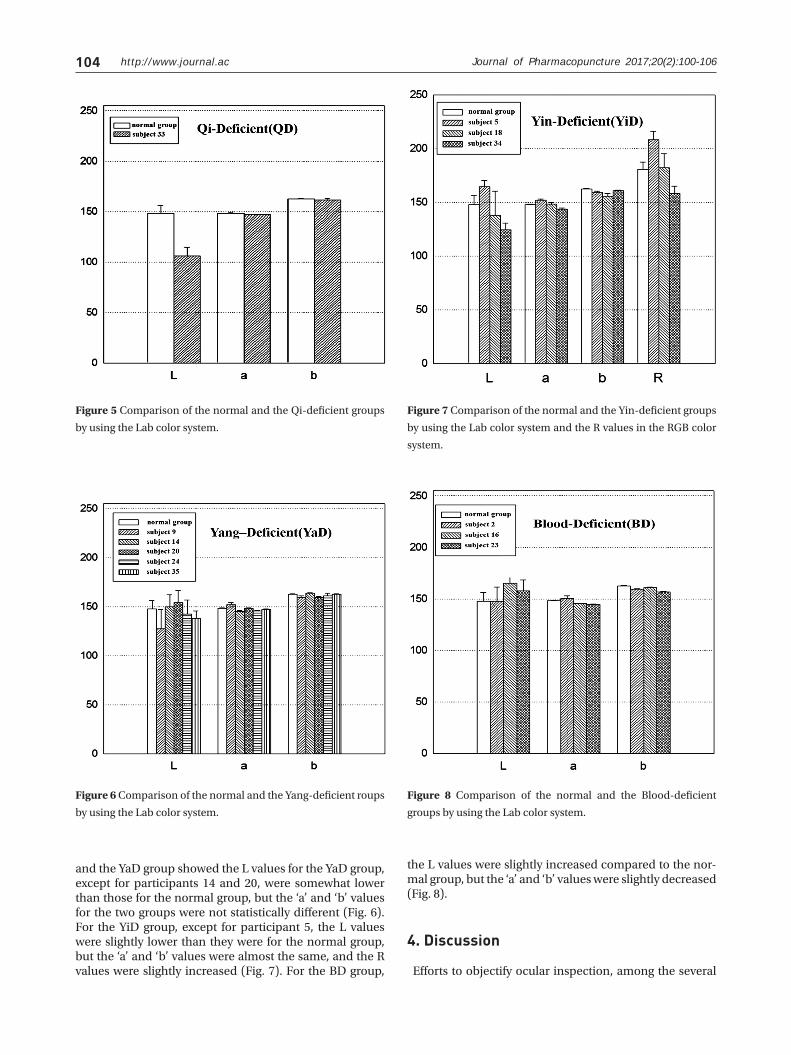
In the face image analysis, three areas, the forehead and
the right and the left cheeks were selected, and the range of the position to be analyzed was the same, 50 pixels in width and 50 pixels in height. After the color analysis pro-gram had been used to calculate the values of R, G, B, L, ‘a’, and ‘b’ as the color-coded factors for the analyzed posi-tions, the average of the three areas was taken (Fig. 4). The average values of the color-coded factors for the 25 normal participants were used as the reference data for the nor-mal group and were compared with the values for the Qi-, Blood-, Yin-, and Yang-deficient participants, respectively.
3. Results
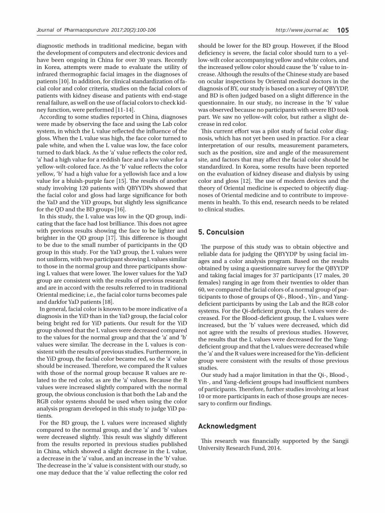
The color analysis for the QD participants revealed that, compared to the normal participants, the L values were fairly decreased, but the ‘a’ and ‘b’ values were almost the same (Fig. 5). A comparison between the normal group
Journal of Pharmacopuncture 2017;20(2):100-106
Figure�3�Lab color system. L represents lightness, with L = 0 being
black and L = 255 being white. ‘a’ represents green to red, ranging
in value from 0 to 255. ‘b’ represents blue to yellow, ranging in
value from 0 to 255.
Figure�4�Color analysis program: (a) analysis range and (b) the L ,
a , b , R , G , B values calculated using the color analysis.
(A)
(B)
http://www.journal.ac104
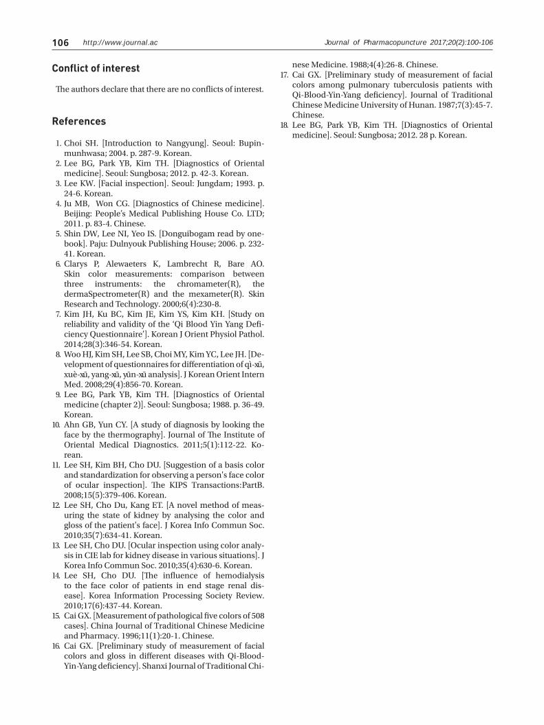
and the YaD group showed the L values for the YaD group, except for participants 14 and 20, were somewhat lower than those for the normal group, but the ‘a’ and ‘b’ values for the two groups were not statistically different (Fig. 6). For the YiD group, except for participant 5, the L values were slightly lower than they were for the normal group, but the ‘a’ and ‘b’ values were almost the same, and the R values were slightly increased (Fig. 7). For the BD group,
the L values were slightly increased compared to the nor-mal group, but the ‘a’ and ‘b’ values were slightly decreased (Fig. 8).
4. Discussion
Efforts to objectify ocular inspection, among the several
Figure�5�Comparison of the normal and the Qi-deficient groups
by using the Lab color system.
Figure�7�Comparison of the normal and the Yin-deficient groups
by using the Lab color system and the R values in the RGB color
system.
Figure�6�Comparison of the normal and the Yang-deficient roups
by using the Lab color system.
Figure� 8� Comparison of the normal and the Blood-deficient
groups by using the Lab color system.
Journal of Pharmacopuncture 2017;20(2):100-106
http://www.journal.ac 105
diagnostic methods in traditional medicine, began with the development of computers and electronic devices and have been ongoing in China for over 30 years. Recently in Korea, attempts were made to evaluate the utility of infrared thermographic facial images in the diagnoses of patients [10]. In addition, for clinical standardization of fa-cial color and color criteria, studies on the facial colors of patients with kidney disease and patients with end-stage renal failure, as well on the use of facial colors to check kid-ney function, were performed [11-14].
According to some studies reported in China, diagnoses were made by observing the face and using the Lab color system, in which the L value reflected the influence of the gloss. When the L value was high, the face color turned to pale white, and when the L value was low, the face color turned to dark black. As the ‘a’ value reflects the color red, ‘a’ had a high value for a reddish face and a low value for a yellow-wilt-colored face. As the ‘b’ value reflects the color yellow, ‘b’ had a high value for a yellowish face and a low value for a bluish-purple face [15]. The results of another study involving 120 patients with QBYYDPs showed that the facial color and gloss had large significance for both the YaD and the YiD groups, but slightly less significance for the QD and the BD groups [16].
In this study, the L value was low in the QD group, indi-cating that the face had lost brilliance. This does not agree with previous results showing the face to be lighter and brighter in the QD group [17]. This difference is thought to be due to the small number of participants in the QD group in this study. For the YaD group, the L values were not uniform, with two participant showing L values similar to those in the normal group and three participants show-ing L values that were lower. The lower values for the YaD group are consistent with the results of previous research and are in accord with the results referred to in traditional Oriental medicine; i.e., the facial color turns becomes pale and darkfor YaD patients [18].
In general, facial color is known to be more indicative of a diagnosis in the YiD than in the YaD group, the facial color being bright red for YiD patients. Our result for the YiD group showed that the L values were decreased compared to the values for the normal group and that the ‘a’ and ‘b’ values were similar. The decrease in the L values is con-sistent with the results of previous studies. Furthermore, in the YiD group, the facial color became red, so the ‘a’ value should be increased. Therefore, we compared the R values with those of the normal group because R values are re-lated to the red color, as are the ‘a’ values. Because the R values were increased slightly compared with the normal group, the obvious conclusion is that both the Lab and the RGB color systems should be used when using the color analysis program developed in this study to judge YiD pa-tients.
For the BD group, the L values were increased slightly compared to the normal group, and the ‘a’ and ‘b’ values were decreased slightly. This result was slightly different from the results reported in previous studies published in China, which showed a slight decrease in the L value, a decrease in the ‘a’ value, and an increase in the ‘b’ value. The decrease in the ‘a’ value is consistent with our study, so one may deduce that the ‘a’ value reflecting the color red
should be lower for the BD group. However, if the Blood deficiency is severe, the facial color should turn to a yel-low-wilt color accompanying yellow and white colors, and the increased yellow color should cause the ‘b’ value to in-crease. Although the results of the Chinese study are based on ocular inspections by Oriental medical doctors in the diagnosis of BY, our study is based on a survey of QBYYDP, and BD is often judged based on a slight difference in the questionnaire. In our study, no increase in the ‘b’ value was observed because no participants with severe BD took part. We saw no yellow-wilt color, but rather a slight de-crease in red color.
This current effort was a pilot study of facial color diag-nosis, which has not yet been used in practice. For a clear interpretation of our results, measurement parameters, such as the position, size and angle of the measurement site, and factors that may affect the facial color should be standardized. In Korea, some results have been reported on the evaluation of kidney disease and dialysis by using color and gloss [12]. The use of modern devices and the theory of Oriental medicine is expected to objectify diag-noses of Oriental medicine and to contribute to improve-ments in health. To this end, research needs to be related to clinical studies.
5. Conculsion
The purpose of this study was to obtain objective and reliable data for judging the QBYYDP by using facial im-ages and a color analysis program. Based on the results obtained by using a questionnaire survey for the QBYYDP and taking facial images for 37 participants (17 males, 20 females) ranging in age from their twenties to older than 60, we compared the facial colors of a normal group of par-ticipants to those of groups of Qi-, Blood-, Yin-, and Yang-deficient participants by using the Lab and the RGB color systems. For the Qi-deficient group, the L values were de-creased. For the Blood-deficient group, the L values were increased, but the ’b’ values were decreased, which did not agree with the results of previous studies. However, the results that the L values were decreased for the Yang-deficient group and that the L values were decreased while the ‘a’ and the R values were increased for the Yin-deficient group were consistent with the results of those previous studies.
Our study had a major limitation in that the Qi-, Blood-, Yin-, and Yang-deficient groups had insufficient numbers of participants. Therefore, further studies involving at least 10 or more participants in each of those groups are neces-sary to confirm our findings.
Acknowledgment
This research was financially supported by the Sangji University Research Fund, 2014.
Journal of Pharmacopuncture 2017;20(2):100-106
http://www.journal.ac106
Choi SH. [Introduction to Nangyung]. Seoul: Bupin-munhwasa; 2004. p. 287-9. Korean.Lee BG, Park YB, Kim TH. [Diagnostics of Oriental medicine]. Seoul: Sungbosa; 2012. p. 42-3. Korean.Lee KW. [Facial inspection]. Seoul: Jungdam; 1993. p. 24-6. Korean.Ju MB, Won CG. [Diagnostics of Chinese medicine]. Beijing: People’s Medical Publishing House Co. LTD; 2011. p. 83-4. Chinese.Shin DW, Lee NI, Yeo IS. [Donguibogam read by one-book]. Paju: Dulnyouk Publishing House; 2006. p. 232-41. Korean.Clarys P, Alewaeters K, Lambrecht R, Bare AO. Skin color measurements: comparison between three instruments: the chromameter(R), the dermaSpectrometer(R) and the mexameter(R). Skin Research and Technology. 2000;6(4):230-8.Kim JH, Ku BC, Kim JE, Kim YS, Kim KH. [Study on reliability and validity of the ‘Qi Blood Yin Yang Defi-ciency Questionnaire’]. Korean J Orient Physiol Pathol. 2014;28(3):346-54. Korean.Woo HJ, Kim SH, Lee SB, Choi MY, Kim YC, Lee JH. [De-velopment of questionnaires for differentiation of qì-xu, xuè-xu, yang-xu, yun-xu analysis]. J Korean Orient Intern Med. 2008;29(4):856-70. Korean.Lee BG, Park YB, Kim TH. [Diagnostics of Oriental medicine (chapter 2)]. Seoul: Sungbosa; 1988. p. 36-49. Korean.Ahn GB, Yun CY. [A study of diagnosis by looking the face by the thermography]. Journal of The Institute of Oriental Medical Diagnostics. 2011;5(1):112-22. Ko-rean.Lee SH, Kim BH, Cho DU. [Suggestion of a basis color and standardization for observing a person's face color of ocular inspection]. The KIPS Transactions:PartB. 2008;15(5):379-406. Korean.Lee SH, Cho Du, Kang ET. [A novel method of meas-uring the state of kidney by analysing the color and gloss of the patient's face]. J Korea Info Commun Soc. 2010;35(7):634-41. Korean.Lee SH, Cho DU. [Ocular inspection using color analy-sis in CIE lab for kidney disease in various situations]. J Korea Info Commun Soc. 2010;35(4):630-6. Korean.Lee SH, Cho DU. [The influence of hemodialysis to the face color of patients in end stage renal dis-ease]. Korea Information Processing Society Review. 2010;17(6):437-44. Korean. Cai GX. [Measurement of pathological five colors of 508 cases]. China Journal of Traditional Chinese Medicine and Pharmacy. 1996;11(1):20-1. Chinese. Cai GX. [Preliminary study of measurement of facial colors and gloss in different diseases with Qi-Blood-Yin-Yang deficiency]. Shanxi Journal of Traditional Chi-
1.
2.
3.
4.
5.
6.
7.
8.
9.
10.
11.
12.
13.
14.
15.
16.
nese Medicine. 1988;4(4):26-8. Chinese.Cai GX. [Preliminary study of measurement of facial colors among pulmonary tuberculosis patients with Qi-Blood-Yin-Yang deficiency]. Journal of Traditional Chinese Medicine University of Hunan. 1987;7(3):45-7. Chinese. Lee BG, Park YB, Kim TH. [Diagnostics of Oriental medicine]. Seoul: Sungbosa; 2012. 28 p. Korean.
17.
18.
Conflict of interest
The authors declare that there are no conflicts of interest.
References
Journal of Pharmacopuncture 2017;20(2):100-106