Embed Size (px)
Citation preview

OBALON® BALLOON SYSTEM – INSTRUCTIONS FOR USE
LIT-7500-0011-01 Page 1
Balloon System
Instructions for Use
Caution: Federal (USA) law restricts this device to sale by or on the order of a physician
Rx Only

LIT-7500-0011-01 Page 2
TABLE OF CONTENTS TABLE OF CONTENTS 2 INTRODUCTION 2 OBALON BALLOON-EZFILL INFLATION SYSTEM OVERVIEW 2 FACILITY REQUIREMENTS 3 INDICATIONS FOR USE 3 CONTRAINDICATIONS 3 WARNINGS 3 PRECAUTIONS 4 ADVERSE REACTIONS 4 WEIGHT LOSS PROGRAM REQUIREMENTS 5 HOW SUPPLIED 6 ADDITIONAL ITEMS REQUIRED FOR DEVICE USE 6 PRE-BALLOON ADMINISTRATION 6 PREPARATION FOR USE 7 BALLOON ADMINISTRATION 7 BALLOON INFLATION 9 BALLOON USE 10 BALLOON REMOVAL 11 TROUBLESHOOTING 12 STORAGE AND DISPOSAL 14 CLINICAL STUDY DESIGN 15 PRODUCT SPECIFICATIONS 24
INTRODUCTION The Obalon Balloon System (the “System”) is designed to assist weight loss by partially filling the stomach. The System consists of up to 3 intragastric balloons placed during a 6-month period. The balloons are swallowable in that they are delivered via capsule. Each balloon is placed individually within the first three months. All three balloons are removed 6 months after the first balloon was placed.
For administration, a Balloon Kit is used, which includes a balloon and catheter assembly. Each balloon is contained within a USP grade porcine gelatin capsule, which is attached to a catheter. The balloon capsule delivers the balloon in a similar manner that a medicinal capsule delivers pharmaceuticals. The catheter comes pre-attached to the compacted balloon’s radio-opaque, resealing valve.
The administration (placement) procedure requires no sedation. The catheter/capsule is swallowed by the patient. The catheter is then attached to the EzFill Dispenser that contains an EzFill Can (a can containing nitrogen-sulfur hexafluoride gas mixture) to fill the balloon. After the patient swallows a balloon capsule, radiography must be done prior to inflation to ensure the balloon is in the stomach (visualized by the radio-opaque marker). The preferred radiographic method is fluoroscopy or digital x-ray since both provide real-time image of the balloon using low levels of radiation with immediate imaging feedback. Once there is radiographic confirmation that the balloon
(radiopaque marker) is below the gastroesophageal junction, then the balloon is then inflated.
A fully inflated single balloon is an ellipsoid with a volume of approximately 250cc. When 3 balloons are placed, the total balloon volume is approximately 750cc.
After inflation is complete, the catheter is ejected from the balloon valve and retrieved, leaving each balloon free-floating in the patient’s stomach.
Balloon use requires the concurrent use of Proton Pump Inhibitors for the duration of implantation. Clinical studies have shown that use of 40 mg/day of omeprazole or an equivalent dosage of similar medications is required over the duration of use. Anti-emetic and anti-spasmodic medication is required at least 24 hours prior to administration and should be prescribed in conjunction with balloon use for up to 5 days beyond balloon administration. Pre-existing GI pathology must be ruled out prior to placement of any balloons by conducting a comprehensive medical history and Upper GI to determine a patient’s suitability for the procedure and to ensure the patient is not contraindicated for device use.
The balloon therapy must be used in conjunction with a moderate intensity diet and lifestyle modification program to achieve weight loss. The components of the program that are necessary to ensure device effectiveness are provided in more detail in the following sections.
The balloons are only intended to remain in the stomach for 6 months from the time of placement of the first balloon. There should be no less than 14 days between each balloon placement. All balloons placed must be removed at the end of 6 months after placement of the first balloon using endoscopy per the specified tool dimensions. All placed balloons must be removed by a credentialed physician trained in endoscopy and foreign object retrieval.
OBALON BALLOON-EZFILL INFLATION SYSTEM OVERVIEW The Obalon Balloons must only be inflated utilizing the EzFill Inflation System. The EzFill Inflation system consists of an EzFill Can and EzFill Dispenser. The EzFill Can is intended to provide a gas source for transfer of a fixed volume of inflation gas to the Obalon Balloon and the EzFill Dispenser is intended to serve as a method for transferring the fixed volume of the inflation gas to the Obalon Balloon.
The EzFill Inflation System is prepared prior to balloon administration away from the patient. The Extension Tube from the Accessory Kit is attached on the proximal end to the EzFill Dispenser using a luer connection, while a 3-way stopcock valve on the distal end of the Extension tube remains closed. The green valve on the EzFill Dispenser is turned to the on position and the EzFill Can is inserted into the Dispenser. The lever on the Dispenser is closed to secure the Can in place and actuate a valve in the Can pressurizing the system; the Dispenser audibly releases excess pressure in the Can via its mechanical pressure relief system to ensure a starting pressure appropriate for the altitude at which the system is operated. The Dispenser contains a digital pressure gauge that provides a continuous read-out of the pressure inside the Can. Prior to initiating each step in the inflation process, the pressure gauge value is verified to ensure that it is stable for at least 30 seconds (not changing more than 0.3 kPa) at each decision point to ensure there are no leaks in the system.
LIT-7500-0011-01 Page 3
After the capsule is swallowed, the EzFill Inflation System is connected to the catheter by way of the Extension tube. All entries and exits within the Dispenser and Catheter connections are sealed and it is imperative that all system connections are fully secured during the procedure to maintain a closed gas pathway between the Can and Balloon.
The pressure gauge on the Dispenser must be monitored to ensure there are no leaks in the system during inflation. All decision points require that prior to moving to the next step that the measured value is stable and does not change more than 0.3 kPa in a 30 second period. Any value provided that is not stable or unexpected could indicate that there is a leak in the closed loop system.
FACILITY REQUIREMENTS The health care setting in which the device is to be used must have access to fluoroscopy or digital x-ray at the time the device is administered in order to ascertain the balloon/capsule placement in the stomach prior to inflation. In addition, the prescribing physician must have access to an endoscopy suite and physicians credentialed in endoscopy and foreign object retrieval should problems arise during administration. Endoscopy equipment and credentialed physicians trained in endoscopy and foreign object retrieval is required for device removal.
INDICATIONS FOR USE The Obalon Balloon System (the “System”) is a swallowable intragastric balloon system indicated for temporary use to facilitate weight loss in adults with obesity (BMI of 30 – 40 kg/m2) who have failed to lose weight through diet and exercise. The System is intended to be used as an adjunct to a moderate intensity diet and behavior modification program. All balloons must be removed 6 months after the first balloon is placed.
CONTRAINDICATIONS The following contraindications apply to the Obalon Gastric Balloon System:
• Anatomical abnormalities or functional disorders that may inhibit swallowing or passage through any portion of the entire Gastrointestinal (GI) Tract.
• Prior surgeries that may have resulted in intestinal adhesions, narrowing of any portion of the digestive tract or any other condition that may inhibit passage through any portion of the GI tract.
• Persons whom have undergone any bariatric surgery procedure.
• Inflammatory and other pathophysiological conditions of the GI tract.
• Chronic or acute use of medications known to be gastric irritants or to otherwise alter function or integrity of any portion of the GI tract, including but not limited to NSAIDs and aspirin.
• Untreated Helicobacter pylori infection.
• Patients who are unable or unwilling to take prescribed proton pump inhibitor medication for the duration of the device implant.
• Allergies to products/foods of porcine origin.
• Patients diagnosed with bulimia, binge eating, compulsive overeating, high liquid calorie intake habits or similar eating related psychological disorders.
• Patients with known history of structural or functional disorders of the stomach including, gastroparesis, gastric ulcer, chronic gastritis, gastric varices, hiatal hernia (> 2 cm), cancer or any other disorder of the stomach.
• Patients requiring the use of anti-platelet drugs or other agents affecting the normal clotting of blood.
• Pregnant or lactating women, or women with an intention to become pregnant.
• Known history of duodenal ulcer, intestinal diverticula (diverticulitis), intestinal varices, intestinal stricture/stenosis, small bowel obstruction, or any other obstructive disorder of the gastrointestinal tract.
• Known history of irritable bowel syndrome, radiation enteritis, or other inflammatory bowel disease, such as Crohn’s disease.
• Patients taking medications on specified hourly intervals that may be affected by changes in gastric emptying, such as anti-seizure or anti-arrhythmic medications.
• Alcoholism or drug addiction.
WARNINGS • To minimize radiation, during administration, if fluoroscopy
is utilized instead of digital x-ray, monitoring of the actual swallow process is not required to ensure successful placement and is not recommended. Radiation exposure should be minimized to the lowest possible level during confirmation after swallow and balloon inflation. The balloon must not be inflated until the capsule can be clearly identified to be in the stomach.
• The risk of balloon deflation is significantly higher with balloons that are left longer than 6 months.
• Death due to intestinal obstruction is possible and has been reported with other intragastric balloons.
• Patients reporting a loss of fullness, increased hunger, and/or weight gain should be examined by radiograph, as this may be a sign of balloon deflation. Additionally, any increase in nausea, vomiting and/or cramping after initial symptoms have subsided may indicate a deflated balloon. Patients should be evaluated by Radiograph and Endoscopic visualization might be required if the state of inflation cannot be determined radiographically. In the event of balloon deflation, the balloon should be removed as soon as possible.
• Each patient should be monitored closely during the entire device therapy period in order to detect the development of possible complications. Patients should be instructed regarding symptoms of deflation, gastrointestinal obstruction, ulceration, esophageal injury, and other possible complications that could occur, and should be advised to contact their physician if these symptoms worsen over time or persist for more than 24 hours.
LIT-7500-0011-01 Page 4
• Do not place more than 3 balloons in one patient across the 6-month therapy cycle.
• Do not place more than one device simultaneously. There should be no less than 14 days between Balloon placements. Risk of intolerance due to too much initial volume may occur.
• Endoscopic retrieval might be required in the event that a capsule is swallowed, but not completely inflated. A foreign body retriever should be immediately available in the endoscopic suite.
• Patients must not use gastric irritant medications including but not limited to NSAIDs or Aspirin during use. This can lead to an increase in ulcerations and gastric bleeding events while balloons are in residence.
• Do not place balloons if the patient expects to permanently reside at an elevation greater than 4000 ft. from balloon placement elevation or lower than 2500 ft. from the balloon placement elevation. The risk of balloon deflation increases with significant change in elevation during balloon use.
• The pressure gauge on the Dispenser must be monitored continuously to ensure there are no leaks in the system during inflation. All decision points require that prior to moving to the next step, that the measured value is stable and does not change more than 0.3 kPa in a 30 second period. Any value provided that is not stable or unexpected could indicate that there is a leak in the closed loop system.
PRECAUTIONS • The Obalon Balloon System procedure should only be
conducted by physicians specifically trained to perform the Obalon Balloon System procedure.
• Prior to usage of the Obalon Gastric Balloon System, patients should have previously attempted to lose weight unsuccessfully using a medically supervised or non-medically supervised diet.
• Patients using medications known to affect weight or who are undergoing chronic steroid immunosuppressive therapy should not use the treatment.
• Patients should be advised not to undertake scuba diving or travel in an unpressurized airplane cabin as these activities might cause the balloons to deflate.
• The safety and effectiveness of the Obalon Balloon System has not been established in patients with:
• Type 1 diabetes.
• Type 2 diabetes requiring insulin or other hypoglycemic oral agents.
• Uncontrolled hypothyroidism or Cushing's disease or syndrome.
• Severe, unstable/uncontrolled medical conditions of major organ systems.
• Patients with known cardiovascular disease such as recent acute coronary syndrome or clinically unstable ischaemic cardiac disease including
evolving or ongoing myocardial infarction, typical angina at rest, recent coronary intervention, recent deterioration of ECG, laboratory or clinical findings.
• Poorly controlled hypertension (≥160 mm Hg Systolic and ≥100 mm Hg Diastolic).
• End stage renal disease or requiring hemodialysis within the past 6 months.
ADVERSE REACTIONS The following patient complications associated with use of the Obalon Balloon System were noted in clinical studies:
Most frequently occurring events (> 50%): • Abdominal Pain • Nausea
Frequently occurring events (10-20%):
• Vomiting • Indigestion/Heartburn • Bloating
Less frequently reported events (1%-9.9%):
• Burping/Belching • Diarrhea • Gastric Irritation • Gastric Bleeding/Abrasion • Esophageal Bleeding/Abrasion • Esophagastric Bleeding/Abrasion • Constipation • Difficulty in Sleeping • Excessive Gas • Esophagitis • Headache • Oxygen Desaturation
Rarely reported events include (<1%):
• Chest Pain • Gastric Ulcer • Hypersalivation • Device Intolerance • Shortness of Breath • Sore Throat • Vocal Cord Spasm • Allergic Reaction • Asthma • Coughing • Dizziness • Dry Heaving • Fatigue • Food Passage Difficulty • Fullness • Hiccups • Hypertension • Peptic Ulcer Disease • Retaining Food & Fluid • Shoulder Pain • Swollen Lips • Syncope • Halitosis-bad breath

LIT-7500-0011-01 Page 5
Extremely Rarely reported events observed in global experience:
Less than 0.05%:
• Balloon deflation, migration that lead to a bowel Obstruction requiring surgery to remove
Less than 0.01%:
• Esophageal Rupture Requiring Surgical repair (Europe)• Esophageal Rupture requiring surgical repair
resulting in sepsis and ultimately death (Mexico)
Possible complications resulting from balloon therapy that have not been reported in the US study or Globally to date include:
• Perforation or rupture of the stomach
Additional complications that can be associated with endoscopy include:
• Abdominal cramps or discomfort from the air used to distend the stomach
• Allergic or Adverse Reaction to sedation or anesthesia
• Aspiration (of liquid or food if present in stomach duringballoon removal procedure)
• Cardiac or respiratory arrest (These are extremely rare and are usually related to severe underlying medical problems)
• Digestive tract injury or perforation
• Sore or irritated throat following the procedure
• Excessive Sweating
• Hypotension
• Impaired judgment or reactions after sedation or anesthesia
• Laryngospasm
WEIGHT LOSS PROGRAM REQUIREMENTS The Obalon Balloon System is intended to be used as an adjunct to a moderate intensity diet and behavior modification program. In order to produce weight loss with the system patients that are prescribed the Obalon System must also participate in a diet and behavior modification program that focuses on the following principles:
• A balanced low calorie diet
• Education on identifying nutritional content and determining appropriate portion sizes
• Behavior modification techniques to promote healthy eating habits
• Medically appropriate use of physical activity
The program should be, at a minimum, a Moderate Intensity program where the intensity of a program is defined as the number of patient contacts/interactions per AHA/ACC/TOS 2013 Guidelines for the Management of Overweight and Obesity. Per the 2013 Guidelines, a moderate intensity program is defined as 1-2 patient interactions per month for at least 25-30 minutes each visit. The designated weight loss behavior modification program must be directed by a “Qualified Practitioner” or
Physician. At a minimum, the Qualified Practitioner is required to have a Registered Dietician certification or Medical Doctor who both possess a degree with adequate training to prescribe caloric adjustments based upon subject age, starting weight, gender, activity levels, and weight loss progress as delineated in Table 1.
Programs utilized with the Obalon Balloon System should be consistent with AHA/ACC/TOS 2013 Guidelines for the Management of Overweight and Obesity. At each visit, at a minimum, calorie and protein recommendations should be reviewed and adjusted when necessary and lifestyle education should be conducted. Lifestyle education should include elements inclusive of topics related to diet, exercise, and behavior change, as well as provide a mechanism for delivering individual lifestyle therapy based on subjects’ unique barriers to weight loss.
An example starting calorie prescription recommendation for a 5’5” woman with a BMI 30-40 with a goal of losing 1.5-2 lbs/weeks is listed in Table 1:
Table 1. Sample Starting Calorie Recommendation
150-199 lbs 200-249 lbs 250-320 lbs
Kcal Protein (g)
Kcal Protein (g)
Kcal Protein (g)
1200-1300 60 1300-
1500 70 1500-1800 70-80
*To be adjusted based upon age, sex, and activity level
The qualified practitioner should use his/her judgment based on weight loss and hunger or fatigue issues to adjust the calories up or down. In general, if a subject is not losing 1.5 lbs per week the calorie prescription can be lowered by 100-200 kcal/day. Decreasing calories below the recommendations listed above can be considered if a patient is not losing 1.5 lbs/week at 1200 kcal/day and the patient is not exhibiting symptoms (fatigue) that interfere with activities of daily living. Exercise goals should also be included in the weight loss and behavior modification program. An exercise program based upon the 2013 American Heart Association/American College of Cardiology/The Obesity Society Guideline for the Management of Overweight and Obesity in Adults recommends ≥150 minutes/week of moderate intensity (e.g. brisk walk) exercise per week for weight loss. The exercise program should have an emphasis on aerobic exercise which is associated with weight loss and weight loss maintenance. Additionally, it is suggested that if a subject has plateaued, re-evaluating the exercise regimen should facilitate additional weight loss.
LIT-7500-0011-01 Page 6
HOW SUPPLIED All components are supplied non-sterile. The Balloon System consists of the following:
CAUTION
If the balloon capsule becomes separated from the catheter prior to placement, do not attempt to use the capsule or
reattach the catheter to the balloon.
1. Balloon Kit (All components in this kit are for single use only)
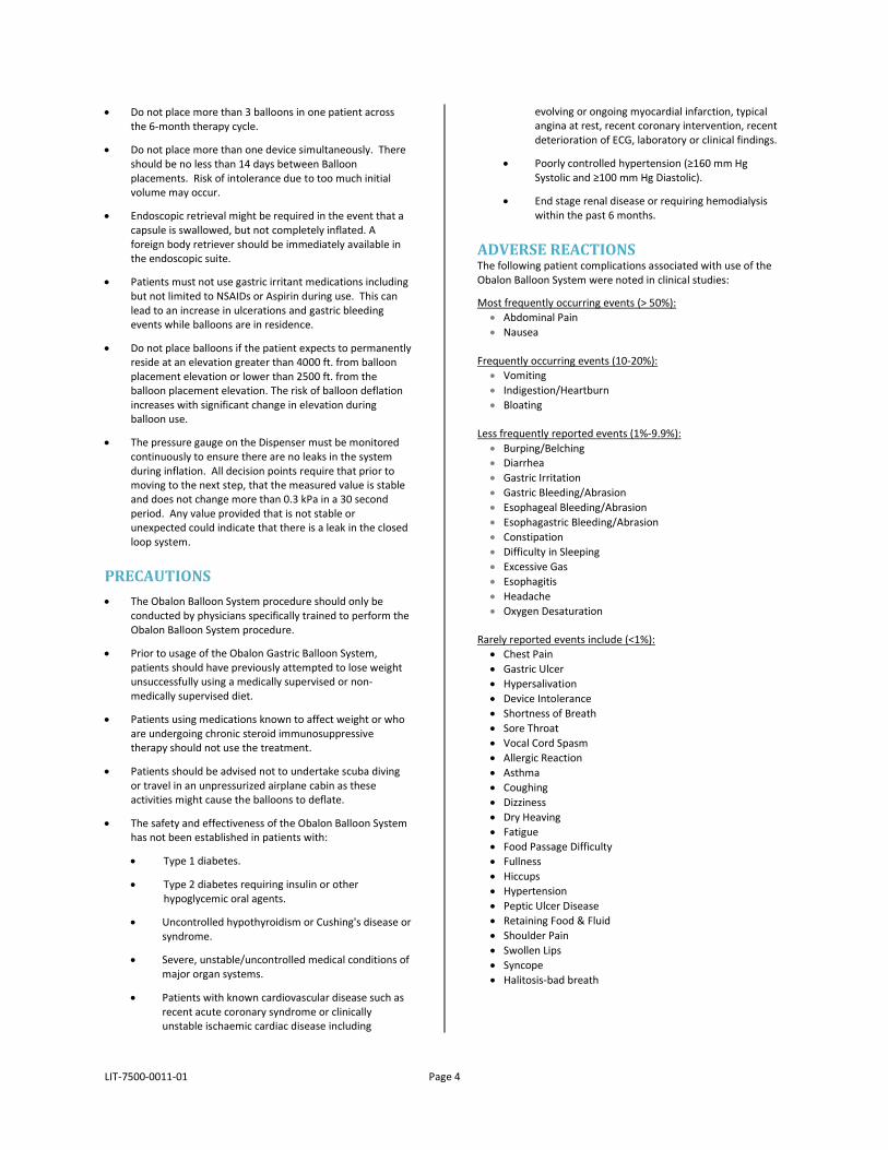
• Obalon Balloon Assembly: 1 folded balloon contained in a swallowable Gelatin Capsule that is attached to 1 disposable, flexible Catheter Delivery System
2. Accessory Kit: (All components in this kit are for single use only)
• 3cc Syringes (QTY: 2)
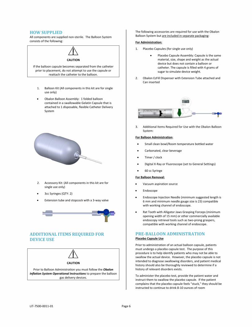
• Extension tube and stopcock with a 3-way valve
ADDITIONAL ITEMS REQUIRED FOR DEVICE USE
CAUTION
Prior to Balloon Administration you must follow the Obalon Inflation System Operational Instructions to prepare the balloon
gas delivery devices.
The following accessories are required for use with the Obalon Balloon System but are included in separate packaging:
For Administration:
1. Placebo Capsules (for single use only)
• Placebo Capsule Assembly: Capsule is the same material, size, shape and weight as the actual device but does not contain a balloon or catheter. The capsule is filled with 4 grams of sugar to simulate device weight.
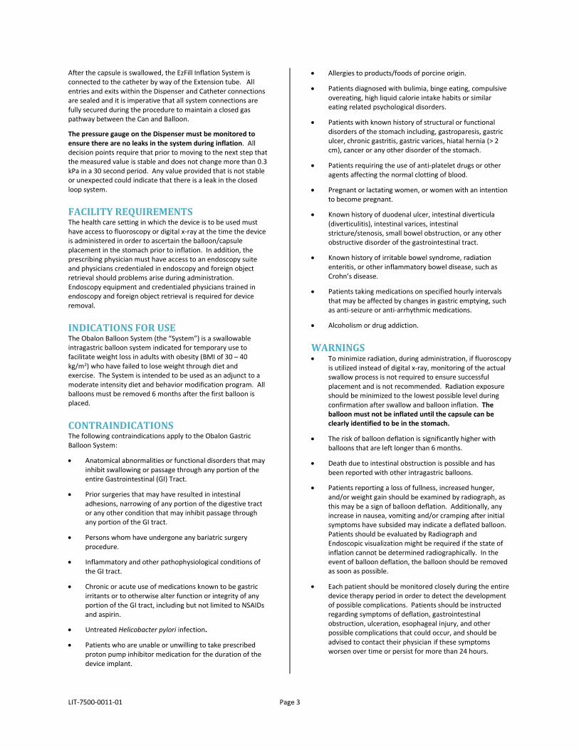
2. Obalon EzFill Dispenser with Extension Tube attached and Can inserted
3. Additional Items Required for Use with the Obalon Balloon System:
For Balloon Administration:
• Small clean bowl/Room temperature bottled water
• Carbonated, clear beverage
• Timer / clock
• Digital X-Ray or Fluoroscope (set to General Settings)
• 60 cc Syringe
For Balloon Removal:
• Vacuum aspiration source
• Endoscope
• Endoscope Injection Needle (minimum suggested length is 6 mm and minimum needle gauge size is 23) compatible with working channel of endoscope.
• Rat Tooth with Alligator Jaws Grasping Forceps (minimum opening width of 15 mm) or other commercially available endoscopy retrieval tools such as two-prong graspers, compatible with working channel of endoscope.
PRE-BALLOON ADMINISTRATION Placebo Capsule Use
Prior to administration of an actual balloon capsule, patients must undergo a placebo capsule test. The purpose of this procedure is to help identify patients who may not be able to swallow the actual device. However, the placebo capsule is not intended to diagnose swallowing disorders, and patient medical history should also be thoroughly reviewed to determine if a history of relevant disorders exists.
To administer the placebo test, provide the patient water and instruct them to swallow the placebo capsule. If the patient complains that the placebo capsule feels “stuck,” they should be instructed to continue to drink 8-10 ounces of room
LIT-7500-0011-01 Page 7
temperature water. The capsule will eventually dissolve and pass through the gastro-esophageal junction.
If the patient has any difficulty swallowing the placebo capsule, they may be poor candidates for the actual balloon device. The actual balloon capsule is attached to a catheter and patients experiencing swallowing difficulties with the placebo capsule may have even more difficulty with the actual balloon capsule.
If the patient swallows the placebo capsule without any difficulty, they may proceed with the balloon therapy. Swallowing the actual balloon device should not be done immediately after the placebo test. The placebo test should be done at least 8 hours prior to balloon placement.
PREPARATION FOR USE You must follow the Obalon Inflation System Directions for Use in their entirety prior to administering the Obalon Balloon. Then you may proceed to preparing the System as discussed below:
3cc Balloon Ejection Syringe (Accessory Kit)
1. Remove the 3cc syringe from its packaging.
2. Fill each 3ml syringe with 1.5ml of room temperature water and set aside.
CAUTION
Do not fill the syringe more than half way (1.5 ml). Doing so may compromise the amount of force needed to push the syringe
plunger for detachment of balloon from the catheter.
Use the provided syringes. Use of a larger syringe might not create enough hydrostatic pressure to eject the balloon.
Balloon Capsule/Catheter (Balloon Kit)
1. Fill a small, clean bowl half-way with water.
2. Carefully remove the balloon by gently pouring out the components of the package. Trying to pull out the capsule/catheter may cause more tangling. Examine all components for bends, kinks or other damage.
EzFill Dispenser
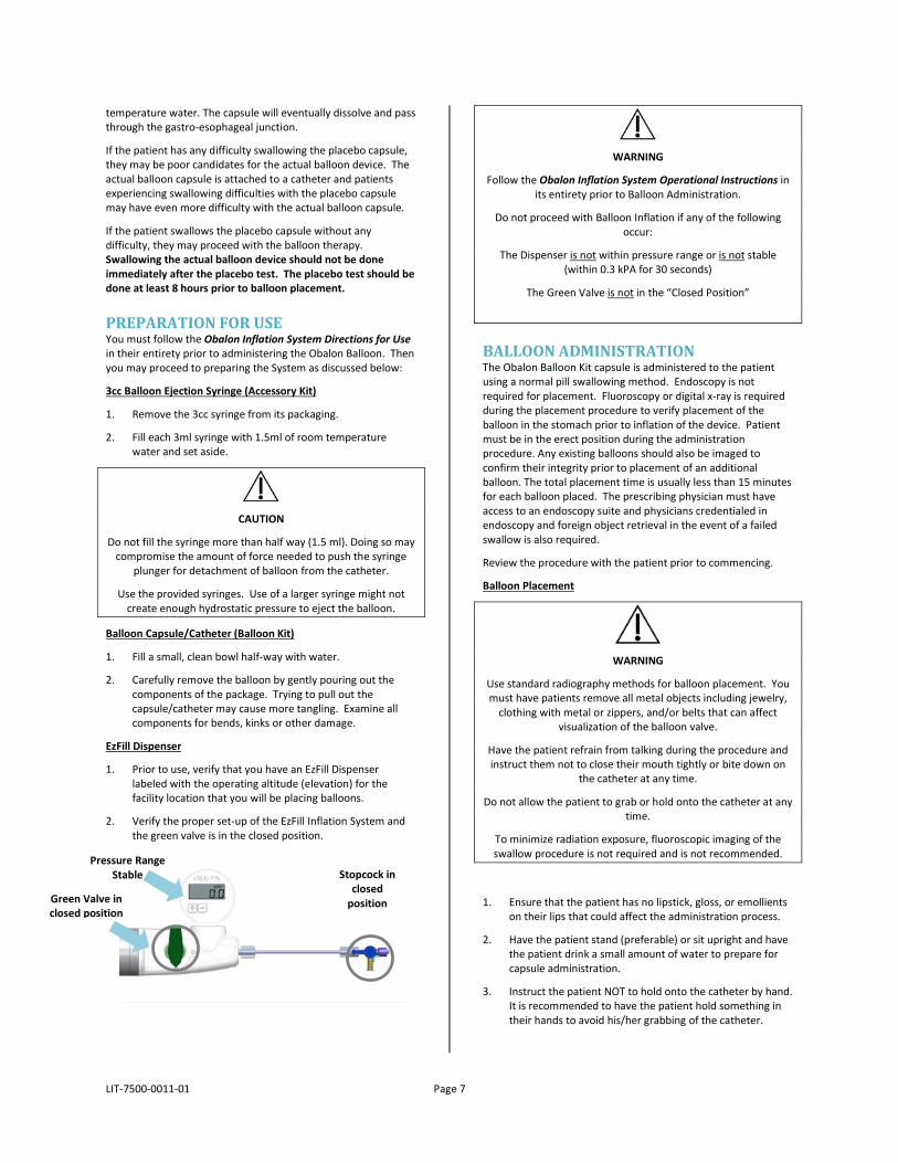
1. Prior to use, verify that you have an EzFill Dispenser labeled with the operating altitude (elevation) for the facility location that you will be placing balloons.
2. Verify the proper set-up of the EzFill Inflation System and the green valve is in the closed position.
WARNING
Follow the Obalon Inflation System Operational Instructions in its entirety prior to Balloon Administration.
Do not proceed with Balloon Inflation if any of the following occur:
The Dispenser is not within pressure range or is not stable (within 0.3 kPA for 30 seconds)
The Green Valve is not in the “Closed Position”
BALLOON ADMINISTRATION The Obalon Balloon Kit capsule is administered to the patient using a normal pill swallowing method. Endoscopy is not required for placement. Fluoroscopy or digital x-ray is required during the placement procedure to verify placement of the balloon in the stomach prior to inflation of the device. Patient must be in the erect position during the administration procedure. Any existing balloons should also be imaged to confirm their integrity prior to placement of an additional balloon. The total placement time is usually less than 15 minutes for each balloon placed. The prescribing physician must have access to an endoscopy suite and physicians credentialed in endoscopy and foreign object retrieval in the event of a failed swallow is also required.
Review the procedure with the patient prior to commencing.
Balloon Placement
WARNING
Use standard radiography methods for balloon placement. You must have patients remove all metal objects including jewelry,
clothing with metal or zippers, and/or belts that can affect visualization of the balloon valve.
Have the patient refrain from talking during the procedure and instruct them not to close their mouth tightly or bite down on
the catheter at any time.
Do not allow the patient to grab or hold onto the catheter at any time.
To minimize radiation exposure, fluoroscopic imaging of the swallow procedure is not required and is not recommended.
1. Ensure that the patient has no lipstick, gloss, or emollients on their lips that could affect the administration process.
2. Have the patient stand (preferable) or sit upright and have the patient drink a small amount of water to prepare for capsule administration.
3. Instruct the patient NOT to hold onto the catheter by hand. It is recommended to have the patient hold something in their hands to avoid his/her grabbing of the catheter.
Stopcock in closed
position Green Valve in closed position
Pressure Range Stable
LIT-7500-0011-01 Page 8
4. Wet the capsule/catheter by submerging into the bowl of water for no more than 10 seconds.
WARNING
Do Not Use a balloon capsule assembly that has been wetted for more than 10 seconds. This may cause the balloon capsule to
dissolve prior to passing through the gastro-esophageal junction and reaching the stomach.
If this happens go to the Troubleshooting Section of these Instructions for Use.
5. Within 1 minute of submerging the capsule in the water, hand the patient the capsule/catheter and instruct him/her to place the capsule immediately in the mouth and swallow the capsule with another large glass of room temperature water. Hold the Proximal Catheter Port outside of the patient’s mouth.
6. Patients should be given additional water (at least 100 ml) after swallow. Patients must remain in an upright sitting or standing position the entire time.
7. Ask the patient to continually drink water or carbonated beverage to facilitate peristalsis of the capsule/catheter if the balloon has not visibly passed into the stomach.
8. If the patient is having difficulty swallowing the capsule (not passing the pharynx) go to the Troubleshooting Section for additional swallowing tips.
9. Once swallowed, the proximal end of the capsule/catheter assembly will remain outside of the patients’ mouth until after the balloon is filled.
10. Approximately 1-2 minutes after swallow, perform digital x-ray or fluoroscopy with the patient standing (preferred) or sitting upright (if necessary) to determine the location of the radio-opaque balloon marker in the stomach.
11. If the capsule is still in the esophagus, proceed to the Troubleshooting section for esophageal transit difficulties.
Balloon Placement “Pre-Pulse” Detection
WARNING
Do not connect the balloon catheter before verifying that the Green Valve and the Stopcock Lock are in the CLOSED position.
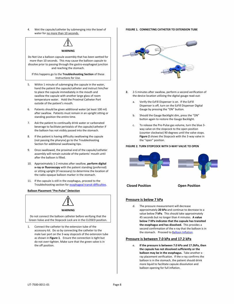
1. Connect the catheter to the extension tube of the accessory kit. Do so by connecting the catheter to the male luer port on the 3-way stopcock of the extension tube as shown in Figure 1. Ensure the connection is tight but do not over-tighten. Make sure that the green valve is in the off position.
FIGURE 1. CONNECTING CATHETER TO EXTENSION TUBE
2. 2-5 minutes after swallow, perform a second verification of the device location utilizing the digital gauge read-out:
a. Verify the EzFill Dispenser is on. If the EzFill Dispenser is off, turn on the EzFill Dispenser Digital Gauge by pressing the “ON” button.
b. Should the Gauge Backlight dim, press the “ON” button again to restore the Gauge Backlight.
c. To release the Pre-Pulse gas volume, turn the blue 3-way valve on the stopcock to the open position (counter clockwise) 90 degrees until the valve stops. Figure 2 shows the Stopcock with the 3-way valve in the “open” position.
FIGURE 2. TURN STOPCOCK WITH 3-WAY VALVE TO OPEN
Closed Position Open Position
Pressure is below 7 kPa
d. The pressure measurement will decrease approximately 20 kPa and continue to decrease to a value below 7 kPa. This should take approximately 45 seconds but no longer than 4 minutes. A value below 7 KPa indicates that the capsule has transited the esophagus and has dissolved. This provides a second confirmation of the x-ray that the balloon is in the stomach. Proceed to Balloon Inflation.
Pressure is between 7.0 kPa and 17.2 kPa
e. If the pressure is between 7.0 kPa and 17.2kPa, then the capsule has not dissolved sufficiently or the balloon may be in the esophagus. Take another x-ray placement verification. If the x-ray confirms the balloon is in the stomach, the patient should drink more liquid to facilitate capsule dissolution and balloon opening for full inflation.

LIT-7500-0011-01 Page 9
Pressure is above 17.2 kPa
f. If the pre-pulse pressure remains above 17.2 kPa for an extended period of time after swallow and the gauge stays constant without significant changes in pressure, the capsule may be constricted in the esophagus. Do not attempt balloon inflation. Continue to observe patient for signs of discomfort and immediately refer to the use of the 60 cc syringe in the Troubleshooting Section
3. Continue to monitor pressure for up to 4 minutes. If after this time the pressure has not dropped below 7 kPa, then the capsule has not dissolved or may be constrained in the esophagus. Do not proceed with Balloon Inflation. Re-confirm placement in the stomach with another x-ray or fluoroscopic viewing.
WARNING
Do not rely solely on the gauge measurement for confirmation of placement in the stomach. You must confirm all pressure
measurements with x-ray or fluoroscopy verification.
4. If the gauge reads less than 7 kPa and you have re-confirmed that the balloon valve is in the stomach by radiographic imaging, you can proceed to BALLOON INFLATION.
BALLOON INFLATION
1. To fill the balloon, rotate the Green Valve on the EzFill Dispenser to the right (open position) as shown in Figure 3.
FIGURE 3. BALLOON FILL: GREEN VALVE OPEN & STOPCOCK WITH A 3-WAY VALVE OPEN
Open positions
2. The balloon will inflate completely approximately 2 minutes after the Green Valve is opened.
WARNING
During inflation, if there is indication of inflation in a constrained space (by pressure readings or patient
symptoms), shut off the gas flow by closing the green valve, detach the catheter and evacuate the gas from the
balloon with a 60cc syringe. Proceed to the Troubleshooting Section.
Inflation in the esophagus can cause serious injury or death.
Do not disconnect the balloon from the catheter prior to balloon fill completion. In the event of a premature disconnection, retrieve the catheter by pulling it out.
Document the gauge pressure measurement and inform and contact Obalon Customer Service immediately. The
Balloon may require endoscopic removal.
3. Wait a minimum of 2 minutes from when the Green Valve is opened in step 2. Verify that the readout pressure on the digital gauge is stable by monitoring the gauge for at least 30 seconds to assess balloon pressure stability. The balloon pressure is stable if it does not steadily decrease by more than 0.3 kPa within a 30 second period.
4. Close the Green Valve and verify that the final readout pressure on the digital gauge reads between 9.0 and 13.0 kPa. Repeat monitoring the gauge for at least 30 seconds to assess balloon pressure stability. The balloon pressure is considered stable if it does not steadily decrease by more than 0.3 kPa within a 30 second period. If the balloon pressure is greater than 13.0, refer to “Final Pressure Setting Adjustment” in the Troubleshooting section of these Instructions for Use.
CAUTION
IF THE GAUGE DOES NOT STABILIZE, the balloon inflation process is not complete or the balloon system may have a leak. Monitor pressure for additional 30 seconds. If pressure is stable (does not
drop more than 0.3 kPa) and reads between 9.0 and 13.0 kPa, proceed to the next step. If the pressure continues to decrease,
contact Obalon Customer Service. The Balloon may require endoscopic removal.
5. Verify the location of all balloons using x-ray or fluoroscopy imaging, in addition to being at a stable pressure between 9.0-13.0 kPa. A fully inflated balloon will look like the x-ray presented in Figure 4. Evaluate the image by examining the shape of each balloon, confirming that the outline appears to be elliptical or circular. Also note if the radiopaque balloon valve appears to be in the stomach. A second x-ray view may be useful in the event that balloons are obscured from each other in a single x-ray view. Note that partial balloon deflations may not be visible on x-ray until a substantial portion of gas has left the balloon.

LIT-7500-0011-01 Page 10
FIGURE 4. EXAMPLE X-RAY SHOWING 3 BALLOONS INFLATED IN THE STOMACH
6. Attach the pre-filled Syringe to the Stopcock with the 3-way Valve still open as shown in Figure 3.
7. Rotate the 3-way Valve back to the closed position (or 90 degrees clockwise) to close the flow of gas from the EzFill Dispenser (see Figure 5).
FIGURE 5. STOPCOCK WITH A 3-WAY VALVE CLOSED/SYRINGE ATTACHED
CAUTION
Do not close the 3-way stopcock valve until you have attached the pre-filled syringe. Doing so will reduce the final starting
pressure in the balloon, and the balloon may not maintain its volume for the 6-month period.
Catheter Retrieval
1. Push the plunger of the 3cc syringe filled with 1.5ml of room temperature water in a single rapid and deliberate motion. The catheter will detach from the balloon valve in the stomach. If the catheter does not detach after the first attempt, a second attempt will be necessary.
2. Prior to removing the first syringe, rotate the stopcock valve to open the flow of gas. Attach the second 3cc syringe and rotate the stopcock back to the closed position. Use the second water filled syringe to again attempt to remove the catheter. On the second attempt, ensure that the catheter is straight and has no kinks and that the plunger is pressed in a rapid and deliberate motion.
3. If more than two attempts are unsuccessful go to the Troubleshooting Section of these Instructions for Use.
4. Slowly pull the catheter out of the patient’s mouth. Having the patient put their chin down may facilitate this process with less of a gag reflex.
5. Visually inspect the catheter and visualize the needle inside the needle sleeve (which is the white protective hub that came attached to the capsule device) to ensure the needle is intact. If the needle is not inside the needle sleeve, remove the balloon endoscopically.
6. Separate the catheter from the Extension Tube by unscrewing the luer lock.
BALLOON USE Post-Administration Guidelines
Advise patients to drink liquids for the first 24 hours and then transition to soft solids on the 2nd day after administration. Patients should not drink sodas or other “fizzy” or carbonated drinks. On the 3rd day after the administration, patients should be able to return to solid foods and follow the diet and behavior modification program provided to them by their physician.
Patients should be advised that some degree of nausea, vomiting and cramping are normal within the first week of each administration procedure. Patients should also be reminded to take medications intended to help minimize symptoms exactly as prescribed by their physician. Patients should be informed of who to contact in the event that they experience symptoms that are intolerable, new symptoms that start after the first week post-administration, or a sudden loss in fullness or increase in hunger. These symptoms should be further evaluated by the following physician and potential balloon deflations should be ruled out.
WARNING
Patients reporting a loss of fullness, increased hunger, and/or weight gain should be examined by radiograph, as this may also
be a sign of balloon deflation. Balloon deflation can be evaluated using radiography (film x-ray, digital x-ray, or
fluoroscopy) and endoscopy as appropriate. Evident balloon deflation may require early balloon removal.
Additionally, an increase in nausea, vomiting, and/or cramping after initial symptoms have subsided may indicate a deflated
balloon.
Patients should also be advised to contact their physician if the frequency of adverse events experienced is more than
anticipated or becomes intolerable.
Patients must not use gastric irritant medications including but not limited to NSAIDs or Aspirin during use. This can lead to an
increase in ulcerations and gastric bleeding events while balloons are in residence.

LIT-7500-0011-01 Page 11
Diet and Behavior Modification Program
The Obalon Balloon System is intended to be an adjunct to weight loss behavior modification program. All subjects will participate in an adjunctive weight loss program focusing on the following principles:
• A balanced low calorie diet
• Education on identifying nutritional content and determining appropriate portion sizes
• Behavior modification techniques to promote healthy eating habits
• Medically appropriate use of physical activity
Additional detail on the recommended program elements is provided in WEIGHT LOSS PROGRAM REQUIREMENTS Section of this Instruction for Use.
Additional Balloon Placements
A single Obalon Balloon does not provide enough volume for maximum weight loss. Clinical studies have shown that three balloons help achieve more effective weight loss over the course of the 6-month intended use period.
WARNING
DO NOT place more than 3 balloons in one patient across the 6-month therapy cycle.
DO NOT place more than one device simultaneously. Risk of intolerance due to too much initial volume may occur.
DO NOT place balloons with less than 14 days between balloon placements.
BALLOON REMOVAL After 6 months from the first implantation, all balloons must be removed from the patient.
This procedure should be conducted using a working length endoscope that is less than 1200 mm, and the inner diameter must be compatible with the accessory tools suggested for puncture and retrieval of the balloon.
Suggested Tools:
• A needle instrument: Injector needle in a Teflon Sleeve 23G x 6mm or similar having a lumen for suction
• Rat Tooth Grasper with Alligator Jaws or Two Jaw Grasping Forceps (with a minimum opening width of 15 mm); two prong graspers with same minimum opening may work as well.
WARNING
Use of tools that are not within the specifications above could result in patient injury.
The above mentioned tools are suggested, but there may be other retrieval tools acceptable for retrieving the balloons. Retrieval procedures in general should be conducted per the endoscope manufacturer’s instructions for retrieving foreign objects. The endoscopy procedure performed is similar to that of an interventional or therapeutic procedure; however, tailoring the endoscopic approach according to the unique product features is important:
• Balloons should only be punctured once, so that the maximum amount of gas can be aspirated (via vacuum)
• A lesser degree of stomach inflation (less air insufflation) allows for easier puncture of the balloon
• Once deflated and all gas is aspirated, the balloon should be optimally grasped at the 6:00 position as seen below. The image below shows a cross section of an inflated balloon through the balloon seam
• The balloon should be optimally grasped at the 6:00 position on the seam, which may result in a more secure hold on the balloon given the increased strength of the overlapping seam
WARNING
Potential excessive bleeding is possible at device removal for those patients requiring the use of anti-platelet drugs or other
agents that affect the normal clotting of blood.
The removal procedure must be conducted by a physician credentialed in endoscopy and foreign object retrieval. Patients should be fasted at least 24 hours or per hospital protocol for endoscope procedures to ensure the stomach is empty and the balloon(s) are therefore easily visible.
1. Anesthetize per hospital and physician recommendations for endoscope procedures.
2. Insert the endoscope into the patient’s stomach.
3. Get a clear view of the filled balloons through the endoscope.
4. Insert the needle instrument down the working channel of the endoscope.
5. Locate the valve of the balloon and puncture the balloon with the needle only once (at the opposite end of the valve if possible for easier removal).
12:00Good
6:00Optimal
3:00Poor
9:00Poor
Apex(Top or Bottom)

LIT-7500-0011-01 Page 12
6. Apply suction and aspirate balloon gas using a large 60cc syringe or aspiration tube.
7. Remove the needle from the working channel.
8. Quickly insert the graspers through the working channel.
9. Grab the balloon with the graspers at the opposite end of the valve.
10. With a firm grasp on the balloon, slowly extract the balloon up through the esophagus removing the balloon through the mouth.
11. Repeat for the remainder of the balloons.
TROUBLESHOOTING Placement of the balloon does not require endoscopy; however, it is highly suggested that a trained Endoscopist be readily available should there be a problem with placement of the balloon. Physicians should contact Obalon Customer Support for technical support with device operation or to report an issue. The following should be considered during balloon placement:
Swallowing Difficulties:
• Always use the Obalon Placebo Capsule prior to attempting administration of an Obalon Balloon Capsule.
• Make sure the patient has not had large volumes of liquid prior to the administration procedure. This usually provides them a feeling of “too full” and makes it more difficult to ingest anything else.
• Create a relaxing, calm environment for the patient. Minimize the number of people in the room. Provide calm, confident words of encouragement. Have the patient take a few deep breaths before swallowing. If failure to swallow is due to anxiety, standard methods to reduce the patient’s anxiety should be used.
• Guide the patient to place the capsule on the back of the patient’s tongue and orient the catheter to the side of the patient’s mouth.
• Encourage the patient to drink large gulps of water rather than small sips of water.
• If the patient fails two attempts, you should discuss this issue with the patient and determine if the patient remains a good candidate for the therapy.
• If the device does not pass the pharynx in the patient’s mouth after 30 seconds of attempting swallow, the capsule must be removed from the mouth. A new wetted balloon capsule/catheter assembly must be used.
• 4 oz. of a “smoothie” (thick consistency/flavored beverage) can aid in swallowing difficulties. This method helps to mask the flavor and consistency of the device.
Esophageal Transit Difficulties:
• Attempting to remove the balloon by traction on the catheter could lead to separation of the balloon. If this occurs, it could occlude the upper airway requiring emergency procedures to relieve the obstruction.
• Esophageal transit of the device can be facilitated by use of clear carbonated beverages or a small piece of banana.
• When the capsule clears the esophagus, return to the Balloon Placement.
• If the capsule does not clear the esophagus, immediately proceed to endoscopic removal.
60cc Syringe Use:
If radiography or x-ray indicates that the radio-opaque marker has not passed completely into the stomach within 2 minutes after swallow, the patient may be having difficulty transiting the capsule. If pre-pulse has already been administered and the pressure gauge is still above 17.2 kPa, ensure that the green valve is turned to the closed position and proceed below:
• To relieve gas from the balloon, attach the 60cc Syringe to the stopcock. Rotate the stopcock to the closed position. Pull back the syringe and hold the plunger to maintain the vacuum. Note that the pressure might be strong. This will help constrict the balloon as much as possible.
Attempt to use additional beverages or a piece of banana to encourage transit of the balloon into the stomach.
WARNING
DO NOT use banana more than once to encourage transit as aspiration might occur during the endoscopic removal of the
balloon capsule.
Catheter Ejection Difficulties:
• Catheter ejection requires a single, rapid push of the plunger. It is recommended to use two thumbs on the plunger. Performing this step in a less forceful manner may lead to a failed ejection. Ensure the 3 ml syringe is always filled to 1.5 ml and perform an additional attempt.
• If the catheter fails to eject there may be a kink in the catheter. Gently tug the catheter to straighten the line and repeat the syringe ejection procedure.
Final Pressure Setting Adjustment:
If during balloon placement the final balloon pressure reads greater than 13.0 kPa, the balloon pressure must be adjusted. You can do this using the following method:
1. Ensure the current pressure reading is stable for at least 30 seconds and close the green valve.
2. Rotate the stopcock to the Closed Position for 2 seconds.
Open Position Closed Position

LIT-7500-0011-01 Page 13
3. Then rotate the stopcock back to the Open position.
4. Wait 10 seconds to verify that the pressure is now within a range of 9.0-13.0 kPa. If the pressure does not meet this range, repeat steps 2-3, waiting 10 seconds between attempts until the balloon falls within the required 9-13 kPa pressure range. Once the pressure reading meets the required 9.0-13.0 kPa range, please make sure it remains stable for at least 30 seconds.
CAUTION
If the balloon pressure does not fall within 9.0-13.0 kPa after several attempts, or drops below 9.0 kPa, contact Obalon Customer Service. In the event the balloon pressure does
not fall within this range you will have to remove the balloon endoscopically.
Unstable Pressure Measurements:
For each pressure measurement review, you must verify that the measurement is “stable.” A stable measurement is one that does not steadily decrease by more than 0.3 kPa for a minimum of 30 seconds. If the gauge measurement is not stable, there may be a leak in the System. Please call Obalon customer service. If the expected measurement is not stable or not within the expected measurement range, you will have to perform an endoscopic removal of the balloon.
The expected stabilized pressure measurements at balloon placement to be verified during the procedure prior to moving to the next step are in Table 2 as follows:
Table 2. Important Pressure Measurements
Instructions for Use Section
Expected Pressure Reading
Preparation for Use Review Dispenser Label:
Operating Altitude xxx-xxx ft.
Pressure XXX.X – XXX.X kPa
City, State
Balloon Administration - Balloon Placement “Pre-Pulse” Detection
Less than 7.0 kPa and approaching 0.0 kPa after Radiographic Confirmation. Re-verify radiographically if pressure exceeds 7.0 kPa a second X-ray verification is required.
Balloon Inflation 9.0-13.0 kPa
Use of radiography to rule out potential balloon deflation: It is expected for patients to experience some degree of nausea, vomiting, and cramping within the first week after each balloon administration. Severe symptoms during that time or new symptoms occurring after the first week could indicate a premature balloon deflation. A sudden loss of fullness or a sudden increase in feelings of hunger may also indicate a potential balloon deflation.
In these circumstances, radiographic imaging should be considered to rule out a potential balloon deflation. Balloon valves are radiopaque and the outline of an inflated balloon will have an elliptical or circular perimeter. If all balloons cannot be visualized with a single x-ray view, a second x-ray view should also be evaluated.
If a previously placed balloon is not in the stomach and has not been excreted, patients should be closely monitored for symptoms suggestive of a bowel obstruction. Serial imaging at 24 hour intervals should be considered if the patient is asymptomatic in an effort to ascertain if the balloon is progressing through the digestive tract. Deflated balloons may eventually be excreted and invasive intervention (e.g. colonoscopy, surgery) to remove the balloon should be considered if the physician believes that a bowel obstruction may occur and the benefits of the intervention outweigh the risks.
Closed Position
Open Position

LIT-7500-0011-01 Page 14
STORAGE AND DISPOSAL Obalon Balloon Kit and Placebo Capsule
• Keep the Obalon Balloon Kit and Placebo Capsule in its packaging until you are ready to use it.
• Do not use the Obalon Balloon Kit or Placebo Capsule past the expiration date printed on the packaging; it must be used before the last day of the month if only the month and year are printed. Storage temperature should be 59o to 77o F (15 o to 25o C) for the length of its shelf life.
• Discard the Catheter per standard biohazard methods and instructions after use.
Accessory Kit
• Keep the Accessory Kit in its packaging until you are ready
to use it. • Store under normal conditions.• Discard the Accessory Kit per standard biohazard methods
and instructions after use.
OBALON® BALLOON SYSTEM– INSTRUCTIONS FOR USE
LIT-7500-0011-01 Page 15
CLINICAL STUDY DESIGN The SMART Trial is a prospective, multicenter, double-blinded, randomized sham controlled trial designed to generate safety and effectiveness data for the Obalon 6-Month Balloon System. The trial was conducted from March 9, 2015 to May 19, 2016. The trial was designed to evaluate the effects of a six-month course of balloon therapy in subjects with a starting BMI in the range of 30 – 40 kg/m2 and who participated in a moderate intensity weight loss behavior management program. Treatment Subjects were then followed in an observational portion of the Study (Phase II) for 6-months and previously assigned Sham subjects were provided an elective option to receive the Obalon Balloon for a 6-month period if they still met the BMI qualification criteria. Males and females (22 – 64 years old) with a baseline BMI of 30 – 40 kg/m2 meeting all study inclusion/exclusion criteria were eligible for participation. Those with a history of gastrointestinal abnormalities, previous gastrointestinal surgery, prior use of a weight loss device, were excluded as well as other exclusions such as Type 1 diabetes, uncontrolled hypertension and chronic use of NSAIDs and other gastric irritants. 387 subjects were treated with the Obalon Balloon (198) or sham device (189) in Phase I of the Study. After completion of Phase I, 170 subjects of those previously treated with the balloon (Treatment Subjects) had at least 1 visit in Phase II and 138 sham device subjects meeting the BMI criteria, elected to have the balloon therapy and successfully swallowed a balloon capsule in Phase II. Safety data is based on the 336 subjects who had at least one device placed in Phase I or Phase II of the study.
Adverse Events
One (1) subject had one (1) Device-Related Serious Adverse Event (SAE). Therefore, the Device-Related SAE rate for the study was 0.3% (1/336) with 95% confidence interval of 0.0% to 1.6%.
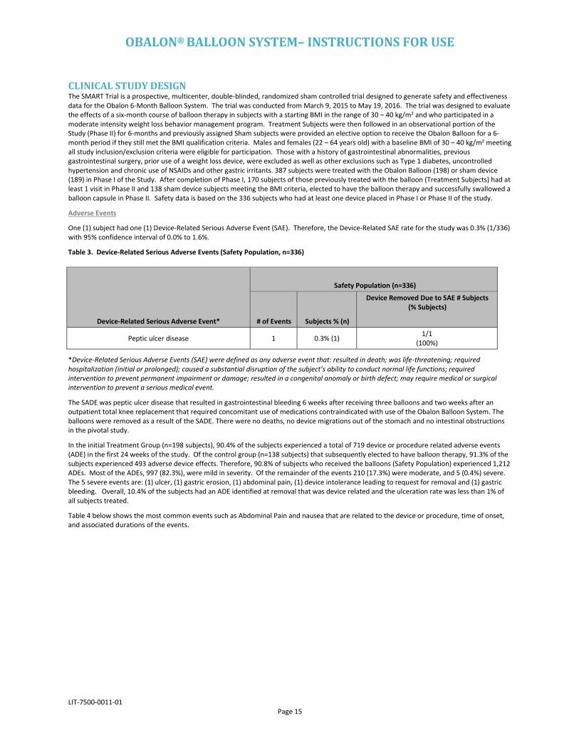
Table 3. Device-Related Serious Adverse Events (Safety Population, n=336)
*Device-Related Serious Adverse Events (SAE) were defined as any adverse event that: resulted in death; was life-threatening; required hospitalization (initial or prolonged); caused a substantial disruption of the subject’s ability to conduct normal life functions; required intervention to prevent permanent impairment or damage; resulted in a congenital anomaly or birth defect; may require medical or surgical intervention to prevent a serious medical event.
The SADE was peptic ulcer disease that resulted in gastrointestinal bleeding 6 weeks after receiving three balloons and two weeks after an outpatient total knee replacement that required concomitant use of medications contraindicated with use of the Obalon Balloon System. The balloons were removed as a result of the SADE. There were no deaths, no device migrations out of the stomach and no intestinal obstructions in the pivotal study.
In the initial Treatment Group (n=198 subjects), 90.4% of the subjects experienced a total of 719 device or procedure related adverse events (ADE) in the first 24 weeks of the study. Of the control group (n=138 subjects) that subsequently elected to have balloon therapy, 91.3% of the subjects experienced 493 adverse device effects. Therefore, 90.8% of subjects who received the balloons (Safety Population) experienced 1,212 ADEs. Most of the ADEs, 997 (82.3%), were mild in severity. Of the remainder of the events 210 (17.3%) were moderate, and 5 (0.4%) severe. The 5 severe events are: (1) ulcer, (1) gastric erosion, (1) abdominal pain, (1) device intolerance leading to request for removal and (1) gastric bleeding. Overall, 10.4% of the subjects had an ADE identified at removal that was device related and the ulceration rate was less than 1% of all subjects treated.
Table 4 below shows the most common events such as Abdominal Pain and nausea that are related to the device or procedure, time of onset, and associated durations of the events.
Device-Related Serious Adverse Event*
Safety Population (n=336)
# of Events Subjects % (n)
Device Removed Due to SAE # Subjects (% Subjects)
Peptic ulcer disease 1 0.3% (1) 1/1 (100%)

LIT-7500-0011-01 Page 16
Table 4. GI-System Device Related Adverse Events Occurring in 10% or More of Subjects treated (Safety Population, n=336)
Device Related Adverse Event
Events Subjects (%)
N=336
Mild*
#Events/ %Events
Moderate**
#Events/ %Events
Severe***
#Events/ %Events
Onset****
(Days)
Event Duration (Days)
#Events (% of Events)
Abdominal Pain 494 244 (72.6%)
414 (83.8%)
79 (16.0%)
1 (0.2%)
Median: 0
Mean: 10
Range: 0-112
0-7: 323 (65.4%)
8-14: 37 (7.5%)
>14: 134 (27.1%)
Nausea 311 188 (56.0%)
261 (83.9%)
50 (16.1%)
0 (0.0%)
Median: 0
Mean: 11
Range: 0-90
0-7: 225 (72.3%)
8-14: 25 (8.0%)
>14: 61 (19.6%)
Vomiting 71 58 (17.3%)
56 (78.9%)
15 (21.1%)
0 (0.0%)
Median: 1
Mean: 14
Range: 0-134
0-7: 59 (83.1%)
8-14: 5 (7.0%)
>14: 7 (9.9%)
Indigestion/
Heartburn
69 57 (17.0%)
48 (69.6%)
20 (30.4%)
0 (0.0%)
Median: 5
Mean: 15
Range: 0-67
0-7: 22 (31.9%)
8-14: 4 (5.8%)
>14: 43 (62.3%)
Bloating 54 49 (14.6%)
49 (90.7%)
5 (9.3%)
0 (0.0%)
Median: 2
Mean: 14
Range: 0-61
0-7: 22 (40.7%)
8-14: 3 (5.6%)
>14: 29 (53.7%)
*Mild: Subject has an awareness of signs or symptoms, which are easily tolerated and causing no loss of time from normal daily activities;symptoms do not require prescription medications, other than those previously specified; actions taken are limited to clinical observations or diagnostic tests. ** Moderate: Subject is experiencing transient periods of discomfort, interfering with normal daily activities; actions taken may include prescription medications beyond what is pre-specified; actions taken do not require hospitalization or invasive interventions. *** Severe: Subject is experiencing non-transient discomfort inhibiting performance of normal daily activities; actions taken require hospitalization or invasive interventions. **** Onset: Number of days from the time of balloon administration or removal that the Adverse Event began.
Of the abdominal pain events, no additional treatment beyond the use of the daily PPI and first 5 days of antispasmodic was necessary for treatment for 72.6% of the events. Thirty-three (33) events (6.7%) were treated with natural antispasmodics such as foods containing peppermint oil (e.g. Altoids or Peppermint Tea). Five events (1.0%) were treated with narcotics and 14.0% of events were treated by extending the use of the prescription antispasmodic. Similar to the abdominal pain events, 75.6% of the nausea events were not treated beyond the antiemetic prescribed for five days post each balloon placement, 9.0% of events utilized chamomile tea for soothing the symptoms, and 13.2% of events required the extension of the use of the anti-emetic and/or antispasmodic to alleviate the symptoms. One event was prescribed a narcotic. For vomiting, 78.9% of events required no additional medications other than the 5-days of use of the antiemetic post-each balloon placement. In 12.7% of events, the anti-emetic was extended beyond the 5-days of use. No injectable anti-emetics were used to treat nausea or vomiting at any point in the study. For indigestion/heartburn events, the majority (58.0%) did not require any medical intervention, and about a quarter of the events (27.5%) were prescribed an increase in the PPI medication from 40 mg to 80 mg.
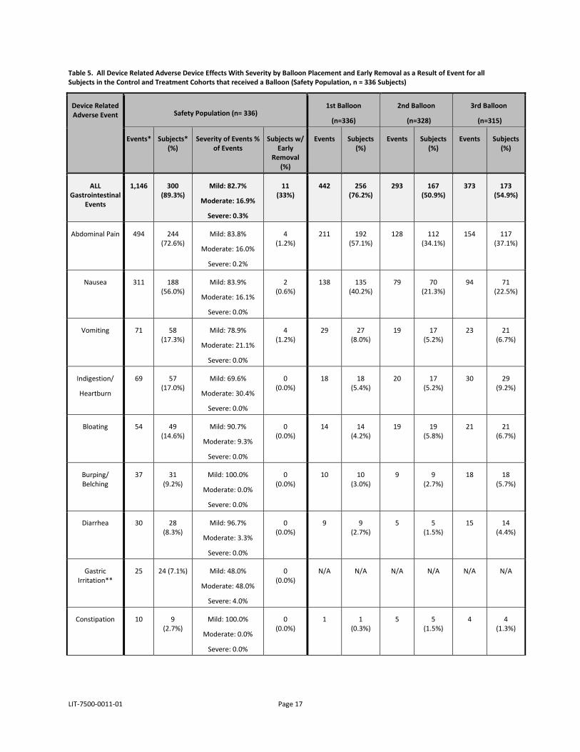
Table 5 below presents the adverse event frequency by number of balloons placed (after 1st, 2nd, and 3rd) and identified at removal, the severity of events and number (%) of subjects that required a device removal as a result of the symptoms experienced during balloon residence. None of the balloon removals were emergent, but were due to a request by the subject usually as a result of the symptoms.
LIT-7500-0011-01 Page 17
Table 5. All Device Related Adverse Device Effects With Severity by Balloon Placement and Early Removal as a Result of Event for all Subjects in the Control and Treatment Cohorts that received a Balloon (Safety Population, n = 336 Subjects)
Device Related Adverse Event Safety Population (n= 336)
1st Balloon
(n=336)
2nd Balloon
(n=328)
3rd Balloon
(n=315)
Events* Subjects* (%)
Severity of Events % of Events
Subjects w/ Early
Removal (%)
Events Subjects (%)
Events Subjects (%)
Events Subjects (%)
ALL Gastrointestinal
Events
1,146 300 (89.3%)
Mild: 82.7%
Moderate: 16.9%
Severe: 0.3%
11 (33%)
442 256 (76.2%)
293 167 (50.9%)
373 173 (54.9%)
Abdominal Pain 494 244 (72.6%)
Mild: 83.8%
Moderate: 16.0%
Severe: 0.2%
4 (1.2%)
211 192 (57.1%)
128 112 (34.1%)
154 117 (37.1%)
Nausea 311 188 (56.0%)
Mild: 83.9%
Moderate: 16.1%
Severe: 0.0%
2 (0.6%)
138 135 (40.2%)
79 70 (21.3%)
94 71 (22.5%)
Vomiting 71 58 (17.3%)
Mild: 78.9%
Moderate: 21.1%
Severe: 0.0%
4 (1.2%)
29 27 (8.0%)
19 17 (5.2%)
23 21 (6.7%)
Indigestion/
Heartburn
69 57 (17.0%)
Mild: 69.6%
Moderate: 30.4%
Severe: 0.0%
0 (0.0%)
18 18 (5.4%)
20 17 (5.2%)
30 29 (9.2%)
Bloating 54 49 (14.6%)
Mild: 90.7%
Moderate: 9.3%
Severe: 0.0%
0 (0.0%)
14 14 (4.2%)
19 19 (5.8%)
21 21 (6.7%)
Burping/ Belching
37 31 (9.2%)
Mild: 100.0%
Moderate: 0.0%
Severe: 0.0%
0 (0.0%)
10 10 (3.0%)
9 9 (2.7%)
18 18 (5.7%)
Diarrhea 30 28 (8.3%)
Mild: 96.7%
Moderate: 3.3%
Severe: 0.0%
0 (0.0%)
9 9 (2.7%)
5 5 (1.5%)
15 14 (4.4%)
Gastric Irritation**
25 24 (7.1%) Mild: 48.0%
Moderate: 48.0%
Severe: 4.0%
0 (0.0%)
N/A N/A N/A N/A N/A N/A
Constipation 10 9 (2.7%)
Mild: 100.0%
Moderate: 0.0%
Severe: 0.0%
0 (0.0%)
1 1 (0.3%)
5 5 (1.5%)
4 4 (1.3%)

LIT-7500-0011-01 Page 18
Device Related Adverse Event Safety Population (n= 336)
1st Balloon
(n=336)
2nd Balloon
(n=328)
3rd Balloon
(n=315)
Events* Subjects* (%)
Severity of Events % of Events
Subjects w/ Early
Removal (%)
Events Subjects (%)
Events Subjects (%)
Events Subjects (%)
Difficulty in Sleeping
9 9 (2.7%)
Mild: 66.7%
Moderate: 33.3%
Severe: 0.0%
0 (0.0%)
3 3 (0.9%)
1 1 (0.3%)
5 5 (1.6%)
Excessive Gas 8 8 (2.4%)
Mild: 100.0%
Moderate: 0.0%
Severe: 0.0%
0 (0.0%)
3 3 (0.9%)
0 0 (0.0%)
5 5 (1.6%)
Esophagitis** 6 6 (1.8%)
Mild: 33.3%
Moderate: 66.7%
Severe: 0.0%
0 (0.0%)
N/A N/A N/A N/A N/A N/A
Hypersalivation 4 3 (0.9%)
Mild: 100.0%
Moderate: 0.0%
Severe: 0.0%
0 (0.0%)
1 1 (0.3%)
2 1 (0.3%)
1 1 (0.3%)
Chest Pain 3 3 (0.9%)
Mild: 100.0%
Moderate: 0.0%
Severe: 0.0%
0 (0.0%)
1 1 (0.3%)
1 1 (0.3%)
1 1 (0.3%)
Gastric Ulcer** 3 3 (0.9%)
Mild: 0.0%
Moderate: 66.7%
Severe: 33.3%
1 (3.3%)
N/A N/A N/A N/A N/A N/A
Device Intolerance
2 2 (0.6%)
Mild: 0.0%
Moderate: 50.0%
Severe: 50.0%
0 (0.0%)
0 0 (0.0%)
1 1 (0.3%)
1 1 (0.3%)
Hiccups 2 1 (0.3%)
Mild: 100.0%
Moderate: 0.0%
Severe: 0.0%
0 (0.0%)
0 0 (0.0%)
1 1 (0.3%)
1 1 (0.3%)
Sore Throat 2 1 (0.3%)
Mild: 100.0%
Moderate: 0.0%
Severe: 0.0%
0 (0.0%)
2 1 (0.3%)
0 0 (0.0%)
0 0 (0.0%)
Dry Heaving 1 1 (0.3%)
Mild: 100.0%
Moderate: 0.0%
Severe: 0.0%
0 (0.0%)
1 1 (0.3%)
0 0 (0.0%)
0 0 (0.0%)
Food Passage Difficulty
1 1 (0.3%)
Mild: 100.0%
Moderate: 0.0%
Severe: 0.0%
0 (0.0%)
0 0 (0.0%)
1 1 (0.3%)
0 0 (0.0%)

LIT-7500-0011-01 Page 19
Device Related Adverse Event Safety Population (n= 336)
1st Balloon
(n=336)
2nd Balloon
(n=328)
3rd Balloon
(n=315)
Events* Subjects* (%)
Severity of Events % of Events
Subjects w/ Early
Removal (%)
Events Subjects (%)
Events Subjects (%)
Events Subjects (%)
Fullness 1 1 (0.3%)
Mild: 100.0%
Moderate: 0.0%
Severe: 0.0%
0 (0.0%)
0 0 (0.0%)
1 1 (0.3%)
0 0 (0.0%)
Retaining Food & Fluid**
1 1 (0.3%)
Mild: 100.0%
Moderate: 0.0%
Severe: 0.0%
0 (0.0%)
N/A N/A N/A N/A N/A N/A
Syncope 1 1 (0.3%)
Mild: 0.0%
Moderate: 100.0%
Severe: 0.0%
0 (0.0%)
1 1 (0.3%)
0 0 (0.0%)
0 0 (0.0%)
Upper Body Injury/ Pain
1 1 (0.3%)
Mild: 100.0%
Moderate: 0.0%
Severe: 0.0%
0 (0.0%)
0 0 (0.0%)
1 1 (0.3%)
0 0 (0.0%)
ALL Metabolic/ Nutritional
Events
9 8 (2.4%)
Mild: 66.7%
Moderate: 33.0%
Severe: 0.0%
0 (0.0%)
5 5 (1.5%)
2 2 (0.6%)
2 2 (0.6%)
Headache/
Migraines
7 6 (1.8%)
Mild: 71.4%
Moderate: 28.6%
Severe: 0.0%
0 (0.0%)
4 4 (1.2%)
2 2 (0.6%)
1 1 (0.3%)
Dizziness 1 1 (0.3%)
Mild: 100.0%
Moderate: 0.0%
Severe: 0.0%
0 (0.0%)
1 1 (0.3%)
0 0 (0.0%)
0 0 (0.0%)
Fatigue 1 1 (0.3%)
Mild: 100.0%
Moderate: 0.0%
Severe: 0.0%
0 (0.0%)
0 0 (0.0%)
0 0 (0.0%)
1 1 (0.3%)
ALL Respiratory Events
3 3 (0.9%)
Mild: 66.7%
Moderate: 33.3%
Severe: 0.0%
0 (0.0%)
0 0 (0.0%)
0 0 (0.0%)
3 3 (0.9%)
Shortness of Breath
2 2 (0.6%)
Mild: 100.0%
Moderate: 0.0%
Severe: 0.0%
0 (0.0%)
0 0 (0.0%)
0 0 (0.0%)
2 2 (0.6%)

LIT-7500-0011-01 Page 20
Device Related Adverse Event Safety Population (n= 336)
1st Balloon
(n=336)
2nd Balloon
(n=328)
3rd Balloon
(n=315)
Events* Subjects* (%)
Severity of Events % of Events
Subjects w/ Early
Removal (%)
Events Subjects (%)
Events Subjects (%)
Events Subjects (%)
Asthma 1 1 (0.3%)
Mild: 0.0%
Moderate: 100.0%
Severe: 0.0%
0 (0.0%)
0 0 (0.0%)
0 0 (0.0%)
1 1 (0.3%)
ALL Other Events 1 1 (0.3%)
Mild: 100.0%
Moderate: 0.0%
Severe: 0.0%
0 (0.0%)
1 1 (0.3%)
0 0 (0.0%)
0 0 (0.0%)
Allergic Reaction 1 1 (0.3%)
Mild: 100.0%
Moderate: 0.0%
Severe: 0.0%
0 (0.0%)
1 1 (0.3%)
0 0 (0.0%)
0 0 (0.0%)
ALL 1,159 300 (89.3%)
Mild: 82.6%
Moderate: 17.1%
Severe: 0.3%
11 (3.3%)
448 256 (76.2%)
295 167 (50.9%)
378 175 (55.6%)
*Total Events/%Subjects include 1st, 2nd, 3rd balloon placements as well as those identified at removal. **ADE onset date is not known
Adverse events identified at removal due to the removal procedure were also captured and the corresponding severity was assigned using the same Mild, Moderate, Severe and Serious definitions presented in Table 4. Table 6 presents the frequency of ADEs identified at balloon removal due to the removal procedure. Overall, 13.4% of the subjects had an ADE related to the removal procedure. More than three quarters of procedural related events (75.5%) were mild and required no treatment. Moderate events typically required PPI therapy for an additional period. The Gastric Mucosal damage in these cases was determined to be caused by the balloon removal procedure. Table 7 presents all ADEs in the study and by severity.
Table 6. All Removal Procedure Related Adverse Device Effects With Severity for all Subjects in the Control and Treatment Cohorts that received a Balloon (Safety Population, n = 336 Subjects)
Device Related Adverse Event Safety Population (n=336 Subjects)
Events Subjects (%)
Mild Moderate Severe
Gastric Bleeding/ Abrasion 17 17 (5.1%) 15 (88.2%) 1 (5.9%) 1 (5.9%)
Esophageal Bleeding/ Abrasion 14 14 (4.2%) 11 (78.6%) 3 (21.4%) 0 (0.0%)
Esophagastric Bleeding/ Abrasion 12 12 (3.6%) 9 (75.0%) 3 (25.0%) 0 (0.0%)
Oxygen Desaturation 4 4 (1.2%) 2 (50.0%) 2 (50.0%) 0 (0.0%)
Vocal Cord Spasm 2 2 (0.6%) 0 (0.0%) 2 (100.0%) 0 (0.0%)
Hypertension 1 1 (0.3%) 0 (0.0%) 1 (100.0%) 0 (0.0%)
Coughing 1 1 (0.3%) 1 (100.0%) 0 (0.0%) 0 (0.0%)
Sore Throat 1 1 (0.3%) 1 (100.0%) 0 (0.0%) 0 (0.0%)
Swollen Lips 1 1 (0.3%) 1 (100.0%) 0 (0.0%) 0 (0.0%)
LIT-7500-0011-01 Page 21
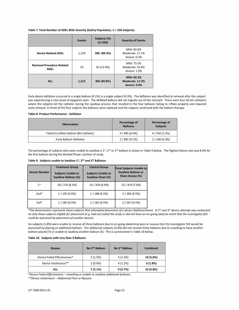
Table 7: Total Number of ADEs With Severity (Safety Population, n = 336 Subjects)
Events Subjects (%) (n=336) Severity of Events
Device Related ADEs 1,159 300 (89.3%) Mild: 82.6%
Moderate: 17.1% Severe: 0.3%
Removal Procedure Related ADEs 53 45 (13.4%)
Mild: 75.5% Moderate: 22.6%
Severe: 1.9%
ALL 1,212 305 (90.8%) Mild: 82.3%
Moderate: 17.3% Severe: 0.4%
Early device deflation occurred in a single balloon (0.1%) in a single subject (0.3%). The deflation was identified at removal after the subject was experiencing a new onset of epigastric pain. The deflated balloon did not migrate out of the stomach. There were four (4) bit catheters where the subjects bit the catheter during the swallow process that resulted in the four balloons failing to inflate properly and required early removal. In three of the four subjects the balloons were replaced and the subjects continued with the balloon therapy.
Table 8: Product Performance - Deflation
Observation Percentage of Balloons
Percentage of Subjects
Failed to inflate balloon (Bit Catheter) 4 / 985 (0.4%) 4 / 336 (1.2%)
Early Balloon Deflation 1 / 981 (0.1%) 1 / 336 (0.3%)
The percentage of subjects who were unable to swallow a 1st, 2nd or 3rd balloon is shown in Table 9 below. The highest failure rate was 8.3% for the first balloon during the blinded Phase I portion of study.
Table 9. Subjects unable to Swallow 1st, 2nd and 3rd Balloons
Device Number
Treatment Group
Subjects Unable to Swallow Balloon (%)
Control Group
Subjects Unable to Swallow Sham (%)
Total Subjects Unable to Swallow Balloon or
Sham Device (%)
1st 18 / 216 (8.3%) 14 / 203 (6.9%) 32 / 419 (7.6%)
2nd* 1 / 195 (0.5%) 1 / 188 (0.5%) 2 / 383 (0.5%)
3rd* 1 / 185 (0.5%) 1 / 182 (0.5%) 2 / 367 (0.5%)
*The denominator represents those subjects that attempted placement of a device (balloon/sham). A 2nd and 3rd device attempt was conductedon only those subjects eligible for placement (e.g. had not exited the study or did not have an on-going adverse event that the investigator felt could be worsened by placement of another device).
Six subjects (1.8%) were unable to receive all three balloons due to on-going abdominal pain or nausea that the investigator felt would be worsened by placing an additional balloon. Ten additional subjects (3.0%) did not receive three balloons due to unwilling to have another balloon placed (7) or unable to swallow another balloon (3). This is summarized in Table 10 below.
Table 10. Subjects with Less than 3 Balloons
Reason No 2nd Balloon No 3rd Balloon Combined
Device Failed Effectiveness* 5 (1.5%) 5 (1.5%) 10 (3.0%)
Device Intolerance** 2 (0.6%) 4 (1.2%) 6 (1.8%)
ALL 7 (2.1%) 9 (2.7%) 16 (4.8%)
*Device Failed Effectiveness – Unwilling or unable to swallow additional balloons**Device Intolerance – Abdominal Pain or Nausea
LIT-7500-0011-01 Page 22
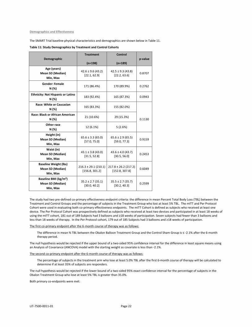
Demographics and Effectiveness
The SMART Trial baseline physical characteristics and demographics are shown below in Table 11.
Table 11: Study Demographics by Treatment and Control Cohorts
Demographic Treatment
(n=198)
Control
(n=189) p-value
Age (years) Mean SD (Median)
Min, Max
42.6 ± 9.6 (43.2) [22.1, 62.9]
42.5 ± 9.3 (43.8) [22.2, 63.6] 0.8707
Gender: Female N (%)
171 (86.4%) 170 (89.9%) 0.2762
Ethnicity: Not Hispanic or Latino N (%)
183 (92.4%) 165 (87.3%) 0.0943
Race: White or Caucasian N (%)
165 (83.3%) 155 (82.0%)
0.1130
Race: Black or African American N (%)
21 (10.6%) 29 (15.3%)
Other race N (%)
12 (6.1%) 5 (2.6%)
Height (in) Mean SD (Median)
Min, Max
65.6 ± 3.3 (65.0) [57.0, 75.0]
65.6 ± 2.9 (65.5) [59.0, 77.3] 0.9159
Waist (in) Mean SD (Median)
Min, Max
43.1 ± 3.8 (43.0) [31.5, 52.8]
43.6 ± 4.0 (43.7) [30.5, 56.0] 0.2453
Baseline Weight (lbs) Mean SD (Median)
Min, Max
216.3 ± 29.1 (210.1) [156.8, 301.2]
217.8 ± 26.2 (217.2) [152.8, 307.8] 0.6049
Baseline BMI (kg/m²) Mean SD (Median)
Min, Max
35.2 ± 2.7 (35.1) [30.0, 40.2]
35.5 ± 2.7 (35.7) [30.2, 40.3] 0.2599
The study had two pre-defined co-primary effectiveness endpoint criteria: the difference in mean Percent Total Body Loss (TBL) between the Treatment and Control Groups and the percentage of subjects in the Treatment Group who lost at least 5% TBL. The mITT and Per Protocol Cohort were used in evaluating both co-primary effectiveness endpoints. The mITT Cohort is defined as subjects who received at least one device. The Per Protocol Cohort was prospectively defined as subjects who received at least two devices and participated in at least 18 weeks of using the mITT cohort, 181 out of 189 Subjects had 3 balloons and ≥18 weeks of participation. Seven subjects had fewer than 3 balloons and less than 18 weeks of therapy. In the Per Protocol cohort, 179 out of 185 Subjects had 3 balloons and ≥18 weeks of participation.
The first co-primary endpoint after the 6-month course of therapy was as follows:
The difference in mean % TBL between the Obalon Balloon Treatment Group and the Control Sham Group is ≤ -2.1% after the 6-month therapy period.
The null hypothesis would be rejected if the upper bound of a two-sided 95% confidence interval for the difference in least square means using an Analysis of Covariance (ANCOVA) model with the starting weight as covariate is less than -2.1%.
The second co-primary endpoint after the 6-month course of therapy was as follows:
The percentage of subjects in the treatment arm who lose at least 5.0% TBL after the first 6-month course of therapy will be calculated to determine if at least 35% of subjects are responders.
The null hypothesis would be rejected if the lower bound of a two-sided 95% exact confidence interval for the percentage of subjects in the Obalon Treatment Group who lose at least 5% TBL is greater than 35.0%.
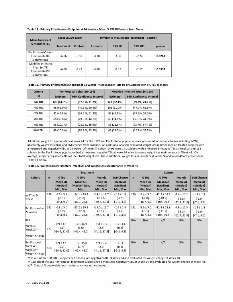
Both primary co-endpoints were met:
LIT-7500-0011-01 Page 23
Table 12. Primary Effectiveness Endpoint at 24 Weeks – Mean % TBL Difference from Sham
Main Analysis of 6-Month %TBL
Least Square Mean Difference in LS Means (Treatment – Control)
Treatment Control Estimate 95% LCL 95% UCL p-value
Per Protocol Cohort Treatment=185
Control=181 -6.86 -3.59 -3.28 -4.32 -2.24 0.0261
Modified Intent to Treat (mITT)
Treatment=198 Control=189
-6.60 -3.42 -3.18 -4.19 -2.17 0.0354
Table 13. Primary Effectiveness Endpoint at 24 Weeks - % Responder Rate (% of Subjects with 5% TBL or more)
Criteria (≤)
Per Protocol Cohort (n=185) Modified Intent to Treat (n=198)
Estimate 95% Confidence Interval Estimate 95% Confidence Interval
-5% TBL 120 (64.9%) (57.5 %, 71.7%) 123 (62.1%) (59.2%, 73.2 %)
-6% TBL 98 (53.0%) (45.5 %, 60.3%) 101 (51.0%) (47.1%, 61.9%)
-7% TBL 81 (43.8%) (36.5 %, 51.3%) 83 (41.9%) (37.6%, 52.3%)
-8% TBL 68 (36.8%) (29.8 %, 44.1%) 69 (34.8%) (30.3%, 44.7%)
-9% TBL 55 (29.7%) (23.2 %, 36.9%) 56 (28.3%) (23.7%, 37.4 %)
-10% TBL 49 (26.5%) (20.3 %, 33.5%) 49 (24.7%) (20.3%, 33.5%)
Additional weight loss parameters at week 24 for the mITT and Per Protocol populations are presented in the table below including %EWL, absolutely weight loss (lbs), and BMI change from baseline. An additional analysis evaluated weight loss maintenance on treated subjects with a measured and negative %TBL at 24 weeks. Of the mITT cohort, there were 171 subjects with a measured negative TBL at Week 24 and 168 subjects in the Per Protocol population had a measured negative TBL at week 24 value to assess weight loss maintenance at Week 48. On average, subjects re-gained ≈2lbs of their total weight lost. These additional weight loss parameters at Week 24 and Week 48 are presented in Table 14 below.
Table 14. Weight Loss Parameters - Week 24 and Weight Loss Maintenance at Week 48
Treatment Control
Cohort n % TBL Mean SD (Median) Min, Max
% EWL Mean SD
(Median) Min, Max
Pounds Mean SD (Median) Min, Max
BMI Change Mean SD (Median) Min, Max
n % TBL Mean SD (Median) Min, Max
% EWL Mean SD (Median) Min, Max
Pounds Mean SD (Median) Min, Max
BMI Change Mean SD (Median) Min, Max
mITT at 24 weeks
198 -6.6 ± 5.1 (-6.1)
[-19.3, 9.5]
-24.1 ± 19.2 (-21.8)
[-80.7, 28.8]
-14.4 ± 11.7 (-12.6)
[-49.7, 21.1]
-2.3 ± 1.8 (-2.1)
[-7.1, 3.6]
189 -3.4 ± 5.0 (-2.8)
[-18.7, 9.6]
-12.2 ± 18.8 (-10.2)
[-104, 30.4]
-7.4 ± 11.2 (-5.8)
[-52.4, 21.8]
-1.2 ± 1.8 (-1.0)
[-7.1, 3.5]
Per Protocol at 24 weeks
185 -6.9 ± 5.0 (-6.2)
[-19.3, 9.5]
-25.2 ± 19.2 (-22.7)
[-80.7, 28.8]
-15.0 ± 11.7 (-13.2)
[-49.7, 21.1]
-2.4 ± 1.8 (-2.2)
[-7.1, 3.6]
181 -3.6 ± 5.0 (-3.1)
[-18.7, 9.6]
-12.8 ± 18.9 (-11.2)
[-104, 30.4]
-7.8 ± 11.3 (-6.2)
[-52.4, 21.8]
-1.3 ± 1.8 (-1.0)
[-7.1, 3.5] mITT
Week 48 – Week 24*
Weight Change
171
0.9 ± 4.1 (1.1)
[-14.4, 12.0]
3.2 ± 14.9 (3.6)
[-46.4, 43.2]
1.8 ± 9.3 (2.6)
[-41.6, 27.8]
0.3 ± 1.4 (0.4)
[-5.2, 4.2]
N/A N/A N/A N/A N/A
Per Protocol Week 48 – Week 24* Weight Change
168
0.9 ± 4.1 (1.2)
[-14.4, 12.0]
3.3 ± 15.0 (3.7)
[-46.4, 43.2]
1.9 ± 9.4 (2.6)
[-41.6, 27.8]
0.3 ± 1.5 (0.4)
[-5.2, 4.2]
N/A N/A N/A N/A N/A
*171 out of the 198 mITT Subjects had a measured negative %TBL at Week 24 and evaluated for weight change at Week 48. ** 168 out of the 185 Per Protocol Treatment subjects had a measured negative %TBL at Week 24 and evaluated for weight change at Week 48. N/A: Control Group weight loss maintenance was not evaluated
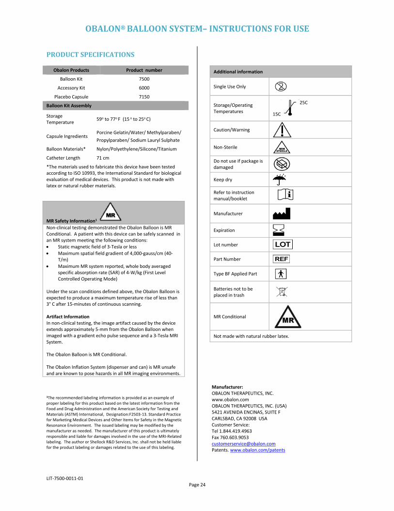
OBALON® BALLOON SYSTEM– INSTRUCTIONS FOR USE
LIT-7500-0011-01 Page 24
PRODUCT SPECIFICATIONS
1The recommended labeling information is provided as an example of proper labeling for this product based on the latest information from the Food and Drug Administration and the American Society for Testing and Materials (ASTM) International, Designation:F2503-13. Standard Practice for Marketing Medical Devices and Other Items for Safety in the Magnetic Resonance Environment. The issued labeling may be modified by the manufacturer as needed. The manufacturer of this product is ultimately responsible and liable for damages involved in the use of the MRI-Related labeling. The author or Shellock R&D Services, Inc. shall not be held liable for the product labeling or damages related to the use of this labeling.
Manufacturer: OBALON THERAPEUTICS, INC. www.obalon.com OBALON THERAPEUTICS, INC. (USA) 5421 AVENIDA ENCINAS, SUITE F CARLSBAD, CA 92008 USA Customer Service: Tel 1.844.419.4963 Fax 760.603.9053 [email protected] Patents. www.obalon.com/patents
Obalon Products Product number
Balloon Kit 7500
Accessory Kit 6000
Placebo Capsule 7150
Balloon Kit Assembly
Storage Temperature 59o to 77o F (15 o to 25o C)
Capsule Ingredients Porcine Gelatin/Water/ Methylparaben/ Propylparaben/ Sodium Lauryl Sulphate
Balloon Materials* Nylon/Polyethylene/Silicone/Titanium
Catheter Length 71 cm
*The materials used to fabricate this device have been tested according to ISO 10993, the International Standard for biological evaluation of medical devices. This product is not made with latex or natural rubber materials.
Additional information
Single Use Only
Storage/Operating Temperatures 15C
25C
Caution/Warning
Non-Sterile
Do not use if package is damaged
Keep dry
Refer to instruction manual/booklet
Manufacturer
Expiration
Lot number
Part Number
Type BF Applied Part
Batteries not to be placed in trash
MR Conditional
Not made with natural rubber latex.
MR Safety Information1 Non-clinical testing demonstrated the Obalon Balloon is MR Conditional. A patient with this device can be safely scanned in an MR system meeting the following conditions: • Static magnetic field of 3-Tesla or less • Maximum spatial field gradient of 4,000-gauss/cm (40-
T/m) • Maximum MR system reported, whole body averaged
specific absorption rate (SAR) of 4-W/kg (First Level Controlled Operating Mode)
Under the scan conditions defined above, the Obalon Balloon is expected to produce a maximum temperature rise of less than 3° C after 15-minutes of continuous scanning. Artifact Information In non-clinical testing, the image artifact caused by the device extends approximately 5-mm from the Obalon Balloon when imaged with a gradient echo pulse sequence and a 3-Tesla MRI System. The Obalon Balloon is MR Conditional. The Obalon Inflation System (dispenser and can) is MR unsafe and are known to pose hazards in all MR imaging environments.