Embed Size (px)
DESCRIPTION
o
Citation preview
Osteoarthritis of Osteoarthritis of Knee Knee
Diagnosis and ManagementDiagnosis and Management
Agus Adiantono, MDAgus Adiantono, MD
Lamongan, Oktober 2010 Lamongan, Oktober 2010
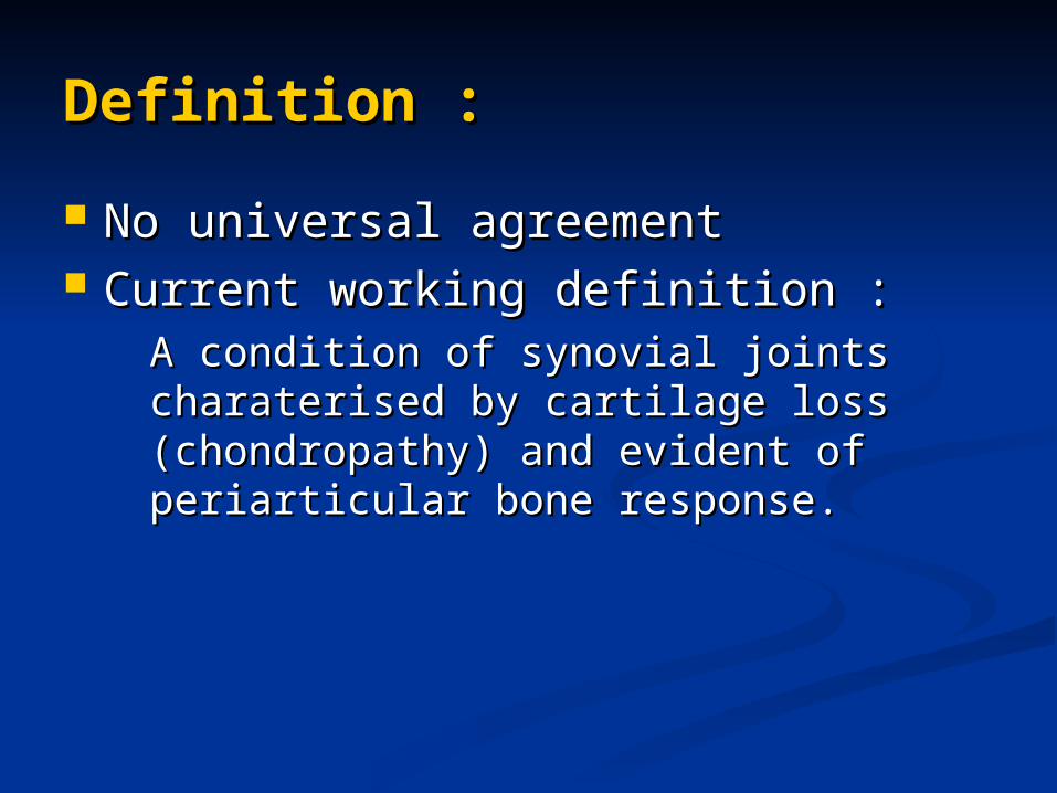
Definition :Definition :
No universal agreementNo universal agreement Current working definition :Current working definition :
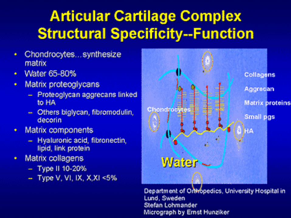
A condition of synovial joints A condition of synovial joints charaterised by cartilage loss charaterised by cartilage loss (chondropathy) and evident of (chondropathy) and evident of periarticular bone response.periarticular bone response.
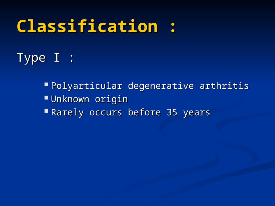
Classification :Classification :
Type I :Type I :
Polyarticular degenerative arthritisPolyarticular degenerative arthritis Unknown originUnknown origin Rarely occurs before 35 yearsRarely occurs before 35 years
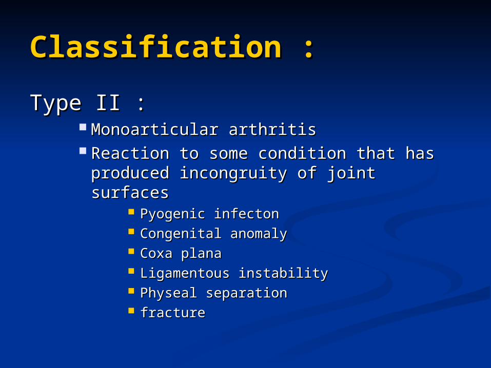
Classification :Classification :
Type II :Type II : Monoarticular arthritisMonoarticular arthritis Reaction to some condition that has Reaction to some condition that has
produced incongruity of joint surfacesproduced incongruity of joint surfaces Pyogenic infectonPyogenic infecton Congenital anomalyCongenital anomaly Coxa planaCoxa plana Ligamentous instabilityLigamentous instability Physeal separationPhyseal separation fracturefracture
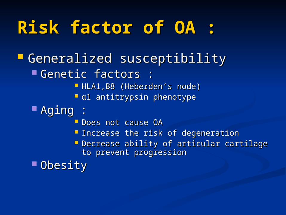
Risk factor of OA :Risk factor of OA :
Generalized susceptibilityGeneralized susceptibility Genetic factors :Genetic factors :
HLA1,B8 (Heberden’s node)HLA1,B8 (Heberden’s node) α1 α1 antitrypsin phenotypeantitrypsin phenotype
Aging :Aging : Does not cause OADoes not cause OA Increase the risk of degenerationIncrease the risk of degeneration Decrease ability of articular cartilage to Decrease ability of articular cartilage to
prevent progressionprevent progression
ObesityObesity
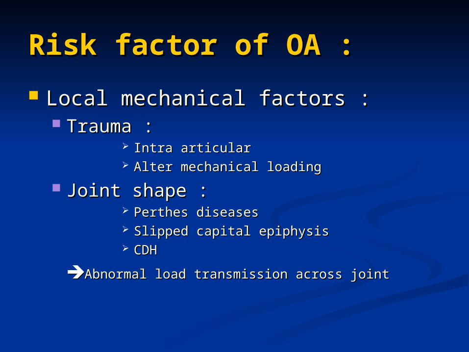
Risk factor of OA :Risk factor of OA :
Local mechanical factors :Local mechanical factors : Trauma :Trauma :
Intra articular Intra articular Alter mechanical loadingAlter mechanical loading
Joint shape :Joint shape : Perthes diseases Perthes diseases Slipped capital epiphysisSlipped capital epiphysis CDHCDH
Abnormal load transmission across jointAbnormal load transmission across joint
Risk factor of OA :Risk factor of OA :
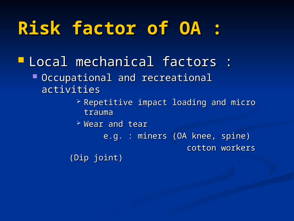
Local mechanical factors :Local mechanical factors : Occupational and recreational activitiesOccupational and recreational activities
Repetitive impact loading and micro traumaRepetitive impact loading and micro trauma Wear and tear Wear and tear
e.g. : miners (OA knee, spine)e.g. : miners (OA knee, spine)
cotton workers (Dip joint)cotton workers (Dip joint)
PathologyPathology
ChondropathyChondropathy
Periarticular bone responsePeriarticular bone response
PathologyPathology ChondropathyChondropathy
Early :Early : Surface irregularities (fibrilation)Surface irregularities (fibrilation) Small cleft beyond the superficial zoneSmall cleft beyond the superficial zone Slight hypercellularitySlight hypercellularity Minimum loss of proteoglycan, not Minimum loss of proteoglycan, not
extending beyond the transitional zoneextending beyond the transitional zone Moderately Advance :Moderately Advance :
More extensive loss of surfaceMore extensive loss of surface Clefts extend into middle zoneClefts extend into middle zone Loss of proteoglycan extend to middle Loss of proteoglycan extend to middle
zonezone HypercellularHypercellular
PathologyPathology
ChondropathyChondropathy
Advance :Advance : Reduce of cartilage thicknessReduce of cartilage thickness Reduce proteoglycan throughout entire Reduce proteoglycan throughout entire
thicknessthickness Complete loss of articular cartilage Complete loss of articular cartilage
(eburnated of sub chondral bone)(eburnated of sub chondral bone) Focal pressure necrosisFocal pressure necrosis
PathologyPathology
Periarticular bone response :Periarticular bone response :
New bone formation in sub chondral New bone formation in sub chondral bone bone
New cartilage formation and New cartilage formation and enchondral ossificationenchondral ossification
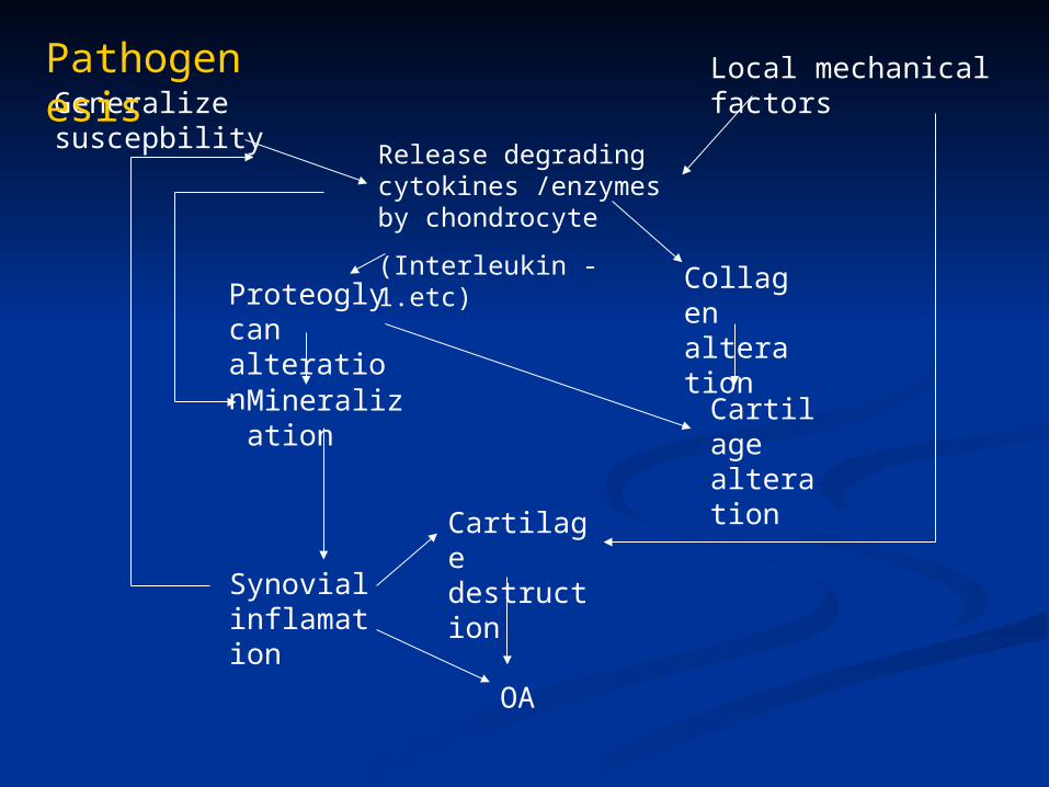
Release degrading cytokines /enzymes by chondrocyte
(Interleukin - 1.etc)
Collagen alteration
Proteoglycan alteration
Mineralization Cartilage alteration
Cartilage destruction
Synovial inflamation
OA
Local mechanical factorsGeneralize suscepbility
Pathogenesis
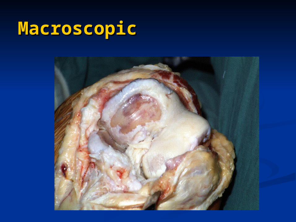
MacroscopicMacroscopic
OA KneeOA Knee
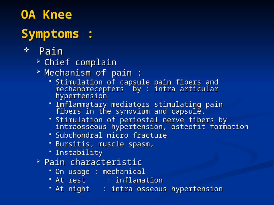
Symptoms :Symptoms : Pain Pain
Chief complainChief complain Mechanism of pain :Mechanism of pain :
Stimulation of capsule pain fibers and Stimulation of capsule pain fibers and mechanorecepters by : intra articular mechanorecepters by : intra articular hypertensionhypertension
Imflammatary mediators stimulating pain fibers in Imflammatary mediators stimulating pain fibers in the synovium and capsule.the synovium and capsule.
Stimulation of periostal nerve fibers by Stimulation of periostal nerve fibers by intraosseous hypertension, osteofit formationintraosseous hypertension, osteofit formation
Subchondral micro fractureSubchondral micro fracture Bursitis, muscle spasm,Bursitis, muscle spasm, Instability Instability
Pain characteristicPain characteristic On usage : mechanicalOn usage : mechanical At rest : inflamationAt rest : inflamation At night : intra osseous hypertensionAt night : intra osseous hypertension
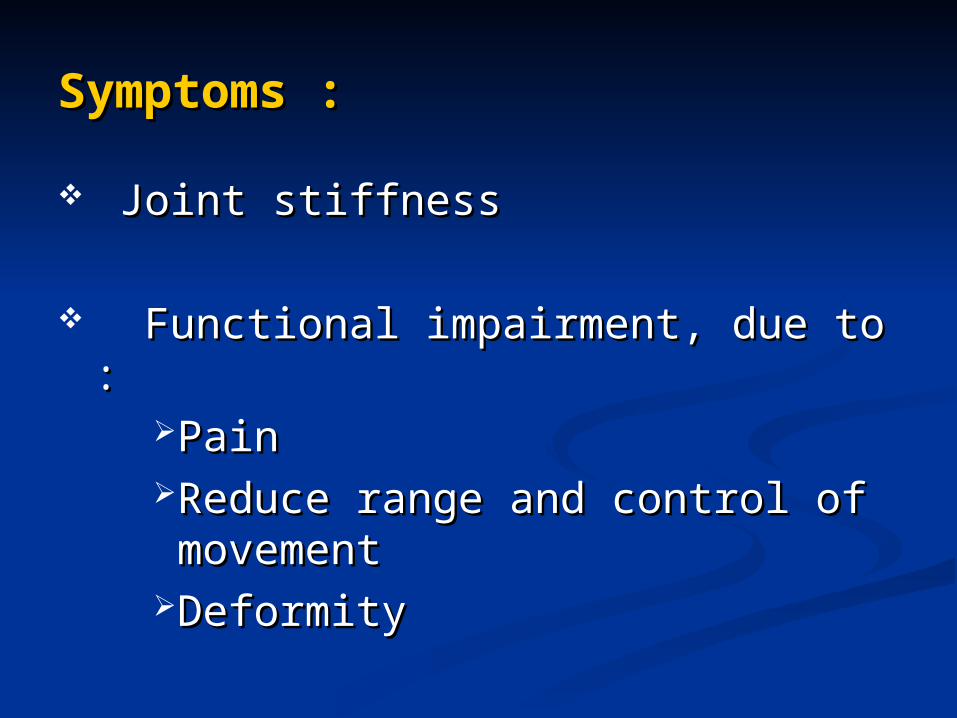
Symptoms :Symptoms :
Joint stiffnessJoint stiffness
Functional impairment, due to :Functional impairment, due to :Pain Pain Reduce range and control of Reduce range and control of movementmovement
DeformityDeformity
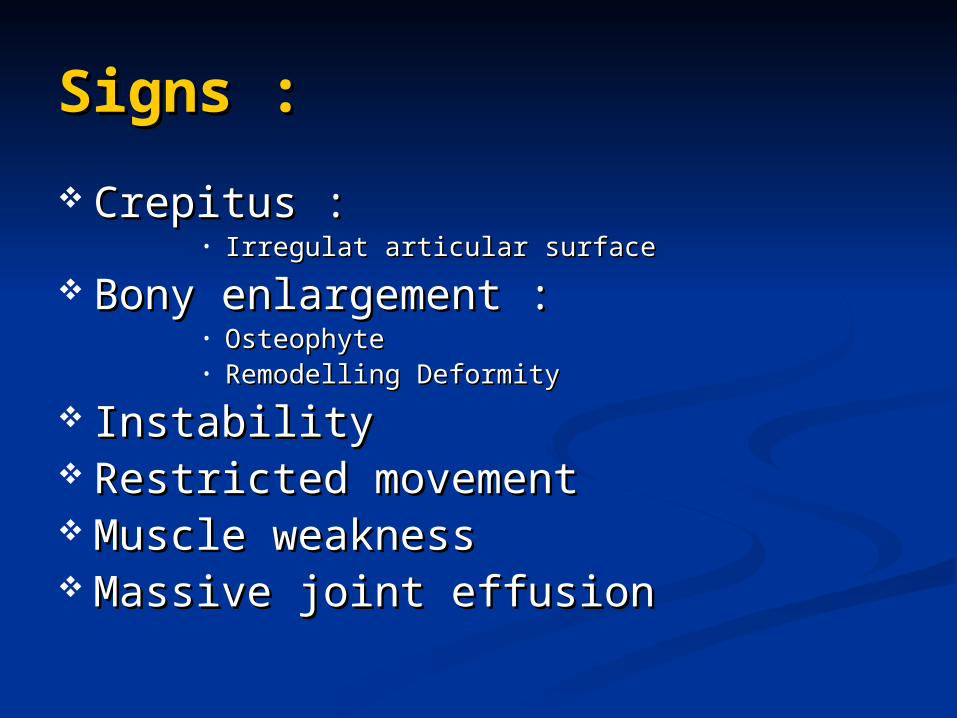
Signs :Signs :
Crepitus :Crepitus :• Irregulat articular surfaceIrregulat articular surface
Bony enlargement :Bony enlargement :• OsteophyteOsteophyte• Remodelling DeformityRemodelling Deformity
InstabilityInstability Restricted movementRestricted movement Muscle weaknessMuscle weakness Massive joint effusionMassive joint effusion
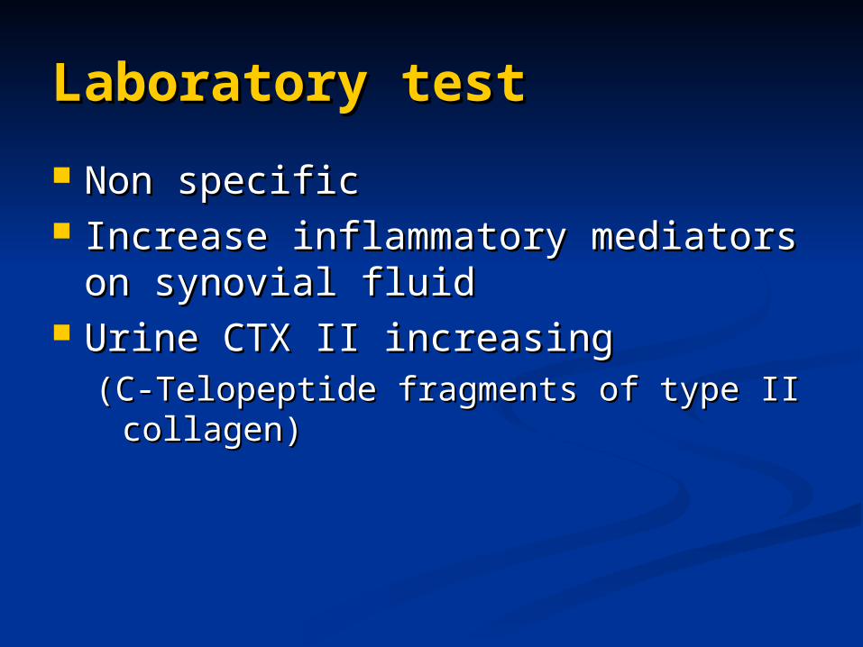
Laboratory testLaboratory test
Non specificNon specific Increase inflammatory mediators on Increase inflammatory mediators on
synovial fluidsynovial fluid Urine CTX II increasingUrine CTX II increasing
(C-Telopeptide fragments of type II (C-Telopeptide fragments of type II collagen)collagen)
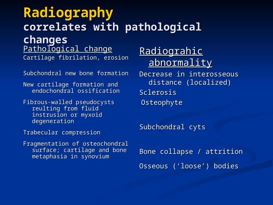
RadiographyRadiographycorrelates with pathological changescorrelates with pathological changes
Pathological changePathological changeCartilage fibrilation, erosionCartilage fibrilation, erosion
Subchondral new bone Subchondral new bone formationformation
New cartilage formation and New cartilage formation and endochondral ossificationendochondral ossification
Fibrous-walled pseudocysts Fibrous-walled pseudocysts reulting from fluid instrusion reulting from fluid instrusion or myxoid degenerationor myxoid degeneration
Trabecular compressionTrabecular compression
Fragmentation of osteochondral Fragmentation of osteochondral surface; cartilage and bone surface; cartilage and bone metaphasia in synovium metaphasia in synovium
Radiograhic Radiograhic abnormalityabnormality
Decrease in interosseous Decrease in interosseous distance (localized)distance (localized)
SclerosisSclerosis
OsteophyteOsteophyte
Subchondral cytsSubchondral cyts
Bone collapse / attrition Bone collapse / attrition
Osseous (‘loose’) bodiesOsseous (‘loose’) bodies
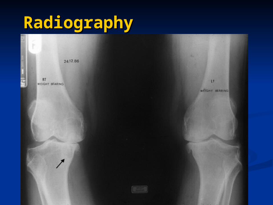
RadiographyRadiography
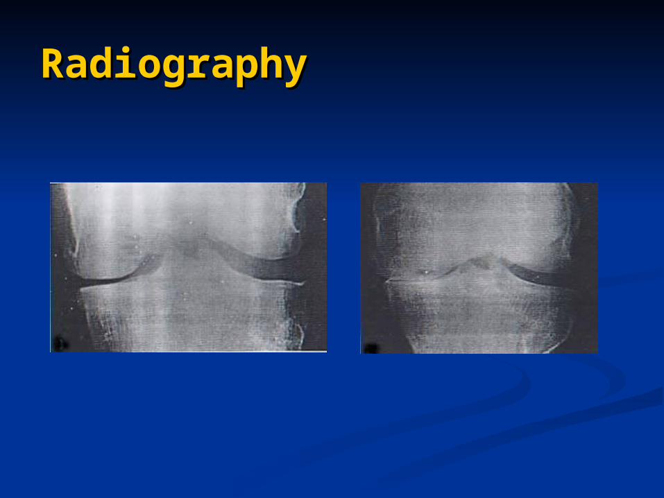
RadiographyRadiography
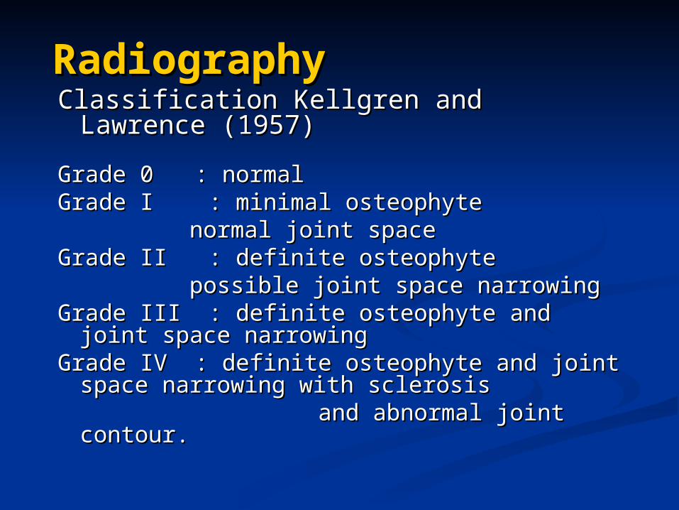
RadiographyRadiographyClassification Kellgren and Lawrence Classification Kellgren and Lawrence
(1957)(1957)
Grade 0 : normalGrade 0 : normalGrade I : minimal osteophyte Grade I : minimal osteophyte
normal joint spacenormal joint spaceGrade II : definite osteophyteGrade II : definite osteophyte
possible joint space narrowing possible joint space narrowing Grade III : definite osteophyte and joint Grade III : definite osteophyte and joint
space narrowingspace narrowingGrade IV : definite osteophyte and joint Grade IV : definite osteophyte and joint
space narrowing with sclerosis space narrowing with sclerosis and abnormal joint contour.and abnormal joint contour.
RadiographyRadiography
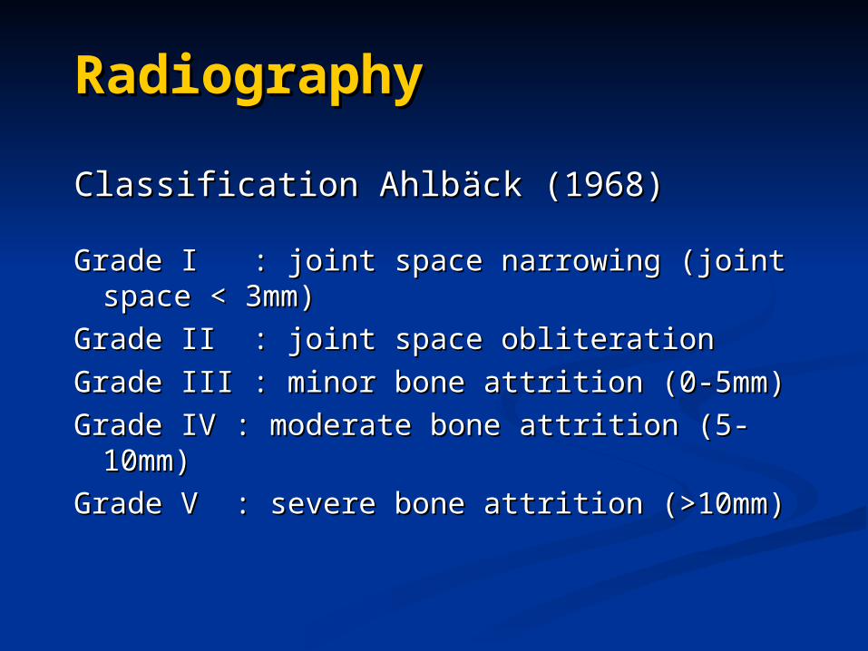
Classification AhlbClassification Ahlbääck (1968)ck (1968)
Grade I : joint space narrowing (joint Grade I : joint space narrowing (joint space < 3mm)space < 3mm)
Grade II : joint space obliterationGrade II : joint space obliteration
Grade III : minor bone attrition (0-5mm)Grade III : minor bone attrition (0-5mm)
Grade IV : moderate bone attrition (5-Grade IV : moderate bone attrition (5-10mm)10mm)
Grade V : severe bone attrition (>10mm)Grade V : severe bone attrition (>10mm)
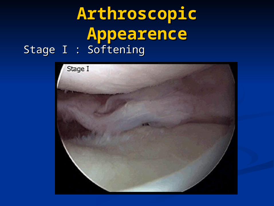
Arthroscopic Arthroscopic AppearenceAppearence
Stage I : SofteningStage I : Softening
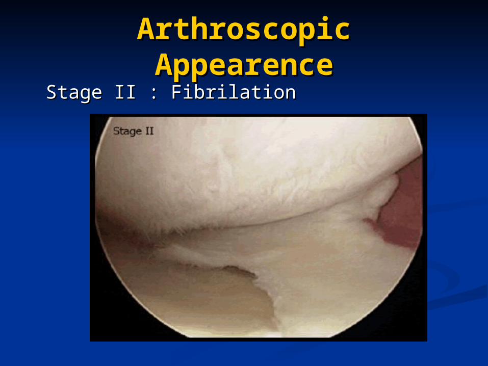
Arthroscopic Arthroscopic AppearenceAppearence
Stage II : FibrilationStage II : Fibrilation
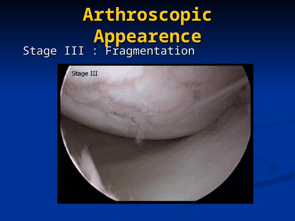
Arthroscopic Arthroscopic AppearenceAppearence
Stage III : FragmentationStage III : Fragmentation
Arthroscopic Arthroscopic AppearenceAppearence
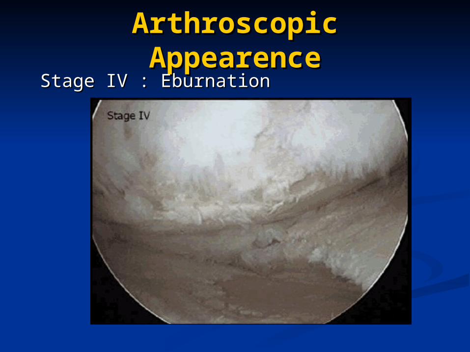
Stage IV : EburnationStage IV : Eburnation
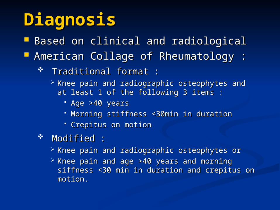
DiagnosisDiagnosis Based on clinical and radiological Based on clinical and radiological American Collage of Rheumatology :American Collage of Rheumatology :
Traditional format :Traditional format : Knee pain and radiographic osteophytes and at Knee pain and radiographic osteophytes and at
least 1 of the following 3 items :least 1 of the following 3 items : Age >40 yearsAge >40 years Morning stiffness <30min in durationMorning stiffness <30min in duration Crepitus on motionCrepitus on motion
Modified :Modified : Knee pain and radiographic osteophytes orKnee pain and radiographic osteophytes or Knee pain and age >40 years and morning Knee pain and age >40 years and morning
siffness <30 min in duration and crepitus on siffness <30 min in duration and crepitus on motion.motion.
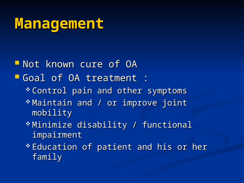
ManagementManagement
Not known cure of OANot known cure of OA Goal of OA treatment :Goal of OA treatment :
Control pain and other symptomsControl pain and other symptoms Maintain and / or improve joint mobilityMaintain and / or improve joint mobility Minimize disability / functional Minimize disability / functional
impairmentimpairment Education of patient and his or her Education of patient and his or her
familyfamily
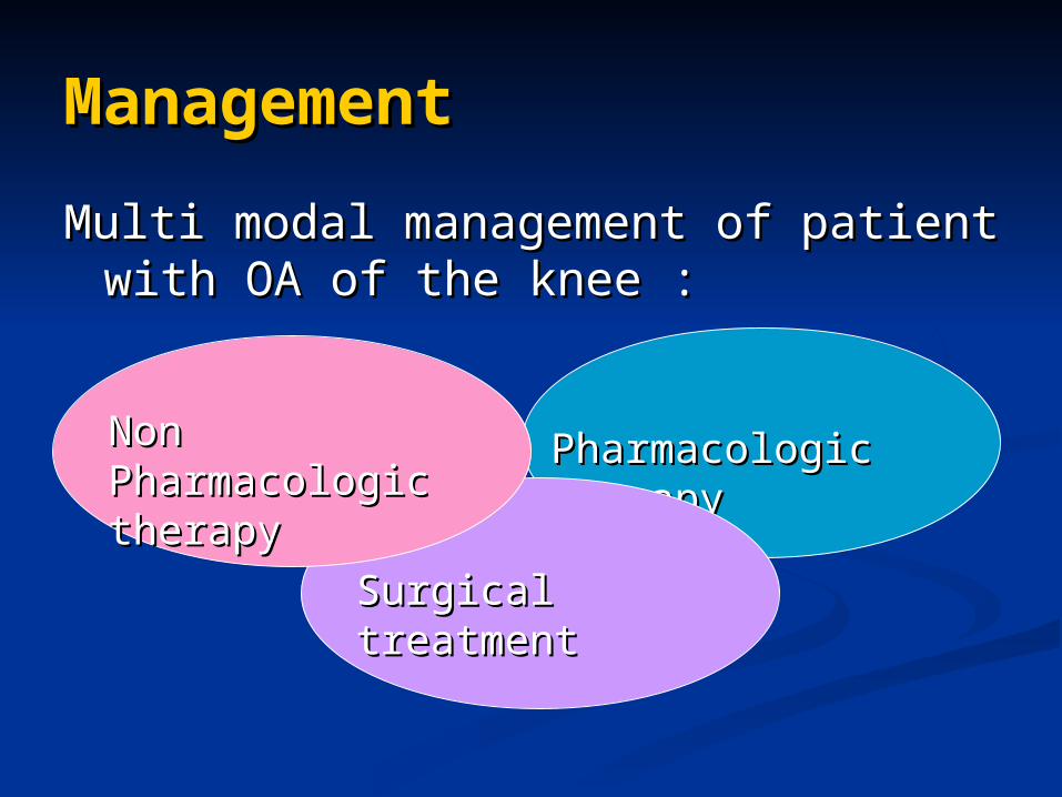
ManagementManagement
Multi modal management of patient Multi modal management of patient with OA of the knee :with OA of the knee :
Pharmacologic Pharmacologic therapytherapy
Surgical treatmentSurgical treatment
Non Non Pharmacologic Pharmacologic therapytherapy
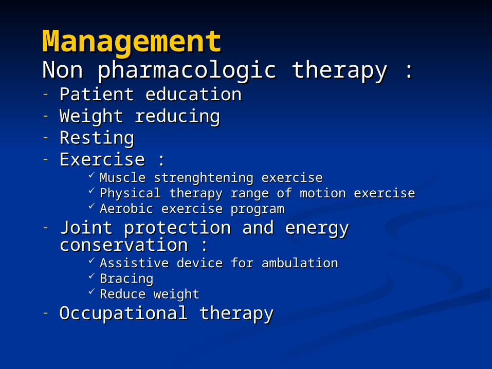
ManagementManagement Non pharmacologic therapy :Non pharmacologic therapy :- Patient education Patient education - Weight reducingWeight reducing- RestingResting- Exercise :Exercise :
Muscle strenghtening exerciseMuscle strenghtening exercise Physical therapy range of motion exercisePhysical therapy range of motion exercise Aerobic exercise programAerobic exercise program
- Joint protection and energy Joint protection and energy conservation :conservation :
Assistive device for ambulationAssistive device for ambulation Bracing Bracing Reduce weightReduce weight
- Occupational therapyOccupational therapy
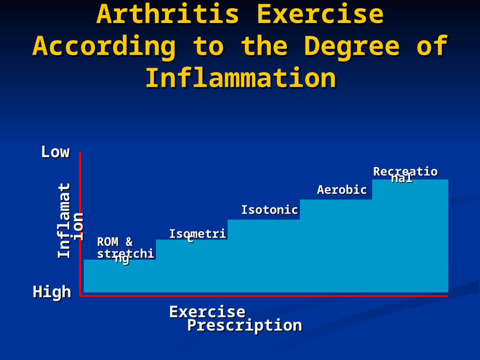
Arthritis Exercise Arthritis Exercise According to the Degree of According to the Degree of
InflammationInflammation
ROM & ROM & stretchinstretchingg
IsometriIsometricc
IsotonicIsotonic
AerobicAerobic
RecreatioRecreationalnal
Exercise Exercise PrescriptionPrescription
HigHighh
LoLoww
Infl
am
ati
Infl
am
ati
on
on
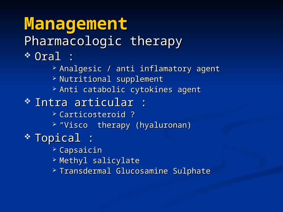
ManagementManagementPharmacologic therapyPharmacologic therapy Oral :Oral :
Analgesic / anti inflamatory agentAnalgesic / anti inflamatory agent Nutritional supplementNutritional supplement Anti catabolic cytokines agentAnti catabolic cytokines agent
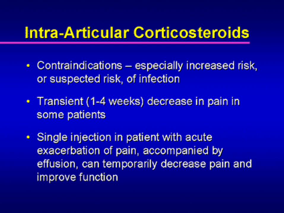
Intra articular :Intra articular : Carticosteroid ?Carticosteroid ? ““Visco” therapy (hyaluronan)Visco” therapy (hyaluronan)
Topical :Topical : Capsaicin Capsaicin Methyl salicylateMethyl salicylate Transdermal Glucosamine SulphateTransdermal Glucosamine Sulphate
ManagementManagement
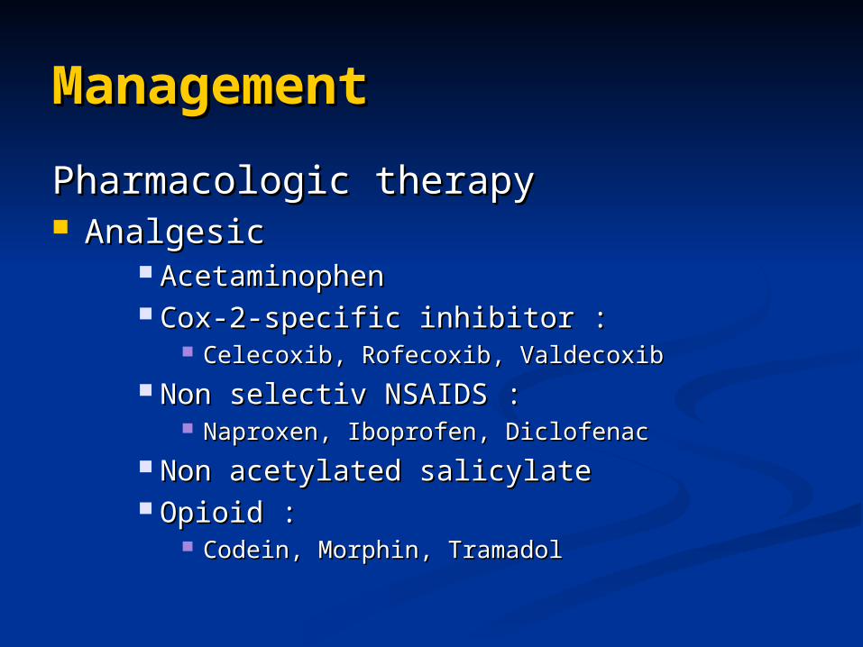
Pharmacologic therapyPharmacologic therapy AnalgesicAnalgesic
AcetaminophenAcetaminophen Cox-2-specific inhibitor :Cox-2-specific inhibitor :
Celecoxib, Rofecoxib, ValdecoxibCelecoxib, Rofecoxib, Valdecoxib Non selectiv NSAIDS :Non selectiv NSAIDS :
Naproxen, Iboprofen, DiclofenacNaproxen, Iboprofen, Diclofenac Non acetylated salicylateNon acetylated salicylate Opioid :Opioid :
Codein, Morphin, TramadolCodein, Morphin, Tramadol
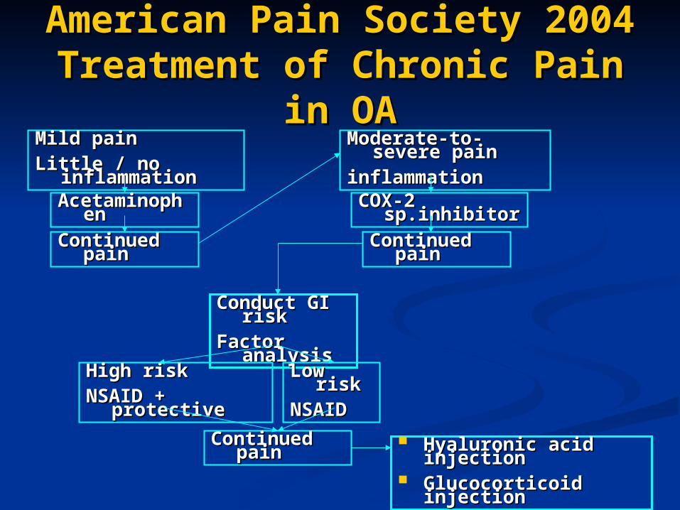
American Pain Society 2004American Pain Society 2004Treatment of Chronic Pain Treatment of Chronic Pain
in OAin OAMild pain Mild pain Little / no Little / no
inflammationinflammationAcetaminopAcetaminop
hen hen Continued Continued
painpain
Conduct GI Conduct GI riskrisk
Factor Factor analysisanalysis
Moderate-to-Moderate-to-severe painsevere pain
inflammationinflammationCOX-2 COX-2
sp.inhibitorsp.inhibitorContinued Continued
painpain
High riskHigh riskNSAID + NSAID +
protectiveprotective
Low Low riskrisk
NSAIDNSAID
Continued Continued painpain
Hyaluronic acid Hyaluronic acid injectioninjection
Glucocorticoid Glucocorticoid injectioninjection
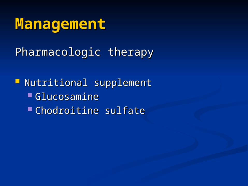
ManagementManagement
Pharmacologic therapyPharmacologic therapy
Nutritional supplementNutritional supplement GlucosamineGlucosamine Chodroitine sulfateChodroitine sulfate
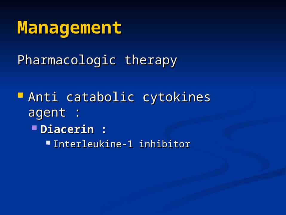
ManagementManagement
Pharmacologic therapyPharmacologic therapy
Anti catabolic cytokines agent :Anti catabolic cytokines agent : Diacerin : Diacerin :
Interleukine-1 inhibitorInterleukine-1 inhibitor
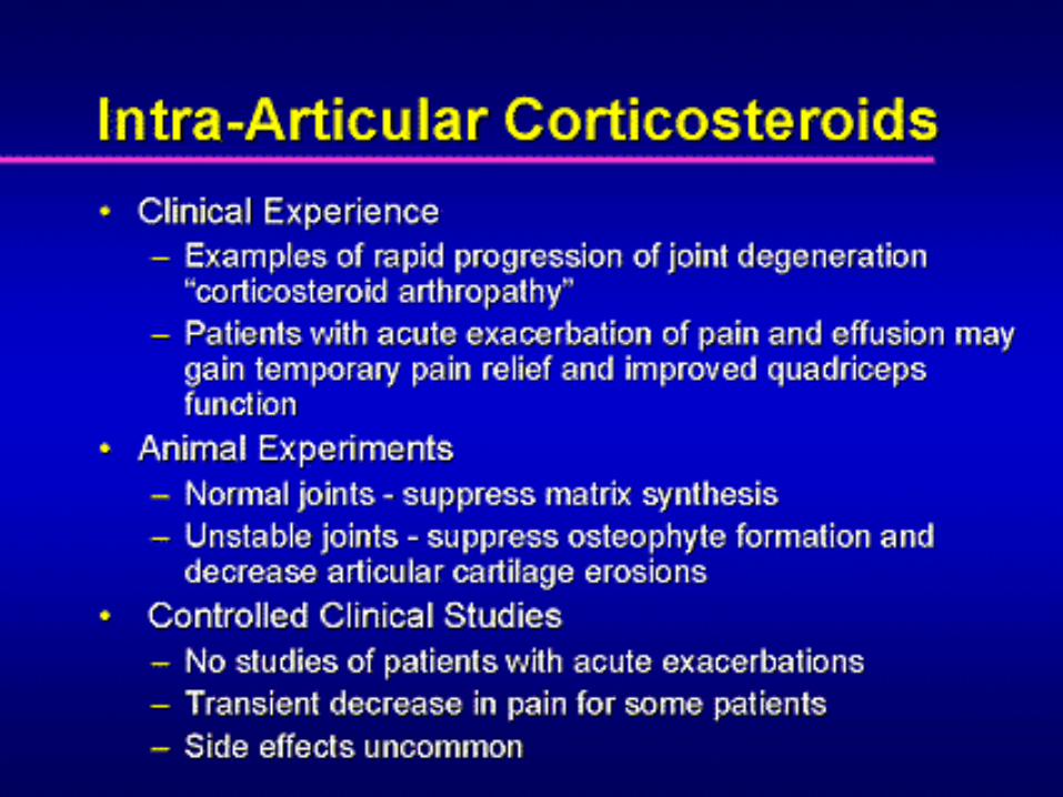

Intra-Articular Intra-Articular “Visco”therapy“Visco”therapy
Hyaluronan (hyaluronic acid ): Hyaluronan (hyaluronic acid ): Attempts to restore synovial fluid protective, Attempts to restore synovial fluid protective,
lubricating, shock absorbing, barrier and lubricating, shock absorbing, barrier and rheologic affects. rheologic affects.
The agents are :The agents are : Non immunologicNon immunologic Permeabel to metabolites and macromolecules.Permeabel to metabolites and macromolecules. Qualitatively similar to human synovial fluid. Qualitatively similar to human synovial fluid. Retained longer than endogenus hyaluronic acid.Retained longer than endogenus hyaluronic acid.
Effects on joint pain and mobility last longer Effects on joint pain and mobility last longer than the agent is retained in the joint (improved than the agent is retained in the joint (improved endogenous production?)endogenous production?)

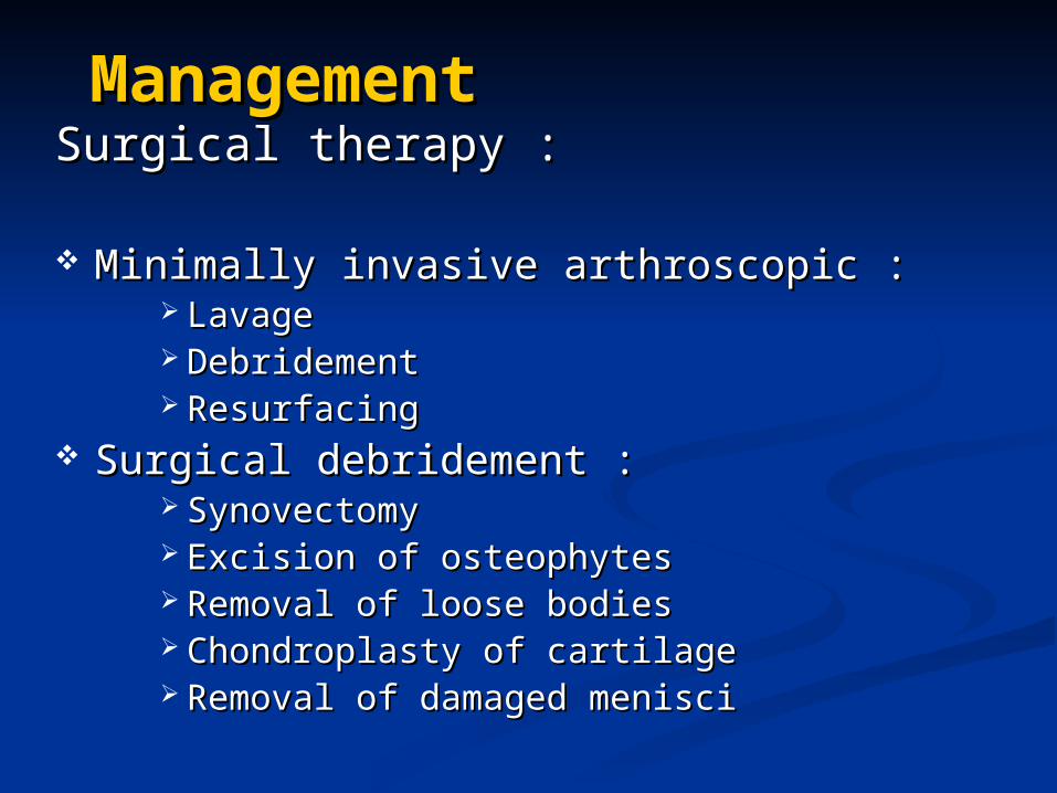
ManagementManagementSurgical therapy :Surgical therapy :
Minimally invasive arthroscopic :Minimally invasive arthroscopic : LavageLavage Debridement Debridement Resurfacing Resurfacing
Surgical debridement :Surgical debridement : Synovectomy Synovectomy Excision of osteophytes Excision of osteophytes Removal of loose bodiesRemoval of loose bodies Chondroplasty of cartilageChondroplasty of cartilage Removal of damaged menisci Removal of damaged menisci
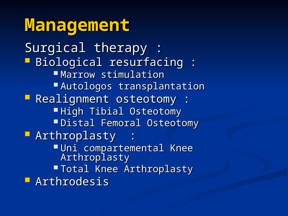
ManagementManagementSurgical therapy :Surgical therapy : Biological resurfacing :Biological resurfacing :
Marrow stimulationMarrow stimulation Autologos transplantationAutologos transplantation
Realignment osteotomy :Realignment osteotomy : High Tibial OsteotomyHigh Tibial Osteotomy Distal Femoral OsteotomyDistal Femoral Osteotomy
Arthroplasty :Arthroplasty : Uni compartemental Knee ArthroplastyUni compartemental Knee Arthroplasty Total Knee ArthroplastyTotal Knee Arthroplasty
ArthrodesisArthrodesis
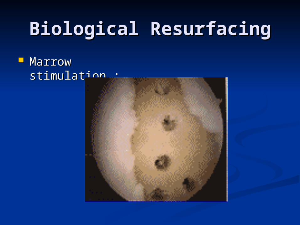
Biological ResurfacingBiological Resurfacing
Marrow Marrow stimulation :stimulation :
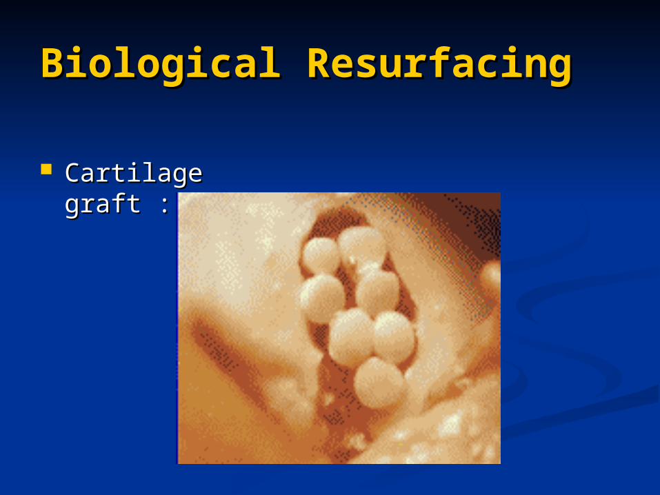
Biological ResurfacingBiological Resurfacing
Cartilage graft : Cartilage graft :
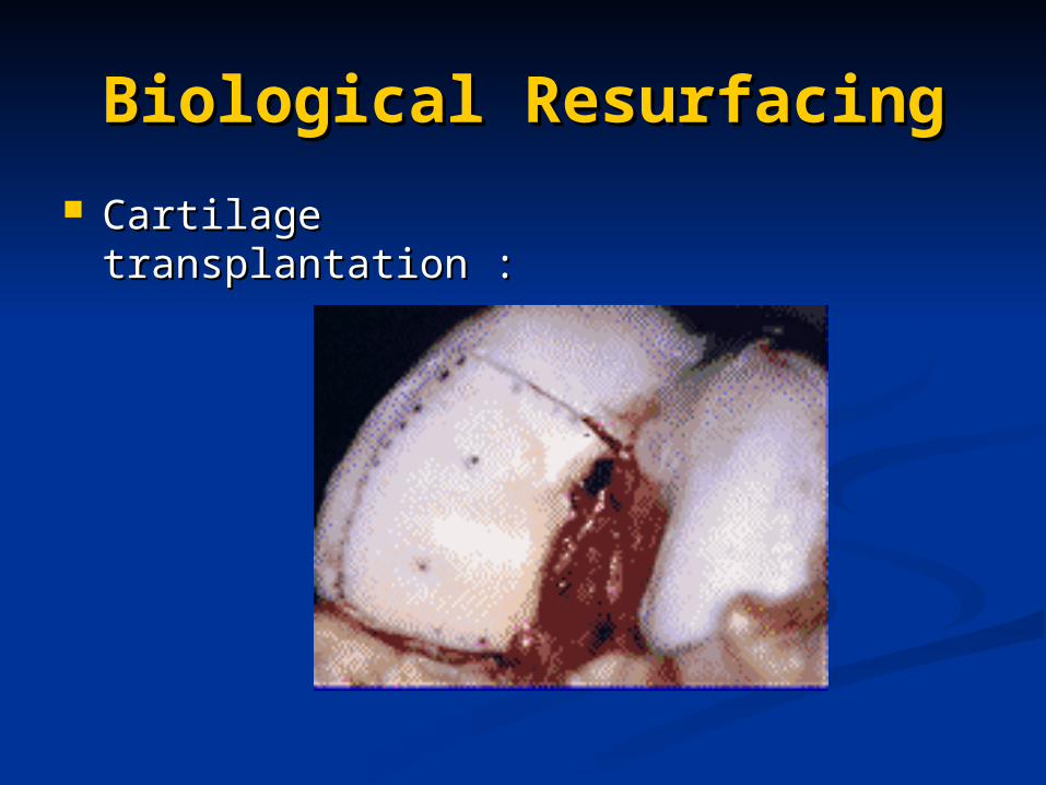
Biological ResurfacingBiological Resurfacing
Cartilage Cartilage transplantation :transplantation :
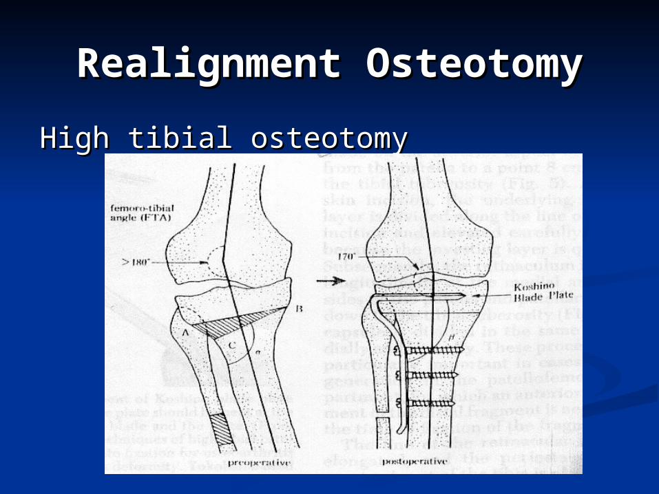
Realignment OsteotomyRealignment Osteotomy
High tibial osteotomyHigh tibial osteotomy
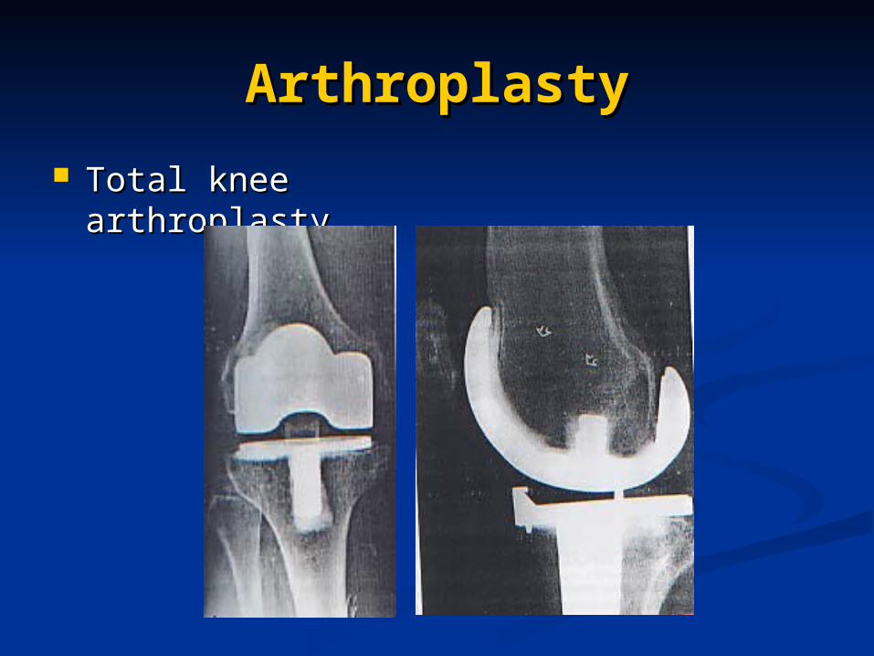
ArthroplastyArthroplasty
Total knee Total knee arthroplastyarthroplasty
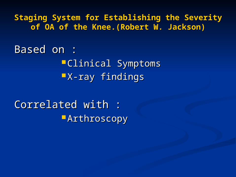
Staging System for Establishing the Severity Staging System for Establishing the Severity of OA of the Knee.(Robert W. Jackson)of OA of the Knee.(Robert W. Jackson)
Based on :Based on : Clinical SymptomsClinical Symptoms X-ray findingsX-ray findings
Correlated with :Correlated with : ArthroscopyArthroscopy
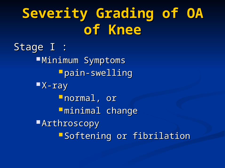
Severity Grading of OA of Severity Grading of OA of KneeKnee
Stage I :Stage I : Minimum SymptomsMinimum Symptoms
pain-swellingpain-swelling X-rayX-ray
normal, ornormal, or minimal changeminimal change
ArthroscopyArthroscopy Softening or fibrilationSoftening or fibrilation
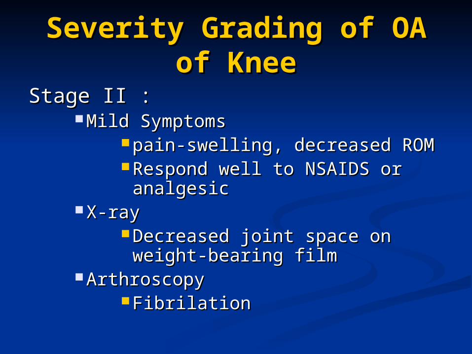
Severity Grading of OA of Severity Grading of OA of KneeKnee
Stage II :Stage II : Mild SymptomsMild Symptoms
pain-swelling, decreased ROMpain-swelling, decreased ROM Respond well to NSAIDS or Respond well to NSAIDS or
analgesicanalgesic X-rayX-ray
Decreased joint space on weight-Decreased joint space on weight-bearing filmbearing film
ArthroscopyArthroscopy FibrilationFibrilation
Severity Grading of OA of Severity Grading of OA of KneeKnee
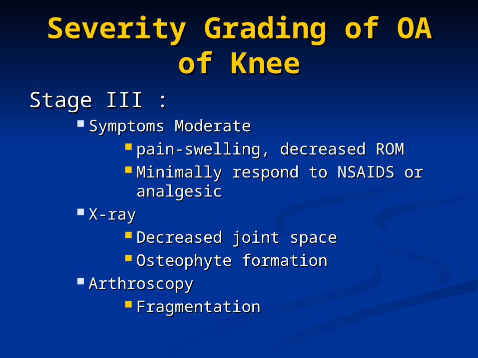
Stage III :Stage III : Symptoms ModerateSymptoms Moderate
pain-swelling, decreased ROMpain-swelling, decreased ROM Minimally respond to NSAIDS or Minimally respond to NSAIDS or
analgesicanalgesic X-rayX-ray
Decreased joint space Decreased joint space Osteophyte formationOsteophyte formation
ArthroscopyArthroscopy FragmentationFragmentation
Severity Grading of OA of Severity Grading of OA of KneeKnee
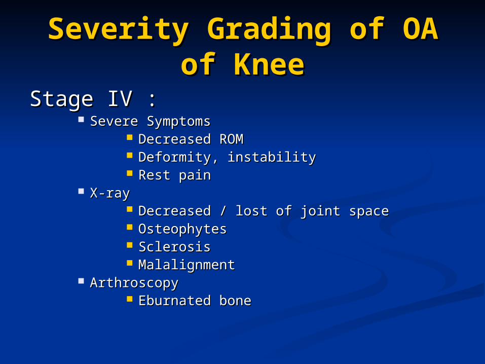
Stage IV :Stage IV : Severe SymptomsSevere Symptoms
Decreased ROMDecreased ROM Deformity, instabilityDeformity, instability Rest painRest pain
X-rayX-ray Decreased / lost of joint spaceDecreased / lost of joint space OsteophytesOsteophytes Sclerosis Sclerosis MalalignmentMalalignment
ArthroscopyArthroscopy Eburnated boneEburnated bone
Recommended Recommended TreatmentTreatment
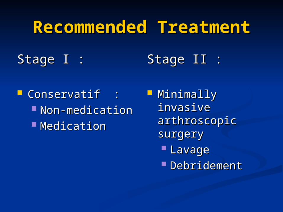
Stage I :Stage I :
Conservatif :Conservatif : Non-medicationNon-medication MedicationMedication
Stage II :Stage II :
Minimally invasive Minimally invasive arthroscopic arthroscopic surgerysurgery LavageLavage DebridementDebridement
Recommended Recommended TreatmentTreatment
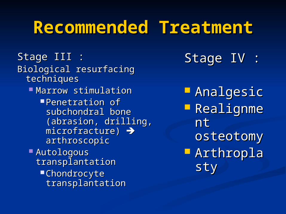
Stage III :Stage III :Biological resurfacing Biological resurfacing
techniquestechniques Marrow stimulationMarrow stimulation
Penetration of Penetration of subchondral bone subchondral bone (abrasion, drilling, (abrasion, drilling, microfracture) microfracture) arthroscopicarthroscopic
Autologous transplantationAutologous transplantation Chondrocyte Chondrocyte
transplantationtransplantation
Stage IV :Stage IV :
AnalgesicAnalgesic RealignmeRealignme
nt nt osteotomyosteotomy
ArthroplastArthroplastyy

Thank youThank you