Embed Size (px)
Citation preview
10/18/2011
1
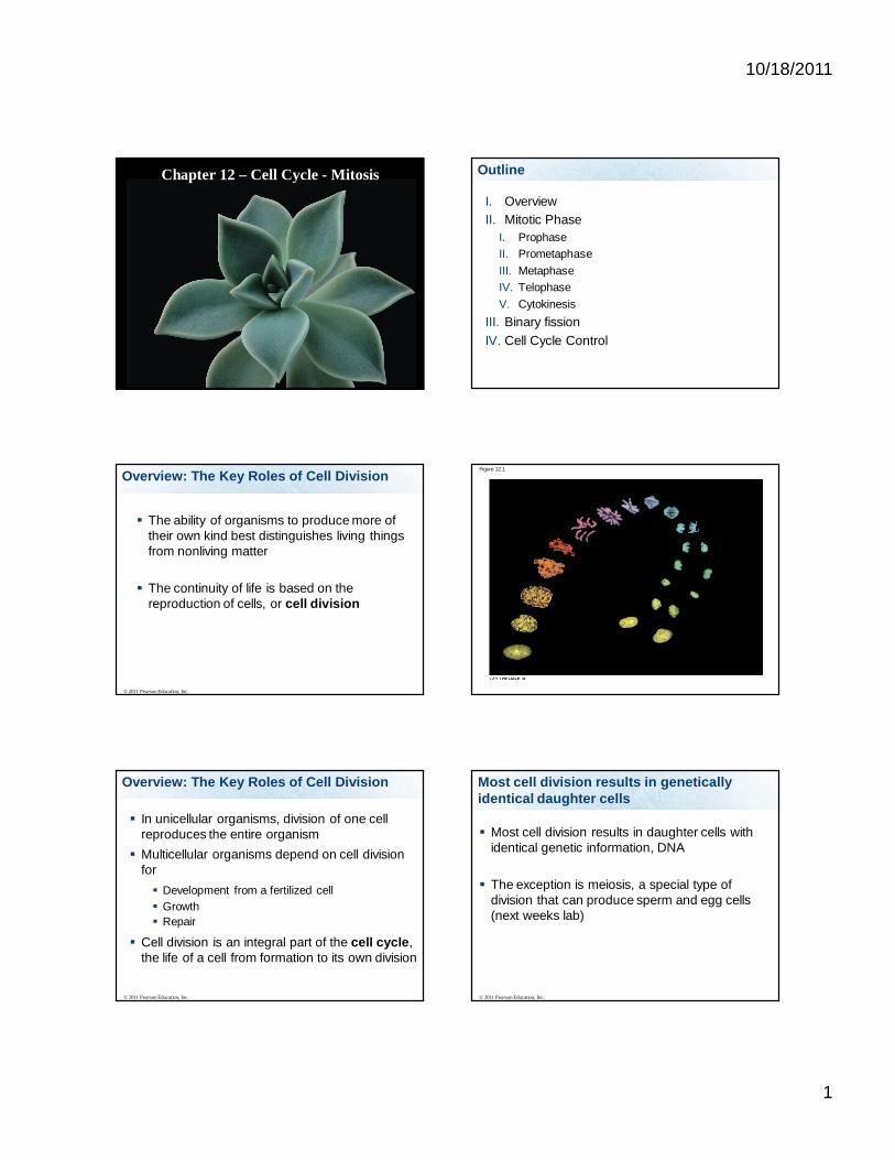
Chapter 12 – Cell Cycle - Mitosis Outline
I. OverviewII. Mitotic Phase
I. ProphaseII. PrometaphaseIII. MetaphaseIV. TelophaseV. Cytokinesis
III. Binary fissionIV. Cell Cycle Control
Overview: The Key Roles of Cell Division
The ability of organisms to produce more of their own kind best distinguishes living things from nonliving matter
The continuity of life is based on the reproduction of cells, or cell division
© 2011 Pearson Education, Inc.
Figure 12.1
In unicellular organisms, division of one cell reproduces the entire organism
Multicellular organisms depend on cell division for Development from a fertilized cell Growth Repair
Cell division is an integral part of the cell cycle, the life of a cell from formation to its own division
© 2011 Pearson Education, Inc.
Overview: The Key Roles of Cell Division Most cell division results in genetically identical daughter cells
Most cell division results in daughter cells with identical genetic information, DNA
The exception is meiosis, a special type of division that can produce sperm and egg cells (next weeks lab)
© 2011 Pearson Education, Inc.

10/18/2011
2
Cellular Organization of the Genetic Material
All the DNA in a cell constitutes the cell’s genome
A genome can consist of a single DNA molecule (common in prokaryotic cells) or a number of DNA molecules (common in eukaryotic cells)
DNA molecules in a cell are packaged into chromosomes
© 2011 Pearson Education, Inc.
Figure 12.3
20 m
Chromosomes
Eukaryotic chromosomes consist of chromatin, a complex of DNA and protein that condenses during cell division
Every eukaryotic species has a characteristic number of chromosomes in each cell nucleus
Somatic cells (nonreproductive cells) have two sets of chromosomes
Gametes (reproductive cells: sperm and eggs) have half as many chromosomes as somatic cells
© 2011 Pearson Education, Inc.
Distribution of Chromosomes During Eukaryotic Cell Division
In preparation for cell division, DNA is replicated and the chromosomes condense
Each duplicated chromosome has two sister chromatids (joined copies of the original chromosome), which separate during cell division
The centromere is the narrow “waist” of the duplicated chromosome, where the two chromatids are most closely attached
© 2011 Pearson Education, Inc.
Figure 12.4
0.5 mCentromere
Sisterchromatids
During cell division, the two sister chromatids of each duplicated chromosome separate and move into two nuclei
Once separate, the chromatids are called chromosomes
© 2011 Pearson Education, Inc.
Distribution of Chromosomes During Eukaryotic Cell Division

10/18/2011
3
Figure 12.5-1
ChromosomesChromosomalDNA molecules
Centromere
Chromosomearm
1
Figure 12.5-2
ChromosomesChromosomalDNA molecules
Centromere
Chromosomearm
Chromosome duplication(including DNA replication)and condensation
Sisterchromatids
1
2
Figure 12.5-3
ChromosomesChromosomalDNA molecules
Centromere
Chromosomearm
Chromosome duplication(including DNA replication)and condensation
Sisterchromatids
Separation of sisterchromatids intotwo chromosomes
1
2
3
The region where the chromatids are tightly associated is called
1 2 3 4
25% 25%25%25%1. Centrosome2. Chromatin3. Centromere4. Kinetochore
Microtubule-organizing centers have two
1 2 3 4
25% 25%25%25%1. Centrioles2. Chromatins3. Centromeres4. Kinetochores
Eukaryotic cell division consists of Mitosis, the division of the genetic material in the
nucleus Cytokinesis, the division of the cytoplasm
Gametes are produced by a variation of cell division called meiosis
Meiosis yields nonidentical daughter cells that have only one set of chromosomes, half as many as the parent cell
© 2011 Pearson Education, Inc.
Eukaryotic Cell Division

10/18/2011
4
The mitotic phase alternates with interphase in the cell cycle
In 1882, the German anatomist Walther Flemming developed dyes to observe chromosomes during mitosis and cytokinesis
© 2011 Pearson Education, Inc.
When does DNA replicate?
Just prior to cell division
Cells need to divide for organisms to grow or to replace old cells, or for one celled organisms to reproduce
Before a cell divides it needs to replicate its DNA
Phases of the Cell Cycle
The cell cycle consists of
Mitotic (M) phase (mitosis and cytokinesis) Interphase (cell growth and copying of
chromosomes in preparation for cell division)
© 2011 Pearson Education, Inc.
Interphase (about 90% of the cell cycle) can be divided into subphases G1 phase (“first gap”) S phase (“synthesis”) G2 phase (“second gap”)
The cell grows during all three phases, but chromosomes are duplicated only during the S phase
© 2011 Pearson Education, Inc.
Phases of the Cell Cycle - Interphase
Figure 12.6
INTERPHASE
G1
G2
S(DNA synthesis)
Mitotic Phase
Mitosis is conventionally divided into five phases Prophase Prometaphase Metaphase Anaphase Telophase
Cytokinesis overlaps the latter stages of mitosis
© 2011 Pearson Education, Inc.

10/18/2011
5
© 2011 Pearson Education, Inc. BioFlix: Mitosis
Figure 12.7
G2 of Interphase Prophase PrometaphaseCentrosomes(with centriole pairs)
Chromatin(duplicated)
Nucleolus Nuclearenv elope
Plasmamembrane
Early mitoticspindle
AsterCentromere
Chromosome, consistingof two sister chromatids
Fragments of nuclearenv elope
Nonkinetochoremicrotubules
Kinetochore Kinetochoremicrotubule
Metaphase
Metaphase plate
Anaphase Telophase and Cytokinesis
Spindle Centrosome atone spindle pole
Daughterchromosomes
Cleav agefurrow
Nucleolusforming
Nuclearenv elopeforming
10
m
Figure 12.7a
G2 of Interphase Prophase Prometaphase
Centrosomes(with centriole pairs)
Chromatin(duplicated)
NucleolusNuclearenvelope
Plasmamembrane
Early mitoticspindle
AsterCentromere
Chromosome, consistingof two sister chromatids
Fragments of nuclearenvelope
Nonkinetochoremicrotubules
Kinetochore Kinetochoremicrotubule
Figure 12.7b
Metaphase
Metaphase plate
Anaphase Telophase and Cytokinesis
Spindle Centrosome atone spindle pole
Daughterchromosomes
Cleavagefurrow
Nucleolusforming
Nuclearenvelopeforming
Figure 12.7c
G2 of Interphase Prophase Prometaphase
10
m
Figure 12.7d
10
m
Metaphase Anaphase Telophase and Cytokinesis

10/18/2011
6
G2 of Interphase Prophase Prometaphase
Centrosomes(with centriole pairs)
Chromatin(duplicated)
NucleolusNuclearenvelope
Plasmamembrane
G2 of Interphase
Nuclear envelope present Centrosomes are duplicated Chromosomes are in the
duplicated state (DNA replication occurred during S phase of interphase)
Prophase
Early mitoticspindle
AsterCentromere
Chromosome, consistingof two sister chromatids
Prophase
Chromosomes condense Nucleolus disappear Sister chromatids are joined
at the centromeres Mitotic spindle appears,
composed of centrosomes, spindle microtubles and asters
Centrosomes move to opposite poles of the cell
PrometaphaseFragments of nuclearenvelope
Nonkinetochoremicrotubules
Kinetochore Kinetochoremicrotubule
Prometaphase
Nuclear envelope fragments
Microtubles attach to the kinetochore
Metaphase
Metaphase plate
Telophase and Cytokinesis
Spindle Centrosome atone spindle pole
Metaphase
Centrosomes are at opposite ends of cell
Chromosomes line up at the metaphase plate
Anaphase
Daughterchromosomes
Metaphase Cohesion proteins are cleaved,
sister chromatids separate, now each are a chromosome
Kinetochore microtubules shorten, moving the daughter chromosomes to opposite poles of the cell
Cell elongates due to nonkinetochore microtubules lengthening
Telophase and Cytokinesis
Cleavagefurrow
Nucleolusforming
Nuclearenvelopeforming
Telophase
Two identical daughter nuclei form, nuclear envelopes form
Nucleolus reappears Chromosomes uncondense Spindle microtubles
depolymerize

10/18/2011
7
Telophase and Cytokinesis
Cleavagefurrow
Nucleolusforming
Nuclearenvelopeforming
Cytokinesis
Cytoplasm divides In animal cells, cleavage
furrow pinches the cell in two
The Mitotic Spindle: A Closer Look
The mitotic spindle is a structure made of microtubules that controls chromosome movement during mitosis
In animal cells, assembly of spindle microtubules begins in the centrosome, the microtubule organizing center
The centrosome replicates during interphase, forming two centrosomes that migrate to opposite ends of the cell during prophase and prometaphase
© 2011 Pearson Education, Inc.
An aster (a radial array of short microtubules) extends from each centrosome
The spindle includes the centrosomes, the spindle microtubules, and the asters
© 2011 Pearson Education, Inc.
The Mitotic Spindle: A Closer Look
During prometaphase, some spindle microtubules attach to the kinetochores of chromosomes and begin to move the chromosomes
Kinetochores are protein complexes associated with centromeres
At metaphase, the chromosomes are all lined up at the metaphase plate, an imaginary structure at the midway point between the spindle’s two poles
© 2011 Pearson Education, Inc.
The Mitotic Spindle: A Closer Look
Figure 12.8
Sisterchromatids
AsterCentrosome
Metaphaseplate(imaginary)
Kineto-chores
Overlappingnonkinetochoremicrotubules Kinetochore
microtubules
Microtubules
Chromosomes
Centrosome
0.5 m
1 m
In anaphase, sister chromatids separate and move along the kinetochore microtubules toward opposite ends of the cell
The microtubules shorten by depolymerizing at their kinetochore ends
© 2011 Pearson Education, Inc.
The Mitotic Spindle: A Closer Look
10/18/2011
8
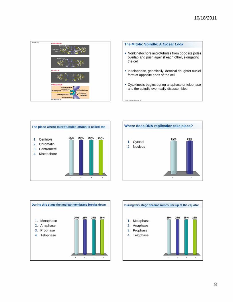
Figure 12.9
Chromosomemovement
Microtubule
Motor protein
Chromosome
Kinetochore
Tubulinsubunits
Kinetochore
Mark
Spindlepole
EXPERIMENT
RESULTS
CONCLUSION
Nonkinetochore microtubules from opposite poles overlap and push against each other, elongating the cell
In telophase, genetically identical daughter nuclei form at opposite ends of the cell
Cytokinesis begins during anaphase or telophase and the spindle eventually disassembles
© 2011 Pearson Education, Inc.
The Mitotic Spindle: A Closer Look
The place where microtubules attach is called the
1 2 3 4
25% 25%25%25%1. Centriole2. Chromatin3. Centromere4. Kinetochore
Where does DNA replication take place?
1 2
50%50%1. Cytosol2. Nucleus
During this stage the nuclear membrane breaks down
1 2 3 4
25% 25%25%25%1. Metaphase2. Anaphase3. Prophase4. Telophase
During this stage chromosomes line up at the equator
1 2 3 4
25% 25%25%25%1. Metaphase2. Anaphase3. Prophase4. Telophase
10/18/2011
9
During this stage the chromosomes begin to uncondense
1 2 3 4
25% 25%25%25%1. Metaphase2. Anaphase3. Prophase4. Telophase
At the end of Mitosis the chromosomes are in the duplicated state
1 2
50%50%1. Yes2. No
© 2011 Pearson Education, Inc.
Animation: Cytokinesis Right-click slide / select ”Play”
© 2011 Pearson Education, Inc.
Animation: Animal Mitosis Right-click slide / select ”Play”
© 2011 Pearson Education, Inc.
Animation: Sea Urchin (Time Lapse) Right-click slide / select ”Play”
Videos
Youtube Mitosis in embryo Mitosis
10/18/2011
10
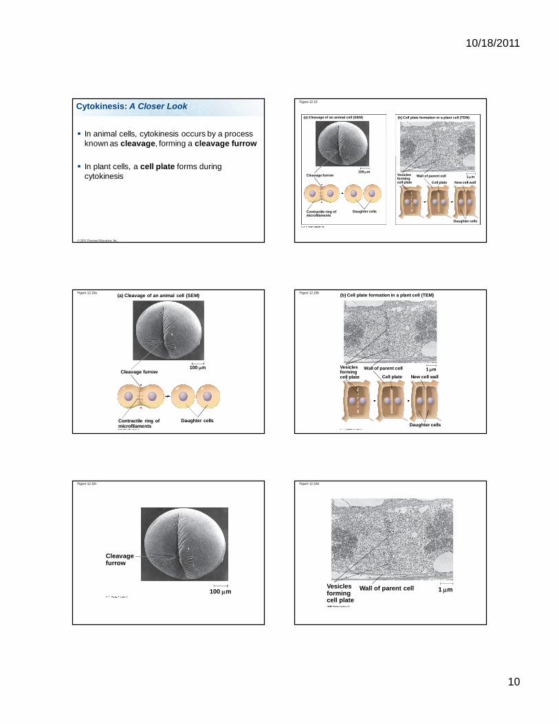
Cytokinesis: A Closer Look
In animal cells, cytokinesis occurs by a process known as cleavage, forming a cleavage furrow
In plant cells, a cell plate forms during cytokinesis
© 2011 Pearson Education, Inc.
Figure 12.10
(a) Cleavage of an animal cell (SEM) (b) Cell plate formation in a plant cell (TEM)
Cleavage furrow
Contractile ring ofmicrofilaments
Daughter cells
Vesiclesformingcell plate
Wall of parent cell
Cell plate New cell wall
Daughter cells
100 m1 m
Figure 12.10a (a) Cleavage of an animal cell (SEM)
Cleavage furrow
Contractile ring ofmicrofilaments
Daughter cells
100 m
Figure 12.10b (b) Cell plate formation in a plant cell (TEM)
Vesiclesformingcell plate
Wall of parent cell
Cell plate New cell wall
Daughter cells
1 m
Figure 12.10c
Cleavage furrow
100 m
Figure 12.10d
Vesiclesformingcell plate
Wall of parent cell 1 m
10/18/2011
11
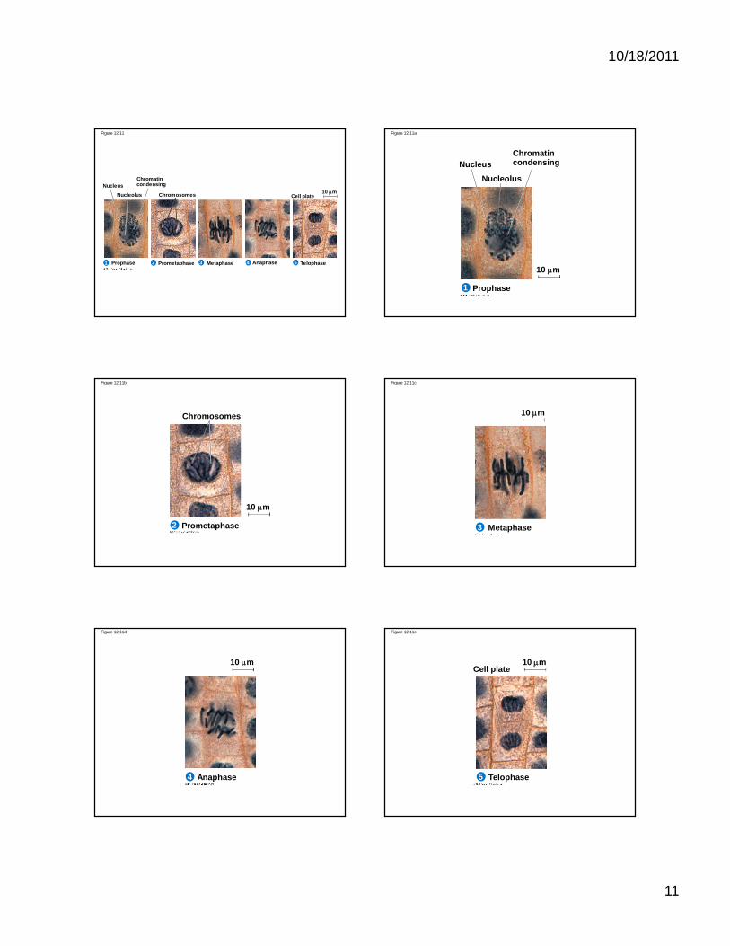
Figure 12.11
ChromatincondensingNucleus
Nucleolus Chromosomes Cell plate10 m
Prophase Prometaphase Metaphase Anaphase Telophase1 2 3 4 5
Figure 12.11a
ChromatincondensingNucleus
Nucleolus
Prophase1
10 m
Figure 12.11b
Chromosomes
Prometaphase2
10 m
Figure 12.11c
10 m
Metaphase3
Figure 12.11d
Anaphase4
10 m
Figure 12.11e
10 m
Telophase5
Cell plate

10/18/2011
12
Binary Fission in Bacteria
Prokaryotes (bacteria and archaea) reproduce by a type of cell division called binary fission
In binary fission, the chromosome replicates (beginning at the origin of replication), and the two daughter chromosomes actively move apart
The plasma membrane pinches inward, dividing the cell into two
© 2011 Pearson Education, Inc.
Figure 12.12-1
1
Origin ofreplication
E. coli cell
Two copies of origin
Cell wallPlasma membrane
Bacterial chromosomeChromosomereplicationbegins.
1
Origin ofreplication
E. coli cell
Two copies of origin
Cell wallPlasma membrane
Bacterial chromosome
Origin Origin
Chromosomereplicationbegins.
Replicationcontinues.
2
Figure 12.12-2
1
Origin ofreplication
E. coli cell
Two copies of origin
Cell wallPlasma membrane
Bacterial chromosome
Origin Origin
Chromosomereplicationbegins.
Replicationcontinues.
Replicationfinishes.
2
3
Figure 12.12-3
1
Origin ofreplication
E. coli cell
Two copies of origin
Cell wallPlasma membrane
Bacterial chromosome
Origin Origin
Chromosomereplicationbegins.
Replicationcontinues.
Replicationfinishes.
Two daughtercells result.
2
3
4
Figure 12.12-4
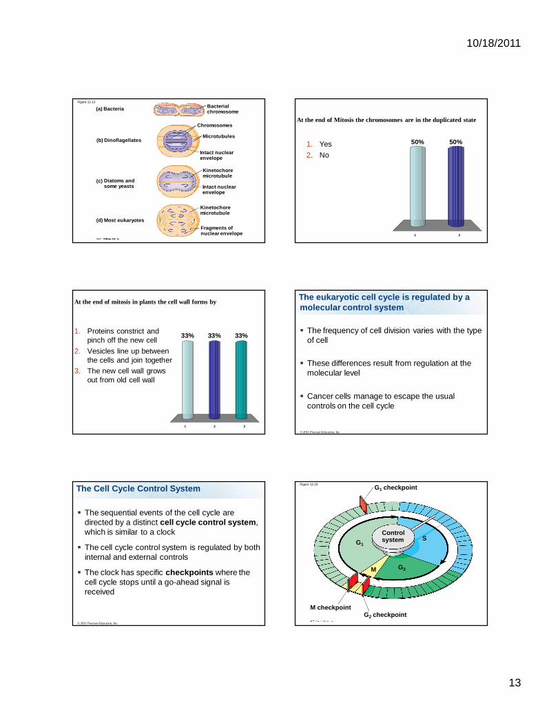
The Evolution of Mitosis
Since prokaryotes evolved before eukaryotes, mitosis probably evolved from binary fission
Certain protists exhibit types of cell division that seem intermediate between binary fission and mitosis
© 2011 Pearson Education, Inc.
10/18/2011
13
Figure 12.13
(a) Bacteria
(b) Dinoflagellates
(d) Most eukaryotes
Intact nuclearenvelope
Chromosomes
Microtubules
Intact nuclearenvelope
Kinetochoremicrotubule
Kinetochoremicrotubule
Fragments ofnuclear envelope
Bacterialchromosome
(c) Diatoms andsome yeasts
At the end of Mitosis the chromosomes are in the duplicated state
1 2
50%50%1. Yes2. No
At the end of mitosis in plants the cell wall forms by
1 2 3
33% 33%33%1. Proteins constrict and pinch off the new cell
2. Vesicles line up between the cells and join together
3. The new cell wall grows out from old cell wall
The eukaryotic cell cycle is regulated by a molecular control system
The frequency of cell division varies with the type of cell
These differences result from regulation at the molecular level
Cancer cells manage to escape the usual controls on the cell cycle
© 2011 Pearson Education, Inc.
The Cell Cycle Control System
The sequential events of the cell cycle are directed by a distinct cell cycle control system, which is similar to a clock
The cell cycle control system is regulated by both internal and external controls
The clock has specific checkpoints where the cell cycle stops until a go-ahead signal is received
© 2011 Pearson Education, Inc.
G1 checkpoint
G1
G2
G2 checkpointM checkpoint
M
SControlsystem
Figure 12.15
10/18/2011
14
For many cells, the G1 checkpoint seems to be the most important
If a cell receives a go-ahead signal at the G1checkpoint, it will usually complete the S, G2, and M phases and divide
If the cell does not receive the go-ahead signal, it will exit the cycle, switching into a nondividing state called the G0 phase
© 2011 Pearson Education, Inc.
The Cell Cycle Control System Figure 12.16
G1 checkpoint
G1 G1
G0
(a) Cell receives a go-aheadsignal.
(b) Cell does not receive ago-ahead signal.
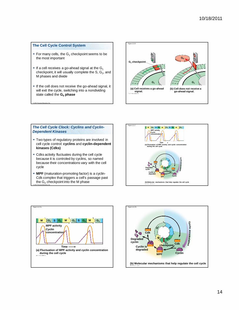
The Cell Cycle Clock: Cyclins and Cyclin-Dependent Kinases
Two types of regulatory proteins are involved in cell cycle control: cyclins and cyclin-dependent kinases (Cdks)
Cdks activity fluctuates during the cell cycle because it is controled by cyclins, so named because their concentrations vary with the cell cycle
MPF (maturation-promoting factor) is a cyclin-Cdk complex that triggers a cell’s passage past the G2 checkpoint into the M phase
© 2011 Pearson Education, Inc.
Figure 12.17
(a) Fluctuation of MPF activity and cyclin concentration during the cell cycle
(b) Molecular mechanisms that help regulate the cell cycle
MPF activityCyclinconcentration
Time
M M MS SG1 G2 G1 G2 G1
Cdk
Degradedcyclin
Cyclin isdegraded
MPF
G2checkpoint Cdk
Cyclin
Figure 12.17a
(a) Fluctuation of MPF activity and cyclin concentration during the cell cycle
MPF activityCyclinconcentration
Time
M M MS SG1 G2 G1 G2 G1
(b) Molecular mechanisms that help regulate the cell cycle
Cdk
Degradedcyclin
Cyclin isdegraded
MPF
G2checkpoint Cdk
Cyclin
Figure 12.17b

10/18/2011
15
Stop and Go Signs: Internal and External Signals at the Checkpoints An example of an internal signal is that
kinetochores not attached to spindle microtubules send a molecular signal that delays anaphase
Some external signals are growth factors, proteins released by certain cells that stimulate other cells to divide
For example, platelet-derived growth factor (PDGF) stimulates the division of human fibroblast cells in culture
© 2011 Pearson Education, Inc.
Cancer Genes
Cancer is often caused by mutations in two types of genes: Proto-Oncogenes
Tumor-Suppressor Genes
24-5
Proto-Oncogenes
Proto-Oncogenes – Part of the DNA that codes for “gas pedal” proteins.
When proto-oncogenes mutate they become cancer-causing genes called oncogenes.
Example of a proto-oncogene = ras gene The ras protein is a G protein that relays a signal from
a growth factor receptor to a cascade of protein kinases, the end result of the cascade is a the synthesis of a protein that stimulates the cell cycle
24-5
Tumor-Suppressor Genes
Tumor-Suppressor Genes – Part of the DNA that codes for the “brake” proteins. Usually recessive
When tumor-suppressor genes mutate their products no longer inhibit the cell cycle nor promote apoptosis.
An example is the p53 gene. This gene codes for a transcription factor that promotes the synthesis of cell cycle-inhibiting proteins. p53 transcription factor promotes the p21 gene, a cell
cycle inhibitor p53 also activates apoptosis genes
24-5
Causes of Cancer - Heredity BRCA1 and BRCA2—mutated tumor
suppressor genes that may cause breast and ovarian cancer.
RB gene—mutated tumor suppressor gene that may cause retinoblastoma tumor.
RET gene—proto-oncogene which predisposes one to thyroid cancer.
24-8

10/18/2011
16
Figure 12.18
A sample of humanconnective tissue iscut up into smallpieces.
Enzymes digestthe extracellularmatrix, resulting ina suspension offree fibroblasts.
Cells are transferred toculture vessels.
Scalpels
Petridish
PDGF is addedto half thevessels.
Without PDGF With PDGF
10 m
1
2
34
Stop and Go Signs: Internal and External Signals at the Checkpoints
A clear example of external signals is density-dependent inhibition, in which crowded cells stop dividing
Most animal cells also exhibit anchorage dependence, in which they must be attached to a substratum in order to divide
Cancer cells exhibit neither density-dependent inhibition nor anchorage dependence
© 2011 Pearson Education, Inc.
Figure 12.19
Anchorage dependence
Density-dependent inhibition
Density-dependent inhibition
(a) Normal mammalian cells (b) Cancer cells20 m 20 m
Loss of Cell Cycle Controls in Cancer Cells
Cancer cells do not respond normally to the body’s control mechanisms
Cancer cells may not need growth factors to grow and divide They may make their own growth factor They may convey a growth factor’s signal without
the presence of the growth factor They may have an abnormal cell cycle control
system
© 2011 Pearson Education, Inc.
A normal cell is converted to a cancerous cell by a process called transformation
Cancer cells that are not eliminated by the immune system form tumors, masses of abnormal cells within otherwise normal tissue
If abnormal cells remain only at the original site, the lump is called a benign tumor
Malignant tumors invade surrounding tissues and can metastasize, exporting cancer cells to other parts of the body, where they may form additional tumors
© 2011 Pearson Education, Inc.
Loss of Cell Cycle Controls in Cancer CellsFigure 12.20
Glandulartissue
Tumor
Lymph vesselBloodvessel
Cancercell
Metastatictumor
A tumor growsfrom a singlecancer cell.
Cancer cells invade neighboringtissue.
Cancer cells spreadthrough lymph andblood vessels to other parts of the body.
Cancer cells may survive and establisha new tumor in another part of the body.
4321

10/18/2011
17
Figure 12.UN05 Important concepts
Know all the vocabulary presented in the lecture
Know the form (condensed vs uncondensed) that DNA is in during cell division vs interphase
Know the roles of microtubules, and their structure
Know the role of microtubule-organizing centers Know the two stages of cell cycle (interphase
and mitotic phase
Important concepts
Know what happens during the stages of interphase: G1 phase, S phase, G2 phase.
Know what happens during each checkpoint
Know the role of microfilaments in cell division of animal cells, the structure of microfilaments
Important concepts
Understand how cytokinesis takes place in animal and plant cells
Know the basics of cell division in prokaryotic cells
Understand the controls on cell cycle including the role of cdks and cyclin, MFP, tumor suppressor genes and proto-oncogenes
Important Concepts for Lab Exam
For Mitosis: Know each stage, the order of the stages, and what happens in each stage.
Know what the end result is of mitosis Know what state the cell and the
chromosomes are in at the beginning and end of mitosis. For example: Are the cells haploid or diploid? Are the chromosomes duplicated, or not duplicated? How many chromosomes are there in the cell? Are they in pairs?
Be able to identify the stages using models and slides