Embed Size (px)
Citation preview
YEASTBOOK
GENE EXPRESSION & METABOLISM
Nutritional Control of Growth and Developmentin YeastJames R. BroachDepartment of Molecular Biology, Princeton University, Princeton, New Jersey 08544
ABSTRACT Availability of key nutrients, such as sugars, amino acids, and nitrogen compounds, dictates the developmental programsand the growth rates of yeast cells. A number of overlapping signaling networks—those centered on Ras/protein kinase A, AMP-activated kinase, and target of rapamycin complex I, for instance—inform cells on nutrient availability and influence the cells’transcriptional, translational, posttranslational, and metabolic profiles as well as their developmental decisions. Here I review ourcurrent understanding of the structures of the networks responsible for assessing the quantity and quality of carbon and nitrogensources. I review how these signaling pathways impinge on transcriptional, metabolic, and developmental programs to optimizesurvival of cells under different environmental conditions. I highlight the profound knowledge we have gained on the structure ofthese signaling networks but also emphasize the limits of our current understanding of the dynamics of these signaling networks.Moreover, the conservation of these pathways has allowed us to extrapolate our finding with yeast to address issues of lifespan, cancermetabolism, and growth control in more complex organisms.
TABLE OF CONTENTS
Abstract 73
Introduction 74
Nutrient Sensing Pathways 75Regulatory networks responsive to carbon sources 75
The Ras/protein kinase A pathway: 75Sch9, a protein kinase B homolog: 78Yak1, a proquiescence kinase: 78SNF1 and the use of alternative carbon sources: 79
Components of SNF1: 79Transcriptional regulation by SNF1: 80Metabolic regulation by SNF1: 82
The HAP2/3/4/5 complex and mitochrondrial biogenesis: 82The Rgt network and glucose transport: 83Protein phosphatase 1: 83
Regulatory networks responsive to nitrogen source 84Nitrogen regulation: 84
Continued
Copyright © 2012 by the Genetics Society of Americadoi: 10.1534/genetics.111.135731Manuscript received October 23, 2011; accepted for publication April 6, 2012Available freely online through the author-supported open access option.Present address: Department of Biochemistry and Molecular Biology, Penn State Hershey College of Medicine, Hershey, PA 17033.Author e-mail: [email protected]
Genetics, Vol. 192, 73–105 September 2012 73
CONTENTS, continued
Growth control: 84Nitrogen catabolite repression: 84Retrograde regulation: 84
Nitrogen regulatory pathways: 85The TORC1 pathway and cellular growth control: 85
Regulation of TORC1: 85Downstream effectors of TORC1: 86
A second nitrogen regulatory pathway: 87
The Response of Cells to Nutrient Availability 88Growth control 88
Ribosome biogenesis: 88RNA polymerase I: 89RNA polymerase II: 89RNA polymerase III: 90
Metabolism: 90Metabolic flux: 90Glucose sparing: 91Metabolic cycles: 91
Stress response: 91Autophagy: 92
Development 93Filamentous growth: 93Quiescence: 94Meiosis: 96
Conclusions and Prospectives 98Key unanswered questions 98
YEAST cells finely tune their growth and behavior inaccordance with available nutrients. They can adjust
their growth rate in response to their nutritional environ-ment by altering the length of their cell cycle over at leasta 10-fold range (Brauer et al. 2008). They can adapt tonutritional depletion by engaging one of a number of alter-native developmental programs depending on the particularnutritional circumstances. These programs can range fromrapid mitotic growth in rich media, to filamentous growthallowing foraging under limiting nutrient conditions, to var-ious distinct quiescent states that reversibly shut down thecell in response to starvation for a single nutrient, to theextreme state of biological stasis following sporulation uponsevere starvation.
In metazoans, in which cells are continuously bathed ina uniform sea of nutrients, regulation of metabolic activity,cell growth, or developmental progression at the cellularlevel is dictated by growth factors, hormones, and modu-lators. For yeast, nutrients supply not only the substrates forgrowth but also the signals for growth. That is, nutrientsserve not only as the resources by which the cell increasesmass and generates energy to propel its biosynthetic activitybut also as the signals dictating the metabolic, transcrip-tional, and developmental programs that optimize survivalunder the particular nutritional state in which the cell finds
itself. Thus, understanding nutrient regulation in yeastrequires understanding the dual role of nutrients as metab-olites and as signaling molecules and appreciating howthose two roles are interconnected.
In this review I describe our current understanding of howthe yeast Saccharomyces responds to the two major classes ofnutrients, carbon and nitrogen. I will focus on the means bywhich yeast cells perceive the amount and quality of theseclasses of nutrients and how they use that information, bothsingly and in combination, to alter their cellular, metabolic,transcriptional, and developmental landscapes. Other chap-ters in this series address the means by which Saccharomycesresponds to other nutrient classes, including phosphate, sulfur,and amino acids. Moreover, other chapters address the meta-bolic flow in the cell as well as the various developmentalprograms yeast can engage. Finally, several excellent reviewshave recently appeared that have addressed glucose-inducedsignaling (Schuller 2003; Johnston and Kim 2005; Santangelo2006), nitrogen regulation (Magasanik and Kaiser 2002; DeVirgilio and Loewith 2006a; De Virgilio and Lowith 2006b),nutrient sensing in fungi (Bahn et al. 2007), and the responseof Saccharomyces to starvation (Smets et al. 2010; De Virgilio2011). Many of the details of topics covered in this chapter,particularly with regard to earlier studies, are elaborated ina recent review (Zaman et al. 2008).
74 J. R. Broach
Nutrient Sensing Pathways
Regulatory networks responsive to carbon sources
Yeast cells grow on a wide variety of compounds as sourcesof energy and as carbon-containing precursors of anabolicmetabolism and biomass accumulation (Johnston and Carlson1992). However, yeast cells consume glucose or fructose inpreference to other mono-, di-, and trisaccharides, such assucrose, raffinose, or trehalose, and prefer any fermentablecarbon source over any source, such as glycerol, ethanol, oracetate, that has to be catabolized by oxidative phosphoryla-tion. This hierarchical pattern of consumption is establishedby allosteric regulation of various key enzymes in glycolysisand gluconeogenesis, described below, and by an extensivetranscriptional regulatory network in which glucose repressestranscription of genes required for initial catabolism of lessfavorable sugars and of genes encoding components of theelectron transport chain and other mitochrondrial proteins.This latter regulatory process precludes metabolism by oxida-tive phosphorylation of any nonfermentable carbon sources inthe presence of glucose.
Glucose repression of mitochrondrial function is the basisof the Crabtree effect, whereby Saccharomyces ferments glu-cose to produce ethanol even under aerobic conditions. TheCrabtree effect distinguishes Saccharomyces from closely re-lated yeasts such as Kluyveromyces, for example, which donot perform aerobic fermentation. Such fermentation fromglucose to ethanol, which yields 2 ATP molecules per mole-cule of glucose, is much less efficient in energy productionthan funneling pyruvate, the primary product of glycolysis,into the tricarboxylic acid cycle, which optimally can yield32 molecules of ATP from each glucose molecule. Aerobicfermentation to ethanol is particularly energetically unfavor-able for Saccharomyces since the subsequent introductioninto the TCA cycle of the ethanol produced by fermentationrequires ATP consumption. Thus, at first glance, aerobic fer-mentation would appear to be maladaptive.
Several explanations have been proposed to accountfor aerobic fermentation in Saccharomyces. One hypothesisholds that Saccharomyces cells, which are relatively resistantto ethanol toxicity, may generate ethanol to defend its nichefrom competing microorganisms in its normal ecological set-ting of rotting fruit (Thomson et al. 2005). A second ex-planation is that growth by fermentation minimizes theproduction of reactive oxygen species that could increaseincorporation of mutagenic errors during DNA replication.This explanation has been invoked to explain the presenceof metabolic cycles by which Saccharomyces cells promotea burst of fermentation and suppress oxidative phosphory-lation during DNA replication even in cells growing on a non-fermentable carbon source (Chen et al. 2007; Silvermanet al. 2010). Finally, the Crabtree effect bears striking re-semblance to the Warburg effect observed in a variety ofcancer cells, a process in which cells consume more glucosethan can be funneled through the tricarboxylic acid (TCA)cycle and shunt the excess metabolized glucose into lactate,
even under aerobic conditions. A recent hypothesis pro-posed to account for the Warburg effect is that this en-ergy-inefficient process may actually be quite efficient inproducing both reducing potential and anabolic precursors,namely acetyl-CoA, required for the biosynthetic capacitynecessary for producing macromolecular components ofa new cell. In this model, aerobic fermentation serves asa means of accelerating rapid growth by facilitating massaccumulation (Vander Heiden et al. 2009). Moreover, themodel suggests that Saccharomyces in rich medium sub-scribes to the same exigencies as cancer cells—the need toproduce as many progeny in as short a period of time aspossible—and aerobic fermentation fulfills that exigency inboth settings. Further studies will be required to determinewhich, if any, of these explanations account for the Crabtreeeffect and its restriction to the Saccharomyces clade of yeastspecies (Pfeiffer et al. 2001).
Reflecting the hierarchical pattern of carbon sourceutilization in yeast, addition of glucose to cells growing ona nonfermentable carbon source results in rapid andsweeping changes in the phosphorylation profile of yeastproteins and the pattern of yeast gene transcripts. Phos-phorylation changes occur on a variety of metabolic and cell-cycle–associated proteins as well as a number of transcrip-tion factors and chromatin modifiers, consistent with meta-bolic, proliferative, and transcriptional reprogramming ofthe cell in response to carbon source changes. More than40% of genes change their transcript levels by more thantwo fold within minutes of a shift of cells from glycerol toglucose (Wang et al. 2004; Kresnowati et al. 2006; Zamanet al. 2009). This transition results in activation of genesrequired for mass accumulation, such as ribosomal proteinand ribosomal biogenesis genes, and repression of genesassociated with stress response or required for use of alter-nate carbon sources. A similarly widespread transcriptionalreprogramming occurs following depletion of glucose incells growing on rich medium and the ensuing transitionto growth on ethanol (Derisi et al. 1997; Young et al.2003; Brauer et al. 2005).
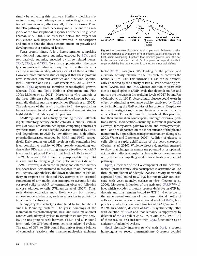
A variety of signaling networks mediate this reprogram-ming of the metabolic, proliferative, and transcriptionalcapacity of cells (Figure 1). Different pathways appear to beassociated with different processes responsive to the qualityand amount of carbon source. For instance, glucose effectson biosynthetic capacity and stress responses are mediatedby the protein kinase A pathway, while repression of genesinvolved in use of alternative carbon sources are mediatedpredominantly by Snf1 and tuning of the glucose uptakemachinery to match glucose levels is effected through theRgt/Snf3 circuit.
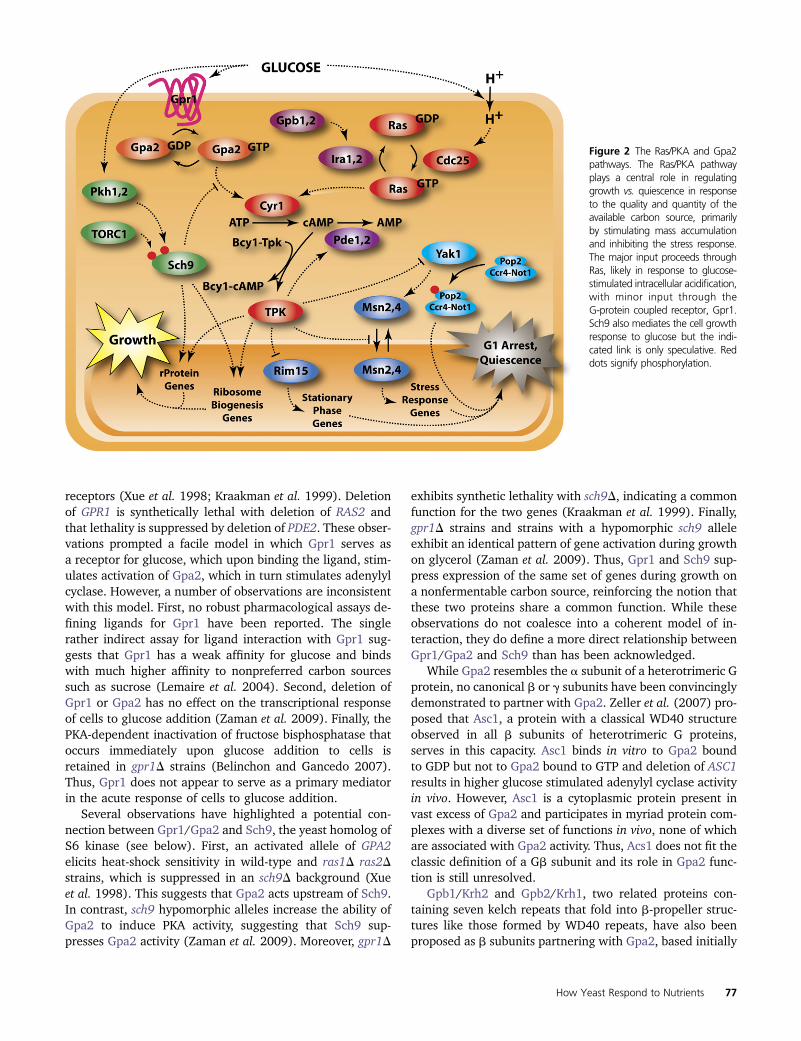
The Ras/protein kinase A pathway: Most of the glucose-induced signaling in yeast cells proceeds through the Ras/protein kinase A (PKA) pathway (Figure 2). Ninety per-cent of the transcriptional changes that occur on additionof glucose- to glycerol-grown cells can be recapitulated
How Yeast Respond to Nutrients 75
simply by activating this pathway. Similarly, blocking sig-naling through the pathway concurrent with glucose addi-tion eliminates most, albeit not all, of the responses. Thus,the PKA pathway is both necessary and sufficient for a ma-jority of the transcriptional responses of the cell to glucose(Zaman et al. 2009). As discussed below, the targets forPKA extend well beyond those involved in transcriptionand indicate that the kinase exerts effects on growth anddevelopment at a variety of levels.
Yeast protein kinase A is a heterotetramer comprisingtwo identical regulatory subunits, encoded by BCY1, andtwo catalytic subunits, encoded by three related genes,TPK1, TPK2, and TPK3. To a first approximation, the cata-lytic subunits are redundant: any one of the three is suffi-cient to maintain viability, whereas loss of all three is lethal.However, more nuanced studies suggest that these proteinshave somewhat different activities and functional specific-ities (Robertson and Fink 1998; Ptacek et al. 2005). For in-stance, Tpk2 appears to stimulate pseudohyphal growth,whereas Tpk3 and Tpk1 inhibit it (Robertson and Fink1998; Malcher et al. 2011). Moreover, in vitro analysis ofthe three different subunits indicated overlapping but sub-stantially distinct substrate specificities (Ptacek et al. 2005).The relevance of the in vitro studies to in vivo specificitieshas not been explored and most genetic and genomic studieshighlight the redundancy of the proteins.
cAMP regulates PKA activity by binding to Bcy1, alleviat-ing its inhibitory activity on the catalytic subunits. Cellularlevels of cAMP are determined by the competing activities ofsynthesis from ATP via adenylyl cyclase, encoded by CYR1,and degradation to AMP by low-affinity and high-affinityphosphodiesterases, encoded by PDE1 and PDE2, respec-tively. Early studies on cAMP levels in cells containing lowlevel constitutive activity of PKA provide compelling evi-dence that PKA exerts a strong negative feedback on cAMPlevels and implicated Pde’s in that feedback (Nikawa et al.1987). Moreover, Pde1 can be phosphorylated by PKAin vitro and following a glucose pulse in vivo (Ma et al.1999). However, a decrease in phosphodiesterase activityhas never been demonstrated in response to an increase inPKA activity. Nonetheless, the down modulation of Pde ac-tivity in response to elevated PKA activity is an essentialcomponent of any model that attempts to account for theobserved spike in cAMP concentration observed followingglucose addition to cells (Williamson et al. 2009). Thus,such down-modulation most likely occurs but througha more subtle mechanism, such as alteration in protein in-teraction or localization.
Adenylyl cyclase activity is stimulated by two families ofsmall GTP-binding proteins. The yeast homologs of themammalian ras protooncogene, Ras1 and Ras2, make directcontact with adenylyl cyclase to stimulate its catalytic activ-ity. The Ras proteins cycle between a GDP- and GTP-boundform; only the GTP-bound form activates adenylyl cyclase.The ratio of GTP- to GDP-bound Ras derives from a balanceof competing reactions: the guanine nucleotide exchange
factor, Cdc25, catalyzes GTP loading of the protein anda GTPase activity intrinsic to the Ras proteins converts thebound GTP to GDP. This intrinsic GTPase can be dramati-cally enhanced by the activity of two GTPase activating pro-teins (GAPs), Ira1 and Ira2. Glucose addition to yeast cellselicits a rapid spike in cAMP levels that depends on Ras andmirrors the increase in intracellular levels of GTP-bound Ras(Colombo et al. 1998). Accordingly, glucose could exert itseffect by stimulating exchange activity catalyzed by Cdc25or by inhibiting the GAP activity of Ira proteins. Despite ex-tensive investigations, the mechanism by which glucoseaffects Ras GTP levels remains unresolved. Ras proteins,like their mammalian counterparts, undergo extensive post-translational modification—including C-terminal proteolyticcleavage, farnesylation, palmitoylation, and carboxymethyla-tion—and are deposited on the inner surface of the plasmamembrane by a specialized transport mechanism (Dong et al.2003; Wang and Deschenes 2006). Addition of glucose tocells elicits a rapid acidification of the yeast cytoplasm(Dechant et al. 2010). While no direct evidence has emergedto show that changes in membrane potential or cytoplasmicacidification affects adenylyl cyclase activity, these are cur-rently the most compelling models for activation of the PKApathway.
Gpa2, a member of the Ga component of the heterotri-meric G-protein family, also participates in activation of PKAthrough stimulation of adenylyl cyclase activity. Bacteriallyexpressed Gpa2 bound to GTPgS but not to GDP can asso-ciate with yeast adenylyl cyclase in vitro (Peeters et al.2006). Moreover, induction of the activated GPA2Q300L al-lele, which encodes a mutant protein defective in GTP hy-drolysis and thus remains bound to GTP in vivo, results inthe same reconfiguration of the transcriptional profile ofcells as does induction of an activated allele of RAS2, bothprofiles of which depend on a functional PKA (Zaman et al.2009). In addition, deletion of GPA2 is synthetically lethalwith deletion of RAS2 and that lethality is suppressed bydeletion of PDE2 (Kubler et al. 1997; Xue et al. 1998). Allof these results are consistent with Gpa2 functioning as anactivator of adenylyl cyclase.
Gpa2 physically interacts in vivo with Gpr1, a proteinhomologous to seven transmembrane G-protein–coupled
Figure 1 An overview of glucose signaling pathways. Different signalingnetworks respond to availability of fermentable sugars and regulate dis-tinct, albeit overlapping, functions that optimize growth under the par-ticular nutrient status of the cell. Sch9 appears to respond directly tosugar availability but the mechanistic connection is not well defined.
76 J. R. Broach
receptors (Xue et al. 1998; Kraakman et al. 1999). Deletionof GPR1 is synthetically lethal with deletion of RAS2 andthat lethality is suppressed by deletion of PDE2. These obser-vations prompted a facile model in which Gpr1 serves asa receptor for glucose, which upon binding the ligand, stim-ulates activation of Gpa2, which in turn stimulates adenylylcyclase. However, a number of observations are inconsistentwith this model. First, no robust pharmacological assays de-fining ligands for Gpr1 have been reported. The singlerather indirect assay for ligand interaction with Gpr1 sug-gests that Gpr1 has a weak affinity for glucose and bindswith much higher affinity to nonpreferred carbon sourcessuch as sucrose (Lemaire et al. 2004). Second, deletion ofGpr1 or Gpa2 has no effect on the transcriptional responseof cells to glucose addition (Zaman et al. 2009). Finally, thePKA-dependent inactivation of fructose bisphosphatase thatoccurs immediately upon glucose addition to cells isretained in gpr1D strains (Belinchon and Gancedo 2007).Thus, Gpr1 does not appear to serve as a primary mediatorin the acute response of cells to glucose addition.
Several observations have highlighted a potential con-nection between Gpr1/Gpa2 and Sch9, the yeast homolog ofS6 kinase (see below). First, an activated allele of GPA2elicits heat-shock sensitivity in wild-type and ras1D ras2Dstrains, which is suppressed in an sch9D background (Xueet al. 1998). This suggests that Gpa2 acts upstream of Sch9.In contrast, sch9 hypomorphic alleles increase the ability ofGpa2 to induce PKA activity, suggesting that Sch9 sup-presses Gpa2 activity (Zaman et al. 2009). Moreover, gpr1D
exhibits synthetic lethality with sch9D, indicating a commonfunction for the two genes (Kraakman et al. 1999). Finally,gpr1D strains and strains with a hypomorphic sch9 alleleexhibit an identical pattern of gene activation during growthon glycerol (Zaman et al. 2009). Thus, Gpr1 and Sch9 sup-press expression of the same set of genes during growth ona nonfermentable carbon source, reinforcing the notion thatthese two proteins share a common function. While theseobservations do not coalesce into a coherent model of in-teraction, they do define a more direct relationship betweenGpr1/Gpa2 and Sch9 than has been acknowledged.
While Gpa2 resembles the a subunit of a heterotrimeric Gprotein, no canonical b or g subunits have been convincinglydemonstrated to partner with Gpa2. Zeller et al. (2007) pro-posed that Asc1, a protein with a classical WD40 structureobserved in all b subunits of heterotrimeric G proteins,serves in this capacity. Asc1 binds in vitro to Gpa2 boundto GDP but not to Gpa2 bound to GTP and deletion of ASC1results in higher glucose stimulated adenylyl cyclase activityin vivo. However, Asc1 is a cytoplasmic protein present invast excess of Gpa2 and participates in myriad protein com-plexes with a diverse set of functions in vivo, none of whichare associated with Gpa2 activity. Thus, Acs1 does not fit theclassic definition of a Gb subunit and its role in Gpa2 func-tion is still unresolved.
Gpb1/Krh2 and Gpb2/Krh1, two related proteins con-taining seven kelch repeats that fold into b-propeller struc-tures like those formed by WD40 repeats, have also beenproposed as b subunits partnering with Gpa2, based initially
Figure 2 The Ras/PKA and Gpa2pathways. The Ras/PKA pathwayplays a central role in regulatinggrowth vs. quiescence in responseto the quality and quantity of theavailable carbon source, primarilyby stimulating mass accumulationand inhibiting the stress response.The major input proceeds throughRas, likely in response to glucose-stimulated intracellular acidification,with minor input through theG-protein coupled receptor, Gpr1.Sch9 also mediates the cell growthresponse to glucose but the indi-cated link is only speculative. Reddots signify phosphorylation.
How Yeast Respond to Nutrients 77
on two-hybrid interaction. However, substantial evidencehas accumulated discounting Gpb1/Gpb2 as b subunits(Peeters et al. 2007), including the fact that the site onGpa2 at which the proteins bind does not correspond tothe classic Gb-binding domain (Niranjan et al. 2007). None-theless, Gpb1 and Gpb2 play redundant roles in negativelyregulating the activity of the Ras/PKA pathway, either byinterference with the Gpr1/Gpa2 interaction (Harashimaand Heitman 2005), or through stabilization of the Ras–GAP proteins, Ira1 and Ira2 (Harashima et al. 2006), orby stabilization of the interaction between the regulatorysubunit, Bcy1, and the catalytic subunits, Tpk1–3, of pro-tein kinase A (Lu and Hirsch 2005; Peeters et al. 2006;Budhwar et al. 2010), or by some combination of all threemechanisms.
One should note that the studies on Gpr1, Gpa2, andGpb1/2 have not examined the dynamic nature of thesecomponents in the context of signal transduction. Rather,these studies exclusively provide a static view of the roleof these proteins in signal output. Thus, we do not knowwhether these components serve a dynamic function in thesignaling cascade or simply function as structural elementsof the signaling machinery, imparting stability to the Ras–GAP proteins or the Bcy1–Tpk interaction, for example.
Sch9, a protein kinase B homolog: SCH9 encodes an AGCfamily protein kinase homologous to the mammalian S6kinase and the prosurvival protein kinase, Akt. It was iden-tified as a high-copy suppressor of strains lacking proteinkinase A. Overexpression of Sch9 results in essentially iden-tical transcriptional reprogramming as does activation ofprotein kinase A, which suggests that the ability of Sch9 tosuppress loss of PKA activity is a consequence of overlappingsubstrate specificities of the two kinases. In fact, their rec-ognition motifs are quite similar and many of the identifiedsubstrates of Sch9 are substrates of PKA, although the set ofsubstrates and the precise phosphorylation sites are notcompletely congruent (Huber et al. 2009; Mok et al.2010). Nonetheless, Sch9 and PKA appear to perform simi-lar functions in the cell by targeting overlapping substrates.
As noted below, Sch9 is activated by direct phosphoryla-tion by TORC1 and, as such, is responsible for many of thechanges in cellular protein phosphorylation elicited byTORC1 (Urban et al. 2007). Glucose also regulates Sch9activity, both by increasing its level in the cell and by in-ducing its phosphorylation (Jorgensen et al. 2004). ThePkh1,2 kinases are activated by sphingolipids and phosphor-ylate Sch9 on its activation loop (Jacinto and Lorberg 2008).In addition, the AMP-activated protein kinase SNF1 phos-phorylates Sch9 and apparently enhances its activity (Luet al. 2011). Whether these or other kinases or phosphatasesmediate glucose activation of Sch9 is not clear.
An acute increase in Sch9 activity substantially recapitu-lates transcriptional responses to glucose addition to cells,suggesting that Sch9 activation is sufficient to elicit the glu-cose transcriptional response (Zaman et al. 2009). However,
inactivation of Sch9 concurrent with glucose additiondoes not diminish glucose-induced transcriptional changes,whereas inactivation of PKA concurrent with glucose addi-tion reduces the magnitude of the transcriptional responseby 75%. This indicates that Sch9 per se is not necessary forthe glucose response, whereas PKA plays a requisite role.Nonetheless, the residual transcriptional response to glucosein the absence of PKA activity depends on Sch9 (Zamanet al. 2009). In short, while Sch9 certainly participates innutrient signaling downstream of TORC1, it also plays a sig-nificant role in glucose regulation of cell growth.
Yak1, a proquiescence kinase: Yak1 is a member of the con-served dual-specificity tyrosine-phosphorylation–regulated pro-tein kinase. It functions in a PKA pathway but it inhibitsrather than stimulates cell proliferation. YAK1 was identi-fied as a loss-of-function suppressor of PKA deficiency andYAK1 overexpression inhibits cell proliferation, suggestingthat it functions downstream of PKA (Garrett et al. 1991).Yak1 localizes to the nucleus following glucose depletionor rapamycin treatment but becomes phosphorylated andlocalized to the cytoplasm following glucose addition tocells (Moriya et al. 2001; Martin et al. 2004). Cytoplasmic14-3-3 proteins bind phosphorylated Yak1 and inhibit itsprotein kinase activity. Thus, while PKA phosphorylationdoes not alter Yak1 kinase activity in vitro, 14-3-3 interac-tion resulting from PKA phosphorylation in vivo reducesits activity (Budovskaya et al. 2005; Ptacek et al. 2005;Lee et al. 2011). Consistently, Yak1 without its PKA phos-phorylation sites accumulates in the nucleus, even in cellsgrown on glucose (Lee et al. 2011). Thus, Yak1 activityis regulated in response to glucose at least in part throughPKA-dependent subcellular localization (Moriya et al.2001).
A major downstream target of Yak1 is Pop2/Caf1, amember of the Ccr4–Caf1–Not1 deadenylation complex thatcontrols the stability and/or translation of a variety of tran-scripts involved in stress response and use of alternativecarbon sources (Moriya et al. 2001). Blocking Yak1 phos-phorylation of Pop2 prevents cells from arresting in G1 atthe end of postdiauxie prior to entry into stationary phase.Yak1 also exhibits genetic interaction with Msi1/Cac3,a high-copy suppressor of hyperactive PKA signaling anda member of the CAK chromatin deposition complex (Prattet al. 2007). Msi1 and Yak1 work in parallel to promotecessation of growth that counteracts the effects of PKA.Yak1 impinges on the stress response pathways by directlyphosphorylating the heat-shock transcription factor, Hsf1,and the major stress response transcription factors, Msn2and Msn4 (Lee et al. 2008). Phosphorylation of Hsf1 byYak1 increases its DNA-binding activity and Yak1 is requiredfor full transcriptional activity of Hsf1. While Yak1 isrequired for full activity of Msn2/4, the absence of Yak1-induced phosphorylation does not affect nuclear localizationof Msn2 in vivo or its DNA binding affinity in vitro. Finally,transcriptional profiling and genetic studies suggest that
78 J. R. Broach
Yak1 inhibits the filamentation-antagonizing transcriptionfactor, Sok2 (Malcher et al. 2011).
In sum, Yak1 appears to function in concert with PKAbut in the opposite direction: PKA promotes cell growthand inhibits the stress response while Yak1 inhibits cellgrowth and stimulates the stress response. Yak1 may ac-complish this by impinging directly on the PKA pathwaythrough phosphorylation of Bcy1 (Griffioen et al. 2001)but more likely through an independent process of activat-ing stress-responsive transcription factors and stabilizing orpromoting the translation of growth-inhibitory, stationary-phase–promoting mRNAs. Moreover, glucose influencesYak1 function through a mechanism at least partially de-pendent on PKA. Thus, Yak1 represents a branch of thePKA pathway by which glucose regulates the growth anddevelopment commitment of the cell.
SNF1 and the use of alternative carbon sources: Thepreferential use of glucose as carbon and energy source byyeast results from glucose-induced transcriptional repres-sion of genes required for catabolism of other sugars as wellas those involved in central carbon metabolism. In addition,glucose causes repression of mitochrondrial function, whichis required for oxidative phosphorylation necessary formetabolism of nonfermentable carbon sources. These pro-cesses are regulated by glucose through the combinedactions of the Snf1 kinase and the Hap regulatory complex.
Components of SNF1: SNF1 was identified as a generequired for glucose repression, for growth on sucrose as solecarbon source, and for induction of invertase in response toglucose depletion. Snf1 is the catalytic subunit and foundingmember of the eukaryotic family of AMP-activated proteinkinases (AMPKs). In mammalian cells, AMPK responds to de-clining energy charge of the cell by stimulating increasedglucose uptake and oxidation, increased fatty acid oxidation,inhibition of anabolic reactions, and stimulation of reactionsthat generate ATP. Thus, AMPK serves as a guardian of en-ergy homeostasis in cells, promoting increased energy pro-duction and reduced energy demand by a multiplicity ofmeans when energy reserves are depleted (Hardie et al.1998, 2011). In yeast, Snf1 kinase performs similar functionsbut may do so in direct response to declining glucose levelsrather than energy charge, reflecting the fact that yeast cellsassess their nutrient sufficiency predominantly through theirperception of glucose rather than their metabolism of it.
Like other members of the AMPK family, SNF1 proteinkinase is a heterotrimer comprising the Snf1 catalytic (a)subunit, a regulatory (g) subunit, Snf4, and one of three b
subunits—Gal83, Sip1 or Sip2—that function as scaffoldand localization determinants. In this review, I will refer tothe complex as SNF1, distinct from the catalytic subunitSnf1 and the gene SNF1. The Snf1 catalytic subunit containsan N-terminal kinase domain and a C-terminal autoinhibi-tory domain. In mammalian AMPKs, binding of AMP to the gsubunit stimulates kinase activity via allosteric alteration ofinteraction of the autoinhibitory domain with the kinase
domain (Chen et al. 2009). Snf4 is required for SNF functionin yeast cells but deletion of the autoinhibitory domain ofSnf1 eliminates the requirement for Snf4 for kinase activityin vivo and in vitro, suggesting that the primary function ofSnf4 is to alleviate Snf1 autoinhibition.
Snf4 consists of two pairs of repeats, termed Batemandomains, which in other proteins bind adenosine deriva-tives. The structure of this domain in Snf4 is quite similarto the Schizosaccharomyces pombe g subunit, which bindsa single molecule of AMP or ATP (Townley and Shapiro2007). Recent results have shown that amino acid substitu-tions within the Bateman domain of Snf4, analogous tosome disease causing activating alleles in the human AMPKg subunit, alleviate to some extent the inhibitory effects ofglucose on SNF1 activity (Momcilovic et al. 2008). Thiswould suggest that allosteric changes in Snf4 resulting fromthese substitutions can result in reduced deactivation of thecatalytic subunit by glucose. However, AMP fails to activateSNF1 in vitro, suggesting that AMP does not bind or stimu-late SNF1 in vivo (Mitchelhill et al. 1994; Woods et al. 1994;Wilson et al. 1996). Rather, ADP binds to Snf4 and, at leastin vitro, protects against dephosphorylation of Thr210 (seebelow) (Mayer et al. 2011). But, since 2-deoxyglucose,which can be phosphorylated by hexokinase but cannot befurther metabolized, inhibits SNF1 activity in vivo, glucosedoes not have to be extensively metabolized to affect SNF1function. Finally, glucose regulates phosphorylation of theSnf1 activation domain in vivo (see below) even in the ab-sence of Snf4 and the Snf1 autoinhibitory domain (Jiangand Carlson 1996; Leech et al. 2003). Thus, while Snf4 isrequired to alleviate autoinhibition of Snf1 and mutations inSnf4 can attenuate glucose inhibition of SNF1, Snf4 does notseem to appreciably regulate SNF1 in response to energycharge in the cell.
Activation of SNF1 kinase activity results from phosphor-ylation of threonine 210 in the activation loop of Snf1. Threekinases—Elm1, Tos3, and Sak1—serve as redundant SNF1-activating kinases (Hong et al. 2003; Nath et al. 2003;Sutherland et al. 2003). In mammalian cells, this functionis performed redundantly by LKB1, Ca2+/calmodulin-dependent kinase (CaCDK) and TGFb-activated kinase,the identities of which were revealed by heterologous com-plementation in yeast (Hong et al. 2005; Momcilovic et al.2006). The action of the three SNF1-activating kinases iscounteracted by the essential protein phosphatase 1, Glc7,in conjunction with its specificity subunit, Reg1 (Tu andCarlson 1995). Low glucose levels correlate with increasedphosphorylation of Thr210 and enhanced SNF1 activity(McCartney and Schmidt 2001). Several results suggest thatglucose does not act through the upstream kinases: SNF1activity exhibits normal regulation in strains in which thethree yeast kinases are functionally replaced by mammalianLKB1. Moreover, the three upstream kinases exhibit thesame activity in extracts of cells grown in glucose-limitedor glucose-replete media (Hong et al. 2005; Rubensteinet al. 2008). Unlike mammalian AMPK, AMP does not
How Yeast Respond to Nutrients 79
stimulate SNF1 in yeast, although ADP binding to Snf4 pro-tects Thr210 from dephosphorylation, at least in vitro(Mayer et al. 2011). Rather, glucose must regulate SNF1activity either by inhibiting one or more of the upstreamkinases, or by activating the Reg1/Glc7 phosphatase, or byrendering Thr210 more accessible to dephosphorylation.Finally, while high glucose levels accelerate the rate ofThr210 dephosphorylation in vivo, Reg1/Glc7 activity in vivoappears unaffected by changes in glucose levels (Rubensteinet al. 2008). Thus, glucose may regulate SNF1 activity bymodifying the accessibility of the complex to the Reg1/Glc7phosphatase, perhaps through reduction in ADP levels orthrough modulation of the interaction between SNF1 andReg1 (Dombek et al. 2004; Rubenstein et al. 2008; vonPlehwe et al. 2009; Mayer et al. 2011).
While Snf1 activation has been studied predominantly inthe context of glucose repression, Snf1 is phosphorylatedand activated in response to a number of environmentalstresses. Alkaline pH, high sodium chloride, or oxidativeagents, but not high sorbitol or heat shock, result in in-creased Thr210 phosphorylation and SNF1 activity as wellas nuclear relocalization (Hong and Carlson 2007). All threeupstream kinases contribute to this stress-induced phosphor-ylation with Sak1 playing the predominant role. However, aswith glucose regulation of SNF1 activity, SNF1 responds tothese stresses even in elm1D sak1D tos3D strains expressingmammalian CaCDK. Thus, activation of SNF1 in responseto stress appears to result from inactivation of Reg1/Glc7phosphatase rather than activation of the upstream kinase.Finally, SNF1 is activated by Thr210 phosphorylation in re-sponse to nitrogen starvation and TORC1 inactivation (seebelow) (Orlova et al. 2006). In this case, phosphorylation issolely dependent on Sak1, suggesting that TORC1 mightregulate this Snf1-activating kinase directly (Orlova et al.2010).
The b subunits all contain domains for binding Snf1 andSnf4 and as such provide a scaffold for assembly of thekinase complex. In addition, both Gal83 and Sip2 containa glycogen binding domain, although Gal83 binds glycogenavidly, while Sip2 does so only weakly. Mutations within theglycogen-binding domain of Gal83 or deletion of the domainalleviate glucose-induced inhibition of SNF1 kinase activityin vivo although elimination of glycogen in the cell does not(Momcilovic et al. 2008). This suggests that this domainmay alter the structure of the complex in a way that allowsglucose-induced inhibition of kinase activity but does notprovide a means for regulation of the complex in responseto glycogen levels.
The b subunits confer distinct functions and subcelllularlocalizations of the SNF1 complex (Schmidt and McCartney2000; Vincent et al. 2001). In glucose grown cells, all threecomplexes reside in the cytoplasm. In limiting glucose,Gal83-containing SNF1 complexes relocate to the nucleus,where they participate in transcriptional activation; Sip1-containing complexes relocate to the vacuolar periphery;and the Sip2-containing complexes remain in the cytoplasm.
In response to alkaline stress, SNF1 relocates to the nucleus,while in response to salt stress, it remains in the cytoplasm.This suggests that regulation of subcellular location maycontribute to the specificity of SNF1 action.
While b subunits usually promote increased SNF1 activitytoward selected substrates, Sip2 appears to function as aninhibitor of SNF1 function, at least in older yeast cells (Ashrafiet al. 2000). SNF1 activity increases in older cells, resultingin diminished replicative aging; sip2 mutants exhibit short-ened replicative lifespan, an effect that is reversed by con-currently deleting SNF1, suggesting that Sip2 inhibits Snf1function in older cells. Recent results demonstrate that Sip2is acetylated in vivo by the NuA4 acetyl transferase complex,a modification that enhances its interaction with Snf1 andincreases replicative lifespan, likely through inhibition ofSNF1 activity (Lu et al. 2011). SNF1 phosphorylates andactivates Sch9, which serves as the critical downstream tar-get in SNF1’s effect on replicative aging in older cells, andacetylated Sip2 diminishes the activity of SNF1 towardSch9. Thus, Sip2 inhibits SNF1, reducing activation ofSch9 and extending replicative lifespan.
Transcriptional regulation by SNF1: Activated SNF1 pro-motes expression of hundreds of genes involved in use ofalternate carbon sources through a variety of transcriptionfactors and promotes repression of a number of genes in-volved in amino acid metabolism through Gcn4 (Figure 3)(Young et al. 2003; Shirra et al. 2008; Zaman et al. 2009).Genes required for metabolism of alternative sugars, such assucrose, galactose, and maltose, respond to Snf1 throughthe Mig1 transcriptional repressor, a C2H2 zinc finger proteinthat binds to a GC-rich consensus sequence (reviewed inSchuller 2003). In cells grown in the absence of glucose,Snf1 phosphorylates Mig1 to inhibit Mig1’s repressor activ-ity. In the presence of glucose, Mig1 becomes dephosphory-lated and localizes to the nucleus repressing expression oftarget genes such as SUC2. Mig1 acts as a repressor in asso-ciation with Hxk2, one of the two yeast hexokinases(Ahuatzi et al. 2004, 2007). Hxk2 forms a complex in vitrowith a SUC2 DNA and Mig1, suggesting that Hxk2 interactswith Mig1 as part of the repressor complex on the SUC2promoter. Moreover, Hxk2 interacts specifically throughthe S311 residue of Mig1, mutation to a nonphosphorylat-able form of which results in constitutive localization ofMig1 to the nucleus and constitutive inhibition of SUC2 ex-pression. Finally, certain mutants of Hxk2 defective in cata-lytic activity retain full corepressor activity and certainmutants defective in repressor activity retain catalytic activ-ity (Pelaez et al. 2010). Thus, Hxk2 participates in regula-tion independently of its metabolic activity.
Snf1 regulates expression of genes involved in ethanolmetabolism and b oxidation of fatty acids through modula-tion of the Adr1 transcription factor (Ratnakumar andYoung 2010). Deletion of ADR1 reduced the expression of�100 genes in cells grown on low glucose (Young et al.2003). This study showed that Adr1 also affected expressionof genes in other functions, such as amino acid transport and
80 J. R. Broach
metabolism, meiosis, and sporulation. However, since only30 genes are tightly bound by Adr1 in cells grown in glu-cose-free media, altered regulation of most genes in an adr1could be the consequence of secondary regulatory or meta-bolic effects (Tachibana et al. 2005; Zaman et al. 2009).
Adr1 is negatively regulated by phosphorylation on ser-ine 230 in glucose-grown cells and activated by dephosphor-ylation of that site in a Snf1-dependent manner in cellsgrown in the absence of glucose (reviewed in Schuller2003). While PKA and CaCDK can phosphorylate this sitein vitro, neither is essential for its phosphorylation in vivo,suggesting that redundant and/or some other kinases servein that capacity (Ratnakumar et al. 2009). Moreover, themechanism by which Snf1 induces dephosphorylation ofS230 is unknown. Adr1 is also under negative regulationof Reg1, as deletion of REG1 increases the protein level ofAdr1 and leads to induction of Adr1-regulated genes, suchas ADH2 (Dombek et al. 2004). The yeast 14-3-3 proteins,Bmh1 and Bmh2, likely act in a pathway parallel to Reg1 toinhibit expression of Adr1-regulated genes. Bmh1 and Bmh2bind to Adr1 phosphorylated on S230 (Parua et al. 2010)and expression of ADH2 under repressed conditions is in-creased in a bmh1 bmh2 strain and even further increased ina reg1 bmh1 bmh2 strain. Thus, Adr1 is sensitive to a numberof glucose-dependent inputs.
Several unrelated transcription factors, including Cat8,Sip4, and Rds2, activate expression of genes required for
gluconeogenesis during growth in the absence of glucoseby binding carbon source response elements (CSRE). De-repression of genes having CSRE motifs is completely abol-ished in cat8 sip4 mutants, suggesting that these twoproteins are the major activators (reviewed in Schuller2003; Turcotte et al. 2010). However, Cat8 and Sip4 donot equally contribute to activation of genes in the absenceof glucose: cat8 cells cannot grow on nonfermentable car-bon sources, whereas sip4 mutants can. This hierarchy isfurther supported by the fact that Sip4 has much stricterrequirement for the consensus CSRE motifs than does Cat8(Roth et al. 2004). Of the 255 genes whose expressionis reduced in the cat8 relative to CAT8 in low glucose me-dia, only 48 are bound by Cat8 in vivo, again suggestinga large contribution of secondary events in microarraystudies. During growth of cells in ethanol, Rds2 binds toa set of CSRE-containing genes distinct from, but partiallyoverlapping with, those bound by Cat8. Rds2 activity asa transcriptional activator is enhanced during growth onnonfermentable carbon sources and correlates with Snf1-dependent hyperphosphorylation. Similarly, Sip4 respondsto glucose starvation through Gal83-mediated phosphory-lation by Snf1 (Vincent and Carlson 1999). CAT8 transcrip-tion is inhibited by Mig1 and activated by Hap2/3/4/5,while Rds2 activates expression of Hap4. Thus, the induc-tion of gluconeogenic, TCA cycle, and glyoxylate shuntgenes in response to glucose limitation involves a complex
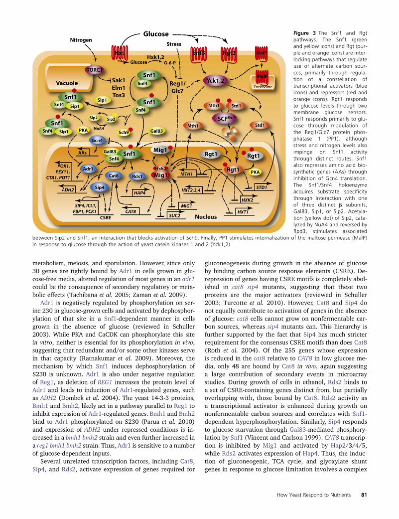
Figure 3 The Snf1 and Rgtpathways. The Snf1 (greenand yellow icons) and Rgt (pur-ple and orange icons) are inter-locking pathways that regulateuse of alternate carbon sour-ces, primarily through regula-tion of a constellation oftranscriptional activators (blueicons) and repressors (red andorange icons). Rgt1 respondsto glucose levels through twomembrane glucose sensors.Snf1 responds primarily to glu-cose through modulation ofthe Reg1/Glc7 protein phos-phatase 1 (PP1), althoughstress and nitrogen levels alsoimpinge on Snf1 activitythrough distinct routes. Snf1also represses amino acid bio-synthetic genes (AAs) throughinhibition of Gcn4 translation.The Snf1/Snf4 holoenzymeacquires substrate specificitythrough interaction with oneof three distinct b subunits,Gal83, Sip1, or Sip2. Acetyla-tion (yellow dot) of Sip2, cata-lyzed by NuA4 and reversed byRpd3, stimulates associated
between Sip2 and Snf1, an interaction that blocks activation of Sch9. Finally, PP1 stimulates internalization of the maltose permease (MalP)in response to glucose through the action of yeast casein kinases 1 and 2 (Yck1,2).
How Yeast Respond to Nutrients 81
interplay of interacting transcription factors downstream ofSNF1.
SNF1 protein kinase complex regulates certain stress re-sponse genes during carbon source downshift. Phosphoryla-tion of Hsf1 and its subsequent binding to heat-shockelements (HSE) and activation of genes in response to car-bon stress, such as HSP82, CUP1, HSP30, and SSA3, dependin part on SNF1 (Sanz 2003; Hahn and Thiele 2004). SNF1also attenuates the Msn2 response to carbon stress. Msn2 isdephosphorylated by Reg1–Glc7 immediately following glu-cose depletion and localizes to the nucleus to induce expres-sion of target genes such as CTT1 (De Wever et al. 2005).However, long-term carbon stress induces rephosphoryla-tion of Msn2 in a SNF1-dependent manner leading to reloc-alization of Msn2 to the cytoplasm and inhibition of CTT1expression (De Wever et al. 2005). This suggests that SNF1is involved in long-term adaptation to carbon stress by neg-atively regulating Msn2 transcriptional activity.
SNF1 also affects gene expression by stimulating chroma-tin remodeling. Glucose depletion yields Snf1-dependentphosphorylation of S10 on histone H3 at the INO1 promoter(Lo et al. 2001, 2005), resulting in recruitment of the SAGAcomplex and acetylation of histone H3 K14. Glucose de-pletion results in a similar Snf1-dependent recruitment ofthe SAGA complex to the HXT2 and HXT4 promoters underglucose limitation (van Oevelen et al. 2006). Moreover,SNF1 phosphorylates Gcn5 in vitro, the histone acetyl trans-ferase component of SAGA, and stimulates its activity(Liu et al. 2010). Thus, Snf1 promotes transcriptional acti-vation through both mobilization of transcription factorsand remodeling of chromatin structure of target promoters.
Finally, SNF1 impinges on the Gcn4 control of amino acidbiosynthesis genes (Ljungdahl and Daignan-Fornier, 2012).In addition to repression of the genes involved in carbonmetabolism noted above, inactivation of Snf1 unexpectedlyresults in induction of dozens of genes involved in aminoacid metabolism regulated by Gcn4 (Shirra et al. 2008;Zaman et al. 2009). This suggests that under glucose-de-pleted conditions, SNF1 inhibits Gcn4 production or tran-scriptional activation. Subsequent studies have indicatedthat SNF1 plays additional roles in activating Gcn4, depend-ing on the condition: under amino-acid-limiting conditionsin the presence of glucose, SNF1 collaborates with un-charged tRNA to activate Gcn2, which ultimately leads toincreased Gcn4 translation through increased phosphoryla-tion of eIF2a. In glucose-limiting conditions, active SNF1inhibits two protein phosphatases responsible for dephos-phorylating eIF2a, Sit4 (see below), and Glc7. This SNF1-promoted increase in eIF2a phosphorylation also results inincreased Gcn4 translation (Cherkasova et al. 2010). Thus,SNF1 appears to both stimulate and inhibit Gcn4, perhapsindicating a subtle interplay between energy homeostasisand amino acid biosynthesis coordinated by Snf1.
Metabolic regulation by SNF1: While most of the studies ofSNF1 have focused on its transcriptional targets, SNF1 alsomodulates energy consumption and generation through
direct regulation of metabolic activity, most notably of lipidbiosynthesis and catabolism. SNF1 directly phosphorylatesand inactivates acetyl coenzyme A (acetyl-CoA) carboxylase(Acc1), the enzyme that catalyzes the rate-limiting step infatty acid biosynthesis, and thus minimize lipid biosynthesisin carbon-limiting conditions (Woods et al. 1994). SNF1promotes fatty acid degradation through b oxidation in partby promoting biogenesis of peroxisomes (Hiltunen et al.2003; Ratnakumar and Young 2010). Whether SNF1 hasan additional direct role in modulating the biochemical ac-tivity of the peroxisome is not known, but free fatty acidsaccumulate in snf1 strains under glucose-limiting conditions,demonstrating the requirement for SNF1 in stimulating b
oxidation to generate energy under nutrient-limited condi-tions (Usaite et al. 2009).
In sum, SNF1 couples the absence of glucose or otherstresses to the suppression of energy-consuming activitiesand the induction of energy-generating processes. This isaccomplished primarily through induction of a limited num-ber of genes required for metabolism of carbon sources otherthan glucose as well as activation of genes required for glu-coneogenesis and fatty acid oxidation. In the absence ofSNF1 function, �400 genes normally induced by glucosedepletion show diminished induction, although only 10%of these are direct targets of transcription factors regulatedby SNF1. In addition, SNF1 likely affects the metabolic fluxin the cell through modulation of the activities of key bio-synthetic and catabolic enzymes, particularly in fatty acidmetabolism. Unlike mammalian cells, yeast cells regulateSnf1 activity not in response to energy charge but ratherthrough phosphorylation of the activation loop catalyzedredundantly by several upstream kinases and counteractedby protein phosphatase 1, albeit recent work has implicatedADP as a potential modulator of SNF1 activity. Current ev-idence supports the conclusion that glucose impinges onSNF1 through modulation of the phosphatase. We still donot understand how glucose alters the activity of the phos-phatase, although glucose has to be phosphorylated, albeitnot metabolized, to affect SNF1 function.
The HAP2/3/4/5 complex and mitochrondrial biogenesis:A number of genes, particularly those involved in respirationand oxidative phosphorylation, are repressed by glucoseindependently of PKA and Snf1. Many of these are regulatedby the Hap2/3/4/5 transcription complex, suggesting thatthe Hap complex may provide an independent route forglucose regulation of gene expression (Zaman et al. 2009).The Hap2/3/4/5 complex plays a central role in convertingcells from fermentative to respiratory growth following thediauxic shift by inducing genes required for mitochondrialfunction upon glucose depletion. Hap2, -3, and -5 forma DNA-binding complex and are constitutively expressed.Hap4 provides the activation domain of the complex andits levels increase upon glucose depletion (Forsburg andGuarente 1989; Derisi et al. 1997). Increased expression ofHap4 alone yields induction of those genes under control of
82 J. R. Broach
the complex (Lascaris et al. 2003). While Hap4 could bea target of Rds2 transcriptional induction in response toSNF1 activation, the fact that induction of Hap complex re-sponsive genes is independent of SNF1 activity suggests anindependent mechanism for Hap complex activation. Thenature of the connection between glucose depletion andHap complex activation remains to be determined.
The Rgt network and glucose transport: The expression ofmany hexose transporter genes (HXTs) is precisely tuned toglucose levels available to cells to insure that the glucosetransporters produced provide the most efficient import ofavailable glucose, over a wide range of external glucoseconcentrations (Kaniak et al. 2004; Zaman et al. 2009). Thistuning is achieved through two intertwined signaling net-works, one mediated by Snf1 and one mediated by Rgt1(Figure 3). Rgt1 is a zinc cluster DNA-binding protein that,in association with corepressors, Mth1 and Std1, repressesHXT gene expression, such as HXT1–4, as well as the hexo-kinase gene, HXK2 (Lakshmanan et al. 2003; Mosley et al.2003). The corepressors, Mth1 and Std1, play partially re-dundant roles in regulation: they each bind to a commonsite on Rgt1 to suppress transcriptional activation and blockaccess to PKA, whose hyperphosphorylation of Rgt1 elicitsits eviction from promoters (Palomino et al. 2006). Rgt re-pression activity is alleviated by binding of external glucoseto two membrane-spanning glucose sensors, Snf3 and Rgt2.These sensors likely detect the relative external-to-internalglucose concentrations (Wu et al. 2006; Karhumaa et al.2010). Glucose activation of the sensors induces functionalrecruitment of Mth1 and Std1 to the plasma membrane,where they are phosphorylated by casein kinases, Yck1and Yck2. Once phosphorylated, the corepressors are tar-geted by the SCFGrr1 E2/E3 ubiquitin-conjugating complexfor degradation by the proteosome (Schmidt et al. 1999;Flick et al. 2003; Moriya and Johnston 2004; Spielewoyet al. 2004). Elimination of these corepressors by proteolysisexposes Rgt1 to phosphorylation and alleviates its repressiveactivity (Palomino et al. 2006).
The repression activity of Rgt1 is stimulated by directphosphorylation by Snf1. In contrast, some of the hexosetransporter genes are repressed by Mig1, whose nuclear lo-calization is blocked by SNF1 phosphorylation. Thus, SNF1both promotes and attenuates repression. Moreover, STD1expression is autoregulated by the Rgt1 network, and thusinduced by high glucose, whereas MTH1 expression is re-pressed at high glucose by the Snf1-regulated Mig1 repres-sor. These observations prompt a model in which Mth1serves primarily to maintain repression, while Std1 func-tions predominantly in establishment of repression duringtransition to the absence of glucose (Kim et al. 2006; Sabinaand Johnston 2009). This complex interplay between thecomponents of the Rgt network and Snf1/Mig1 providesa graded derepression of the different hexose transportersin response to different glucose levels, such that cells ex-press only those transporters with the appropriate affinity
for the available glucose (Johnston and Kim 2005). Albeitquite complex, with both feed-forward and feed-back regu-latory loops, this network is sufficiently well defined to al-low predictive modeling of its behavior both in a steady stateand kinetic representations (Figure 3) (Kuttykrishnan et al.2010).
Protein phosphatase 1: While assignment of direct roles ofprotein phosphatases in various biological processes hasbeen notoriously difficult, growing evidence suggests thatthe Glc7 protein phosphatase 1 plays a central role in glu-cose signaling. Glc7, which encodes the sole and essentialprotein phosphatase 1 in yeast, has little specificity on itsown but associates with a large number of regulatory sub-units that target its activity to different subsets of proteins.One such regulatory subunit, Reg1, binds to Glc7 to promoteglucose repression predominantly through inactivation ofSnf1 by dephosphorylation of its activation loop leading toactivation of Mig1 (Tu and Carlson 1995). Consistent withthis model, deletion of REG1 results in constitutive activa-tion of Snf1 and hyperphosphorylation of its activation loop(McCartney and Schmidt 2001). As noted above, glucosestimulates dephosphorylation of Snf1 either by direct acti-vation of Reg1/Glc7 or by promoting the productive inter-action of Snf1 with Reg1/Glc7.
Glucose induces internalization and degradation ofmaltose permeases through a process that requires Yck1,2-induced phosphorylation of the permeases. Surprisingly,phosphorylation and degradation of the permeases also re-quire Reg1/Glc7 acting upstream of the Yck1,2 kinases: reg1mutants are defective in glucose-induced internalization anddegradation of maltose permeases, a defect that is sup-pressed by overexpression of Yck1 (Gadura et al. 2006).These results are consistent with the idea that Reg1/Glc7enhances Yck1,2 activity, although a mechanistic link is cur-rently lacking. Rgt2 is also required for glucose-inducedmaltose permease turnover: rgt2 mutants exhibit reducedinternalization and an RGT2 constitutive allele induces turn-over even in the absence of glucose (Jiang et al. 1997;Gadura et al. 2006). These observations suggest that glucoseimpinges on maltose permease internalization and degrada-tion through two routes, one by direct binding to Rgt2 andone through activation of Yck1,2 via Reg1/Glc7. Whetherthis second route involves direct activation of Reg1/Glc7by glucose has not been established but is consistent withthe observations.
Msn2 and Msn4, the major stress-responsive transcriptionfactors, are regulated predominantly through their nuclearlocalization as a result of phosphorylation of a nuclear lo-calization site (NLS) on the proteins (Gorner et al. 2002).Phosphorylation of this domain, catalyzed by PKA, restrictsthe proteins to the cytoplasm while dephosphorylation ofthe domain, catalyzed by Glc7, renders the site functionaland promotes nuclear entry and activation of stress-responsivegenes (Gorner et al. 1998, 2002). Dephosphorylation of theNLS occurs much too quickly upon glucose downshift to be
How Yeast Respond to Nutrients 83
explained solely as an inhibition of PKA activity (De Weveret al. 2005). Rather, the kinetics suggest that glucose deple-tion induces Msn2 nuclear localization through activationof Glc7. Neither deletion of Reg1 nor of Bub14, another reg-ulatory subunit of Glc7 implicated in activation of Msn2 upondiauxic shift (Lenssen et al. 2005), alleviated the glucose-depletion–induced nuclear localization of Msn2 (De Weveret al. 2005). Accordingly, some as yet unidentified specificitysubunit likely mediates the effects of glucose depletion onactivation of Glc7 toward Msn2.
There are certainly other glucose-regulated processes,such as glycogen and trehalose accumulation and, as notedabove, eIF2a phosphorylation, in which protein phosphataseplays a role, although whether as a direct conduit of theglucose signal or as a foil to glucose-regulated kinasesremains to be determined.
Regulatory networks responsive to nitrogen source
Nitrogen regulation: Growth control: Yeast cells recognize thenature and availability of nitrogen compounds and activelyadjust their transcriptional, metabolic, and biosynthetic capa-bilities to match that perception. When nitrogen is limiting, cellsslow their growth, primarily through reduction in ribosomalbiogenesis and translation, resulting in expansion of the G1phase of the cell cycle (Brauer et al. 2008). In the extreme caseof nitrogen depletion, cells cease growing, even with all othernutrients available in excess, and enter a nitrogen-specific qui-escent state (Klosinska et al. 2011). Unlike auxotrophic cellsstarved for their required amino acid, such quiescent cells retainviability for an extended period of time and suppress catabolismin a way that prevents consumption of ambient glucose in themedium (Brauer et al. 2008). Thus, yeast cells couple theirsynthetic capacity and growth rate to the quality and amountof available metabolizable nitrogen.
Nitrogen catabolite repression: While yeast cells can usea variety of nitrogen-containing compounds as sole nitrogensource, they exhibit a hierarchical preference for thosesources. Most laboratory strains prefer glutamine or ammo-nia but will use other nitrogen sources, albeit with a reducedgrowth rate. Moreover, yeast exhibit nitrogen cataboliterepression (NCR) in which preferred nitrogen sourcesrepress expression of genes required for uptake and catab-olism of less preferred nitrogen sources (Magasanik andKaiser 2002). Nitrogen catabolite repression is further man-ifest by post-translational regulation of the spectrum ofamino acid permeases residing in the plasma membrane,such that the high capacity general amino acid permease,Gap1, is maintained at the cell surface only under poornitrogen conditions (Magasanik and Kaiser 2002). Finally,availability of a readily metabolizable nitrogen source sup-presses the process of autophagy, by which the cell deliverscytoplasmic macromolecular components to the vacuole forproteolytic recycling of the component parts (Yang andKlionsky 2009). Thus, nitrogen accessibility regulates me-tabolism, growth, transcription, post-transcriptional proteinsorting, and protein turnover in yeast.
The addition of glutamine or ammonia to cells growingon a poor nitrogen source results in a number of transcrip-tional changes, including induction of genes required forgrowth and repression of genes for use of poorly metabo-lized nitrogen sources. This latter category comprises the�90 genes subject to NCR, which are regulated by an in-terplay of four GATA family zinc-finger transcription factors:two transcriptional activators, Gln3 and Gat1 (Nil1 andMep80), and two repressors, Dal80 and Gzf3 (Deh1 andNil2) (Cooper 2002; Magasanik and Kaiser 2002; Scherenset al. 2006). Cells regulate NCR genes primarily by modu-lating subcellular localization of the transcriptional activa-tors: during growth on poor nitrogen sources, Gln3 andGat1 localize to the nucleus where they bind to GATAsequences in promoters of NCR genes, while during growthon ammonium or glutamine, the transcription factors residein the cytoplasm. Ure2 serves as an anchor to sequester Gln3in the cytoplasm: Gln3 resides in the nucleus and fully acti-vates NCR transcription in a ure2 mutant, regardless of ni-trogen source. This observation demonstrates not only thatUre2 serves as a cytoplasmic anchor for Gln3 but also thatnitrogen deprivation acts on Gln3 solely to liberate it fromsequestration by Ure2. Gat1 does not localize to the nucleusin a ure2 mutant. This suggests that a separate as yet un-identified protein may anchor Gat1 in the cytoplasm in cellsgrown on glutamine or ammonia or that Gat1 phosphoryla-tion directly regulates its interaction with the nuclear importmachinery.
Retrograde regulation: Yeast cells assimilate nitrogen fromsources other than glutamate and glutamine by convertingthem to ammonium and then condensing the ammonia witha-ketoglutarate to form glutamate. a-ketoglutarate can begenerated from pyruvate and acetyl-CoA by an anapleuoticpathway catalyzed by the first three enzymes of the citricacid cycle. However, since genes of the citric acid cycle arerepressed during growth on glucose, genes encoding en-zymes of this portion of the citric acid cycle are specificallyupregulated during growth on certain poor nitrogen sourcesby activators of the RTG pathway, which responds bothto mitochondrial dysfunction and to growth on nitrogensources requiring a-ketoglutarate for assimilation (Liuand Butow 1999). In this way, the RTG pathway providesa means of ammonium assimilation from poor nitrogensources and a source of glutamate in the absence of mito-chrondrial function (Figure 4).
The RTG regulatory pathway consists of four positiveregulators—Rtg1, Rtg3, Rtg2, and Grr1 and four negativeregulators—Mks1, Bmh1, Bmh2, and Lst8 (Liu and Butow2006). Rtg1 and Rtg3 form a heterodimeric transcriptionalactivator whose nuclear localization is regulated by theother components of the pathway in response to mitochron-drial integrity and nitrogen availability. When mitochrondriaare functional and sufficient nitrogen is available, the tran-scription factors are cytoplasmic; disruption of mitochron-drial function or nitrogen depletion results in nuclearlocalization of the factors and subsequent transcriptional
84 J. R. Broach
activation of target genes. Regulation of the nuclear/cytoplasmictrafficking of Rtg1/Rtg3 involves complex interactionsamong Mks1, Rtg2, and Bmh1/2 (Dilova et al. 2004). Whenphosphorylated, Mks1 complexes with Bmh1/2 to form ananchor that sequesters Rtg3/Rtg1 in the cytoplasm. Rtg2can compete for Bmh1/2 binding to Mks1 and thereby re-lieve the cytoplasmic sequestration and promote nuclear en-try and transcriptional activation by Rtg1/3. Release ofMks1 from Bmh1/2 is associated with reduced phosphory-lation of Mks1. Grr1, the SCF-targeting subunit, promotesubiquitination and subsequent degradation of Mks1, pro-viding a long-term modulation of the pathway, while Lst8,a subunit of the TOR complexes, renders the RTG pathwaysensitive to Tor inhibition (Figure 4).
Nitrogen regulatory pathways: At least two pathwaysmediate the response of yeast cells to nitrogen availability.The rapamycin-sensitive TORC1 complex, universally con-served among eukaryotic cells, is the central mediator andcoordinator of physiological responses of the cell tochanges in nitrogen source and availability (De Virgilioand Loewith 2006a). The yeast TOR complex I (TORC1)comprises a phosphatidylinsotiol kinase-related protein ki-nase, Tor1 (or in its absence, Tor2), Kog1 (homolog ofmammalian raptor), Lst8, and Tco89 and exerts its biolog-ical function as a protein kinase. In yeast cells, TORC1responds predominantly to nitrogen availability, likelysensed as the level of intracellular amino acids. The pri-mary evidence positing a central role for TORC1 in nitro-gen signaling is the strong correlation in the responses of
cells to nitrogen starvation and the responses of cells torapamycin addition, which specifically inhibits TORC1 ac-tivity (Cardenas et al. 1999; Bertram et al. 2000; Shamjiet al. 2000). However, the fact that rapamycin additiondoes not fully phenocopy nitrogen depletion, particularlywith regard to retrograde transcription and nitrogen catab-olite repression (Tate and Cooper 2003; Tate et al. 2009,2010), demands the existence of at least one other nitro-gen signaling pathway. Neither the constituents nor thestructure of that pathway has been defined.
The TORC1 pathway and cellular growth control: Regula-tion of TORC1: In mammalian cells, TORC1 provides a nexusfor integrating energy charge, growth factor signaling,amino acid availability, and other nutritional inputs. Signal-ing pathways for energy charge and growth factors impingeon TORC1 through the heterodimeric Tsc1/2 tubularsclerosis complex, which stimulates the GTPase activity ofthe Rheb small G protein, whose binding to TORC1 in itsGTP-bound state is necessary for TORC1 kinase activity(Sarbassov et al. 2005). However, stimulation of mammalianTORC1 by amino acids occurs independently of Tsc1/2 andis mediated instead by a heterodimer of two small GTP-binding protein, consisting of either RagA or RagB andeither RagC or RagD. The Rag complex, activated by thepresence of amino acids, promotes relocalization of TORC1from discrete cytoplasmic sites to a late endosomal or lyso-somal compartment at which Rheb resides (Sancak et al.2010). Thus, amino acid availability regulates mammalianTORC1 in a manner distinct from other inputs.
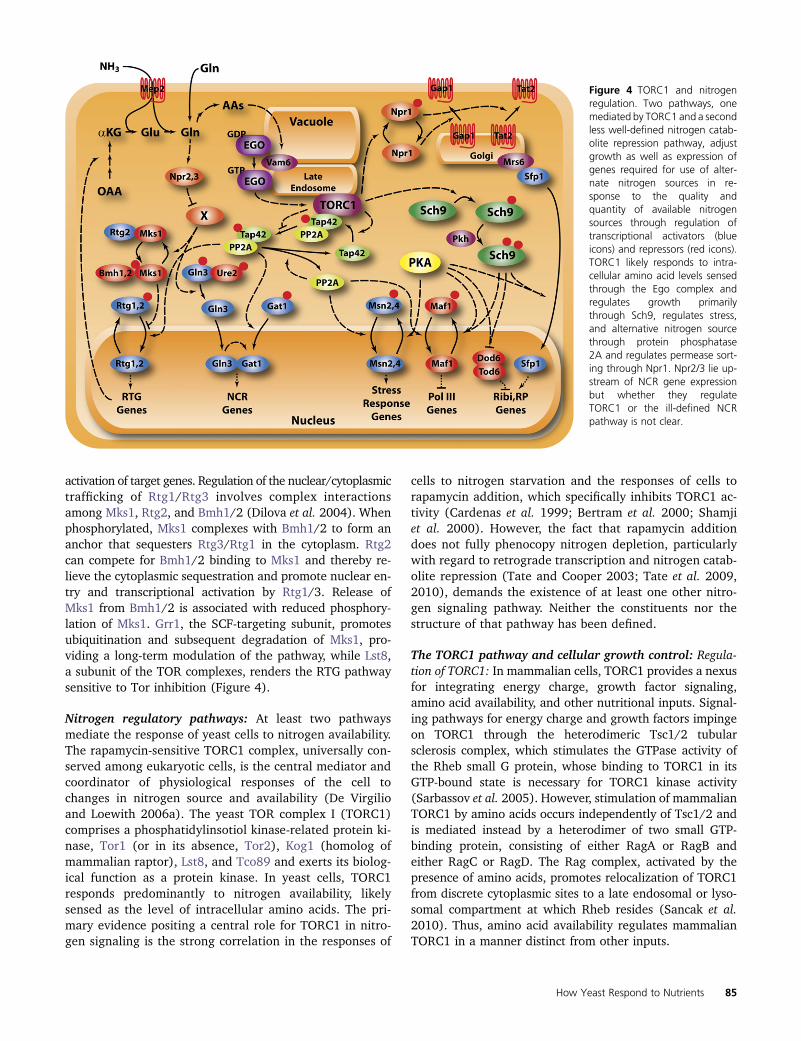
Figure 4 TORC1 and nitrogenregulation. Two pathways, onemediated by TORC1 and a secondless well-defined nitrogen catab-olite repression pathway, adjustgrowth as well as expression ofgenes required for use of alter-nate nitrogen sources in re-sponse to the quality andquantity of available nitrogensources through regulation oftranscriptional activators (blueicons) and repressors (red icons).TORC1 likely responds to intra-cellular amino acid levels sensedthrough the Ego complex andregulates growth primarilythrough Sch9, regulates stress,and alternative nitrogen sourcethrough protein phosphatase2A and regulates permease sort-ing through Npr1. Npr2/3 lie up-stream of NCR gene expressionbut whether they regulateTORC1 or the ill-defined NCRpathway is not clear.
How Yeast Respond to Nutrients 85
The yeast TORC1 responds primarily to the quality andamount of nitrogen in the environment (Figure 4). DeceasedTORC1 activity occurs upon nitrogen starvation or down-shift and increased activity results from nitrogen sourceupshifts or from cycloheximide treatment, which causes anincrease in intracellular amino acids as a result of dimin-ished protein synthesis (Binda et al. 2009). Previous resultshave suggested that the quantity and quality of nitrogensource is perceived as the level of intracellular glutamine:mutations in GLN1 that result in a partially active glutaminesynthetase elicit transcriptional patterns similar to thoseobtained by inhibition of TORC1 (Magasanik and Kaiser2002). Similarly, treatment of cells with the glutamine syn-thetase-specific inhibitor, methionine sulfoximine, yieldsresponses similar to those following treatment of cells withrapamycin (Crespo et al. 2002). However, more detailedanalysis indicates that for several responses, such as Gln3or Gat1 localization under certain conditions (see below),inhibition of glutamine synthetase has the opposite effect ofthat of rapamycin treatment (Tate et al. 2010). The likelyconclusion is that glutamine levels provide input to the ni-trogen catabolite repression pathway described above,which functions in parallel with TORC1 to effect overlap-ping downstream responses. Thus, the actual intracellularsignal for TORC1 remains undefined but may be, as withmammalian cells, intracellular amino acid levels (Figure 4).
Saccharomyces cerevisiae regulates TORC1 using only aportion of the machinery used by mammalian cells. Saccha-romyces does not encode homologs of Tsc1 or Tsc2 and itsRheb homolog is not involved in regulating TORC1. Theabsence of these regulatory elements may reflect eliminationin yeast of input to TORC1 from growth factor receptors orAMP kinase. However, yeast TORC1 does respond to aminoacid levels and the Rag family of GTP-binding proteins areretained in yeast and appear to help couple TORC1 activityto nitrogen quality and quantity, as reflected by amino acidavailability.
Gtr1 and Gtr2 are yeast orthologs of RagA/B and RagC/D,respectively. These proteins, along with Meh1/Ego1 andSlm4/Ego3, form the EGO complex, which is required formicroautophagy and recovery of cells from treatment withrapamycin (Dubouloz et al. 2005). Recovery from rapamycintreatment requires Gtr1 to be bound to GTP and Gtr2 to bebound to GDP, suggesting that as is the case with the mam-malian Rag orthologs, the specific nucleotide binding states ofGtr1 and Gtr2 dictates function of the complex in which itacts (Binda et al. 2009). Moreover, Gtr1 locked in the GTP-bound state stimulates TORC1 in vivo and blocks the ability ofcells to grow on poor nitrogen sources, which requires re-duced TORC1 activity. Both genetic and biochemical evidenceindicates that Gtr1, particularly when bound to GTP, physi-cally interacts with the TORC1 components, Tco89 and Kog1,and this interaction is diminished under leucine starvation.The nucleotide binding status of Gtr1 is regulated by theVam6 guanine nucleotide exchange factor, which is a compo-nent of the homotypic fusion and vacuole protein sorting
complex in which it promotes nucleotide exchange of Ypt7,the yeast homology of mammalian Rab-7. Consistent with thebiochemical role of Vam6 in Gtr1 function, vam6mutants aredefective in recovery from rapamycin treatment and exhibitreduced TORC1 activity.
Components of the TORC1 complex as well as Gtr1 andVam6 localize predominantly to the vacuolar membrane, tothe late endosome and to the intersection of those two struc-tures. These positions remain the same regardless ofwhether cells are growing in nitrogen-replete medium orunder leucine or nitrogen starvation. In sum, the EGO com-plex possesses many of the characteristics of machinery cou-pling amino acid levels in the cell to TORC1 activity andshare many properties with the mammalian Rag complex.However, unlike the mammalian complex, regulation is noteffected by EGO-dependent relocalization of TORC1 toa subcellular activation region. Rather, EGO appears to cou-ple amino acid levels directly to TORC1 activity. The local-ization of the TORC1 and EGO complex to the vacuole raisesthe possibility that the key upstream signal for TORC1involves mobilization of amino acids from their stores inthe vacuole.
While the above model seems to account for upstreamregulation of TORC1, it is likely incomplete, since gtr1 de-letion strains are not defective in several TORC1-dependentcellular responses, such as transcriptional activation of ni-trogen catabolite repression genes or phosphorylation con-trol of Npr1, a protein kinase that regulates plasmamembrane sorting of amino acid permeases. One alternativepathway involves a direct interaction of the cell wall integ-rity pathway component, Rho1, with TORC1, inducing re-lease of Tap42 (see below) in response to various stresses,including nitrogen downshift (Yan et al. 2012). Anothercandidate for upstream regulation is the conserved Npr2/Npr3 complex, identified as mutants in yeast defective ininduction of DAL80, a gene subject to nitrogen cataboliterepression, specifically in response to nitrogen starvation(Neklesa and Davis 2009). Mutations of NPR2 or NPR3 aredefective in nuclear localization of the NCR transcriptionfactors Gat1 and Gln3 and retain Npr1 in a highly phosphor-ylated state in response to nitrogen starvation. These phe-notypes are consistent with a model in which the Npr2/Npr3complex inhibits TORC1 and that inhibition is alleviated bynitrogen availability, perhaps as monitored by intracellularamino acid levels. However, a direct physical link betweenNpr2/Npr3 and TORC1 has not been established. Given thelikely existence of a second pathway working in parallel toTORC1 to effect nitrogen catabolite repression, it is not clearwhether the Npr2/3 complex acts on TORC1 or on thisalternative pathway.
Downstream effectors of TORC1: Two distinct effectors—Sch9, the protein kinase B homolog discussed above, andprotein phosphatase 2A—function as intermediaries be-tween TORC1 activity and the various downstream cellularcomponents that affect growth, metabolism, and development.The TORC1 connection to Sch9 is relatively straightforward:
86 J. R. Broach
TORC1 directly phosphorylates Sch9 and that phosphorylationstimulates the protein kinase activity of Sch9 (Urban et al.2007). Thus, although Sch9 requires activation by addi-tional upstream protein kinases (see above) that perhapsprovide input on other environmental conditions, TORC1and Sch9 function as a kinase cascade connecting growthpromotion to nitrogen status.
The mechanism by which PP2A transmits TORC1 activitystatus is less clear. TORC1 phosphorylates the essentialprotein Tap42, which, in its phosphorylated state forms het-erodimers with the protein phosphatase 2A catalytic sub-unit, encoded redundantly by PPH21 and PPH22, and withthe protein phosphatase 2A-like catalytic subunit, Sit4 (DiComo and Arndt 1996; Jiang and Broach 1999; Duvel et al.2003). Pph21/22 separately forms a heterotrimeric complexwith a scaffolding subunit, Tpd3, and one of two regulatorysubunits, Cdc55 or Rts1, which impart different substratespecificities to the complex. Similarly, Sit4 also forms a het-erodimer with one of three regulatory subunits, Sap155,Sap185, or Sap190 (Luke et al. 1996). Given the vast excessof Pph21/Pph22 and Sit4 relative to Tap42, all of thesecomplexes likely exist concurrently within the cell. Thus,Tap42 most likely acts to direct protein phosphatase activityto specific targets rather than simply to inhibit phosphataseactivity. The Tap42 interacting protein Tip41 collaborateswith Tap42 in executing the phosphatase-mediated down-stream functions of TORC1 (Jacinto et al. 2001; Santhanamet al. 2004; Kuepfer et al. 2007).
Analysis of biochemical studies has prompted the follow-ing working model for the role of phosphatases in TORC1signaling (see Figure 4) (Kuepfer et al. 2007; Tate et al.2009). Active TORC1 phosphorylates and binds Tap42 incomplexes with Pph21/22 and Sit4 at the endosomal/vacu-olar membrane (Yan et al. 2006). In the TORC1-boundstate, these complexes remain essentially inactive due totheir spatial restriction. Upon starvation or treatment withrapamycin, the complexes are released in the cytoplasmwhere Tap42/Tip41 directs the phosphatase activities tovarious downstream substrates, such as Gat1 and Mks1.The intrinsic phosphatase activity of the complexes, or otherphosphatases in the cytoplasm, results in dephosphorylationof Tap42 and dissociation of the complexes with time,resulting in a self-limiting signal elicited following inactiva-tion of TORC1.
Tap42 appears to function as a specificity factor for thecatalytic phosphatase subunits, directing the phosphatasesto certain substrates. For instance, rapamycin induces a Sit4-and Pph21/22-dependent dephosphorylation of the tran-scription factor Gln3 and Gat1 and subsequent translocationof the factors to the nucleus, where they induce transcrip-tion of NCR target genes (Beck and Hall 1999; Cardenaset al. 1999; Tate et al. 2009). Inactivation of Tap42 has noeffect on NCR gene expression under normal growth con-ditions but significantly attenuates induction of these genesby rapamycin (Duvel et al. 2003). These results suggest thatTap42 is required for dephosphorylation of Gln3 and Gat1
following rapamycin treatment, an event catalyzed by Sit4and Pph21/22 (Beck and Hall 1999; Tate et al. 2009). Thus,Tap42 acts in concert with phosphatase catalytic subunits todephosphorylate downstream targets in response to rapamy-cin treatment, placing Tap42 as a positive regulator of phos-phatase activity. Tap42 plays a similar role in rapamycininduction of RTG target genes (Duvel et al. 2003).
Phosphoproteomic studies have highlighted the bifurca-tion of signaling from TORC1 through Sch9 on one branchand Tap42/phosphatases on the other (Huber et al. 2009).In particular, this study examined the changes in phosphor-ylation following rapamycin treatment of a large number ofproteins and identified changes dependent on Sch9 orTap42. While some proteins exhibited rapamycin-inducedchanges that were dependent on both Sch9 and Tap42,many proteins exhibited phosphorylation changes depen-dent only on one or the other activity, highlighting the in-dependence of the two downstream pathways. As notedbelow, the primary targets of Sch9-mediated TORC1 phos-phorylation are those proteins involved in regulation of massaccumulation, including transcriptional regulators of ribo-some biogenesis and ribosomal protein genes, rRNA expres-sion and tRNA synthesis. Finally, some rapamycin-inducedchanges in phosphorylation occurred independently of Sch9and Tap42, suggesting either a limit in the sensitivity of theanalysis or the existence of other pathways emanating fromTORC1 (Breitkreutz et al. 2010).
A second nitrogen regulatory pathway: A variety of ob-servations, primarily from the Cooper laboratory, have pro-vided strong evidence that nitrogen-source regulation ofNCR and RTG genes does not proceed solely through theTORC1 pathway. For instance, induction by rapamycin of theRTG responsive gene, CIT2, is nitrogen-source dependent,occurring in ammonia or glutamine but not proline or gluta-mate grown cells (Tate and Cooper 2003). In addition, thepattern of Gln3 phosphorylation differs in rapamycin-treatedvs. nitrogen starved or methionine sulfoximine treated cells,indicating that nitrogen deprivation and rapamycin impingeon Gln3 phosphorylation status in different ways (Tate et al.2009). Finally, Gln3 and Gat1 both regulate NCR genes butGln3 nuclear localization occurs in response predominantlyto nitrogen limitation or methionine sulfoximine treatmentrather than rapamycin treatment, whereas Gat1 nuclear lo-calization occurs in response predominantly to rapamycintreatment and is immune to nitrogen starvation or methio-nine sulfoximine treatment (Tate et al. 2010). These obser-vations suggest that nitrogen availability does not regulatethe TORC1 activity with regard to nitrogen response so muchas provides a permissive state in which TORC1 may or maynot influence nitrogen catabolite repression. For instance,Gat1 activation depends on the presence of Gln3 but Gln3can promote transcriptional activation on its own (Georiset al. 2009). This suggests that this alternative nitrogen re-sponse pathway plays the predominant role in NCR and RTGregulation and that TORC1 inhibition can reinforce that
How Yeast Respond to Nutrients 87
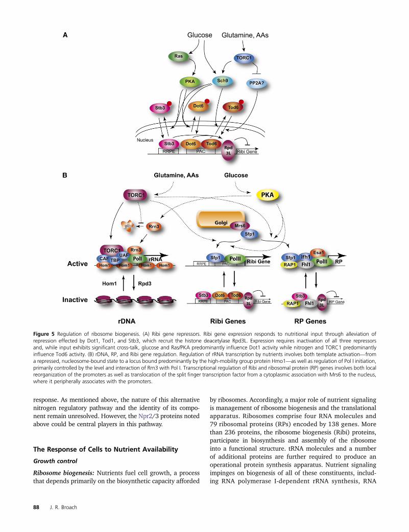
response. As mentioned above, the nature of this alternativenitrogen regulatory pathway and the identity of its compo-nent remain unresolved. However, the Npr2/3 proteins notedabove could be central players in this pathway.
The Response of Cells to Nutrient Availability
Growth control
Ribosome biogenesis: Nutrients fuel cell growth, a processthat depends primarily on the biosynthetic capacity afforded
by ribosomes. Accordingly, a major role of nutrient signalingis management of ribosome biogenesis and the translationalapparatus. Ribosomes comprise four RNA molecules and79 ribosomal proteins (RPs) encoded by 138 genes. Morethan 236 proteins, the ribosome biogenesis (Ribi) proteins,participate in biosynthesis and assembly of the ribosomeinto a functional structure. tRNA molecules and a numberof additional proteins are further required to produce anoperational protein synthesis apparatus. Nutrient signalingimpinges on biogenesis of all of these constituents, includ-ing RNA polymerase I-dependent rRNA synthesis, RNA
Figure 5 Regulation of ribosome biogenesis. (A) Ribi gene repressors. Ribi gene expression responds to nutritional input through alleviation ofrepression effected by Dot1, Tod1, and Stb3, which recruit the histone deacetylase Rpd3L. Expression requires inactivation of all three repressorsand, while input exhibits significant cross-talk, glucose and Ras/PKA predominantly influence Dot1 activity while nitrogen and TORC1 predominantlyinfluence Tod6 activity. (B) rDNA, RP, and Ribi gene regulation. Regulation of rRNA transcription by nutrients involves both template activation—froma repressed, nucleosome-bound state to a locus bound predominantly by the high-mobility group protein Hmo1—as well as regulation of Pol I initiation,primarily controlled by the level and interaction of Rrn3 with Pol I. Transcriptional regulation of Ribi and ribosomal protein (RP) genes involves both localreorganization of the promoters as well as translocation of the split finger transcription factor from a cytoplasmic association with Mrs6 to the nucleus,where it peripherally associates with the promoters.
88 J. R. Broach
polymerase II-dependent RP, and Ribi protein productionand RNA polymerase III-dependent synthesis of tRNAs andthe 5S ribosomal RNA (Figure 5).
RNA polymerase I: Nutrient availability regulates the rateof rRNA production. Three of the rRNAs, 25S, 18S, and5.8S, are processed from a single 35S transcript encoded in150–200 tandem repeats of the 9.1-kb rDNA gene (Johnstonet al. 1997), which also encodes the RNA polymerase IIItranscribed 5S RNA. Four general transcription factor com-plexes promote transcription initiation of the 35S gene byPol I: the upstream activation factor (UAF), core factor (CF),TATA binding protein (TBP, encoded by SPT15), and themonomeric factor Rrn3, which forms a complex with Pol I.In the current model for Pol I initiation, UAF recruits CF andTBP to the Pol I promoter, which together provide a platformfor transcriptional initiation by the Rrn3–Pol I complex(Keys et al. 1996; Goetze et al. 2010). The level of theinitiation competent Rrn3–Pol I complex likely serves asa key regulatory process affecting the rate of rDNA synthesisin response to nutrient availability (Grummt 2003; Claypoolet al. 2004; Mayer et al. 2004; Philippi et al. 2010). Specif-ically, glucose depletion decreases synthesis of Rrn3 whileTORC1 inhibition stimulates its proteolytic degradation anddestabilizes the Rrn3–Pol I complex. Li et al. (2006) showedthat Tor1 directly binds to the 35S rDNA promoter in a nu-trient- and rapamycin-sensitive manner and that this physi-cal interaction is necessary for 35S rRNA synthesis. Finally,artificially stabilizing the Rrn3–Pol I complex by physicallytethering Rrn3 to the polymerase renders rDNA expressionresistant to repression by nutritional downshift or rapamycintreatment (Laferte et al. 2006). In sum, these observationsdemonstrate that Pol I activity responds to nutritional levelsvia modulation of its interaction with Rrn3 (Figure 5).
Nutrient signaling also regulates RNA Pol I-dependentrRNA production at the level of template availability (Figure5). Even under nutrient-rich conditions and rapid growth,the rDNA repeats in each cell exist in two different chroma-tin states, with approximately half in a transcriptionallyactive “open” configuration and the others in a transcription-ally inactive “closed” configuration (Conconi et al. 1989;Dammann et al. 1993). Following transition into stationaryphase, the number of open repeats decreases, a process de-pendent on the histone deacetylase, Rpd3 (Sandmeier et al.2002). Transcriptionally active repeats are essentially de-void of nucleosomes and instead are coated with the HMGbox protein, Hmo1, while the inactive repeats are packagedin canonical nucleosomes (Merz et al. 2008; Wittner et al.2011). The proportion of active vs. inactive repeats appearsto be established by a dynamic equilibrium reset at each cellcycle. DNA replication converts most repeats into the closed,nucleosome-packaged state, a subset of which are reacti-vated shortly after S phase by Pol I-dependent transcription,which evicts nucleosomes and recruits Hmo1. Hmo1 main-tains the open configuration for the duration of the cellcycle, even in the absence of additional transcription. Thus,the proportion of active loci likely reflects the amount of one
or more limiting Pol I initiation factors responsible for tran-scriptional activation following S phase (Wittner et al. 2011).Accordingly, nutrient regulation of active repeat numberdevolves into regulation of transcriptional initiation byRNA PolI.
RNA polymerase II: Nutritional status regulates expressionof RP and Ribi genes using overlapping but distinct mecha-nisms (Figure 5). Ribi gene promoters possess various combi-nations of RRPE and PAC motifs, which serve as binding sitesfor Stb3 and Dot6/Tod6 factors, respectively (Jorgensen et al.2004; Badis et al. 2008; Freckleton et al. 2009; Zhu et al.2009). Dot6 and Tod6 function as transcriptional repressorswhose repressive activities are alleviated by phosphorylationby PKA and TOR, respectively (Lippman and Broach 2009).As noted in the previous section, PKA responds to glucoseavailability and TOR to nitrogen availability. Thus, to a firstapproximation, regulation of Ribi gene expression followsa simple logic: activity of both pathways is required for elim-ination of both repressors; the absence of either signal resultsin Ribi gene repression. In other words, any single nutritionaldeprivation is sufficient to cause repression of ribosome bio-genesis. This process is more complex in that Sch9 respondsto both glucose and nitrogen availability and phosphorylatesboth Dot6 and Tod6, perhaps as a means of reinforcing reg-ulation through PKA and TORC1 (Huber et al. 2009; Lippmanand Broach 2009). Moreover, the nuclear localization of Stb3,which also serves primarily as a repressor of both RP and Ribigene expression, the latter through association with the RRPEmotif, is regulated by PKA and TORC1: elimination of eitheractivity results in cytoplasmic localization of Stb3 (Liko et al.2010). Dot6, Tod6, and Stb3 appear to effect repression ofRibi gene expression at least in part by recruitment of theRpd3L histone deacetylatase complex to Ribi gene promoters(Humphrey et al. 2004; Huber et al. 2011).
RP gene transcription is quite sensitive to the growthpotential of the cell, rapidly increasing during nutrientupshifts and rapidly decreasing during nutrient downshiftsor in response to a variety of stresses. Most of the RP genepromoters bind transcriptional factors Rap1 or Abf1, whichgenerate a nucleosome-free domain and recruit the forkhead-like transcription factor, Fhl1, which in turn recruits the tran-scriptional activator Ifh1 to drive gene expression (Morse2000; Boeger et al. 2003; Lee et al. 2004; Wade et al.2004; Zhao et al. 2006). In the absence of Ifh1, Fhl1 actsa repressor. Nutrient regulation of RP gene expression isnot effected at the level of Rap1 binding. Rather, TORC1controls RP gene expression in part by regulating the inter-action between Fhl1 and Ifhl via the forkhead-associated(FHA) domain of Fhl1 (Schawalder et al. 2004; Wade et al.2004; Rudra et al. 2005). Disruption of this domain or TORC1inactivation by rapamycin results in loss of Ifhl from RP genepromoters and severe reduction in RP gene expression(Martin et al. 2004; Zhao et al. 2006). Moreover, althoughStb3 was identified as a factor with affinity for the RRPEelement in Ribi gene promoters, Stb3 associates with RPpromoters in vivo and deletion of STB3 alleviates RP
How Yeast Respond to Nutrients 89
repression following Sch9 inactivation (Huber et al. 2011).These factors function in part by promoting chromatinremodeling. Under nutrient-replete conditions, the histoneacetyl transferase, Esa1, resides at RP promoters throughassociation with Rap1 (Reid et al. 2000; Rohde and Cardenas2003). Under starvation conditions, Esa1 is released and theRpd3L complex binds to the promoter, recruited predomi-nantly by Stb3 (Huber et al. 2011).
The split finger transcription factor Sfp1 provides an ad-ditional layer of regulation of Ribi and RP genes in responseto nutrients. Sfp1 is required for efficient expression of Ribiand RP genes: deletion of SFP1 results in slow growth,repression of Ribi/RP genes and very small cell volume(Jorgensen et al. 2002, 2004; Marion et al. 2004). Over-expression of Sfp1 elicits a rapid increase in Ribi geneexpression and a delayed increase in RP gene expression,suggesting that the latter response is indirect. Sfp1 shows nospecific binding to Ribi or RP promoter motifs in vitro and isassociated only with RP promoters in vivo (Marion et al.2004). Subcellular localization of Sfp1 is highly sensitiveto nutrient status. In unstressed cells growing in rich me-dium, Sfp1 is located predominantly in the nucleus. Glucoseor nitrogen depletion induces exodus of Sfp1 from the nu-cleus within minutes, a process mimicked by rapamycintreatment and blocked by hyperactivation of the PKA path-way (Jorgensen et al. 2004). Cytoplasmic Sfp1 binds to theessential Rab escort protein, Mrs6, a protein that promotesprenylation and membrane delivery of a number of constit-uents that control vesicle trafficking at various stages of thesecretory process, particularly ER to Golgi and intra-Golgitrafficking. Sfp1–Mrs6 interaction dictates the nuclear/cytoplasmic distribution of Sfp1: increasing or decreasingMrs6 levels leads to a corresponding increase or decrease inthe fraction of Sfp1 localized to the cytoplasm (Singh andTyers 2009). Sfp1 also binds to, and is directly phosphory-lated by, TORC1 (Lempiainen et al. 2009). However, nutri-ent depletion does not disrupt the binding of TORC1 toSfp1, even though (1) nutrient depletion results in evictionof Sfp1 from the nucleus and (2) rapamycin treatment dis-rupts the interaction. Thus, nutrients regulate Sfp1’s inter-action with Mrs6. Finally, these results highlight a growingconsensus that TORC1 regulatory complexes are assembledon internal membrane structures.
In short, the mechanistic basis of RP and Ribi generegulation is still somewhat enigmatic, despite accumulationof substantial details. Regulators such as Stb3 and Sfp1 bindto one set of genes but affect expression of the other set ofgenes. This may indicate a cross regulation between RP andRibi gene expression that has not been fully clarified. How-ever, it is certainly the case that TORC1 regulates ribosomebiogenesis through two parallel branches: TORC1 modu-lates Sch9 activity, which affects Ifh1–Fhl1 interaction at,as well as Tod6 and Stb3 binding to, Ribi gene promoters,and TORC1 influences RP gene expression by regulatingSfp1 localization. Similarly, PKA promotes Ribi gene expres-sion through inactivation of Dot6 and Stb3 and promotes RP
gene expression by stimulating Sfp1 nuclear localizationthrough modulation of its interaction with Mrs6. Thus, carbon-source signaling primarily through PKA and nitrogen-sourcesignaling primarily through TORC1 impinge on Ribi and RPgene regulation in a way that both activities are required forgene expression.
RNA polymerase III: The Maf1 repressor couples expres-sion of tRNA and other Pol III transcribed genes to nutrientavailability (Willis and Moir 2007). Cells lacking Maf1 grownormally in rich media but fail to repress Pol III transcriptionfollowing exit from exponential growth or following nutri-ent starvation or various environmental stresses. Nutrientdepletion leads to relocalization of Maf1 to the nucleuswhere it either disengages or dislodges Pol III from tran-scribing genes (Oficjalska-Pham et al. 2006; Roberts et al.2006). Signals affect Maf1 at two levels. Phosphorylation ofPKA consensus sites adjacent to a nuclear localization do-main on Maf1, catalyzed by either PKA or Sch9 and re-versed by protein phosphatase 2A, precludes nuclearimport (Oficjalska-Pham et al. 2006; Lee et al. 2009). How-ever, nuclear localization of Maf1 is not sufficient to es-tablish repression; phosphorylated Maf1 does not effectrepression even when restricted to the nucleus (Towpiket al. 2008). Thus, phosphorylation both prevents nuclearimport and blocks the ability of Maf1 to effect repressiononce in the nucleus. However, this model falls short ofaccounting for the logic of nutrient control of Pol III activ-ity. Available data suggest that either active signalingthrough PKA in response to glucose or active signalingvia TORC1/Sch9 in response to nitrogen should blockMaf1 activity, thus requiring inactivation of both signalingpathways to effect repression. However, starvation for ei-ther nutrient alone is sufficient to elicit repression. Accord-ingly, some pieces of the puzzle are missing.
Metabolism: Metabolic flux: Central to cell growth is themetabolism of nutrients to generate energy, create buildingblocks for macromolecular biosynthesis, and promote syn-thesis of the panoply of molecules needed to make two cellsfrom one. Yeast cells must recognize what nutrients areavailable and adapt their metabolic activity to match thenature and levels of those available nutrients. Severalstudies have documented substantial changes in the levelsof various metabolites following nutrient transitions (Braueret al. 2006; Kresnowati et al. 2006; Boer et al. 2010).For instance, phosphoenol pyruvate levels spike followingglucose starvation and a-ketoglutarate levels significantlyincrease following nitrogen starvation. Surprisingly, the cor-relation between gene expression and metabolism in yeast isquite poor: metabolite levels are little influenced by theexpression levels of genes encoding enzymes for their syn-thesis or consumption (Brauer et al. 2006; Bradley et al.2009; Boer et al. 2010; Klosinska et al. 2011). Accordingly,metabolic flow is likely controlled by mass action combinedwith allosteric regulation and post-translational modifica-tion of key metabolic enzymes. The extent to which each
90 J. R. Broach
of these processes contributes to metabolic regulation is onlypartially understood.
Carbon-source signaling through PKA clearly plays a rolein certain metabolic processes (Figure 6). High glucose lev-els block synthesis and accumulation of trehalose and gly-cogen through PKA-dependent inhibition of the enzymesresponsible for synthesis of these storage carbohydratesand activation of the enzymes responsible for their degrada-tion (see Broach and Deschenes 1990 for references). PKA,in conjunction with protein kinase C, phosphorylates severalenzymes involved in phospholipid biosynthesis, resultingin increased phosphatidyl serine production and a redirec-tion of synthesis through the triacylglycerol biosynthetic(Kennedy) pathway relative to the diacylglycerol pathway(Carman and Han 2010). PKA signaling has also been impli-cated in activation of metabolic flux through the glycolyticpathway. PKA phosphorylates and activates phosphofructosekinase 2 (Dihazi et al. 2003), which synthesizes fructose-2,6bisphosphate, an allosteric activator of phosphofructose ki-nase 1, a rate-limiting step in glycolysis and whose product,fructose-1,6 bisphosphate (FBP), is itself an allosteric activa-tor of pyruvate kinase, the second rate-limiting step in glycol-ysis. Moreover, PKA phosphorylates pyruvate kinase directly,a modification that renders its activity less dependent onallosteric activation by FBP (Portela et al. 2002, 2006). Fi-nally, PKA-dependent phosphorylation of the key gluconeo-genic enzyme, FBP bisphosphatase, targets the enzyme forvacuolar import and degradation. Accordingly, inactivationof PKA would be expected to reduce flux through the glyco-lytic pathway and enhance gluconeogenesis while hyperacti-vation would be expected to have the opposite effect. To date,this hypothesis has not been tested. Finally, although meta-bolic changes attendant on nutrient transitions in snf1 andSNF1 strain have been reported (Usaite et al. 2009; Humstonet al. 2011), little information is available on the possibledirect effects on metabolic flux of the SNF1 signaling pathwayor others, such as TORC1. Exploration of these possible con-nections between signaling and metabolism in yeast couldprove informative, especially given the emerging awarenessof the role of signaling-mediated changes in metabolic activ-ity in diseases such as cancer (Dang et al. 2011).
Glucose sparing: Yeast cells starved for nitrogen, phos-phate, or sulfur arrest in a quiescent state in which fer-mentation of glucose is suppressed: external glucose is notdepleted and ethanol does not accumulate. In contrast,auxotrophs starved for the required amino acid arrestgrowth but continue to ferment glucose, thereby depletingexternal glucose and accumulating ethanol in the medium(Brauer et al. 2008). This observation suggests that cellsexert cross control in metabolism such that starvation foran essential nutrient elicits a coherent growth cessation inwhich the cell’s metabolic activity toward other nutrients issuppressed. This cross-metabolic control is not simply theconsequence of growth arrest, since “unnatural” starvationdoes not elicit this cross-metabolic regulation. Mutants de-fective in TORC1 signaling protect auxotrophs from rapid
loss of viability as well as glucose wasting upon starvationfor the required amino acid (Boer et al. 2008), suggestingthat reduced TORC1 signaling attendant on starvation in-hibits glucose metabolism.
Metabolic cycles: Yeast cells in high-density cultures canexhibit synchronous metabolic cycles between conditionsof high-oxygen consumption and low-oxygen consump-tion. These cycles are correlated with specific cyclicalchanges in expression of a large number of genes (Kleveczet al. 2004; Tu et al. 2005). The same gene expressionchanges can be observed in individual cells in continuousnutrient-limited chemostats where culture-wide synchronyis not ongoing, suggesting that metabolic cycling likelyoccurs in a cell autonomous fashion even in low-densitycultures under nutrient limitation (Silverman et al. 2010).The metabolic cycle time is generally shorter than that ofthe cell cycle although harmonically coupled to it, suchthat DNA replication in any cell in the culture alwaysoccurs at the same point in the metabolic cycle but notat every metabolic cycle. The observed correlation be-tween the specific phase of the metabolic cycle at whichDNA replication occurred suggests that the metabolic cycleprovided a mechanism by which DNA replication can betemporally segregated from oxidative respiration to avoidpotential mutagenic effects of reactive oxygen species gen-erated during respiration (Klevecz et al. 2004; Tu et al.2005; Chen et al. 2007; Silverman et al. 2010). However,a recent report may suggest that this conclusion may notbe universally applicable (Slavov and Botstein 2011).Moreover, the coupling between gene expression and met-abolic cycling and the role of nutrient signaling in thisprocess has yet to be resolved.
Stress response: Yeast cells subjected to starvation for anynutrient exhibit a stereotypic pattern of gene expressionchanges, referred to as the environmental stress response(ESR), elicited by any of a large number of environmentalinsults, such as heat, oxidative stress, or high osmolarity(Gasch et al. 2000). The predominant components of the setof genes that are repressed in the ESR include those requiredfor mass accumulation, primarily the Ribi and RP clustersdescribed previously. That these genes are repressed follow-ing a nutrient downshift can be readily understood on thebasis of their regulation by nutrient signaling pathways de-scribed above. That other stresses also elicit a similar repres-sion suggests that the individual stressors either engagenutrient signaling pathways, such as PKA and TORC1, orinteract with the same transcriptional regulatory apparatusthat responds to nutrients. One potential candidate for thefocal point for these convergent signals is Sfp1, which con-trols both Ribi and RP gene expression and whose nuclearlocalization responds to nutrient levels as well as to stressessuch as oxidizing agents (Jorgensen et al. 2004; Singh andTyers 2009).
Several different transcription factors are responsible forthe stereotypic induction of genes in the ESR. The redundant
How Yeast Respond to Nutrients 91
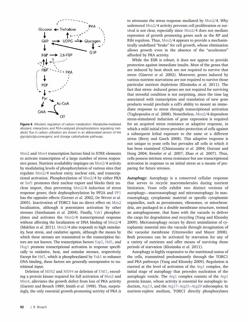
Msn2 and Msn4 transcription factors bind to STRE elementsto activate transcription of a large number of stress respon-sive genes. Nutrient availability impinges on Msn2/4 activityby modulating levels of phosphorylation of various sites thatregulate Msn2/4 nuclear entry, nuclear exit, and transcrip-tional activation. Phosphorylation of Msn2/4 by either PKAor Snf1 promotes their nuclear export and blocks their nu-clear import, thus preventing Msn2/4 induction of stressresponse genes; their dephosphorylation by PP2A and PP1has the opposite effects (Gorner et al. 2002; De Wever et al.2005). Inactivation of TORC1 has no direct effect on Msn2localization, although it potentiates activation by otherstresses (Santhanam et al. 2004). Finally, Yak1 phosphor-ylates and activates the Msn2/4 transcriptional responsewithout affecting the localization or DNA binding of Msn2/4(Malcher et al. 2011). Msn2/4 also responds to high osmolar-ity, heat stress, and oxidative agents, although the means bywhich these stresses are transmitted to the transcription fac-tors are not known. The transcription factors Yap1, Hsf1, andHog1 promote transcriptional activation in response specifi-cally to oxidative, heat, and osmolar stresses, respectively.Except for Hsf1, which is phosphorylated by Yak1 to enhanceDNA binding, these factors are generally unresponsive to nu-tritional input.
Deletion of MSN2 and MSN4 or deletion of YAK1, encod-ing a protein kinase required for full activation of Msn2 andMsn4, alleviates the growth defect from loss of PKA activity(Garrett and Broach 1989; Smith et al. 1998). Thus, surpris-ingly, the only essential growth-promoting activity of PKA is
to attenuate the stress response mediated by Msn2/4. Whyunfettered Msn2/4 activity prevents cell proliferation or sur-vival is not clear, especially since Msn2/4 does not mediaterepression of growth promoting genes such as the RP andRibi regulons. Thus, Msn2/4 appears to provide a mechanis-tically undefined “brake” for cell growth, whose eliminationallows growth even in the absence of the “accelerator”afforded by PKA activity.
While the ESR is robust, it does not appear to provideprotection against immediate insults. Most of the genes thatare induced by heat shock are not required to survive thatstress (Giaever et al. 2002). Moreover, genes induced byvarious nutrient starvations are not required to survive thoseparticular nutrient depletions (Klosinska et al. 2011). Thefact that stress -induced genes are not required for survivingthat stressful condition is not surprising, since the time lagassociated with transcription and translation of new geneproducts would preclude a cell’s ability to mount an imme-diate response to stress through transcriptional activation(Tagkopoulos et al. 2008). Nonetheless, Msn2/4-dependentstress-stimulated induction of gene expression is requiredfor an acquired stress resistance or adaptive response, inwhich a mild initial stress provides protection of cells againsta subsequent lethal exposure to the same or a differentstress (Berry and Gasch 2008). This adaptive response isnot unique to yeast cells but pervades all cells in which ithas been examined (Chinnusamy et al. 2004; Durrant andDong 2004; Kensler et al. 2007; Zhao et al. 2007). Thus,cells possess intrinsic stress resistance but use transcriptionalactivation in response to an initial stress as a means of pre-paring for future stresses.
Autophagy: Autophagy is a conserved cellular responsethat serves to recycle macromolecules during nutrientlimitation. Yeast cells exhibit two distinct versions ofautophagy—macroautophagy and microautophagy. In mac-roautophagy, cytoplasmic material or specific cytoplasmicorganelles, such as peroxisomes, ribosomes, or mitochron-dria, are packaged in a double membrane structure, termedan autophagosome, that fuses with the vacuole to deliverthe cargo for degradation and recycling (Yang and Klionsky2009). Microautophagy occurs by direct assimilation of cy-toplasmic material into the vacuole through invagination ofthe vacuolar membrane (Uttenweiler and Mayer 2008).Both processes can be activated by starvation for any ofa variety of nutrients and offer means of surviving thoseperiods of starvation (Klosinska et al. 2011).
Autophagy is highly responsive to the nutritional status ofthe cells, transmitted predominantly through the TORC1and PKA pathways (Yang and Klionsky 2009). Regulation isexerted at the level of activation of the Atg1 complex, theinitial stage of autophagy that precedes nucleation of theautophagic vesicle. The Atg1 complex consists of the Atg1protein kinase, whose activity is essential for autophagic in-duction, Atg13, and the Atg17–Atg31–Atg29 subcomplex. Innutrient-replete medium, TORC1 directly phosphorylates
Figure 6 Allosteric regulation of carbon metabolism. Metabolite-mediatedallosteric interactions and PKA-catalyzed phosphorylations regulating met-abolic flux in carbon utilization are shown in an abbreviated version of theglycolytic/gluconeogenic and storage carbohydrate pathways.
92 J. R. Broach
Atg13 on multiple sites, preventing Atg1 recruitment to thecomplex (Kamada et al. 2010). Under starvation conditionsor following treatment with rapamycin, Atg13 becomesdephosphorylated, allowing association of Atg1 with thecomplex and activation of its protein kinase activity. Expres-sion of a nonphosphorylatable version of Atg13 yields in-duction of autophagy in cells growing in rich medium,indicating that dephosphorylation of Atg13 is alone suffi-cient for initiating the autophagic process (Kamada et al.2010).
The PKA pathway also influences autophagy. Activationof PKA by deletion of Bcy1, by expression of an activatedRAS2 allele or by addition of cAMP, prevents induction ofautophagy by rapamycin treatment or nitrogen starvation(Budovskaya et al. 2004; Yorimitsu et al. 2007). Consistentwith those observations, simultaneous inactivation of PKAand Sch9 yields partial activation of the autophagic re-sponse. The locus of action of PKA and Sch9 is not entirelyclear: inactivation of PKA and Sch9 does not significantlyalter the phosphorylation state of Atg13. Moreover, the ef-fect on autophagy of inactivating PKA and Sch9 is synergisticwith TORC1 inactivation, suggesting that the PKA pathwayacts in parallel with the TORC1 pathway. Finally, the dom-inant role of TORC1 over PKA in regulating autophagy mayreflect the greater significance of autophagy in protectingcells from nitrogen starvation than from carbon starvation,given that the product of autophagy-induced turnover ofproteins is amino acids. In fact, mutants defective in autoph-agy are much more sensitive to nitrogen or phosphate star-vation than to glucose starvation (Klosinska et al. 2011).
Microautophagy is less extensively studied than macro-autophagy and has been characterized primarily frommicroscopic morphological analysis and from biochemicalstudies (Uttenweiler and Mayer 2008). The process involvestubular invaginations into the vacuole followed by scissionof the tubular structure to form vesicles within the lumen ofthe vacuole. It is related to piecemeal microautophagy of thenucleus, in which nuclear ER makes direct contact with thevacuole, followed by invaginations into the vacuole at thepoint of contact that are pinched off to generate luminalvesicles filled with nuclear material (Roberts et al. 2003;Levine and Klionsky 2004). Like macroautophagy, microau-tophagy is induced by nitrogen or carbon starvation or byTORC1 inactivation. The machinery for microautophagyis distinct from but overlaps that for macroautophagy(Uttenweiler and Mayer 2008; Krick et al. 2009; Dawalibyand Mayer 2010). In addition to its role in the starvationresponse, microautophagy serves to restore cell growth fol-lowing rapamycin treatment, at least in part by reducing theamount of vacuolar membrane that accumulates as a resultof starvation-induced macroautophagy. This process requiresthe previously described EGO complex, comprising the smallG proteins, Gtr1 and Gtr2, along with Meh1/Ego1 andSlm4/Ego3 (Dubouloz et al. 2005; Binda et al. 2009), whichis also required for activation of TORC1 by amino acids. Infact, the effect of the EGO complex on microautophagy may
result solely from its function as an activator of TORC1activity.
Development
In addition to controlling cell growth, nutrients dictatewhich of a number of developmental programs a cellchooses to pursue. For instance, under nutrient-limitingconditions cells can engage in pseudohyphal growth andinvasion of the substratum, perhaps as a method of “forag-ing” for nutrients. Haploid cells starved for any essentialnutrient can exit the mitotic cycle and assume a nutrient-specific quiescent state, which allows extended survival un-der the particular starvation condition. Finally, diploid cellssubject to substantial starvation can undergo meiosis andsporulation. The spores that emerge from this process arecapable of weathering extreme conditions for extendedperiods of time. The particular developmental programpursued is dictated by a complex interplay of the signalingnetworks responsive to nutrient availability and to stress.The specific developmental pathways are discussed in moredetail in other chapters in this volume. Accordingly, thefollowing focuses primarily on nutrient regulation of thoseprocesses.
Filamentous growth: Diploid cells subjected to limitingnitrogen or haploid cells subjected to limiting glucosebecome elongated, exhibit polar budding, suppress buddingin mother cells, remain attached after cytokinesis, andelaborate extracellular glucanases (Gancedo 2001; Paleceket al. 2002). This ensemble of features yields chains of cells,referred to as pseudohyphae in diploids or filaments in hap-loids, capable of invading the substratum. Depending on thenature of the strain and whether the cells are in liquid or ona solid surface, such cells could exhibit other behaviors, in-cluding biofilm formation, flocculation, or surface flotation(Bruckner and Mosch 2011). While these programs havebeen viewed as distinct processes responding to differentnutritional cues, recent studies have shown that the sig-naling pathways responsible for all these programs over-lap significantly. Nutrient regulation of filamentous growthimpinges on many cellular processes and affects a largenumber of genes. This regulation has been reviewed re-cently (Zaman et al. 2008; Bruckner and Mosch 2011), soI simply highlight a few of the more recent observations,focusing primarily on the regulation of FLO11, a major ad-hesion gene whose expression is tightly associated withfilamentation.
The transcriptional changes associated with filamenta-tion are coordinated by a collection of transcription factorsresponsive to the various nutrient signaling pathways de-scribed in the previous sections (Figure 7). These factorsinclude the transcriptional activators Ste12, Tec1, Ash1,Flo8, Phd1, Mss11, Msn1, Haa1, and Mga1 and the tran-scriptional repressors Sok2, Nrg1, Nrg2, and Sfl1. Thesefactors comprise a highly connected and complex net-work with Mga1 and Phd1 serving as the predominant
How Yeast Respond to Nutrients 93
modulators of the filamentous transitions (Borneman et al.2006). These transcription factors mediate expressionof several hundred genes that change expression duringtransition from yeast to pseudohyphal growth (Prinz et al.2004). In some cases, these factors all converge on thepromoter of a gene, such as FLO11, whose expressionis required for pseudohyphal growth but more oftenthese factors regulate distinct but overlapping suites ofgenes whose concerted action orchestrates the filamen-tous program.
Carbon source regulation of filamentation and the sig-naling pathways mediating that regulation is confounding.Haploid invasiveness and diploid filamentation are stimu-lated by carbon starvation through the Snf1 pathway, whichinactivates the transcriptional repressors, Nrg1 and Nrg2.However, stimulation of the Ras/PKA pathway, by an acti-vated RAS2 allele, an activated GPA2 allele, inactivation ofIra1, or addition of cAMP, also stimulates filamentation,even though enhanced PKA activity is associated with highglucose levels. One possible explanation for this discrepancyis that PKA regulation of filamentation may not be related toits function in glucose signaling, which is realized on a rela-tively short time scale, but rather to a long time-scale rolefor PKA, perhaps in cell–cell communication on the basis ofdiffusion of metabolic byproducts, not unlike quorum sens-ing in bacteria (Chen and Fink 2006; Wuster and Babu2010). The role of PKA in filamenation has an added com-plexity in that Tpk2 stimulates FLO11 expression by activat-ing the Flo8 transcriptional activator but Tpk1 inhibitsFLO11 expression by inhibiting Yak1 kinase, which allowsSok2 to inhibit, and prevents Phd1 from activating, FLO11transcription. Thus, activation of PKA sends both positiveand negative regulatory signals, at least for adhesion (Rob-ertson and Fink 1998; Malcher et al. 2011), perhaps allow-ing fine tuning of the adhesive response under a variety ofconditions.
As discussed previously, several signaling systemsinfluence diploid filamentation in response to nitrogenlimitation, although no coherent model for nitrogen sensingemerges from these anecdotal observations. Mutations ina number of genes involved in nitrogen regulation andassimilation affect filamentation in response to nitrogenlimitation. These include genes encoding the high-affinityMep2 ammonia permease, the regulators—Gln3 andUre2—of the NCR, those involved in amino acid permeaseinduction, and one of the glutamine tRNA genes. How-ever, rapamycin treatment inhibits rather than stimulatesfilamentation, even at sublethal doses, suggesting atmost an indirect role of TORC1 in filamenation. Finally,Snf1 T210 phosphorylation is stimulated not only byglucose limitation but also by nitrogen limitation, evenin the presence of high levels of glucose (Orlova et al.2006, 2010). Thus, Snf1 activation provides the mostconsistent connection between filamentation and themultiple forms of nutritional deprivation required forfilamentation.
Filamentation is also subject to epigenetic regulation(Figure 7). Even under conditions promoting filamenta-tion, the FLO11 gene is persistently inactive in a subset ofcells in the population while active in others (Halme et al.2004). Inactivation of HDA1, encoding one of the yeasthistone deacetylases, results in uniformly high levelFLO11 expression in all cells, indicating that the inactivestate of the gene results from a heritable, chromatin-basedrepression, similar to telomere position effect. The regula-tion of FLO11 expression can best be appreciated by postu-lating that the FLO11 promoter can exist in three distinctstates—a closed or silenced state, an open competent state,and a transcriptionally engaged state—and that the regu-latory factors influence transitions between those states(Octavio et al. 2009). The silenced state is impervious totranscriptional activation but can undergo an infrequenttransition into a competent state, which, while transcrip-tionally inactive, can be rapidly converted to the activestate by the appropriate combination of transcription fac-tors. Some transcription factors influence the transition be-tween silence and competence, while others serve only tostimulate transcription of competent promoters, whileother transcription factors can perform both functions.Moreover, this transition is influenced by two upstreamnoncoding RNAs, whose expression responds to Rpd3L,an observation that accounts for the unexpected role ofa histone deacetylase in activating gene expression (Bum-garner et al. 2009). In short, like rRNA expression, chro-matin structure dictates transcription factor accessibility tothe template while the signaling pathways can both influ-ence the distribution of open vs. closed templates and thetranscriptional activity of the open templates.
Quiescence: Yeast cells, like all other living cells, spend mostof their time in a quiescent state, which in yeast results fromstarvation for one or more nutrients. Haploid or diploidyeast cells starved for carbon, nitrogen, phosphate, or sulfurcease accumulating mass, arrest cell cycle progression priorto “start,” and enter the poorly defined G0 state. Investiga-tors have ascribed to G0 yeast cells a number of distinguish-ing characteristics, including a thickened cell wall; increasedstorage carbohydrates; compacted chromatin; substantiallyreduced translation; a specific transcriptional profile; andenhanced resistance to heat, oxidative stress, and high os-molarity (Gray et al. 2004; Smets et al. 2010). However, thesole unequivocal trait of quiescence is the ability to maintainviability when starved and to resume growth following res-toration of the missing nutrient. This characteristic distin-guishes cells suffering from “natural” starvation, for a carbonsource, for instance, from those subject to an unnatural star-vation, such as auxotrophic cells deprived of the requiredamino acid. In the latter case, cells rapidly lose viability eventhough they arrest uniformly as unbudded cells (Saldanhaet al. 2004; Boer et al. 2008; Brauer et al. 2008). Thus, thequiescent G0 state requires the coordinated and deliberateadaptation of cells to depletion of a core nutrient and not
94 J. R. Broach
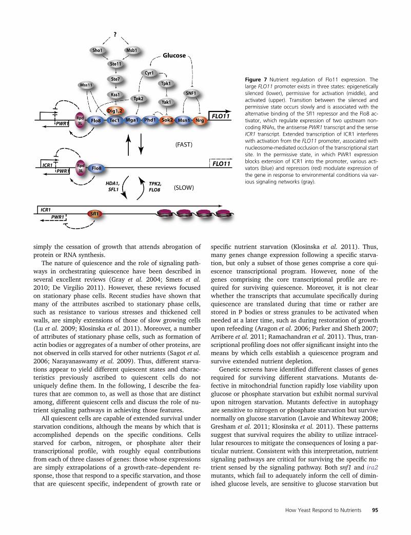
simply the cessation of growth that attends abrogation ofprotein or RNA synthesis.
The nature of quiescence and the role of signaling path-ways in orchestrating quiescence have been described inseveral excellent reviews (Gray et al. 2004; Smets et al.2010; De Virgilio 2011). However, these reviews focusedon stationary phase cells. Recent studies have shown thatmany of the attributes ascribed to stationary phase cells,such as resistance to various stresses and thickened cellwalls, are simply extensions of those of slow growing cells(Lu et al. 2009; Klosinska et al. 2011). Moreover, a numberof attributes of stationary phase cells, such as formation ofactin bodies or aggregates of a number of other proteins, arenot observed in cells starved for other nutrients (Sagot et al.2006; Narayanaswamy et al. 2009). Thus, different starva-tions appear to yield different quiescent states and charac-teristics previously ascribed to quiescent cells do notuniquely define them. In the following, I describe the fea-tures that are common to, as well as those that are distinctamong, different quiescent cells and discuss the role of nu-trient signaling pathways in achieving those features.
All quiescent cells are capable of extended survival understarvation conditions, although the means by which that isaccomplished depends on the specific conditions. Cellsstarved for carbon, nitrogen, or phosphate alter theirtranscriptional profile, with roughly equal contributionsfrom each of three classes of genes: those whose expressionsare simply extrapolations of a growth-rate–dependent re-sponse, those that respond to a specific starvation, and thosethat are quiescent specific, independent of growth rate or
specific nutrient starvation (Klosinska et al. 2011). Thus,many genes change expression following a specific starva-tion, but only a subset of those genes comprise a core qui-escence transcriptional program. However, none of thegenes comprising the core transcriptional profile are re-quired for surviving quiescence. Moreover, it is not clearwhether the transcripts that accumulate specifically duringquiescence are translated during that time or rather arestored in P bodies or stress granules to be activated whenneeded at a later time, such as during restoration of growthupon refeeding (Aragon et al. 2006; Parker and Sheth 2007;Arribere et al. 2011; Ramachandran et al. 2011). Thus, tran-scriptional profiling does not offer significant insight into themeans by which cells establish a quiescence program andsurvive extended nutrient depletion.
Genetic screens have identified different classes of genesrequired for surviving different starvations. Mutants de-fective in mitochondrial function rapidly lose viability uponglucose or phosphate starvation but exhibit normal survivalupon nitrogen starvation. Mutants defective in autophagyare sensitive to nitrogen or phosphate starvation but survivenormally on glucose starvation (Lavoie and Whiteway 2008;Gresham et al. 2011; Klosinska et al. 2011). These patternssuggest that survival requires the ability to utilize intracel-lular resources to mitigate the consequences of losing a par-ticular nutrient. Consistent with this interpretation, nutrientsignaling pathways are critical for surviving the specific nu-trient sensed by the signaling pathway. Both snf1 and ira2mutants, which fail to adequately inform the cell of dimin-ished glucose levels, are sensitive to glucose starvation but
Figure 7 Nutrient regulation of Flo11 expression. Thelarge FLO11 promoter exists in three states: epigeneticallysilenced (lower), permissive for activation (middle), andactivated (upper). Transition between the silenced andpermissive state occurs slowly and is associated with thealternative binding of the Sfl1 repressor and the Flo8 ac-tivator, which regulate expression of two upstream non-coding RNAs, the antisense PWR1 transcript and the senseICR1 transcript. Extended transcription of ICR1 interfereswith activation from the FLO11 promoter, associated withnucleosome-mediated occlusion of the transcriptional startsite. In the permissive state, in which PWR1 expressionblocks extension of ICR1 into the promoter, various acti-vators (blue) and repressors (red) modulate expression ofthe gene in response to environmental conditions via var-ious signaling networks (gray).
How Yeast Respond to Nutrients 95
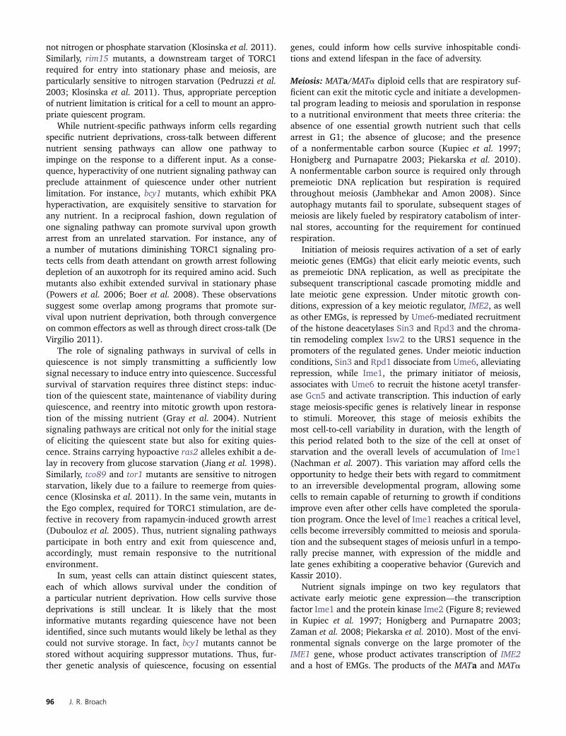
not nitrogen or phosphate starvation (Klosinska et al. 2011).Similarly, rim15 mutants, a downstream target of TORC1required for entry into stationary phase and meiosis, areparticularly sensitive to nitrogen starvation (Pedruzzi et al.2003; Klosinska et al. 2011). Thus, appropriate perceptionof nutrient limitation is critical for a cell to mount an appro-priate quiescent program.
While nutrient-specific pathways inform cells regardingspecific nutrient deprivations, cross-talk between differentnutrient sensing pathways can allow one pathway toimpinge on the response to a different input. As a conse-quence, hyperactivity of one nutrient signaling pathway canpreclude attainment of quiescence under other nutrientlimitation. For instance, bcy1 mutants, which exhibit PKAhyperactivation, are exquisitely sensitive to starvation forany nutrient. In a reciprocal fashion, down regulation ofone signaling pathway can promote survival upon growtharrest from an unrelated starvation. For instance, any ofa number of mutations diminishing TORC1 signaling pro-tects cells from death attendant on growth arrest followingdepletion of an auxotroph for its required amino acid. Suchmutants also exhibit extended survival in stationary phase(Powers et al. 2006; Boer et al. 2008). These observationssuggest some overlap among programs that promote sur-vival upon nutrient deprivation, both through convergenceon common effectors as well as through direct cross-talk (DeVirgilio 2011).
The role of signaling pathways in survival of cells inquiescence is not simply transmitting a sufficiently lowsignal necessary to induce entry into quiescence. Successfulsurvival of starvation requires three distinct steps: induc-tion of the quiescent state, maintenance of viability duringquiescence, and reentry into mitotic growth upon restora-tion of the missing nutrient (Gray et al. 2004). Nutrientsignaling pathways are critical not only for the initial stageof eliciting the quiescent state but also for exiting quies-cence. Strains carrying hypoactive ras2 alleles exhibit a de-lay in recovery from glucose starvation (Jiang et al. 1998).Similarly, tco89 and tor1 mutants are sensitive to nitrogenstarvation, likely due to a failure to reemerge from quies-cence (Klosinska et al. 2011). In the same vein, mutants inthe Ego complex, required for TORC1 stimulation, are de-fective in recovery from rapamycin-induced growth arrest(Dubouloz et al. 2005). Thus, nutrient signaling pathwaysparticipate in both entry and exit from quiescence and,accordingly, must remain responsive to the nutritionalenvironment.
In sum, yeast cells can attain distinct quiescent states,each of which allows survival under the condition ofa particular nutrient deprivation. How cells survive thosedeprivations is still unclear. It is likely that the mostinformative mutants regarding quiescence have not beenidentified, since such mutants would likely be lethal as theycould not survive storage. In fact, bcy1 mutants cannot bestored without acquiring suppressor mutations. Thus, fur-ther genetic analysis of quiescence, focusing on essential
genes, could inform how cells survive inhospitable condi-tions and extend lifespan in the face of adversity.
Meiosis: MATa/MATa diploid cells that are respiratory suf-ficient can exit the mitotic cycle and initiate a developmen-tal program leading to meiosis and sporulation in responseto a nutritional environment that meets three criteria: theabsence of one essential growth nutrient such that cellsarrest in G1; the absence of glucose; and the presenceof a nonfermentable carbon source (Kupiec et al. 1997;Honigberg and Purnapatre 2003; Piekarska et al. 2010).A nonfermentable carbon source is required only throughpremeiotic DNA replication but respiration is requiredthroughout meiosis (Jambhekar and Amon 2008). Sinceautophagy mutants fail to sporulate, subsequent stages ofmeiosis are likely fueled by respiratory catabolism of inter-nal stores, accounting for the requirement for continuedrespiration.
Initiation of meiosis requires activation of a set of earlymeiotic genes (EMGs) that elicit early meiotic events, suchas premeiotic DNA replication, as well as precipitate thesubsequent transcriptional cascade promoting middle andlate meiotic gene expression. Under mitotic growth con-ditions, expression of a key meiotic regulator, IME2, as wellas other EMGs, is repressed by Ume6-mediated recruitmentof the histone deacetylases Sin3 and Rpd3 and the chroma-tin remodeling complex Isw2 to the URS1 sequence in thepromoters of the regulated genes. Under meiotic inductionconditions, Sin3 and Rpd1 dissociate from Ume6, alleviatingrepression, while Ime1, the primary initiator of meiosis,associates with Ume6 to recruit the histone acetyl transfer-ase Gcn5 and activate transcription. This induction of earlystage meiosis-specific genes is relatively linear in responseto stimuli. Moreover, this stage of meiosis exhibits themost cell-to-cell variability in duration, with the length ofthis period related both to the size of the cell at onset ofstarvation and the overall levels of accumulation of Ime1(Nachman et al. 2007). This variation may afford cells theopportunity to hedge their bets with regard to commitmentto an irreversible developmental program, allowing somecells to remain capable of returning to growth if conditionsimprove even after other cells have completed the sporula-tion program. Once the level of Ime1 reaches a critical level,cells become irreversibly committed to meiosis and sporula-tion and the subsequent stages of meiosis unfurl in a tempo-rally precise manner, with expression of the middle andlate genes exhibiting a cooperative behavior (Gurevich andKassir 2010).
Nutrient signals impinge on two key regulators thatactivate early meiotic gene expression—the transcriptionfactor Ime1 and the protein kinase Ime2 (Figure 8; reviewedin Kupiec et al. 1997; Honigberg and Purnapatre 2003;Zaman et al. 2008; Piekarska et al. 2010). Most of the envi-ronmental signals converge on the large promoter of theIME1 gene, whose product activates transcription of IME2and a host of EMGs. The products of the MATa and MATa
96 J. R. Broach
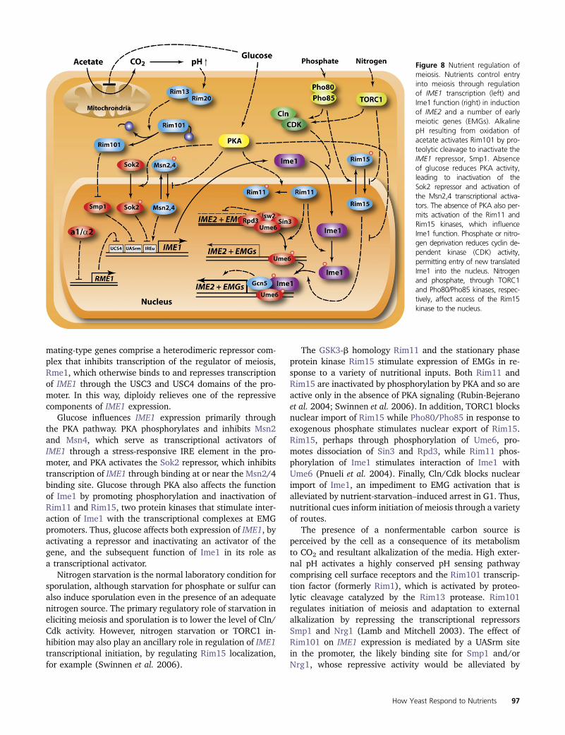
mating-type genes comprise a heterodimeric repressor com-plex that inhibits transcription of the regulator of meiosis,Rme1, which otherwise binds to and represses transcriptionof IME1 through the USC3 and USC4 domains of the pro-moter. In this way, diploidy relieves one of the repressivecomponents of IME1 expression.
Glucose influences IME1 expression primarily throughthe PKA pathway. PKA phosphorylates and inhibits Msn2and Msn4, which serve as transcriptional activators ofIME1 through a stress-responsive IRE element in the pro-moter, and PKA activates the Sok2 repressor, which inhibitstranscription of IME1 through binding at or near the Msn2/4binding site. Glucose through PKA also affects the functionof Ime1 by promoting phosphorylation and inactivation ofRim11 and Rim15, two protein kinases that stimulate inter-action of Ime1 with the transcriptional complexes at EMGpromoters. Thus, glucose affects both expression of IME1, byactivating a repressor and inactivating an activator of thegene, and the subsequent function of Ime1 in its role asa transcriptional activator.
Nitrogen starvation is the normal laboratory condition forsporulation, although starvation for phosphate or sulfur canalso induce sporulation even in the presence of an adequatenitrogen source. The primary regulatory role of starvation ineliciting meiosis and sporulation is to lower the level of Cln/Cdk activity. However, nitrogen starvation or TORC1 in-hibition may also play an ancillary role in regulation of IME1transcriptional initiation, by regulating Rim15 localization,for example (Swinnen et al. 2006).
The GSK3-b homology Rim11 and the stationary phaseprotein kinase Rim15 stimulate expression of EMGs in re-sponse to a variety of nutritional inputs. Both Rim11 andRim15 are inactivated by phosphorylation by PKA and so areactive only in the absence of PKA signaling (Rubin-Bejeranoet al. 2004; Swinnen et al. 2006). In addition, TORC1 blocksnuclear import of Rim15 while Pho80/Pho85 in response toexogenous phosphate stimulates nuclear export of Rim15.Rim15, perhaps through phosphorylation of Ume6, pro-motes dissociation of Sin3 and Rpd3, while Rim11 phos-phorylation of Ime1 stimulates interaction of Ime1 withUme6 (Pnueli et al. 2004). Finally, Cln/Cdk blocks nuclearimport of Ime1, an impediment to EMG activation that isalleviated by nutrient-starvation–induced arrest in G1. Thus,nutritional cues inform initiation of meiosis through a varietyof routes.
The presence of a nonfermentable carbon source isperceived by the cell as a consequence of its metabolismto CO2 and resultant alkalization of the media. High exter-nal pH activates a highly conserved pH sensing pathwaycomprising cell surface receptors and the Rim101 transcrip-tion factor (formerly Rim1), which is activated by proteo-lytic cleavage catalyzed by the Rim13 protease. Rim101regulates initiation of meiosis and adaptation to externalalkalization by repressing the transcriptional repressorsSmp1 and Nrg1 (Lamb and Mitchell 2003). The effect ofRim101 on IME1 expression is mediated by a UASrm sitein the promoter, the likely binding site for Smp1 and/orNrg1, whose repressive activity would be alleviated by
Figure 8 Nutrient regulation ofmeiosis. Nutrients control entryinto meiosis through regulationof IME1 transcription (left) andIme1 function (right) in inductionof IME2 and a number of earlymeiotic genes (EMGs). AlkalinepH resulting from oxidation ofacetate activates Rim101 by pro-teolytic cleavage to inactivate theIME1 repressor, Smp1. Absenceof glucose reduces PKA activity,leading to inactivation of theSok2 repressor and activation ofthe Msn2,4 transcriptional activa-tors. The absence of PKA also per-mits activation of the Rim11 andRim15 kinases, which influenceIme1 function. Phosphate or nitro-gen deprivation reduces cyclin de-pendent kinase (CDK) activity,permitting entry of new translatedIme1 into the nucleus. Nitrogenand phosphate, through TORC1and Pho80/Pho85 kinases, respec-tively, affect access of the Rim15kinase to the nucleus.
How Yeast Respond to Nutrients 97
Rim101. The fact that deletion of SMP1, but not NRG1,alleviates the sporulation defect in rim101D strains wouldargue that Smp1 is the immediate regulator of IME1 (Lamband Mitchell 2003). Finally, glucose also likely influencesthis signaling pathway through repression of respiration,thereby blocking alkalization.
As noted above, addition of nutrients to cells undergoingmeiosis can abrogate the developmental program andrestore cells to mitotic growth. The targets for this nutri-tional control of meiotic progression have not been fullydefined, although some information has emerged. Forinstance, Ime2, which is required at multiple stages duringmeiotic progression, is destabilized by glucose additionthrough degradation targeted by the SCFGrr1 ubiquitin ligase(Purnapatre et al. 2005). However, while expression ofa degradation-resistant version of Ime2 renders cells resis-tant to the glucose-induced block to meiotic DNA replica-tion, it does not render later steps in meiosis resistant toglucose. Thus, additional nutritionally sensitive processeshave yet to be identified.
Conclusions and Prospectives
We have a detailed understanding of some of the cir-cuitry underlying nutritional sensing in yeast, but we arestill somewhat vague on others. For instance, the interplayof positive and negative regulators and the various feed-forward and feed-back loops in regulating expression ofglucose transporters is so well described that modelingefforts have yielded highly predictive dynamic descriptionsof its behavior. On the other hand, we still have no clearunderstanding of the upstream components of glucosesignaling regulating protein kinase A or the interplay be-tween the various small G proteins in that process. Evenmore poorly described are the pathways sensing andresponding to nitrogen levels. While many of the compo-nents of the TORC1 signaling network have been identifiedand their interactions defined, we have less understandingof the pathways emanating from TORC1, particularlythrough protein phosphatases. Moreover, we can infer theexistence of a second nitrogen-sensing pathway from thelimits of TORC1 effects, but this pathway is poorly defined.Finally, we appreciate that significant cross-talk existsbetween and among the various nutrient signaling path-ways—for instance, glucose sparing in nitrogen- or phos-phate-starved cells—but the nature of that interplay isundefined. Thus, we have a number of important detailsto fill in regarding the structure of the nutrient-sensing net-works. In addition, several fundamental questions regardingthe interplay of nutrient availability and growth have yet tobe solved.
Key unanswered questions
How is cell growth controlled in response to nutrients? Cellscan adapt their growth rate over at least a 10-fold range inresponse to limiting nutrients and diminished growth rate is
associated with downregulation of biosynthetic capacity andupregulation of stress response genes. What is cause andeffect in this growth control? Do limiting nutrients result inlimiting metabolic capacity, which is sensed through re-duced energy charge or the inability to synthesize keycomponents required for continued biomass accumulation?Or, do nutrient levels through signaling pathways set thebiosynthetic, metabolic, and transcriptional program appro-priate for the perceived levels of nutrients. This question isfundamental to understanding regulation of cell growth inany organism.
How have evolutionary pressures shaped the growthcapacity of yeast cells? Most of the studies on yeast growthand metabolism have been conducted in nutrient-repleteconditions with cells undergoing exponential growth.However, yeast cells in the wild seldom experience sucha lush environment but rather struggle under nutrient-limited or -depleted conditions. Accordingly, survival andevolution likely depended more on the ability of cells towithstand adverse conditions, through developmental pro-grams such as filamentation, quiescence, and sporulation,than on the ability to grow rapidly under nutrient-repleteconditions. Survival under these conditions also requiresthat cells be capable of responding effectively to changingnutritional status by rapidly sensing those changes andtransitioning from one state to another. Moreover, cellshave survived in a highly variable and uncertain environ-ment. Mounting a response that allows cells to “hunkerdown” in response to stress may be the appropriate behav-ior if the stress continues but would be disadvantageous ifconditions improved and the stress response precludedcells from rapidly taking advantage of the improved con-ditions. Thus, cell populations that exhibit a diverse set ofbehaviors to a given environmental stress may be betterpositioned to survive a variable environment than thosethat mount uniform responses. All these issues suggest thatunderstanding the biology of yeast, considering the pres-sures that shaped its biology, require exploring the waysyeast cells respond to challenging conditions and themeans by which they make transitions between differentnutrient states. These areas offer rich topics for futureresearch.
Acknowledgments
Work from my laboratory described in this review wassupported by National Institutes of Health (NIH) grant RO1GM076562 with additional support from a Center forQuantitative Biology/NIH grant P50 GM071508.
Literature Cited
Ahuatzi, D., P. Herrero, T. de la Cera, and F. Moreno, 2004 Theglucose-regulated nuclear localization of hexokinase 2 in Sac-charomyces cerevisiae is Mig1-dependent. J. Biol. Chem. 279:14440–14446.
98 J. R. Broach
Ahuatzi, D., A. Riera, R. Pelaez, P. Herrero, and F. Moreno,2007 Hxk2 regulates the phosphorylation state of Mig1 andtherefore its nucleocytoplasmic distribution. J. Biol. Chem. 282:4485–4493.
Aragon, A. D., G. A. Quinones, E. V. Thomas, S. Roy, and M.Werner-Washburne, 2006 Release of extraction-resistantmRNA in stationary phase Saccharomyces cerevisiae producesa massive increase in transcript abundance in response to stress.Genome Biol. 7: R9.
Arribere, J. A., J. A. Doudna, and W. V. Gilbert, 2011 Reconsider-ing movement of eukaryotic mRNAs between polysomes andP bodies. Mol. Cell 44: 745–758.
Ashrafi, K., S. S. Lin, J. K. Manchester, and J. I. Gordon,2000 Sip2p and its partner snf1p kinase affect aging in S.cerevisiae. Genes Dev. 14: 1872–1885.
Badis, G., E. T. Chan, H. van Bakel, L. Pena-Castillo, D. Tillo et al.,2008 A library of yeast transcription factor motifs revealsa widespread function for Rsc3 in targeting nucleosome exclu-sion at promoters. Mol. Cell 32: 878–887.
Bahn, Y. S., C. Xue, A. Idnurm, J. C. Rutherford, J. Heitman et al.,2007 Sensing the environment: lessons from fungi. Nat. Rev.Microbiol. 5: 57–69.
Beck, T., and M. N. Hall, 1999 The TOR signalling pathway con-trols nuclear localization of nutrient-regulated transcription fac-tors. Nature 402: 689–692.
Belinchon, M. M., and J. M. Gancedo, 2007 Glucose controls mul-tiple processes in Saccharomyces cerevisiae through diversecombinations of signaling pathways. FEMS Yeast Res. 7: 808–818.
Berry, D. B., and A. P. Gasch, 2008 Stress-activated genomic ex-pression changes serve a preparative role for impending stress inyeast. Mol. Biol. Cell 19: 4580–4587.
Bertram, P. G., J. H. Choi, J. Carvalho, W. Ai, C. Zeng et al.,2000 Tripartite regulation of Gln3p by TOR, Ure2p, and phos-phatases. J. Biol. Chem. 275: 35727–35733.
Binda, M., M. P. Peli-Gulli, G. Bonfils, N. Panchaud, J. Urban et al.,2009 The Vam6 GEF controls TORC1 by activating the EGOcomplex. Mol. Cell 35: 563–573.
Boeger, H., J. Griesenbeck, J. S. Strattan, and R. D. Kornberg,2003 Nucleosomes unfold completely at a transcriptionally ac-tive promoter. Mol. Cell 11: 1587–1598.
Boer, V. M., S. Amini, and D. Botstein, 2008 Influence of genotypeand nutrition on survival and metabolism of starving yeast. Proc.Natl. Acad. Sci. USA 105: 6930–6935.
Boer, V. M., C. A. Crutchfield, P. H. Bradley, D. Botstein, and J. D.Rabinowitz, 2010 Growth-limiting intracellular metabolites inyeast growing under diverse nutrient limitations. Mol. Biol. Cell21: 198–211.
Borneman, A. R., J. A. Leigh-Bell, H. Yu, P. Bertone, M. Gersteinet al., 2006 Target hub proteins serve as master regulators ofdevelopment in yeast. Genes Dev. 20: 435–448.
Bradley, P. H., M. J. Brauer, J. D. Rabinowitz, and O. G. Troyanskaya,2009 Coordinated concentration changes of transcripts and me-tabolites in Saccharomyces cerevisiae. PLOS Comput. Biol. 5:e1000270.
Brauer, M. J., A. J. Saldanha, K. Dolinski, and D. Botstein,2005 Homeostatic adjustment and metabolic remodeling inglucose-limited yeast cultures. Mol. Biol. Cell 16: 2503–2517.
Brauer, M. J., J. Yuan, B. D. Bennett, W. Lu, E. Kimball et al.,2006 Conservation of the metabolomic response to starvationacross two divergent microbes. Proc. Natl. Acad. Sci. USA 103:19302–19307.
Brauer, M. J., C. Huttenhower, E. M. Airoldi, R. Rosenstein, J. C.Matese et al., 2008 Coordination of growth rate, cell cycle,stress response, and metabolic activity in yeast. Mol. Biol. Cell19: 352–367.
Breitkreutz, A., H. Choi, J. R. Sharom, L. Boucher, V. Neduva et al.,2010 A global protein kinase and phosphatase interaction net-work in yeast. Science 328: 1043–1046.
Broach, J. R., and R. J. Deschenes, 1990 The function of ras genesin Saccharomyces cerevisiae. Adv. Cancer Res. 54: 79–139.
Bruckner, S., and H. U. Mosch, 2011 Choosing the right lifestyle:adhesion and development in Saccharomyces cerevisiae. FEMSMicrobiol. Rev. 36: 25–58.
Budhwar, R., A. Lu, and J. P. Hirsch, 2010 Nutrient control ofyeast PKA activity involves opposing effects on phosphorylationof the Bcy1 regulatory subunit. Mol. Biol. Cell 21: 3749–3758.
Budovskaya, Y. V., J. S. Stephan, F. Reggiori, D. J. Klionsky, and P.K. Herman, 2004 The Ras/cAMP-dependent protein kinasesignaling pathway regulates an early step of the autophagy pro-cess in Saccharomyces cerevisiae. J. Biol. Chem. 279: 20663–20671.
Budovskaya, Y. V., J. S. Stephan, S. J. Deminoff, and P. K. Herman,2005 An evolutionary proteomics approach identifies sub-strates of the cAMP-dependent protein kinase. Proc. Natl. Acad.Sci. USA 102: 13933–13938.
Bumgarner, S. L., R. D. Dowell, P. Grisafi, D. K. Gifford, and G. R.Fink, 2009 Toggle involving cis-interfering noncoding RNAscontrols variegated gene expression in yeast. Proc. Natl. Acad.Sci. USA 106: 18321–18326.
Cardenas, M. E., N. S. Cutler, M. C. Lorenz, C. J. Di Como, and J.Heitman, 1999 The TOR signaling cascade regulates gene ex-pression in response to nutrients. Genes Dev. 13: 3271–3279.
Carman, G. M., and G. S. Han, 2010 Regulation of phospholipidsynthesis in the yeast Saccharomyces cerevisiae. Annu. Rev. Bio-chem. 80: 859–883.
Chen, H., and G. R. Fink, 2006 Feedback control of morphogen-esis in fungi by aromatic alcohols. Genes Dev. 20: 1150–1161.
Chen, L., Z. H. Jiao, L. S. Zheng, Y. Y. Zhang, S. T. Xie et al.,2009 Structural insight into the autoinhibition mechanism ofAMP-activated protein kinase. Nature 459: 1146–1149.
Chen, Z., E. A. Odstrcil, B. P. Tu, and S. L. McKnight,2007 Restriction of DNA replication to the reductive phase ofthe metabolic cycle protects genome integrity. Science 316:1916–1919.
Cherkasova, V., H. Qiu, and A. G. Hinnebusch, 2010 Snf1 pro-motes phosphorylation of the alpha subunit of eukaryotic trans-lation initiation factor 2 by activating Gcn2 and inhibitingphosphatases Glc7 and Sit4. Mol. Cell. Biol. 30: 2862–2873.
Chinnusamy, V., K. Schumaker, and J. K. Zhu, 2004 Moleculargenetic perspectives on cross-talk and specificity in abiotic stresssignalling in plants. J. Exp. Bot. 55: 225–236.
Claypool, J. A., S. L. French, K. Johzuka, K. Eliason, L. Vu et al.,2004 Tor pathway regulates Rrn3p-dependent recruitment ofyeast RNA polymerase I to the promoter but does not participatein alteration of the number of active genes. Mol. Biol. Cell 15:946–956.
Colombo, S., P. Ma, L. Cauwenberg, J. Winderickx, M. Crauwelset al., 1998 Involvement of distinct G-proteins, Gpa2 and Ras,in glucose- and intracellular acidification-induced cAMP signal-ling in the yeast Saccharomyces cerevisiae. EMBO J. 17: 3326–3341.
Conconi, A., R. M. Widmer, T. Koller, and J. M. Sogo, 1989 Twodifferent chromatin structures coexist in ribosomal RNA genesthroughout the cell cycle. Cell 57: 753–761.
Cooper, T. G., 2002 Transmitting the signal of excess nitrogen inSaccharomyces cerevisiae from the Tor proteins to the GATAfactors: connecting the dots. FEMS Microbiol. Rev. 26: 223–238.
Crespo, J. L., T. Powers, B. Fowler, and M. N. Hall, 2002 TheTOR-controlled transcription activators GLN3, RTG1, andRTG3 are regulated in response to intracellular levels of gluta-mine. Proc. Natl. Acad. Sci. USA 99: 6784–6789.
How Yeast Respond to Nutrients 99
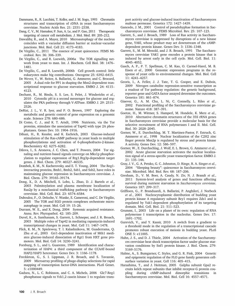
Dammann, R., R. Lucchini, T. Koller, and J. M. Sogo, 1993 Chromatinstructures and transcription of rDNA in yeast Saccharomycescerevisiae. Nucleic Acids Res. 21: 2331–2338.
Dang, C. V., M. Hamaker, P. Sun, A. Le, and P. Gao, 2011 Therapeutictargeting of cancer cell metabolism. J. Mol. Med. 89: 205–212.
Dawaliby, R., and A. Mayer, 2010 Microautophagy of the nucleuscoincides with a vacuolar diffusion barrier at nuclear-vacuolarjunctions. Mol. Biol. Cell 21: 4173–4183.
De Virgilio, C., 2011 The essence of yeast quiescence. FEMS Mi-crobiol. Rev. 36: 306–339.
De Virgilio, C., and R. Loewith, 2006a The TOR signalling net-work from yeast to man. Int. J. Biochem. Cell Biol. 38: 1476–1481.
De Virgilio, C., and R. Loewith, 2006b Cell growth control: littleeukaryotes make big contributions. Oncogene 25: 6392–6415.
De Wever, V., W. Reiter, A. Ballarini, G. Ammerer, and C. Brocard,2005 A dual role for PP1 in shaping the Msn2-dependent tran-scriptional response to glucose starvation. EMBO J. 24: 4115–4123.
Dechant, R., M. Binda, S. S. Lee, S. Pelet, J. Winderickx et al.,2010 Cytosolic pH is a second messenger for glucose and reg-ulates the PKA pathway through V-ATPase. EMBO J. 29: 2515–2526.
DeRisi, J. L., V. R. Iyer, and P. O. Brown, 1997 Exploring themetabolic and genetic control of gene expression on a genomicscale. Science 278: 680–686.
Di Como, C. J., and K. T. Arndt, 1996 Nutrients, via the Torproteins, stimulate the association of Tap42 with type 2A phos-phatases. Genes Dev. 10: 1904–1916.
Dihazi, H., R. Kessler, and K. Eschrich, 2003 Glucose-inducedstimulation of the Ras-cAMP pathway in yeast leads to multiplephosphorylations and activation of 6-phosphofructo-2-kinase.Biochemistry 42: 6275–6282.
Dilova, I., S. Aronova, J. C. Chen, and T. Powers, 2004 Tor sig-naling and nutrient-based signals converge on Mks1p phosphor-ylation to regulate expression of Rtg1.Rtg3p-dependent targetgenes. J. Biol. Chem. 279: 46527–46535.
Dombek, K. M., N. Kacherovsky, and E. T. Young, 2004 The Reg1-interacting proteins, Bmh1, Bmh2, Ssb1, and Ssb2, have roles inmaintaining glucose repression in Saccharomyces cerevisiae. J.Biol. Chem. 279: 39165–39174.
Dong, X., D. A. Mitchell, S. Lobo, L. Zhao, D. J. Bartels et al.,2003 Palmitoylation and plasma membrane localization ofRas2p by a nonclassical trafficking pathway in Saccharomycescerevisiae. Mol. Cell. Biol. 23: 6574–6584.
Dubouloz, F., O. Deloche, V. Wanke, E. Cameroni, and C. De Virgilio,2005 The TOR and EGO protein complexes orchestrate micro-autophagy in yeast. Mol. Cell 19: 15–26.
Durrant, W. E., and X. Dong, 2004 Systemic acquired resistance.Annu. Rev. Phytopathol. 42: 185–209.
Duvel, K., A. Santhanam, S. Garrett, L. Schneper, and J. R. Broach,2003 Multiple roles of Tap42 in mediating rapamycin-inducedtranscriptional changes in yeast. Mol. Cell 11: 1467–1478.
Flick, K. M., N. Spielewoy, T. I. Kalashnikova, M. Guaderrama, Q.Zhu et al., 2003 Grr1-dependent inactivation of Mth1 medi-ates glucose-induced dissociation of Rgt1 from HXT gene pro-moters. Mol. Biol. Cell 14: 3230–3241.
Forsburg, S. L., and L. Guarente, 1989 Identification and charac-terization of HAP4: a third component of the CCAAT-boundHAP2/HAP3 heteromer. Genes Dev. 3: 1166–1178.
Freckleton, G., S. I. Lippman, J. R. Broach, and S. Tavazoie,2009 Microarray profiling of phage-display selections for rapidmapping of transcription factor-DNA interactions. PLoS Genet.5: e1000449.
Gadura, N., L. C. Robinson, and C. A. Michels, 2006 Glc7-Reg1phosphatase signals to Yck1,2 casein kinase 1 to regulate trans-
port activity and glucose-induced inactivation of Saccharomycesmaltose permease. Genetics 172: 1427–1439.
Gancedo, J. M., 2001 Control of pseudohyphae formation in Sac-charomyces cerevisiae. FEMS Microbiol. Rev. 25: 107–123.
Garrett, S., and J. Broach, 1989 Loss of Ras activity in Saccharo-myces cerevisiae is suppressed by disruptions of a new kinasegene, YAKI, whose product may act downstream of the cAMP-dependent protein kinase. Genes Dev. 3: 1336–1348.
Garrett, S., M. M. Menold, and J. R. Broach, 1991 The Saccharo-myces cerevisiae YAK1 gene encodes a protein kinase that isinduced by arrest early in the cell cycle. Mol. Cell. Biol. 11:4045–4052.
Gasch, A. P., P. T. Spellman, C. M. Kao, O. Carmel-Harel, M. B.Eisen et al., 2000 Genomic expression programs in the re-sponse of yeast cells to environmental changes. Mol. Biol. Cell11: 4241–4257.
Georis, I., A. Feller, J. J. Tate, T. G. Cooper, and E. Dubois,2009 Nitrogen catabolite repression-sensitive transcription asa readout of Tor pathway regulation: the genetic background,reporter gene and GATA factor assayed determine the outcomes.Genetics 181: 861–874.
Giaever, G., A. M. Chu, L. Ni, C. Connelly, L. Riles et al.,2002 Functional profiling of the Saccharomyces cerevisiae ge-nome. Nature 418: 387–391.
Goetze, H., M. Wittner, S. Hamperl, M. Hondele, K. Merz et al.,2010 Alternative chromatin structures of the 35S rRNA genesin Saccharomyces cerevisiae provide a molecular basis for theselective recruitment of RNA polymerases I and II. Mol. Cell.Biol. 30: 2028–2045.
Gorner, W., E. Durchschlag, M. T. Martinez-Pastor, F. Estruch, G.Ammerer et al., 1998 Nuclear localization of the C2H2 zincfinger protein Msn2p is regulated by stress and protein kinaseA activity. Genes Dev. 12: 586–597.
Gorner, W., E. Durchschlag, J. Wolf, E. L. Brown, G. Ammerer et al.,2002 Acute glucose starvation activates the nuclear localiza-tion signal of a stress-specific yeast transcription factor. EMBO J.21: 135–144.
Gray, J. V., G. A. Petsko, G. C. Johnston, D. Ringe, R. A. Singer et al.,2004 “Sleeping beauty”: quiescence in Saccharomyces cerevi-siae. Microbiol. Mol. Biol. Rev. 68: 187–206.
Gresham, D., V. M. Boer, A. Caudy, N. Ziv, N. J. Brandt et al.,2011 System-level analysis of genes and functions affectingsurvival during nutrient starvation in Saccharomyces cerevisiae.Genetics 187: 299–317.
Griffioen, G., P. Branduardi, A. Ballarini, P. Anghileri, J. Norbecket al., 2001 Nucleocytoplasmic distribution of budding yeastprotein kinase A regulatory subunit Bcy1 requires Zds1 and isregulated by Yak1-dependent phosphorylation of its targetingdomain. Mol. Cell. Biol. 21: 511–523.
Grummt, I., 2003 Life on a planet of its own: regulation of RNApolymerase I transcription in the nucleolus. Genes Dev. 17:1691–1702.
Gurevich, V., and Y. Kassir, 2010 A switch from a gradient toa threshold mode in the regulation of a transcriptional cascadepromotes robust execution of meiosis in budding yeast. PLoSONE 5: e11005.
Hahn, J. S., and D. J. Thiele, 2004 Activation of the Saccharomy-ces cerevisiae heat shock transcription factor under glucose star-vation conditions by Snf1 protein kinase. J. Biol. Chem. 279:5169–5176.
Halme, A., S. Bumgarner, C. Styles, and G. R. Fink, 2004 Geneticand epigenetic regulation of the FLO gene family generates cell-surface variation in yeast. Cell 116: 405–415.
Harashima, T., and J. Heitman, 2005 Galpha subunit Gpa2 re-cruits kelch repeat subunits that inhibit receptor-G protein cou-pling during cAMP-induced dimorphic transitions inSaccharomyces cerevisiae. Mol. Biol. Cell 16: 4557–4571.
100 J. R. Broach
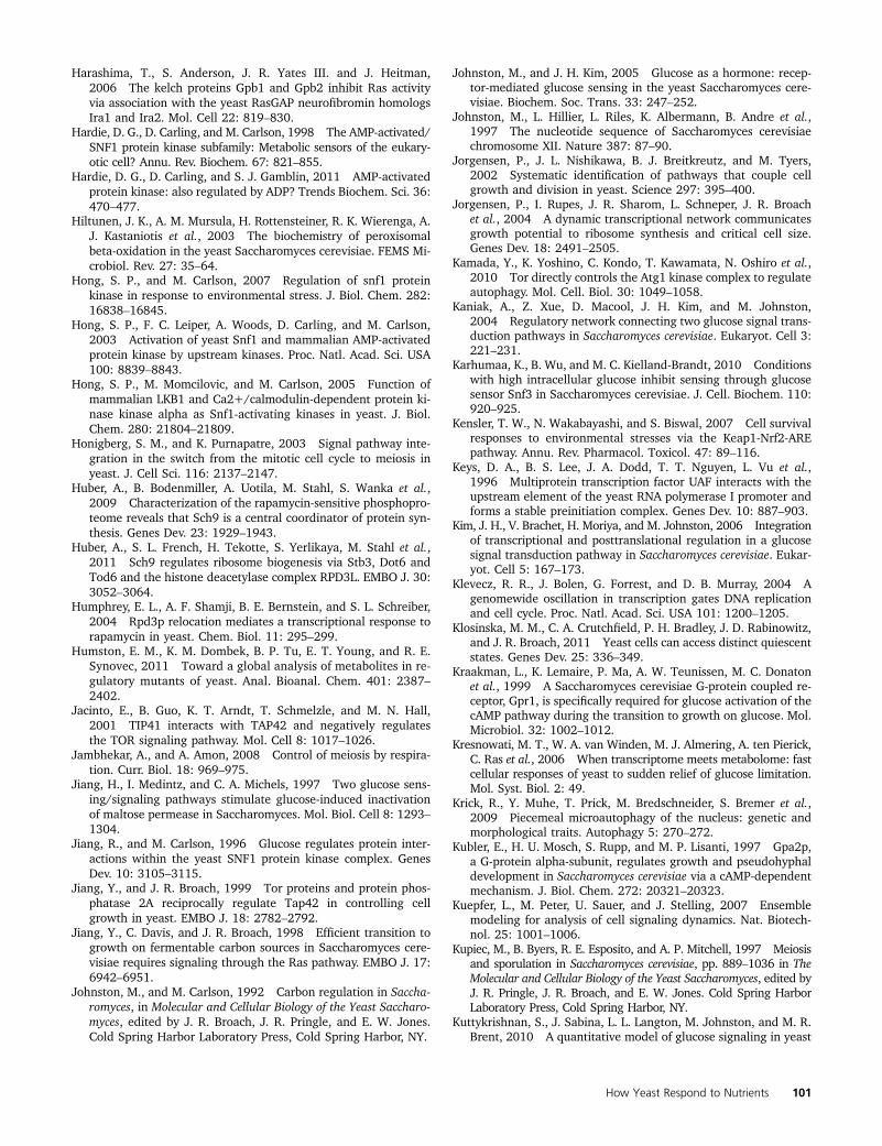
Harashima, T., S. Anderson, J. R. Yates III. and J. Heitman,2006 The kelch proteins Gpb1 and Gpb2 inhibit Ras activityvia association with the yeast RasGAP neurofibromin homologsIra1 and Ira2. Mol. Cell 22: 819–830.
Hardie, D. G., D. Carling, and M. Carlson, 1998 The AMP-activated/SNF1 protein kinase subfamily: Metabolic sensors of the eukary-otic cell? Annu. Rev. Biochem. 67: 821–855.
Hardie, D. G., D. Carling, and S. J. Gamblin, 2011 AMP-activatedprotein kinase: also regulated by ADP? Trends Biochem. Sci. 36:470–477.
Hiltunen, J. K., A. M. Mursula, H. Rottensteiner, R. K. Wierenga, A.J. Kastaniotis et al., 2003 The biochemistry of peroxisomalbeta-oxidation in the yeast Saccharomyces cerevisiae. FEMS Mi-crobiol. Rev. 27: 35–64.
Hong, S. P., and M. Carlson, 2007 Regulation of snf1 proteinkinase in response to environmental stress. J. Biol. Chem. 282:16838–16845.
Hong, S. P., F. C. Leiper, A. Woods, D. Carling, and M. Carlson,2003 Activation of yeast Snf1 and mammalian AMP-activatedprotein kinase by upstream kinases. Proc. Natl. Acad. Sci. USA100: 8839–8843.
Hong, S. P., M. Momcilovic, and M. Carlson, 2005 Function ofmammalian LKB1 and Ca2+/calmodulin-dependent protein ki-nase kinase alpha as Snf1-activating kinases in yeast. J. Biol.Chem. 280: 21804–21809.
Honigberg, S. M., and K. Purnapatre, 2003 Signal pathway inte-gration in the switch from the mitotic cell cycle to meiosis inyeast. J. Cell Sci. 116: 2137–2147.
Huber, A., B. Bodenmiller, A. Uotila, M. Stahl, S. Wanka et al.,2009 Characterization of the rapamycin-sensitive phosphopro-teome reveals that Sch9 is a central coordinator of protein syn-thesis. Genes Dev. 23: 1929–1943.
Huber, A., S. L. French, H. Tekotte, S. Yerlikaya, M. Stahl et al.,2011 Sch9 regulates ribosome biogenesis via Stb3, Dot6 andTod6 and the histone deacetylase complex RPD3L. EMBO J. 30:3052–3064.
Humphrey, E. L., A. F. Shamji, B. E. Bernstein, and S. L. Schreiber,2004 Rpd3p relocation mediates a transcriptional response torapamycin in yeast. Chem. Biol. 11: 295–299.
Humston, E. M., K. M. Dombek, B. P. Tu, E. T. Young, and R. E.Synovec, 2011 Toward a global analysis of metabolites in re-gulatory mutants of yeast. Anal. Bioanal. Chem. 401: 2387–2402.
Jacinto, E., B. Guo, K. T. Arndt, T. Schmelzle, and M. N. Hall,2001 TIP41 interacts with TAP42 and negatively regulatesthe TOR signaling pathway. Mol. Cell 8: 1017–1026.
Jambhekar, A., and A. Amon, 2008 Control of meiosis by respira-tion. Curr. Biol. 18: 969–975.
Jiang, H., I. Medintz, and C. A. Michels, 1997 Two glucose sens-ing/signaling pathways stimulate glucose-induced inactivationof maltose permease in Saccharomyces. Mol. Biol. Cell 8: 1293–1304.
Jiang, R., and M. Carlson, 1996 Glucose regulates protein inter-actions within the yeast SNF1 protein kinase complex. GenesDev. 10: 3105–3115.
Jiang, Y., and J. R. Broach, 1999 Tor proteins and protein phos-phatase 2A reciprocally regulate Tap42 in controlling cellgrowth in yeast. EMBO J. 18: 2782–2792.
Jiang, Y., C. Davis, and J. R. Broach, 1998 Efficient transition togrowth on fermentable carbon sources in Saccharomyces cere-visiae requires signaling through the Ras pathway. EMBO J. 17:6942–6951.
Johnston, M., and M. Carlson, 1992 Carbon regulation in Saccha-romyces, in Molecular and Cellular Biology of the Yeast Saccharo-myces, edited by J. R. Broach, J. R. Pringle, and E. W. Jones.Cold Spring Harbor Laboratory Press, Cold Spring Harbor, NY.
Johnston, M., and J. H. Kim, 2005 Glucose as a hormone: recep-tor-mediated glucose sensing in the yeast Saccharomyces cere-visiae. Biochem. Soc. Trans. 33: 247–252.
Johnston, M., L. Hillier, L. Riles, K. Albermann, B. Andre et al.,1997 The nucleotide sequence of Saccharomyces cerevisiaechromosome XII. Nature 387: 87–90.
Jorgensen, P., J. L. Nishikawa, B. J. Breitkreutz, and M. Tyers,2002 Systematic identification of pathways that couple cellgrowth and division in yeast. Science 297: 395–400.
Jorgensen, P., I. Rupes, J. R. Sharom, L. Schneper, J. R. Broachet al., 2004 A dynamic transcriptional network communicatesgrowth potential to ribosome synthesis and critical cell size.Genes Dev. 18: 2491–2505.
Kamada, Y., K. Yoshino, C. Kondo, T. Kawamata, N. Oshiro et al.,2010 Tor directly controls the Atg1 kinase complex to regulateautophagy. Mol. Cell. Biol. 30: 1049–1058.
Kaniak, A., Z. Xue, D. Macool, J. H. Kim, and M. Johnston,2004 Regulatory network connecting two glucose signal trans-duction pathways in Saccharomyces cerevisiae. Eukaryot. Cell 3:221–231.
Karhumaa, K., B. Wu, and M. C. Kielland-Brandt, 2010 Conditionswith high intracellular glucose inhibit sensing through glucosesensor Snf3 in Saccharomyces cerevisiae. J. Cell. Biochem. 110:920–925.
Kensler, T. W., N. Wakabayashi, and S. Biswal, 2007 Cell survivalresponses to environmental stresses via the Keap1-Nrf2-AREpathway. Annu. Rev. Pharmacol. Toxicol. 47: 89–116.
Keys, D. A., B. S. Lee, J. A. Dodd, T. T. Nguyen, L. Vu et al.,1996 Multiprotein transcription factor UAF interacts with theupstream element of the yeast RNA polymerase I promoter andforms a stable preinitiation complex. Genes Dev. 10: 887–903.
Kim, J. H., V. Brachet, H. Moriya, and M. Johnston, 2006 Integrationof transcriptional and posttranslational regulation in a glucosesignal transduction pathway in Saccharomyces cerevisiae. Eukar-yot. Cell 5: 167–173.
Klevecz, R. R., J. Bolen, G. Forrest, and D. B. Murray, 2004 Agenomewide oscillation in transcription gates DNA replicationand cell cycle. Proc. Natl. Acad. Sci. USA 101: 1200–1205.
Klosinska, M. M., C. A. Crutchfield, P. H. Bradley, J. D. Rabinowitz,and J. R. Broach, 2011 Yeast cells can access distinct quiescentstates. Genes Dev. 25: 336–349.
Kraakman, L., K. Lemaire, P. Ma, A. W. Teunissen, M. C. Donatonet al., 1999 A Saccharomyces cerevisiae G-protein coupled re-ceptor, Gpr1, is specifically required for glucose activation of thecAMP pathway during the transition to growth on glucose. Mol.Microbiol. 32: 1002–1012.
Kresnowati, M. T., W. A. van Winden, M. J. Almering, A. ten Pierick,C. Ras et al., 2006 When transcriptome meets metabolome: fastcellular responses of yeast to sudden relief of glucose limitation.Mol. Syst. Biol. 2: 49.
Krick, R., Y. Muhe, T. Prick, M. Bredschneider, S. Bremer et al.,2009 Piecemeal microautophagy of the nucleus: genetic andmorphological traits. Autophagy 5: 270–272.
Kubler, E., H. U. Mosch, S. Rupp, and M. P. Lisanti, 1997 Gpa2p,a G-protein alpha-subunit, regulates growth and pseudohyphaldevelopment in Saccharomyces cerevisiae via a cAMP-dependentmechanism. J. Biol. Chem. 272: 20321–20323.
Kuepfer, L., M. Peter, U. Sauer, and J. Stelling, 2007 Ensemblemodeling for analysis of cell signaling dynamics. Nat. Biotech-nol. 25: 1001–1006.
Kupiec, M., B. Byers, R. E. Esposito, and A. P. Mitchell, 1997 Meiosisand sporulation in Saccharomyces cerevisiae, pp. 889–1036 in TheMolecular and Cellular Biology of the Yeast Saccharomyces, edited byJ. R. Pringle, J. R. Broach, and E. W. Jones. Cold Spring HarborLaboratory Press, Cold Spring Harbor, NY.
Kuttykrishnan, S., J. Sabina, L. L. Langton, M. Johnston, and M. R.Brent, 2010 A quantitative model of glucose signaling in yeast
How Yeast Respond to Nutrients 101
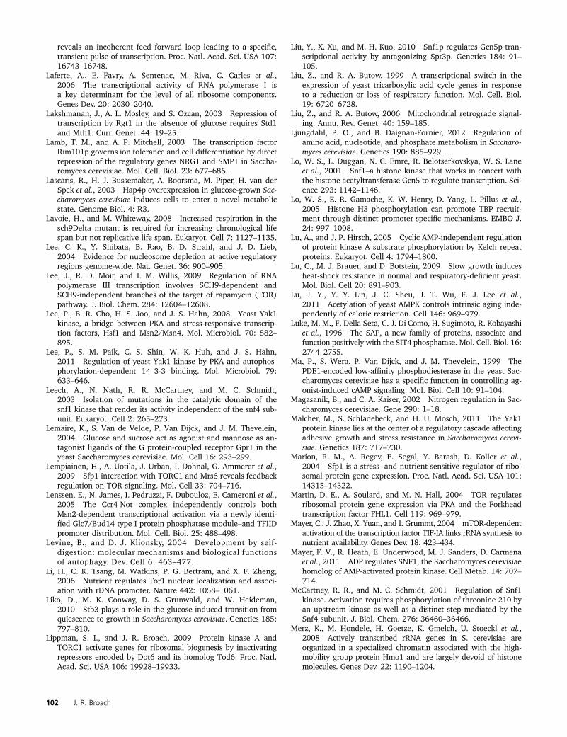
reveals an incoherent feed forward loop leading to a specific,transient pulse of transcription. Proc. Natl. Acad. Sci. USA 107:16743–16748.
Laferte, A., E. Favry, A. Sentenac, M. Riva, C. Carles et al.,2006 The transcriptional activity of RNA polymerase I isa key determinant for the level of all ribosome components.Genes Dev. 20: 2030–2040.
Lakshmanan, J., A. L. Mosley, and S. Ozcan, 2003 Repression oftranscription by Rgt1 in the absence of glucose requires Std1and Mth1. Curr. Genet. 44: 19–25.
Lamb, T. M., and A. P. Mitchell, 2003 The transcription factorRim101p governs ion tolerance and cell differentiation by directrepression of the regulatory genes NRG1 and SMP1 in Saccha-romyces cerevisiae. Mol. Cell. Biol. 23: 677–686.
Lascaris, R., H. J. Bussemaker, A. Boorsma, M. Piper, H. van derSpek et al., 2003 Hap4p overexpression in glucose-grown Sac-charomyces cerevisiae induces cells to enter a novel metabolicstate. Genome Biol. 4: R3.
Lavoie, H., and M. Whiteway, 2008 Increased respiration in thesch9Delta mutant is required for increasing chronological lifespan but not replicative life span. Eukaryot. Cell 7: 1127–1135.
Lee, C. K., Y. Shibata, B. Rao, B. D. Strahl, and J. D. Lieb,2004 Evidence for nucleosome depletion at active regulatoryregions genome-wide. Nat. Genet. 36: 900–905.
Lee, J., R. D. Moir, and I. M. Willis, 2009 Regulation of RNApolymerase III transcription involves SCH9-dependent andSCH9-independent branches of the target of rapamycin (TOR)pathway. J. Biol. Chem. 284: 12604–12608.
Lee, P., B. R. Cho, H. S. Joo, and J. S. Hahn, 2008 Yeast Yak1kinase, a bridge between PKA and stress-responsive transcrip-tion factors, Hsf1 and Msn2/Msn4. Mol. Microbiol. 70: 882–895.
Lee, P., S. M. Paik, C. S. Shin, W. K. Huh, and J. S. Hahn,2011 Regulation of yeast Yak1 kinase by PKA and autophos-phorylation-dependent 14–3-3 binding. Mol. Microbiol. 79:633–646.
Leech, A., N. Nath, R. R. McCartney, and M. C. Schmidt,2003 Isolation of mutations in the catalytic domain of thesnf1 kinase that render its activity independent of the snf4 sub-unit. Eukaryot. Cell 2: 265–273.
Lemaire, K., S. Van de Velde, P. Van Dijck, and J. M. Thevelein,2004 Glucose and sucrose act as agonist and mannose as an-tagonist ligands of the G protein-coupled receptor Gpr1 in theyeast Saccharomyces cerevisiae. Mol. Cell 16: 293–299.
Lempiainen, H., A. Uotila, J. Urban, I. Dohnal, G. Ammerer et al.,2009 Sfp1 interaction with TORC1 and Mrs6 reveals feedbackregulation on TOR signaling. Mol. Cell 33: 704–716.
Lenssen, E., N. James, I. Pedruzzi, F. Dubouloz, E. Cameroni et al.,2005 The Ccr4-Not complex independently controls bothMsn2-dependent transcriptional activation–via a newly identi-fied Glc7/Bud14 type I protein phosphatase module–and TFIIDpromoter distribution. Mol. Cell. Biol. 25: 488–498.
Levine, B., and D. J. Klionsky, 2004 Development by self-digestion: molecular mechanisms and biological functionsof autophagy. Dev. Cell 6: 463–477.
Li, H., C. K. Tsang, M. Watkins, P. G. Bertram, and X. F. Zheng,2006 Nutrient regulates Tor1 nuclear localization and associ-ation with rDNA promoter. Nature 442: 1058–1061.
Liko, D., M. K. Conway, D. S. Grunwald, and W. Heideman,2010 Stb3 plays a role in the glucose-induced transition fromquiescence to growth in Saccharomyces cerevisiae. Genetics 185:797–810.
Lippman, S. I., and J. R. Broach, 2009 Protein kinase A andTORC1 activate genes for ribosomal biogenesis by inactivatingrepressors encoded by Dot6 and its homolog Tod6. Proc. Natl.Acad. Sci. USA 106: 19928–19933.
Liu, Y., X. Xu, and M. H. Kuo, 2010 Snf1p regulates Gcn5p tran-scriptional activity by antagonizing Spt3p. Genetics 184: 91–105.
Liu, Z., and R. A. Butow, 1999 A transcriptional switch in theexpression of yeast tricarboxylic acid cycle genes in responseto a reduction or loss of respiratory function. Mol. Cell. Biol.19: 6720–6728.
Liu, Z., and R. A. Butow, 2006 Mitochondrial retrograde signal-ing. Annu. Rev. Genet. 40: 159–185.
Ljungdahl, P. O., and B. Daignan-Fornier, 2012 Regulation ofamino acid, nucleotide, and phosphate metabolism in Saccharo-myces cerevisiae. Genetics 190: 885–929.
Lo, W. S., L. Duggan, N. C. Emre, R. Belotserkovskya, W. S. Laneet al., 2001 Snf1–a histone kinase that works in concert withthe histone acetyltransferase Gcn5 to regulate transcription. Sci-ence 293: 1142–1146.
Lo, W. S., E. R. Gamache, K. W. Henry, D. Yang, L. Pillus et al.,2005 Histone H3 phosphorylation can promote TBP recruit-ment through distinct promoter-specific mechanisms. EMBO J.24: 997–1008.
Lu, A., and J. P. Hirsch, 2005 Cyclic AMP-independent regulationof protein kinase A substrate phosphorylation by Kelch repeatproteins. Eukaryot. Cell 4: 1794–1800.
Lu, C., M. J. Brauer, and D. Botstein, 2009 Slow growth inducesheat-shock resistance in normal and respiratory-deficient yeast.Mol. Biol. Cell 20: 891–903.
Lu, J. Y., Y. Y. Lin, J. C. Sheu, J. T. Wu, F. J. Lee et al.,2011 Acetylation of yeast AMPK controls intrinsic aging inde-pendently of caloric restriction. Cell 146: 969–979.
Luke, M. M., F. Della Seta, C. J. Di Como, H. Sugimoto, R. Kobayashiet al., 1996 The SAP, a new family of proteins, associate andfunction positively with the SIT4 phosphatase. Mol. Cell. Biol. 16:2744–2755.
Ma, P., S. Wera, P. Van Dijck, and J. M. Thevelein, 1999 ThePDE1-encoded low-affinity phosphodiesterase in the yeast Sac-charomyces cerevisiae has a specific function in controlling ag-onist-induced cAMP signaling. Mol. Biol. Cell 10: 91–104.
Magasanik, B., and C. A. Kaiser, 2002 Nitrogen regulation in Sac-charomyces cerevisiae. Gene 290: 1–18.
Malcher, M., S. Schladebeck, and H. U. Mosch, 2011 The Yak1protein kinase lies at the center of a regulatory cascade affectingadhesive growth and stress resistance in Saccharomyces cerevi-siae. Genetics 187: 717–730.
Marion, R. M., A. Regev, E. Segal, Y. Barash, D. Koller et al.,2004 Sfp1 is a stress- and nutrient-sensitive regulator of ribo-somal protein gene expression. Proc. Natl. Acad. Sci. USA 101:14315–14322.
Martin, D. E., A. Soulard, and M. N. Hall, 2004 TOR regulatesribosomal protein gene expression via PKA and the Forkheadtranscription factor FHL1. Cell 119: 969–979.
Mayer, C., J. Zhao, X. Yuan, and I. Grummt, 2004 mTOR-dependentactivation of the transcription factor TIF-IA links rRNA synthesis tonutrient availability. Genes Dev. 18: 423–434.
Mayer, F. V., R. Heath, E. Underwood, M. J. Sanders, D. Carmenaet al., 2011 ADP regulates SNF1, the Saccharomyces cerevisiaehomolog of AMP-activated protein kinase. Cell Metab. 14: 707–714.
McCartney, R. R., and M. C. Schmidt, 2001 Regulation of Snf1kinase. Activation requires phosphorylation of threonine 210 byan upstream kinase as well as a distinct step mediated by theSnf4 subunit. J. Biol. Chem. 276: 36460–36466.
Merz, K., M. Hondele, H. Goetze, K. Gmelch, U. Stoeckl et al.,2008 Actively transcribed rRNA genes in S. cerevisiae areorganized in a specialized chromatin associated with the high-mobility group protein Hmo1 and are largely devoid of histonemolecules. Genes Dev. 22: 1190–1204.
102 J. R. Broach
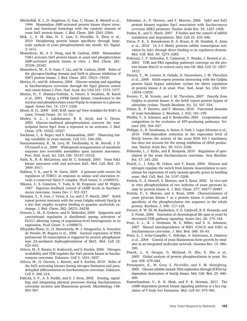
Mitchelhill, K. I., D. Stapleton, G. Gao, C. House, B. Michell et al.,1994 Mammalian AMP-activated protein kinase shares struc-tural and functional homology with the catalytic domain ofyeast Snf1 protein kinase. J. Biol. Chem. 269: 2361–2364.
Mok, J., P. M. Kim, H. Y. Lam, S. Piccirillo, X. Zhou et al.,2010 Deciphering protein kinase specificity through large-scale analysis of yeast phosphorylation site motifs. Sci. Signal.3: ra12.
Momcilovic, M., S. P. Hong, and M. Carlson, 2006 MammalianTAK1 activates Snf1 protein kinase in yeast and phosphorylatesAMP-activated protein kinase in vitro. J. Biol. Chem. 281:25336–25343.
Momcilovic, M., S. H. Iram, Y. Liu, and M. Carlson, 2008 Roles ofthe glycogen-binding domain and Snf4 in glucose inhibition ofSNF1 protein kinase. J. Biol. Chem. 283: 19521–19529.
Moriya, H., and M. Johnston, 2004 Glucose sensing and signalingin Saccharomyces cerevisiae through the Rgt2 glucose sensorand casein kinase I. Proc. Natl. Acad. Sci. USA 101: 1572–1577.
Moriya, H., Y. Shimizu-Yoshida, A. Omori, S. Iwashita, M. Katohet al., 2001 Yak1p, a DYRK family kinase, translocates to thenucleus and phosphorylates yeast Pop2p in response to a glucosesignal. Genes Dev. 15: 1217–1228.
Morse, R. H., 2000 RAP, RAP, open up! New wrinkles for RAP1 inyeast. Trends Genet. 16: 51–53.
Mosley, A. L., J. Lakshmanan, B. K. Aryal, and S. Ozcan,2003 Glucose-mediated phosphorylation converts the tran-scription factor Rgt1 from a repressor to an activator. J. Biol.Chem. 278: 10322–10327.
Nachman, I., A. Regev, and S. Ramanathan, 2007 Dissecting tim-ing variability in yeast meiosis. Cell 131: 544–556.
Narayanaswamy, R., M. Levy, M. Tsechansky, G. M. Stovall, J. D.O’Connell et al., 2009 Widespread reorganization of metabolicenzymes into reversible assemblies upon nutrient starvation.Proc. Natl. Acad. Sci. USA 106: 10147–10152.
Nath, N., R. R. McCartney, and M. C. Schmidt, 2003 Yeast Pak1kinase associates with and activates Snf1. Mol. Cell. Biol. 23:3909–3917.
Neklesa, T. K., and R. W. Davis, 2009 A genome-wide screen forregulators of TORC1 in response to amino acid starvation re-veals a conserved Npr2/3 complex. PLoS Genet. 5: e1000515.
Nikawa, J., S. Cameron, T. Toda, K. M. Ferguson, and M. Wigler,1987 Rigorous feedback control of cAMP levels in Saccharo-myces cerevisiae. Genes Dev. 1: 931–937.
Niranjan, T., X. Guo, J. Victor, A. Lu, and J. P. Hirsch, 2007 Kelchrepeat protein interacts with the yeast Galpha subunit Gpa2p ata site that couples receptor binding to guanine nucleotide ex-change. J. Biol. Chem. 282: 24231–24238.
Octavio, L. M., K. Gedeon, and N. Maheshri, 2009 Epigenetic andconventional regulation is distributed among activators ofFLO11 allowing tuning of population-level heterogeneity in itsexpression. PLoS Genet. 5: e1000673.
Oficjalska-Pham, D., O. Harismendy, W. J. Smagowicz, A. Gonzalezde Peredo, M. Boguta et al., 2006 General repression of RNApolymerase III transcription is triggered by protein phosphatasetype 2A-mediated dephosphorylation of Maf1. Mol. Cell 22:623–632.
Orlova, M., E. Kanter, D. Krakovich, and S. Kuchin, 2006 Nitrogenavailability and TOR regulate the Snf1 protein kinase in Saccha-romyces cerevisiae. Eukaryot. Cell 5: 1831–1837.
Orlova, M., H. Ozcetin, L. Barrett, and S. Kuchin, 2010 Roles ofthe Snf1-activating kinases during nitrogen limitation and pseu-dohyphal differentiation in Saccharomyces cerevisiae. Eukaryot.Cell 9: 208–214.
Palecek, S. P., A. S. Parikh, and S. J. Kron, 2002 Sensing, signal-ling and integrating physical processes during Saccharomycescerevisiae invasive and filamentous growth. Microbiology 148:893–907.
Palomino, A., P. Herrero, and F. Moreno, 2006 Tpk3 and Snf1protein kinases regulate Rgt1 association with Saccharomycescerevisiae HXK2 promoter. Nucleic Acids Res. 34: 1427–1438.
Parker, R., and U. Sheth, 2007 P bodies and the control of mRNAtranslation and degradation. Mol. Cell 25: 635–646.
Parua, P. K., S. Ratnakumar, K. A. Braun, K. M. Dombek, E. Armset al., 2010 14–3-3 (Bmh) proteins inhibit transcription acti-vation by Adr1 through direct binding to its regulatory domain.Mol. Cell. Biol. 30: 5273–5283.
Pedruzzi, I., F. Dubouloz, E. Cameroni, V. Wanke, J. Roosen et al.,2003 TOR and PKA signaling pathways converge on the pro-tein kinase Rim15 to control entry into G0. Mol. Cell 12: 1607–1613.
Peeters, T., W. Louwet, R. Gelade, D. Nauwelaers, J. M. Theveleinet al., 2006 Kelch-repeat proteins interacting with the Galphaprotein Gpa2 bypass adenylate cyclase for direct regulationof protein kinase A in yeast. Proc. Natl. Acad. Sci. USA 103:13034–13039.
Peeters, T., M. Versele, and J. M. Thevelein, 2007 Directly fromGalpha to protein kinase A: the kelch repeat protein bypass ofadenylate cyclase. Trends Biochem. Sci. 32: 547–554.
Pelaez, R., P. Herrero, and F. Moreno, 2010 Functional domainsof yeast hexokinase 2. Biochem. J. 432: 181–190.
Pfeiffer, T., S. Schuster, and S. Bonhoeffer, 2001 Cooperation andcompetition in the evolution of ATP-producing pathways. Sci-ence 292: 504–507.
Philippi, A., R. Steinbauer, A. Reiter, S. Fath, I. Leger-Silvestre et al.,2010 TOR-dependent reduction in the expression level ofRrn3p lowers the activity of the yeast RNA Pol I machinery,but does not account for the strong inhibition of rRNA produc-tion. Nucleic Acids Res. 38: 5315–5326.
Piekarska, I., J. Rytka, and B. Rempola, 2010 Regulation of spor-ulation in the yeast Saccharomyces cerevisiae. Acta Biochim.Pol. 57: 241–250.
Pnueli, L., I. Edry, M. Cohen, and Y. Kassir, 2004 Glucose andnitrogen regulate the switch from histone deacetylation to acet-ylation for expression of early meiosis-specific genes in buddingyeast. Mol. Cell. Biol. 24: 5197–5208.
Portela, P., S. Howell, S. Moreno, and S. Rossi, 2002 In vivo andin vitro phosphorylation of two isoforms of yeast pyruvate ki-nase by protein kinase A. J. Biol. Chem. 277: 30477–30487.
Portela, P., S. Moreno, and S. Rossi, 2006 Characterization ofyeast pyruvate kinase 1 as a protein kinase A substrate, andspecificity of the phosphorylation site sequence in the wholeprotein. Biochem. J. 396: 117–126.
Powers, R. W. III. M. Kaeberlein, S. D. Caldwell, B. K. Kennedy, andS. Fields, 2006 Extension of chronological life span in yeast bydecreased TOR pathway signaling. Genes Dev. 20: 174–184.
Pratt, Z. L., B. J. Drehman, M. E. Miller, and S. D. Johnston,2007 Mutual interdependence of MSI1 (CAC3) and YAK1 inSaccharomyces cerevisiae. J. Mol. Biol. 368: 30–43.
Prinz, S., I. Avila-Campillo, C. Aldridge, A. Srinivasan, K. Dimitrovet al., 2004 Control of yeast filamentous-form growth by mod-ules in an integrated molecular network. Genome Res. 14: 380–390.
Ptacek, J., G. Devgan, G. Michaud, H. Zhu, X. Zhu et al.,2005 Global analysis of protein phosphorylation in yeast. Na-ture 438: 679–684.
Purnapatre, K., M. Gray, S. Piccirillo, and S. M. Honigberg,2005 Glucose inhibits meiotic DNA replication through SCFGrr1p-dependent destruction of Ime2p kinase. Mol. Cell. Biol. 25: 440–450.
Ramachandran, V., K. H. Shah, and P. K. Herman, 2011 ThecAMP-dependent protein kinase signaling pathway is a key reg-ulator of P body foci formation. Mol. Cell 43: 973–981.
How Yeast Respond to Nutrients 103
Ratnakumar, S., and E. T. Young, 2010 Snf1 dependence of per-oxisomal gene expression is mediated by Adr1. J. Biol. Chem.285: 10703–10714.
Ratnakumar, S., N. Kacherovsky, E. Arms, and E. T. Young,2009 Snf1 controls the activity of adr1 through dephosphory-lation of Ser230. Genetics 182: 735–745.
Reid, J. L., V. R. Iyer, P. O. Brown, and K. Struhl, 2000 Coordinateregulation of yeast ribosomal protein genes is associated withtargeted recruitment of Esa1 histone acetylase. Mol. Cell 6:1297–1307.
Roberts, D. N., B. Wilson, J. T. Huff, A. J. Stewart, and B. R. Cairns,2006 Dephosphorylation and genome-wide association ofMaf1 with Pol III-transcribed genes during repression. Mol. Cell22: 633–644.
Roberts, P., S. Moshitch-Moshkovitz, E. Kvam, E. O’Toole, M. Wineyet al., 2003 Piecemeal microautophagy of nucleus in Saccha-romyces cerevisiae. Mol. Biol. Cell 14: 129–141.
Robertson, L. S., and G. R. Fink, 1998 The three yeast A kinaseshave specific signaling functions in pseudohyphal growth. Proc.Natl. Acad. Sci. USA 95: 13783–13787.
Rohde, J. R., and M. E. Cardenas, 2003 The tor pathway regulatesgene expression by linking nutrient sensing to histone acetyla-tion. Mol. Cell. Biol. 23: 629–635.
Roth, S., J. Kumme, and H. J. Schuller, 2004 Transcriptional acti-vators Cat8 and Sip4 discriminate between sequence variants ofthe carbon source-responsive promoter element in the yeastSaccharomyces cerevisiae. Curr. Genet. 45: 121–128.
Rubenstein, E. M., R. R. McCartney, C. Zhang, K. M. Shokat, M. K.Shirra et al., 2008 Access denied: Snf1 activation loop phos-phorylation is controlled by availability of the phosphorylatedthreonine 210 to the PP1 phosphatase. J. Biol. Chem. 283: 222–230.
Rubin-Bejerano, I., S. Sagee, O. Friedman, L. Pnueli, and Y. Kassir,2004 The in vivo activity of Ime1, the key transcriptional ac-tivator of meiosis-specific genes in Saccharomyces cerevisiae, isinhibited by the cyclic AMP/protein kinase A signal pathwaythrough the glycogen synthase kinase 3-beta homolog Rim11.Mol. Cell. Biol. 24: 6967–6979.
Rudra, D., Y. Zhao, and J. R. Warner, 2005 Central role of Ifh1p-Fhl1p interaction in the synthesis of yeast ribosomal proteins.EMBO J. 24: 533–542.
Sabina, J., and M. Johnston, 2009 Asymmetric signal transduc-tion through paralogs that comprise a genetic switch for sugarsensing in Saccharomyces cerevisiae. J. Biol. Chem. 284: 29635–29643.
Sagot, I., B. Pinson, B. Salin, and B. Daignan-Fornier, 2006 Actinbodies in yeast quiescent cells: an immediately available actinreserve? Mol. Biol. Cell 17: 4645–4655.
Saldanha, A. J., M. J. Brauer, and D. Botstein, 2004 Nutritionalhomeostasis in batch and steady-state culture of yeast. Mol.Biol. Cell 15: 4089–4104.
Sancak, Y., L. Bar-Peled, R. Zoncu, A. L. Markhard, S. Nada et al.,2010 Ragulator-Rag complex targets mTORC1 to the lyso-somal surface and is necessary for its activation by amino acids.Cell 141: 290–303.
Sandmeier, J. J., S. French, Y. Osheim, W. L. Cheung, C. M. Galloet al., 2002 RPD3 is required for the inactivation of yeast ri-bosomal DNA genes in stationary phase. EMBO J. 21: 4959–4968.
Santangelo, G. M., 2006 Glucose signaling in Saccharomyces cer-evisiae. Microbiol. Mol. Biol. Rev. 70: 253–282.
Santhanam, A., A. Hartley, K. Duvel, J. R. Broach, and S. Garrett,2004 PP2A phosphatase activity is required for stress and Torkinase regulation of yeast stress response factor Msn2p. Eukar-yot. Cell 3: 1261–1271.
Sanz, P., 2003 Snf1 protein kinase: a key player in the response tocellular stress in yeast. Biochem. Soc. Trans. 31: 178–181.
Sarbassov, D. D., S. M. Ali, and D. M. Sabatini, 2005 Growingroles for the mTOR pathway. Curr. Opin. Cell Biol. 17: 596–603.
Schawalder, S. B., M. Kabani, I. Howald, U. Choudhury, M.Werner et al., 2004 Growth-regulated recruitment of the es-sential yeast ribosomal protein gene activator Ifh1. Nature432: 1058–1061.
Scherens, B., A. Feller, F. Vierendeels, F. Messenguy, and E. Dubois,2006 Identification of direct and indirect targets of the Gln3and Gat1 activators by transcriptional profiling in response tonitrogen availability in the short and long term. FEMS Yeast Res.6: 777–791.
Schmidt, M. C., and R. R. McCartney, 2000 beta-subunits of Snf1kinase are required for kinase function and substrate definition.EMBO J. 19: 4936–4943.
Schmidt, M. C., R. R. McCartney, X. Zhang, T. S. Tillman, H. Solimeoet al., 1999 Std1 and Mth1 proteins interact with the glucosesensors to control glucose-regulated gene expression in Saccha-romyces cerevisiae. Mol. Cell. Biol. 19: 4561–4571.
Schuller, H. J., 2003 Transcriptional control of nonfermentativemetabolism in the yeast Saccharomyces cerevisiae. Curr. Genet.43: 139–160.
Shamji, A. F., F. G. Kuruvilla, and S. L. Schreiber, 2000 Partitioningthe transcriptional program induced by rapamycin among the ef-fectors of the Tor proteins. Curr. Biol. 10: 1574–1581.
Shirra, M. K., R. R. McCartney, C. Zhang, K. M. Shokat, M. C.Schmidt et al., 2008 A chemical genomics study identifiesSnf1 as a repressor of GCN4 translation. J. Biol. Chem. 283:35889–35898.
Silverman, S. J., A. A. Petti, N. Slavov, L. Parsons, R. Briehof et al.,2010 Metabolic cycling in single yeast cells from unsynchro-nized steady-state populations limited on glucose or phosphate.Proc. Natl. Acad. Sci. USA 107: 6946–6951.
Singh, J., and M. Tyers, 2009 A Rab escort protein integrates thesecretion system with TOR signaling and ribosome biogenesis.Genes Dev. 23: 1944–1958.
Slavov, N., and D. Botstein, 2011 Coupling among growth rateresponse, metabolic cycle, and cell division cycle in yeast. Mol.Biol. Cell 22: 1997–2009.
Smets, B., R. Ghillebert, P. De Snijder, M. Binda, E. Swinnen et al.,2010 Life in the midst of scarcity: adaptations to nutrientavailability in Saccharomyces cerevisiae. Curr. Genet. 56: 1–32.
Smith, A., M. P. Ward, and S. Garrett, 1998 Yeast PKA repressesMsn2p/Msn4p-dependent gene expression to regulate growth,stress response and glycogen accumulation. EMBO J. 17: 3556–3564.
Spielewoy, N., K. Flick, T. I. Kalashnikova, J. R. Walker, and C.Wittenberg, 2004 Regulation and recognition of SCFGrr1 tar-gets in the glucose and amino acid signaling pathways. Mol.Cell. Biol. 24: 8994–9005.
Sutherland, C. M., S. A. Hawley, R. R. McCartney, A. Leech, M. J.Stark et al., 2003 Elm1p is one of three upstream kinases forthe Saccharomyces cerevisiae SNF1 complex. Curr. Biol. 13:1299–1305.
Swinnen, E., V. Wanke, J. Roosen, B. Smets, F. Dubouloz et al.,2006 Rim15 and the crossroads of nutrient signalling path-ways in Saccharomyces cerevisiae. Cell Div. 1: 3.
Tachibana, C., J. Y. Yoo, J. B. Tagne, N. Kacherovsky, T. I. Lee et al.,2005 Combined global localization analysis and transcriptomedata identify genes that are directly coregulated by Adr1 andCat8. Mol. Cell. Biol. 25: 2138–2146.
Tagkopoulos, I., Y. C. Liu, and S. Tavazoie, 2008 Predictive be-havior within microbial genetic networks. Science 320: 1313–1317.
Tate, J. J., and T. G. Cooper, 2003 Tor1/2 regulation of retro-grade gene expression in Saccharomyces cerevisiae derivesindirectly as a consequence of alterations in ammonia metabo-lism. J. Biol. Chem. 278: 36924–36933.
104 J. R. Broach
Tate, J. J., I. Georis, A. Feller, E. Dubois, and T. G. Cooper,2009 Rapamycin-induced Gln3 dephosphorylation is insuffi-cient for nuclear localization: Sit4 and PP2A phosphatases areregulated and function differently. J. Biol. Chem. 284: 2522–2534.
Tate, J. J., I. Georis, E. Dubois, and T. G. Cooper, 2010 Distinctphosphatase requirements and GATA factor responses to nitro-gen catabolite repression and rapamycin treatment in Saccha-romyces cerevisiae. J. Biol. Chem. 285: 17880–17895.
Thomson, J. M., E. A. Gaucher, M. F. Burgan, D. W. De Kee, T. Liet al., 2005 Resurrecting ancestral alcohol dehydrogenasesfrom yeast. Nat. Genet. 37: 630–635.
Townley, R., and L. Shapiro, 2007 Crystal structures of the adeny-late sensor from fission yeast AMP-activated protein kinase. Sci-ence 315: 1726–1729.
Towpik, J., D. Graczyk, A. Gajda, O. Lefebvre, and M. Boguta,2008 Derepression of RNA polymerase III transcription byphosphorylation and nuclear export of its negative regulator,Maf1. J. Biol. Chem. 283: 17168–17174.
Tu, B. P., A. Kudlicki, M. Rowicka, and S. L. McKnight, 2005 Logicof the yeast metabolic cycle: temporal compartmentalization ofcellular processes. Science 310: 1152–1158.
Tu, J., and M. Carlson, 1995 REG1 binds to protein phosphatasetype 1 and regulates glucose repression in Saccharomyces cer-evisiae. EMBO J. 14: 5939–5946.
Turcotte, B., X. B. Liang, F. Robert, and N. Soontorngun,2010 Transcriptional regulation of nonfermentable carbonutilization in budding yeast. FEMS Yeast Res. 10: 2–13.
Urban, J., A. Soulard, A. Huber, S. Lippman, D. Mukhopadhyayet al., 2007 Sch9 is a major target of TORC1 in Saccharomycescerevisiae. Mol. Cell 26: 663–674.
Usaite, R., M. C. Jewett, A. P. Oliveira, J. R. Yates 3rd. L. Olssonet al., 2009 Reconstruction of the yeast Snf1 kinase regulatorynetwork reveals its role as a global energy regulator. Mol. Syst.Biol. 5: 319.
Uttenweiler, A., and A. Mayer, 2008 Microautophagy in the yeastSaccharomyces cerevisiae. Methods Mol. Biol. 445: 245–259.
Vander Heiden, M. G., L. C. Cantley, and C. B. Thompson,2009 Understanding the Warburg effect: the metabolic re-quirements of cell proliferation. Science 324: 1029–1033.
van Oevelen, C. J., H. A. van Teeffelen, F. J. van Werven, and H. T.Timmers, 2006 Snf1p-dependent Spt-Ada-Gcn5-acetyltrans-ferase (SAGA) recruitment and chromatin remodeling activitieson the HXT2 and HXT4 promoters. J. Biol. Chem. 281: 4523–4531.
Vincent, O., and M. Carlson, 1999 Gal83 mediates the interactionof the Snf1 kinase complex with the transcription activator Sip4.EMBO J. 18: 6672–6681.
Vincent, O., R. Townley, S. Kuchin, and M. Carlson, 2001 Subcellularlocalization of the Snf1 kinase is regulated by specific beta subunitsand a novel glucose signaling mechanism. Genes Dev. 15: 1104–1114.
von Plehwe, U., U. Berndt, C. Conz, M. Chiabudini, E. Fitzke et al.,2009 The Hsp70 homolog Ssb is essential for glucose sensingvia the SNF1 kinase network. Genes Dev. 23: 2102–2115.
Wade, J. T., D. B. Hall, and K. Struhl, 2004 The transcriptionfactor Ifh1 is a key regulator of yeast ribosomal protein genes.Nature 432: 1054–1058.
Wang, G., and R. J. Deschenes, 2006 Plasma membrane localiza-tion of Ras requires class C Vps proteins and functional mito-chondria in Saccharomyces cerevisiae. Mol. Cell. Biol. 26: 3243–3255.
Wang, Y., M. Pierce, L. Schneper, C. G. Guldal, X. Zhang et al.,2004 Ras and Gpa2 mediate one branch of a redundant glu-cose signaling pathway in yeast. PLoS Biol. 2: E128.
Williamson, T., J. M. Schwartz, D. B. Kell, and L. Stateva,2009 Deterministic mathematical models of the cAMP path-way in Saccharomyces cerevisiae. BMC Syst. Biol. 3: 70.
Willis, I. M., and R. D. Moir, 2007 Integration of nutritional andstress signaling pathways by Maf1. Trends Biochem. Sci. 32: 51–53.
Wilson, W. A., S. A. Hawley, and D. G. Hardie, 1996 Glucose re-pression/derepression in budding yeast: SNF1 protein kinase isactivated by phosphorylation under derepressing conditions,and this correlates with a high AMP:ATP ratio. Curr. Biol. 6:1426–1434.
Wittner, M., S. Hamperl, U. Stockl, W. Seufert, H. Tschochner et al.,2011 Establishment and maintenance of alternative chromatinstates at a multicopy gene locus. Cell 145: 543–554.
Woods, A., M. R. Munday, J. Scott, X. Yang, M. Carlson et al.,1994 Yeast SNF1 is functionally related to mammalian AMP-activated protein kinase and regulates acetyl-CoA carboxylasein vivo. J. Biol. Chem. 269: 19509–19515.
Wu, B., K. Ottow, P. Poulsen, R. F. Gaber, E. Albers et al.,2006 Competitive intra- and extracellular nutrient sensingby the transporter homologue Ssy1p. J. Cell Biol. 173: 327–331.
Wuster, A., and M. M. Babu, 2010 Transcriptional control of thequorum sensing response in yeast. Mol. Biosyst. 6: 134–141.
Xue, Y., M. Batlle, and J. P. Hirsch, 1998 GPR1 encodes a putativeG protein-coupled receptor that associates with the Gpa2p Gal-pha subunit and functions in a Ras-independent pathway. EMBOJ. 17: 1996–2007.
Yan, G., X. Shen, and Y. Jiang, 2006 Rapamycin activates Tap42-associated phosphatases by abrogating their association withTor complex 1. EMBO J. 25: 3546–3555.
Yan, G., Y. Lai, and Y. Jiang, 2012 The TOR complex 1 is a directtarget of Rho1 GTPase. Mol. Cell 45: 1–11.
Yang, Z., and D. J. Klionsky, 2009 An overview of the molecularmechanism of autophagy. Curr. Top. Microbiol. Immunol. 335:1–32.
Yorimitsu, T., S. Zaman, J. R. Broach, and D. J. Klionsky,2007 Protein kinase A and Sch9 cooperatively regulate induc-tion of autophagy in Saccharomyces cerevisiae. Mol. Biol. Cell18: 4180–4189.
Young, E. T., K. M. Dombek, C. Tachibana, and T. Ideker,2003 Multiple pathways are co-regulated by the protein ki-nase Snf1 and the transcription factors Adr1 and Cat8. J. Biol.Chem. 278: 26146–26158.
Zaman, S., S. I. Lippman, X. Zhao, and J. R. Broach, 2008 HowSaccharomyces responds to nutrients. Annu. Rev. Genet. 42: 27–81.
Zaman, S., S. I. Lippman, L. Schneper, N. Slonim, and J. R. Broach,2009 Glucose regulates transcription in yeast through a net-work of signaling pathways. Mol. Syst. Biol. 5: 245.
Zeller, C. E., S. C. Parnell, and H. G. Dohlman, 2007 The RACK1ortholog Asc1 functions as a G-protein beta subunit coupled toglucose responsiveness in yeast. J. Biol. Chem. 282: 25168–25176.
Zhao, H., J. Q. Wang, T. Shimohata, G. Sun, M. A. Yenari et al.,2007 Conditions of protection by hypothermia and effects onapoptotic pathways in a rat model of permanent middle cerebralartery occlusion. J. Neurosurg. 107: 636–641.
Zhao, Y., K. B. McIntosh, D. Rudra, S. Schawalder, D. Shore et al.,2006 Fine-structure analysis of ribosomal protein gene tran-scription. Mol. Cell. Biol. 26: 4853–4862.
Zhu, C., K. J. Byers, R. P. McCord, Z. Shi, M. F. Berger et al.,2009 High-resolution DNA-binding specificity analysis of yeasttranscription factors. Genome Res. 19: 556–566.
Communicating editor: A. Hopper
How Yeast Respond to Nutrients 105