Embed Size (px)
Citation preview
37
Nutrition for Enhancing Bone Volume in Mice
Junji Ohtani, Fujita Tadashi, R.A. Marquez Hernandez, Toshitsugu Kawata,
Masato Kaku, Masahide Motokawa and Kazuo Tanne Department of Orthodontics and Craniofacial Developmental Biology, Hiroshima
University Graduate School of Biomedical Sciences, Hiroshima, Japan
1. Introduction
It is well known that postmenopausal osteoporosis is related to severe decreases in the serum estrogen levels. Estrogen deficiency produces an imbalance in the bone remodeling balance which is associated with a reduced bone volume and higher fracture risk. Androgen deficiency also produces osteoporosis by directly affecting bone cells. In addition, androgen was demonstrated to be present during the fetal period, and it is considered to play an essential role in the sexual differentiation of males. Therefore, both sex hormones, androgen and estrogen, are great important for bone homeostasis not only in females but also in males. This is further supported by the fact that both males and females express androgen
receptor (AR), estrogen receptor-alpha (ER) and estrogen receptor-beta (ER). Androgen
and estrogen react with the AR and ER and ERrespectively. It is thus speculated that they mutually regulate themselves during bone development and remodeling. Up to present, various studies have examined the roles of estrogen and its receptors to explore potential methods for preventing or treating postmenopausal osteoporosis (Judd et al., 1983; Kousteni et al., 2002; Martin-Milan et al., 2010; Pietschmann et al., 2008). Recent clinical studies have reported that the prevention of hip bone fractures and vertebral deformities is highly pertinent to improving the quality of life for older people (Chang et al., 2004; O’Neill et al., 2009). To avoid these incidents, we have to try to achieve adequate peak bone mass during adolescent growth, it is also important to understand the bone growth, because this is currently an under-investigated area. In adult animal experiments, orchiectomy and ovariectomy reduced both the bone volume and density. Furthermore, the bone volume loss was induced not only in the long bones but also in the mandibular condyles (Fujita et al., 2001). In addition, we have reported that these phenomena were also shown in immature mice (Fujita et al., 2006). Therefore, the influence of sex hormones on bone remodeling has been demonstrated in the craniofacial region. In orthodontic and orthopedic treatment, it is especially, difficult to predict bone growth including craniofacial and mandibular growth during adolescence. The full nature of bone growth, in association with sex hormones remains to be fully elucidated. The ratio of osteoporosis patient in Japan based on the diagnosed by Japanese society for bone and mineral research is reported 24% of over fifty-year-old people and it is indicated the number of patients is higher than those in USA and European countries (Orimo, 2000).
www.intechopen.com
Osteoporosis
766
Therefore, we all recognize osteoporosis is one of serious social problems in Japan. This fact indicates that the influences of sex hormones on bone metabolism are very complicated, and the mechanism is very difficult to be understood. Peak bone mass, in general, is acquired from childhood to adolescence, but 35% of cortical bone and 50% of trabecular bone are lost gradually thereafter after. The importance of preventive medicine thus has been gradually recognized in the field of orthopaedic surgery with a concept that peak bone mass is to be increased and saved during childhood as much as possible (Lapauw et al., 2009; Kaufman, 2009). Such an idea has been attracting a special attention in the field of clinical medicine for the prevention of osteoporosis. The importance of normal development and growth of bone until adolescence has also been reported (Karlsson et al., 2008; Wang et al., 2008). This fact suggested that if we are able to increase the peak bone mass during adolescence as possible by providing some effective nutrition for increasing bone volume, it may leads to prevent the risk and to delay the appearance of osteoporosis. According to the prevention and treatment of guideline in osteoporosis, it was
recommended to intake daily dose of calcium is higher (Rosen Gallagher, 2011). However, the amount of calcium intake is approximately 500mg per one day in practically, and even added the consumption applied by supplement, could not reach daily objective intake. A newly developed snack used in our experiments contains appropriate amounts of minerals (calcium and magnesium ) and casein phosphopeptide (CPP) as well as soybean isoflavone which has a sex hormone-like action, which is recognized as a specified food for health, is also included. The objective of this chapter was to introduce the effects of bone remodeling including bone volume, density and strength in osteoporosis and healthy growing mice fed a special composition nutrition.
2. Nutrition
2.1 Function of nutrition
Recently, it was focused on the prevention of osteoporosis and life style related disease like a diabetic and heart disease. Under such situation, people are well considered the food function. It is well known that the function of foodstuff has mainly for nutrition and taste. Additionally, it has also the important function as a decreasing of risk in some disease. The development of foodstuff which has the specific function for living body adjustment is going forward actively in nowadays, therefore the daily food which we intake were diversify. Now it was reported that there are detected and demonstrated in some nutrition that the function of anti-thrombosis, anti-oxidation, encouragement to assimilate calcium, controlling of blood pressure and improvement of cholesterol readings as a food for special health uses and Japanese government had approved it (Yamada et al., 2008). For example, beta-carotene was known as a great nutrition because of its essential function, foodstuff factor as a provitamin, taste factor by its color in vegetable and fruit, and the role of anti-oxidation (Kim et al., 2010).
2.2 Casein phosphopeptide
CPP is also well known as a factor of encouragement to assimilate calcium. Milk and dairy products contains CPP together with functional protein like a milky basic protein and lactoferrin. Casein as a milk protein is a protein containing phosphoric acid, and is decomposed into oligopeptide of various sizes by the digestive enzyme. It became
www.intechopen.com
Nutrition for Enhancing Bone Volume in Mice
767
clarified that special protein containing many phosphoric acid called CPP. Generally, calcium is the fifth abundant element by mass in the human body, where it is a common cellular ionic messenger with many functions, and serves also as a structural element in bone. And it has four stable isotopes (40Ca, 42Ca, 43Ca and 44Ca) plus two more isotopes (46Ca and 48Ca). 97% of naturally occurring calcium is in the form of 40Ca which is a one of
daughter products of 40K decay, along with 40Ar (Mueller Boehm, 2011; Skulan DePaolo, 1999). Calcium is hard to be absorbed in human body since it combines with negative ion such as a phosphoric acid in a small intestine. However, CPP prevents own insoluble and promote its efficiency of absorption in a small intestine due to a part of serine residue is phosphorylated and the ionic bond of CPP is carried out to calcium with the negative ion of phosphate group. Moreover, the function in the promotion of absorption efficacy of CPP, it is positively
applied in the form of casein phosphopeptide-amorphous calcium phosphate (CPP-ACP),
and is widely well-known as Recaldent. CPP from the major protein of milk have the
remarkable ability to stabilize calcium, phosphate and fluoride ions as water soluble
amorphous complexes that provide bioavairable calcium, phosphate and fluoride ions to
the tooth. Animal and short-term human clinical trials to repair early staged of tooth
decay by replacing the calcium and phosphate ions lost due to decay (Reynolds, 2009;
Llena et al., 2009). Long-term controlled clinical trials also have demonstrated the efficacy
of CPP-ACP is slowing the progression of dental caries and in regressing early stages of
tooth decay (Andersson et al., 2007; Morgan et al., 2008). Moreover, it was reported that it
is effective rather than the 500ppm sodium fluoride paste which the dentists have used
conventionally for the purpose of prevention of tooth decay (Zhang et al., 2011;
Altenburger et al., 2010).
On the other hand, it was reported that the calcium in food exists 50~60% in milk,
approximately 20% in small fish and approximately 30% in vegetable. But, absorption
efficacy of calcium in human body is not much excellent. Therefore, CPP is further
developed as “food suitable for the people of calcium shortage” such as suger-free gum,
tooth cream, dairy milk and another drink and Japanese tofu. However, with such
professional tool, food and drink form, we speculated that it will be probably difficult to
take freely and daily for the purpose of increasing the absorption efficacy of calcium.
3. Animal study
3.1 Materials and methods
Fifty male and fifty female C57BL/6J mice of twelve-week-old and thirty male and thirty
female C57BL/6J mice of five-day-old (Jackson Laboratory, Bar Harbar, ME, USA) were
used in this experiment and divided respectively into eight groups with ten in each sex. In
the experimental groups, orchiectomy (ORX) and ovariectomy (OVX) were performed for
ten mice of twelve-week-old in each gender at the beginning of experiment to create the
situation of osteoporosis model. ORX and OVX were performed by means of a stereoscopic
microscope (SZX9, Olympus Optical Co., Tokyo, Japan) under general anesthesia with
sodium pentobarbital, whereas the control mice underwent sham operation. The detail of
time schedules in adult and growing mice during a series of experiment are shown in Table
1 and 2, respectively. These animals were treated under the ethical regulations defined by
the Ethics Committee, Hiroshima University Faculty of Dentistry.
www.intechopen.com
Osteoporosis
768
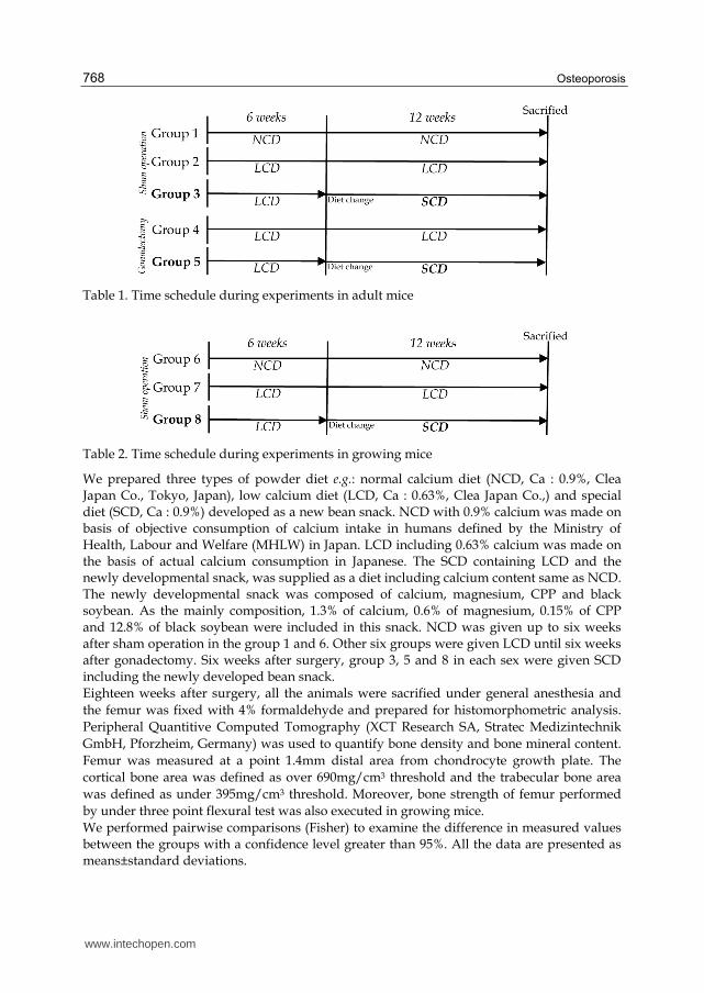
Table 1. Time schedule during experiments in adult mice
Table 2. Time schedule during experiments in growing mice
We prepared three types of powder diet e.g.: normal calcium diet (NCD, Ca : 0.9%, Clea Japan Co., Tokyo, Japan), low calcium diet (LCD, Ca : 0.63%, Clea Japan Co.,) and special diet (SCD, Ca : 0.9%) developed as a new bean snack. NCD with 0.9% calcium was made on basis of objective consumption of calcium intake in humans defined by the Ministry of Health, Labour and Welfare (MHLW) in Japan. LCD including 0.63% calcium was made on the basis of actual calcium consumption in Japanese. The SCD containing LCD and the newly developmental snack, was supplied as a diet including calcium content same as NCD. The newly developmental snack was composed of calcium, magnesium, CPP and black soybean. As the mainly composition, 1.3% of calcium, 0.6% of magnesium, 0.15% of CPP and 12.8% of black soybean were included in this snack. NCD was given up to six weeks after sham operation in the group 1 and 6. Other six groups were given LCD until six weeks after gonadectomy. Six weeks after surgery, group 3, 5 and 8 in each sex were given SCD including the newly developed bean snack. Eighteen weeks after surgery, all the animals were sacrified under general anesthesia and
the femur was fixed with 4% formaldehyde and prepared for histomorphometric analysis.
Peripheral Quantitive Computed Tomography (XCT Research SA, Stratec Medizintechnik
GmbH, Pforzheim, Germany) was used to quantify bone density and bone mineral content.
Femur was measured at a point 1.4mm distal area from chondrocyte growth plate. The
cortical bone area was defined as over 690mg/cm3 threshold and the trabecular bone area
was defined as under 395mg/cm3 threshold. Moreover, bone strength of femur performed
by under three point flexural test was also executed in growing mice.
We performed pairwise comparisons (Fisher) to examine the difference in measured values between the groups with a confidence level greater than 95%. All the data are presented as means±standard deviations.
www.intechopen.com
Nutrition for Enhancing Bone Volume in Mice
769
3.2 In ORX and OVX mice
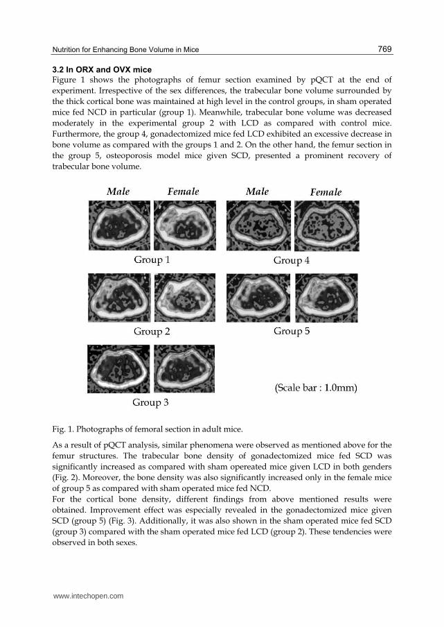
Figure 1 shows the photographs of femur section examined by pQCT at the end of
experiment. Irrespective of the sex differences, the trabecular bone volume surrounded by
the thick cortical bone was maintained at high level in the control groups, in sham operated
mice fed NCD in particular (group 1). Meanwhile, trabecular bone volume was decreased
moderately in the experimental group 2 with LCD as compared with control mice.
Furthermore, the group 4, gonadectomized mice fed LCD exhibited an excessive decrease in
bone volume as compared with the groups 1 and 2. On the other hand, the femur section in
the group 5, osteoporosis model mice given SCD, presented a prominent recovery of
trabecular bone volume.
Fig. 1. Photographs of femoral section in adult mice.
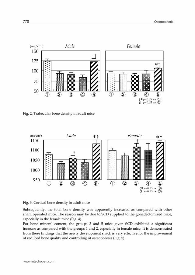
As a result of pQCT analysis, similar phenomena were observed as mentioned above for the
femur structures. The trabecular bone density of gonadectomized mice fed SCD was
significantly increased as compared with sham opereated mice given LCD in both genders
(Fig. 2). Moreover, the bone density was also significantly increased only in the female mice
of group 5 as compared with sham operated mice fed NCD.
For the cortical bone density, different findings from above mentioned results were
obtained. Improvement effect was especially revealed in the gonadectomized mice given
SCD (group 5) (Fig. 3). Additionally, it was also shown in the sham operated mice fed SCD
(group 3) compared with the sham operated mice fed LCD (group 2). These tendencies were
observed in both sexes.
www.intechopen.com
Osteoporosis
770
Fig. 2. Trabecular bone density in adult mice
Fig. 3. Cortical bone density in adult mice
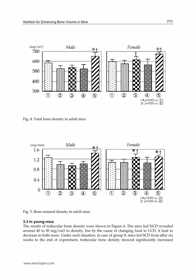
Subsequently, the total bone density was apparently increased as compared with other
sham operated mice. The reason may be due to SCD supplied to the gonadectomized mice,
especially in the female mice (Fig. 4).
For bone mineral content, the groups 3 and 5 mice given SCD exhibited a significant
increase as compared with the groups 1 and 2, especially in female mice. It is demonstrated
from these findings that the newly development snack is very effective for the improvement
of reduced bone quality and controlling of osteoporosis (Fig. 5).
www.intechopen.com
Nutrition for Enhancing Bone Volume in Mice
771
Fig. 4. Total bone density in adult mice
Fig. 5. Bone mineral density in adult mice
3.3 In young mice
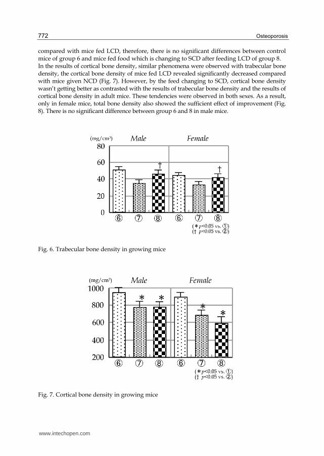
The results of trabecular bone density were shown in Figure 6. The mice fed NCD revealed around 40 to 50 mg/cm3 in density, but by the cause of changing food to LCD, it lead to decrease in both sexes. Under such situation, in case of group 8, mice fed SCD from after six weeks to the end of experiment, trabecular bone density showed significantly increased
www.intechopen.com
Osteoporosis
772
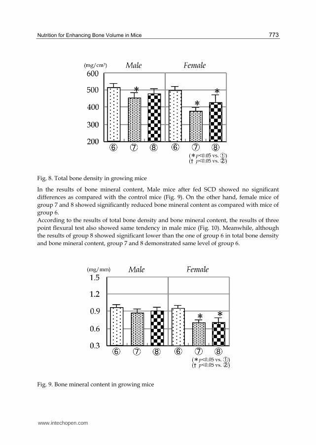
compared with mice fed LCD, therefore, there is no significant differences between control mice of group 6 and mice fed food which is changing to SCD after feeding LCD of group 8. In the results of cortical bone density, similar phenomena were observed with trabecular bone density, the cortical bone density of mice fed LCD revealed significantly decreased compared with mice given NCD (Fig. 7). However, by the feed changing to SCD, cortical bone density wasn’t getting better as contrasted with the results of trabecular bone density and the results of cortical bone density in adult mice. These tendencies were observed in both sexes. As a result, only in female mice, total bone density also showed the sufficient effect of improvement (Fig. 8). There is no significant difference between group 6 and 8 in male mice.
Fig. 6. Trabecular bone density in growing mice
Fig. 7. Cortical bone density in growing mice
www.intechopen.com
Nutrition for Enhancing Bone Volume in Mice
773
Fig. 8. Total bone density in growing mice
In the results of bone mineral content, Male mice after fed SCD showed no significant
differences as compared with the control mice (Fig. 9). On the other hand, female mice of
group 7 and 8 showed significantly reduced bone mineral content as compared with mice of
group 6.
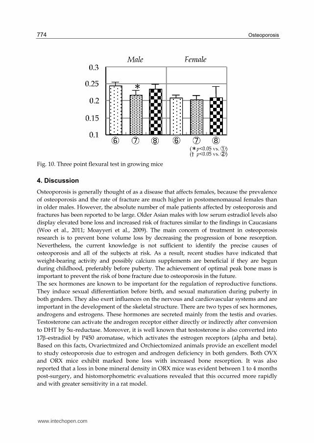
According to the results of total bone density and bone mineral content, the results of three
point flexural test also showed same tendency in male mice (Fig. 10). Meanwhile, although
the results of group 8 showed significant lower than the one of group 6 in total bone density
and bone mineral content, group 7 and 8 demonstrated same level of group 6.
Fig. 9. Bone mineral content in growing mice
www.intechopen.com
Osteoporosis
774
Fig. 10. Three point flexural test in growing mice
4. Discussion
Osteoporosis is generally thought of as a disease that affects females, because the prevalence
of osteoporosis and the rate of fracture are much higher in postomenomausal females than
in older males. However, the absolute number of male patients affected by osteoporosis and
fractures has been reported to be large. Older Asian males with low serum estradiol levels also
display elevated bone loss and increased risk of fractures similar to the findings in Caucasians
(Woo et al., 2011; Moayyeri et al., 2009). The main concern of treatment in osteoporosis
research is to prevent bone volume loss by decreasing the progression of bone resorption.
Nevertheless, the current knowledge is not sufficient to identify the precise causes of
osteoporosis and all of the subjects at risk. As a result, recent studies have indicated that
weight-bearing activity and possibly calcium supplements are beneficial if they are begun
during childhood, preferably before puberty. The achievement of optimal peak bone mass is
important to prevent the risk of bone fracture due to osteoporosis in the future.
The sex hormones are known to be important for the regulation of reproductive functions.
They induce sexual differentiation before birth, and sexual maturation during puberty in
both genders. They also exert influences on the nervous and cardiovascular systems and are
important in the development of the skeletal structure. There are two types of sex hormones,
androgens and estrogens. These hormones are secreted mainly from the testis and ovaries.
Testosterone can activate the androgen receptor either directly or indirectly after conversion
to DHT by 5-reductase. Moreover, it is well known that testosterone is also converted into
17-estradiol by P450 aromatase, which activates the estrogen receptors (alpha and beta).
Based on this facts, Ovariectmized and Orchiectomized animals provide an excellent model
to study osteoporosis due to estrogen and androgen deficiency in both genders. Both OVX
and ORX mice exhibit marked bone loss with increased bone resorption. It was also
reported that a loss in bone mineral density in ORX mice was evident between 1 to 4 months
post-surgery, and histomorphometric evaluations revealed that this occurred more rapidly
and with greater sensitivity in a rat model.
www.intechopen.com
Nutrition for Enhancing Bone Volume in Mice
775
This snack we had developed was composed of calcium, magnesium, CPP and black
soybean. We have examined into body weight and height of all mice during experimental
period. However, all mice of supplied food in these measurements were no significant
difference compared with the control groups in both sexes (data were not shown). In
addition, the amount of all nutrition including supplied food was restrained according the
instructions of MHLW in Japan. Therefore, we assumed that supplied special diet food has
no occurrence of side effect following snack administration.
It is well known that calcium intake decreases the risk of bone fracture. It was also reported
that the calcium supplementation suppress bone formation when magnesium is deficient
(Mora Gilsanz, 2003). Based on these reports, we designed to mix calcium and magnesium
at an appropriate proportion of 2 to 1. These contents in this snack were compounded to
fulfill ninety percentage of the daily-required nutrition defined by MHLW in Japan.
It was also reported that addition of CPP will have a beneficial effect on the absorption of
calcium (Dontas Yiannakopoulos, 2007). In Japan, CPP was accredited as a food for
qualified health uses by MHLW. Accordingly, we add a certain amount of CPP to absorb
mineral content easily.
The black soybean contains isoflavone which is widely accepted to have a weak estrogen
activity, and to be able to bind estrogen receptor. Actually, Brandi and Miyauchi et al.,
reported that isoflavone had either effect for the suppression of bone resorption or
enhancement of bone formation, affecting directly both osteoclasts and osteoblasts (Brandi,
2003; Miyauchi et al., 1996). In addition, isoflavone was revealed to affect bone metabolism
similarly to the sex hormone-like effect in male (Chavarro et al., 2008). Recently, it was also
reported that isoflavones have revealed inhibition of bone loss in castrated male mice and
growing male mice respectively (Fujioka et al., 2007; Ishimi et al., 2002; Khalil et al., 2005).
Under such background, a new snack was developed by use of black soybean. The amount
of isoflavone in this snack was determined with a special reference to the amount of safety
surplus nutrition per day defined by MHLW.
Decrease in the trabecular and cortical bone volumes after gonadectomy has already been
demonstrated by our previously reports (Fujita et al., 2001, 2004). We also reported that
bone growth was significantly suppressed in the gonadectomized mice immediately after
birth (Fujita et al., 2006). Moreover, we clarified that decrease in bone volume was occurred
four weeks after gonadectomy. According to these results, we examined bone density of the
femur six weeks after ovariectomy and orchiectomy.
The density of trabecular and cortical bones in the gonadectomized mice given LCD was
significantly lower than in the sham operated mice given NCD and LCD, respectively. It
was revealed that the deficiency of calcium intake caused decrease in bone density, under
sex hormone disturbances in particular. Therefore, it is speculated that the bone density is
below the optimal level in Japanese and fracture risk may become higher for aged people.
These findings also support that the incidence of primary osteoporosis is higher in Japan
than in American and European countries.
The bone metabolism is classified into two types, high turnover type to accelerate both bone
formation and resorption and low turnover type caused by degradation of bone formation.
In this study, twelve-week-old mice with sham operation fed LCD are regarded as young
growing humans with low turnover type, whereas gonadectomy mice to simulate sex
hormone disturbance in the experimental groups are assumed to be under the condition of
www.intechopen.com
Osteoporosis
776
postmenopausal osteoporosis as a high turnover type. Irrespective of the turnover types,
however, the bone density in the group 5 given appropriate amount of calcium by
supplying SCD exhibited a remarkable increase. It is thus suggested that sufficient calcium
quantity through nutrition of newly developed bean snack enhanced bone formation
irrespective of age.
It is reported that postmenopausal women with daily calcium intake of less than 400mg
experience significant bone loss and that calcium intake of 800mg per day is effective for
improving postmenopausal bone loss (Dawson-Hughes et al., 1990; Reid et al., 1993). On the
other hand, it is well known that improvement effect against bone volume loss by calcium
intake is available only at the initial stage of treatment. In addition, Riggs et al., clarified that
the effect on bone loss is weaker than those reported for estrogen and bisphosphonates
therapy, indicating that calcium supplements alone can’t substitute treatment for
osteoporosis (Riggs et al., 1998). In this study, longitudinal effect of newly developmental
snack intake was not examined. However, It is hopefully anticipated that new bean snack
could contributed to the enrichment of QOL as a nutrition function food, because of it was
contain of several ingredients to promote assimilation efficiency of calcium.
5. Conclusions
The new snack we developed included proper amount of calcium, magnesium, CPP and
black soybean. When this product was given to the osteoporosis model mice, bone density
of the femur was significantly increased. From these results, it is suggested that this product
supplement promote bone formation irrespective of gender and age. We demonstrated that
newly developmental snack supplements may be a useful preventive measure for the
people whose bone mineral density values are less than the ideal condition.
6. Acknowledgement
This investigation was supported by Grants-in Aid for Scientific Research of the Ministry of Education, Culture, Sports, Science and Technology in Japan (19890131) and Hiroshima University collaborative research (052010).
7. References
Judd, H. L., Meldrum, D. R., Deftos, L. J., Henderson, B. E. (1983). Estrogen replacement
therapy: indications and complications. Annual of Internal Medicine, Vol. 98, No. 2,
(February 1983), pp. 195-205, ISSN 1539-3704
Kousteni, S., Chen, J., Bellido R. T., Han, L., Ali, A. A., O'Brien, C. A., Plotkin, L., Fu, Q.,
Mancino, A. T., Wen, Y., Vertino, A.M., Powers, C.C., Stewart, S.A., Ebert, R.,
Parfitt, A.M., Weinstein, R.S., Jilka, R.L., Manolagas, S.C. (2002). Reversal of bone
loss in mice by nongenotropic signaling of sex steroids. Science, Vol. 298, No. 5594,
(February 2003), pp. 843-846, ISSN 1095-9203
Martin-Milan, M., Almeida, M., Ambrogini, E., Han, L., Zhao, H., Weinstein, R. S., Jilka, R.
L., O'Brien, C. A., Manolagas, S. C. (2010). The estrogen receptor-alpha in
osteoclasts mediates the protective effects of estrogens on cancellous but not
www.intechopen.com
Nutrition for Enhancing Bone Volume in Mice
777
cortical bone. Molecular endocrinology, Vol. 24, No. 2, (January 2010), pp. 323-334,
ISSN 0888-8809
Pietschmann, P., Rauner, M., Sipos, W., Kerschan-Schindl, K. P. (2008). Osteoporosis: an
age-related and gender-specific disease: a mini-review. Gerontology, Vol. 55, No. 1,
(October 2008), pp. 3-12, ISSN 1423-0003
Chang, K. P., Center, J. R., Nguyen, T. V., Eisman, J. A. (2004). Incidence of hip and other
osteoporotic fractures in elderly men and women: Dubbo Osteoporosis
Epidemiology Study. Journal of Bone and Mineral Research, Vol. 19, No. 4, (January
2004), pp. 532-536 ISSN 1523-4681
O'Neill, T. W., Felsenberg, D., Varlow, J., Cooper, C., Kanis, J. A., Silman, A. J. (1996). The
prevalence of vertebral deformity in european men and women: the European
Vertebral Osteoporosis Study. Journal of Bone and Mineral Research, Vol. 11, No. 7,
(December 2009), pp. 1010-1018, ISSN 1523-4681
Fujita, T., Kawata, T., Tokimasa, C., Tanne, K. (2001). Influence of oestrogen and androgen
on modelling of the mandibular condylar bone in ovariectomized and
orchiectomized growing mice. Archives Oral Biology, Vol. 46, No. 1, (January 2001),
pp. 57-65, ISSN 1879-1506
Fujita, T., Ohtani, J., Shigekawa, M., Kawata, T., Kaku, M., Kohno, S., Tsutsui, K., Tenjo, K.,
Motokawa, M., Tohma, Y., Tanne, K. (2006) Effects of sex hormone disturbances
on craniofacial growth in newborn mice. Journal of Dental Research, Vol. 83, No. 3,
(January 2004), pp. 950-254, ISSN 1554-0591
Orimo, H. (2001) New diagnostic criteria of primary osteoporosis. Clinical Calcium, Vol. 11,
No. 9, (September 2001), pp. 1133-1139. ISSN 20010917-5857
Lapauw, B., Taes, Y., Bogaert, V., Vanbillemont, G., Goemaere, S., Zmierczak, H., De
Bacquer, D., Kaufman, J. (2009). Serum estradiol is associated with volumetric
bone mineral density and modulates the impact of physical activity on bone size at
the age of peak bone mass; a study in healty male siblings. Journal of Bone and
Mineral Research, Vol. 24, No. 6, (December 2008), pp. 1075-1085, ISSN 1523-4681
Karlsson, M. K., Nordqvist, A., Karlsson, C. (2008). Physical activity increase bone mass
during growth. Food and Nutrition Research, Vol. 52 doi: 10.3402/fnr.v52i0.1871.
(October 2008), ISSN 1654-661X
Wang, Q., Seeman, E. (2008). Skeletal growth and peak bone mass. Best Practice and
Research. Clinical Endocrinology and Metabolism, Vol. 22, No. 5, (October 2008), pp.
687-700, ISSN 1532-1908
Rosen, C. J., Gallagher, J. C. (2011). The 2011 IOM report on vitamin D and calcium
requirements for north America: clinical implications for providers treating
patients with low bone mineral density. Journal of Clinical Densitometry, Vol. 14, No.
2, (April 2011), pp. 79-84, ISSN 1094-6950
Schoenau, E., Fricke, O. (2008). Mechanical influences on bone development in children.
European Journal of Endoclinology, Vol. 159, No. 1, (September 2008), pp. 27-31 ISSN
1479-683X
Yamada, K., Sato-Mito, N., Nagata, J., Umegaki, K. (2008). Health claim evidence
requirements in Japan. Journal of Nutrition, Vol. 138, No. 6, (January 2008), pp.
1192S-8S ISSN 1541-6100
www.intechopen.com
Osteoporosis
778
Kim, Y. K., Wassef, L., Chung, S., Jiang, H., Wyss, A., Blaner, W. S., Quadro, L. (2011).
Beta-carotene and its cleavage enzyme beta-carotene-15, 15’-oxygenase (CMOI)
affect retinoid metabolism in developing tissues. Official Publication of the Federation
of American Societies for Experimental Biology, Vol. 25, No. 5, (May 2011), pp. 1641-
1652, ISSN 1530-6860
Mueller, L., Boehm, V. (2011). Antioxidant activity of beta-carotene compounds in
different in vitro assays. Molecules. Vol. 25, No. 16, (January 2011), pp. 1055-69 ISSN
1420-3049
Skulan, J. DePaolo, D. J. (1999). Calcium isotope fractionation between soft and
mineralized tissues as a monitor of calcium use in vertebrates. Proceedings of the
National Academy of Sciences of the United States of America, Vol. 96, No. 24,
(November 1993), pp. 13709–13713, ISSN 1091-6490
Reynolds, E. C. (2009). Casein phosphopeptide-amorphous calcium phosphate: the scientific
evidence. Advances in Dental Research, Vol. 21, No. 1, (August 2009), pp. 25-29, ISSN
1544-0737
Llena, C., Forner, L., Baca, P. (2009). Anticariogenicity of casein phosphopeptide-
amorphous calcium phosphate: a review of the literature. Journal of Contemporary
Dental Practice, Vol. 10, No. 3, (May 2009), pp. 1-9, ISSN 1602-1622
Andersson, A., Sköld-Larsson, K., Hallgren, A., Petersson, L. G., Twetman, S. (2007).
Effect of a dental cream containing amorpous complexs on white spot lesion
regression assessed by laser fluorescence. Oral Health and Preventive Dentistry, Vol.
5, No. 3, (January 2007), pp. 229-233, ISSN 1602-1622
Morgan, M. V., Adams, G. G., Bailey, D. L., Tsao, C. E., Fischman, S. L., Reynolds, E. C.
(2008). The anticariogenic effect of sugar-free gum containing CPP-ACP
nanocomplexes on approximal caries determined using digital bitewing
radiography. Caries Research, Vol. 42, No. 3, (April 2008), pp. 171-184, ISSN 1421-
976X
Zhang, Q., Zou, J., Yang, R., Zhou, X. (2011). Remineralization effects of casein
phosphopeptide-amorphous calcium phosphate crème on artificial early enamel
lesions of primary teeth. International Journal of Pediatric Dentistry, Vol. 21, No. 5,
(January 2011), pp. 374-381, ISSN 1365-263X
Altenburger, M. J., Gmeiner, B., Hellwig, E., Wrbas, K. T., Schirrmeister, J. F. (2010). The
evaluation of fluorescence changes after application of casein phosphopeptides
(CPP) and amorphous calcium phosphate (ACP) on early carious lesions. American
Journal of Dentistry, Vol. 23, No. 4, (August 2010), pp. 188-192, ISSN 0894-8275
Woo, J., Kwok, T., Leung, J. C., Ohlsson, C., Vandenput, L., Leung, P. C. (2011). Sex
steroids and bone health in older Chinese men. Osteoporosis International, doi:
10.1007/s00198-001-1552-y
Moayyeri, A., Kaptoge, S., Luben, R. N., Wareham, N. J., Bingham, S., Reeve, J., Khaw, K.
T. (2009). Estimation of absolute fracture risk among middle-aged and older men
and women: the EPIC-Norfolk population cohort study. European Journal of
Epidemiology, Vol. 24, No. 5, (April 2009), pp. 259-266 ISSN 1573-7284
Mora, S., Gilsanz, V. (2003). Establishment of peak bone mass. Endocrinology and
Metabolism Clinics of North America, Vol. 32, No. 1, (March 2003), pp. 39-63, ISSN
1558-4410
www.intechopen.com
Nutrition for Enhancing Bone Volume in Mice
779
Dontas, I. A., Yiannakopoulos, C. K. (2007). Risk factors and prevention of osteoporosis-
related fractures. Journal of musculoskelet Neuronal Interact, Vol. 7, No. 3, (September
2009), pp. 268-272, ISSN 1108-7161
Brandi, M. L. (2003). Management of post-menopausal osteoporosis: something new on the
horizon?. Journal of Endocrinology Investigation, Vol. 26, No. 2, (Feburary 2003), pp.
170-173, ISSN 1720-8386
Miyauchi, A., Notoya, K., Taketomi, S., Takagi, Y., Fujii, Y., Jinnai, K., Takahashi, K.,
Chihara, K., Fujita, T. (1996). Novel ipriflavone receptors coupled to calcium
influx regulate osteoclast differentiation and function. Endocrinology, Vol. 137, No.
8, (August 1996), pp. 3544-3550, ISSN 1945-7170
Chavarro, J. E., Toth, T. L., Sadio, S. M., Hauser, R. (2008). Soy food and isoflavone intake
in relation to semen quality parameters among men from an infertility clinic.
Human Reproduction, Vol. 23, No. 11, (November 2008), pp. 2584-2590, ISSN 1460-
2350
Fujioka, M., Sudo, Y., Okumura, M., Wu, J., Uehara, M., Takeda, K., Hosokawa, Y., Yamada,
K., Ikegami, S., Ishimi, Y. (2007). Differential effects of isoflavones on bone
formation in growing male and female mice. Metabolism Clinical and Experiment,
Vol. 56, No. 8, (August 2008), pp. 1142-1148, ISSN 1532-8600
Ishimi, Y., Yoshida, M., Wakimoto, S., Wu, J., Chiba, H., Wang, X., Takeda, K., Miyaura, C.
(2002). Genistein, a soybean isoflavone, affects bone marrow lymphopoiesis and
prevents bone loss in castrated male mice. Bone, Vol. 31, No. 1, (July 2002), pp. 180-
185, ISSN 1873-2763
Khalil, D. A., Lucas, E.A., Smith, B. J., Soung, D. Y., Devareddy, L., Juma, S., Akhter, M. P.,
Recker, R., Arjmandi, B. H. (2005). Soy isoflavones may protect against
orchidectomy-induced bone loss in aged male rats. Calcified Tissue International, Vol.
76, No. 1, (January 2005), pp. 56-62, ISSN 1432-0827
Fujita, T., Kawata, T., Ohtani, J., Kaku, M., Tokimasa, C., Kohno, S., Tsutsui, K., Tenjo, K.,
Motokawa, M., Tanne, K. (2001). Breadth of the mandibular condyle affected by
disturbances of the sex hormones in ovariectomized and orchiectomized mice.
Clinical Orthodontic Research, Vol. 4, No. 3, (August 2001), pp. 291-294, ISSN 1600-
0544
Fujita, T., Ohtani, J., Shigekawa, M., Kawata, T., Kaku, M., Kohno, S., Motokawa, M.,
Tohma, Y., Tanne, K. (2006). Influence of sex hormone disturbances on the
internal structure of the mandible in newborn mice. European Journal of
Orthodontics, Vol. 28, No. 2, (April 2006), pp. 190~193, ISSN 1460-2210
Fujita, T., Ohtani, J., Shigekawa, M., Kawata, T., Kaku, M., Kohno, S., Tsutsui, K., Tenjo, K.,
Motokawa, M., Tohma, Y., Tanne, K. (2006). Effects of sex hormone disturbances
on craniofacial growth in newborn mice. Journal of Dental Research, Vol. 83, No. 3,
(March 2006), pp. 250-254, ISSN 1544-0591
Dawson-Hughes, B., Dallal, G. E., Krall, E. A., Sadowski, L., Sahyoun, N., Tannenbaum, S.
(1990). A controlled trial of the effect of calcium supplementation on bone density
in postmenopausal women. The New England Journal of Medicine, Vol. 323, No. 13,
(September 1990), pp. 878-883, ISSN 1533-4406
www.intechopen.com
Osteoporosis
780
Reid, I. R., Ames, R. W., Evans, M. C., Gamble, G. D., Sharpe, S. J. (1993). Effect of calcium
supplementation on bone loss in postmenopausal women. The New England Journal
of Medicine, Vol. 328, No.7, (February 1993), pp. 460-464, ISSN 1533-4406
Riggs, B. L., O’Fallon W. M., Muhs, J., O’Connor M. K., Kumar, R., Melton L. J. (1998).
Long-term effects of calcium supplementation on serum parathyroid hormone
level, bone turnover, and bone loss in elderly women. Journal of Bone and Mineral
Research, Vol. 13, No. 2, (February, 1998), pp. 168-174, ISSN 1523-4681
www.intechopen.com
OsteoporosisEdited by PhD. Yannis Dionyssiotis
ISBN 978-953-51-0026-3Hard cover, 864 pagesPublisher InTechPublished online 24, February, 2012Published in print edition February, 2012
InTech EuropeUniversity Campus STeP Ri Slavka Krautzeka 83/A 51000 Rijeka, Croatia Phone: +385 (51) 770 447 Fax: +385 (51) 686 166www.intechopen.com
InTech ChinaUnit 405, Office Block, Hotel Equatorial Shanghai No.65, Yan An Road (West), Shanghai, 200040, China
Phone: +86-21-62489820 Fax: +86-21-62489821
Osteoporosis is a public health issue worldwide. During the last few years, progress has been madeconcerning the knowledge of the pathophysiological mechanism of the disease. Sophisticated technologieshave added important information in bone mineral density measurements and, additionally, geometrical andmechanical properties of bone. New bone indices have been developed from biochemical and hormonalmeasurements in order to investigate bone metabolism. Although it is clear that drugs are an essentialelement of the therapy, beyond medication there are other interventions in the management of the disease.Prevention of osteoporosis starts in young ages and continues during aging in order to prevent fracturesassociated with impaired quality of life, physical decline, mortality, and high cost for the health system. Anumber of different specialties are holding the scientific knowledge in osteoporosis. For this reason, we havecollected papers from scientific departments all over the world for this book. The book includes up-to-dateinformation about basics of bones, epidemiological data, diagnosis and assessment of osteoporosis,secondary osteoporosis, pediatric issues, prevention and treatment strategies, and research papers fromosteoporotic fields.
How to referenceIn order to correctly reference this scholarly work, feel free to copy and paste the following:
Junji Ohtani, Fujita Tadashi, R.A. Marquez Hernandez, Toshitsugu Kawata, Masato Kaku, MasahideMotokawa and Kazuo Tanne (2012). Nutrition for Enhancing Bone Volume in Mice, Osteoporosis, PhD. YannisDionyssiotis (Ed.), ISBN: 978-953-51-0026-3, InTech, Available from:http://www.intechopen.com/books/osteoporosis/nutrition-for-enhancing-bone-volume-in-mice

© 2012 The Author(s). Licensee IntechOpen. This is an open access articledistributed under the terms of the Creative Commons Attribution 3.0License, which permits unrestricted use, distribution, and reproduction inany medium, provided the original work is properly cited.