Embed Size (px)
Citation preview

YANG ET AL.: NUMERICAL COUPLED RECONSTRUCTION AND REGISTRATION IN DBT 1Annals of the BMVA Vol. 2012, No. x, pp 1–29 (2012)
Numerical Methods for CoupledReconstruction and Registration inDigital Breast TomosynthesisGuang Yang, John H. Hipwell, David J. Hawkes and Simon R. Arridge
Centre for Medical Image Computing, Department of Computer Science andMedical Physics, University College London, Gower Street, London, UK.〈[email protected]〉www.cs.ucl.ac.uk/people/G.Yang.html
Abstract
Digital Breast Tomosynthesis (DBT) provides an insight into the fine details of normal fibrog-landular tissues and abnormal lesions by reconstructing a pseudo-3D image of the breast. Inthis respect, DBT overcomes a major limitation of conventional X-ray mammography by re-ducing the confounding effects caused by the superposition of breast tissue. In a breast cancerscreening or diagnostic context, a radiologist is interested in detecting change, which might beindicative of malignant disease. To help automate this task image registration is required to es-tablish spatial correspondence between time points. Typically, images, such as MRI or CT, arefirst reconstructed and then registered. This approach can be effective if reconstructing using acomplete set of data. However, for ill-posed, limited-angle problems such as DBT, estimatingthe deformation is complicated by the significant artefacts associated with the reconstruction,leading to severe inaccuracies in the registration.
This paper presents a mathematical framework, which couples the two tasks and jointly es-timates both image intensities and the parameters of a transformation. Under this framework,we compare an iterative method and a simultaneous method, both of which tackle the problemof comparing DBT data by combining reconstruction of a pair of temporal volumes with theirregistration.
We evaluate our methods using various computational digital phantoms, uncompressedbreast MR images, and in-vivo DBT simulations. Firstly, we compare both iterative and simul-taneous methods to the conventional, sequential method using an affine transformation model.We show that jointly estimating image intensities and parametric transformations gives supe-rior results with respect to reconstruction fidelity and registration accuracy. Also, we incor-porate a non-rigid B-spline transformation model into our simultaneous method. The resultsdemonstrate a visually plausible recovery of the deformation with preservation of the recon-struction fidelity.
1 Introduction
Limited angle transmission tomography, i.e., tomosynthesis, is playing an increasingly significantresearch role across a wide range of clinical imaging tasks, including coronary angiography, cere-bral angiography, and chest, breast, dental and orthopaedic applications [DobbinsIII and God-frey, 2003].
c© 2012. The copyright of this document resides with its authors.It may be distributed unchanged freely in print or electronic forms.
arX
iv:1
307.
6008
v1 [
cs.C
V]
23
Jul 2
013
2 YANG ET AL.: NUMERICAL COUPLED RECONSTRUCTION AND REGISTRATION IN DBTAnnals of the BMVA Vol. 2012, No. x, pp 1–29 (2012)
Digital Breast Tomosynthesis (DBT) involves acquiring a small number of low dose X-ray im-ages, over a limited angle, and reconstructing this data into a pseudo-3D image of the breast.This offers potential sensitivity and specificity gains to be made over conventional X-ray mam-mography in the management of breast cancer, by reducing the confounding effects associatedwith superimposed breast tissue. Increased sensitivity would increase survival rates, which areknown to be associated with early detection of the disease, whilst increased specificity would re-duce recall rates, the associated patient anxiety and clinical costs [Poplack et al., 2007, Gur et al.,2009, Spangler et al., 2011].
In a breast cancer screening or diagnostic setting, radiologists routinely compare conventionalcurrent and prior mammograms to detect suspicious changes that might be indicative of malig-nancy. The workflow in which DBT would be used clinically, involves two key tasks: reconstruc-tion, to generate a 3D image of the breast, and registration, to enable images from different visitsto be compared, as is routinely performed by radiologists working with conventional mammo-grams. In established medical image modalities these tasks are normally performed sequentially;the images are reconstructed and then registered. In this paper, we hypothesise that, for DBT inparticular, combining the optimisation processes of reconstruction and registration into a sin-gle algorithm will offer benefits for both tasks. Based on this hypothesis, we have devised amathematical framework to combine these two tasks iteratively and simultaneously, and haveimplemented both affine and non-rigid B-spline transformation models as plug-ins. By applyingour algorithm to various simulated data, we demonstrate the success of our method in termsof both reconstruction fidelity and in the registration accuracy of the recovered transformationparameters.
This paper is organised as follows. Section 2 surveys previous studies on tomographic recon-struction and image registration techniques, which are applicable to DBT. In Section 3, we brieflyrecapitulate the conventional sequential method. Section 4 describes our iterative reconstruction andregistration method and in Section 5, we propose a simultaneous method to solve the fully-coupledreconstruction and registration problem. Section 6 describes the experimental results obtained,and this is followed by the discussion, Section 7, and conclusion, Section 8.
2 Related Work
2.1 Tomographic Reconstruction in DBT
The development and performance of algorithms for the reconstruction of DBT have been exten-sively investigated over the last two decades. Most existing tomographic reconstruction algo-rithms fall into four categories: back-projection (BP) [Kak and Slaney, 2001, Herman, 2010] basedmethods including filtered back-projection (FBP); algebraic reconstruction techniques (ART) [Kakand Slaney, 2001, Herman, 2010] such as simultaneous algebraic reconstruction technique (SART)[Mueller et al., 1999]; least squares (LS) based optimisation methods [Fessler, 1994] and maximumlikelihood (ML) techniques [Shepp and Vardi, 1982, Hudson and Larkin, 1994].
BP-based algorithms are also classified as analytical or transform methods. They are natu-rally simple and operate by smearing line integral values of the forward projections back intothe image volume. In [Kolitsi et al., 1992] Kolitsi et al. carried out an early study of BP-baseddigital tomosynthesis (DTS) reconstruction. They achieved an optimised efficiency by dividingthe reconstruction process into discrete groups of pixels rather than performing a pixelwise oper-ation. The traditional shift-and-add (SAA) method and the BP method are equivalent exceptinga spatial scaling factor in the context of DTS reconstruction [Wu et al., 2004a]. This equivalenceis only valid when the motion of the X-ray focal-spot is parallel to the detector, i.e., a linear mo-tion at a fixed height above the detector. One of the major disadvantages of the BP method isthat the reconstructed images are over-smooth. The FBP method, which is the most widely-usedmethod in parallel beam tomographic reconstruction, is a means of correcting this blurring ef-fect. In the early 1990s, Matsuo et al. [Matsuo et al., 1993] proposed a reconstruction methodthat utilised a 3D convolution process with an inverse filter function. This process was analyti-
YANG ET AL.: NUMERICAL COUPLED RECONSTRUCTION AND REGISTRATION IN DBT 3Annals of the BMVA Vol. 2012, No. x, pp 1–29 (2012)
cally derived from the point spread function of the projection geometry, and was well adapted toboth phantom experiments and clinical evaluations. Stevens et al. [Stevens et al., 2001] deviseda filtering technique to blur out-of-plane objects whilst preserving in-plane features using a cir-cular tomosynthesis setup. Recent studies on FBP are mainly divided into two categories: newfilter designs and hardware acceleration, e.g., field-programmable gate array (FPGA), graphicsprocessing unit (GPU) and others. For example, Mertelmeier et al. [Mertelmeier et al., 2006]published the filter design for their FBP reconstruction of DBT, and in [Yan et al., 2007] Yan etal. adopted GPU programming for high performance DTS reconstruction using commercial PCgraphics hardware.
Unlike one-step BP and FBP algorithms, iterative methods are deliberately modelled andmathematically complex. They recursively update the reconstructed estimation until the modelreaches convergence according to a given criteria, e.g., objective function tolerance, optimisedvalue tolerance, or maximum number of iterations. ART, LS or Maximum Likelihood [Demp-ster et al., 1977] (ML) algorithms can be used to build the model and instantiate the objectivefunction mathematically. The proponents of iterative methods claim superior reconstruction ac-curacy compared to analytical methods e.g., [Wu et al., 2004a, Zhang et al., 2006]; but their highercomputational cost has been a major impediment to their adoption in commercial systems. Wuet al. [Wu et al., 2004b] developed an iterative ML based method to reconstruct DBT using par-allel computing. This method reduced the execution time from 187 minutes using a CPU to 6.5minutes without reducing restoration quality. In [Zhang et al., 2006] Zhang et al. concluded thatboth the SART and ML-convex methods increased the contrast and edges of high-contrast fea-tures, but decreased the signal to noise ratio. In addition, Kastanis et al. [Kastanis et al., 2008]and Sidky et al. [Sidky et al., 2009] implemented total variation based reconstruction methodsfor DBT.
A recent investigation by Candès, Romberg and Tao [Candès et al., 2006] into compressedsensing (CS), indicates that it is possible to recover the original signal exactly, using a linearmeasurement model with incomplete data. This theoretical derivation is applicable to DBT re-constructions, which are computed given incomplete forward projections. Therefore, mathe-matically, we can solve the DBT reconstruction problem perfectly, with a limited angle set ofprojections, given judicious choice of appropriate constraints such as regularisation.
Most recently, Van de Sompel et al. [Van de Sompel et al., 2011] have developed a task-drivenevaluation study of FBP, SART and ML for DBT reconstructions. They have concluded that DBTreconstructions are highly dependent on the choice of particular acquisitions and reconstructionparameters. This is an expected but also a non-trivial observation. Although numerous iterativemethods have been proposed for DBT application, FBP types of methods still dominate the in-dustry. The reason for this is the ease of implementation and computation, combined with theefficacious reconstructions of these FBP methods. Quantitative comparison of DBT reconstruc-tion methods using clinical data is still an open topic for research however.
2.2 DBT Registration
Early breast cancer detection requires the recognition of subtle pathological changes, such asthose due to tumour growth, over time. These abnormal changes and deformations of the breasttissue must be distinguished from normal deformations caused by differences in breast posi-tion, compression and other imaging acquisition parameters between time-points. In the highthroughput breast screening context, the greater volume of data generated by DBT must be inte-grated into the workflow in a way that enhances performance but does not increase the workloadof the clinicians involved [CRUK, 2010]. In this respect, image registration could play an im-portant role in eliminating differences between temporal DBT data sets due to patient position,allowing the observer to focus on identifying those changes that might be indicative of disease.
Previous work on DBT image registration is limited. Sinha et al. [Sinha et al., 2009] de-scribed an application of a thin-plate spline registration of corresponding manually selected con-trol points, using mutual information as the objective function. They applied this method toseven subjects’ data sets, which were acquired between one year and a few minutes apart and
4 YANG ET AL.: NUMERICAL COUPLED RECONSTRUCTION AND REGISTRATION IN DBTAnnals of the BMVA Vol. 2012, No. x, pp 1–29 (2012)
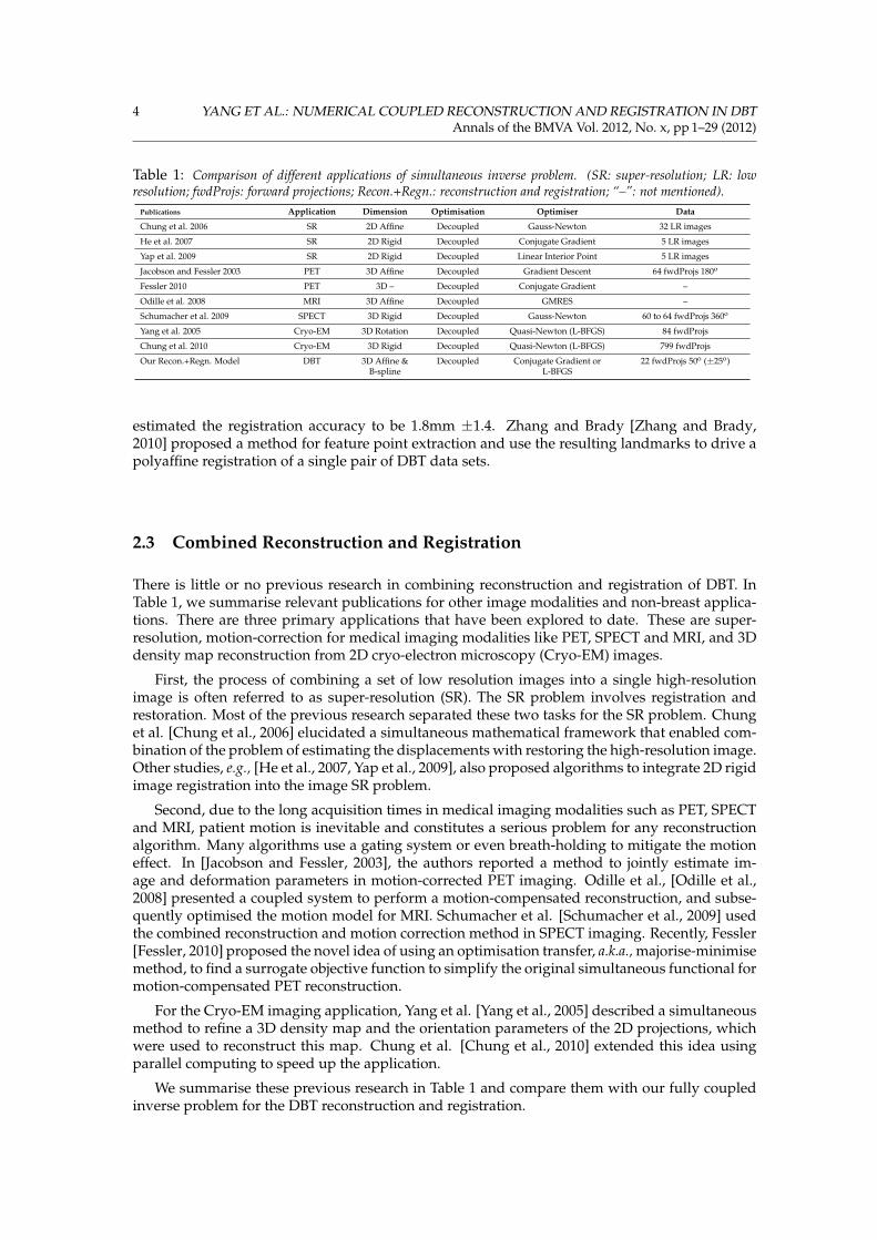
Table 1: Comparison of different applications of simultaneous inverse problem. (SR: super-resolution; LR: lowresolution; fwdProjs: forward projections; Recon.+Regn.: reconstruction and registration; “–”: not mentioned).
Publications Application Dimension Optimisation Optimiser Data
Chung et al. 2006 SR 2D Affine Decoupled Gauss-Newton 32 LR images
He et al. 2007 SR 2D Rigid Decoupled Conjugate Gradient 5 LR images
Yap et al. 2009 SR 2D Rigid Decoupled Linear Interior Point 5 LR images
Jacobson and Fessler 2003 PET 3D Affine Decoupled Gradient Descent 64 fwdProjs 180o
Fessler 2010 PET 3D – Decoupled Conjugate Gradient –
Odille et al. 2008 MRI 3D Affine Decoupled GMRES –
Schumacher et al. 2009 SPECT 3D Rigid Decoupled Gauss-Newton 60 to 64 fwdProjs 360o
Yang et al. 2005 Cryo-EM 3D Rotation Decoupled Quasi-Newton (L-BFGS) 84 fwdProjs
Chung et al. 2010 Cryo-EM 3D Rigid Decoupled Quasi-Newton (L-BFGS) 799 fwdProjs
Our Recon.+Regn. Model DBT 3D Affine &B-spline
Decoupled Conjugate Gradient orL-BFGS
22 fwdProjs 50o (±25o)
estimated the registration accuracy to be 1.8mm ±1.4. Zhang and Brady [Zhang and Brady,2010] proposed a method for feature point extraction and use the resulting landmarks to drive apolyaffine registration of a single pair of DBT data sets.
2.3 Combined Reconstruction and Registration
There is little or no previous research in combining reconstruction and registration of DBT. InTable 1, we summarise relevant publications for other image modalities and non-breast applica-tions. There are three primary applications that have been explored to date. These are super-resolution, motion-correction for medical imaging modalities like PET, SPECT and MRI, and 3Ddensity map reconstruction from 2D cryo-electron microscopy (Cryo-EM) images.
First, the process of combining a set of low resolution images into a single high-resolutionimage is often referred to as super-resolution (SR). The SR problem involves registration andrestoration. Most of the previous research separated these two tasks for the SR problem. Chunget al. [Chung et al., 2006] elucidated a simultaneous mathematical framework that enabled com-bination of the problem of estimating the displacements with restoring the high-resolution image.Other studies, e.g., [He et al., 2007, Yap et al., 2009], also proposed algorithms to integrate 2D rigidimage registration into the image SR problem.
Second, due to the long acquisition times in medical imaging modalities such as PET, SPECTand MRI, patient motion is inevitable and constitutes a serious problem for any reconstructionalgorithm. Many algorithms use a gating system or even breath-holding to mitigate the motioneffect. In [Jacobson and Fessler, 2003], the authors reported a method to jointly estimate im-age and deformation parameters in motion-corrected PET imaging. Odille et al., [Odille et al.,2008] presented a coupled system to perform a motion-compensated reconstruction, and subse-quently optimised the motion model for MRI. Schumacher et al. [Schumacher et al., 2009] usedthe combined reconstruction and motion correction method in SPECT imaging. Recently, Fessler[Fessler, 2010] proposed the novel idea of using an optimisation transfer, a.k.a., majorise-minimisemethod, to find a surrogate objective function to simplify the original simultaneous functional formotion-compensated PET reconstruction.
For the Cryo-EM imaging application, Yang et al. [Yang et al., 2005] described a simultaneousmethod to refine a 3D density map and the orientation parameters of the 2D projections, whichwere used to reconstruct this map. Chung et al. [Chung et al., 2010] extended this idea usingparallel computing to speed up the application.
We summarise these previous research in Table 1 and compare them with our fully coupledinverse problem for the DBT reconstruction and registration.
YANG ET AL.: NUMERICAL COUPLED RECONSTRUCTION AND REGISTRATION IN DBT 5Annals of the BMVA Vol. 2012, No. x, pp 1–29 (2012)
3 Conventional Method
3.1 Forward Problem
A 3D image, fg ∈ RD3 , two sets of temporal data, p1, p2 ∈ Rpnum×D2 , the parametric transforma-tions, T g
ζ , and the system matrix, A ∈ Rpnum×D2×D3 : RD3 7→ RD2 , can be related via
p1 = Afg = AR(x); (1)
p2 = AT gζ fg = AT[Tζ(x)], (2)
where D2 and D3 denote the dimensions of 2D projection space and 3D volume space, respec-tively. In addition, fg and T g
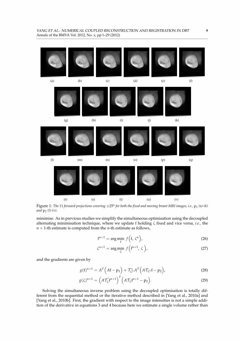
ζ are the ground truth of the reconstruction and the parametric trans-formations respectively, whilst R and T represent the interpolations at original coordinates x andtransformed coordinates Tζ(x). Forward projections, i.e., p1, p2, are acquired using a limitedangle DBT geometry with pnum = 11 projections covering ±25◦ (Figure 1).
3.2 Conventional Sequential Method
In the conventional sequential method, the reconstruction of Equations 1 and 2 can be solved byminimising
f?1 = arg minf1
(f (f1) =
12
∥∥Af1 − p1∥∥2)
; (3)
f?2 = arg minf2
(f (f2) =
12
∥∥Af2 − p2∥∥2)
, (4)
where f1 = R(x) and f2 = T[Tζ(x)].Following reconstruction, volumes f?1 and f?2 , i.e., the fixed and moving images, are registered
with respect to the registration parameters ζ:
ζ? = arg minζ
(f (ζ) =
12
∥∥Tζ(f?2)− f?1∥∥2)
(5)
= arg minζ
(f (ζ) =
12
∥∥T?[Tζ(x)]− R?(x)∥∥2)
,
in which T? and R? denote the interpolations using reconstructed intensities, and the similaritymeasurement is described by a sum of squared difference.
4 Iterative Method
In our novel iterative reconstruction and registration method [Yang et al., 2010a,b], we solveEquations 1 and 2 with respect to estimates f1 and f2 of f and the registration parameters ζ, byalternating an incomplete optimisation (i.e., j iterations) of the reconstructed volumes f̂1 and f̂2:
f̂1 = j STEPS of arg minf1
(f (f1)
)(6)
f̂2 = j STEPS of arg minf2
(f (f2)
)(7)
with the registration of the current estimates f̂1 and f̂2 with respect to the registration parametersζ:
ζ̂ = arg minζ
(f (ζ) =
12
∥∥Tζ(f̂2)− f̂1∥∥2)
(8)
= arg minζ
(f (ζ) =
12
∥∥T̂[Tζ(x)]− R̂(x)∥∥2)
. (9)
6 YANG ET AL.: NUMERICAL COUPLED RECONSTRUCTION AND REGISTRATION IN DBTAnnals of the BMVA Vol. 2012, No. x, pp 1–29 (2012)
Algorithm 1: Iterative Method
Input: p1, p2.Output: f?1 , f?2 , T̂ζ f̂2.
begin% Initialise f1 and f2 to zero vectors;% Initialise ζ to a vector of identity matrix when we use affine transformation model.f1 := 0; f2 := 0; ζ := I;
% Outer loop for the registration runs k timesfor k iterations do
% Inner loop for the reconstruction runs j timesfor j iterations do
f̂1 = j STEPS of arg minf1
(f (f1)
);
f̂2 = j STEPS of arg minf2
(f (f2)
);
ζ̂ = arg minζ
(f (ζ) = 1
2
∥∥Tζ f̂2 − f̂1∥∥2 );
f1 = T̂ζ f̂2; f2 = f̂2;
% Output f?1 , f?2 , and T̂ζ f̂2
f?1 = f̂1;f?2 = f̂2;T̂ζ f̂2.
After each registration iteration (Equation 9), and prior to the next iteration of the reconstruc-tions (Equations 6 and 7), the reconstruction estimates are updated as follows (Equations 10 and11).
f1 = T̂ζ(f̂2) = T̂[T̂ζ(x)] (10)
f2 = f̂2. (11)
This “outer loop” of reconstruction followed by registration is repeated k times. The last iterationoutputs f?1 = f̂1, f?2 = f̂2 and T̂ζ f̂2.
In addition, the following analytical gradients are used to calculate f̂1 and f̂2 for the recon-struction
g(f1) = AT(Af1 − p1) (12)
g(f2) = AT(Af2 − p2). (13)
Similarly, by the chain rule, the analytical gradient for the registration is
g(ζ) =(
f̂1 − Tζ(f̂2))∂Tζ(f̂2)
∂ζ(14)
=(
R̂(x)− T̂[Tζ(x)])∂T̂[Tζ(x)]
∂ζ(15)
=(
R̂(x)− T̂[Tζ(x)])∂T̂[Tζ(x)]
∂Tζ(x)∂Tζ(x)
∂ζ. (16)
It consists of three parts, i.e., the image difference(R̂(x)− T̂[Tζ(x)]
), the partial derivative of
the moving image ∂T̂[Tζ(x)]∂Tζ(x)
evaluated at location Tζ(x), and the partial derivative of the transfor-
mation ∂Tζ(x)∂ζ . As we employed the gradient information to get the updated parameters ζ, the
YANG ET AL.: NUMERICAL COUPLED RECONSTRUCTION AND REGISTRATION IN DBT 7Annals of the BMVA Vol. 2012, No. x, pp 1–29 (2012)
moving image used to calculate the partial derivative is the original moving image (not the up-dated or transformed moving image). In addition, this partial derivative, i.e., spatial derivative,of the original moving image is calculated using the image gradient defined as
∇T̂(yζ) =∂T̂[Tζ(x)]
∂Tζ(x)=(∂T̂
∂xyζ,
∂T̂∂y
yζ,∂T̂∂z
yζ
)T, (17)
in which yζ = Tζ(x) = (x, y, z)T .The preceding iterative reconstruction and registration method is summarised in Algorithm
1, and in our implementation we can use a non-linear conjugate gradient or Limited MemoryBFGS (L-BFGS) optimiser to solve the steps in Equations 6 and 7.
At each update of the volumes after the outer loop registration, we use the transformation off2 to correct f1 that is
f1 = T̂ζ f̂2; (18)
however, an alternative method is updating f1 using the average of the transformed f̂2 and recon-structed f̂1 that is
f1 =12(T̂ζ f̂2 + f̂1), (19)
in which we gather information of both f̂1 and f̂2. Furthermore, we can also incorporate theinverse transformation of f1 into the correction of f2. However, not all transformations have ananalytical inverse.
To sum up, our iterative method alternately performs incomplete reconstructions for twotemporal data sets, followed by a registration.
5 Simultaneous Method
5.1 Formulation of the Simultaneous Method
Whilst combined reconstruction and registration algorithms have been applied to other modali-ties (e.g., PET, SPECT and MRI), little has been published on applying these techniques to DBT.We have proposed an iterative method, which partially coupled the two tasks by alternating be-tween optimising image intensities and parametric transformations to obtain a reduced objectivefunctional [Yang et al., 2010a,b]. An alternative to registering the images after reconstruction orpartially coupling them, is to perform the two tasks simultaneously (fully coupled). This avoidsthe assumptions of missing data being equal to zero (implicit in algorithms such as FBP). Thework in this section hypothesises that the two tasks are not independent but reciprocal, and thatcombining them will enhance the performance of both [Yang et al., 2011].
Using this hypothesis, we have developed an algorithm, which outputs one unified result forthe reconstruction and registration (Algorithm 2). However, the introduction of the nonlinearparametric transformation renders the solution of the inverse problem more complex.
We solve the inverse problem by forming the objective function given by
{f?, ζ?} = arg minf,ζ
(f (f, ζ)
), (20)
f (f, ζ) =12
(∥∥Af− p1∥∥2
+∥∥ATζf− p2
∥∥2)
, (21)
in which f denotes the estimation of the unknown volume, and ζ is the estimation of the unknownparametric transformations [Yang et al., 2012a,b, Yang, 2012].
A minimiser {f?, ζ?} of f (f, ζ) is characterised by the necessary condition that the partialderivative with respect to f and ζ equals zero. The partial derivative with respect to f is straight-
8 YANG ET AL.: NUMERICAL COUPLED RECONSTRUCTION AND REGISTRATION IN DBTAnnals of the BMVA Vol. 2012, No. x, pp 1–29 (2012)
Algorithm 2: Simultaneous Method
Input: p1, p2.Output: f?, ζ?.
begin% Initialise f to a vector with all zero entries;% Initialise ζ to a vector of identity matrix when we use affine transformation model;% (Initialise ζ to a vector with all zero entries when we use B-spline model).f := 0;ζ := I;
% Simultaneous reconstruction and registration loopfor k iterations do
{f?, ζ?} = arg minf,ζ
(f (f, ζ)
);
% Output f? and ζ?
f?;ζ?.
forward, and is given by
g(f) =∂ f (f, ζ)
∂f= AT(Af− p1) + T ∗ζ AT(ATζf− p2),
(22)
in which g(f) is the gradient with respect to f, and T ∗ζ is the adjoint operator of Tζ.To derive the partial derivative with respect to ζ, we apply a small perturbation to the objec-
tive function. The linearisation via the norm then yields,
f(
f, ζ+ ∆ζ)
=12
(∥∥Af− p1∥∥2
+∥∥ATζ+∆ζf− p2
∥∥2)
(23)
≈ 12
(∥∥Af− p1∥∥2
+∥∥ATζf + A
∂Tζ∂ζ
f∆ζ− p2∥∥2)
.
By taking the derivative with respect to ∆ζ, and equating the result to zero, we obtain:(A
∂Tζ∂ζ
f)T(
ATζf + A∂Tζ∂ζ
f∆ζ− p2
)= 0; (24)
If g(ζ) denotes the gradient then we have,
g(ζ) =∂ f (f, ζ)
∂ζ=(
A∂Tζ∂ζ
f)T(
ATζf− p2
)=(
AT ′ζf)T(
ATζf− p2
).
(25)
5.2 Decoupled Solver for the Fully-coupled System
Our survey of published simultaneous methods indicates that none of these studies solved thesimultaneous reconstruction and registration problem directly (Table 1). Similarly, we adopt adecoupled approach to solve the combined problem because the objective function in Equation21 is a nonconvex function of the transformation parameters ζ and therefore very challenging to
YANG ET AL.: NUMERICAL COUPLED RECONSTRUCTION AND REGISTRATION IN DBT 9Annals of the BMVA Vol. 2012, No. x, pp 1–29 (2012)
(a) (b) (c) (d) (e) (f)
(g) (h) (i) (j) (k)
(l) (m) (n) (o) (p) (q)
(r) (s) (t) (u) (v)
Figure 1: The 11 forward projections covering±25o for both the fixed and moving breast MRI images, i.e., p1 (a)-(k)and p2 (l)-(v).
minimise. As in previous studies we simplify the simultaneous optimisation using the decoupledalternating minimisation technique, where we update f holding ζ fixed and vice versa, i.e., then + 1-th estimate is computed from the n-th estimate as follows,
fn+1 = arg minf
f(
f, ζn)
, (26)
ζn+1 = arg minζ
f(
fn+1, ζ)
, (27)
and the gradients are given by
g(f)n+1 = AT(
Af− p1
)+ T ∗ζn AT
(ATζn f− p2
), (28)
g(ζ)n+1 =(
AT ′ζfn+1)T(
ATζfn+1 − p2
). (29)
Solving the simultaneous inverse problem using the decoupled optimisation is totally dif-ferent from the sequential method or the iterative method described in [Yang et al., 2010a] and[Yang et al., 2010b]. First, the gradient with respect to the image intensities is not a simple addi-tion of the derivative in equations 3 and 4 because here we estimate a single volume rather than
10 YANG ET AL.: NUMERICAL COUPLED RECONSTRUCTION AND REGISTRATION IN DBTAnnals of the BMVA Vol. 2012, No. x, pp 1–29 (2012)
two as in the sequential or iterative methods. Accordingly, we have one unified result instead oftwo reconstructions, which need to be registered in a further step. More significantly, because ofthe presence of the system matrix A in the gradient formulation in equation 29, the simultane-ous concept is more challenging than a typical image registration problem described in equation5, i.e., registering two complete reconstructions in the sequential method or registering the twocurrent estimates of the incomplete reconstructions in the iterative method.
Table 2: Comparison of the gradient information used in the iterative and simultaneous methods.
Reconstruction Part Registration Part
Iterative Method g(f1) = AT(Af1 − p1) g(ζ) =(R̂(x)− T̂[Tζ(x)]
) ∂T̂[Tζ(x)]∂Tζ(x)
∂Tζ(x)∂ζ
g(f2) = AT(Af2 − p2)
Intensity Part Transformation Part
Simultaneous Method g(f) = AT(Af− p1) + T ∗ζ AT(ATζf− p2) g(ζ) =(
∂T[Tζ(x)]∂Tζ(x)
∗) ∂Tζ(x)∂ζ AT(ATζf− p2
)
5.3 Derivative Operator of the Transformations
The derivative of the transformation operation is a key component of the algorithm and has greatimpact on the result of the optimisation. Deriving an analytical derivative of the transformationis desirable because it would be fast to compute but is complicated by the need to formulatethe derivative of the underlying interpolation. In addition, some interpolation schemes have noanalytical derivative. For this reason therefore, we use the Finite Difference Method (FDM) toapproximate the derivative operation:
T ′ζ ≈Tζ+ε + Tζ−ε
2ε(30)
where ε is a small number.
5.4 Optimisation
The optimisation is performed using a quasi-Newton based L-BFGS method, which is describedas a generic form in Algorithm 3. This approximates the inverse of the Hessian matrix whilstavoiding the considerable memory overhead (for large DBT data sets) associated with computing2nd order derivatives or their fully dense approximations directly.
6 Results
In this section, we demonstrate the performance of our combined reconstruction and registrationframework using both iterative and simultaneous methods. Both affine and B-spline transforma-tion models have been considered. First, we combine optimisation of the two temporal recon-structions with the 12 degrees of freedom, of an affine transformation, which globally describesthe translation, scaling, rotation and shearing between the two time points. Second, we can alsosubstitute non-rigid B-spline deformations for the affine transformation in this framework. Webegin in Section 6.1 using an affine transformation model, and test this using a software synthetictoroidal phantom image. We can compare the final transformed moving image T̂ζ f̂2 with theoriginal fixed image fg
1 , which is the ground truth of the reconstruction, to analyse the accuracyof our iterative method. The difference image, between T̂ζ f̂2 and fg
1 , is compared with the dif-ference image of the conventional sequential method, i.e., differences between T ?
ζ f?2 and fg1 , in
which T ? is calculated using ζ? from Equation 5, and f?2 is obtained from Equation 4. In Section6.2, we qualitatively and quantitatively assess the performance of our simultaneous method us-ing the affine transformation model, and test on various simulated data sets. In addition, both
YANG ET AL.: NUMERICAL COUPLED RECONSTRUCTION AND REGISTRATION IN DBT 11Annals of the BMVA Vol. 2012, No. x, pp 1–29 (2012)
Algorithm 3: quasi-Newton Method
Input: k, finitial.Output: foptimised.
Hinitial := I; % Initialise the inverse Hessian matrix as identity matrixdinitial := −Hinitialg(finitial); % Initial search direction is the negative gradientbegin
while stopping criterion unfulfilled doτk := arg minτ>0 f (fk + τdk); % Line Searchfk+1 := fk + τkdk; % Update the fsk := fk+1 − fk;zk := g(fk+1)− g(fk);
Hk+1 := Hk +1
sTk zk
[(1 + zT
k HkzksT
k zk
)sksT
k − HkzksTk − skzT
k Hk
]; % BFGS
dk+1 := −Hk+1g(fk+1); % Update the search direction
k := k + 1;
foptimised := fk+1;
our iterative and simultaneous methods are compared with the conventional sequential method.Finally, we analyse the efficacy of incorporating a non-rigid B-spline transformation model intoour simultaneous method in Section 6.3.
6.1 Sequential Method vs. Iterative Method
In this experiment, we created a software synthetic toroidal phantom, which is embedded in a 3Dvolume of 70× 70× 70mm3 with 1mm resolution in each direction (Figure 2 (a)-(c)). The groundtruth affine transformation is a translation of [10, 0,−20] mm and a rotation about the y axis of−30o (Figure 2 (d)-(f)). The reconstructions of the fixed and moving images without registrationare shown in Figure 3 (a)-(c) and (d)-(f) respectively. As seen in Figure 4 (a)-(c), the iterativeresults are more compact and accurate than the sequential results, i.e., the transformed movingimage reconstruction T ?
ζ f?2 (Figure 4 (d)-(f)), and the out of plane blurring is reduced. The meansquared error (MSE = 1
D3‖f?1 − fg‖2) is decreased from 106 to 104 in order of magnitude; however,
for the iterative method this value of 1.26× 10⁴ is superior to the sequential result of 2.01× 10⁴ (D3is the total number of voxels). In addition, from lower value of the objective function, we canconclude that our iterative method outperformed the sequential method (Figure 5).
6.2 Sequential Method vs. Simultaneous Method
6.2.1 Test on a Toroid Phantom Image
We performed 20 different set of randomly simulated affine transformations to test the robustnessof our simultaneous method. Affine test case 1 is presented here as an example (Figure 6 (a)-(c)). Results of the two different methods, i.e., sequential vs. simultaneous, were compared. Wefound that there were fewer artefacts in the results of our simultaneous method compared to thesequential method (Figure 8 (a)-(c) vs. (d)-(f)).
Similarly the difference images indicate that the simultaneous method is superior to the se-quential method. The absolute errors between the recovered affine parameters and the groundtruth of the transformations were also calculated. The results show that the recovery of the pa-rameters were accurate and consistent for all the 20 tests (Figure 7).
12 YANG ET AL.: NUMERICAL COUPLED RECONSTRUCTION AND REGISTRATION IN DBTAnnals of the BMVA Vol. 2012, No. x, pp 1–29 (2012)
(a) (b) (c)
(d) (e) (f)
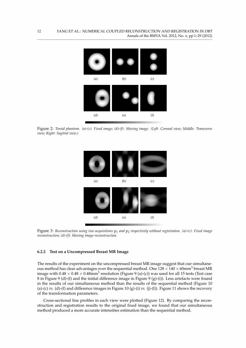
Figure 2: Toroid phantom. (a)-(c): Fixed image; (d)-(f): Moving image. (Left: Coronal view; Middle: Transverseview; Right: Sagittal view.)
(a) (b) (c)
(d) (e) (f)
Figure 3: Reconstruction using two acquisitions p1 and p2 respectively without registration. (a)-(c): Fixed imagereconstruction; (d)-(f): Moving image reconstruction.
6.2.2 Test on a Uncompressed Breast MR Image
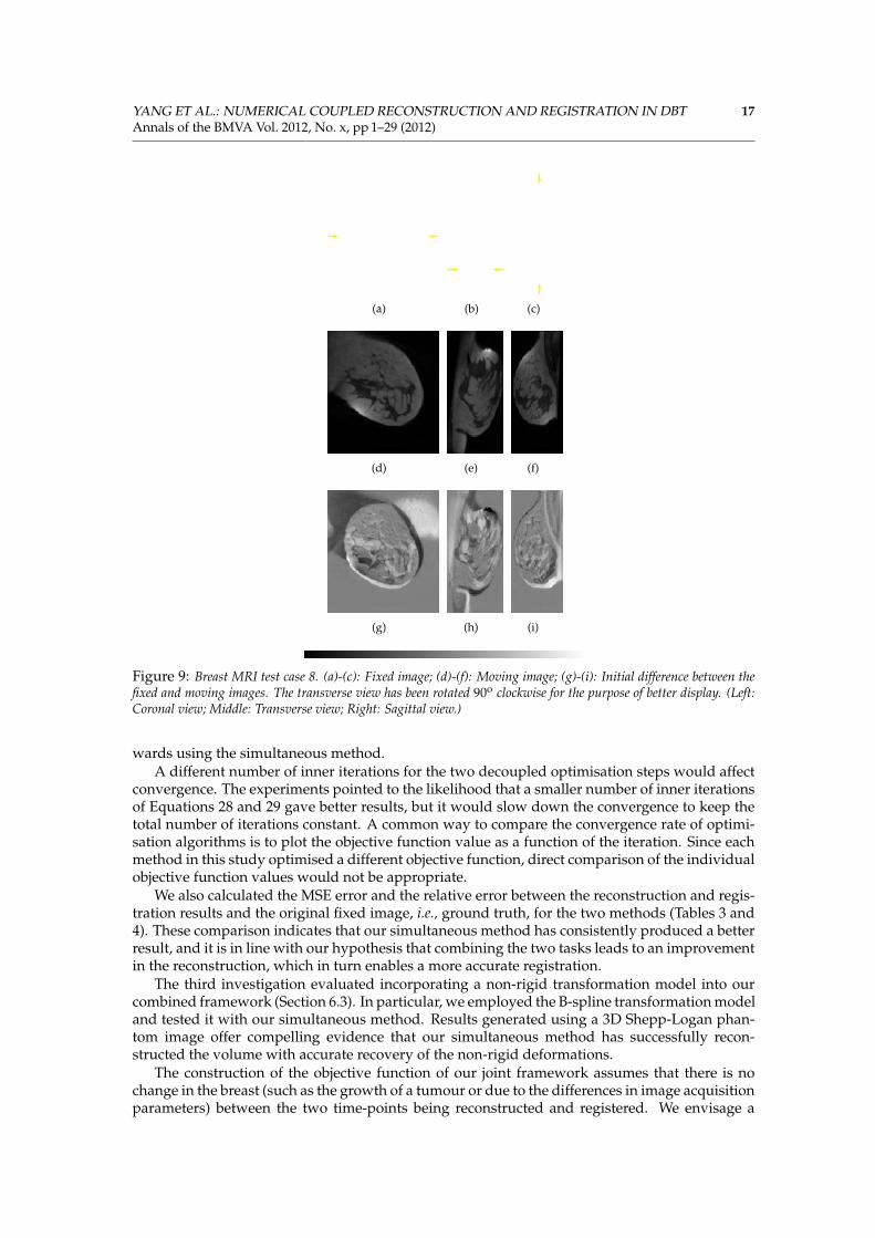
The results of the experiment on the uncompressed breast MR image suggest that our simultane-ous method has clear advantages over the sequential method. One 128× 140× 60mm3 breast MRimage with 0.48× 0.48× 0.48mm3 resolution (Figure 9 (a)-(c)) was used for all 15 tests (Test case8 in Figure 9 (d)-(f) and the initial difference image in Figure 9 (g)-(i)). Less artefacts were foundin the results of our simultaneous method than the results of the sequential method (Figure 10(a)-(c) vs. (d)-(f) and difference images in Figure 10 (g)-(i) vs. (j)-(l)). Figure 11 shows the recoveryof the transformation parameters.
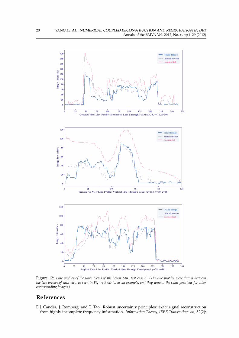
Cross-sectional line profiles in each view were plotted (Figure 12). By comparing the recon-struction and registration results to the original fixed image, we found that our simultaneousmethod produced a more accurate intensities estimation than the sequential method.
YANG ET AL.: NUMERICAL COUPLED RECONSTRUCTION AND REGISTRATION IN DBT 13Annals of the BMVA Vol. 2012, No. x, pp 1–29 (2012)
(a) (b) (c)
(d) (e) (f)
(g) (h) (i)
(j) (k) (l)
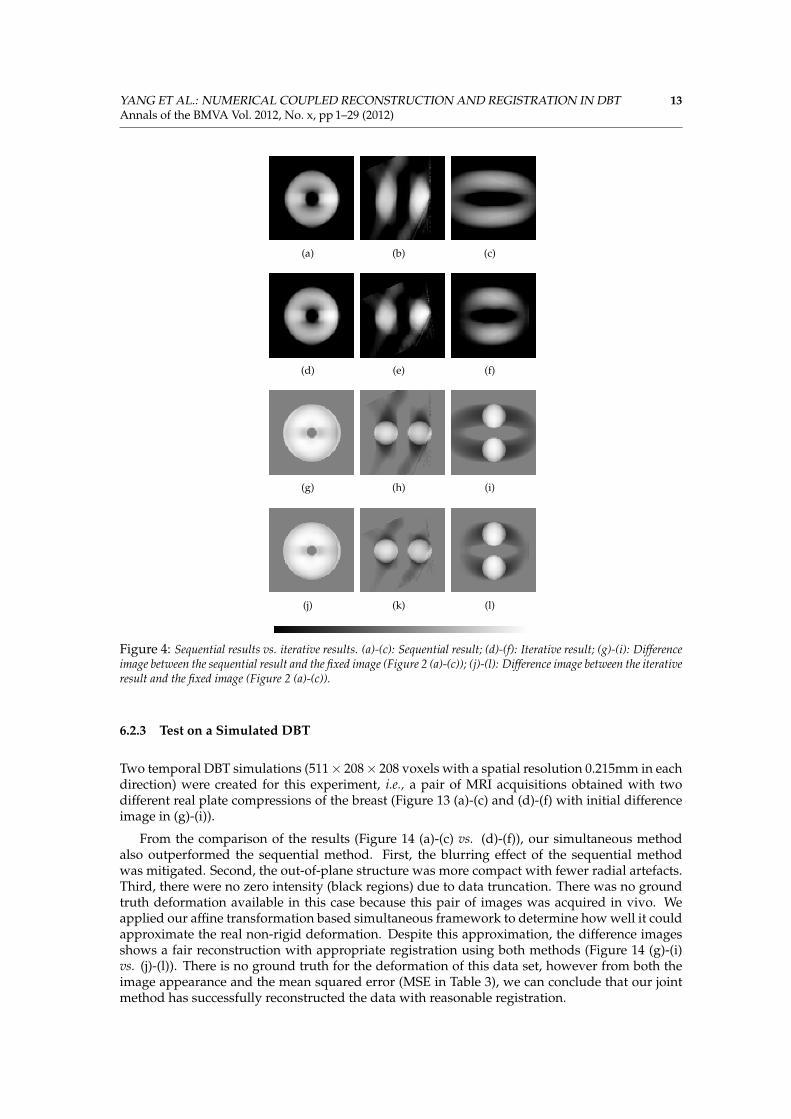
Figure 4: Sequential results vs. iterative results. (a)-(c): Sequential result; (d)-(f): Iterative result; (g)-(i): Differenceimage between the sequential result and the fixed image (Figure 2 (a)-(c)); (j)-(l): Difference image between the iterativeresult and the fixed image (Figure 2 (a)-(c)).
6.2.3 Test on a Simulated DBT
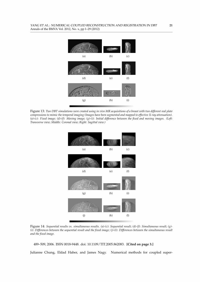
Two temporal DBT simulations (511× 208× 208 voxels with a spatial resolution 0.215mm in eachdirection) were created for this experiment, i.e., a pair of MRI acquisitions obtained with twodifferent real plate compressions of the breast (Figure 13 (a)-(c) and (d)-(f) with initial differenceimage in (g)-(i)).
From the comparison of the results (Figure 14 (a)-(c) vs. (d)-(f)), our simultaneous methodalso outperformed the sequential method. First, the blurring effect of the sequential methodwas mitigated. Second, the out-of-plane structure was more compact with fewer radial artefacts.Third, there were no zero intensity (black regions) due to data truncation. There was no groundtruth deformation available in this case because this pair of images was acquired in vivo. Weapplied our affine transformation based simultaneous framework to determine how well it couldapproximate the real non-rigid deformation. Despite this approximation, the difference imagesshows a fair reconstruction with appropriate registration using both methods (Figure 14 (g)-(i)vs. (j)-(l)). There is no ground truth for the deformation of this data set, however from both theimage appearance and the mean squared error (MSE in Table 3), we can conclude that our jointmethod has successfully reconstructed the data with reasonable registration.
14 YANG ET AL.: NUMERICAL COUPLED RECONSTRUCTION AND REGISTRATION IN DBTAnnals of the BMVA Vol. 2012, No. x, pp 1–29 (2012)
0 50 1001.0 1́0 0 8
1.0 1́0 0 9
1.0 1́0 1 0
1.0 1́0 1 1
1.0 1́0 1 2
Sequential Fixed f(f1)
Iterative Fixed f(f1)
Iteration Number of the Sequential and the Iterative Reconstruction and Registration
Obj
ecti
ve F
unct
ion
Val
ues
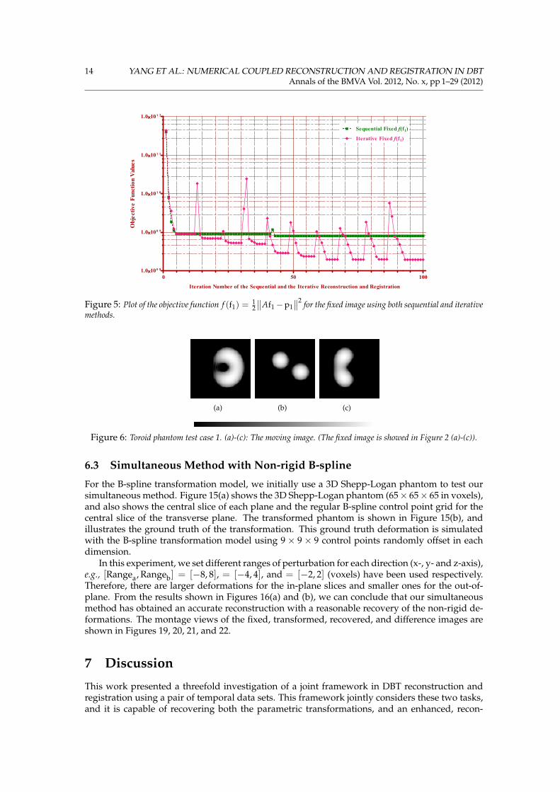
Figure 5: Plot of the objective function f (f1) =12
∥∥Af1−p1∥∥2 for the fixed image using both sequential and iterative
methods.
(a) (b) (c)
Figure 6: Toroid phantom test case 1. (a)-(c): The moving image. (The fixed image is showed in Figure 2 (a)-(c)).
6.3 Simultaneous Method with Non-rigid B-spline
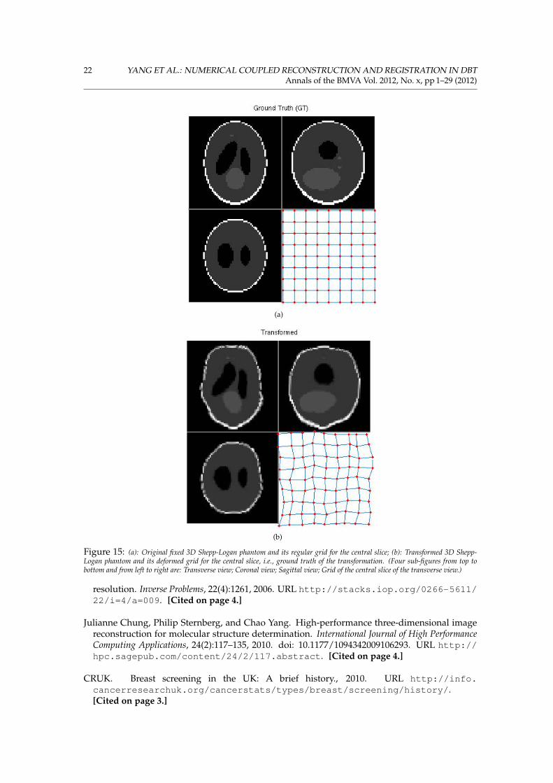
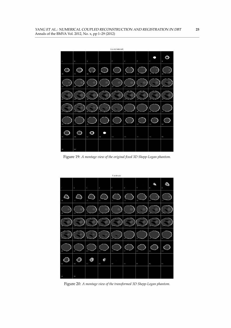
For the B-spline transformation model, we initially use a 3D Shepp-Logan phantom to test oursimultaneous method. Figure 15(a) shows the 3D Shepp-Logan phantom (65× 65× 65 in voxels),and also shows the central slice of each plane and the regular B-spline control point grid for thecentral slice of the transverse plane. The transformed phantom is shown in Figure 15(b), andillustrates the ground truth of the transformation. This ground truth deformation is simulatedwith the B-spline transformation model using 9× 9× 9 control points randomly offset in eachdimension.
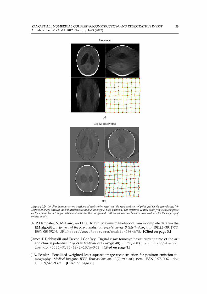
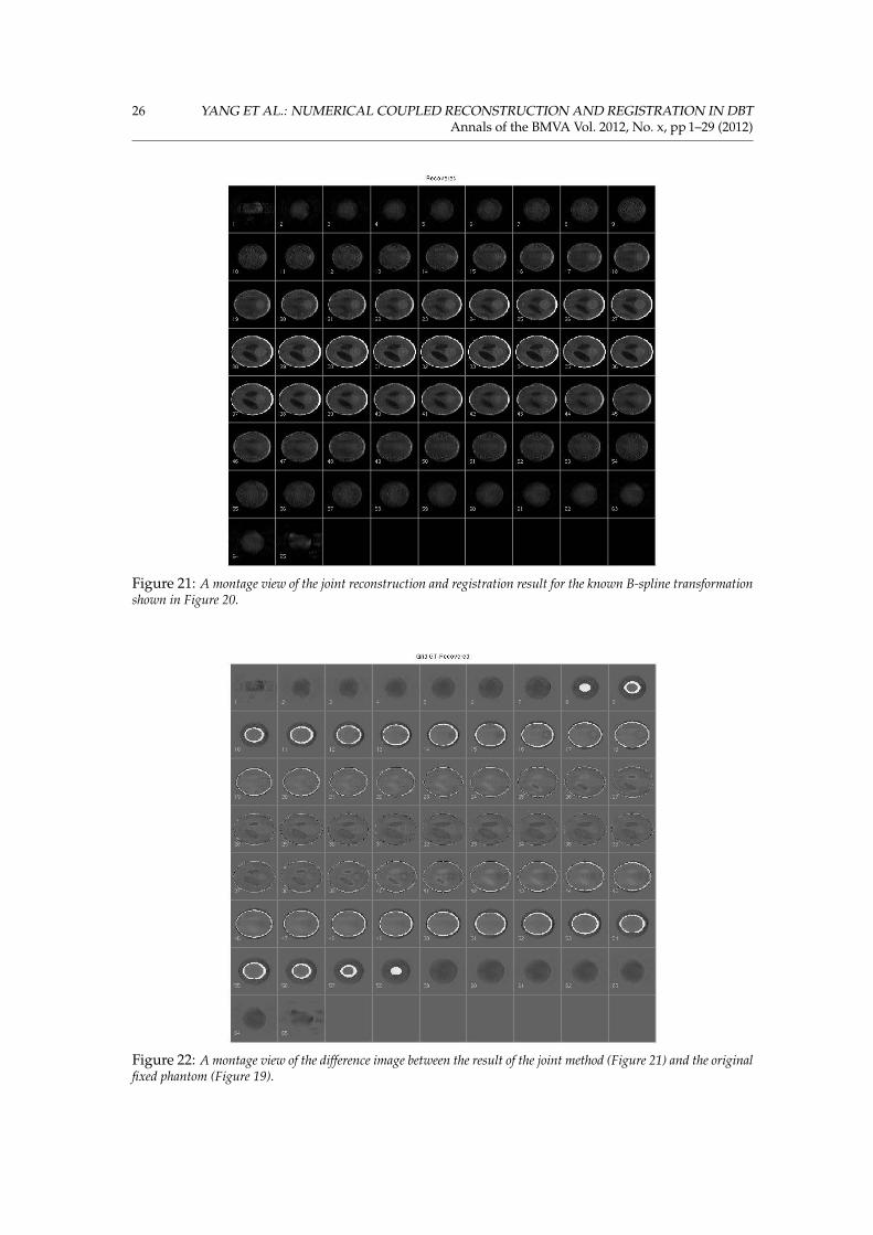
In this experiment, we set different ranges of perturbation for each direction (x-, y- and z-axis),e.g., [Rangea, Rangeb] = [−8, 8], = [−4, 4], and = [−2, 2] (voxels) have been used respectively.Therefore, there are larger deformations for the in-plane slices and smaller ones for the out-of-plane. From the results shown in Figures 16(a) and (b), we can conclude that our simultaneousmethod has obtained an accurate reconstruction with a reasonable recovery of the non-rigid de-formations. The montage views of the fixed, transformed, recovered, and difference images areshown in Figures 19, 20, 21, and 22.
7 Discussion
This work presented a threefold investigation of a joint framework in DBT reconstruction andregistration using a pair of temporal data sets. This framework jointly considers these two tasks,and it is capable of recovering both the parametric transformations, and an enhanced, recon-
YANG ET AL.: NUMERICAL COUPLED RECONSTRUCTION AND REGISTRATION IN DBT 15Annals of the BMVA Vol. 2012, No. x, pp 1–29 (2012)
1 2 3 4 5 6 7 8 9 10 11 12-0.5
0.0
0.5
1.0
Recovery of 20 Different Set of the 12 Affine Registration Parameters
Mea
n an
d SD
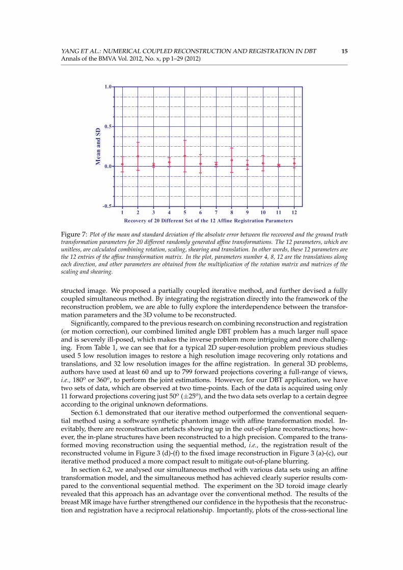
Figure 7: Plot of the mean and standard deviation of the absolute error between the recovered and the ground truthtransformation parameters for 20 different randomly generated affine transformations. The 12 parameters, which areunitless, are calculated combining rotation, scaling, shearing and translation. In other words, these 12 parameters arethe 12 entries of the affine transformation matrix. In the plot, parameters number 4, 8, 12 are the translations alongeach direction, and other parameters are obtained from the multiplication of the rotation matrix and matrices of thescaling and shearing.
structed image. We proposed a partially coupled iterative method, and further devised a fullycoupled simultaneous method. By integrating the registration directly into the framework of thereconstruction problem, we are able to fully explore the interdependence between the transfor-mation parameters and the 3D volume to be reconstructed.
Significantly, compared to the previous research on combining reconstruction and registration(or motion correction), our combined limited angle DBT problem has a much larger null spaceand is severely ill-posed, which makes the inverse problem more intriguing and more challeng-ing. From Table 1, we can see that for a typical 2D super-resolution problem previous studiesused 5 low resolution images to restore a high resolution image recovering only rotations andtranslations, and 32 low resolution images for the affine registration. In general 3D problems,authors have used at least 60 and up to 799 forward projections covering a full-range of views,i.e., 180o or 360o, to perform the joint estimations. However, for our DBT application, we havetwo sets of data, which are observed at two time-points. Each of the data is acquired using only11 forward projections covering just 50o (±25o), and the two data sets overlap to a certain degreeaccording to the original unknown deformations.
Section 6.1 demonstrated that our iterative method outperformed the conventional sequen-tial method using a software synthetic phantom image with affine transformation model. In-evitably, there are reconstruction artefacts showing up in the out-of-plane reconstructions; how-ever, the in-plane structures have been reconstructed to a high precision. Compared to the trans-formed moving reconstruction using the sequential method, i.e., the registration result of thereconstructed volume in Figure 3 (d)-(f) to the fixed image reconstruction in Figure 3 (a)-(c), ouriterative method produced a more compact result to mitigate out-of-plane blurring.
In section 6.2, we analysed our simultaneous method with various data sets using an affinetransformation model, and the simultaneous method has achieved clearly superior results com-pared to the conventional sequential method. The experiment on the 3D toroid image clearlyrevealed that this approach has an advantage over the conventional method. The results of thebreast MR image have further strengthened our confidence in the hypothesis that the reconstruc-tion and registration have a reciprocal relationship. Importantly, plots of the cross-sectional line
16 YANG ET AL.: NUMERICAL COUPLED RECONSTRUCTION AND REGISTRATION IN DBTAnnals of the BMVA Vol. 2012, No. x, pp 1–29 (2012)
(a) (b) (c)
(d) (e) (f)
(g) (h) (i)
(j) (k) (l)
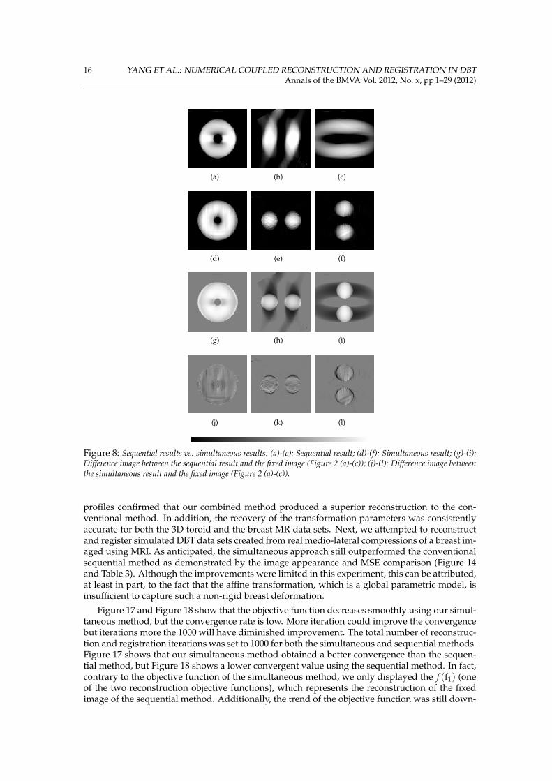
Figure 8: Sequential results vs. simultaneous results. (a)-(c): Sequential result; (d)-(f): Simultaneous result; (g)-(i):Difference image between the sequential result and the fixed image (Figure 2 (a)-(c)); (j)-(l): Difference image betweenthe simultaneous result and the fixed image (Figure 2 (a)-(c)).
profiles confirmed that our combined method produced a superior reconstruction to the con-ventional method. In addition, the recovery of the transformation parameters was consistentlyaccurate for both the 3D toroid and the breast MR data sets. Next, we attempted to reconstructand register simulated DBT data sets created from real medio-lateral compressions of a breast im-aged using MRI. As anticipated, the simultaneous approach still outperformed the conventionalsequential method as demonstrated by the image appearance and MSE comparison (Figure 14and Table 3). Although the improvements were limited in this experiment, this can be attributed,at least in part, to the fact that the affine transformation, which is a global parametric model, isinsufficient to capture such a non-rigid breast deformation.
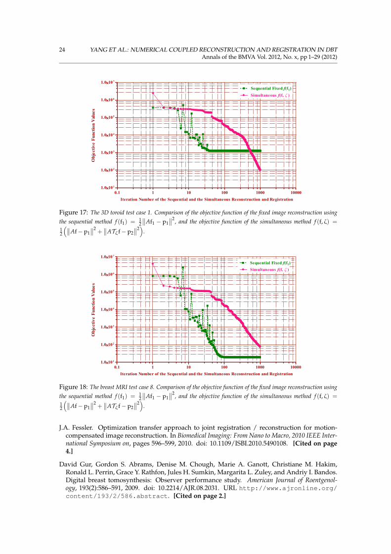
Figure 17 and Figure 18 show that the objective function decreases smoothly using our simul-taneous method, but the convergence rate is low. More iteration could improve the convergencebut iterations more the 1000 will have diminished improvement. The total number of reconstruc-tion and registration iterations was set to 1000 for both the simultaneous and sequential methods.Figure 17 shows that our simultaneous method obtained a better convergence than the sequen-tial method, but Figure 18 shows a lower convergent value using the sequential method. In fact,contrary to the objective function of the simultaneous method, we only displayed the f (f1) (oneof the two reconstruction objective functions), which represents the reconstruction of the fixedimage of the sequential method. Additionally, the trend of the objective function was still down-
YANG ET AL.: NUMERICAL COUPLED RECONSTRUCTION AND REGISTRATION IN DBT 17Annals of the BMVA Vol. 2012, No. x, pp 1–29 (2012)
(a) (b) (c)
(d) (e) (f)
(g) (h) (i)
Figure 9: Breast MRI test case 8. (a)-(c): Fixed image; (d)-(f): Moving image; (g)-(i): Initial difference between thefixed and moving images. The transverse view has been rotated 90o clockwise for the purpose of better display. (Left:Coronal view; Middle: Transverse view; Right: Sagittal view.)
wards using the simultaneous method.A different number of inner iterations for the two decoupled optimisation steps would affect
convergence. The experiments pointed to the likelihood that a smaller number of inner iterationsof Equations 28 and 29 gave better results, but it would slow down the convergence to keep thetotal number of iterations constant. A common way to compare the convergence rate of optimi-sation algorithms is to plot the objective function value as a function of the iteration. Since eachmethod in this study optimised a different objective function, direct comparison of the individualobjective function values would not be appropriate.
We also calculated the MSE error and the relative error between the reconstruction and regis-tration results and the original fixed image, i.e., ground truth, for the two methods (Tables 3 and4). These comparison indicates that our simultaneous method has consistently produced a betterresult, and it is in line with our hypothesis that combining the two tasks leads to an improvementin the reconstruction, which in turn enables a more accurate registration.
The third investigation evaluated incorporating a non-rigid transformation model into ourcombined framework (Section 6.3). In particular, we employed the B-spline transformation modeland tested it with our simultaneous method. Results generated using a 3D Shepp-Logan phan-tom image offer compelling evidence that our simultaneous method has successfully recon-structed the volume with accurate recovery of the non-rigid deformations.
The construction of the objective function of our joint framework assumes that there is nochange in the breast (such as the growth of a tumour or due to the differences in image acquisitionparameters) between the two time-points being reconstructed and registered. We envisage a
18 YANG ET AL.: NUMERICAL COUPLED RECONSTRUCTION AND REGISTRATION IN DBTAnnals of the BMVA Vol. 2012, No. x, pp 1–29 (2012)
(a) (b) (c)
(d) (e) (f)
(g) (h) (i)
(j) (k) (l)
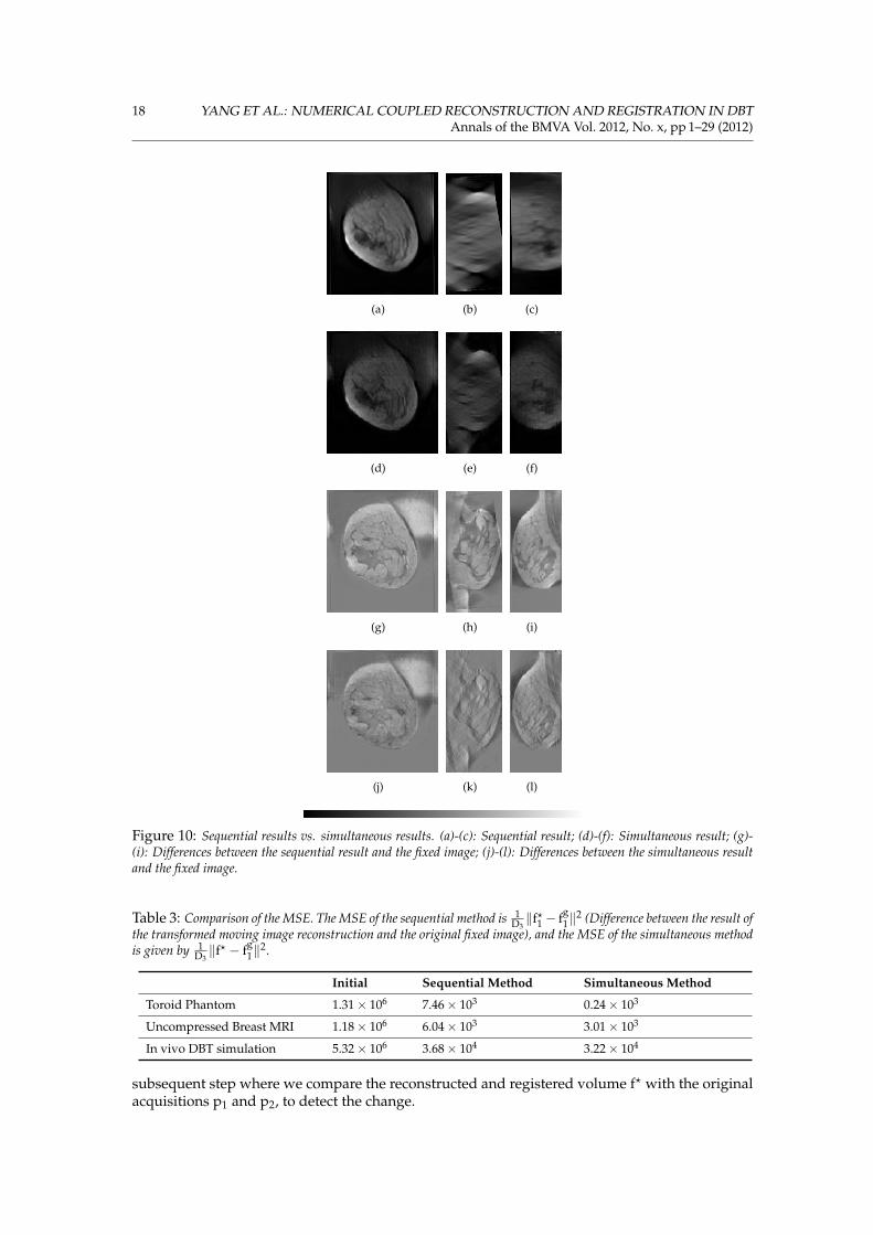
Figure 10: Sequential results vs. simultaneous results. (a)-(c): Sequential result; (d)-(f): Simultaneous result; (g)-(i): Differences between the sequential result and the fixed image; (j)-(l): Differences between the simultaneous resultand the fixed image.
Table 3: Comparison of the MSE. The MSE of the sequential method is 1D3‖f?1 − fg
1‖2 (Difference between the result of
the transformed moving image reconstruction and the original fixed image), and the MSE of the simultaneous methodis given by 1
D3‖f? − fg
1‖2.
Initial Sequential Method Simultaneous Method
Toroid Phantom 1.31× 10⁶ 7.46× 10³ 0.24× 10³
Uncompressed Breast MRI 1.18× 10⁶ 6.04× 10³ 3.01× 10³
In vivo DBT simulation 5.32× 10⁶ 3.68× 10⁴ 3.22× 10⁴
subsequent step where we compare the reconstructed and registered volume f? with the originalacquisitions p1 and p2, to detect the change.
YANG ET AL.: NUMERICAL COUPLED RECONSTRUCTION AND REGISTRATION IN DBT 19Annals of the BMVA Vol. 2012, No. x, pp 1–29 (2012)
1 2 3 4 5 6 7 8 9 10 11 12-0.5
0.0
0.5
1.0
Recovery of 15 Different Set of the 12 Affine Registration Parameters
Mea
n an
d SD
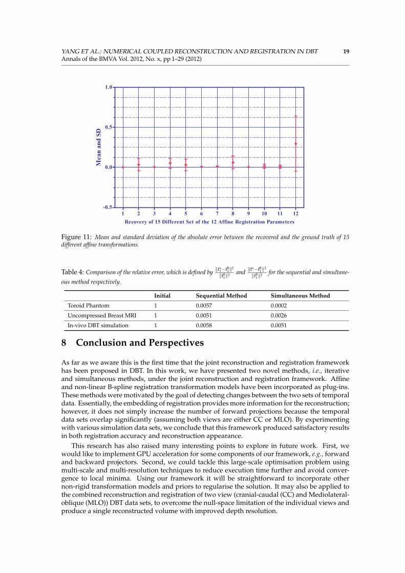
Figure 11: Mean and standard deviation of the absolute error between the recovered and the ground truth of 15different affine transformations.
Table 4: Comparison of the relative error, which is defined by ‖f?1−fg
1‖2
‖fg1‖2 and ‖f
?−fg1‖2
‖fg1‖2 for the sequential and simultane-
ous method respectively.
Initial Sequential Method Simultaneous Method
Toroid Phantom 1 0.0057 0.0002
Uncompressed Breast MRI 1 0.0051 0.0026
In-vivo DBT simulation 1 0.0058 0.0051
8 Conclusion and Perspectives
As far as we aware this is the first time that the joint reconstruction and registration frameworkhas been proposed in DBT. In this work, we have presented two novel methods, i.e., iterativeand simultaneous methods, under the joint reconstruction and registration framework. Affineand non-linear B-spline registration transformation models have been incorporated as plug-ins.These methods were motivated by the goal of detecting changes between the two sets of temporaldata. Essentially, the embedding of registration provides more information for the reconstruction;however, it does not simply increase the number of forward projections because the temporaldata sets overlap significantly (assuming both views are either CC or MLO). By experimentingwith various simulation data sets, we conclude that this framework produced satisfactory resultsin both registration accuracy and reconstruction appearance.
This research has also raised many interesting points to explore in future work. First, wewould like to implement GPU acceleration for some components of our framework, e.g., forwardand backward projectors. Second, we could tackle this large-scale optimisation problem usingmulti-scale and multi-resolution techniques to reduce execution time further and avoid conver-gence to local minima. Using our framework it will be straightforward to incorporate othernon-rigid transformation models and priors to regularise the solution. It may also be applied tothe combined reconstruction and registration of two view (cranial-caudal (CC) and Mediolateral-oblique (MLO)) DBT data sets, to overcome the null-space limitation of the individual views andproduce a single reconstructed volume with improved depth resolution.
20 YANG ET AL.: NUMERICAL COUPLED RECONSTRUCTION AND REGISTRATION IN DBTAnnals of the BMVA Vol. 2012, No. x, pp 1–29 (2012)
0 25 50 75 100 125 150 175 200 225 250 275
0
20
40
60
80
100
120
140
160
180
200 Fixed Image
Simultaneous
Sequential
Coronal View Line Profile: Horizontal Line Through Voxel (x=28, y=71, z=30)
Imag
e In
tens
itie
s
0 25 50 75 100 125
0
20
40
60
80
100
120Fixed Image
Simultaneous
Sequential
Transverse View Line Profile: Vertical Line Through Voxel (x=102, y=70, z=18)
Imag
e In
tens
itie
s
0 25 50 75 100 125 150 175 200 225 250 275 300
0
20
40
60
80
100
120Fixed Image
Simultaneous
Sequential
Sagittal View Line Profile: Vertical Line Through Voxel (x=64, y=78, z=30)
Imag
e In
tens
itie
s
Figure 12: Line profiles of the three views of the breast MRI test case 8. (The line profiles were drawn betweenthe two arrows of each view as seen in Figure 9 (a)-(c) as an example, and they were at the same positions for othercorresponding images.)
References
E.J. Candès, J. Romberg, and T. Tao. Robust uncertainty principles: exact signal reconstructionfrom highly incomplete frequency information. Information Theory, IEEE Transactions on, 52(2):
YANG ET AL.: NUMERICAL COUPLED RECONSTRUCTION AND REGISTRATION IN DBT 21Annals of the BMVA Vol. 2012, No. x, pp 1–29 (2012)
(a) (b) (c)
(d) (e) (f)
(g) (h) (i)
Figure 13: Two DBT simulations were created using in vivo MR acquisitions of a breast with two different real platecompressions to mimic the temporal imaging (Images have been segmented and mapped to effective X-ray attenuation).(a)-(c): Fixed image; (d)-(f): Moving image; (g)-(i): Initial difference between the fixed and moving images. (Left:Transverse view; Middle: Coronal view; Right: Sagittal view.)
(a) (b) (c)
(d) (e) (f)
(g) (h) (i)
(j) (k) (l)
Figure 14: Sequential results vs. simultaneous results. (a)-(c): Sequential result; (d)-(f): Simultaneous result; (g)-(i): Differences between the sequential result and the fixed image; (j)-(l): Differences between the simultaneous resultand the fixed image.
489–509, 2006. ISSN 0018-9448. doi: 10.1109/TIT.2005.862083. [Cited on page 3.]
Julianne Chung, Eldad Haber, and James Nagy. Numerical methods for coupled super-
22 YANG ET AL.: NUMERICAL COUPLED RECONSTRUCTION AND REGISTRATION IN DBTAnnals of the BMVA Vol. 2012, No. x, pp 1–29 (2012)
(a)
(b)
Figure 15: (a): Original fixed 3D Shepp-Logan phantom and its regular grid for the central slice; (b): Transformed 3D Shepp-Logan phantom and its deformed grid for the central slice, i.e., ground truth of the transformation. (Four sub-figures from top tobottom and from left to right are: Transverse view; Coronal view; Sagittal view; Grid of the central slice of the transverse view.)
resolution. Inverse Problems, 22(4):1261, 2006. URL http://stacks.iop.org/0266-5611/22/i=4/a=009. [Cited on page 4.]
Julianne Chung, Philip Sternberg, and Chao Yang. High-performance three-dimensional imagereconstruction for molecular structure determination. International Journal of High PerformanceComputing Applications, 24(2):117–135, 2010. doi: 10.1177/1094342009106293. URL http://hpc.sagepub.com/content/24/2/117.abstract. [Cited on page 4.]
CRUK. Breast screening in the UK: A brief history., 2010. URL http://info.cancerresearchuk.org/cancerstats/types/breast/screening/history/.[Cited on page 3.]
YANG ET AL.: NUMERICAL COUPLED RECONSTRUCTION AND REGISTRATION IN DBT 23Annals of the BMVA Vol. 2012, No. x, pp 1–29 (2012)
(a)
(b)
Figure 16: (a): Simultaneous reconstruction and registration result and the registered control point grid for the central slice; (b):Difference image between the simultaneous result and the original fixed phantom. The registered control point grid is superimposedon the ground truth transformation and indicates that the ground truth transformation has been recovered well for the majority ofcontrol points.
A. P. Dempster, N. M. Laird, and D. B. Rubin. Maximum likelihood from incomplete data via theEM algorithm. Journal of the Royal Statistical Society. Series B (Methodological), 39(1):1–38, 1977.ISSN 00359246. URL http://www.jstor.org/stable/2984875. [Cited on page 3.]
James T DobbinsIII and Devon J Godfrey. Digital x-ray tomosynthesis: current state of the artand clinical potential. Physics in Medicine and Biology, 48(19):R65, 2003. URL http://stacks.iop.org/0031-9155/48/i=19/a=R01. [Cited on page 1.]
J.A. Fessler. Penalized weighted least-squares image reconstruction for positron emission to-mography. Medical Imaging, IEEE Transactions on, 13(2):290–300, 1994. ISSN 0278-0062. doi:10.1109/42.293921. [Cited on page 2.]
24 YANG ET AL.: NUMERICAL COUPLED RECONSTRUCTION AND REGISTRATION IN DBTAnnals of the BMVA Vol. 2012, No. x, pp 1–29 (2012)
0.1 1 10 100 1000 100001.0 1́0 1
1.0 1́0 2
1.0 1́0 3
1.0 1́0 4
1.0 1́0 5
1.0 1́0 6
1.0 1́0 7
Simultaneous f(f, z )
Sequential Fixed f(f1)
Iteration Number of the Sequential and the Simultaneous Reconstruction and Registration
Obj
ecti
ve F
unct
ion
Val
ues
Figure 17: The 3D toroid test case 1. Comparison of the objective function of the fixed image reconstruction usingthe sequential method f (f1) = 1
2
∥∥Af1 − p1∥∥2, and the objective function of the simultaneous method f (f, ζ) =
12
(∥∥Af− p1∥∥2
+∥∥ATζf− p2
∥∥2)
.
0.1 1 10 100 1000 100001.0 1́0 1
1.0 1́0 2
1.0 1́0 3
1.0 1́0 4
1.0 1́0 5
1.0 1́0 6
1.0 1́0 7
Simultaneous f(f, z )
Sequential Fixed f(f1)
Iteration Number of the Sequential and the Simultaneous Reconstruction and Registration
Obj
ecti
ve F
unct
ion
Val
ues
Figure 18: The breast MRI test case 8. Comparison of the objective function of the fixed image reconstruction usingthe sequential method f (f1) = 1
2
∥∥Af1 − p1∥∥2, and the objective function of the simultaneous method f (f, ζ) =
12
(∥∥Af− p1∥∥2
+∥∥ATζf− p2
∥∥2)
.
J.A. Fessler. Optimization transfer approach to joint registration / reconstruction for motion-compensated image reconstruction. In Biomedical Imaging: From Nano to Macro, 2010 IEEE Inter-national Symposium on, pages 596–599, 2010. doi: 10.1109/ISBI.2010.5490108. [Cited on page4.]
David Gur, Gordon S. Abrams, Denise M. Chough, Marie A. Ganott, Christiane M. Hakim,Ronald L. Perrin, Grace Y. Rathfon, Jules H. Sumkin, Margarita L. Zuley, and Andriy I. Bandos.Digital breast tomosynthesis: Observer performance study. American Journal of Roentgenol-ogy, 193(2):586–591, 2009. doi: 10.2214/AJR.08.2031. URL http://www.ajronline.org/content/193/2/586.abstract. [Cited on page 2.]
YANG ET AL.: NUMERICAL COUPLED RECONSTRUCTION AND REGISTRATION IN DBT 25Annals of the BMVA Vol. 2012, No. x, pp 1–29 (2012)
Figure 19: A montage view of the original fixed 3D Shepp-Logan phantom.
Figure 20: A montage view of the transformed 3D Shepp-Logan phantom.
26 YANG ET AL.: NUMERICAL COUPLED RECONSTRUCTION AND REGISTRATION IN DBTAnnals of the BMVA Vol. 2012, No. x, pp 1–29 (2012)
Figure 21: A montage view of the joint reconstruction and registration result for the known B-spline transformationshown in Figure 20.
Figure 22: A montage view of the difference image between the result of the joint method (Figure 21) and the originalfixed phantom (Figure 19).
YANG ET AL.: NUMERICAL COUPLED RECONSTRUCTION AND REGISTRATION IN DBT 27Annals of the BMVA Vol. 2012, No. x, pp 1–29 (2012)
Yu He, Kim-Hui Yap, Li Chen, and Lap-Pui Chau. A nonlinear least square technique for simul-taneous image registration and super-resolution. Image Processing, IEEE Transactions on, 16(11):2830–2841, 2007. ISSN 1057-7149. doi: 10.1109/TIP.2007.908074. [Cited on page 4.]
Gabor T. Herman. Fundamentals of Computerized Tomography: Image Reconstruction from Projections.Springer London, second edition, 2010. ISBN 978-1-85233-617-2. [Cited on page 2.]
H.M. Hudson and R.S. Larkin. Accelerated image reconstruction using ordered subsets of pro-jection data. Medical Imaging, IEEE Transactions on, 13(4):601–609, 1994. ISSN 0278-0062. doi:10.1109/42.363108. [Cited on page 2.]
M.W. Jacobson and J.A. Fessler. Joint estimation of image and deformation parameters in motion-corrected PET. In Nuclear Science Symposium Conference Record, 2003 IEEE, volume 5, pages3290–3294, 2003. doi: 10.1109/NSSMIC.2003.1352599. [Cited on page 4.]
A. C. Kak and M. Slaney. Principles of computerized tomographic imaging. Society for Industrial andApplied Mathematics, Philadelphia, PA, USA, 2001. ISBN 0-89871-494-X. [Cited on page 2.]
Iason Kastanis, Simon Arridge, Alex Stewart, Spencer Gunn, Christer Ullberg, and TomFrancke. 3D digital breast tomosynthesis using total variation regularization. In ElizabethKrupinski, editor, Digital Mammography, volume 5116 of Lecture Notes in Computer Science,pages 621–627. Springer Berlin / Heidelberg, 2008. URL http://dx.doi.org/10.1007/978-3-540-70538-3_86. [Cited on page 3.]
Z. Kolitsi, G. Panayiotakis, V. Anastassopoulos, A. Scodras, and N. Pallikarakis. A multipleprojection method for digital tomosynthesis. Medical Physics, 19(4):1045–1050, 1992. doi: 10.1118/1.596822. URL http://link.aip.org/link/?MPH/19/1045/1. [Cited on page 2.]
H. Matsuo, A. Iwata, I. Horiba, and N. Suzumura. Three-dimensional image reconstruction bydigital tomo-synthesis using inverse filtering. Medical Imaging, IEEE Transactions on, 12(2):307–313, 1993. ISSN 0278-0062. doi: 10.1109/42.232260. [Cited on page 2.]
Thomas Mertelmeier, Jasmina Orman, Wolfgang Haerer, and Mithun K. Dudam. Optimizing fil-tered backprojection reconstruction for a breast tomosynthesis prototype device. In Michael J.Flynn and Jiang Hsieh, editors, Medical Imaging 2006: Physics of Medical Imaging, volume 6142,page 61420F. SPIE, 2006. doi: 10.1117/12.651380. URL http://link.aip.org/link/?PSI/6142/61420F/1. [Cited on page 3.]
K. Mueller, R. Yagel, and J.J. Wheller. Anti-aliased three-dimensional cone-beam reconstructionof low-contrast objects with algebraic methods. Medical Imaging, IEEE Transactions on, 18(6):519–537, 1999. ISSN 0278-0062. doi: 10.1109/42.781017. [Cited on page 2.]
Freddy Odille, Pierre-André Vuissoz, Pierre-Yves Marie, and Jacques Felblinger. Generalized re-construction by inversion of coupled systems (GRICS) applied to free-breathing mri. MagneticResonance in Medicine, 60(1):146–157, 2008. ISSN 1522-2594. doi: 10.1002/mrm.21623. URLhttp://dx.doi.org/10.1002/mrm.21623. [Cited on page 4.]
Steven P. Poplack, Tor D. Tosteson, Christine A. Kogel, and Helene M. Nagy. Digital breasttomosynthesis: Initial experience in 98 women with abnormal digital screening mammogra-phy. American Journal of Roentgenology, 189(3):616–623, 2007. doi: 10.2214/AJR.07.2231. URLhttp://www.ajronline.org/content/189/3/616.abstract. [Cited on page 2.]
H. Schumacher, J. Modersitzki, and B. Fischer. Combined reconstruction and motion correctionin SPECT imaging. Nuclear Science, IEEE Transactions on, 56(1):73–80, 2009. ISSN 0018-9499.doi: 10.1109/TNS.2008.2007907. [Cited on page 4.]
L. A. Shepp and Y. Vardi. Maximum likelihood reconstruction for emission tomography. MedicalImaging, IEEE Transactions on, 1(2):113–122, 1982. ISSN 0278-0062. doi: 10.1109/TMI.1982.4307558. [Cited on page 2.]
28 YANG ET AL.: NUMERICAL COUPLED RECONSTRUCTION AND REGISTRATION IN DBTAnnals of the BMVA Vol. 2012, No. x, pp 1–29 (2012)
Emil Y. Sidky, Xiaochuan Pan, Ingrid S. Reiser, Robert M. Nishikawa, Richard H. Moore, andDaniel B. Kopans. Enhanced imaging of microcalcifications in digital breast tomosynthesisthrough improved image-reconstruction algorithms. Medical Physics, 36(11):4920–4932, 2009.doi: 10.1118/1.3232211. URL http://link.aip.org/link/?MPH/36/4920/1. [Cited onpage 3.]
Sumedha P. Sinha, Ramkrishnan Narayanan, Bing Ma, Marilyn A. Roubidoux, He Liu, andPaul L. Carson. Image registration for detection and quantification of change on digital to-mosynthesis mammographic volumes. American Journal of Roentgenology, 192(2):384–387, 2009.doi: 10.2214/AJR.08.1388. URL http://www.ajronline.org/cgi/content/abstract/192/2/384. [Cited on page 3.]
M. Lee Spangler, Margarita L. Zuley, Jules H. Sumkin, Gordan Abrams, Marie A. Ganott, Chris-tiane Hakim, Ronald Perrin, Denise M. Chough, Ratan Shah, and David Gur. Detection andclassification of calcifications on digital breast tomosynthesis and 2d digital mammography: Acomparison. American Journal of Roentgenology, 196(2):320–324, 2011. doi: 10.2214/AJR.10.4656.URL http://www.ajronline.org/content/196/2/320.abstract. [Cited on page 2.]
Grant M. Stevens, Rebecca Fahrig, and Norbert J. Pelc. Filtered backprojection for modifying theimpulse response of circular tomosynthesis. Medical Physics, 28(3):372–380, 2001. doi: 10.1118/1.1350588. URL http://link.aip.org/link/?MPH/28/372/1. [Cited on page 3.]
Dominique Van de Sompel, Sir Michael Brady, and John Boone. Task-based performance anal-ysis of FBP, SART and ML for digital breast tomosynthesis using signal CNR and chan-nelised hotelling observers. Medical Image Analysis, 15(1):53–70, 2011. ISSN 1361-8415.doi: DOI:10.1016/j.media.2010.07.004. URL http://www.sciencedirect.com/science/article/pii/S1361841510000964. [Cited on page 3.]
Tao Wu, Richard H. Moore, Elizabeth A. Rafferty, and Daniel B. Kopans. A comparison ofreconstruction algorithms for breast tomosynthesis. Medical Physics, 31(9):2636–2647, 2004a.doi: 10.1118/1.1786692. URL http://link.aip.org/link/?MPH/31/2636/1. [Cited onpages 2 and 3.]
Tao Wu, Juemin Zhang, Richard Moore, Elizabeth Rafferty, Daniel Kopans, Waleed Meleis, andDavid Kaeli. Digital tomosynthesis mammography using a parallel maximum-likelihood re-construction method. In Martin J. Yaffe and Michael J. Flynn, editors, Medical Imaging 2004:Physics of Medical Imaging, volume 5368, pages 1–11. SPIE, 2004b. doi: 10.1117/12.534446. URLhttp://link.aip.org/link/?PSI/5368/1/1. [Cited on page 3.]
Hui Yan, Lei Ren, Devon J. Godfrey, and Fang-Fang Yin. Accelerating reconstruction of referencedigital tomosynthesis using graphics hardware. Medical Physics, 34(10):3768–3776, 2007. doi:10.1118/1.2779945. URL http://link.aip.org/link/?MPH/34/3768/1. [Cited on page3.]
Chao Yang, Esmond G. Ng, and Pawel A. Penczek. Unified 3-d structure and projection orienta-tion refinement using quasi-newton algorithm. Journal of Structural Biology, 149(1):53–64, 2005.ISSN 1047-8477. doi: DOI:10.1016/j.jsb.2004.08.010. URL http://www.sciencedirect.com/science/article/pii/S1047847704001716. [Cited on page 4.]
Guang Yang. Numerical Approaches for Solving the Combined Reconstruction and Registration of Dig-ital Breast Tomosynthesis. Doctoral thesis, UCL (University College London)., Feb 2012. [Citedon page 7.]
Guang Yang, John Hipwell, Matthew Clarkson, Christine Tanner, Thomy Mertzanidou, SpencerGunn, Sebastien Ourselin, David Hawkes, and Simon Arridge. Combined reconstruction andregistration of digital breast tomosynthesis. In Digital Mammography, volume 6136 of LectureNotes in Computer Science, pages 760–768. Springer Berlin/Heidelberg, 2010a. URL http://dx.doi.org/10.1007/978-3-642-13666-5_102. [Cited on pages 5, 7, and 9.]
YANG ET AL.: NUMERICAL COUPLED RECONSTRUCTION AND REGISTRATION IN DBT 29Annals of the BMVA Vol. 2012, No. x, pp 1–29 (2012)
Guang Yang, John H. Hipwell, Matthew J. Clarkson, Christine Tanner, Thomy Mertzanidou,Spencer Gunn, Sebastien Ourselin, David J. Hawkes, and Simon R. Arridge. Combined re-construction and registration of digital breast tomosynthesis: Sequential method versus itera-tive method. In Medical Image Understanding and Analysis, pages 1–5, University of Warwick,Coventry., 2010b. [Cited on pages 5, 7, and 9.]
Guang Yang, John H. Hipwell, David J. Hawkes, and Simon R. Arridge. Unconstrained simul-taneous scheme to fully couple reconstruction and registration for digital breast tomosyn-thesis: A feasible study. In Workshop on Breast Image Analysis, pages 25–32, In conjunctionwith MICCAI 2011, September 2011. ISBN 978-87-981270-9-3. URL http://www.diku.dk/BIA2011proceedings.pdf. [Cited on page 7.]
Guang Yang, John Hipwell, Christine Tanner, David Hawkes, and Simon Arridge. Joint registra-tion and limited-angle reconstruction of digital breast tomosynthesis. In Digital Mammography,IWDM’12, Lecture Notes in Computer Science. Springer Berlin / Heidelberg, 2012a. Submitted.[Cited on page 7.]
Guang Yang, John H. Hipwell, David J. Hawkes, and Simon R. Arridge. A nonlinear least squaresmethod for solving the joint reconstruction and registration problem in digital breast tomosyn-thesis. In Medical Image Understanding and Analysis, pages 75–80, Swansea University, UK,2012b. [Cited on page 7.]
Kim-Hui Yap, Yu He, Yushuang Tian, and Lap-Pui Chau. A nonlinear L1-norm approach for jointimage registration and super-resolution. Signal Processing Letters, IEEE, 16(11):981–984, 2009.ISSN 1070-9908. doi: 10.1109/LSP.2009.2028106. [Cited on page 4.]
Weiwei Zhang and Sir Brady. Feature point detection for non-rigid registration of digitalbreast tomosynthesis images. In Joan Martí, Arnau Oliver, Jordi Freixenet, and RobertMartí, editors, Digital Mammography, volume 6136 of Lecture Notes in Computer Science,pages 296–303. Springer Berlin / Heidelberg, 2010. URL http://dx.doi.org/10.1007/978-3-642-13666-5_40. [Cited on page 4.]
Yiheng Zhang, Heang-Ping Chan, Berkman Sahiner, Jun Wei, Mitchell M. Goodsitt, Lubomir M.Hadjiiski, Jun Ge, and Chuan Zhou. A comparative study of limited-angle cone-beam re-construction methods for breast tomosynthesis. Medical Physics, 33(10):3781–3795, 2006. doi:10.1118/1.2237543. URL http://link.aip.org/link/?MPH/33/3781/1. [Cited on page3.]


![DRAFT: COMPUTATIONAL OPTIMIZATION OF ARC WELDING … · 2015. 2. 12. · welding distortion over the years using FEM. Michalaras and Debiccari [3] applied decoupled 2D and 3D finite](https://img.pdfslide.us/doc/110x75/5fbfd8ddb8304b37c23f5985/draft-computational-optimization-of-arc-welding-2015-2-12-welding-distortion.jpg)