Embed Size (px)
Citation preview

© 2015 The Korean Society of Rheology and Springer 163
Korea-Australia Rheology Journal, 27(2), 163-171 (May 2015)DOI: 10.1007/s13367-015-0016-x
www.springer.com/13367
pISSN 1226-119X eISSN 2093-7660
Numerical investigation on the structural characteristics of multiple RBCs
in a stenotic microcapillary under plasma-alcohol solution
Aleksey Ni1, Taqi Ahmad Cheema
2 and Cheol Woo Park
1,*1School of Mechanical Engineering, Kyungpook National University, Daegu 702-701, Republic of Korea
2Department of Mechanical Engineering, GIK Institute of Engineering Science and Technology, Topi, Swabi Khabar Pakhtoon Khwa 23460, Pakistan
(Received January 27, 2015; final revision received April 14, 2015; accepted April 15, 2015)
Alcohol significantly affects blood rheology and influences the mechanical behavior of red blood cells(RBCs). Previous studies indicate that plasma-RBC and multiple RBC interaction are important indicatorsfor atherosclerosis progression. Therefore, multiphysics interactions under highly viscous conditions ofalcohol consumption and stenosis structure must be investigated. A 2D microcapillary model, wherein mul-tiple RBCs float through a stenotic structure, was established to investigate the effect of alcohol on cellmotion and deformability under various flow conditions. Results show that the deformed cells inside thestenosis increase flow resistance and increase plasma velocity gradient in the pre and post stenotic regions.Moreover, the effect of the initial RBC position is important describing the RBC deformation pattern. Thestructural properties of RBCs may be significantly affected when the stenosis path is filled by cells thatincrease flow resistance.
Keywords: red blood cell, alcohol, stenotic structure, microcapillary, plasma, deformability
1. Introduction
Blood is a non-homogeneous fluid that mainly consists
of blood cells, plasma, and nutrients. Blood circulation in
microvessels delivers oxygen and nutrients to living tis-
sues and removes metabolic wastes. Red blood cells
(RBCs) have biconcave discoid shapes of 8 µm diameter
and form 40 to 45% of the blood volume (Stoltz et al.,
1999). When the diameter of RBCs are comparable with
the vessel dimensions of capillaries, the co-interaction of
RBCs and the interaction between RBCs and plasma sig-
nificantly affects the overall properties of blood (Fung,
1993). For instance, the deformed shape of blood cells or
the changes in plasma viscosity is a symptom of various
diseases, such as acute myocardial infarction, malaria, and
sickle cell anemia (Popel and Johnson, 2005; Gokturk et
al., 2009; Cranston et al., 1984). The RBC membrane can
undergo large deformations and the cell shape is squeezed
to a diameter of 3µm while flowing through the vessel.
Deformed RBCs may increase blood viscosity and flow
resistance, thus resulting in myocardial infarction and apo-
plexy (Mi et al., 2006). The circulation phenomenon can
be aggravated under the presence of a pathological con-
dition such as stenosis inside the microvessel, such as ste-
nosis (Pozrikidis, 2003; Bagchi, 2007).
In-vitro experiments have shown that alcohol changes
the morphology of RBCs by changing plasma viscosity
when mixed with blood plasma thus affecting the mechan-
ical behavior of RBCs (Deng et al., 2005; Homaidan et
al., 1984). In microcirculation, the effects of alcohol on
RBCs increase in severity because of comparable dimen-
sions within the vessel. Numerous researchers have
investigated RBC deformability and blood flow velocity
under the influence of alcohol (Hillbom et al., 1983;
Shiraishi et al., 1994). However, most of these studies
rely on the cation-osmotic hemolysis method to determine
the relationship between plasma flow velocity and eryth-
rocyte deformability (Gdovinova, 2001; 2002). Erythro-
cyte deformability is plotted in terms of the percentage of
hemolysis as a function of various concentrations of
sodium chloride (NaCl) present in the incubated media
(Gdovinova, 2006). Therefore, the influence of alcohol on
local RBC deformation in microvessels has not been
investigated.
Recent developments in the field of computer simulation
have enabled researchers to investigate local RBC defor-
mation by using fluid structure interaction (FSI) methods.
Researchers have numerically investigated RBC behavior
without considering the effect of alcohol by using the FSI
method (Secomb et al., 2007; Tsubota et al., 2006; Guo
and Xiwen, 2011; Cho et al., 2011). These studies used
the moving mesh-based arbitrary Lagrangian-Eulerian
(ALE) method. However, moving mesh-based ALE dis-
torts the mesh thus affecting solution accuracy and stabil-
ity. One-way to avoid this situation is to place deformable
objects into a frame, whereas the object deformation is
calculated inside the frame. This method works well for a
straight channel; however, some convergence problems
may occur for complex geometries (e.g., a stenotic struc-*Corresponding author; E-mail: [email protected];

Aleksey Ni, Taqi Ahmad Cheema and Cheol Woo Park
164 Korea-Australia Rheology J., 27(2), 2015
ture in the channel). The only solution to restore the mesh
quality of a distorted mesh is to use the automatic re-
meshing method. Some, the numerical studies that have
investigated RBC deformation in microvessels do not con-
sider the use of the automatic re-meshing method. More-
over, most previous studies use single cells to describe the
structural properties of RBC while floating in plasma;
therefore, such studies are limited to the single cell
behavior. On the contrary, the actual morphology of RBCs
is influenced by mutual RBC interaction (Chen et al.,
2011). Therefore, multiple RBC motion and deformation
should be investigated, including the dominant viscous
effects caused by alcohol in microvessel (Vahidkhah and
Fatouraee, 2012).
This study aims to address the aforementioned concerns
by using a 2-D stenotic capillary model. The present work
is the extension of our previous research on the deform-
ability and behavior of a single RBC in the stenotic micro-
channel under the influence of plasma-alcohol solution
(Ni et al., 2013). The previous work purposed ALE mov-
ing mesh method to solve the fluid structure interaction of
RBC and alcohol. The present study used the same mov-
ing mesh methodology to model the motion of multiple
RBCs in the stenotic micro capillary with the effect of
highly viscous alcohol conditions. The problem of reduced
mesh quality because of the distorted mesh is addressed
by implementing an automatic re-meshing method. The
mechanical behavior of multiple cells with fluid dynamic
characteristics are computed and compared with the nor-
mal blood flow. The results present the importance of the
alcoholic effect on RBC behavior in a stenotic channel by
evaluating RBC deformation, RBC mutual interaction and
RBC-plasma interaction.
2. Model development and methods
The 2D biconcave RBC shape is considered in this study
because of its optimal combination of surface area to vol-
ume ratio (Guo and Xiwen, 2011). Moreover some of the
distinct deformation characteristics of RBCs can be eval-
uated easily by using this model (Zhodi and Kuypers,
2006). Any loss in the shape of the RBC may indicate a
problem in RBC function. From a computational point of
view, the use of a 2D simulation model is advantageous
because of its reduced computational time and complex-
ities. The contraction effect in stenotic blood vessel with
single or multiple RBCs can be completely analyzed by
3Dmodeling. However, 3D modeling will result in larger
distorted mesh that will reduce the mesh quality and affect
the accuracy of the results with large computational time.
In contrast, the 2D modeling with taking account the lim-
itations and certain assumptions is well suited for such
fluid-solid problems and can reproduce the investigated
phenomena more precisely (Guo and Xiwen, 2011). More-
over, it was also reported by many researchers that the
deformation and behavior of RBCs depend on shear rate
of the surrounding plasma and viscosity ratio between
external (plasma) and internal fluids (cytoplasm) (Mohan-
das et al., 1980; Linderkamp and Meiselman, 1982).
Therefore, in the present study, an efficient and numer-
ically robust 2D RBC model was used to study mechan-
ical behavior of RBCs in an alcoholic plasma solution
inside a microcapillary with a stenotic structure. More-
over, the present study considered 2D computational
model instead of axisymmetric simulation due to the
involvement of nonsymmetrical initial positions of RBCs
and to analyze the effect of RBCs distortion in stenotic
region to fully replicate the actual blood vessel geomet-
rical effects.
Fig. 1 shows the schematic of the 2D model (diameter
= 10 µm) used in this study with 45% stenosis. RBCs
move from left to right in the plasma-alcohol solution via
a microcapillary vessel with a length of 100 µm. The ste-
notic structure is 5 μm high and 20 μm long. The RBCs
are expected to deform during its motion by external shear
flow. Two factors may influence the deformation intensity:
a) Ratio of internal and external fluids viscosity
b) Shear rate
RBCs are considered as capsules made of an elastic
membrane that encloses a cytoplasm and are suspended in
a Newtonian fluid (plasma). Biconcave-shaped RBCs with
8 µm average diameter and 1 µm thickness were intro-
duced from the inlet of the channel (Evans and Fung,
1972). Gdovinova reported that alcohol changes the prop-
erties of plasma and decreases shear rate (Gdovinova,
2001; 2006). Therefore, the effect of alcohol on the
motion and deformation of RBCs could be modeled with
an increased plasma viscosity and decreased shear rate,
(i.e. the RBCs move slowly and exhibit a slight deforma-
tion).
Previous studies reported that the primary deformation
mode of RBC is folding or bending about the longitudinal
axis of the capillary rather than compression or stretching
in the plane of the original disk due to the nature of RBC
membrane(Secomb and Hsu, 1996; Bagge et al., 1980).
Past researchers also stated that the RBCs behavior and
position in microchannel depend on the shear rate (Fed-
osov et al., 2014; Tsukada et al., 2001; Lee et al., 2004).
Different values of shear rate resulted in different shapes
of RBC like the tumbling, slipper, snaking and so called
parachute shape which is considered stable for cell motion
and it is orthogonal to the channel wall under different
value of shear rate (Secomb and Hsu, 1996; Bagge et al.,
1980; Fedosov et al., 2014). Based on the above men-
tioned research findings, the present study assumed the
initial position of RBCs to be perpendicular to stream
lines. Three different types of initial positions for RBC
were considered in the present study i.e., a) the center of

Numerical investigation on the structural characteristics of multiple RBCs in a stenotic microcapillary......
Korea-Australia Rheology J., 27(2), 2015 165
RBC at the symmetry axis, b) the center of RBC above the
symmetry axis and c) the center of RBC below the sym-
metry axis.
The model geometry can be classified into elastic
deformable structures (i.e., for RBC membrane modeling
and fluids for plasma and cytoplasm). Therefore, the prob-
lem under consideration is a fluid-structure interaction
problem. The fluid flow was described by the incompress-
ible Navier-Stokes equations in the spatial coordinate sys-
tem:
, (1)
(2)
where uf is fluid velocity, ρf is fluid density, p is pressure
and f is the body force per unit volume, which is assumed
to be zero in the fluid dynamic calculation of this study.
The variable τ can be written as a function of shear
strain rate in a Newtonian fluid:
(3)
where ε is the strain rate tensor and can be evaluated as
follows:
. (4)
A homogeneous, incompressible, and Newtonian
plasma with a viscosity of µ = 1.2×10⁻³ Pa·s and density
of ρ = 1,060 kg/m3 was considered in a rigid vessel. The
cytoplasm of plasma density has a viscosity of µ = 6×10⁻³
Pa·s (Ramanujan and Pozdrikidis, 1998). To include the
effect of alcohol, plasma viscosity was increased to µ =
5×10⁻³ Pa·s while density remain the same ρ = 1,060 kg/
m3 (assuming the presence of alcohol at 1 mL/kg). The
viscosity ratio (λ) for normal blood was 5, whereas that for
alcohol-mixed blood was 1.2. The Reynolds numbers for
normal blood and alcohol-mixed blood were 0.0088 and
0.00021, respectively. The RBC capillary numbers in nor-
mal and alcoholic plasma solutions were 0.52 and 2.17,
respectively. In this study, the RBC models consisted of
cytoplasm enclosed by a hyperelastic membrane. The
interaction of the membrane with the fluid could be
described by the following elastodynamic equation:
(5)
where σ is the Cauchy stress tensor, and Fi represents the
component of body force acting at the fluid solid interface.
The ALE method was used to handle the dynamics of
the deforming geometry and moving boundaries to study
the structural deformation of cell membranes by fluid
flow. A hyperelastic smoothing method was applied to
model the motion of solid materials following the
deformed mesh by using a neo-Hookean material model.
(6)
where G and K are the artificial shear and bulk modulus,
respectively; J is the ratio of the deformed elastic volume
to the un-deformed volume; is the first invariant of the
left Cauchy–Green deformation; λ denotes the Lamé con-
stant.
The energy stored in the solid body after the structure
experiences deformation could be evaluated by using thes-
train energy density function W:
(7)
where S and ε are the second Piola–Kirchhoff stress and
Green–Lagrange strain tensor components, respectively.
A direct two-way coupling formulation between RBC
and plasma was made wherein the RBC surface represents
the fluid-solid interface. The following boundary condi-
tions were assigned on this interface:
Displacement: , (8)
Traction: , (9)
No-slip: (10)
where df, ds and σf, σs are the displacement and stress ten-
sors of the fluid and solid domains, respectively; rep-
resents the mean fluid velocity.
The RBC membrane was considered hyperelastic and an
average RBC Young’s modulus of 30 kPa was chosen in
this study (Rotsch et al., 1999). The literature value of
Poison’s ratio used during the simulation was 0.49.
3. Numerical implementation
Commercial software COMSOL Multiphysics (4.3) was
used to simulate the 2D model of a microcapillary channel
with a stenotic structure. The 2D model was discretized by
using triangular elements. Fig. 1 shows the schematics of
the model used to simulate the motion and deformation of
multiple RBCs. A fully coupled time-dependent solver
with different time steps was implemented to simulate the
motion and deformation process of RBCs. The use of
commercial code allowed the possibility to solve the tran-
sient problem of cells moving through the microvessel.
Cell movement was restricted to small displacements
when the mesh quality became too low to obtain accurate
∂ρ∂t------ + ∇ ρf uf( )⋅ = 0
ρf
∂uf
∂t------- uf ∇uf⋅+⎝ ⎠⎛ ⎞ = −∇p + ∇ τ⋅ + f
τ = 2με
ε = 1
2--- ∇uf( ) ∇uf( )T+( )
ρs
∂2us
∂t2
---------- = ∇ σ⋅ + ρsFi
W = G
2---- I 3–( ) +
K
2---- J 1–( )2
I
S = ∂W
∂ε--------
df = ds
σf = σs
U = d·s
U
Fig. 1. Schematic outlines of the 2-D model used in the calcu-
lations.

Aleksey Ni, Taqi Ahmad Cheema and Cheol Woo Park
166 Korea-Australia Rheology J., 27(2), 2015
computational results. The mesh quality was a dimension-
less parameter between zero and one; the quality of the
elements is better when the value is closer to the one (Frey
and Borouchaki, 1999). Another problem with low mesh
quality is the appearance of inverted mesh elements and
convergence issues, particularly in iterative solvers. There-
fore, the automatic remeshing procedure with a mesh
quality limit of 0.2 was used to overcome such problem.
In this case, the solver stops the calculation when the qual-
ity of the mesh reached the limit, restores the initial qual-
ity of the mesh, and then continues. Fig. 2 shows the
schematic of the simulated model with moving triangular
meshes.
In this study, the generalized minimal residual (GMRES)
method was adopted to avoid the stability and conver-
gence problems that often appear while solving FSI prob-
lems by using sub-iteration techniques. However, GMRES
is a computationally expensive solver and requires large
memory requirements. To obtain the forces acted on the
cell, the entire surface was integrated and the deformation
plots were obtained by using various post-processing tools.
4. Results and Discussion
The motion and mechanical behavior of multiple RBCs
while moving through the microvessel with stenosis were
studied under various conditions of decreased shear rate
and increased plasma viscosity under the influence of
alcohol. The validation of the computational model was
conducted with a single RBC moving through a 6.2 µm
diameter capillary from the experiment of Jeong et al.
(2006) can be observed on Fig. 3. RBC deformation was
computed and the results were in agreement with the
experiment. The experimental value deviated from the
numerical value, particularly at high RBC velocities
because of the effect of retrograde flow in the non-uni-
form microcapillary.
4.1. Effect of initial RBC position, viscosity, and inletvelocity
The initial RBC position is important for the rheology of
plasma, and motion and the deformability of RBC. This
Fig. 2. Schematics of the moving mesh used for calculating the
motion of multiple cells through a microvessel. (a) Initial con-
dition of RBCs. (b) Condition of RBCs before entering the ste-
notic structure. (c) Condition of RBCs while entering the stenotic
structure. Fig. 3. (Color online) Comparison of numerically computed
RBC deformation with the in-vivo results of Jeong et al. (2006).
Fig. 4. (Color online) RBC motion and deformation at normal
plasma velocity and of viscosity (v=1×10⁻³ m/s, µ=1.2×10⁻³
Pa·s) with initialpositionsof RBCs centers. (a) At the symmetry
axis. (b) Above the symmetry axis. (c) Below the symmetry axis.

Numerical investigation on the structural characteristics of multiple RBCs in a stenotic microcapillary......
Korea-Australia Rheology J., 27(2), 2015 167
effect has been studied under various boundary condi-
tions. At the start of the simulation, multiple RBCs were
horizontally located at a distance of 11, 13.6, 16.2, and
18.8 µm from the inlet. Three different vertical positions
of RBCs were examined by placing the center of the RBC
at the channel symmetric axis and at a distance 0.6 µm
above and below the axis. The motion of the RBCs was
traced at four different times of 0, 0.015, 0.025, and 0.38
s. Fig. 4 shows the comparison between RBC motion and
deformation at a normal plasma viscosity of 1.2×10⁻³ Pa·s
with a velocity of 1×10⁻³ m/s at the inlet. The alcoholic
effect was studied by using plasma viscosity of 5×10⁻³
Pa·s while keeping other conditions constant (Fig. 5).
Moreover the effects of reduced velocity because of alco-
hol with the same viscosity was also investigated (Fig. 6).
RBCs start to deform as they approach the stenotic
structure because of the convergence of plasma stream-
lines in the narrow section of the channel. In all the cases
discussed in this study, RBCs tend to increase the plasma
velocity gradient not only in the stenotic region but also in
the pre and post stenotic regions by offering increased
flow resistance, when RBC reach the narrow section (t =
0.015 s). RBCs flow through the stenosis at a high plasma
shear rate with relative ease. However, RBCs require
increased time when the shear rate is low. In the stenosis,
the RBC shape was completely distorted; however, the
shape was restored to its original shape in the post stenotic
region. RBCs initially positioned with their centers at the
symmetric axis are symmetrically deformed at the center
irrespective of the fluid velocity and viscosity. This con-
dition can also be interpreted by a symmetric velocity gra-
dient increase in the plasma throughout the channel.
However, the deforming pattern of RBCs is different in
the other cases. The intensity of the deforming pattern
increases in the stenotic region with a non-symmetric
plasma velocity gradient. One main reason for this non-
parachute shape in the stenotic region is a ±45o turn before
their entrance into this region. Moreover, the deformation
of RBCs placed away from the channel symmetry axis is
small when the inlet velocity is reduced (Figs. 4-6). This
result shows the poor microcirculation that may become a
cause of deficiency of oxygen and nutrient in tissues and
organs. The deformed parachute shape of RBCs can be
observed in the non-symmetric cases when the plasma vis-
cosity is increased, thus indicating, that shear forces dom-
inate the stenotic region to increase membrane deformability.
This condition could be one of the main reasons for the
progression of various diseases depending on RBC
deformability (Késmárky et al., 2008).
In the two non-symmetric cases, the cells tend to rotate
in the post stenotic region and the low shear rate made the
cell motion unstable and lead to cell collision. Similar
observations of RBC deformation and rotation have been
reported in previous studies under situations with compa-
rable viscosities of plasma and cytoplasm (Korin et al.,
2007; Wang et al., 2008). The tendency of RBCs to rotate
Fig. 5. (Color online) RBC motion and deformation under alco-
hol-plasma viscosity (v=1×10⁻³
m/s, µ= 5×10⁻³
Pa·s) with initial
positions of RBCs centers. (a) At the symmetry axis. (b) Above
the symmetry axis. (c) Below the symmetry axis.
Fig. 6. (Color online) RBC motion and deformation under alco-
hol-plasma velocity (v=1×10⁻⁴
m/s, µ=1.2×10⁻³
Pa·s) with initial
positions of RBCs centers. (a) At the symmetry axis. (b) Above
the symmetry axis. (c) Below the symmetry axis.

Aleksey Ni, Taqi Ahmad Cheema and Cheol Woo Park
168 Korea-Australia Rheology J., 27(2), 2015
toward the channel walls is caused by the plasma velocity
distribution, which is similar to a pipe flow, and the geo-
metric imbalance of RBCs during deformation (Wang et
al., 2008).
4.2. Effects on RBC velocity in the stenotic structureFig. 7 shows the velocity magnitudes for the plasma and
the four RBCs while passing through the stenotic region
under various conditions. The RBCs were placed initially
with their centers at the symmetry axis, and the first RBC
was designated as Cell 1, i.e. the first cell to enter into ste-
nosis. In a similar manner, the last cell to enter stenosis is
Cell 4. The cells between Cell 1 and Cell 4, are designated
as Cell 2 and Cell 3, respectively. All RBCs have similar
velocity distribution in the stenotic structure with a slight
difference at the entrance and exit of the narrow section.
Each velocity profile can be divided into three main sec-
tions starting with an increasing, uniform, and decreasing
velocity.
The length of the increasing and decreasing velocity
regions varies from Cell 1 to Cell 4 and depends on the
flow resistance in the stenotic structure. For example, the
increasing velocity region for Cell 4 starts at the time
when the velocity of Cell 4 starts to decrease. Therefore,
a strong relative velocity relationship exists between the
cells while moving and deforming through stenosis for all
conditions employed in this study. A slight decrease in
plasma velocity was observed because of the flow resis-
tance offered by RBCs in the stenosis. No significant
effect of increased viscosity on RBCs velocity distribution
because of the addition of alcohol was observed (Fig. 7b).
However, a slow inlet plasma flow results in 10-fold
decrease in the maximum velocity magnitude of RBCs
while following the previous pattern of velocity distribu-
tion (Fig. 7c).
4.3. Effects of initial RBC position on the von-misesstress
To study the effect of the initial position of RBCs on
their structural characteristics, the von-Mises stress was
defined as a function of principle stresses σ1, σ2 and σ3 to
represent the maximum distortion energy:
. (11)
The computation of the von-Mises stresses of RBCs was
conducted in the stenotic regions for the normal plasma
and highly viscous alcoholic plasma solution (Fig. 6). The
placement of RBCs with their centers displaced from the
symmetric line has an insignificant effect on the stress dis-
tribution pattern (Figs. 8a, 8b). However, the magnitude of
the stress increased when the RBCs moved through the
alcoholic plasma solution (Figs. 8c, 8d). This result shows
that the cell offers increased resistance to deformation in
viscous flows, thus achieving the peak stress value in the
middle of stenosis.
At this stage, the pressure of the plasma is maintained at
the minimum value, to apply a deforming force on the
RBC membrane structure. An important feature that
affects the stress profile is the resistance offered by Cell 1
to the flow in the stenosis and the following cells. This
σVM = σ1 σ2–( )2 σ1 σ3–( )2 σ2 σ3–( )2+ +
2----------------------------------------------------------------------------
Fig. 7. (Color online) Velocity magnitudes of plasma and RBCs
inside the stenotic region. (a) Normal plasma solution. (b) Alco-
hol-plasma solution. (c) Normal plasma solution with reduced
inlet velocity.

Numerical investigation on the structural characteristics of multiple RBCs in a stenotic microcapillary......
Korea-Australia Rheology J., 27(2), 2015 169
flow resistance initially decreases the deformation resis-
tance of the cell structure depending on the cell number,
thus making the cell prone to rupture. Therefore, a stenotic
structure may initiate RBC rupture if a cell is blocked
inside the narrow section.
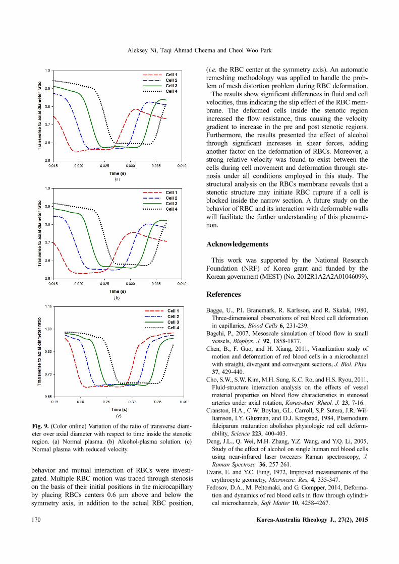
4.4. Effects on RBC transverse to the axial diame-ter ratio in the stenotic structure
The deformation characteristics of RBCs and their
mutual deforming interaction have been computed in
terms of the ratio of the RBCs transverse diameter to the
axial diameter. The RBCs were placed initially with their
centers at the symmetry axis. Fig. 9 shows the distribution
of this ratio with respect to time inside the stenosis region.
The graphs represent an initial drop in the deformation at
the entrance of the stenosis followed by a constant diam-
eter ratio in the middle of stenosis. The stenosis causes in
a significant pressure drop, thus leading to reduced mem-
brane deformation while moving fast.
On the contrary, the increase in plasma pressure results
in a sharp increase in RBC deformation at the end of the
stenosis region. The fast moving Cell 1 had the smallest
ratio of transverse to axial diameter for normal and highly
viscous plasma flows. For the case of reduced inlet plasma
velocity, the deformation pattern is reversed at the exit of
the stenotic region, which is a clear sign of the shear
effects for reduced plasma velocity. Other cells show sim-
ilar deformation patterns with relatively high magnitudes
at the start and exit of stenosis for normal and highly vis-
cous plasma flows, thus indicating the effects of flow
resistance caused by the number of cell ahead of one par-
ticular cell. However, the small inlet flow velocity at the
inlet has a dominant shear effect on RBC deformation
after the middle of stenosis.
5. Conclusions
We conducted a 2D numerical simulation to investigate
the motion and deformation of multiple RBCs moving in
a microcapillary filled with plasma alcohol solution. The
presented model can help explain the pathologies as a
result of alcohol consumption, which increases plasma
viscosity and reduces blood flow to a significant extent.
The effects of rheological properties on the mechanical
Fig. 8. (Color online) Von Mises stress distribution for multiple RBCs inside the stenosis region for normal plasma viscosity when
the cell centers are displaced toward the (a) top wall and (b) bottom wall. The alcohol-plasma solution viscosity caused by alcohol
when the cell centers are displaced toward the (c) top wall and (d) bottom wall.
Aleksey Ni, Taqi Ahmad Cheema and Cheol Woo Park
170 Korea-Australia Rheology J., 27(2), 2015
behavior and mutual interaction of RBCs were investi-
gated. Multiple RBC motion was traced through stenosis
on the basis of their initial positions in the microcapillary
by placing RBCs centers 0.6 µm above and below the
symmetry axis, in addition to the actual RBC position,
(i.e. the RBC center at the symmetry axis). An automatic
remeshing methodology was applied to handle the prob-
lem of mesh distortion problem during RBC deformation.
The results show significant differences in fluid and cell
velocities, thus indicating the slip effect of the RBC mem-
brane. The deformed cells inside the stenotic region
increased the flow resistance, thus causing the velocity
gradient to increase in the pre and post stenotic regions.
Furthermore, the results presented the effect of alcohol
through significant increases in shear forces, adding
another factor on the deformation of RBCs. Moreover, a
strong relative velocity was found to exist between the
cells during cell movement and deformation through ste-
nosis under all conditions employed in this study. The
structural analysis on the RBCs membrane reveals that a
stenotic structure may initiate RBC rupture if a cell is
blocked inside the narrow section. A future study on the
behavior of RBC and its interaction with deformable walls
will facilitate the further understanding of this phenome-
non.
Acknowledgements
This work was supported by the National Research
Foundation (NRF) of Korea grant and funded by the
Korean government (MEST) (No. 2012R1A2A2A01046099).
References
Bagge, U., P.I. Branemark, R. Karlsson, and R. Skalak, 1980,
Three-dimensional observations of red blood cell deformation
in capillaries, Blood Cells 6, 231-239.
Bagchi, P., 2007, Mesoscale simulation of blood flow in small
vessels, Biophys. J. 92, 1858-1877.
Chen, B., F. Guo, and H. Xiang, 2011, Visualization study of
motion and deformation of red blood cells in a microchannel
with straight, divergent and convergent sections, J. Biol. Phys.
37, 429-440.
Cho, S.W., S.W. Kim, M.H. Sung, K.C. Ro, and H.S. Ryou, 2011,
Fluid-structure interaction analysis on the effects of vessel
material properties on blood flow characteristics in stenosed
arteries under axial rotation, Korea-Aust. Rheol. J. 23, 7-16.
Cranston, H.A., C.W. Boylan, G.L. Carroll, S.P. Sutera, J.R. Wil-
liamson, I.Y. Gluzman, and D.J. Krogstad, 1984, Plasmodium
falciparum maturation abolishes physiologic red cell deform-
ability, Science 223, 400-403.
Deng, J.L., Q. Wei, M.H. Zhang, Y.Z. Wang, and Y.Q. Li, 2005,
Study of the effect of alcohol on single human red blood cells
using near-infrared laser tweezers Raman spectroscopy, J.
Raman Spectrosc. 36, 257-261.
Evans, E. and Y.C. Fung, 1972, Improved measurements of the
erythrocyte geometry, Microvasc. Res. 4, 335-347.
Fedosov, D.A., M. Peltomaki, and G. Gompper, 2014, Deforma-
tion and dynamics of red blood cells in flow through cylindri-
cal microchannels, Soft Matter 10, 4258-4267.
Fig. 9. (Color online) Variation of the ratio of transverse diam-
eter over axial diameter with respect to time inside the stenotic
region. (a) Normal plasma. (b) Alcohol-plasma solution. (c)
Normal plasma with reduced velocity.

Numerical investigation on the structural characteristics of multiple RBCs in a stenotic microcapillary......
Korea-Australia Rheology J., 27(2), 2015 171
Frey, P.J. and H. Borouchaki, 1999, Surface mesh quality eval-
uation, Int. J. Numer. Meth. Eng. 45, 101-118.
Fung, Y.C., 1993, Biomechanics: Mechanical Properties of Liv-
ing Tissues, Springer-Verlag, New York.
Gdovinova, Z., 2001, Blood flow velocity in the middle cerebral
artery in heavy alcohol drinkers, Alcohol Alcohol. 36, 346-348.
Gdovinova, Z., 2002, Cerebral blood flow velocity, erythrocyte
deformability and alcohol intake, Comp. Clin. Pathol. 11, 77-
81.
Gdovinova, Z., 2006, Cerebral blood flow velocity and erythro-
cyte deformability in heavy alcohol drinkers at the acute stage
and two weeks after withdrawal, Drug Alcohol Depend. 81,
207-213.
Gokturk, H.S., M. Demir, N.A. Ozturk, G.K. Unler, S. Kulak-
sizoglu, I. Kozanoglu, E. Serin, and U. Yilmaz, 2009, Plasma
viscosity changes in patients with liver cirrhosis, South Med. J.
102, 1013-1018.
Guo, Z.Z. and Z. Xiwen, 2011, Mechanical behavior of the eryth-
rocyte in micro vessel stenosis, Sci. China Life Sci. 54, 450-
458.
Hillbom, M., M. Kaste, L. Tarssanen, and R. Johnsson, 1983,
Effect of ethanol on blood viscosity and erythrocyte flexibility
in healthy men, Eur. J. Clin. Invest. 13, 45-48.
Homaidan, F.R., L.J. Kricka, and T.P. Whitehead, 1984, Mor-
phology of red blood cells in alcoholics, Lancet 1, 913-914.
Jeong, J.H., Y. Sugii, M. Minamiyama, and K. Okamoto, 2006,
Measurement of RBC deformation and velocity in capillaries
in vivo, Microvasc. Res. 71, 212-217.
Késmárky, G., P. Kenyeres, M. Rábai, and K. Tóth, 2008, Plasma
viscosity: a forgotten variable, Clin. Hemorheol. Microcirc. 39,
243-246.
Korin, N., A. Bransky, and U. Dinnar, 2007, Theoretical model
and experimental study of red blood cell (RBC) deformation in
microchannels, J. Biomech. 40, 2088-2095.
Lee, S.S., K.H. Ahn, S.J. Lee, K. Sun, P.T. Goedgart, and R. Har-
deman, 2004, Shear induced damage of red blood cells mon-
itored by the decrease of their deformability, Korea-Aust.
Rheol. J. 16, 141-146.
Linderkamp, O. and H.J. Meiselman, 1982, Geometric, osmotic
and membrane mechanical properties of density-separated
human red cells, Blood 59, 1121-1127.
Mi, X.Q., J.Y. Chen, and L.W. Zhou, 2006, Effect of low power
laser irradiation on disconnecting the membrane attached
hemoglobin from erythrocyte membrane, J. Photochem. Pho-
tobiol. B-Biol. 83, 146-150.
Mohandas, N., M.R. Clark, M.S. Jacobs, and S.B. Shohet, 1980,
Analysis of factors regulating erythrocyte deformability, J.
Clin. Invest. 66, 563-573.
Ni, A., T.A. Cheema, M.K. Kwak, and C.W. Park, 2013, Two-
dimensional numerical simulation of the red blood cell floating
in a plasma-alcohol solution through stenosis in a microvessel,
Korea-Aust. Rheol. J. 26, 293-301.
Popel, A.S. and P.C. Johnson, 2005, Microcirculation and hemor-
heology, Annu. Rev. Fluid. Mech. 37, 43-69.
Pozrikidis, C., 2003, Numerical simulation of the flow-induced
deformation of red blood cells, Ann. Biomed. Eng. 31, 1194-
1205.
Ramanujan, S. and C. Pozdrikidis, 1998, Deformation of liquid
capsules enclosed by elastic membranes in simple shear flow:
Large deformations and the effect of capsule viscosity, J. Fluid
Mech. 361, 117-143.
Rotsch, C., K. Jacobson, and M. Radmacher, 1999, Dimensional
and mechanical dynamics of active and stable edges in motile
fibroblasts investigated by using atomic force microscopy,
Proc. Natl. Acad. Sci. U.S.A. 3, 921-926.
Secomb, T.W. and B.S. Rekowska, A.R. Pries, 2007, Two-dimen-
sional simulation of red blood cell deformation and lateral
migration in microvessels, Ann. Biomed. Eng. 35, 755-765.
Secomb, T.W. and R. Hsu, 1996, Analysis of red blood cell
motion through cylindrical micropores: effects of cell proper-
ties, Biophys. J. 71, 1095-1101.
Shiraishi, K., M. Watanabe, M. Itakura, S. Matsuzaki, and H.
Ishida, 1994, Influence of plasma composition on erythrocyte
filterability in alcoholic liver disease, Alcohol Alcohol. 29, 1-4.
Stoltz, J.F., M. Singh, and P. Riha, 1999, Hemorheology in Prac-
tice, IOS Press, Amsterdam.
Tsukada, K., E. Sekizuka, C. Oshio, and H. Minamitani, 2001,
Direct measurement of erythrocyte deformability n diabetes
mellitus with a transparent microchannel capillary model and
high-speed video camera system, Microvasc. Res. 61, 231-239.
Tsubota, K., S. Wada, and T. Ymaguchi, 2006, Particle method
for computer simulation of red blood cell motion in blood flow,
Comput. Meth. Programs Biomed. 83, 139-146.
Vahidkhah, K. and N. Fatouraee, 2012, Numerical simulation of
red blood cell behavior in a stenosed arteriole using immersed
boundary-lattice Boltzmann method, Int. J. Numer. Meth.
Biomed. Eng. 28, 239-256.
Wang, C., X. Wang, and P. Ye, 2008, The transport and defor-
mation of blood cells in microchannel, 3rd IEEE International
Conference on Nano/Micro Engineered and Molecular Sys-
tems, Sanya, China, pp.116-119.
Zhodi, T.I. and F.A. Kuypers, 2006, Modelling and rapid simu-
lation of multiplered blood cell light scattering, J. R. Soc. Inter-
face 3, 823-831.