Embed Size (px)
Citation preview
NUMBERS
Estimated number of proteins in the human body: 100 000
Primary structure analysis (F. Sanger, 1953)
1953-1978 (25 years) 10811979-1991 (13 years) 16 0001992- 1000/year
Three-dimensional (3D) structure (J. Kendrew, 1962)
1962-1985 (20 years) 2001986-1991 ( 5 years) 4801992- 100/years
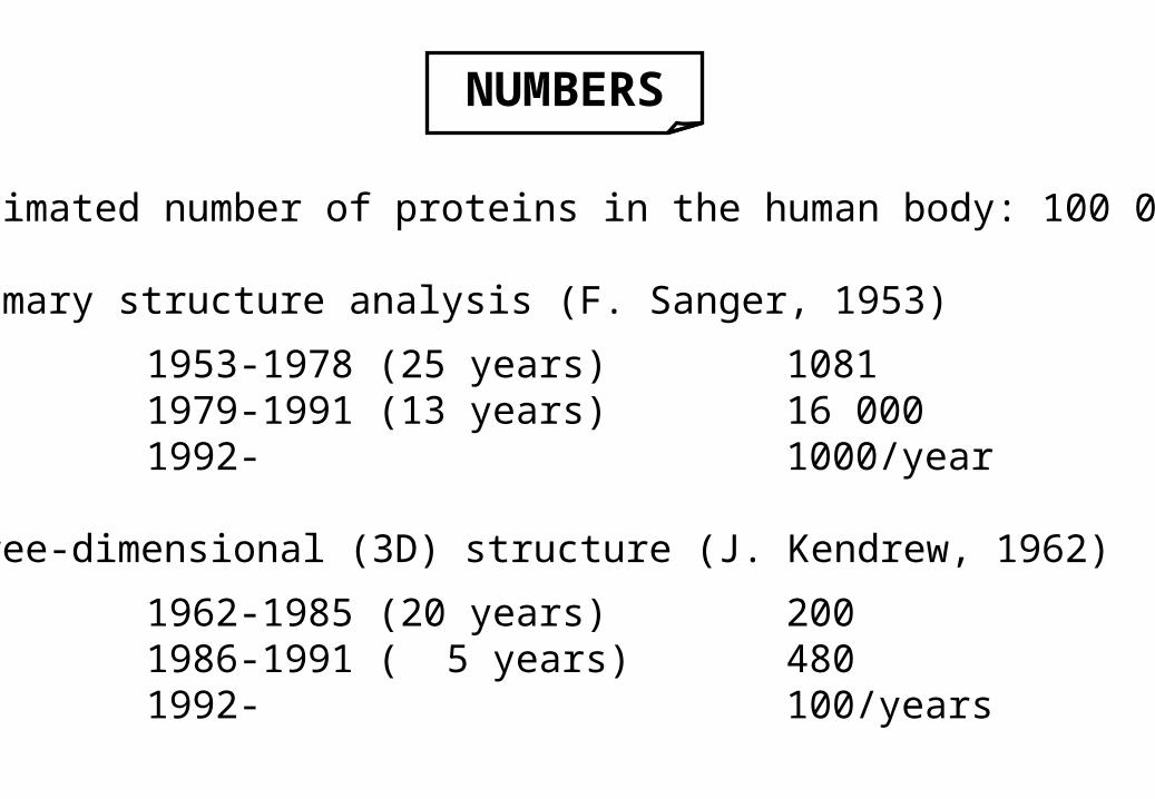
CLASSIFICATION OF PROTEINSACCORDING TO THEIR FUNCTION
1. Enzymatic catalysis (e.g. Ser proteases)2. Transport (e.g. transferrin for iron, serum albumin for fatty acids)3. Storage (e.g. ferrin for iron in liver, casein in milk)4. Protection
• toxins (e.g. ricin [plant], diphteria [bacteria])• self and non-self discrimination, immune protection
(e.g. antibodies, antigenes)5. Signal transduction (e.g. hormones, receptors)
• nerve impulses• growth• differentiation
6. Cell to cell communication (e.g. adhesion, molecules; factors, acceptors)7. Coordinated motion (e.g. muscle proteins)8. Mechanical support
• at cellular level (e.g. Membrane proteins)• at tissue level (e.g. structural proteins, e.g. collagen in skin, bone)
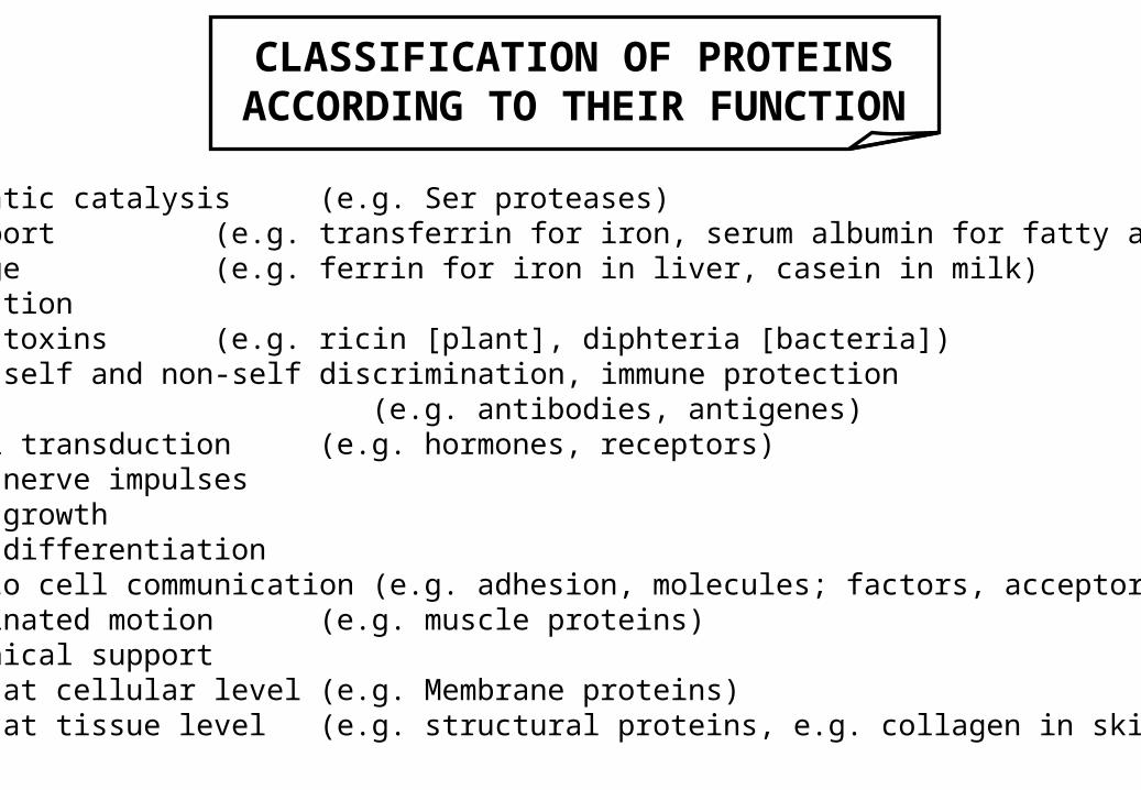
RECOGNITION PHENOMENA
Interaction Kd[M]
1. Enzyme – substrate 10-3 – 10-5
2. Transporter – ligand 10-6 – 10-8
3. Hormone – receptor 10-9
4. Antibody – antigen 10-7 – 10-11
5. Storage protein – ligand
6. Toxin – receptor
7. Protein – protein (in a contractile superassembly)
8. Lectin – carbohydrate 10-4 – 10-7
9. Avidin – biotin 10-15
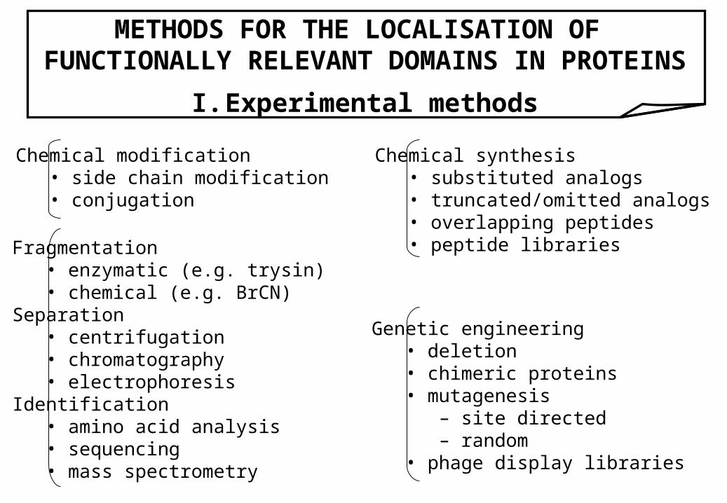
METHODS FOR THE LOCALISATION OF FUNCTIONALLY RELEVANT DOMAINS IN PROTEINS
I. Experimental methods
Chemical modification• side chain modification• conjugation
Fragmentation• enzymatic (e.g. trysin)• chemical (e.g. BrCN)
Separation• centrifugation• chromatography• electrophoresis
Identification• amino acid analysis• sequencing• mass spectrometry
Genetic engineering• deletion• chimeric proteins• mutagenesis
– site directed– random
• phage display libraries
Chemical synthesis• substituted analogs• truncated/omitted analogs• overlapping peptides• peptide libraries
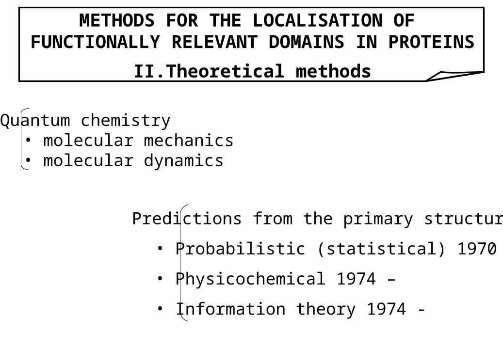
METHODS FOR THE LOCALISATION OF FUNCTIONALLY RELEVANT DOMAINS IN PROTEINS
II. Theoretical methods
Quantum chemistry• molecular mechanics• molecular dynamics
Predictions from the primary structure
• Probabilistic (statistical) 1970 –
• Physicochemical 1974 –
• Information theory 1974 -
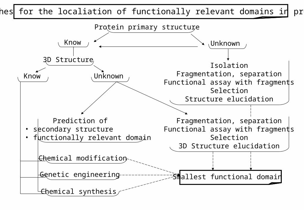
Approaches for the localiation of functionally relevant domains in proteins
Protein primary structure
Know Unknown
3D Structure
Know Unknown
IsolationFragmentation, separation
Functional assay with fragmentsSelection
Structure elucidation
Prediction of• secondary structure• functionally relevant domain
Fragmentation, separationFunctional assay with fragments
Selection3D Structure elucidation
Chemical modification
Genetic engineering
Chemical synthesis
Smallest functional domain
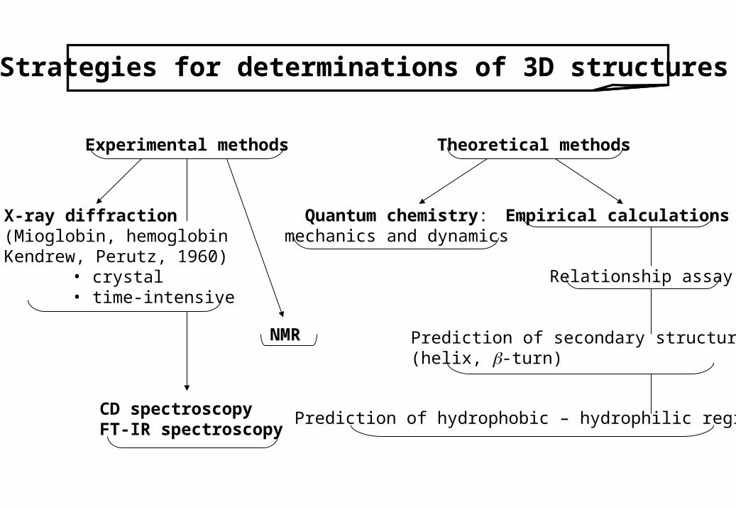
Strategies for determinations of 3D structures
Experimental methods Theoretical methods
X-ray diffraction(Mioglobin, hemoglobin Kendrew, Perutz, 1960)
• crystal• time-intensive
NMR
CD spectroscopyFT-IR spectroscopy
Quantum chemistry:mechanics and dynamics
Empirical calculations
Relationship assay
Prediction of secondary structures(helix, -turn)
Prediction of hydrophobic – hydrophilic regions
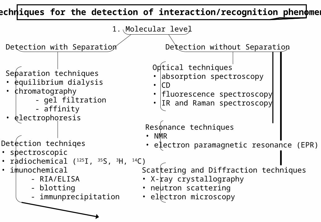
Techniques for the detection of interaction/recognition phenomena
1. Molecular level
Detection with Separation Detection without Separation
Separation techniques• equilibrium dialysis• chromatography
- gel filtration- affinity
• electrophoresis
Detection techniqes• spectroscopic• radiochemical (125I, 35S, 3H, 14C)• imunochemical
- RIA/ELISA- blotting- immunprecipitation
Optical techniques• absorption spectroscopy• CD• fluorescence spectroscopy• IR and Raman spectroscopy
Resonance techniques• NMR• electron paramagnetic resonance (EPR)
Scattering and Diffraction techniques• X-ray crystallography• neutron scattering• electron microscopy
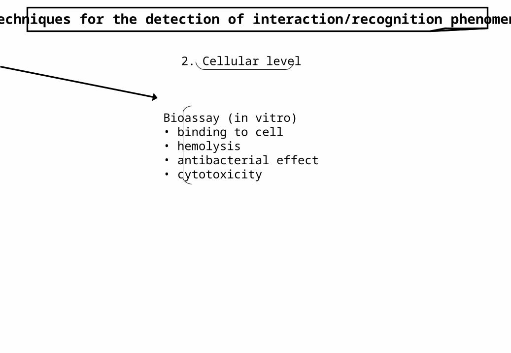
Techniques for the detection of interaction/recognition phenomena
2. Cellular level
Bioassay (in vitro)• binding to cell• hemolysis• antibacterial effect• cytotoxicity
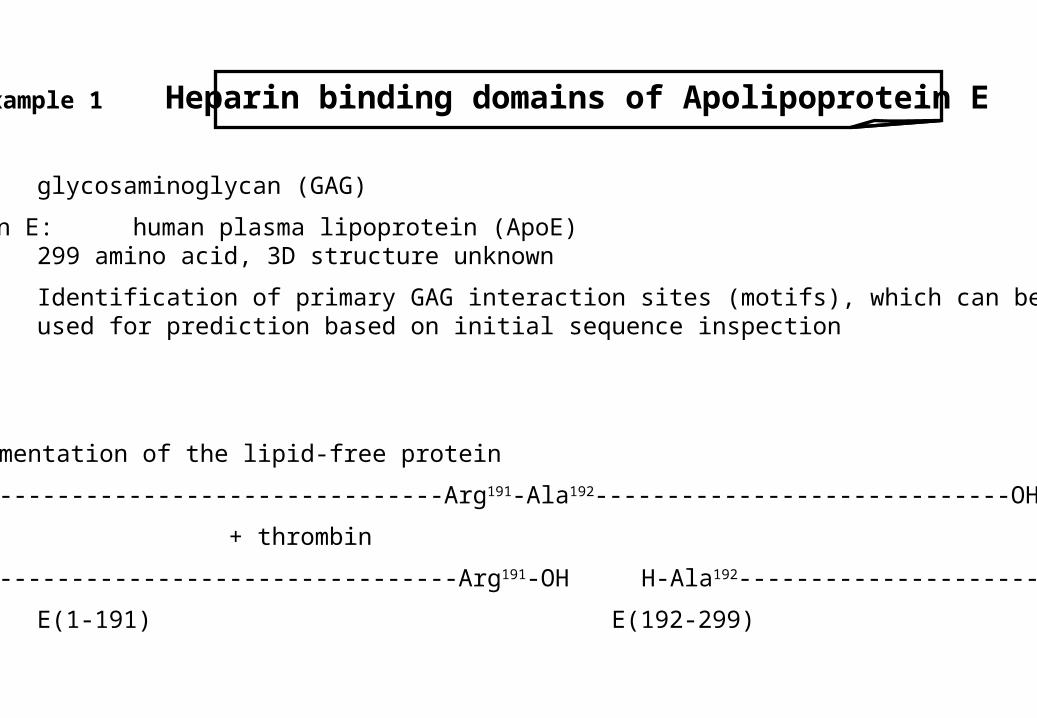
Example 1 Heparin binding domains of Apolipoprotein E
Heparin: glycosaminoglycan (GAG)
Apolipoprotein E: human plasma lipoprotein (ApoE)299 amino acid, 3D structure unknown
Aim: Identification of primary GAG interaction sites (motifs), which can beused for prediction based on initial sequence inspection
Phase I
1. step Fragmentation of the lipid-free protein
H----------------------------------Arg191-Ala192-----------------------------OH
+ thrombin
H ----------------------------------Arg191-OH H-Ala192-----------------------------OH
E(1-191) E(192-299)
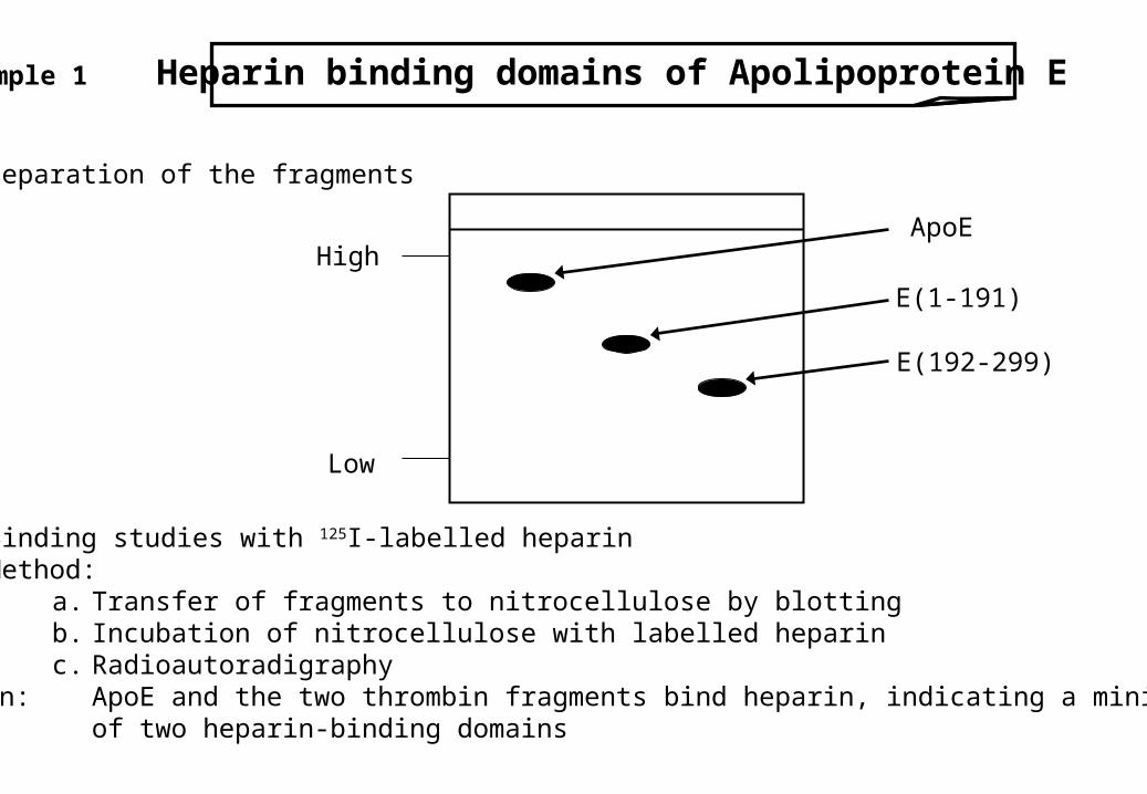
Example 1 Heparin binding domains of Apolipoprotein E
Phase I
2. step Separation of the fragments
3. step Binding studies with 125I-labelled heparinMethod:
a. Transfer of fragments to nitrocellulose by blottingb. Incubation of nitrocellulose with labelled heparinc. Radioautoradigraphy
Observation: ApoE and the two thrombin fragments bind heparin, indicating a minimum of two heparin-binding domains
High
Low
ApoE
E(1-191)
E(192-299)
Example 1 Heparin binding domains of Apolipoprotein E
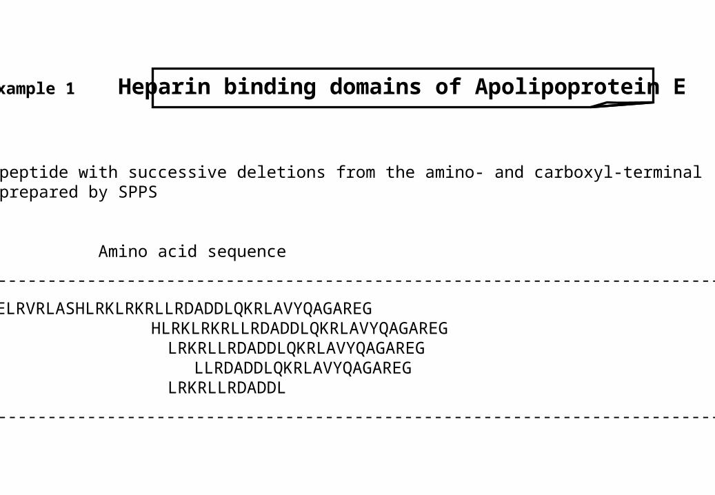
Phase II
1. step Synthetic peptide with successive deletions from the amino- and carboxyl-terminal ends were prepared by SPPS
Residue Amino acid sequence
-------------------------------------------------------------------------------------------------------------
129-169 STEELRVRLASHLRKLRKRLLRDADDLQKRLAVYQAGAREG139-169 HLRKLRKRLLRDADDLQKRLAVYQAGAREG144-169 LRKRLLRDADDLQKRLAVYQAGAREG148-169 LLRDADDLQKRLAVYQAGAREG141-155 LRKRLLRDADDL
-------------------------------------------------------------------------------------------------------------
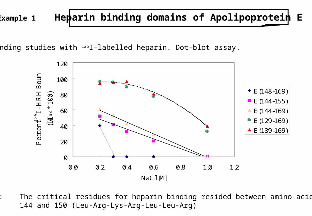
Example 1 Heparin binding domains of Apolipoprotein E
Phase II
2. step Binding studies with 125I-labelled heparin. Dot-blot assay.
Observation: The critical residues for heparin binding resided between amino acid144 and 150 (Leu-Arg-Lys-Arg-Leu-Leu-Arg)
0
20
40
60
80
100
120
0.0 0.2 0.4 0.6 0.8 1.0 1.2
NaCl [M]
Perc
ent 1
25 I
- H
RH
Bound
(I/Im
ax
* 100) E(148-169)
E(144-155)
E(144-169)
E(129-169)
E(139-169)
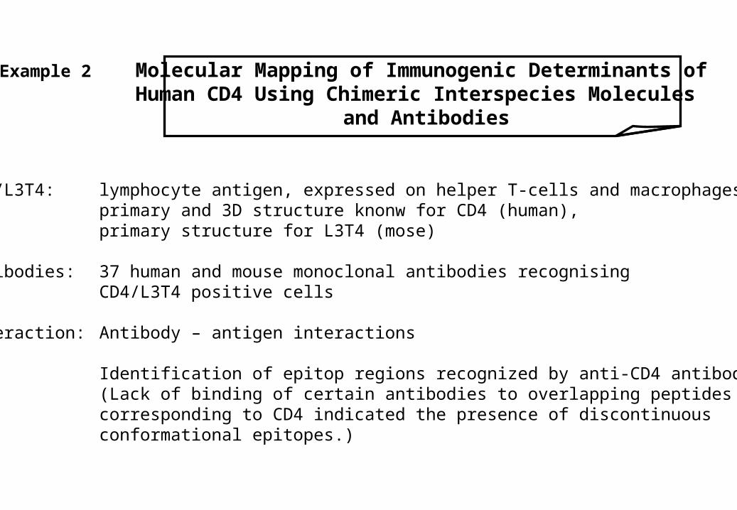
Example 2 Molecular Mapping of Immunogenic Determinants ofHuman CD4 Using Chimeric Interspecies Molecules
and Antibodies
CD4/L3T4: lymphocyte antigen, expressed on helper T-cells and macrophages,primary and 3D structure knonw for CD4 (human), primary structure for L3T4 (mose)
Antibodies: 37 human and mouse monoclonal antibodies recognising CD4/L3T4 positive cells
Interaction: Antibody – antigen interactions
Aim: Identification of epitop regions recognized by anti-CD4 antibodies.(Lack of binding of certain antibodies to overlapping peptides corresponding to CD4 indicated the presence of discontinuous conformational epitopes.)
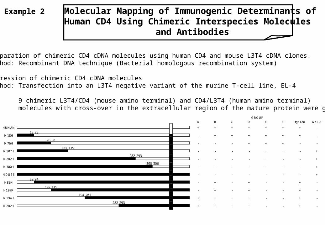
Example 2 Molecular Mapping of Immunogenic Determinants ofHuman CD4 Using Chimeric Interspecies Molecules
and Antibodies
1. step Preparation of chimeric CD4 cDNA molecules using human CD4 and mouse L3T4 cDNA clones.Method: Recombinant DNA technique (Bacterial homologous recombination system)
2. step Expression of chimeric CD4 cDNA moleculesMethod: Transfection into an L3T4 negative variant of the murine T-cell line, EL-4
Observation: 9 chimeric L3T4/CD4 (mouse amino terminal) and CD4/L3T4 (human amino terminal) molecules with cross-over in the extracellular region of the mature protein were generated
A B C D E F rgp120 GK1.5
M76H
M107H
H107M
M194H
M282H
18
M282H
M300H
MOUSE
H89M
HUMAN
M18H
293
300
23
76 80
107
89 94
107 119
194
282
201
-
-
-
+
306
119
282
293
GROUP
+ + + + + + + -
+ + + + + + -
- - - + + + - -
- - - - + + - +
- - - + - - +
- - - - + - - +
- - - - - - - +
+ - + - - + -
- + - + - - + -
+ + + + - - + -
- + -+ + + -
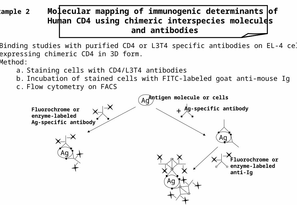
Example 2 Molecular mapping of immunogenic determinants ofHuman CD4 using chimeric interspecies molecules
and antibodies
3. step Binding studies with purified CD4 or L3T4 specific antibodies on EL-4 cellsexpressing chimeric CD4 in 3D form.Method:
a. Staining cells with CD4/L3T4 antibodiesb. Incubation of stained cells with FITC-labeled goat anti-mouse Igc. Flow cytometry on FACS
Ag
Ag
+
Ag
Ag
Antigen molecule or cells
Ag-specific antibody
Fluorochrome orenzyme-labeledanti-Ig
Fluorochrome orenzyme-labeledAg-specific antibody
3. step
Observation: All chimeric molecules analys ??? in transfectants detectable withhuman and/or mouse specific anti-CD4 antibodies.Using the chimerics, it was possible to localise most of the CD4epitopes to specific region of the CD4 protein.
NB.: 1. CD4/L3T4 recognise antigen in the context of class II MHC antigens.2. As expected from their functional similarities, the human and the mouse CD4 molecules are highly homologous at both the DNA (70%) and amino acid (54%) levels.
Reference: P. Estess et al. Current Research in Protein Chemistry (ED.: J. J. Villafranca, Academic Press,San Diego, p. 499 (1990))
Example 2 Molecular mapping of immunogenic determinants ofHuman CD4 using chimeric interspecies molecules
and antibodies

Example 3 Localization of Immunogenic Determinants (Epitopes) ofhuman epithelial mucin glycoprotein, MUC-1
Using synthetic Peptides and MUC-1 specific Antibodies
MUC-1: high molecular weight, MUC-1 gene related glycoprotein,associated with human breast and ovarian carcinoma,primary structure is known
Antibodies: mouse monoclonal antibodies recognizing MUC-1 glycoprotein[HMFG-1, C595, B55, etc.]
Interaction: Antibody – antigen interactions
Aim: Identification of epitopes recognized by anti-MUC-1 antibodies
Phase I
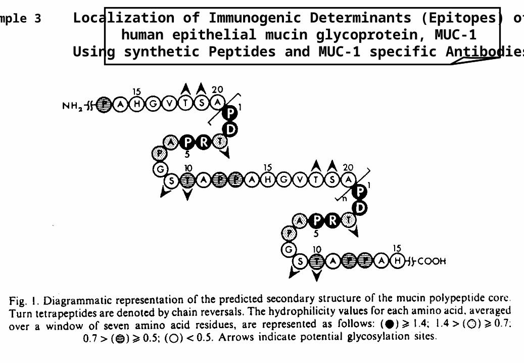
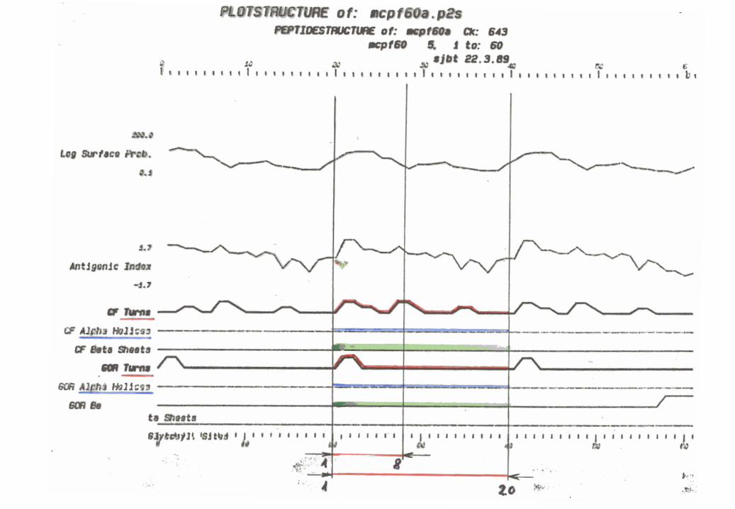
1. step Analysis of the primary structure of MUC-1 glycoprotein.
Method: Prediction of B-cell epitopes using various algorithms searching fora. hydrophilic region andb. -turn secondary structure
Example 3 Localization of Immunogenic Determinants (Epitopes) ofhuman epithelial mucin glycoprotein, MUC-1
Using synthetic Peptides and MUC-1 specific Antibodies
Example 3 Localization of Immunogenic Determinants (Epitopes) ofhuman epithelial mucin glycoprotein, MUC-1
Using synthetic Peptides and MUC-1 specific Antibodies
Phase I
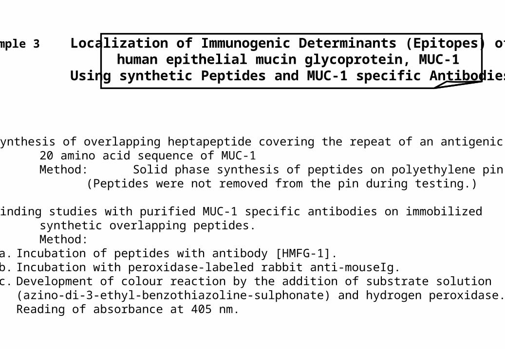
2. step Synthesis of overlapping heptapeptide covering the repeat of an antigenic20 amino acid sequence of MUC-1Method: Solid phase synthesis of peptides on polyethylene pin support.
(Peptides were not removed from the pin during testing.)
3. step Binding studies with purified MUC-1 specific antibodies on immobilizedsynthetic overlapping peptides.Method:
a. Incubation of peptides with antibody [HMFG-1].b. Incubation with peroxidase-labeled rabbit anti-mouseIg.c. Development of colour reaction by the addition of substrate solution
(azino-di-3-ethyl-benzothiazoline-sulphonate) and hydrogen peroxidase.Reading of absorbance at 405 nm.
Example 3 Localization of Immunogenic Determinants (Epitopes) ofhuman epithelial mucin glycoprotein, MUC-1
Using synthetic Peptides and MUC-1 specific Antibodies
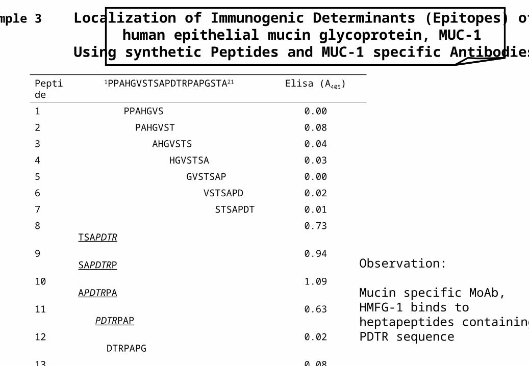
Peptide 1PPAHGVSTSAPDTRPAPGSTA21 Elisa (A405)
1 PPAHGVS 0.00
2 PAHGVST 0.08
3 AHGVSTS 0.04
4 HGVSTSA 0.03
5 GVSTSAP 0.00
6 VSTSAPD 0.02
7 STSAPDT 0.01
8 TSAPDTR 0.73
9 SAPDTRP 0.94
10 APDTRPA 1.09
11 PDTRPAP 0.63
12 DTRPAPG 0.02
13 TRPAPGS 0.08
14 RPAPGST 0.03
15 PAPGSTA 0.03
16 APGSTAP 0.02
Observation:
Mucin specific MoAb,HMFG-1 binds toheptapeptides containingPDTR sequence
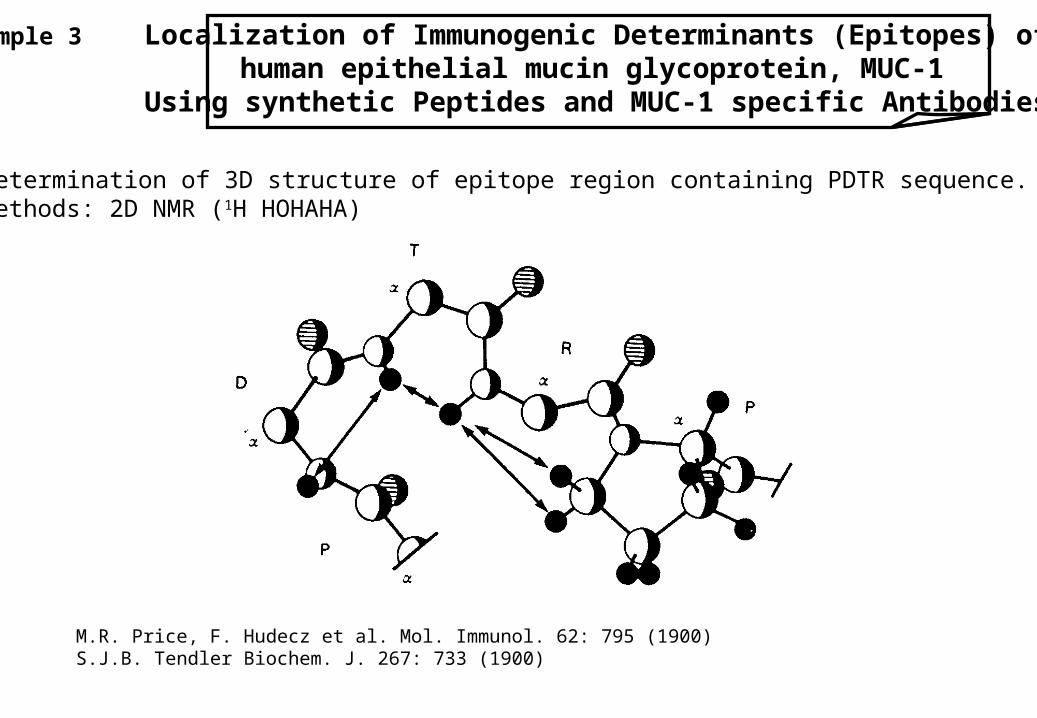
Phase I Determination of 3D structure of epitope region containing PDTR sequence.Methods: 2D NMR (1H HOHAHA)
Reference: M.R. Price, F. Hudecz et al. Mol. Immunol. 62: 795 (1900)S.J.B. Tendler Biochem. J. 267: 733 (1900)
Example 3 Localization of Immunogenic Determinants (Epitopes) ofhuman epithelial mucin glycoprotein, MUC-1
Using synthetic Peptides and MUC-1 specific Antibodies
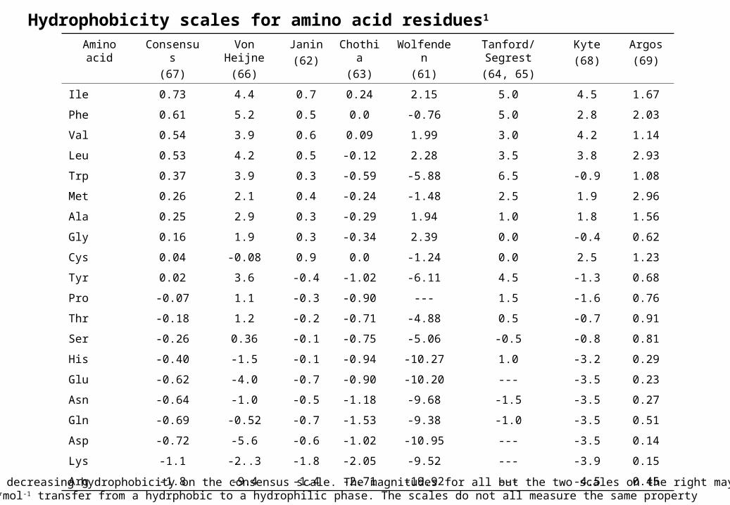
Hydrophobicity scales for amino acid residues1
Amino acid Consensus
(67)
Von Heijne
(66)
Janin
(62)
Chothia
(63)
Wolfenden
(61)
Tanford/Segrest
(64, 65)
Kyte
(68)
Argos
(69)
Ile 0.73 4.4 0.7 0.24 2.15 5.0 4.5 1.67
Phe 0.61 5.2 0.5 0.0 -0.76 5.0 2.8 2.03
Val 0.54 3.9 0.6 0.09 1.99 3.0 4.2 1.14
Leu 0.53 4.2 0.5 -0.12 2.28 3.5 3.8 2.93
Trp 0.37 3.9 0.3 -0.59 -5.88 6.5 -0.9 1.08
Met 0.26 2.1 0.4 -0.24 -1.48 2.5 1.9 2.96
Ala 0.25 2.9 0.3 -0.29 1.94 1.0 1.8 1.56
Gly 0.16 1.9 0.3 -0.34 2.39 0.0 -0.4 0.62
Cys 0.04 -0.08 0.9 0.0 -1.24 0.0 2.5 1.23
Tyr 0.02 3.6 -0.4 -1.02 -6.11 4.5 -1.3 0.68
Pro -0.07 1.1 -0.3 -0.90 --- 1.5 -1.6 0.76
Thr -0.18 1.2 -0.2 -0.71 -4.88 0.5 -0.7 0.91
Ser -0.26 0.36 -0.1 -0.75 -5.06 -0.5 -0.8 0.81
His -0.40 -1.5 -0.1 -0.94 -10.27 1.0 -3.2 0.29
Glu -0.62 -4.0 -0.7 -0.90 -10.20 --- -3.5 0.23
Asn -0.64 -1.0 -0.5 -1.18 -9.68 -1.5 -3.5 0.27
Gln -0.69 -0.52 -0.7 -1.53 -9.38 -1.0 -3.5 0.51
Asp -0.72 -5.6 -0.6 -1.02 -10.95 --- -3.5 0.14
Lys -1.1 -2..3 -1.8 -2.05 -9.52 --- -3.9 0.15
Arg -1.8 -9.4 -1.4 -2.71 -19.92 --- -4.5 0.451The order is by decreasing hydrophobicity on the consensus scale. The magnitudes for all but the two scales on the right may be consideredRoughly in kcal*mol-1 transfer from a hydrphobic to a hydrophilic phase. The scales do not all measure the same property
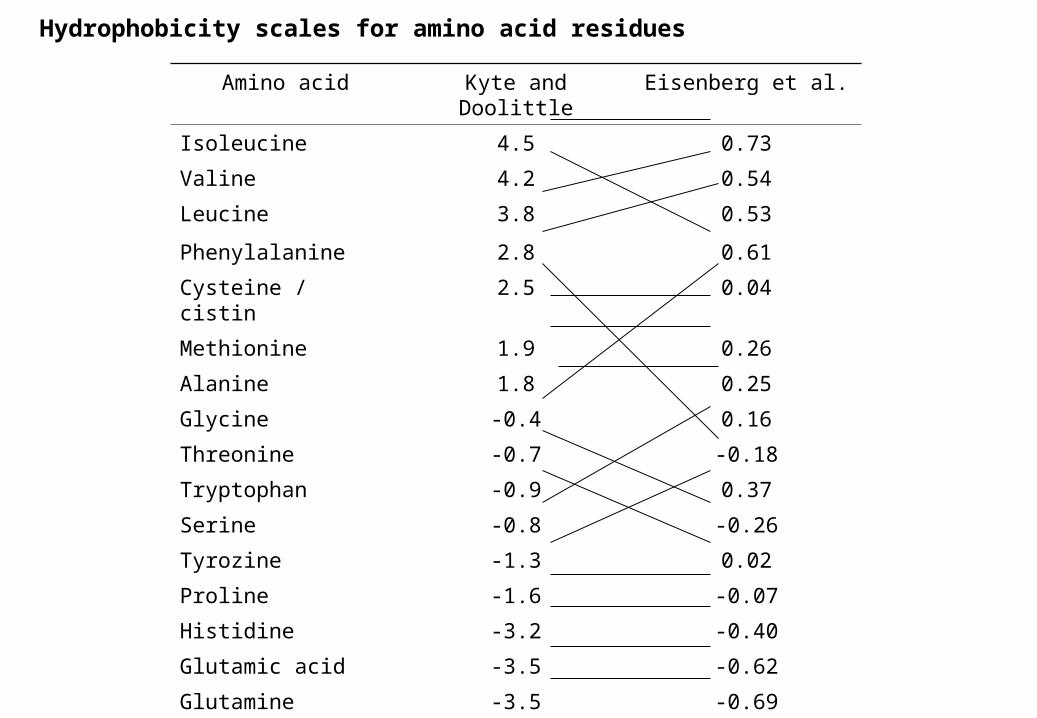
Hydrophobicity scales for amino acid residues
Amino acid Kyte and Doolittle Eisenberg et al.
Isoleucine 4.5 0.73
Valine 4.2 0.54
Leucine 3.8 0.53
Phenylalanine 2.8 0.61
Cysteine / cistin 2.5 0.04
Methionine 1.9 0.26
Alanine 1.8 0.25
Glycine -0.4 0.16
Threonine -0.7 -0.18
Tryptophan -0.9 0.37
Serine -0.8 -0.26
Tyrozine -1.3 0.02
Proline -1.6 -0.07
Histidine -3.2 -0.40
Glutamic acid -3.5 -0.62
Glutamine -3.5 -0.69
Aspartic acid -3.5 -0.72
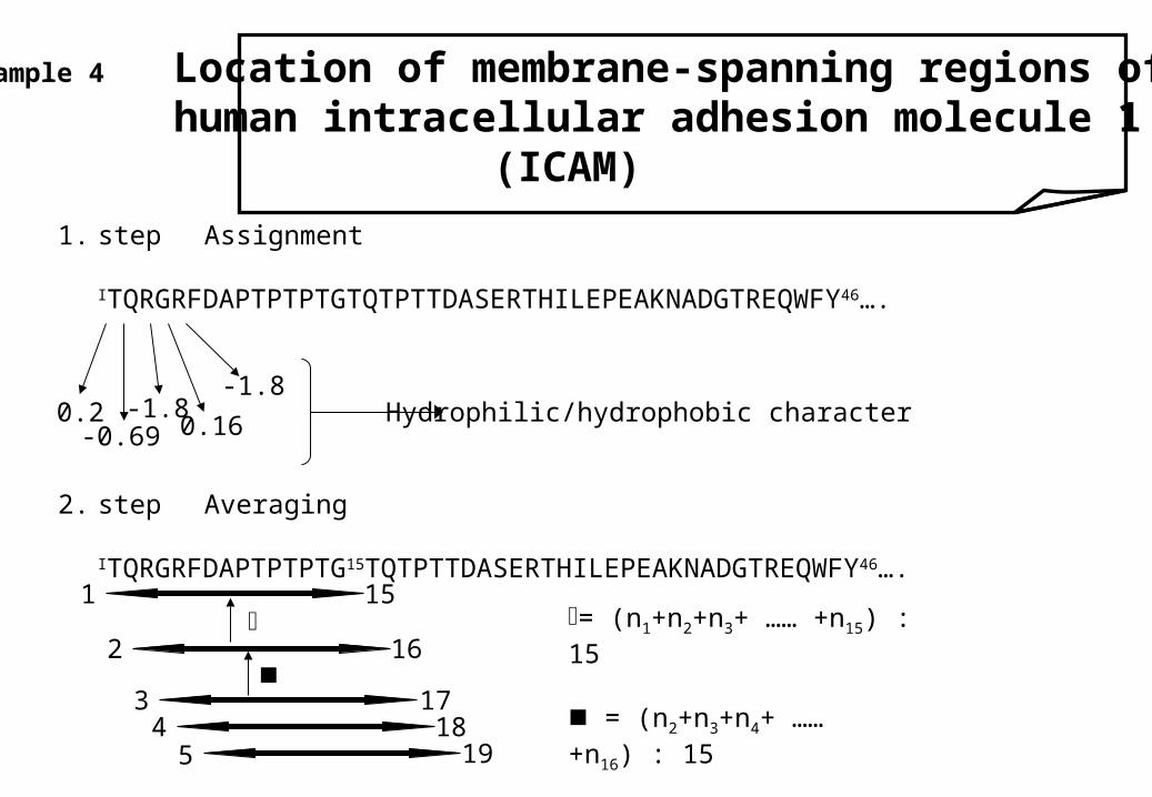
1. step Assignment
ITQRGRFDAPTPTPTGTQTPTTDASERTHILEPEAKNADGTREQWFY46….
2. step Averaging
ITQRGRFDAPTPTPTG15TQTPTTDASERTHILEPEAKNADGTREQWFY46….
Example 4 Location of membrane-spanning regions of human intracellular adhesion molecule 1
(ICAM)
0.2-0.69
-1.80.16
-1.8Hydrophilic/hydrophobic character
1 15
2 16
3 174 18
5 19
= (n1+n2+n3+ …… +n15) : 15
= (n2+n3+n4+ …… +n16) : 15
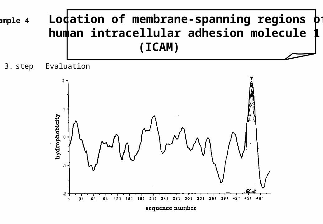
3. step Evaluation
Example 4 Location of membrane-spanning regions of human intracellular adhesion molecule 1
(ICAM)
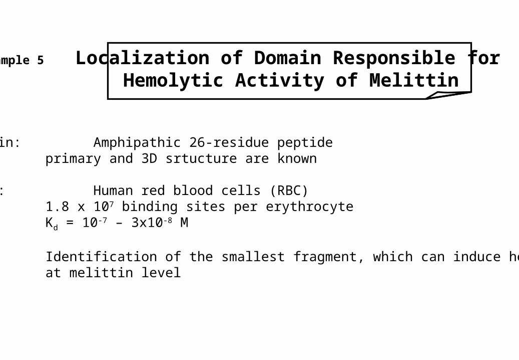
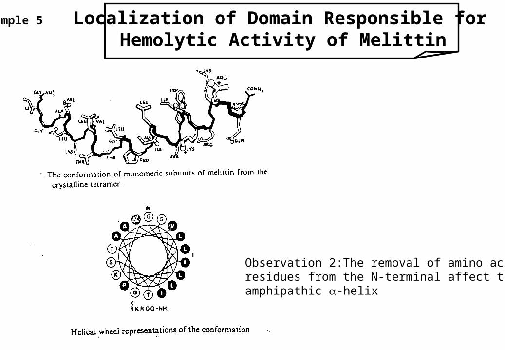
Example 5 Localization of Domain Responsible forHemolytic Activity of Melittin
Melittin: Amphipathic 26-residue peptideprimary and 3D srtucture are known
Target: Human red blood cells (RBC)1.8 x 10⁷ binding sites per erythrocyteKd = 10-7 – 3x10⁻⁸ M
Aim: Identification of the smallest fragment, which can induce hemolysisat melittin level
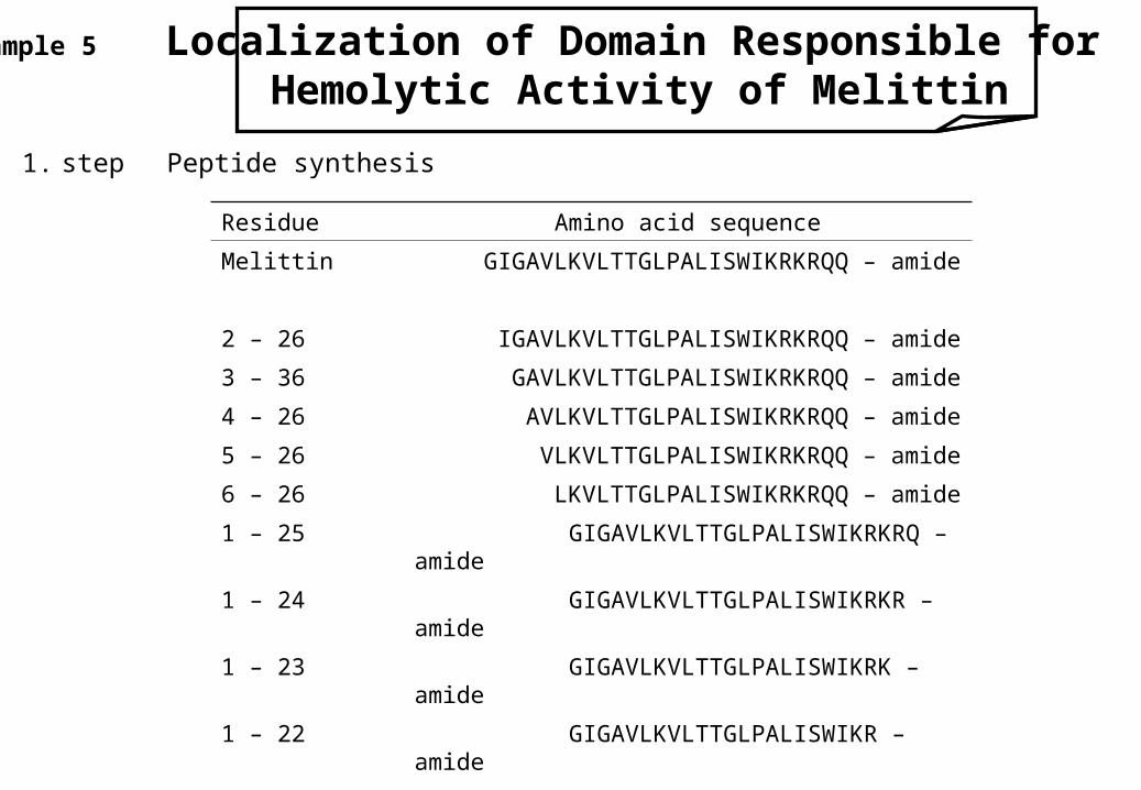
Example 5 Localization of Domain Responsible forHemolytic Activity of Melittin
1. step Peptide synthesis
Residue Amino acid sequence
Melittin GIGAVLKVLTTGLPALISWIKRKRQQ – amide
2 – 26 IGAVLKVLTTGLPALISWIKRKRQQ – amide
3 – 36 GAVLKVLTTGLPALISWIKRKRQQ – amide
4 – 26 AVLKVLTTGLPALISWIKRKRQQ – amide
5 – 26 VLKVLTTGLPALISWIKRKRQQ – amide
6 – 26 LKVLTTGLPALISWIKRKRQQ – amide
1 – 25 GIGAVLKVLTTGLPALISWIKRKRQ – amide
1 – 24 GIGAVLKVLTTGLPALISWIKRKR – amide
1 – 23 GIGAVLKVLTTGLPALISWIKRK – amide
1 – 22 GIGAVLKVLTTGLPALISWIKR – amide
1 – 21 GIGAVLKVLTTGLPALISWIK – amide
1 – 20 GIGAVLKVLTTGLPALISWI – amide
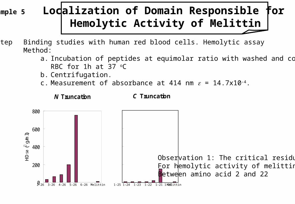
Example 5 Localization of Domain Responsible forHemolytic Activity of Melittin
2. step Binding studies with human red blood cells. Hemolytic assayMethod:
a. Incubation of peptides at equimolar ratio with washed and countedRBC for 1h at 37 oC
b. Centrifugation.c. Measurement of absorbance at 414 nm = 14.7x10⁻⁴.
N Truncation
0
200
400
600
800
HD
50 (m g
/ml)
C Truncation
0
200
400
600
800
Melittin2-26 3-26 4-26 5-26 6-26 1-25 1-24 1-23 1-22 1-21 1-20 Melittin
Observation 1: The critical residuesFor hemolytic activity of melittin areBetween amino acid 2 and 22
Example 5 Localization of Domain Responsible forHemolytic Activity of Melittin
Observation 2:The removal of amino acid residues from the N-terminal affect the amphipathic -helix