Embed Size (px)
Citation preview
VIROLOGY 181, 353-358 (1991)
Nucleotide Sequence of the Genes Encoding the Major Tail Sheath and Tail Tube Proteins of Bacteriophage P2
LOUISE M. TEMPLE,* SUSAN L. FORSBURG,t*’ RICHARD CALENDAR,t AND GAIL E. CHRISTIE*+
*Department of Microbiology and Immunology, Virginia Commonwealth University, Box 678 MCV Station, Richmond, Virginia 23298-0678, and tDepartmenr of Molecular Biology, University of California. Berkeley, California 94720
Received October 30, 1990; accepted November 20, 1990
The major structural components of the contractile tail of bacteriophage P2 are proteins F, and F,,, which are believed to be the tail sheath and tube proteins, respectively. Both proteins were mapped previously to the P2 late gene F, based on the pattern of protein synthesis in various P2 amber mutants. In order to clarify the gene arrangement and to provide a basis for structural comparisons with other contractile phage tails, we have determined the nucleotide sequence of the region of the P2 genome encoding these two proteins. The coding regions were confirmed by location of the Fam4 mutation and by N-terminal amino acid sequencing of both proteins. The molecular weight and amino acid composition predicted by each of the coding regions correspond well to those determined experimentally for each protein. F,, is encoded by a newly identified P2 late gene. These proteins bear little resemblance to their functional homologues in bacteriophage T4. 0 1991 Academic Press, Inc.
INTRODUCTION
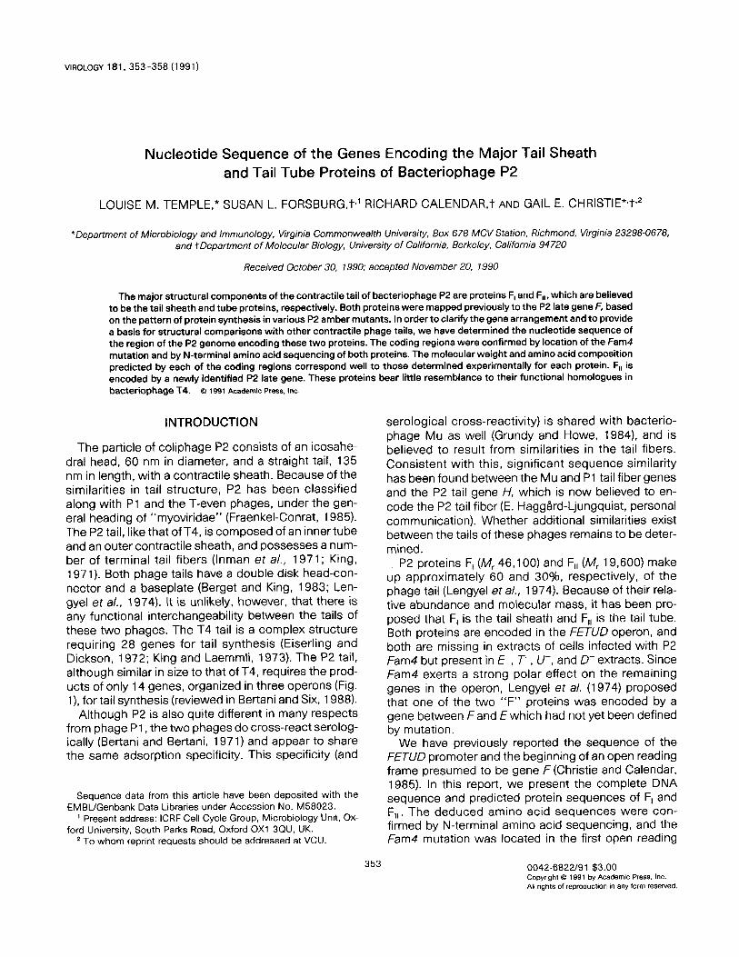
The particle of coliphage P2 consists of an icosahe- dral head, 60 nm in diameter, and a straight tail, 135 nm in length, with a contractile sheath. Because of the similarities in tail structure, P2 has been classified along with Pl and the T-even phages, under the gen- eral heading of “myoviridae” (Fraenkel-Conrat, 1985). The P2 tail, like that of T4, is composed of an inner tube and an outer contractile sheath, and possesses a num- ber of terminal tail fibers (Inman et a/., 1971; King, 1971). Both phage tails have a double disk head-con- nector and a baseplate (Berget and King, 1983; Len- gyel ef a/., 1974). It is unlikely, however, that there is any functional interchangeability between the tails of these two phages. The T4 tail is a complex structure requiring 28 genes for tail synthesis (Eiserling and Dickson, 1972; King and Laemmli, 1973). The P2 tail, although similar in size to that of T4, requires the prod- ucts of only 14 genes, organized in three operons (Fig. l), for tail synthesis (reviewed in Bertani and Six, 1988).
Although P2 is also quite different in many respects from phage Pl , the two phages do cross-react serolog- ically (Bertani and Bertani, 1971) and appear to share the same adsorption specificity. This specificity (and
Sequence data from this article have been deposited with the EMBUGenbank Data Libraries under Accession No. M58023.
’ Present address: ICRF Cell Cycle Group, Microbiology Unit, Ox- ford University, South Parks Road, Oxford OX1 3QU, UK.
2 To whom reprint requests should be addressed at VCU
serological cross-reactivity) is shared with bacterio- phage Mu as well (Grundy and Howe, 1984), and is believed to result from similarities in the tail fibers. Consistent with this, significant sequence similarity has been found between the Mu and Pl tail fiber genes and the P2 tail gene H, which is now believed to en- code the P2 tail fiber (E. HaggBrd-Ljungquist, personal communication). Whether additional similarities exist between the tails of these phages remains to be deter- mined.
P2 proteins F, (IV!, 46,100) and F,, (AI!~ 19,600) make up approximately 60 and 30%, respectively, of the phage tail (Lengyel et al., 1974). Because of their rela- tive abundance and molecular mass, it has been pro- posed that F, is the tail sheath and F,, is the tail tube. Both proteins are encoded in the FETUD operon, and both are missing in extracts of cells infected with P2 Fam4 but present in E-. T-, U-, and D- extracts. Since Fam4 exerts a strong polar effect on the remaining genes in the operon, Lengyel et al. (1974) proposed that one of the two “F” proteins was encoded by a gene between F and E which had not yet been defined by mutation.
We have previously reported the sequence of the FETUD promoter and the beginning of an open reading frame presumed to be gene F (Christie and Calendar, 1985). In this report, we present the complete DNA sequence and predicted protein sequences of F, and F,,. The deduced amino acid sequences were con- firmed by N-terminal amino acid sequencing, and the Fam4 mutation was located in the first open reading
353 QO42-6822191 $3.00 Copyright Q 1991 by Academic Press. Inc. All rights of reproducrlon I” any form resewed.
354 TEMPLE ET AL.
att ti-. b
Q P ON MLKRSVWJIHG Z$F,F,,E T “Dir ::
Co B A old
ori
L head _I L tail -I L tail -I lykae;;JY
L -I lysis late
control replication
FIG. 1. Genetrc map of phage P2. In addition to the genes previously defined (see Haggard-Ljungquist, 1990) thus map contains the tail tube gene F,, described in this paper and two new genes in the VJHG tail gene cluster. An open reading frame between late genes/and H, which contains the wild-type allele of P2 am78, has been designated gene I, and an open reading frame between genes Wand /, which contains the wild-type allele of P2 am50. has been designated gene W(E. Haggard-Ljungquist. personal communicatron). Arrows indicate the four late gene transcription units.
frame in the operon, which encodes F,. We have also determined that F,, is encoded by a previously unidenti- fied gene which is upstream of gene E.
METHODS
Bacterial and phage strains
The strains used for the growth of P2 and plasmids were derivatives of Escherichia co/i strain C (Wiman et a/., 1970). C-l a is prototrophic (Sasaki and Bertani, 1965) and was used for propagation of phage P2 virl (Ber-tani, 1957). The supD strain C-l 757 (Sunshine et a/., 197 1) was used as the host for P2 virl Eam30(Sun- shine eta/., 1971) and P2 virl Fam4 (Lindahl, 1971). E. co/i strain JM 101 (Messing, 1979) was used to propa- gate derivatives of phage M 13.
Growth and preparation of bacteria and phage
LB broth (Bertani and Bertani, 1970) was used for routine propagation of bacterial strains; for growth of P2 this was supplemented with 0.5 mlVl CaCI,. Ampi- cillin was used at 50 pg/ml. Phage stocks were pre- pared as described by Lengyel et a/. (1973) and purified by equilibrium sedimentation in 0.637 g/ml CsCl fol- lowed by extensive dialysis against P2 buffer (20 mn/l Tris HCI, pH 7.3, 10 m/l/l MgCI,, 1% ammonium ace- tate). M 13 and derivative phages were grown as de- scribed by Messing et a/. (1977).
DNA manipulations
P2 phage DNA was purified by extracting virions three times with phenol/chloroform (l/l) and once with chloroform alone, and concentrated by ethanol precipi- tation. Plasmid DNA was extracted by the alkaline lysis method of Maniatis et al. (1982) and purified by equilib- rium sedimentation in CsCl and ethidium bromide. The minilysis procedure of Hattori and Sakaki (1986) was used to prepare DNA for double-stranded sequencing.
Replicative form Ml 3mpl8 and Ml 3mpl9 was pur- chased from Bethesda Research Labs. Restriction en- zymes and other DNA modification enzymes were used as recommended by the suppliers. Nick-transla- tion, end-labeling, ligation, and gel electrophoresis were performed as described in Maniatis et al. (1982).
Preparation and analysis of protein
Proteins F, and F,, were resolved by SDS-polyacryl- amide gel electrophoresis (SDS-PAGE) of phage par-ti- cles. CsCI-banded preparations of phage were con- centrated by precipitation of approximately 5 X 10” phage (0.3 pg protein) with 10 vol of cold 10% trichloro- acetic acid. After centrifugation for 5 min at 10,000 g, phage pellets were washed twice by vortexing for 1 min in cold acetone. The final pellet was dried briefly under vacuum, and subjected to discontinuous SDS- PAGE as described by Laemmli (1970) using acrylam- ide concentrations of 6, 10, and 12Ob. The separated proteins were trans-blotted onto polyvinylidene difluor- ide (PVDF) membranes essentially as described by Matsudaira (1987), except that the Genie electrophore- sis unit (Idea Scientific) was used as the blotting appa- ratus. Amino acid analysis and N-terminal amino acid sequence determination were carried out directly from the PVDF membrane, in the Protein Sequencing Facil- ity of the Department of Biochemistry and Molecular Biophysics, Virginia Commonwealth University. Amino acids were detected by postcolumn derivatization with o-phthaldehyde. N-terminal sequences were obtained by Edman degradation on an Applied Biosystem Model 470A gas-phase sequencer.
DNA sequence analysis
Initial sequencing of the Fam4 mutation was carried out by the method of Maxam and Gilbert (1980). Di- deoxy sequencing of single-stranded recombinant M 13 phage DNAs and double-stranded plasmid DNA
BACTERIOPHAGE P2 TAIL TUBE AND SHEATH GENES 355
0 0 cqg 8
$ g
;;;j 6 % I I 2
II BgE; A ; E P
, -.
,’ -.
-.
,’ -.
-.
,’ -.
-.
,’ -.
-.
RT R R TB R TT TA TTTT T
F, gene F,, gene
--- b e--o--b
b ‘L-----+ b b
4 +
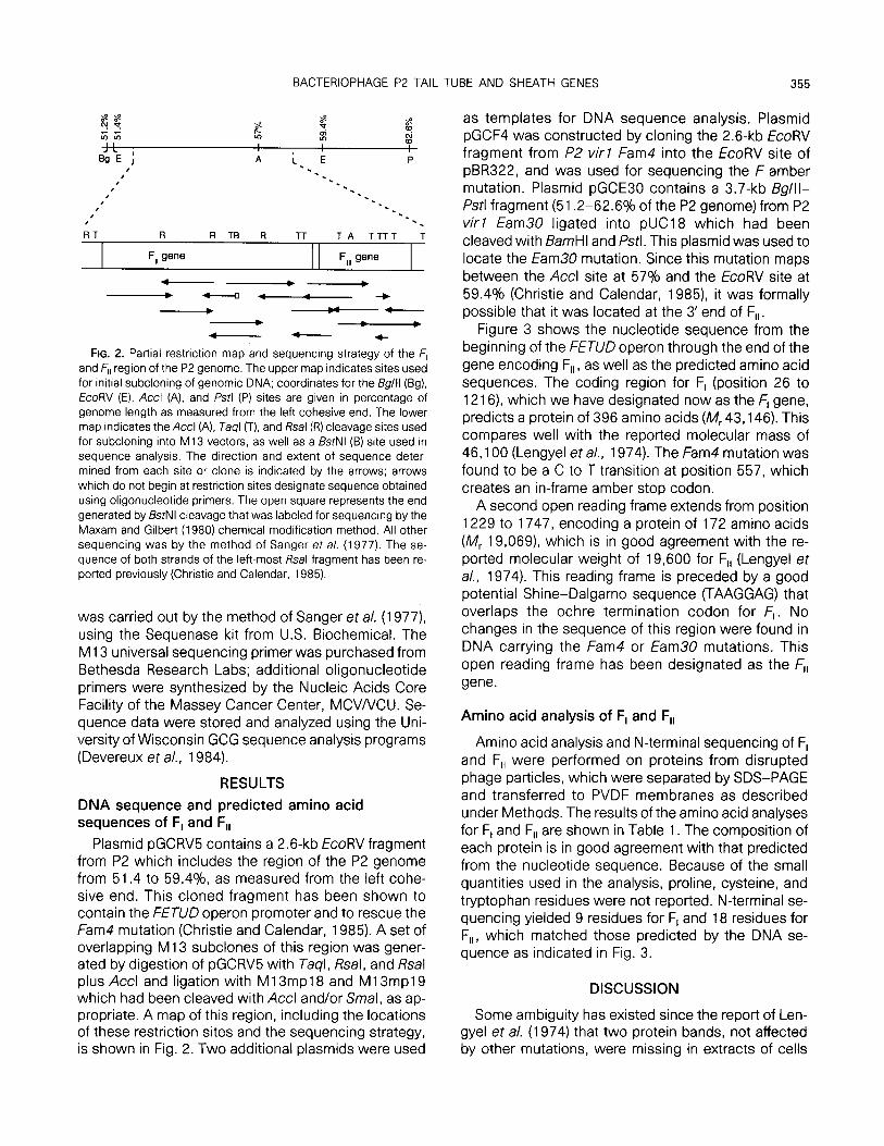
FIG. 2. Partial restriction map and sequencing strategy of the F, and F,, region of the P2 genome. The upper map indicates sites used for initial subcloning of genomic DNA; coordinates for the Bg/ll (Bg), EcoRV (E), Accl (A), and fstl (P) sites are given in percentage of genome length as measured from the left cohesive end. The lower map indicates the Accl (A), Taql (T), and Rsal (R) cleavage sites used for subcloning into Ml3 vectors, as well as a BstNl (B) site used in sequence analysis. The direction and extent of sequence deter- mined from each site or clone is indicated by the arrows; arrows which do not begin at restriction sites designate sequence obtained using oligonucleotide primers. The open square represents the end generated by BstNl cleavage that was labeled for sequencing by the Maxam and Gilbert (1980) chemical modification method. All other sequencing was by the method of Sanger et a/. (1977). The se- quence of both strands of the left-most &al fragment has been re- ported previously (Christie and Calendar, 1985).
was carried out by the method of Sanger et al. (1977) using the Sequenase kit from U.S. Biochemical. The M 13 universal sequencing primer was purchased from Bethesda Research Labs; additional oligonucleotide primers were synthesized by the Nucleic Acids Core Facility of the Massey Cancer Center, MCV/VCU. Se- quence data were stored and analyzed using the Uni- versity of Wisconsin GCG sequence analysis programs (Devereux et al., 1984).
RESULTS
DNA sequence and predicted amino acid sequences of F, and F,,
Plasmid pGCRV5 contains a 2.6-kb EcoRV fragment from P2 which includes the region of the P2 genome from 51.4 to 59.4%, as measured from the left cohe- sive end. This cloned fragment has been shown to contain the FETUD operon promoter and to rescue the Fam4 mutation (Christie and Calendar, 1985). A set of overlapping M 13 subclones of this region was gener- ated by digestion of pGCRV5 with Taql, Rsal, and Rsal plus Accl and ligation with M13mp18 and M13mp19 which had been cleaved with Accl and/or Smal, as ap- propriate. A map of this region, including the locations of these restriction sites and the sequencing strategy, is shown in Fig. 2. Two additional plasmids were used
as templates for DNA sequence analysis. Plasmid pGCF4 was constructed by cloning the 2.6-kb EcoRV fragment from PZ virl Fam4 into the EcoRV site of pBR322, and was used for sequencing the F amber mutation. Plasmid pGCE30 contains a 3.7-kb Bg/ll- Pstl fragment (51.2-62.69/o of the P2 genome) from P2 virl Earn30 ligated into pUC18 which had been cleaved with BarnHI and Pstl. This plasmid was used to locate the Earn30 mutation. Since this mutation maps between the Accl site at 57% and the EcoRV site at 59.4% (Christie and Calendar, 1985) it was formally possible that it was located at the 3’ end of F,, .
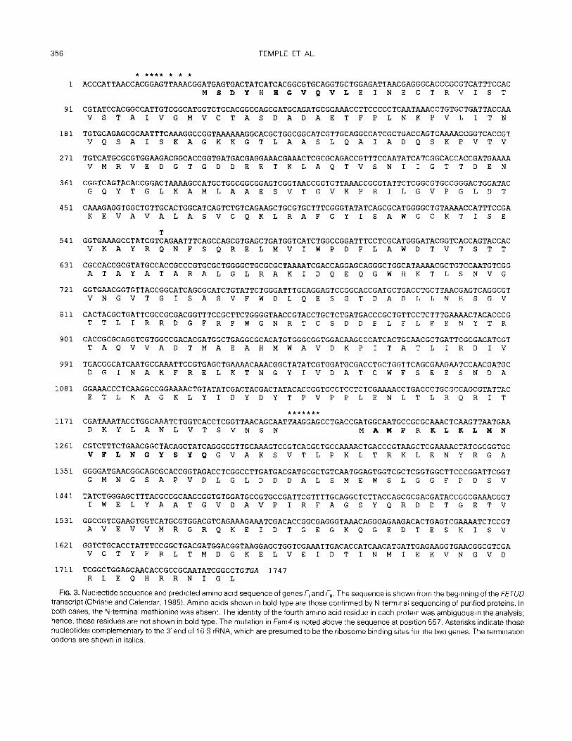
Figure 3 shows the nucleotide sequence from the beginning of the FETUD operon through the end of the gene encoding F,, , as well as the predicted amino acid sequences. The coding region for F, (position 26 to 1216) which we have designated now as the F, gene, predicts a protein of 396 amino acids (AJ 43,146). This compares well with the reported molecular mass of 46,100 (Lengyel et a/., 1974). The Fam4 mutation was found to be a C to T transition at position 557, which creates an in-frame amber stop codon.
A second open reading frame extends from position 1229 to 1747, encoding a protein of 172 amino acids (R/1, 19,069) which is in good agreement with the re- ported molecular weight of 19,600 for F,, (Lengyel et a/., 1974). This reading frame is preceded by a good potential Shine-Dalgarno sequence (TAAGGAG) that overlaps the ochre termination codon for F,. No changes in the sequence of this region were found in DNA carrying the Fam4 or Earn30 mutations. This open reading frame has been designated as the F,, gene.
Amino acid analysis of F, and F,,
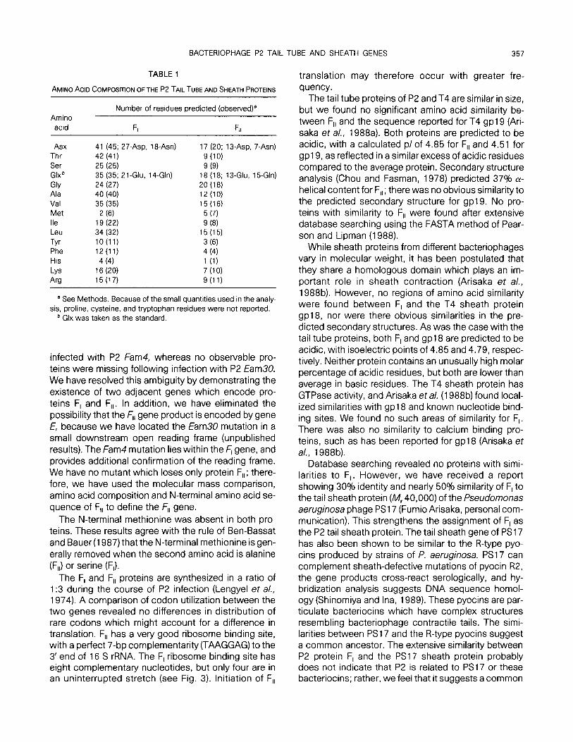
Amino acid analysis and N-terminal sequencing of F, and F,, were performed on proteins from disrupted phage particles, which were separated by SDS-PAGE and transferred to PVDF membranes as described under Methods. The results of the amino acid analyses for F, and F,, are shown in Table 1. The composition of each protein is in good agreement with that predicted from the nucleotide sequence. Because of the small quantities used in the analysis, proline, cysteine, and tryptophan residues were not reported. N-terminal se- quencing yielded 9 residues for F, and 18 residues for F,,, which matched those predicted by the DNA se- quence as indicated in Fig. 3.
DISCUSSION
Some ambiguity has existed since the report of Len- gyel et a/. (1974) that two protein bands, not affected by other mutations, were missing in extracts of cells
356 TEMPLE ET At
* **** * * * 1 ACCCATTAACCACGGAGTTAAACGGATGAGTGACTATCATCACGGCGTGCAGGTGCTGGAGATTAACGAGGGCACCCGCGTCATTTCCAC
MSDYHBGVQVLE INEGTRVIST
91 CGTATCCACGGCCATTGTCGGCATGGTCTGCACGGCCAGCGATGCAGATGCGG~CCTTCCCCCTCAATAAACCTGTGCTGATTACCAA VSTAIVGMVCTASDADAETFPLNKPVL I T N
181 TGTGCAGAGCGCAATTTCAGCCGGT AAAAAAGGCACGCTGGCGGCATCGTTGCAGGCCATCGCTGACCAGTCAAAACCGGTCACCGT VQSAISKAGKKGTLAASLQAIADQS K P VT V
271 TGTCATGCGCGTGGAAGACGGCACCGGTGATGACGAGGAATCGCGCAGACCGTTTCCAATATCATCGGCACCACCGATGA VMRVEDGTGDDEETKLAQTVSN IIGTTDEN
361 CGGTCAGTACACCGGACTAAAAGCCATGCTGGCGGCGGAGTCGGTAACCGGTGTTAAACCGCGTATTCTCGGCGTGCCGGGACTGGATAC GQYTGLKAMLAAESVTGVKP RILGVPGLDT
451 CAAAGAGGTGGCTGTTGCACTGGCATCAGTCTGTCAGAAGCTGCGTGCTTTCGGGTATATCAGCGCATGGGGCTGTAAAACCATTTCCGA KEVAVALASVCQKLRAFGY ISAWGCKT I S E
T 541 GGTGAAAGCCTATCGTCAGAATTTCAGCCAGCGTGAGCTGATGGTCATCTGGCCGGATTTCCTCGCATGGGATACGGTCACCAGTACCAC
VKAYRQNF SQRELMVIWP DFLAWDTVTSTT
631 CGCCACCGCGTATGCCACCGCCCGTGCGCTGGGGCTGCGCGCTAAAATCGACCAGGAGCAGGGCTGGCATAAAACGCTGTCCAATGTCGG ATAYATARALGLRAKIDQEQGWHKTLSNVG
721 GGTGAACGGTGTTACCGGCATCAGCGCATCTGTATTCTGGGATTTGCAGGAGTCCGGCACCGATGCTGACCTGCTTAACGAGTCAGGCGT VNGVTGISASVFWDLQESGTDADLLNESGV
811 CACTACGCTGATTCGCCGCGACGGTTTCCGCTTCTGGGGTAACCGTACCCGCTGTTCCTCTTTGAAAACTACACCCG TTLIRRDGFRFWGNRTCSDDPLFLFENYTR
901 CACCGCGCAGGTCGTGGCCGACACGATGGCTGAGGCGCACATGTGGGCGGTGGACAAGCCCATCACTGCAACGCTGATTCGCGACATCGT TAQVVADTMAEAHMWAVDKP ITATLIRDIV
991 TGACGGCATCAATGCCAAATTCCGTGAGCTGAAAACAAACGGCTATATCGTGGATGCGACCTGCTGGTTCAGCGAAGAATCCAACGATGC DGINAKFRELKTNGYIVDATCWFSEESNDA
1081 GGAAACCCTCAAGGCCGGAZlAACTGTATATCGACTACGACTATACACCGGTGCCTCCTCTCGAAAACCTGACCCTGCGCCAGCGTATTAC ETLKAGKLYIDYDYTPVPPLENLTLRQRIT
1171 CGATAAATACCTGGCAAATCTGGTCACCTCGGTTAACAGCAATTAAGGAGCCTGACCGATGGCAATGCCGCGCAAACTCAAGTTAATGAA DKYLANLVTSVNSN MAMPRKLKLWN
1261 CGTCTTTCTGAACGGCTACAGCTATCAGGGCGTTGCAAAGTCCGTCACGCTGCCAAAACTGACCCGTAAGCTCGAAAACTATCGCGGTGC VFLNGYSYQGVAKSVTLP KLTRKLENYRGA
1351 GGGGATGAACGGCAGCGCACCGGTAGACCTCGGCCTTGATGACGATGCGCTGTCAATGGAGTGGTCGCTCGGTGGCTTCCCGGATTCGGT GMNGSAPVDLGLDDDALSMEWSLGGFPDSV
1441 TATCTGGGAGCTTTACGCCGCAACCGGTGTGGATGCCGTGCCGATTCGTTTTGCAGGCTCTTACCAGCGCGACGATACCGGCGAAACGGT IWELYAATGVDAVPIRFAGSYQRDDTGETV
1531 GGCCGTCGAAGTGGTCATGCGTGGACGTCAGAAAGAAATCGACACCGGCGAGGGTAAACAGGGAGAACTGAGTCGAAAATCTCCGT AVEVVMRGRQKE IDTGEGKQGEDTESK I s v
1621 GGTCTGCACCTATTTCCGGCTGACGATGGACGGTAAGGAGCTGGTCGAAATTGACACCATCAACATGATTGAGAAGGTGAACGGCGTCGA VCTYFRLTMDGKELVE IDTINMIEKVNGVD
1711 TCGGCTGGAGCAACACCGCCGCAATATCGGCCTGTGTGA 1747 RLEQHRRN I G L
FIG. 3. Nucleotide sequence and predicted amino acid sequence of genes F, and F,, The sequence is shown from the begtnning of the FETUD transcript (Christie and Calendar, 1985). Amino acids shown In bold type are those confirmed by N-terminal sequencing of purified proterns. In both cases, the N-terminal methionine was absent. The identity of the fourth amino acid residue in each protein was ambiguous in the analysis; hence, these residues are not shown in bold type. The mutation in Fam4 is noted above the sequence at position 557. Asterisks indicate those nucleotides complementary to the 3’end of 16 S rRNA, which are presumed to be the nbosome binding sites for the two genes. The termrnation codons are shown in italics.
BACTERIOPHAGE P2 TAIL TUBE AND SHEATH GENES 357
TABLE 1
AMINO ACID COMPOSITION OF THE P2 TAIL TUBE AND SHEATH PROTEINS
Number of residues predicted (observed)8 Amino
acid 6 F,,
Asx 41 (45; 27.Asp, 18-Asn) 17 (20; 1 ~-ASP, 7-Asn) Thr 42 (41) 9 (10) Ser 25 (25) 9 (9) Glxb 35 (35; 2 1 -Glu, 14-Gin) 18 (18; 13-Glu, 15-Gln)
GIY 24 (27) 20 (18) Ala 40 (40) 12 (10) Val 35 (35) 15 (16) Met 2 (6) 5 (7) Ile 19 (22) 9 (8) Leu 34 (32) 15 (15)
Tyr lO(l1) 3 (6) Phe 12(11) 4 (4) His 4 (4) 1 (1) LYS 16 (20) 7 (10) Au 15 (17) 9 (11)
a See Methods. Because of the small quantities used in the analy- sis, proline, cysteine. and tryptophan residues were not reported.
b Glx was taken as the standard.
infected with P2 Fam4, whereas no observable pro- teins were missing following infection with P2 Earn30 We have resolved this ambiguity by demonstrating the existence of two adjacent genes which encode pro- teins F, and F,,. In addition, we have eliminated the possibility that the Fll gene product is encoded by gene E, because we have located the Earn30 mutation in a small downstream open reading frame (unpublished results). The fam4 mutation lies within the fl gene, and provides additional confirmation of the reading frame. We have no mutant which loses only protein F,,; there- fore, we have used the molecular mass comparison, amino acid composition and N-terminal amino acid se- quence of F,, to define the Fll gene.
The N-terminal methionine was absent in both pro- teins. These results agree with the rule of Ben-Bassat and Bauer (1987) that the N-terminal methionine is gen- erally removed when the second amino acid is alanine (F,,) or serine (F,).
The F, and F,, proteins are synthesized in a ratio of 1:3 during the course of P2 infection (Lengyel et al., 1974). A comparison of codon utilization between the two genes revealed no differences in distribution of rare codons which might account for a difference in translation. F,, has a very good ribosome binding site, with a perfect 7-bp complementarity (TAAGGAG) to the 3’ end of 16 S rRNA. The F, ribosome binding site has eight complementary nucleotides, but only four are in an uninterrupted stretch (see Fig. 3). Initiation of F,,
translation may therefore occur with greater fre- quency.
The tail tube proteins of P2 and T4 are similar in size, but we found no significant amino acid similarity be- tween F,, and the sequence reported for T4 gpl9 (Ari- saka et al., 1988a). Both proteins are predicted to be acidic, with a calculated pl of 4.85 for F,, and 4.51 for gpl9, as reflected in a similar excess of acidic residues compared to the average protein. Secondary structure analysis (Chou and Fasman, 1978) predicted 37% CY- helical content for F,,; there was no obvious similarity to the predicted secondary structure for gp19. No pro- teins with similarity to F,, were found after extensive database searching using the FASTA method of Pear- son and Lipman (1988).
While sheath proteins from different bacteriophages vary in molecular weight, it has been postulated that they share a homologous domain which plays an im- portant role in sheath contraction (Arisaka et a/., 1988b). However, no regions of amino acid similarity were found between F, and the T4 sheath protein gp18, nor were there obvious similarities in the pre- dicted secondary structures. As was the case with the tail tube proteins, both F, and gp18 are predicted to be acidic, with isoelectric points of 4.85 and 4.79, respec- tively. Neither protein contains an unusually high molar percentage of acidic residues, but both are lower than average in basic residues. The T4 sheath protein has GTPase activity, and Arisaka et al. (198813) found local- ized similarities with gp18 and known nucleotide bind- ing sites. We found no such areas of similarity for F,. There was also no similarity to calcium binding pro- teins, such as has been reported for gp18 (Arisaka et a/., 1988b).
Database searching revealed no proteins with simi- larities to F,. However, we have received a report showing 30% identity and nearly 509/o similarity of F, to the tail sheath protein (M, 40,000) of the Pseudomonas aeruginosa phage PS17 (Fumio Arisaka, personal com- munication). This strengthens the assignment of F, as the P2 tail sheath protein. The tail sheath gene of PS17 has also been shown to be similar to the R-type pyo- tins produced by strains of P. aeruginosa. PS17 can complement sheath-defective mutations of pyocin R2, the gene products cross-react serologically, and hy- bridization analysis suggests DNA sequence homol- ogy (Shinomiya and Ina, 1989). These pyocins are par- ticulate bacteriocins which have complex structures resembling bacteriophage contractile tails. The simi- larities between PS 17 and the R-type pyocins suggest a common ancestor. The extensive similarity between P2 protein F, and the PS17 sheath protein probably does not indicate that P2 is related to PS17 or these bacteriocins; rather, we feel that it suggests a common
358 TEMPLE ET AL
motif for structure of the contractile tails. The lack of similarity to the T4 sheath protein may indicate a differ- ent structural motif for the T4 contractile tail. It will be interesting to compare the sequences of these tail genes to those encoding the tail tube and sheath pro- teins of the Pl and Mu contractile tails when they be- come available, given the antigenic cross-reactivity be- tween these phages and P2. Comparison to the se- quences of additional genes encoding contractile tail proteins should provide insights into the structure- function relationship of the sheath proteins and the mechanism of sheath contraction.
ACKNOWLEDGMENTS
We are grateful to Fumio Arisaka and Elisabeth HaggBrd-Ljung- quist for sharing unpublished results. This work was supported by NIH Grants GM-34651 (to G.E.C.) and Al-08722 (to R.C.).
REFERENCES
ARISAKA, F., ISHIMOTO, L., KASSAVETIS, G., KUMAZAKI. T., and ISHII, S.-l. (1988a). Nucleotide sequence of the tail tube structural gene of bacteriophage T4. /. viral. 62, 882-886.
ARISAKA, F., NAKAKO, T., TAKAHASHI, H., and ISHII, S.-l. (198813). Nu- cleotide sequence of the tail sheath gene of bacteriophage T4 and amino acid sequence of its product. /. Viral. 62, 1 186-l 193.
BEN-BASSAT, A., and BAUER, K. (1987). Amino-terminal processing of proteins. Nature (London) 326, 315.
BERGET, P. G., and KING, I. (1983). T4 tail morphogenesis. In “Bacte- riophage T4” (C. K. Mathews, E. M. Kutter, G. Mosig, and P. B. Berget, Eds.), pp. 246-258. Amer. Sot. Microbial. Washington, D.C.
BERTANI, L. E. (1957). The effect of the inhibition of protein synthesis on the establishment of lysogeny. Virology 4, 53-7 1.
BERTANI, L. E., and BERTANI, G. (1970). Preparation and characteriza- tion of temperate, non-inducible bacteriophage P2 (host E. co/i). J. Gen. Viral. 6, 201-212.
BERTANI, L. E., and BERTANI. G. (1971). Genetics of P2 and related phages. Adv. Genet. 16, 199-237.
BERTANI, L. E., and SIX, E. W. (1988). The P2-like phages and their parasite, P4. ln “The Bacteriophages” (R. Calendar, Ed.), Vol. 2. pp. 73-143. Plenum, New York.
CHOU, P. Y., and FASMAN, G. D. (1978). Empirical prediction of pro- tein conformation. Annu. Rev. Biochem. 47, 251-276.
CHRISTIE, G. E., and CALENDAR, R. (1985). Bacteriophage P2 late pro- moters. II. Comparison of the four late promoter sequences. 1. Mol. Biol. 181, 373-382.
DEVEREUX, J., HAEBERLI, P., and SMITHIES, 0. (1984). A comprehen- sive set of sequence analysis programs for the VAX. Nucleic Acids Res. 12, 387-395.
EISERLING, F. A., and DICKSON, R. C. (1972). Assembly of viruses. Annu. Rev. Biochem. 41, 467-502.
FRAENKEL-CONRAT. H. (1985). Phages of prokaryotes (Bacteria and Cyanobacteria). In “The Viruses: Catalogue, Characterization, and Classification” (H. FraenkeCConrat and R. R. Wagner, Eds.). pp. 171-222, Plenum, New York.
GRUNDY, F. J., and HOWE, M. M. (1984). Involvement of the invertible
G segment in bacteriophage Mu tail fiber biosynthesis. Virology 134,296-317.
HAGGARD-LJUNGQUIST, E. (1990). Bacteriophage P2. In “Genetic Maps 1990, Book 1” (O’Brien, S. J., Ed), pp. 63-69, Cold Spring Harbor Laboratory, Cold Spring Harbor, NY.
HA~ORI, M.. and SAKAKI, Y. (1986). Dideoxy sequencing method us- ing denatured plasmid templates. Anal. Biochem. 152, 232-238.
INMAN, R. B., SCHNBS, M., SIMON, L. D.. SIX, E. W., and WALKER, D. H. (1971). Properties of bacteriophage P4 and its DNA. Virology 44, 67-72.
KING, J. (197 1). Bacteriophage T4 tail assembly: Four steps in core formatlon. J. Mol. Biol. 58, 693-709.
KING, 1.. and LAEMMLI, U. K. (1973). Bacteriophage T4 tail assembly: Structural proteins and their genetic identification. J. Mol. Biol. 75, 315-337.
LAEMMLI, U. K. (1970). Cleavage of structural proteins during the assembly of the head of bacteriophage T4. Nature (London) 227, 680-685.
LENGYEL, J. A., GOLDSTEIN, R. N., MARSH, M., SUNSHINE, M. G.. and CALENDAR, R. (1973). Bacteriophage P2 head morphogenesis: Cleavage of the major capsid protein. virology 53, 1-23.
LENGYEL, J. A., GOLDSTEIN, R. N., MARSH, M., and CALENDAR, R. (1974). Structure of the bacteriophage P2 tail. Virology 62, 16 l- 174.
LINDAHL. G. (1971). On the control of transcription in bacteriophage P2. Virology 46, 620-633.
MANIATIS. T., FRITSCH, E. F., and SAMBROOK, 1. (1982). “Molecular Cloning: A Laboratory Manual.” Cold Spring Harbor Laboratories, Cold Spring Harbor, NY.
MATSUDAIFZA, P. (1987). Sequence from picomole quantities of pro- teins electroblotted onto polyvinylidene difluoride membranes. J. No/. Chem. 262, 10,035-l 0,038.
MAXAM, A. M., and GILBERT, W. (1980). Sequencing end-labeled DNA with base-specific chemical cleavages. In “Methods in Enzymol- ogy” (L. Grossman and K. Moldave, Eds.), Vol. 65, pp. 499-560. Academic Press, San Diego.
MESSING, J., GRONENBORN, B., MULLER-HILL. B., and HOFSCHNEIDER. P. H. (1977). Filamentous coliphage Ml 3 as a cloning vehicle: Insertion of a /-Qndlll fragment of the lac regulatory region in Ml 3 replicative form in vitro. Proc. Natl. Acad. Sci. USA 74, 3642- 4302.
MESSING, J. (1979). A multlpurpose cloning system based on the single-stranded DNA bacteriophage Ml 3. In “Recombinant DNA Technical Bulletin,” NIH Publication No. 79-99. Vol. 2, No. 2, pp. 43-48. NIH, Bethesda, MD.
PEARSON, W. R., and LIPMAN, D. J. (1988). Improved tools for biologi- cal sequence comparison. Proc. Nat/. Acad. Sci. USA 85, 2444- 2448.
SANGER. F.. NICKLEN, S., and COULSON, A. R. (1977). DNA sequenc- ing with chain-terminating inhibitors. Proc. Nat/. Acad. Sci. USA 74,5463-5467.
SASAKI, I., and BERTANI, G. (1965). Growth abnormalities in Hfrderiva- tives of Escherichia co/i strain C. J. Gen. Microbial. 40, 365-376.
SHINOMIYA. T., and INA, S. (1989). Genetic comparison of bacteno- phage PSI 7 and Pseudomonas aeruginosa R-type pyocin. J. Bac- teriol. 171, 2287-2292.
SUNSHINE, M. G., THORN, M., GIBBS, W., CALENDAR, R., and KELLY, B. (1971). P2 phage amber mutants: Characterization by use of a polarity suppressor. wfology 46, 69 l-702.
WIMAN, M.. BERTANI, G., KELLY, B., and SASAKI, T. (1970). Genetic map of Escherichia co/i strain C. Mol. Gen. Gene?. 107, l-31.