Embed Size (px)
Citation preview
Gene, 61 (1987) 339-348
Elsevier
339
GEN 02272
Nucleotide sequence and characterization of a gene encoding the phytochrome polypeptide from Avena
(Regulatory photoreceptor; oat; promoter sequence homology; mung bean nuclease analysis; phyjl; gene structure; recombinant DNA; exons; introns; transcriptional start point)
Howard P. Hersheya*, Richard F. Barker b*, Kenneth B. Idler”*, Michael G. Murray b and Peter H. Quails**
n Departments of Botany and Genetics, University of Wisconsin, Madkon, WI 53706 (U.S.A.) and b Agrikenetics Advanced Research Laboratory, Madison, WI 53716 (U.S.A.) Tel. (408)221-5000
Received 5 October 1987
Accepted 16 October 1987
SUMMARY
We have isolated and characterized a gene encoding the phytochrome polypeptide of Avena. Based on nucleotide sequence identity with previously sequenced cDNA clones this gene is designated as type 3 (phy3). The gene is about 5.9 kb long with six exons and five introns, one each of the latter in the 5’ and 3’-untranslated regions. The largest exon encodes the entire 74-kDa, chromophore-bearing, N-terminal domain of the photo- receptor postulated to be directly involved in its mechanism of action. The transcription start point, identified by mung-bean nuclease digestion, is located 24 to 35 bp downstream from a tandem TATA box. Sequence elements homologous to a number of motifs implicated as upstream regulatory elements in other genes are present in the 5’-flanking DNA of phy3. Particularly intriguing are three elements at positions -140, -470 and -650. These elements share homology with the ‘GT’ motif postulated to be a component of the light-regulatory element of genes encoding the small subunit of ribulose bisphosphate carboxylase.
INTRODUCTION
Phytochrome is a regulatory photoreceptor that controls plant gene expression in response to light (Shropshire and Mohr, 1983; Tobin and Silver- thorne, 1985). The mechanism by which this regu-
lation occurs is not yet known. The photoreceptor molecule is a dimeric chromoprotein, with each sub- unit consisting of a single, linear tetrapyrrole chro- mophore covalently linked to a cysteine residue in the N-terminal domain of a polypeptide of 120 kDa to 127 kDa, depending on plant species (Vierstra
Correspondence to: Dr. P.H. Quail at his present address: Plant
Gene Expression Center, 800 Buchanan Street, Albany, CA
94710 (U.S.A.) Tel. (415)486-3719/486-3662.
* Present addresses: (H.P.H.) E.I. du Pont de Nemours &
Company, Central Research and Development Department, Ex-
perimental Station, Wilmington, DE 19898 (U.S.A.)
Tel. (302)695-7026; (R.F.B.) Plant Breeding Institute, Trum-
pington, Cambridge CB2 2LQ (U.K.) Tel. 44-(223)-840-411;
(K.B.I.) Abbott Laboratories, Department 93-D, AP-9A/3,
Abbott Park, IL 60064 (U.S.A.) Tel. (312)937-6059.
Abbreviations: aa, amino acid(s); bp, base pair(s); kb, kilobases
or 1000 bp; nt, nucleotide(s); SDS, sodium dodecyl sulfate; SSC,
0.15 M NaCI/O.OlS M Na, .citrate pH 7.6.
0378-l 119/87/$03.50 0 1987 Elsevier Science Publishers B.V. (Biomedical Division)
340
et al., 1984; Jones and Quail, 1986; Lagarias, 1985; Lagarias and Mercurio, 1985). Biochemical, spec- tral, ~uno~hemic~ and nucl~tide-sequence ana- lyses have yielded valuable ~fo~ation on a number of structural features of the molecule potentially re- lated to its mechanism of action. These features in- clude regions of the polypeptide involved in photo- conversion-induced conformational changes
(Vierstra et al., 1984; Lagarias and Mercurio, 1985; Jones et al, 1984; Cordonnier et al., 1986; Wong et al., 1986), in interactions with the chromophore (Vierstra et al., 1984; Jones et al., 1984; Grimm et al., 1986; Quail et al., 1987), and in dimerization (Vierstra et al., 1984; Jones and Quail, 1986).
Phytochrome regulates the expression of a number of nuclear genes, including those for the small sub- unit of ribulose bisphosphate carboxylase (r&S) (Stiekema et al., 1983; Thompson et al., 1983; Kaufman et al., 1984; Fh.thr et al., 1986; Fluhr and Chua, 1986; Nagy et al., 1985; 1986a) chlorophyll a/b binding protein (cab) (Stiekema et al., 1983; Thompson et al., 1983; Kaufman et al., 1984; Fluhr et al., 1986; Nagy et al., 1985; 1986a,b; Timko et al., 1985a), protochlorophy~de reductase (Batschauer and Apel, 1984), ph~oc~ome itself (Colbert et al., 1983; 1985; Quail et al., 1986), ferredoxin (Dobres et al., 1987), rRNA (Thien and Schopfer, 1982), and several unidentified gene products (Thompson et al., 1983). This regulation has been shown to occur, at least Pliny, at the level of tr~s~ription by run-on transcription assays (Quail et al., 1986; Silverthorue and Tobin, 1984; Mosinger et al., 1985) and by aua- lysis in transgenic plant tissue of chimeric constructs involving upstream promoter regions of light- regulated genes (Fluhr et al., 1986; Fluhr and Chua, 1986; Nagy et al., 1985; 1986a,b; Timko et al., 1985a,b; Simpson et al., 1985; Paulsen et al., 1986; Kuhlemeier et al., 1987). Progress has been made toward identifying the &-acting elements responsi- ble for ph~oc~ome-related expression of rbcS and cab genes by deletion analysis of the 5’-flanking DNA of these genes in transgenic plants (Fluhr et al., 1986; Fluhr and Chua, 1986; Nagy et al., 1985; 1986a,b; Timko et al., 1985a,b; Simpson et al., 1985; Poulsen et al., 1986; Kuhlemeier et al., 1987).
We have focused on the negative feedback control that phytochrome exerts over the expression of its own genes as an approach to understanding phyto-
chrome-regulated gene expression (Colbert et al., 1983; 1985; Quail et al., 1986; Hershey et al., 1984; 1985a). In Avena there is a decrease in the tran- scription rate of phytochrome genes within 15 min of’ photoconversion, as we11 as evidence for posttran- scriptional regulation (Quail et al., 1986). As a step toward defining the mechanism by which this regu- lation occurs, we report here the first sequence analy- sis of a ph~ochrome gene and its 5’-flanking DNA.
MATERIALS AND METHODS
(a) Isolation of genomic clones and phage DNA
A partial EcoRI library of Avena genomic DNA was constructed in 1 Charon 32 as described by Murray et al. (1984). Appro~ately 1.5 x lo6 phage were plated on E. coli ED8767 and screened according to the method of Benton and Davis (1977) using the cDNA clone pAP3.1 as a hybridization probe (Hershey et al., 1985b). Phage were trans- ferred to nitrocellulose filters and the filters were prehyb~dized in 6 x SSC, 2 x Denhardt’s (Den- hardt, 1966), 0.5% SDS, 100 pg/ml denatured calf thymus DNA, and 20 pg/ml poly(A) at 64°C for 8 h. Filters were hybridized with 2 x lo6 cpm/ml of 32P-labeled pAP3.1 (specific activity 2 x 10’ cpm/pg DNA) for 16 h at 64°C. Filters were washed twice for 15 mm at 20°C in 2 x SSC, 0.5% SDS, and twice for 1 h in 0.1 x SSC, 0.1% SDS at 20°C. All positive clones were picked and rescreened until plaque-pure.
(b) Nucieotide sequence analysis
Sequences of subcloned genomic DNAs were determined by the method of Maxam and Gilbert (1980) with modifications described by Barker et al. (1983). Computer analysis of DNA sequences were performed using programs made available by the University of Wisconsin Genetics Computer Group (Devereux et al., 1984).
(c) Tran~ription start point identi~cation
The transcription start point for the type 3’phyto- chrome gene was identified using mung-bean nu-
341
clease protection following reassociation in 3 M sodium trichloracetate (Murray, 1986). For this pur- pose the genomic subclone pGP8.2-1 was digested with WpaII and the 161-bp fragment corresponding to nt -94 to + 77 was purified (Maxam and Gilbert, 1980) and end-labeled.
RESULTS AND DISCUSSION
(a) Isolation of pb~~hro~e genes
Previous Southern-blot analysis of Avena genomic DNA using the 32P-labeled cDNA clone pAP3.2 showed the presence of a small phytochrome gene family in this plant (Hershey et al., 1985a). Restric- tion site polymorphism among cDNA clones has shown further that at least four members of this family are expressed in etiolated Avena seedlings (Hershey et al., 1985b). Based upon the data from the genomic blots, high-119, Avena DNA was primly
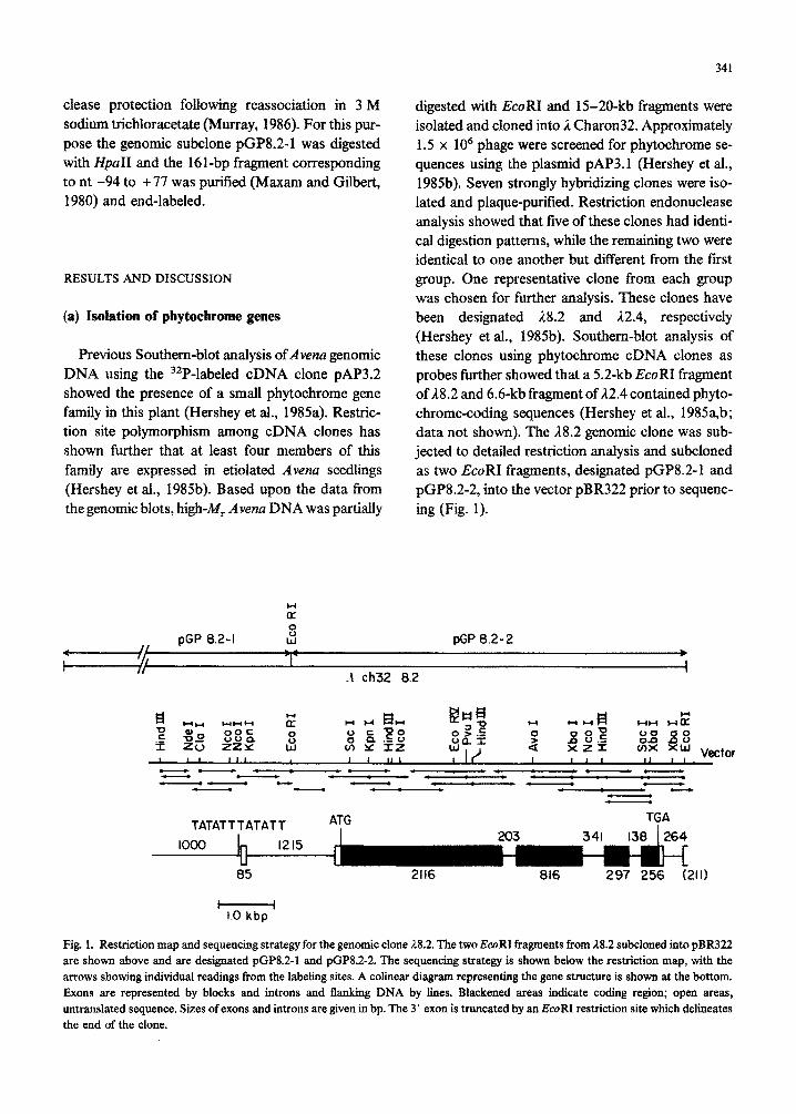
digested with EcoRI and 15-20-kb fragments were isolated and cloned into 1 Charon32. Approximately 1.5 x lo6 phage were screened for phytochrome se- quences using the plasmid pAP3.1 (Hershey et al., 1985b). Seven strongly hybridizing clones were iso- lated and plaque-purified. Restriction endonuclease analysis showed that five of these clones had identi- cal digestion patterns, while the remaining two were identical to one another but different from the frst group. One representative clone from each group was chosen for further analysis. These clones have been designated 1/8.2 and A2.4, respectively (Hershey et al., 1985b). Southern-blot analysis of these clones using phytochrome cDNA clones as probes further showed that a 5.2-kb EcoRI fragment of A8.2 and 6.6-kb fragment of 12.4 contained phyto- chrome-coding sequences (Hershey et al., 1985a,b; data not shown). The A8.2 genomic clone was sub- jected to detailed restriction analysis and subcloned as two EcoRI fragments, designated pGP8.21 and pGP8.2-2, into the vector pBR322 prior to sequenc- ing (Fig. 1).
pGP 8.2-l :: W pGP 8.2- 2
+----q T * II
h ch32 8.2 i
TATATT TAT AT T ATG TGA
1000
85 2116 816 297 256 (211)
Fig. 1. Restriction map and sequencing strategy for the genomic clone 18.2. The two EcoRI fragments from 28.2 subcloned into pBR322 are shown above and are designated pGP8.Z1 and pGP8.2-2. The sequencing strategy is shown below the restriction map, with the arrows showing individual readings from the labeling sites. A colinear diagram representing the gene structure is shown at the bottom. Exons are represented by blocks and introns and flanking DNA by lines. Blackened areas indicate coding region; open areas, untranslated sequence. Sixes ofexons and introns are given in bp. The 3’ exon is truncated by an EcoRI restriction site which delineates the end of the clone.
342
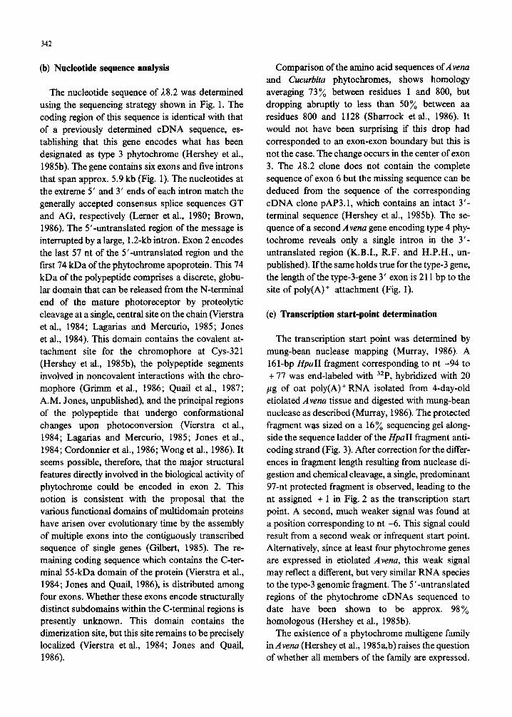
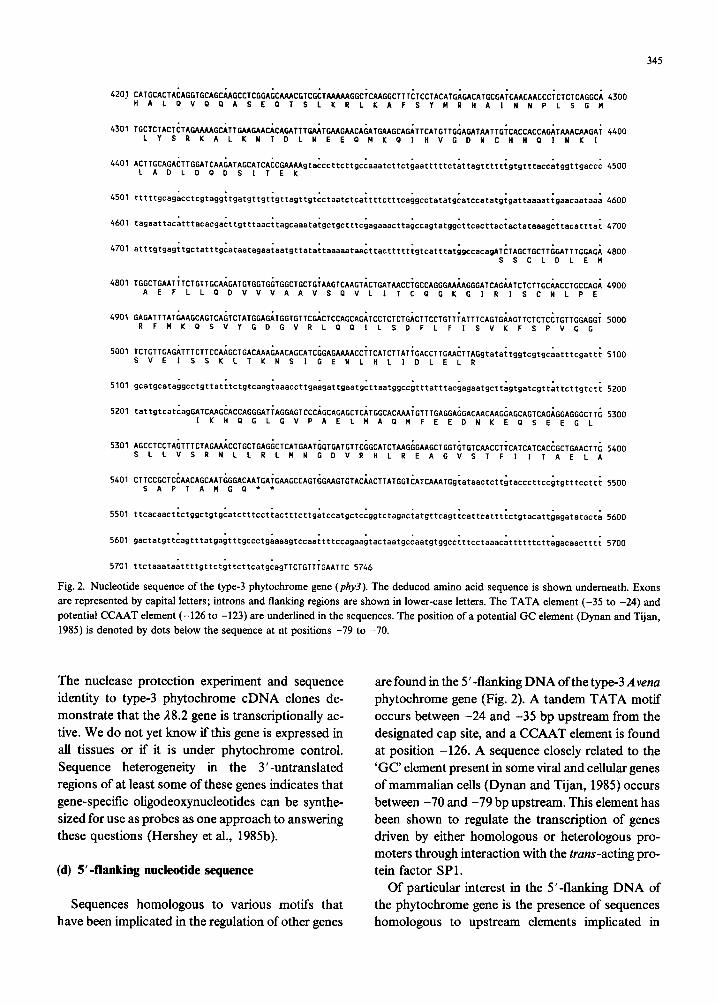
(b) Nucleotide sequence analysis
The nucleotide sequence of U.2 was determined using the sequencing strategy shown in Fig. 1. The coding region of this sequence is identical with that of a previously determined cDNA sequence, es- tablishing that this gene encodes what has been designated as type 3 phytochrome (Hershey et al., 1985b). The gene contains six exons and five introns that span approx. 5.9 kb (Fig. 1). The nucleotides at the extreme 5’ and 3’ ends of each intron match the generally accepted consensus splice sequences GT and AG, respectively (Lerner et al., 1980; Brown, 1986). The 5’untranslated region of the message is interrupted by a large, 1.2-kb intron. Exon 2 encodes the last 57 nt of the 5’-~~~slated region and the frst 74 kDa of the ph~oc~ome apoprotein. This 74 kDa of the polypeptide comprises a discrete, globu- lar domain that can be released from the N-terminal end of the mature photoreceptor by proteolytic cleavage at a single, central site on the chain (Vierstra et al., 1984; Lagarias and Mercurio, 1985; Jones et al., 1984). This domain contains the covalent at- tachment site for the chromophore at Cys-321 (Hershey et al., 1985b), the polypeptide segments involved in noncovalent interactions with the chro- mophore (Grimm et al., 1986; Quail et al, 1987; A.M. Jones, unpublished), and the principal regions of the polypeptide that undergo conformationd changes upon photoconversion (Vierstra et al., 1984; Lagarias and Mercurio, 1985; Jones et al., 1984; Cordonnier et al., 1986; Wong et al., 1986). It seems possible, therefore, that the major structural features directly involved in the biological activity of phytochrome could be encoded in exon 2. This notion is consistent with the proposal that the various functional domains of multidom~n proteins have arisen over evolutions time by the assembly of multiple exons into the contiguously transcribed sequence of single genes (Gilbert, 1985). The re- maining coding sequence which contains the C-ter- minal 55-kDa domain of the protein (Vierstra et al., 1984; Jones and Quail, 1986), is distributed among four exons. Whether these exons encode structurally distinct subdomains within the C-terminal regions is presently unknown. This domain contains the dimerization site, but this site remains to be precisely localized (Vierstra et al., 1984; Jones and Quail, 1986).
Comparison of the amino acid sequences ofAvena and Cucurbita phytochromes, shows homology averaging 73% between residues 1 and 800, but dropping abruptly to less than 50% between aa residues 800 and 1128 (Sharrock et al., 1986). It would not have been surprising if this drop had corresponded to an exon-exon boundary but this is not the case. The change occurs in the center of exon 3. The 18.2 clone does not contain the complete sequence of exon 6 but the missing sequence can be deduced from the sequence of the corresponding cDNA clone pAP3.1, which contains an intact 3’- terminal sequence (Hershey et al., 1985b). The se- quence of a second Avena gene encoding type 4 phy- tochrome reveals only a single intron in the 3’ - ~tr~slated region (K.B.I., R.F. and H.P.H., un- published). If the same holds true for the type-3 gene, the length of the type-3-gene 3’ exon is 211 bp to the site of poly(A)+ attachment (Fig. 1).
(c) Transcription start-point determination
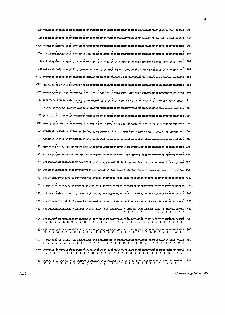
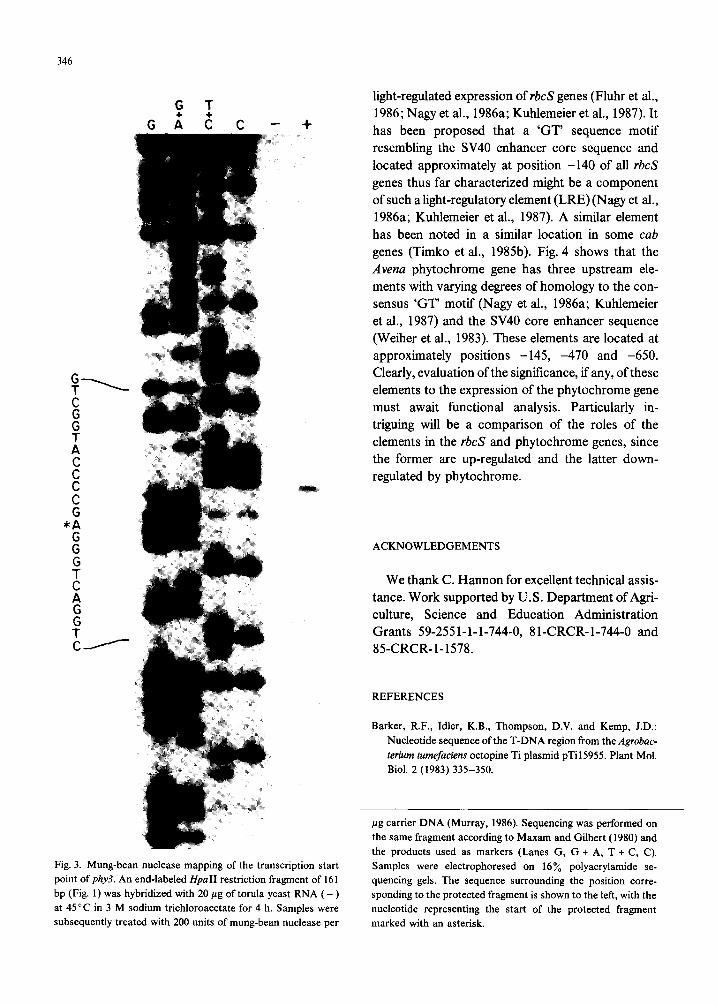
The transcription start point was determined by mung-bean nuclease mapping (Murray, 1986). A 161-bp HpaII fragment corresponding to nt -94 to + 77 was end-labeled with 32P, hybridized with 20
pg of oat poly(A)+ RNA isolated from Cday-old etiolated Avena tissue and digested with mung-bed nuclease as described (Murray, 1986). The protected fragment was sized on a 16% sequencing gel along- side the sequence ladder of the HpaII fragment anti- coding strand (Fig. 3). After correction for the differ- ences in fragment length resulting from nuclease di- gestion and chemical cleavage, a single, predominant 97-nt protected fragment is observed, leading to the nt assigned + 1 in Fig. 2 as the transcription start point. A second, much weaker signal was found at a position corresponding to nt -6. This signal could result from a second weak or infrequent start point. Alternatively, since at least four phytochrome genes are expressed in etiolated Avena, this weak signal may reflect a different, but very similar RNA species to the type-3 genomic fragment. The 5’-untranslated regions of the phytochrome cDNAs sequenced to date have been shown to be approx. 98% homologous (Hershey et al., 1985b).
The existence of a phytochrome multigene family in Avena (Hershey et al., 1985a,b) raises the question of whether all members of the family are expressed.
343
.lOOO
-900
-800
-700
-600
*500
.400
.300
.200
-901
-801
-701
-601
-501
-401
-301
-201
-101
-1
IO0
201
301
CO1
501
601
701
80'1
907
toot
1101
1201
1301
1401
1503
160f
1701
1801
CAGCAGCGAiACACGGGGTilACCACCGT~GAGTGGTlC~ATTACTTGA~GCAGGCGAl~lCClCClCA~GGCCTGCTT~CAGllCTTC~AGCAGGMC~ MSSSRPASSSSSRNR
500
600
700
800
900
TOOD
t100
1200
1300
1400
1500
1600
7700
1800
1900
Fig. 2.
1901 GAGCCTGT~GCClACAG~lllCCTGC~4ClGCTGCT~GGGCTTTGC~GlCCTAC~~ClTGCTGCC~GGC~~AT~CAA~ATCCA~lCATTGCCA~ 2000 EPVKPTEFPATAAGALOSYKLAAKAlSKlPSLPC
2001 GTGGAsGCAiGGAGGTCCT;lGC~TACT~TGGTGAAGG~GTCTTTGA~CTTACCGGGiATGACAGGG~TATGGCTTA~~GTTTCAT~AAGATGACC~ 2100 GSMEVLCNTVVKEVFOLTGYORVMAYKFHEOOH
2201 ATGAAG~C~GTACCGA~GATTTGTW\irGCCGTGCG~GATCCATA~GGTCATTG~GCTGAGGCA~TCCCCTil~TATTAGCCT~TGTGGTTCA~ 2300 MKNKVRNiCDCRARSIKVlEAEALPFD~SLCGSA
2301 CACTCAGGGcACCACACAGiTGTCACCTT~AGTATATGG~G~CATGAA~TCGATTGCAiCCCTTGTCAiGGCTGTTGT~GTTAATGAG~ATGAAGAOG~ 2400 LRAPHSCHLPYMENMNSIASLVMAVV~NENEED
2401 TGATGAAGCiGAGTCTGAACAACCAGCACiCCAGCAGAlUTGTCCCTTTi 2500 DEAESEC!PAOPPKXKKLUGLLVCHHESPRYVPF
2501 CCGCTGCGliATGClTGTG~GTTCTTAGC~CAGGTGTTT~CTGTCCAlG~C~CAGGGA~TTTGAATlA~AGAAACAGTiGCGTGAG~~r\ACATACTG~ 2600 PLRYACEFLAPVFAVHVNREFELEKQLREKNILK
:601 AGATGCAAAcAATGCTCTCiGATATGTTGiTCCGAGAAG~CTCTCCCCT~ACTATCGTAiCAGGGACCC~C~TATCAT~GACCTAGTC~AATGTGATG~ 2700 MOTMLSDt4LFREASPLTIVSGTPNINDLVKCDG
2701 TGCTGCTCTiCTGTATGGGGGAAAAGTATGGCGTCTGCGiAATGCTCCA~CGGAGTClC~GATACATGAiATCGCCTTCiGGCTATCAG~TGTTCACAG~ 2800 AALLYGGKVURLRNAPTESPIHDlAFULSDVHR
2801 tATTCCACT~GCCTGACTA~TGACAGCCT~CATGATGCl~GCTATCCAG~AGCTGCTCCiCTTGGTGAT;TCATTTGTG~AATGGCAGT~GCTAAGATC~ 2900 DSTGLSTDSLHDAGYPGAAALGONICGHAVAK~N
2901 ACTCCAAGG~TATTCTTTliTGGTTCAGGiCACATACAG~lGCTGAAAT~AGATGGGGA~GTGCAAAGA~T~TCCATC~GACATGGAT~ACAGCAGAA~ 3000 SKOlLFUFRSHTAAEIRUGGAKNDPSDHDDSRR
3001 GATGCACCCiAGGTTGTCTiTC~GCTTiCCTTG~CTiGTCAAGAlG~AGAGCTTGC~TTGGAGlGA~TATGAAATG~ATGCTATTC~TTCATTGC~ 3100 MHPRLSFKAFLEVVKHKSLPlJSDYEMDA~HSLO
3101 CTTATACTGSGAGGGACACiAAATGATGC~AGCAAGCCA~AGCGGGAAG~TAGTTTAGAiAACCAGATT~GTGATCTAA~ACTTGATGG~CTTGCTGAA~ 3200 LILRGTLNDASKPKREASLDNQIGDLKLDGLAEL
3201 TGCAGGCCGiGACCAGlGAiATGGTTCGT~T~TGGA~~AGCAAClGTiCCAATCTTG~CAGTAGATG~CAATGGACT~GT~ACGGG~GG~TCAGA~ 3300 PAVTSEKVRLHETATVPILAVDGNGLVNGUNQK
3301 AGCAGCGGA~TTGACTGGG~T~GAGlTGiTGATGCAATiGGAAGGCAC~TACTTACCCiTGTGGAGGA~TCCTCTGTA~CAGTTGlCC~GAGGATGCT~ 3400
3401
3501
360 1
3701
3801
3901
4001
4101
AAELTGLRVDDAIGRHlLlLVEDSSVPVVGRHl
tatttcattgatsaaGceGGTAAACAAGAGAAGGAAGTT~GATTTGAGGi~AGACTCAiGGCCCGAAG~GGGATGATG~TCCAGTTAT~TTGGTTGTG~ 3700 KEEKEVRFEVKTHGPKRDDGPVtLVVN
ATGCTTGTGcCAGTCGGGAcCTTCATGATCATGTTGTTGG 3800 ACASROLHDHVVGVCFVAPDMTVHKLVMDKFTR
tGTTGAGGGiGACTACAAG~CGATCATTC~C~CCCGAAtCTCCTATATiTGGTGCTGA~GAATTTGGAiGGTGTTCGG;GTGGAATGCi 3900 VEGDYKAlIHNPNPLIPPIFGADEFGUCSEUNA
GUUTGACC~GTTWCTGZ;GTGG~TA~~T~GlG~TC~T~GA~GCTTCTTGGi~GTGTlT~ACAGTAGC~TGCTTCCTG~CCTTTG~G~ 4000 AHTKLTGUNRDEVLDKNLLGEVFDSSNASCPLKN
ACAGAGATG~ATTTGTMGiCTTTGTCTT~TTATCMCAfiTGCATTAGC~GGGGAAG~CAGAG~TC~TTTGG~TTCTTCGAC~GMGTGGAA; 4100 ROAFVSLCVLINSALAGEETEKAPFGFFORSGK
345
420.1 CATGCACTA~ACGTGCAGC~GCCTCGGAZiCAMCGTCG~TAGGCiCAAGGCTTT~TCCTACATG;GACATGCGAiCAACAACCCiCTCTCAGGC~ 4300 HALPVPPASEPTSLKRLKAFSYMRHAINNPLSGM
4301 TGCTCTACT~TAGAAAAGCiTTGAAGAACiCAGATTT~TGAG~CA~ATGAAGCAG~TTCATGTTGliAGATAATTGiCACCACCAG~TAAACAAGAi 4400 LYSRKALKNTDLNEEaMKaIHVGDNCHIIaINKI
4401 ACTTGCAGAtTTGGATCAAGATAGCATCACCGAAAA9ta;~~tt~ttg~~~~~t~tt~t~~~ttttt~t~tt~gttttt~gtgttt~~~~tggttg~~~~ 4500 LADLDPDSITEK
4701 atttgtgag;tgctatttg;ataatagaa;aatgtt~t~~t~~~~~t~~~tt~~ttttt~gt~~ttt~t~g~=~~~gAT~TAGCTGCTT~GATTTGGAG~ 4800 SSCLDLEM
4801 TGGCTGAATiTCTGTTGCPjGATGTGGTG~TGGCTGCTGi~GTCAAGT~CTGATAACCiGCCAGGGAA~GGGATCAG~ATCTCTTGC~ACCTGCCAG~ 4900 AEFLLaDVVVAAVSaVLlTCaGKGiRISCNLPE
4901 GAGATTTATGAAGCAGTCAGTCTATGGAGATCGTGTTCG~CTCCAGCAG~TCCTCTCTG~CTTCCTGTTiATTTCAGTG~AGTTCTCTC~TGTTGGAGGi 5000 RFNK~S~YGDG~RL~~ILSDFLFISVKFSPVGG
5001 TCTGTTGAGATTTCTTCCAAGCTGACAAAGAACAGCATCG~TTAGgt~t~~tggt~gtg~~~ttt~g~tt~ 5100 SVEISSKLTKNSIGENLHLIDLELR
5201 tattgtcat;agGATCAAGCACCAGGGATiAGGAGTCCCACAAG~AGCAGTCAG~GGAGGGCTT~ 5300 IKHaGLGVPAELMAaMFEEDNKEaSEEGL
5301 AGCCTCCTA~TTTCTAGAAiCCTGCTGAG~CTCATG~T~GTGATGTTC~GCATCTAAG~GAAGCTGGT~TGTC~CCTiCATCATCAC~GCTGAACTT~ 5400 SLLVSRNLLRLMNGDVRHLREAGVSTFIITAELA
5401 CTTCCGCTCcAACAGCAATcGGACAATGAiG~GCCAGTCGAAGTGTAC~ACTTATGGT~ATCAAATGg~~t~~~t~tt~t~~~~tt~~~tgttt~~tt~ 5500 SAPTAMGa'*
5701 ttctaaet&tttgttct&tcttcatg;egTTCTGTTiGAATTC 5746
Fig. 2. Nucleotide sequence of the type-3 phytochrome gene (phy3). The deduced amino acid sequence is shown underneath. Exons are represented by capital letters; introns and flanking regions are shown in lower-case letters. The TATA element (-35 to -24) and potential CCAAT element (-126 to -123) are underlined in the sequences. The position of a potential GC element (Dynan and Tijan,
1985) is denoted by dots below the sequence at nt positions -79 to -70.
The nuclease protection experiment and sequence identity to type-3 phytochrome cDNA clones de- monstrate that the 18.2 gene is transcriptionally ac- tive. We do not yet know if this gene is expressed in ah tissues or if it is under phytochrome control. Sequence heterogeneity in the 3’-untranslated regions of at least some of these genes indicates that gene-specific oligodeoxynucleotides can be synthe- sized for use as probes as one approach to answering these questions (Hershey et al., 1985b).
(d) 5’-flanking nucleotide sequence
Sequences homologous to various motifs that have been implicated in the regulation of other genes
are found in the 5’-flanking DNA of the type-3 Avena phytochrome gene (Fig. 2). A tandem TATA motif occurs between -24 and -35 bp upstream from the designated cap site, and a CCAAT element is found at position -126. A sequence closely related to the ‘GC’ element present in some viral and cellular genes of mammalian cells (Dynan and Tijan, 1985) occurs between -70 and -79 bp upstream. This element has been shown to regulate the transcription of genes driven by either homologous or heterologous pro- moters through interaction with the trans-acting pro- tein factor SPl.
Of particular interest in the 5’-flanking DNA of the phytochrome gene is the presence of sequences homologous to upstream elements implicated in
346
light-regulated expression of rbcS genes (Fluhr et al., 1986; Nagy et al., 1986a; Kuhlemeier et al., 1987). It has been proposed that a ‘GT’ sequence motif resembling the SV40 enhancer core sequence and located approximately at position -140 of all rbcS genes thus far characterized might be a component of such a light-regulatory element (LRE) (Nagy et al., 1986a; Kuhlemeier et al., 1987). A similar element has been noted in a similar location in some cab genes (Timko et al., 1985b). Fig. 4 shows that the Arena phytochrome gene has three upstream ele- ments with varying degrees of homology to the con- sensus ‘GT’ motif (Nagy et al., 1986a; Kuhlemeier et al., 1987) and the SV40 core enhancer sequence (Weiher et al., 1983). These elements are located at approximately positions -145, -470 and -650. Clearly, evaluation of the significance, if any, of these elements to the expression of the phytochrome gene must await functional analysis. Particularly in- triguing will be a comparison of the roles of the elements in the rbcS and phytochrome genes, since the former are up-regulated and the latter down- regulated by phytochrome.
c E C G
*A
ii ACKNOWLEDGEMENTS
G
+
We thank C. Harmon for excellent technical assis- tance. Work supported by U.S. Department of Agri- culture, Science and Education Administration Grants 59-2551-l-1-744-0, 81-CRCR-1-744-0 and 85-CRCR-1-1578.
REFERENCES
Barker, R.F., Idler, K.B., Thompson, D.V. and Kemp, J.D.:
Nucleotide sequence of the T-DNA region from the Agrobac-
terium tumefaciens octopine Ti plasmid pTi 15955. Plant Mol.
Biol. 2 (1983) 335-350.
Fig. 3. Mung-bean nuclease mapping of the transcription start
point of phy3. An end-labeled HpaII restriction fragment of 161
bp (Fig. 1) was hybridized with 20 pg of torula yeast RNA ( - )
at 45°C in 3 M sodium trichloroacetate for 4 h. Samples were
subsequently treated with 200 units of mung-bean nuclease per
ng carrier DNA (Murray, 1986). Sequencing was performed on
the same fragment according to Maxam and Gilbert (1980) and
the products used as markers (Lanes G, G + A, T + C, C).
Samples were electrophoresed on 16% polyacrylamide se-
quencing gels. The sequence surrounding the position corre-
sponding to the protected fragment is shown to the left, with the
nucleotide representing the start of the protected fragment
marked with an asterisk.
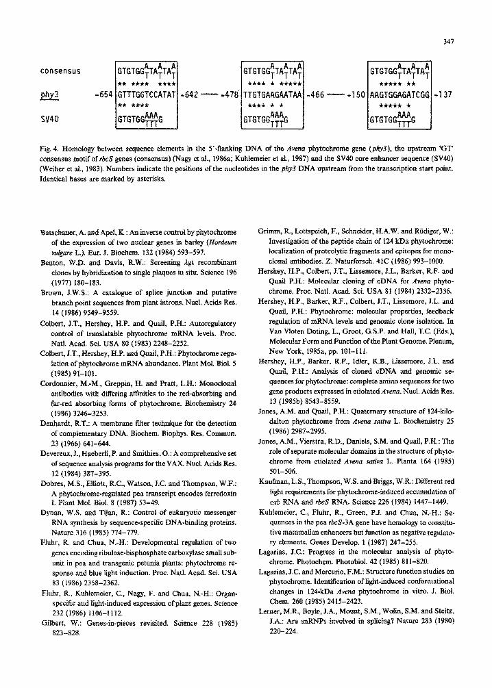
Fig. 4. Homology between sequence elements in tbe 5’-flanking DNA of the Avena phytachrome gene (phy3j, the upstream YW consensus motif of rbcS genes (consensus) (Nagy et aI., 1986a; Kuhlemeier et al., 1987) and the SV40 core enhancer sequence (SV40) (W&her et al., 1983). Numbers indicate the positions of the nucleotides in the phy3 DNA upstream from the transcription start paint.
Identical bases are marked by asterisks.
Batschatier, A. and Ape& KS: An inverse control by phytachrome of the expression of two nuclear genes in barley (born w&are L.). Eur. J. Biochem. 132 (1984) 593-597.
Benton, W.D. and Davis, R.W.: Screening &t recombinant clones by hybridization to single plaques in situ. Science 1%
(1977) 180-183. Brown, J.W.S.: A catalogue of splice junction and putative
branch point sequences from plant introns. Nucl. Acids Res.
14 (1986) 9549-9559.
Colbert, J.T., Hershey, H.P. and Quail, F.H.: Autoregulatory contra1 of translatable phytochrome mRNA levels. Proc. Natl. Acad. Sci. USA 80 (1983) 2248-2252.
Coibert, J.T., Hershey, H.P. and Quail, P.H.: Fhytochrome regu- fation of ph~~~orne mRNA abundance. Plant MoI. Biol. 5 (1985) 91-IOI.
Cordarmier, M.-M., Greppin, H. and Pratt, L.H.: Monoclonat antibodies with differing affinities to the red-absorbing and far-red absorbing forms of phytochrome. Biochemistry 24 (1986) 3246-3253.
Denhardt, R.T.: A membrane filter technique far the detection of complementary DNA. Biochem, Biophys. Res. Commun. 23 (1966) 641-644.
Devereux, J., Haeberli, P. and Smithies, 0.: A comprehensive set of sequence analysis programs for the VAX. Nucl. Acids Res. 12 (1984) 387-395.
Dobres, MS., Elliott, R.C., Watson, J.C. and Thompson, W.F,: A ph~~rome-replated pea transcript encodes ferredoxin I. Plant Mol. Bid. 8 (1987) 53-49.
I&man, W.S. and Tijan, R.: Control of eukaryotic messenger RNA synthesis by sequence-specific DNA-binding proteins. Nature 3 16 (1985) 774-779.
Fluhr, R. and Chua, N.-H.: Developmental regulation of two
genes encading ribulose-bisphosphate carboxylase small sub-
unit in pea and transgenic petunia plants: phytochrome re- sponse and blue light induction, Proc. Natl. Acad. Sci. USA 83 (1986) 2358-2362.
Fluhr, R., Kuhlemeier, C, Nagy, F. and Chua, N.-X.: Organ- specific and light-induced expression of plant genes. Science 232 (1986) 1106-l 112.
Gilbert, W.: Genes-in-pieces revisited. Science 228 (1985) 823-828.
Grimm, R., Lottspeich, I?., Schneider, H.A.W. and Rtldiger, W,: Investigation ofthe peptide chain of 124 kDa phytochrome: localization ofproteolytic fragments and epitopes for mono- clonal antibodies. Z. Naturforsch. 4lC (1986) 993-1000.
Hershey, H.F., Colbert, J.T., Lissemore, J.L., Barker, RF. and Quail P.H.: Molecular cloning of cDNA for Arena phyto- chrome, Prac. Natl. Acad. Sci. USA 81 (1984) 2332-2336.
Hershey, II.F., Barker, R.F., Colbert, J.T., Lissemore, J.L. and Quail, F.H.: Fhytochrome: molecular properties, feedback regulation of mRNA levels and genomic clone isolation. In Van Vloten Doting, L., Groot, G.S.P. and Hall, T.C. (Eds.), Molecular Form and Function of the Plant Genome. Plenum, New York, 1985a, pp. 101-l ii.
Hershey, H.P., Barker, RF., Idler? K.B., Lissemore, J.L. and Quail, P.H.: Analysis of cloned cDNA and genomic se- quences for phytochrome: complete amino sequences for two gene products expressed in etiolated Avena. Nud. Acids Res, 13 (1985b) 8543-8559.
Jones, A.M. and Quail, P.H.: Quaternary structure of l&kilo- dalton phytochrome from Avena sativa L. Biochemistry 25 (1986) 2987-2995.
Jones, A.M., Vierstra, R.D., Dauiels, SM. and Quail, F.H.: The role of separate molecular domains in the structure of phyto- chrome from etiolated Avena sativa L. Planta 164 (1985) 501-506.
Kaufman, L.S., Thompson. W.S. and Briggs, W.R.: Different red
light requirements for ph~~hrome-endued accumulation of cab RNA and rbcS RNA. Science 226 (1984) 1447-1449.
Kuhlemeier, C., Fluhr, R., Green, P.J_ and Chua, N.-H,: Se- quences in the pea rbcS3A gene have homology to constitu- tive mammalian enhancers but function as negative repuiata- ry elements. Genes Develop. 1 (1987) 247-255.
Lagarias, J.C.: Progress in the molecular analysis of phyto-
chrome. Fhotochem. Photobiol. 42 (1985) 811-820. Lagarias, J.C. and Mercurio, F.M.: Structure function studies on
phytochrome. Identification of light-induced conformational changes in 124-kDa Avena phytochrome in vitro. J. Viol. Chem. 260 (1985) 2415-2423.
Lerner, M.R., Boyle, J.A., Mount, SM., Wotm, S.M. and Steitz, J.A.: Are snRNPs involved in splicing? Nature 283 (1980)
220-224.
348
Maxam, A.M. and Gilbert, W.: Sequencing end-labeled DNA with base-specilic chemical cleavages. Methods Enzymoi. 65 (1980) 499-512.
M&singer, E., Batschauer, A., Schafer, E. and Ape], K.: Phyto-
chrome control of in vitro transcription of specific genes in isolated nuclei from barley (Hordeum vulgare). Eur. J. Biochem. 147 (1985) 137-142.
Murray, M.G.: Use of sodium trichloroacetate and mung bean
nuclease to increase sensitivity and precision during tran- script mapping. Anal. Biochem. 158 (1986) 165-170.
Murray, M.G., Kennard, WC., Drong, R.F. and Slightom, J.L.: Use of a recombination-deficient phage lambda system to construct wheat genomic libraries. Gene 30 (1984) 237-240.
Nagy, F., Fluhr, R., Kuhlemeier, C., Kay, S., Boutry, M., Green,
P., Paulsen, C. and Chua, N.-H.: C&acting elements for selective expression of two photosynthetic genes in trans- genie plants. Phil. Trans. Roy. Sot. Lond. B314 (1986a) 493-500.
Nagy, F., Kay, S.A., Boutry, M., Hsu, M.-Y. and Chua, N.-H.: Phytochrome-controlled expression of a wheat cab gene in transgenic tobacco seedlings. EMBO J. 5 (1986b) 1119-l 124.
Nagy, F., Morrelli, G., Frayley, R.T., Rogers, S.G. and Chua,
N.-H.: Photo-regulated expression of a pea rbcS gene in leaves of transgenic plants. EMBO J. 4 (1985) 3063-3068.
Poulsen, C., Fluhr, R., Kauffman, J.M., Boutry, M. and Chua, N.-H.: ~~acte~ation of an r&S gene from ~co~~~p~~-
bug&if&z and expression of an r&&-CAT chimeric gene in homologous and heterologous nuclear background. Mol. Gen. Genet. 205 (1986) 193-200.
Quail, P.H., Christensen, A.H., Jones, A.M., Lissemore, J.L.,
Parks, B.M. and Sharrock, R.A.: The phytochrome molecule and the regulation of its genes. In O.L. Kon (Ed.), 4th FOAB Congress, Integration and Control of Metabolic Processes: Pure and Applied Aspects. ICSU, Miami, 1987, pp. 41-54.
Quail, P.H., Colbert, J.T., Peters, N.K., Christensen, A.H.,
Sharrock, R.A. and Lissemore, J.L.: Phytochrome and the regulation of the expression of its genes. Phil. Trans. Roy. Sot. Lond. B314 (1986) 469-480.
Sharrock, R.A., Lissemore, J.L. and Quail, P.H.: Nucleotide and
amino acid sequence of a Cucurbita phytochrome cDNA clone: identification of conserved features by comparison with Avena phytochrome. Gene 47 (1986) 287-295.
Shropshire Jr., W. and Mohr, H. (Eds.): Photomorphogenesis. Encyclopedia of Plant Physiology, New series, Vols. 16A and B. Springer-Verlag, Heidelberg, 1983.
Siiverthorne, J. and Tobin, E.M.: Demonstration of tran- scriptional regulation of specific genes by phytochrome action. Proc. Natl. Acad. Sci. USA 81 (1984) 1112-l 116.
Simpson, J., Timko, M.P., Cashmore, A.R., Schell, J., Van Montagu, M. and Herrera-Estrella. L.: Light-inducible and tissue-specific expression of a chimeric gene under control of the 5’-flanking sequence of a pea chlorophyll a/b-binding protein gene. EMBO J. 4 (1985) 2723-2729.
Stiekema, W.J., Wimpee, C.F., Silverthome, J. and Tobin, E.M.: Phytochrome control of the expression of two nuclear genes encoding chloroplast proteins in Lemna gibba L. G-3. Plant Physiol. 72 (1983) 717-724.
Thien, W. and Schopfer, P.: Control by ph~~hrome of cytoplas- mic precursor rRNA synthesis in the cotyledons of mustard seedlings. Plant Physiol. 69 (1982) 1156-l 160.
Thompson, W.F., Everett, M., Poians, N.O., Jorgensen, R.A. and Palmer, J.D.: Phytochrome control of RNA levels in developing pea and mung-bean leaves. Planta 158 (1983) 487-500.
Timko, M.P., Kausch, A.P., Castresana, C., Fassler, J., Herrera-
Estrella, L., Van den Broeck, G., Van Montagu, M., Schell, J. and Cashmore, A.R.: Light regulation of plant gene expres- sion by an upstream enhancer-like element. Nature 3l8 (1985a) 579-582.
Timko, M.P., Kausch, A.P., Hand, J.M., Cashmore, A.R., Herrera-Estrella, L., Van den Broeck, G. and Van Montagu, M.: Structure and expression of nuclear genes encoding poly- peptides of the photosynthetic apparatus. In Arntzen, C., Bogorad, L., Bonitz, S. and Steinback, K. (Eds.), Molecular Biology of the Photosynthetic Apparatus. Cold Spring Har-
bor Laboratory, Cold Spring Harbor, 1985b, pp. 381-396. Tobin, E.M. and Silverthorne, J.: Light regulation ofgene expres-
sion in higher plants. Annu. Rev. Plant Physiol. 36 (1985) 569-593.
Vierstra, R.D., Cordomtier, M.-M., Pratt, L.H. and Quail, P.H.: Native phytochrome: Immunoblot analysis of relative molecular mass and in vitro proteolytic degradation for several plant species. Planta 160 (1984) 521-528.
Weiher, H., Konig, M. and Gruss, P.: Multiple point mutations affecting the simian virus 40 enhancer. Science 219 (1983) 626-631.
Wong, Y.-S., Cheng, H.-C., Walsh, D.A. and Lagarias, J.C.: Phosphorylation ofAvena phytochrome in vitro as a probe of light-induced conformational changes. J. Biol. Chem. 261
(1986) 12089-12097.
Communicated by J.L. Slightom.


![Phytochromes and Phytochrome Interacting Factors1[OPEN] · Update on Phytochromes and Phytochrome Interacting Factors Phytochromes and Phytochrome Interacting Factors1[OPEN] Vinh](https://img.pdfslide.us/doc/110x75/5e9224c5cbd0a85457462c45/phytochromes-and-phytochrome-interacting-factors1open-update-on-phytochromes-and.jpg)