Embed Size (px)
Citation preview

Yeast Rrp14p is required for ribosomal subunitsynthesis and for correct positioning of themitotic spindle during mitosisMarlene Oeffinger3, Alessandro Fatica1,2, Michael P. Rout3 and David Tollervey1,*
1Wellcome Trust Centre for Cell Biology, University of Edinburgh, Edinburgh EH9 3JR, Scotland, UK, 2Department ofGenetics and Molecular Biology, University of Rome ‘La Sapienza’, P. Aldo Moro, 5, 00185 Rome, Italy and3Rockefeller University, 1230 York Avenue, New York, NY10021, USA
Received September 11, 2006; Revised and Accepted October 5, 2006
ABSTRACT
Here we report that Rrp14p/Ykl082p is associatedwith pre-60S particles and to a lesser extent withearlier 90S pre-ribosomes. Depletion of Rrp14pinhibited pre-rRNA synthesis on both the 40S and60S synthesis pathways. Synthesis of the 20Sprecursor to the 18S rRNA was largely blocked, aswas maturation of the 27SB pre-rRNA to the 5.8Sand 25S rRNAs. Unexpectedly, Rrp14p-depletedcells also showed apparently specific cell-cycledefects. Following release from synchronization inS phase, Rrp14p-depleted cells uniformly arrestedin metaphase with short mitotic spindles thatwere frequently incorrectly aligned with the site ofbud formation. In the absence of Bub2p, which isrequired for the spindle orientation checkpoint, thismetaphase arrest was not seen in Rrp14p-depletedcells, which then arrested with multiple buds,several SPBs and binucleate mother cells. Thesedata suggest that Rrp14p may play some role in cellpolarity and/or spindle positioning, in addition to itsfunction in ribosome synthesis.
INTRODUCTION
The majority of steps in ribosome synthesis take place withinthe nucleolus, a specialized subnuclear structure. In thebudding yeast Saccharomyces cerevisiae, the nucleolus isformed around the highly repetitive rDNA array on chromo-some XII. Here, the rDNA is transcribed into a large precur-sor RNA (pre-rRNA), which is subsequently modified andthen matured by endonuclease and exonuclease processing toyield the mature 18S, 5.8S and 25S rRNAs (see Figure 1).Ribosome synthesis is a major activity in the Eukaryoticcell and a rapidly growing yeast cell produces around
2000 ribosomes per minute. Both the size of the cell at divi-sion and number of ribosomes per cell, are closely linked togrowth rate [reviewed in (1,2)]. Moreover, both size at divi-sion and ribosome numbers anticipate the future growth ratesuggesting a cross-talk mechanism between ribosome synthe-sis and mitotic cell division (3). Recent studies in yeast haveidentified several connections among the nucleolus, ribosomebiogenesis and cell-cycle progression [reviewed in (4)]. Asmall number of ribosomal processing factors were foundthat appear to facilitate cross-talk between those processes,with mutations in these proteins affecting both ribosome syn-thesis and cell division (5–10).
Ykl082c/Rrp14p is an essential protein that was initiallycharacterized in two-hybrid analyses of a protein interactionnetwork involved in the specification of cell polarity (11).Rrp14p interacted with Bud8p, a component of the distalbud site tag complex, Zds2p, a nucleolar protein with a rolein cell polarity as well as gene silencing (12,13), and withGic1p and Gic2p, which interact directly with the GTPaseCdc42, a key regulator of cell polarity (14–16). Stainscarrying gic1/2D have a depolarized actin and microtubulecytoskeleton, implicating these proteins in microtubule polar-ization and nuclear migration. Rrp14p was therefore proposedto be involved in polarized growth and the establishment ofbud sites, although direct physical interactions were notassessed (11).
YFP-tagged Rrp14p was found to localize to the nucleolus(11), and we subsequently identified Rrp14p as a componentof an early pre-60S complex that was co-purified with taggedSsf1p (17), suggesting a role in ribosome synthesis. Rrp14p isa member of the SURF-6 family of nucleolar proteins, whichhave been predicted from bioinformatic analyses to partici-pate in complex protein–protein and protein–RNA interac-tions within the nucleolus (18–20).
Here we report that Rrp14p functions in ribosome synthe-sis; it is required for the maturation of both small and largesubunit rRNAs and helps to prevent premature cleavage ofthe pre-rRNA at site C2. Strains depleted of Rrp14p also
*To whom correspondence should be addressed. Tel: +44 131 650 7092; Fax: +44 131 650 7040; Email: [email protected] address:Marlene Oeffinger, Rockefeller University, 1230 York Avenue, New York, NY10021, USA
� 2007 The Author(s).This is an Open Access article distributed under the terms of the Creative Commons Attribution Non-Commercial License (http://creativecommons.org/licenses/by-nc/2.0/uk/) which permits unrestricted non-commercial use, distribution, and reproduction in any medium, provided the original work is properly cited.
1354–1366 Nucleic Acids Research, 2007, Vol. 35, No. 4 Published online 1 February 2007doi:10.1093/nar/gkl824

show defects in positioning and elongation of the mitoticspindle during mitosis, which were not previously reportedfor cells depleted for any other ribosome synthesis factor.
MATERIALS AND METHODS
Strains and molecular techniques
Standard techniques were employed for growth and handlingof yeast. Yeast strains used in this work are listed in Supple-mentary Table S1. Strain YAF32 was created from BMA38by use of a one-step PCR strategy as described previously(21). TAP-tagging of Rrp14p was performed as descri-bed in (22). Strain YMO22 was created by integrationof a Cdcc14-GFP-Trp1 construct (kindly provided by
E. Schiebel). Strains YMO111, YMO102 and YMO104 werecreated from KH230 (kindly provided by K. Hardwick) andYMO103 and YMO105 from W303, respectively, by one-step PCR strategy (21) and homologous recombination of thelinearized plasmid PRE10.1, carrying a MAD2::URA3 dele-tion cassette. YMO200 and YMO201 were created by crossingwith YFC2160-16C. YMO202 was created by crossing withVAY371.
Oligonucleotides
For RNA hybridizations, the following oligonucleotides wereused: 001, 50-CCAGTTACGAAAATTCTTG; 002, 50-GCTC-TTTGCTCTTGCC; 003, 50-TGTTACCTCTGGGCCC; 004,CGGTTTTAATTGTCCTA; 005, 50-ATGAAAACTCCACA-
Figure 1. Yeast pre-rRNA and processing. (A) Structure of the yeast pre-rRNA, with locations of oligonucleotides used as hybridization probes. (B) Pre-rRNAprocessing pathway showing the intermediates detected by pulse-chase and northern analyses.
Nucleic Acids Research, 2007, Vol. 35, No. 4 1355

GTG; 006, 50-GGCCAGCAATTTCAAGTTA; 007, 50-CTC-CGCTTATTGATATGC; 008, 50-CATGGCTTAATCTTTG-AGAC; 017, 50-GCGTTGTTCATCGATGC; 020, 50-TGAG-AAGGAAATGACGCT; 033, 50-CGCTGCTCACCAATGG;and 041, 50-CTACTCGGTCAGGCTC.
RNA extraction, northern hybridization andprimer extension
For depletion of the Rrp14p protein, cells were harvested atintervals following a shift from RGS medium (2% raffinose,2% galactose and 2% sucrose), or YPGal medium (2% galac-tose), to YPD medium (2% glucose). Otherwise strains weregrown in YPD medium except for over-expression studiesfor which strains were grown in RGS medium. RNA wasextracted as described previously (23). Northern hybridiza-tions and primer extension analysis were carried out asdescribed in (23). Standard 1.2 or 2% agarose/formaldehydeand 6% acrylamide/urea gels were used to analyze the highand low molecular weight RNA species, respectively.
Sucrose gradient analysis and affinity purification
Sucrose gradient centrifugation was performed as describedpreviously (24,25). RNA was extracted from each fractionand resolved on standard 1.2% agarose/formaldehyde gels.Mature rRNAs and pre-rRNA species were detected byethidium staining and northern hybridization, respectively.Sedimentation of proteins was assayed by SDS–PAGE andTAP-tagged Rrp14p was detected by western immunoblottingwith peroxidase-conjugated rabbit IgG (SIGMA). Affinitypurification of TAP-tagged Rrp14 and analysis of co-purifiedRNAs was performed as described previously (17).
Pulse-chase labeling
Metabolic labeling of RNA was performed as describedpreviously (17). The strains GAL::HA-rrp14 and BMA38were transformed with a plasmid containing the URA3gene, pre-grown in galactose medium lacking uracil andtransferred to glucose minimal medium for 6 h. Cells at0.3 OD600 nm were labeled with [5,6-3H]uracil for 1 min
followed by a chase with excess unlabeled uracil. Standard1.2% agarose/formaldehyde and 6% polyacrylamide/ureagels were used to analyze the high and low molecular weightRNA species, respectively.
Immunofluorescence
GAL::HA-rrp14, GAL::HA-rrp14/CDC14-GFP, GAL::HA-rrp14/mad2D, mad2D, GAL::HA-rrp14/bub2D, bub2Dand wild-type strains with and without Spc42-GFP werepre-grown in RGS medium, containing 2% raffinose, 2%galactose and 2% sucrose, and harvested at intervals follow-ing a shift to medium containing 2% glucose. For arrest inearly S phase, cells were pre-synchronized by a 2 h treatmentwith alpha-factor (10 mM), performed 2 h after shift toglucose containing medium (26). Cells were then transferredto pre-warmed glucose medium lacking alpha-factor butcontaining 0.1 M hydroxyurea for a further 2 h. Cells werereleased from arrest into pre-warmed glucose medium.Samples, taken after 0, 20, 40 and 60 min, were fixed byincubation in 4% (v/v) formaldehyde at 25�C for 3 min forSPB visualization, or 1 h for tubulin, Cdc14–GFP andNop1p detection, and then spheroplasted. Immunofluores-cence was performed as described previously (27,28). Tubu-lin was detected with a rabbit anti-tubulin antibody (Sigma)and a secondary goat anti-rabbit antibody coupled to FITC(Sigma) at 1:1000 and 1:200 dilutions, respectively. Nop1pwas detected with mouse anti-Nop1p (29) and a secondarygoat anti-mouse antibody coupled to FITC (Sigma). Coverslips were mounted using moviol, containing DAPI. Thecells were examined under a Zeiss Axioscop fluorescentmicroscope. Pictures were obtained with Smart Capture VPand Openlab.
FACS analysis
To determine DNA content, cells were harvested 0, 20, 40and 60 min after release from early S phase arrest, andfixed for 1 h in 70% ethanol at RT. Cells were washedtwice in 50 mM Tris (pH 7.5) and resuspended in 1 mg/mlRNase A in 50 mM Tris, pH 7.5. RNase A digestion wasperformed for 4 h at 37�C. Cells were then washed and
Figure 2. Rrp14p cosediments with pre-60S ribosomes. (A) Pre-rRNA species detected and locations of oligonucleotide probes. (B) Lysate from a strainexpressing an Rrp14p–TAP fusion was fractionated on a 10–50% sucrose gradient. Fractions were analyzed by western blotting for the distribution of Rrp14p-TAP and by northern hybridization for the distribution of the 27SA and 27SB pre-rRNA components of the pre-60S ribosome and the 20S component of pre-40S.
1356 Nucleic Acids Research, 2007, Vol. 35, No. 4

resuspended in 40 mg/ml Proteinase K, and incubated for1 h at 55�C. The cells were harvested and resuspended in50 mg/ml propidium iodine in PBS, pH 7.2, and then soni-cated at low power for 5 s to separate loosely associatedcells. Determination of DNA content was performed ona Becton-Dickinson FACScan.
RESULTS
Rrp14p associates with pre-60S particles
Rrp14p (Ykl082c) was identified by mass spectrometry inprecipitates of TAP-tagged ribosome synthesis factors Ssf1p
and Rrp1p, both of which are components of early pre-60Sribosomal particles (17,30,31), suggesting that Rrp14p isalso a component of early pre-60S ribosomes. To determinewhether Rrp14p is associated with pre-ribosomal particles,we constructed a strain expressing a C-terminal fusionbetween a TAP tag (22) and Rrp14p. The fusion constructwas inserted into the genome under the control of theRRP14 promoter and supported wild-type growth, showingit to be functional (data not shown). To assess the associationof Rrp14p with pre-ribosomal particles, a sucrose gradientanalysis was performed with a lysate from the Rrp14-TAPstrain. The sedimentation of Rrp14-TAP was determined bywestern blotting with antibodies that recognize the protein
Figure 3. Depletion of Rrp14p impairs growth and 60S subunit synthesis. (A) Growth of GAL::HA-rrp14 (crosses) and otherwise isogenic wild-type (openboxes) strains following transfer to glucose medium. (B) Western analysis of the depletion of HA–Rrp14p on glucose medium. (C and D) Pulse-chase labeling ofrRNA synthesis in GAL::HA-rrp14 wild-type strains 7 h after transfer to glucose medium. Cells were pulsed with [5,6-3H]uracil for 1 min and then chased with alarge excess of unlabeled uracil for the times indicated. (C) High molecular weight RNA analyzed on a 1.2% agarose/formaldehyde gel. (D) Low molecularweight RNA analyzed on a 6% polyacrylamide/urea gel. RNA species are labeled on the right of the panels. The asterisk in (C) indicates the position of theputative 50ETS-D species.
Nucleic Acids Research, 2007, Vol. 35, No. 4 1357

A moiety of the TAP tag, and compared with the rRNAspecies and pre-rRNAs (Figure 2). Comparison with thepositions of the mature rRNAs detected by ethidium staining(data not shown and indicated by bars on Figure 2B) and thepre-rRNAs indicated that Rrp14-TAP is most enriched in the60S region of the gradient, with a weaker peak �80–90S.No cosedimentation with the 20S pre-rRNA was observed,indicating that Rrp14p is not associated with 40S pre-ribosomes. These analyses are consistent with the previousproteomic data indicating that Rrp14p is present in pre-60Sparticles, but additionally suggest an association with 90Spre-ribosomes, within which the early assembly of the40S subunit occurs.
Rrp14p is required for rRNA synthesis
To determine the requirement for Rrp14p in rRNA synthesis,we integrated an N-terminal HA–Rrp14p fusion constructunder the control of the repressible GAL1 promoter at theendogenous RRP14 locus (21) (see Materials and Methods).The GAL::HA-rrp14 strain grew more slowly than the iso-genic wild type in galactose media, indicating that Rrp14pover-expression is toxic. However, growth of the GAL::HA-rrp14 strains was identical to the wild type in RGS medium(containing raffinose, sucrose and galactose), which results inlower expression levels, and shortly after transfer to glucosemedium (Figure 3A), showing the HA-fusion construct to befunctional. Growth of the GAL::HA-rrp14 strain was strongly
Figure 4. Rrp14p is required for pre-rRNA processing. (A) Schematic diagram of the pre-rRNA showing the processing sites and locations of oligonucleotideprobes used. (B) Northern analyses of high molecular weight RNA separated on a 1.2% agarose/formaldehyde gel. (C) Northern analyses of low molecularweight RNA separated on a 6% polyacrylamide/urea gel. RNA species are labeled on the right of the northern panels. Oligonucleotide probes used are onthe right.
1358 Nucleic Acids Research, 2007, Vol. 35, No. 4

reduced 6 h after transfer to glucose media and had almostceased by 12 h. Consistent with this, western blotting(Figure 3B) showed that HA–Rrp14p was strongly depletedafter 6 h in glucose and undetectable after 12 h.
Pre-rRNA processing was initially assessed by pulse-chaselabeling in vivo with [5,6-3H]uracil 7 h after transfer toglucose medium. Analysis of high molecular weight RNA(Figure 3C) showed that in the Rrp14p-depleted strain35S and 32S pre-rRNAs accumulated, whereas the 27SApre-rRNA was less abundant and its conversion to 27SBand mature 25S rRNA was greatly reduced. The level of20S pre-rRNA was reduced with the concomitant appearanceof the aberrant 23S molecule and synthesis of mature 18S wasboth delayed and reduced. The 23S RNA originates fromdirect cleavage of the 35S pre-rRNA at site A3 when thecleavages at sites A0, A1 and A2 are delayed.
The formation of low molecular weight rRNAs (5S and5.8S) was analyzed by PAGE (Figure 3D). In the Rrp14p-depleted strain, the 7S pre-rRNA was not readily detected
and very low levels of the mature 5.8S rRNA accumulated(Figure 3D). The mature 5.8SL and 5.8SS are the productsof alternative processing pathways. No change in their ratiowas seen, indicating that Rrp14p is required for 27SB pre-rRNA processing in both pathways.
To further characterize pre-rRNA processing in the GAL::HA-rrp14 mutant strain, steady-state levels of matureand precursor rRNA molecules were assessed by northernhybridization of RNAs resolved on agarose gels (Figure 4B)or polyacrylamide gels (Figure 4C). Rrp14p-depletion led tostrong accumulation of the 35S pre-RNA and appearance ofthe 23S RNA, whereas levels of the 27SA2 and 20S pre-rRNAs were reduced (Figure 4B), consistent with inhibitionof cleavage at sites A0, A1 and A2. An aberrant RNA,which is predicted to extend from the end of the 50 ETS tosite D, was also accumulated (50ETS-D in Figure 4B) anda band of appropriate mobility was seen in the pulse-chaselabeling (marked with asterisk in Figure 3C). This speciesis presumably generated by pre-rRNA processing in ITS1and 30 maturation of 18S rRNA, in the absence of processingin the 50ETS. On the pathway of 60S synthesis, the 27SB pre-rRNA was not strongly reduced at the 4 and 8 h depletiontime points, whereas the mature 25S was depleted, showingthat its maturation was prevented. Analyses of low molecularweight RNAs (Figure 4C) showed that levels of the later 7Sand 6S pre-rRNAs were reduced, with a more rapid effect on6S. In addition, an aberrant species that extend from A2 toC2 was accumulated (Figure 4C, panel a), which resultsfrom premature cleavage of the 27SA2 pre-rRNA at site C2.The appearance of the A2–C2 fragment was describedpreviously in strains lacking Ssf1p and Ssf2p (17) and otherpre-60S components (4,32,33).
We conclude that Rrp14 is required for pre-rRNA matura-tion on both the 40S and 60S pathways.
Rrp14p depletion arrests cell-cycle progression
Rrp14p was reported to interact with proteins required forcell polarity (11). We, therefore, inspected cells undergoingRrp14p depletion by microscopy, to determine whether theyarrest at a specific stage of the cell cycle. Following transferto glucose medium for 6 h, 62% of GAL::HA-rrp14 cells(from 650 cells examined) showed a distinctive and unusualmorphology (Figure 5, panel g). The cells were arrested withlarge buds, which were generally more elongated than thoseseen in the wild-type (Figure 5, panels a–d) or in GAL::HA-rrp14 cells growing in permissive, RGS medium(Figure 5, panel e). Cell-cycle arrest with large, elongatedbuds was previously reported for cells defective in the mitoticexit network (MEN) (34,35) or depleted of the ribosome syn-thesis factor Nop15p, which causes an arrest at cytokinesis(6). However, in each of these cases the cells arrest with sepa-rated nuclei whereas Rrp14p-depleted cells arrest with asingle nucleus, as shown by DAPI staining of the DNA(Figure 5, panel h). These observations suggested that, inaddition to its role in ribosome synthesis, Rrp14p is requiredat a specific step in cell-cycle progression, after commitmentto bud formation but before nuclear division.
To better analyze this defect, wild-type and GAL::rrp14cells were synchronized in early S phase using alpha-factor, followed by hydroxyurea (HU) arrest and release.
Figure 5. Rrp14p depleted cells arrest with a distinctive morphology.Cells were pre-grown in RGS medium and depleted of Rrp14p bygrowth of GAL::HA-rrp14 in glucose medium for 6 h. The nucleoplasm wasvisualized by staining of the DNA with DAPI. Cell morphology wasvisualized by differential interference contrast (DIC) microscopy.
Nucleic Acids Research, 2007, Vol. 35, No. 4 1359

Figure 6. Spindle orientation and elongation are impaired in synchronized, Rrp14p-depleted cells. Wild type, GAL::HA-rrp14 and GAL::HA-rrp14/CDC14-GFPcells were shifted to glucose medium for 2 h and then treated with alpha-factor for 2 h, arresting the cells at the G1/S phase boundary. Cells were transferredto glucose medium lacking alpha-factor but containing hydroxyurea (HU) for a further 2 h, arresting cells in early S phase. Cells were fixed and analyzed40 min after release from HU arrest. (A) Rrp14p-depleted cells contain short mitotic spindles that are often miss positioned and lack cytoplasmic microtubules(panels d–f). DNA was visualized by DAPI staining (panels a and d). Tubulin was visualized by staining with rabbit anti-tubulin antibody and goat anti-rabbitcoupled to FITC (panels b and e). Cell outlines are indicated with a dotted line (panels c and f). (B) Cdc14p–GFP remains associated with the nucleolus incells arrested by Rrp14p depletion (panel e). Cell outlines are indicated with a dotted line (panels c and f). (C) Localization of the nucleolus followingdepletion of Rrp14p. The nucleolar marker Nop1p was visualized with mouse anti-Nop1p antibody and goat anti-rabbit coupled to FITC (panel b). Bars represent10 mm.
1360 Nucleic Acids Research, 2007, Vol. 35, No. 4
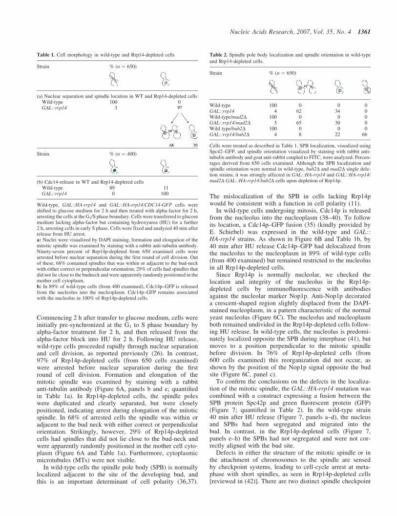
Commencing 2 h after transfer to glucose medium, cells wereinitially pre-synchronized at the G1 to S phase boundary byalpha-factor treatment for 2 h, and then released from thealpha-factor block into HU for 2 h. Following HU release,wild-type cells proceeded rapidly through nuclear separationand cell division, as reported previously (26). In contrast,97% of Rrp14p-depleted cells (from 650 cells examined)were arrested before nuclear separation during the firstround of cell division. Formation and elongation of themitotic spindle was examined by staining with a rabbitanti-tubulin antibody (Figure 6A, panels b and e; quantifiedin Table 1a). In Rrp14p-depleted cells, the spindle poleswere duplicated and clearly separated, but were closelypositioned, indicating arrest during elongation of the mitoticspindle. In 68% of arrested cells the spindle was within oradjacent to the bud neck with either correct or perpendicularorientation. Strikingly, however, 29% of Rrp14p-depletedcells had spindles that did not lie close to the bud-neck andwere apparently randomly positioned in the mother cell cyto-plasm (Figure 6A and Table 1a). Furthermore, cytoplasmicmicrotubules (MTs) were not visible.
In wild-type cells the spindle pole body (SPB) is normallylocalized adjacent to the site of the developing bud, andthis is an important determinant of cell polarity (36,37).
The mislocalization of the SPB in cells lacking Rrp14pwould be consistent with a function in cell polarity (11).
In wild-type cells undergoing mitosis, Cdc14p is releasedfrom the nucleolus into the nucleoplasm (38–40). To followits location, a Cdc14p–GFP fusion (35) (kindly provided byE. Schiebel) was expressed in the wild-type and GAL::HA-rrp14 strains. As shown in Figure 6B and Table 1b, by40 min after HU release Cdc14p–GFP had delocalized fromthe nucleolus to the nucleoplasm in 89% of wild-type cells(from 400 examined) but remained restricted to the nucleolusin all Rrp14p-depleted cells.
Since Rrp14p is normally nucleolar, we checked thelocation and integrity of the nucleolus in the Rrp14p-depleted cells by immunofluorescence with antibodiesagainst the nucleolar marker Nop1p. Anti-Nop1p decorateda crescent-shaped region slightly displaced from the DAPI-stained nucleoplasm, in a pattern characteristic of the normalyeast nucleolus (Figure 6C). The nucleolus and nucleoplasmboth remained undivided in the Rrp14p-depleted cells follow-ing HU release. In wild-type cells, the nucleolus is predomi-nately localized opposite the SPB during interphase (41), butmoves to a position perpendicular to the mitotic spindlebefore division. In 76% of Rrp14p-depleted cells (from600 cells examined) this reorganization did not occur, asshown by the position of the Nop1p signal opposite the budsite (Figure 6C, panel c).
To confirm the conclusions on the defects in the localiza-tion of the mitotic spindle, the GAL::HA-rrp14 mutation wascombined with a construct expressing a fusion between theSPB protein Spc42p and green fluorescent protein (GFP)(Figure 7; quantified in Table 2). In the wild-type strain40 min after HU release (Figure 7, panels a–d), the nucleusand SPBs had been segregated and migrated into thebud. In contrast, in the Rrp14p-depleted cells (Figure 7,panels e–h) the SPBs had not segregated and were not cor-rectly aligned with the bud site.
Defects in either the structure of the mitotic spindle or inthe attachment of chromosomes to the spindle are sensedby checkpoint systems, leading to cell-cycle arrest at meta-phase with short spindles, as seen in Rrp14p-depleted cells[reviewed in (42)]. There are two distinct spindle checkpoint
Table 1. Cell morphology in wild-type and Rrp14-depleted cells
Strain % (n ¼ 650)
(a) Nuclear separation and spindle location in WT and Rrp14-depleted cellsWild-typeGAL::rrp14
1003
0
Strain % (n ¼ 400)
(b) Cdc14-release in WT and Rrp14-depleted cellsWild-type 89 11GAL::rrp14 0 100
Wild-type, GAL::HA-rrp14 and GAL::HA-rrp14/CDC14-GFP cells wereshifted to glucose medium for 2 h and then treated with alpha-factor for 2 h,arresting the cells at the G1/S phase boundary. Cells were transferred to glucosemedium lacking alpha-factor but containing hydroxyurea (HU) for a further2 h, arresting cells in early S phase. Cells were fixed and analyzed 40 min afterrelease from HU arrest.a: Nuclei were visualized by DAPI staining, formation and elongation of themitotic spindle was examined by staining with a rabbit anti-tubulin antibody.Ninety-seven percent of Rrp14p-depleted from 650 examined cells werearrested before nuclear separation during the first round of cell division. Outof these, 68% contained spindles that was within or adjacent to the bud-neckwith either correct or perpendicular orientation; 29% of cells had spindles thatdid not lie close to the budneck and were apparently randomly positioned in themother cell cytoplasm.b: In 89% of wild-type cells (from 400 examined), Cdc14p–GFP is releasedfrom the nucleolus into the nucleoplasm. Cdc14p–GFP remains associatedwith the nucleolus in 100% of Rrp14p-depleted cells.
Table 2. Spindle pole body localization and spindle orientation in wild-type
and Rrp14-depleted cells.
Strain % (n ¼ 650)
Wild-type 100 0 0 0GAL::rrp14 4 62 34 0Wild-type/mad2D 100 0 0 0GAL::rrp14/mad2D 5 65 30 0Wild type/bub2D 100 0 0 0GAL::rrp14/bub2D 4 8 22 66
Cells were treated as described in Table 1. SPB localization, visualized usingSpc42–GFP, and spindle orientation visualized by staining with rabbit anti-tubulin antibody and goat anti-rabbit coupled to FITC, were analyzed. Percen-tages derived from 650 cells examined. Although the SPB localization andspindle orientation were normal in wild-type, bub2D and mad2D single dele-tion strains, it was strongly affected in GAL::HA-rrp14 and GAL::HA-rrp14/mad2D GAL::HA-rrp14/bub2D cells upon depletion of Rrp14p.
Nucleic Acids Research, 2007, Vol. 35, No. 4 1361

pathways in yeast responding to separable MT-dependentevents; the Mad2p pathway or spindle assembly checkpoint,and the Bub2p or spindle orientation checkpoint (43,44). Toassess whether either of these pathways was responsiblefor the observed cell-cycle arrest, GAL::HA-rrp14 wascombined with the mad2D and bub2D deletions. The mad2Dsingle mutation had little effect on spindle formation in theRRP14+ strain (Figures 7, panels i–l, and 8, panels a–c, and
Table 2). When Rrp14p was depleted from the mad2D cells(Figures 7, panels m–p, and 8, panels d–f) the phenotypeclosely resembled the depletion of Rrp14p alone (Figure 7,panels e–h) with no increase in spindle elongation or thefrequency of nuclear division, and a similar fraction of cellscontained a spindle and nucleus that were not located near thebud neck (30% of 650 cells counted) (Figure 7, panels m–p,and Table 2).
Figure 7. The Bub2p spindle orientation check-point is responsible for cell-cycle arrest following Rrp14p depletion. Cells were treated as in Figure 6. SPBlocalization was visualized using Spc42–GFP, in wild type (panel b), GAL::HA-rrp14 (panel f ), mad2D (panel j), GAL::HA-rrp14/mad2D (panel n), bub2D(panel r) and GAL::HA-rrp14/mad2D (panels v and v0) cells 40 min after HU release. Nuclei were visualized by DAPI staining (panels a, e, i, m, q, u and u0).Cell morphology was observed by DIC (panels d, h, l, p, t, x and x0). Bars represent 10 mm.
1362 Nucleic Acids Research, 2007, Vol. 35, No. 4

The bub2D single mutant also showed no effect on spindleformation (Figures 7, panels q–t, and 8, panels g–i, andTable 2), but clearly exacerbated the phenotype whenRrp14p was depleted. Ninety-six percent of cells (from650 cells counted) displayed abnormal spindle orientationwith the majority of cells (66%) also containing multipleSPBs, deformed nuclei or binucleate mother cells (Figures 7,panels u–x, and 8, panels j–l, and Table 2). Many cells appar-ently continued to bud despite the defect in cell division,forming chains of cells (Figures 7, panels x, x0, and 8,panel l). We conclude that activation of the Bub2p spindlecheckpoint is responsible for the cell-cycle arrest in cellsdepleted of Rrp14p.
Recent reports have implicated other 60S ribosome synthe-sis factors in the mechanism of DNA replication (5,45,46). Afailure to undergo DNA replication following HU releasewould potentially explain some, but not all, of the defectsobserved in Rrp14p-depleted cells. DNA replication wasfollowed by fluorescence-activated cell sorting (FACS)analysis at intervals following release from HU arrest
(Figure 9). In unsynchronized cells (Figure 9A) the twomajor peaks correspond to cells with unreplicated (1C)and duplicated (2C) genomes. In both the wild type and mut-ant, the HU-induced block was efficient with most cellsarrested with a 1C genome (Figure 9B and C). In the wildtype, most cells had undergone DNA replication 20 minafter HU release, and almost all after 40 min (Figure 9B).By 60 min after HU release, haploid cells that have com-pleted mitosis were reappearing. In the Rrp14p-depletedcells, DNA replication is slower, with more cells remaining1C or in S phase at 20 and 40 min after release than in thewild type (Figure 9C). However, at 40 min after HU release,the time at which the microscopy presented in Figures 6, 7and 8 was performed, most cells appear to have completedDNA replication, indicating that this is unlikely to be themajor cause of the cell-cycle arrest in Rrp14p-depletedcells. In the GAL::HA-rrp14 strain the absence of Mad2p orBub2p had no clear effects on DNA replication, nor did lossof the genome integrity check point protein Mec1p (47) (datanot shown).
Figure 8. Cells form multiple spindles following Rrp14p depletion in bub2D spindle checkpoint mutants. Cells were treated as in Figure 6. Tubulin wasvisualized by staining with rabbit anti-tubulin antibody and goat anti-rabbit coupled to FITC, in mad2D (panel b), GAL::HA-rrp14/mad2D (panel e), bub2D(panel h) and GAL::HA-rrp14/mad2D (panel k) cells 40 min after HU release. Nuclei were visualized by DAPI staining (panels a, d, g and j). Cell outlines areindicated with a dotted line (panels c, f, i and l).
Nucleic Acids Research, 2007, Vol. 35, No. 4 1363

DISCUSSION
Here we report that yeast Rrp14p functions in the synthesis ofboth 40S and 60S ribosomal subunits and may also play somedirect role in cell-cycle progression through G2/M. Rrp14pwas identified as a putative participant in 60S ribosomalsubunit synthesis by its co-precipitation with early pre-60Sparticles that were associated with TAP-tagged Ssf1p (17)and Rrp1p (30). However, gradient analyses indicate thatRrp14p also associates with the earlier 90S pre-ribosomes,which include many of the factors required for 40S ribosomesynthesis. In addition to inhibiting rRNA maturation, loss ofRrp14p apparently allowed premature cleavage of both the35S and 27SA2 pre-rRNAs. We speculate that Rrp14pbinds late pre-90S particles and then remains associatedwith the pre-60S region. As well as promoting correctmaturation of both subunits, Rrp14p might act to suppressITS1 and ITS2 cleavage until other maturation steps haveoccurred.
In addition to their unusual pre-rRNA processing defects,cells depleted of Rrp14p showed striking cell-cycle-relatedphenotypes. It is difficult to exclude the possibility thatthese are an indirect consequence of reduced ribosomalsubunit abundances. However, this seems unlikely becauseno similar phenotypes have been reported for any of thelarge number of ribosome synthesis factors previously ana-lyzed. This phenotype is certainly not expected to resultfrom the inhibition of translation per se, which leads tocell-cycle arrest at the ‘Start’ check point at the G1–S bound-ary and this is also seen in many strains with ribosome syn-thesis defects [(46,48) and reviewed in (49)]. Rrp14p wasinitially characterized in two hybrid analyses of a proteininteraction network involved in the specification of cell polar-ity, and was predicted to be involved in polarized growth andthe establishment of bud sites (11). Although these authorsdid not demonstrate the physical association of Rrp14p withproteins direct involved in cell polarity, their conclusions areat least consistent with our observations.
Synchronized Rrp14p-depleted cells arrest with an undi-vided nucleus and short spindles, a phenotype characteristicof arrest at the G2/M boundary. Moreover, the spindles areoften misaligned with the bud axis, and the nuclei frequentlyfail to migrate to the bud neck. In wild-type cells the nucleo-lus moves during mitosis from its S phase location oppositethe bud neck to a position perpendicular to the bud neck(50). In most Rrp14p-depleted cells the nucleolus wasincorrectly positioned, remaining located opposite the budneck. The cytoplasmic MT asters, which normally form onthe cytoplasmic face of the SPBs, were also absent inRrp14p-depleted cells.
In several mutant strains cell-cycle arrest during mitosishas been shown to be a consequence of activation of check-points, which respond to defects in DNA replication, spindlestructure or spindle orientation. A mild delay in DNA replica-tion was seen in Rrp14p-depleted cells, but this was verymuch less marked than the inhibition seen in cells depletedof another 60S synthesis factor, Yph1p, which associateswith the DNA origin of replication complex (ORC) (5,51).Moreover, 40 min after release from HU arrest, mostRrp14p-depleted cells have completed DNA replication, asjudged by FACS analysis, but all are arrested at G2/M as
Figure 9. DNA replication is only mildly slowed in Rrp14p-depleted cells butnuclei do not separate. (A) FACS analysis of unsynchronized wild-type cells.The left-hand peak represents the 1N cell population, while the right-handpeak corresponds to 2N cells that have undergone DNA replication. FACSanalyses of wild-type (B) and GAL::HA-rrp14 cells (C), synchronized inearly S phase by alpha-factor and HU treatment in glucose medium, asdescribed for Figure 5. Following HU release, cells were fixed and analyzedafter 0, 20, 40, 60 and 80 min, as indicated. It is likely that a fraction of theRrp14p-depleted cells are arrested at the Start checkpoint before DNAreplication initiation, due to reduced ribosome synthesis. These probablycorrespond to the cells that remain haploid after release from HU.
1364 Nucleic Acids Research, 2007, Vol. 35, No. 4

judged by their morphology. It is therefore unlikely that afailure in DNA replication is responsible for the cell-cyclearrest.
Defects in the structure of the mitotic spindle or itsattachment to chromosomal centromeres are monitored bythe Mad2p-dependent spindle checkpoint, while defects inspindle orientation activate a Bub2p-dependent checkpoint(52,53) [reviewed in (42)]. To determine whether either ofthese checkpoints arrests Rrp14p-depleted cells, we deletedthe genes encoding Mad2p and Bub2p. The absence ofMad2p had no clear effects on the growth, DNA replicationor morphology of Rrp14p-depleted cells. In contrast, theabsence of Bub2p from Rrp14p-depleted cells led to theformation of cells containing multiple SPBs, binucleatemother cells and cell chains, presumably due to ongoingbud formation and SPB duplication without cell division.This observation indicates that the defect in the orientationof the spindles in Rrp14p-depleted cells is responsible forthe cell-cycle arrest.
RNAs have been identified in the centrioles of surf clams(54), whereas RNA species associate with mitotic MTs inXenopus egg extracts and spindle assembly is promoted bythe RNA export factor Rae1p (55), which is structurallyrelated to the spindle checkpoint protein Bub3p (56). YeastRae1p (Gle2p) (57) is implicated in the export of ribosomalsubunits (58,59), but potential interactions with Rrp14phave not been directly assessed. Furthermore, a proteomicanalysis of 60S pre-ribosomal particles isolated from humancells identified MT-associated proteins as components of thecomplex (60).
Together these data suggest that in other systems RNAmolecules associate with mitotic spindles, and it is at leastconceivable that RNAs—and therefore RNA-bindingproteins—also play some role in yeast spindle dynamics.
ACKNOWLEDGEMENTS
We thank Elmar Schiebel for the Cdc14–GFP construct,Mensur Dlakic for Rrp14p alignment studies, VincentArchambault for the mec1D/sml1D strain and Fred Cross forthe bub2D strain. We thank Kevin Hardwick for the Spc42-GFP strain, the mad2D construct and for helpful comments onthe manuscript. M.O. was the recipient of a post-doctoralfellowship by the Charles Revson Foundation. This work wassupported by the Wellcome Trust. Funding to pay the OpenAccess publication charges for this article was provided bythe Wellcome Trust.
Conflict of interest statement. None declared.
REFERENCES
1. Warner,J.R. (1989) Synthesis of Ribosomes in Saccharomycescerevisae. Microbiol. Rev., 53, 256–271.
2. Rudra,D. and Warner,J.R. (2004) What better measure than ribosomesynthesis? Genes Dev., 18, 2431–2436.
3. Ju,Q. and Warner,J.R. (1994) Ribosome synthesis during the growthcycle of Saccharomyces cerevisiae. Yeast, 10, 151–157.
4. Dez,C., Froment,C., Noaillac-Depeyre,J., Monsarrat,B.,Caizergues-Ferrer,M. and Henry,Y. (2004) Npa1p, a component ofvery early Pre-60S ribosomal particles, associates with a subset of
small nucleolar RNPs required for peptidyl transferase centermodification. Mol. Cell. Biol., 24, 6324–6337.
5. Du,Y.C. and Stillman,B. (2002) Yph1p, an ORC-interacting protein.potential links between cell proliferation control, DNA replication, andribosome biogenesis. Cell, 109, 835–848.
6. Oeffinger,M. and Tollervey,D. (2003) Yeast Nop15p is anRNA-binding protein required for pre-rRNA processing andcytokinesis. EMBO J., 22, 6573–6583.
7. Dosil,M. and Bustelo,X.R. (2004) Functional characterization of Pwp2,a WD family protein essential for the assembly of the 90 Spre-ribosomal particle. J. Biol. Chem., 279, 37385–37397.
8. Saracino,F., Bassler,J., Muzzini,D., Hurt,E. and AgostoniCarbone,M.L. (2004) The yeast kinase Swe1 is required for properentry into cell cycle after arrest due to ribosome biogenesis and proteinsynthesis defects. Cell Cycle, 3, 648–654.
9. Shafaatian,R., Payton,M.A. and Reid,J.D. (1996) PWP2, a member ofthe WD-repeat family of proteins, is an essential Saccharomycescerevisiae gene involved in cell separation. Mol. Gen. Genet.,252, 101–114.
10. Bogomolnaya,L.M., Pathak,R., Cham,R., Guo,J., Surovtseva,Y.V.,Jaeckel,L. and Polymenis,M. (2004) A new enrichment approachidentifies genes that alter cell cycle progression in Saccharomycescerevisiae. Curr. Genet., 45, 350–359.
11. Drees,B.L., Sundin,B., Brazeau,E., Caviston,J.P., Chen,G.C., Guo,W.,Kozminski,K.G., Lau,M.W., Moskow,J.J., Tong,A. et al. (2001) Aprotein interaction map for cell polarity development. J. Cell Biol.,154, 549–571.
12. Roy,N. and Runge,K.W. (2000) Two paralogs involved intranscriptional silencing that antagonistically control yeast life span[In Process Citation]. Curr. Biol., 10, 111–114.
13. Yu,Y. and Hirsch,J.P. (1995) An essential gene pair in Saccharomycescerevisiae with a potential role in mating. DNA Cell Biol., 14, 411–418.
14. Hofken,T. and Schiebel,E. (2004) Novel regulation of mitotic exit bythe Cdc42 effectors Gic1 and Gic2. J. Cell Biol., 164, 219–231.
15. Brown,J.L., Jaquenoud,M., Gulli,M.P., Chant,J. and Peter,M. (1997)Novel Cdc42-binding proteins Gic1 and Gic2 control cell polarity inyeast. Genes Dev., 11, 2972–2982.
16. Chen,G.-C., Kim,Y.-J. and Chan,C.S.M. (1997) The Cdc42GTPase-associated proteins Gic1 and Gic2 are required for polarizedcell growth in Saccharomyces cerevisiae. Genes Dev., 11, 2958–2971.
17. Fatica,A., Cronshaw,A.D., Dlakic,M. and Tollervey,D. (2002) Ssf1pprevents premature processing of an early pre-60S ribosomal particle.Mol. Cell, 9, 341–351.
18. Magoulas,C., Zatsepina,O.V., Jordan,P.W., Jordan,E.G. and Fried,M.(1998) The SURF-6 protein is a component of the nucleolar matrix andhas a high binding capacity for nucleic acids in vitro. Eur. J. Cell Biol.,75, 174–183.
19. Hazbun,T.R., Malmstrom,L., Anderson,S., Graczyk,B.J., Fox,B.,Riffle,M., Sundin,B.A., Aranda,J.D., McDonald,W.H., Chiu,C.H. et al.(2003) Assigning function to yeast proteins by integration oftechnologies. Mol. Cell, 12, 1353–1365.
20. Polzikov,M., Zatsepina,O. and Magoulas,C. (2005) Identification of anevolutionary conserved SURF-6 domain in a family of nucleolarproteins extending from human to yeast. Biochem. Biophys. Res.Commun., 327, 143–149.
21. Longtine,M.S., McKenzie,A.,3rd, Demarini,D.J., Shah,N.G., Wach,A.,Brachat,A., Philippsen,P. and Pringle,J.R. (1998) Additional modulesfor versatile and economical PCR-based gene deletion andmodification in Saccharomyces cerevisiae. Yeast, 14, 953–961.
22. Rigaut,G., Shevchenko,A., Rutz,B., Wilm,M., Mann,M. andSeraphin,B. (1999) A generic protein purification method for proteincomplex characterization and proteome exploration. Nat. Biotechnol.,17, 1030–1032.
23. Kufel,J., Allmang,C., Chanfreau,G., Petfalski,E., Lafontaine,D.L.J. andTollervey,D. (2000) Precursors to the U3 snoRNA lack snoRNPproteins but are stabilized by La binding. Mol. Cell Biol., 20,5415–5124.
24. Tollervey,D., Lehtonen,H., Jansen,R., Kern,H. and Hurt,E.C. (1993)Temperature-sensitive mutations demonstrate roles for yeast fibrillarinin pre-rRNA processing, pre-rRNA methylation, and ribosomeassembly. Cell, 72, 443–457.
25. Babler,J., Grandi,P., Gadal,O., Lebmann,T., Petfalski,E., Tollervey,D.,Lechner,J. and Hurt,E. (2001) Identification of a 60S pre-ribosomalparticle that is closely linked to nuclear export. Mol. Cell, 8, 517–529.
Nucleic Acids Research, 2007, Vol. 35, No. 4 1365

26. Fraser,R.S. and Moreno,F. (1976) Rates of synthesis of polyadenylatedmessenger RNA and ribosomal RNA during the cell cycle ofSchizosaccharomyces pombe. With an appendix: calculation of thepattern of protein accumulation from observed changes in the rate ofmessenger RNA synthesis. J. Cell Sci., 21, 497–521.
27. Grandi,P., Doyl,V. and Hurt,E.C. (1993) Purification of NSP1 revealscomplex formation with ‘GLFG’ nucleporins and a novel nuclear poreprotein NIC96. EMBO J., 12, 3061–3071.
28. Berges,T., Petfalski,E., Tollervey,D. and Hurt,E.C. (1994) Syntheticlethality with fibrillarin identifies NOP77p, a nucleolar protein requiredfor pre-rRNA processing and modification. EMBO J., 13, 3136–3148.
29. Aris,J.P. and Blobel,G. (1991) cDNA cloning and sequencing of humanfibrillarin, a conserved nucleolar protein recognized by autoimmuneantisera. Proc. Natl Acad. Sci. USA, 88, 931–935.
30. Horsey,E.W., Jakovljevic,J., Miles,T.D., Harnpicharnchai,P. andWoolford,J.L.,Jr (2004) Role of the yeast Rrp1 protein in the dynamicsof pre-ribosome maturation. RNA, 10, 813–827.
31. Krogan,N.J., Cagney,G., Yu,H., Zhong,G., Guo,X., Ignatchenko,A.,Li,J., Pu,S., Datta,N., Tikuisis,A.P. et al. (2006) Global landscape ofprotein complexes in the yeast Saccharomyces cerevisiae. Nature,440, 637–643.
32. Fatica,A., Oeffinger,M., Tollervey,D. and Bozzoni,I. (2003)Cic1p/Nsa3p is required for synthesis and nuclear export of 60Sribosomal subunits. RNA, 9, 1431–1436.
33. Rosado,I.V. and de la Cruz,J. (2004) Npa1p is an essential trans-actingfactor required for an early step in the assembly of 60S ribosomalsubunits in Saccharomyces cerevisiae. RNA, 10, 1073–1083.
34. Jimenez,J., Cid,V.J., Cenamor,R., Yuste,M., Molero,G., Nombela,C.and Sanchez,M. (1998) Morphogenesis beyond cytokinetic arrest inSaccharomyces cerevisiae. J. Cell Biol., 143, 1617–1634.
35. Hofken,T. and Schiebel,E. (2002) A role for cell polarity proteins inmitotic exit. EMBO J., 21, 4851–4862.
36. Snyder,M., Gehrung,S. and Page,B.D. (1991) Studies concerning thetemporal and genetic control of cell polarity in Saccharomycescerevisiae. J. Cell Biol., 114, 515–532.
37. Jacobs,C.W., Adams,A.E., Szaniszlo,P.J. and Pringle,J.R. (1988)Functions of microtubules in the Saccharomyces cerevisiae cell cycle.J. Cell Biol., 107, 1409–1426.
38. Straight,A.F., Shou,W., Dowd,G.J., Turck,C.W., Deshaies,R.J.,Johnson,A.D. and Moazed,D. (1999) Net1, a Sir2-associated nucleolarprotein required for rDNA silencing and nucleolar integrity. Cell,97, 245–256.
39. Shou,W., Seol,J.H., Shevchenko,A., Baskerville,C., Moazed,D.,Chen,Z.W., Jang,J., Shevchenko,A., Charbonneau,H. and Deshaies,R.J.(1999) Exit from mitosis is triggered by Tem1-dependent release of theprotein phosphatase Cdc14 from nucleolar RENT complex.Cell, 97, 233–244.
40. Visintin,R., Hwang,E.S. and Amon,A. (1999) Cfl1p prevents prematureexit from mitosis by anchoring Cdc14 phosphatase in the nucleolus.Nature, 398, 818–823.
41. Yang,C.H., Lambie,E.J., Hardin,J., Craft,J. and Snyder,M. (1989)Higher order structure is present in the yeast nucleus: autoantibodyprobes demonstrate that nucleolus lies opposite the spindle pole body.Chromosoma, 98, 123–128.
42. Musacchio,A. and Hardwick,K.G. (2002) The spindle checkpoint:structural insights into dynamic signalling. Nature Rev. Mol. Cell Biol.,3, 731–741.
43. Chen,R.H., Brady,D.M., Smith,D., Murray,A.W. and Hardwick,K.G.(1999) The spindle checkpoint of budding yeast depends on a tight
complex between the Mad1 and Mad2 proteins. Mol. Biol. Cell,10, 2607–2618.
44. Daum,J.R., Gomez-Ospina,N., Winey,M. and Burke,D.J. (2000)The spindle checkpoint of Saccharomyces cerevisiae responds toseparable microtubule-dependent events. Curr. Biol., 10,1375–1378.
45. Zhang,Y., Yu,Z., Fu,X. and Liang,C. (2002) Noc3p, a bHLH protein,plays an integral role in the initiation of DNA replication in buddingyeast. Cell, 109, 849–860.
46. Bernstein,K.A. and Baserga,S.J. (2004) The small subunit processomeis required for cell cycle progression at G1. Mol. Biol. Cell,15, 5038–5046.
47. Weinert,T.A., Kiser,G.L. and Hartwell,L.H. (1994) Mitotic checkpointgenes in budding yeast and the dependence of mitosis on DNAreplication and repair. Genes Dev., 8, 652–665.
48. Jorgensen,P., Nishikawa,J.L., Breitkreutz,B.J. and Tyers,M. (2002)Systematic identification of pathways that couple cell growth anddivision in yeast. Science, 297, 395–400.
49. Jorgensen,P., Tyers,M. and Warner,J.R. (2003) In Hall,M.N.,Raff,M. and Thomas,G. (eds), Cell Growth: Control of Cell Size.Cold Spring Harbor Laboratory Press, Cold Spring Harbor,pp. 329–370.
50. Bystricky,K., Laroche,T., van Houwe,G., Blaszczyk,M. andGasser,S.M. (2005) Chromosome looping in yeast: telomere pairingand coordinated movement reflect anchoring efficiency and territorialorganization. J. Cell Biol., 168, 375–387.
51. Kinoshita,Y., Jarell,A.D., Flaman,J.M., Foltz,G., Schuster,J.,Sopher,B.L., Irvin,D.K., Kanning,K., Kornblum,H.I., Nelson,P.S. et al.(2001) Pescadillo, a novel cell cycle regulatory protein abnormallyexpressed in malignant cells. J. Biol. Chem., 276, 6656–6665.
52. Li,R. and Murray,A.W. (1991) Feedback control of mitosis in buddingyeast. Cell, 66, 519–531.
53. Luo,X., Tang,Z., Rizo,J. and Yu,H. (2002) The Mad2 spindlecheckpoint protein undergoes similar major conformationalchanges upon binding to either Mad1 or Cdc20. Mol. Cell,9, 59–71.
54. Alliegro,M.C., Alliegro,M.A. and Palazzo,R.E. (2006)Centrosome-associated RNA in surf clam oocytes. Proc. Natl Acad.Sci. USA, 103, 9034–9038.
55. Blower,M.D., Nachury,M., Heald,R. and Weis,K. (2005) ARae1-containing ribonucleoprotein complex is required for mitoticspindle assembly. Cell, 121, 223–234.
56. Larsen,N.A. and Harrison,S.C. (2004) Crystal structure of thespindle assembly checkpoint protein Bub3. J. Mol. Biol., 344,885–892.
57. Murphy,R., Watkins,J.L. and Wente,S.R. (1996) GLE2,a Saccharomyces cerevisiae homologue of the Schizosaccharomycespombe export factor RAE1, is required for nuclear pore complexstructure and function. Mol. Biol. Cell, 7, 1921–1937.
58. Moy,T.I. and Silver,P.A. (1999) Nuclear export of the small ribosomalsubunit requires the ran-GTPase cycle and certain nucleoporins.Genes Dev., 13, 2118–2133.
59. Stage-Zimmermann,T., Schmidt,U. and Silver,P.A. (2000) Factorsaffecting nuclear export of the 60S ribosomal subunit in vivo.Mol. Biol. Cell, 11, 3777–3789.
60. Fujiyama,S., Yanagida,M., Hayano,T., Miura,Y., Isobe,T. andTakahashi,N. (2002) Isolation and proteomic characterization of humanparvulin-associating preribosomal ribonucleoprotein complexes.J. Biol. Chem., 277, 23773–23780.
1366 Nucleic Acids Research, 2007, Vol. 35, No. 4