Embed Size (px)
Citation preview
Nucleic Acids
• Nucleic acid: are polymers
of Nucleotides linked with
3’, 5’- phosphodiester
bonds
• Nucleotide residues are all
oriented in the same
direction (5’ to 3’) giving
the polymer directionality.
• The sequence of DNA
molecules is always read
in the 5’ to 3’ direction
5'
3'
5'
3'
5'
3'
5'
3'
5'
3'
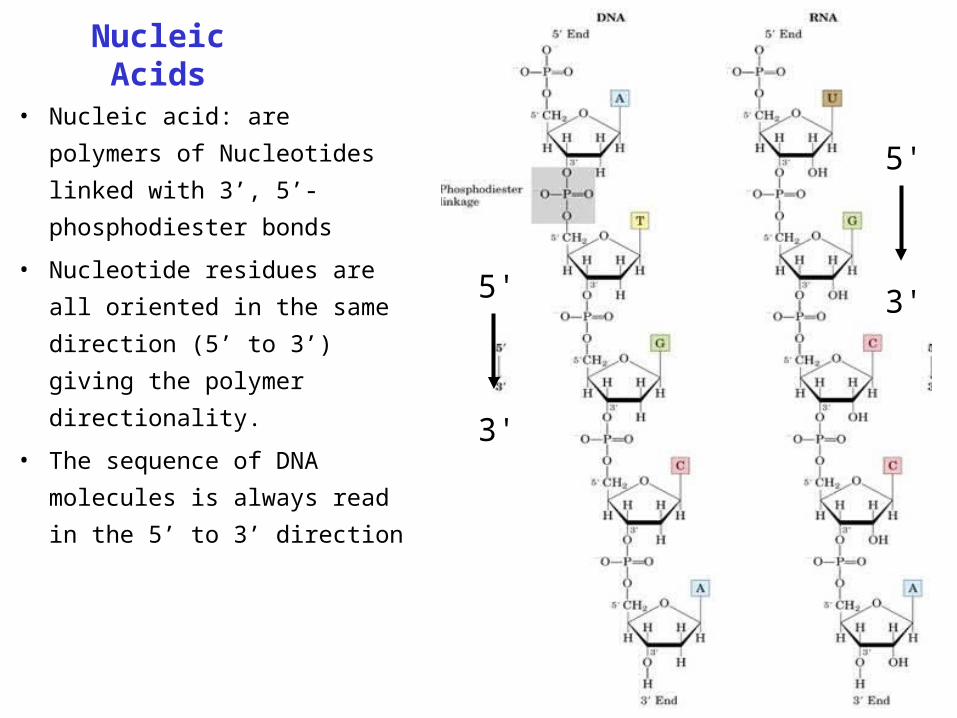
Phospho diester bond formation
3'
5'
Esterification reaction between 2 nucleotides, forming the linkage
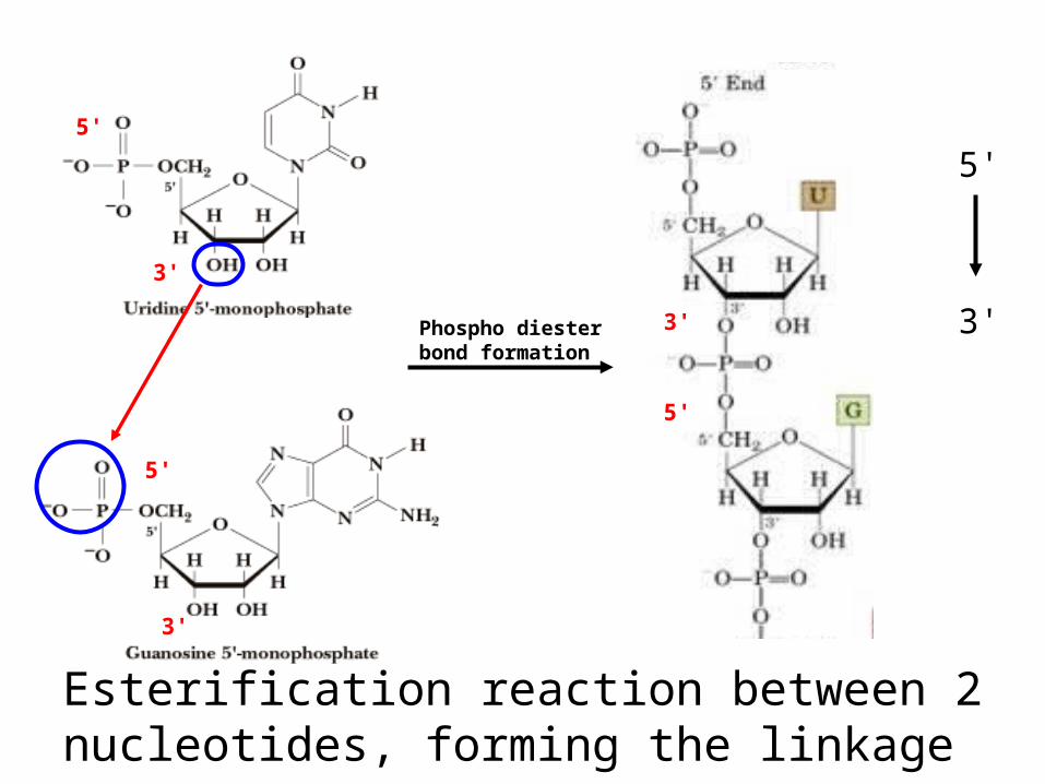
• Nucleotide monomers are joined by 3’-5’ phosphodiester linkages
to form nucleic acid (polynucleotide) polymers
5'
3'
5'
3'
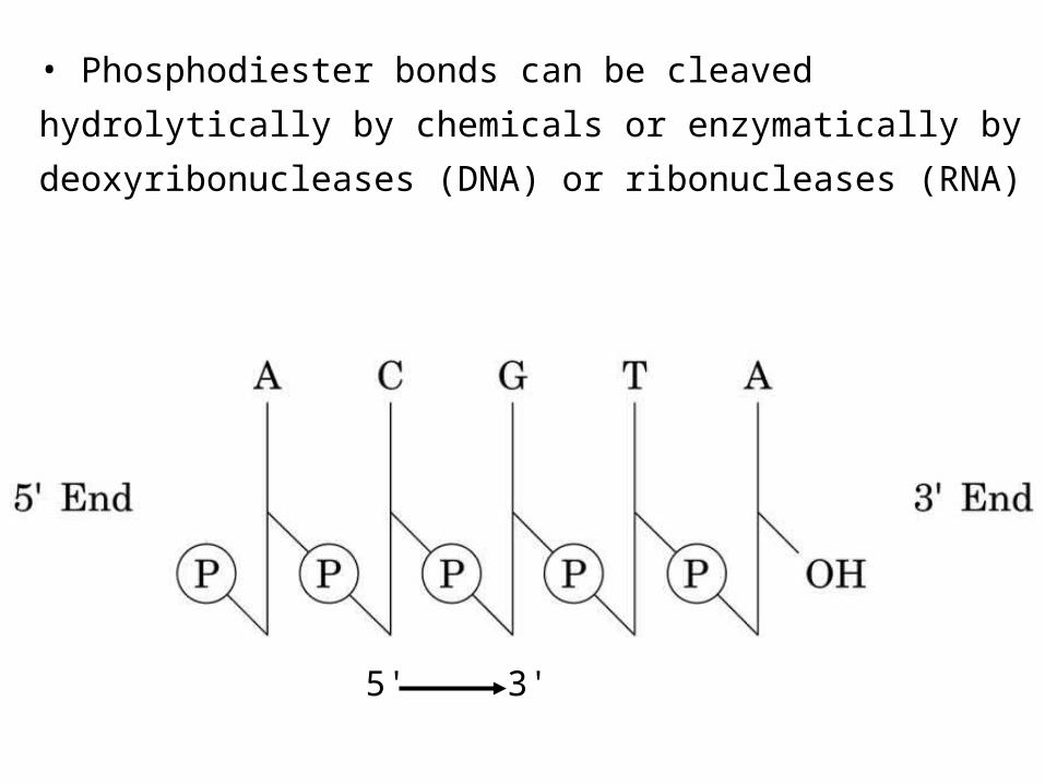
• Phosphodiester bonds can be cleaved hydrolytically by
chemicals or enzymatically by deoxyribonucleases
(DNA) or ribonucleases (RNA)
5' 3'

DNA• 1o Structure - Linear array of nucleotides and their
sequence can be determined by different methods. • 2o Structure – double helix• 3o Structure - Super-coiling, stem-loop formation• 4o Structure – Packaging into chromatin
DNA Secondary structure• DNA is double stranded with antiparallel strands• Right hand double helix• Three different helical forms (A, B and Z DNA.

Properties of DNA Double Helix
* The two chains are coiled around a common
axis
* The chains are paired in an anti-parallel
manner
* Distance between the 2 sugar-phosphate
backbones is always the same, give DNA
molecule a regular shape.
* Plane of bases are oriented perpendicular to
backbone
* Hydrophilic sugar phosphate backbone winds
around outside of helix
* Noncovalent interactions between upper and
lower surfaces of base-pairs (stacking) forms
a closely packed hydrophobic interior.
* Hydrophobic environment makes H-bonding
between bases stronger (no competition with
water)

Bases from two adjacent DNA strands can hydrogen bond•Adenine pairs with thymine using two H-bonds
•Guanine pairs with cytosine using three H-bonds

H-bonding of adjacent antiparallel DNA strands form double helix structure
5'
3'
5'
3'

View down the Double Helix
Sugar-phosphatebackbone
Hydrophobic Interior with
base pair stacking

Structure of Structure of DNA Double DNA Double
HelixHelix• Right handed
helix• Rise = 0.33nm
nm/nucleotide• Pitch = 3.4 nm /
turn• 10.4 nucleotides
per turn• Two groves –
major and minor

* Within groves, functional groups on
the edge of base pairs exposed to
exterior
* involved in interaction with proteins.
Factors stabilizing DNA double
Helix
* Hydrophobic interactions – burying
hydrophobic purine and pyrimidine
rings in interior
* Stacking interactions – van der Waals
interactions between stacked bases.
* Hydrogen Bonding – H-bonding
between bases
* Charge-Charge Interactions –
Electrostatic repulsions of
negatively charged phosphate
groups are minimized by interaction
with cations (e.g. Mg2+)

Three major structural forms of DNA
A: right-handed, short and broad, 2.3 A, 11 bp per turn B: right-handed, longer, thinner, 3.32 A, 10 bp per turn Z: left-handed, longest, thinnest, 3.8 A, 12 bp per turn, Found in G:C-rich regions of DNA

Structural forms of the double helixStructural forms of the double helixThree major structural forms of DNAB-form, described by Watson and CrickIt is right handed helix with 10 residues per 360° turn of the helixThe plane of bases perpendicular to helical axisChromosomal DNA consists primarily of B-DNA
A-DNA form
Is produced by moderately dehydrating the B formIt is right-handed helix Contains 11 base pairs per turnThe planes of the base pairs are tilted 20 ° away from the
perpendicular to helical axis
Z-DNA form contains 12 bp per turnIs left handed helix Contains 12 about 12 base pair per turn The deoxyribose phosphate backbone forms a “Zigzag structure”

Right handed helix
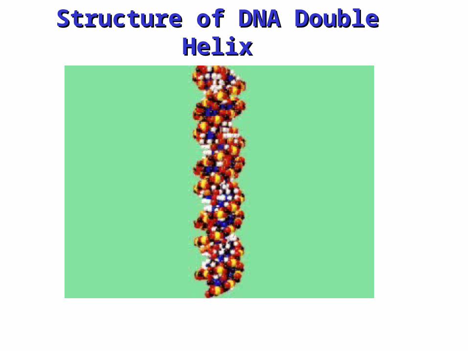
Structure of DNA Double Structure of DNA Double HelixHelix
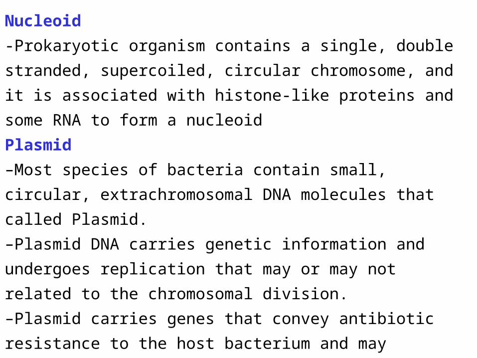
Nucleoid
-Prokaryotic organism contains a single, double
stranded, supercoiled, circular chromosome, and it is
associated with histone-like proteins and some RNA to
form a nucleoid
Plasmid
–Most species of bacteria contain small, circular,
extrachromosomal DNA molecules that called Plasmid.
–Plasmid DNA carries genetic information and undergoes
replication that may or may not related to the
chromosomal division.
–Plasmid carries genes that convey antibiotic resistance
to the host bacterium and may facilitate the transfer of
genetic information from one bacterium to another
–Plasmids used as vectors in recombinant DNA
technology

Heating up to 70 – 90C° the DNA double helix denatures,H-bonds are broken, bases unstack, and the strands separate.
Separation of the two DNA strands in the double helix
Renaturation (annealing) at
lower temperatures occurs in 2
steps.
1. Complementary bases pair.
2. The rest of the structure forms
cooperatively; it “zips-up”.
DNA with high G:C content
denatures at a higher Temp.
than A:T rich segments.

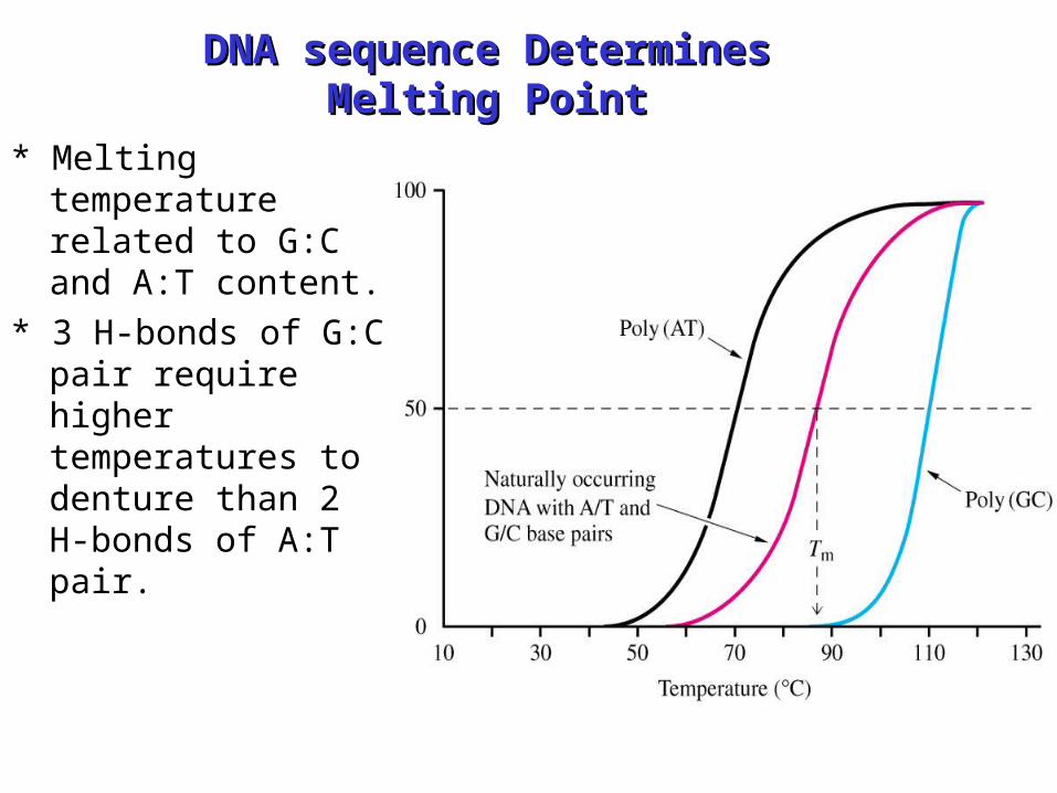
DNA sequence Determines DNA sequence Determines Melting PointMelting Point
• Double Strand DNA can be denatured by heat (get strand separation)
• Can determine degree of denturation by measuring absorbance at 260 nm.
• Conjugated double bonds in bases absorb light at 260 nm.
• Base stacking causes less absorbance.
• Increased single strandedness causes increase in absorbance

Melting Point Single strand has higher relative absorbance at 260 than dose double stranded DNA.Increasing the denaturation increases the absorbanceMelting point: the temperature at which the half of the helical structure is lost
DNA sequence Determines DNA sequence Determines Melting PointMelting Point
* Melting temperature related to G:C and A:T content.
* 3 H-bonds of G:C pair require higher temperatures to denture than 2 H-bonds of A:T pair.

DNA supercoiling:
• Supercoiling: means the
coiling of the coil.
• Typical phone cord is coiled
like a DNA helix and the
coiled cord can itself coil in a
supercoil
• A number of measurable
properties of supercoiling
have been established
DNA 3DNA 3oo Structure Structure• Supercoiling • Cruciform structures
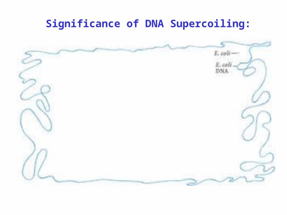
Significance of DNA Supercoiling:

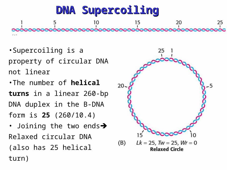
DNA SupercoilingDNA Supercoiling
•Supercoiling is a property
of circular DNA not linear
•The number of helical
turns in a linear 260-bp
DNA duplex in the B-DNA
form is 25 (260/10.4)
• Joining the two ends
Relaxed circular DNA (also
has 25 helical turn)
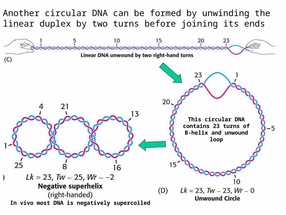
Another circular DNA can be formed by unwinding the linear duplex by two turns before joining its ends
This circular DNA contains 23 turns of B-helix and unwound
loop
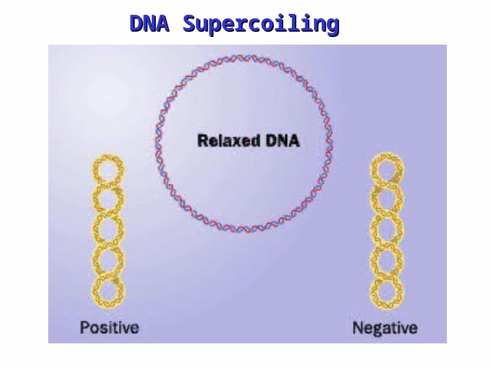
In vivo most DNA is negatively supercoiled

A supercoiled DNA molecule is more compact than a relaxed DNA
molecule of the same length supercoiled DNA moves faster than
relaxed DNA in centrifuged
Topological parameters that describe supercoling Linking number Lk: the number of times one strand of DNA
winds around the other in the right-handed direction. Molecules
that differs only in linking number are called topological isomers
or topoisomers Number of turns of Watson-Crick helix “T” (Twisting number) Number of turns o superhelix “W” (writhing number)
L=T + W
T, W can be non-integral but L should be integral
Supercoiled DNA is favored over unwound DNA because it
contains more paired bases
Enzymes called topoisomerases or gyrases can introduce or
remove supercoils
DNA SupercoilingDNA Supercoiling
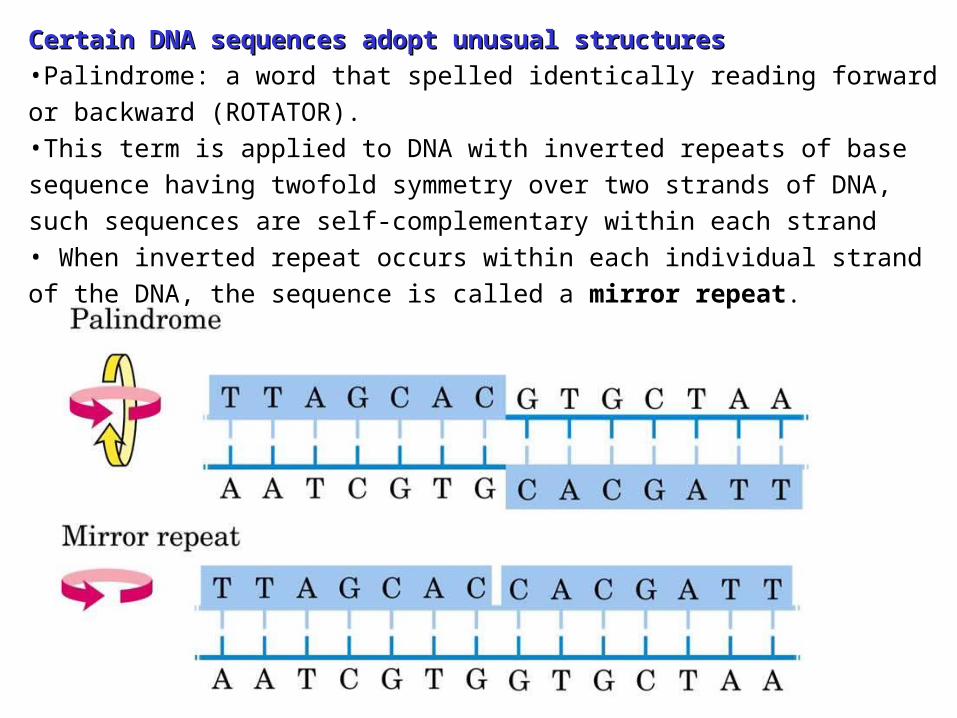
Certain DNA sequences adopt unusual structures Certain DNA sequences adopt unusual structures •Palindrome: a word that spelled identically reading forward or backward (ROTATOR). •This term is applied to DNA with inverted repeats of base sequence having twofold symmetry over two strands of DNA, such sequences are self-complementary within each strand • When inverted repeat occurs within each individual strand of the DNA, the sequence is called a mirror repeat.

Palindromes have potential to form hairpin or Palindromes have potential to form hairpin or cruciform cruciform

Cruciform: cross-shaped structureCan form intrachain base pairing
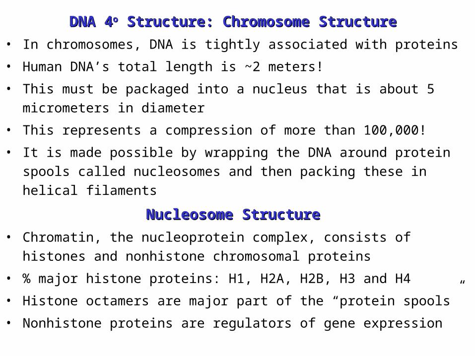
DNA 4DNA 4oo Structure: Chromosome Structure Structure: Chromosome Structure
• In chromosomes, DNA is tightly associated with proteins
• Human DNA’s total length is ~2 meters!
• This must be packaged into a nucleus that is about 5 micrometers in diameter
• This represents a compression of more than 100,000!
• It is made possible by wrapping the DNA around protein spools called nucleosomes and then packing these in helical filaments
Nucleosome StructureNucleosome Structure
• Chromatin, the nucleoprotein complex, consists of histones and nonhistone chromosomal proteins
• % major histone proteins: H1, H2A, H2B, H3 and H4
• Histone octamers are major part of the “protein spools”
• Nonhistone proteins are regulators of gene expression
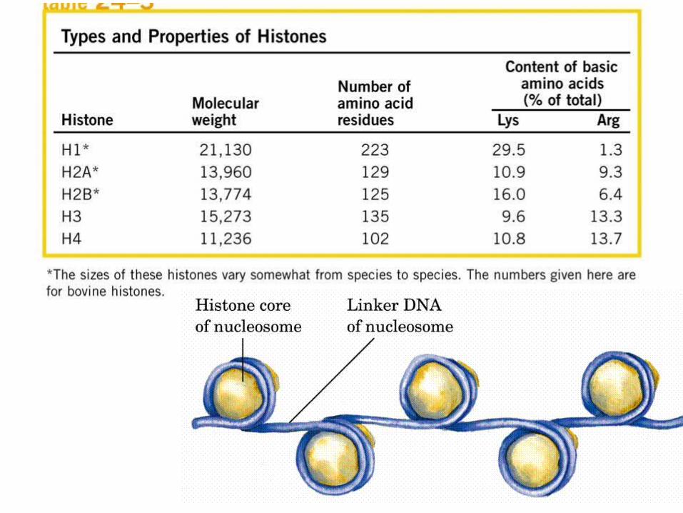

Nucleosome StructureNucleosome Structure
• High content of Lysine and arginine (+ve charge)
•4 major histone (H2A, H2B, H3, H4) proteins for octomer
•200 base pair long DNA strand winds around the octomer
•146 base pair DNA “spacer separates individual nucleosomes
•H1 protein involved in higher-order chromatin structure.
•Chromatin looks like beads on string
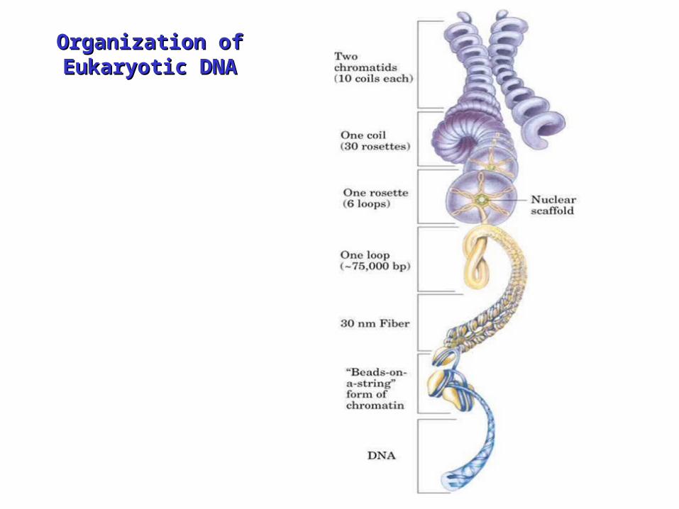
Organization of Organization of Eukaryotic DNAEukaryotic DNA

Organization of Eukaryotic Organization of Eukaryotic DNADNA

The End


•4 major histone (H2A, H2B, H3, H4) proteins for octomer
•200 base pair long DNA strand winds around the octomer
•146 base pair DNA “spacer separates individual nucleosomes
•H1 protein involved in higher-order chromatin structure.
•W/O H1, Chromatin looks like beads on string