Embed Size (px)
Citation preview
Biotechnology Reports 12 (2016) 33–39
CORE Metadata, citation and similar papers at core.ac.uk
Provided by Elsevier - Publisher Connector
Nucleic acid protocols: Extraction and optimization
Saeed El-Ashrama,b,c,d,*, Ibrahim Al Nasre,f, Xun Suoa,b,c,*a State Key Laboratory for Agrobiotechnology, China Agricultural University, Beijing 100193, ChinabNational Animal Protozoa Laboratory & College of Veterinary Medicine, China Agricultural University, Beijing 100193, ChinacKey Laboratory of Animal Epidemiology and Zoonosis of Ministry of Agriculture, Beijing 100193, Chinad Faculty of Science, Kafr El-Sheikh University, Kafr El-Sheikh, EgypteCollege of Science and Arts in Unaizah, Qassim University, Unaizah, Saudi ArabiafCollege of Applied Health Sciences in Ar Rass, Qassim University, Ar Rass 51921, Saudi Arabia
A R T I C L E I N F O
Article history:Received 11 May 2016Received in revised form 25 September 2016Accepted 3 October 2016Available online 5 October 2016
Keywords:Prokaryotic and eukaryotic sourcesDNARNADNaseRNase
A B S T R A C T
Yield and quality are fundamental features for any researchers during nucleic acid extraction. Here, wedescribe a simplified, semi-unified, effective, and toxic material free protocol for extracting DNA and RNAfrom different prokaryotic and eukaryotic sources exploiting the physical and chemical properties ofnucleic acids. Furthermore, this protocol showed that DNA and RNA are under triple protection (i.e. EDTA,SDS and NaCl) during lysis step, and this environment is improper for RNase to have DNA liberated of RNAand even for DNase to degrade the DNA. Therefore, the complete removal of RNA under RNase influence isachieved when RNase is added after DNA extraction, which gives optimal quality with any protocols.Similarly, DNA contamination in an isolated RNA is degraded by DNase to obtain high-quality RNA. Ourprotocol is the protocol of choice in terms of simplicity, recovery time, environmental safety, amount,purity, PCR and RT-PCR applicability.ã 2016 The Authors. Published by Elsevier B.V. This is an open access article under the CC BY license
(http://creativecommons.org/licenses/by/4.0/).
Contents lists available at ScienceDirect
Biotechnology Reports
journal homepage: www.else vie r .com/ locat e/btre
1. Introduction
Biomolecule extraction, such as deoxyribonucleic acid (DNA)and ribonucleic acid (RNA) from a variety of starting biologicalmaterials to be used in downstream applications and otheranalytical or preparative purposes, is the most important first stepin the molecular biology. The widely employed nucleic acidisolation methods can be divided into organic extraction method(phenol/chloroform), inorganic extraction method (salting out)and solid phase extraction method (solid matrix); moreover, fourindispensable steps are generally required for successful nucleicacid purification:
1. Cell lysis through disruption of the cellular membranes, cystwall or egg wall
2. Dehydration and precipitation of the cellular proteins (proteindenaturation)
3. Separation of cellular proteins and other cellular componentsout of the nucleic acid
* Corresponding authors at: State Key Laboratory for Agrobiotechnology, ChinaAgricultural University, Beijing 100193, China.
E-mail addresses: [email protected] (S. El-Ashram),[email protected] (X. Suo).
http://dx.doi.org/10.1016/j.btre.2016.10.0012215-017X/ã 2016 The Authors. Published by Elsevier B.V. This is an open access artic
4. Precipitation and dissolving the nucleic acid
The routinely practised cell lysis step can be divided into threetypes to cope with different tissues, thereby achieving optimumnucleic acid yield:
1. Grinding in liquid nitrogen (mortar and pestle), such as differentanimal and plant tissues
2. Glass-bead grinding, for example, oocysts (e.g. Eimeria spp.),metacercariae (e.g. Fasciola spp.) and nematodes’ eggs (e.g. eggsof Haemonchus contortus)
3. Repetitive pipetting, notable examples of it are animal cells andzoites of apicomplexan parasites, such as sporozoites, mer-ozoites, tahyzoites and bradyzoites, and trypanosomal forms ofTrypanosoma spp. and Leishmania spp., for example, trypomas-tigote, promastigote, amastigote and epimastigote.
In recent years, the development of molecular techniques hascreated a need for establishing simple and efficient novel methodsof DNA and RNA extraction for PCR amplification and other relatedtechniques. Carbohydrates, tannins, polyphenols and proteins inaddition to hazardous organic solvents, such as phenol andchloroform are the major enemies of the embattled researchers. Noexistence for DNA or RNA extraction method that is suitable for allprokaryotic and eukaryotic organisms.
le under the CC BY license (http://creativecommons.org/licenses/by/4.0/).
34 S. El-Ashram et al. / Biotechnology Reports 12 (2016) 33–39
Furthermore, there is an urgent need to address the insuffi-ciency of reasonable environment for RNase to have DNA free ofRNA and even for DNase to degrade the DNA.
2. Materials and methods
2.1. Reagents
Proteinase K, 100% Ethanol, 70% Ethanol, Double distilled (DD)water, Ethylene diamine tetra acetic acid (EDTA), RNase, DNase,Pyrex beads, Agarose, Deoxyribonucleic acid (DNA) Marker,2 � EasyPfu PCR SuperMix, 10% Sodium dodecyl sulfate (SDS),Glacial acetic acid (CH3COOH), Hydrochloric acid (HCl) and Sodiumhydroxide (NaOH).
2.2. Equipments
Mortar, Pestle, PCR machine, Microscope, Refrigerated Bench-top centrifuge (MIKRO200R, Germany), Weighing scale, Pipettes(20, 100, and 1000 ml), 15 and 50 ml falcon tubes, 50 ml centrifugetubes and Disposable Polypropylene micro-centrifuge tubes
2.3. Reagent setup
Tris buffer, Tris-EDTA (TE), DEPC-treated water, Saturated saltsolution (NaCl), Neutral saturated salt solution, Acidic saturatedsalt solution and Lysis buffer:1X STE buffer (50 mM NaCl, 50 mMTris-HCl and 100 mM EDTA; PH 8.0)
2.4. Procedure
2.4.1. Grinding in liquid nitrogen (Mortar and pestle)
2.4.1.1. DNA extraction protocol. Hepatic DNA extraction frommouse can be divided into six steps. These are:
2.4.1.1.1. Homogenization. 1 g of the liver was taken and cut intopieces then ground using a porcelain mortar and pestle in 3 ml oflysis buffer containing 900 ml of 10% SDS. The emulsion wastransferred to micro-centrifuge tubes and 100 mg proteinase K wasadded per ml of emulsion solution, and incubated for 1 h at 50 �C.
Fig. 1. Nucleic acid extraction and downstream application.A. Neutral salting out (DNA extraction).B. Acidic salting out (RNA extraction).C. GoldViewTM Nucleic Acid Stained 1.5% Agarose gel demonstrating the integrity of toD. GoldViewTM Nucleic Acid Stained 1.5% Agarose gel of total RNA of E. tenella.E. Standard PCR amplification of MICII of E. tenella.F. GoldViewTM Nucleic Acid Stained 1.5% Agarose gel of EtMIC-2 RT-PCR amplified prod
2.4.1.1.2. Phase separation. 350 ml of neutral saturated salt solution(NaCl) per ml was added to the previous emulsion, the micro-centrifuge tube was capped and shaken gently by hand for 15 s, andthen incubated at room temperature for 10 min. The micro-centrifuge tube was centrifuged at 590 � g for 15 min at roomtemperature with DNA remaining exclusively in the aqueous phase(see Fig. 1A for illustration).
2.4.1.1.3. DNA precipitation. The resulting aqueous phase wastransferred into another micro-centrifuge tube, and mixed withtwo volumes of room temperature absolute ethyl alcohol. Then themicro-centrifuge tube was inverted several times for 10 s.
2.4.1.1.4. DNA wash. The supernatant was removed; the DNApellet was washed once with 75% ethanol, and the DNA wasprecipitated out of the solution by centrifugation at 9500 � g for5 min.
2.4.1.1.5. DNA dissolving. The DNA pellet was allowed to dry for5 min, and dissolved in DD water. Then the DNA was quantified andaliquoted to be stored at �20 �C.
2.4.1.1.6. Removal of RNA from DNA preparation. 50 mg per mlRNase was added and the mixture was incubated for 1 h at 37 �C.
� Critical step: The treatment of DNA with RNase should be done inTris buffer at the end of the extraction protocol. Salting out step canbe repeated as before according to the protocol to obtain DNA withhighest quality. The DNA can be precipitated and washed with 70%ethanol, and then the pellet can be dissolved in Tris-EDTA (TE) forDNA protection from degradation by metal dependent nucleasesduring storage.
2.4.1.2. RNA extraction protocol. Hepatic RNA extraction methodfrom mouse can be listed as follows:
2.4.1.2.1. Homogenization. 1 g of the liver was taken and cut intopieces then ground using a porcelain mortar and pestle in 3 ml oflysis buffer containing 900 ml of 10% SDS. The emulsion wastransferred to micro-centrifuge tubes.
tal DNA extracted from Eimeria tenella.
uct of E. tenella using Finnzymes phusionTM High-Fidelity DNA Polymerase.
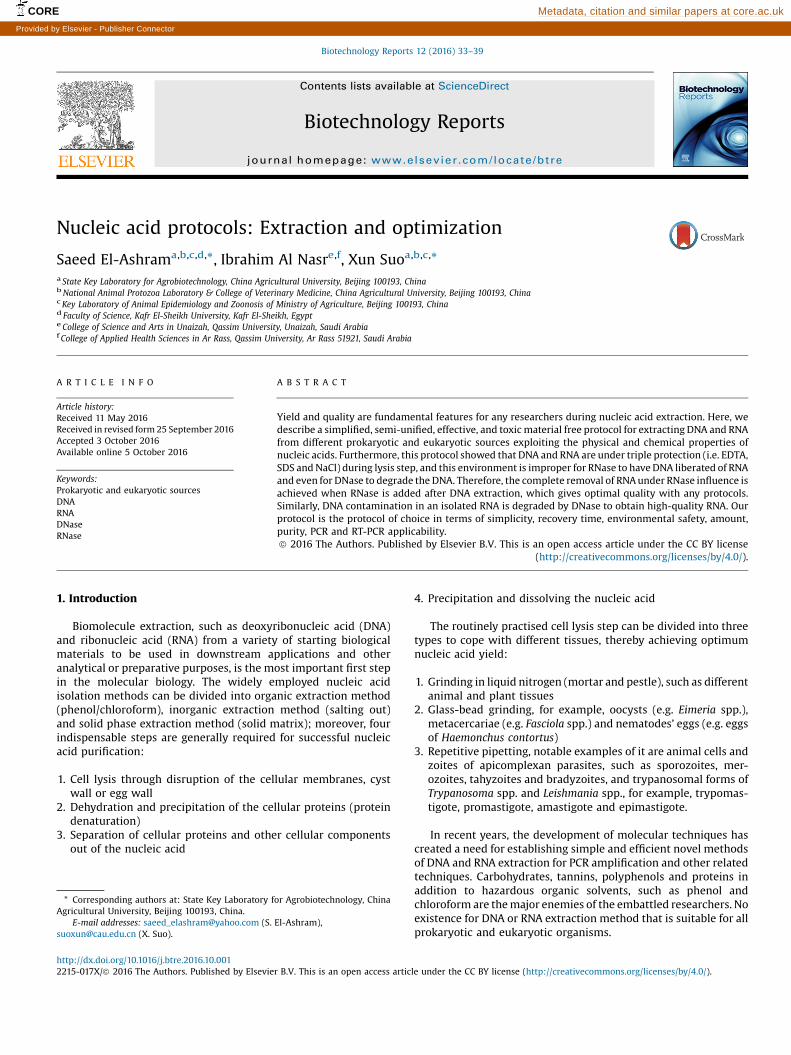
Fig. 2. GoldViewTM Nucleic Acid Stained 1.5% Agarose gel demonstrating.A. The integrity of total DNA extracted from the liver of the BALB/c mice.B. GAPDH amplified product of mouse genome. Lane 1: Trans DNA Marker III, Lane2 and 3: GAPDH amplified product from the genomic DNA of mouse genome.
S. El-Ashram et al. / Biotechnology Reports 12 (2016) 33–39 35
2.4.1.2.2. Phase separation. 350 ml of acidic saturated salt solution(NaCl) was added into each tube of the previous emulsion mixture,and the micro-centrifuge tube was capped and gently shaken byhand for 15 s and then incubated at room temperature for 10 min.The micro-centrifuge tube was centrifuged at 590 � g for 15 min atroom temperature with RNA remaining exclusively in the aqueousphase (see Fig. 1B for elucidation).
2.4.1.2.3. RNA precipitation. The resulting aqueous phase wastransferred into micro-centrifuge tubes and precipitated by mixingthe aqueous phase with two volumes of cold absolute ethyl alcohol.Then the micro-centrifuge tube was inverted several times for 15 s.
2.4.1.2.4. RNA wash. The supernatant was removed; and the RNApellet was washed once with cold 75% ethanol, and the RNA wasprecipitated out of the solution by centrifugation at 9500 � g for5 min.
2.4.1.2.5. RNA dissolving. The RNA pellet was allowed to dry for5 min and dissolved in DEPC-treated water. Then the RNA wasquantified, and aliquoted to be stored at �80 �C.
2.4.1.2.6. DNA removal. DNase I (RNase-free) kit was employed toremove any contaminating DNA from the sample as permanufacturer’s instructions.
� Critical step: The treatment of RNA with DNase should be done inTris buffer at the end of the extraction protocol. Salting out step canbe repeated as before according to the protocol to obtain RNA withhighest quality. The RNA can be precipitated and washed withethanol, and then the pellet can be dissolved in DEPC-treated wateror Tris-EDTA (TE) for DNA protection from degradation by metal-dependent nucleases during storage.
2.4.2. Repetitive pipetting: prokaryotes, for example E. coli
2.4.2.1. DNA extraction protocol. Bacterial DNA extraction can belisted as follows:
2.4.2.1.1. Homogenization. 1 ml microbial culture was transferredinto micro-centrifuge tubes and pelleted by centrifugation at380 � g for 5 min at room temperature. Then the supernatant wasdiscarded, and the pellet was resuspended with repetitivepipetting in 1 ml lysis buffer containing 100 ml of 10% SDSand 100 mg proteinase K. The mixture was incubated for 1 h at50 �C.
2.4.2.1.2. Phase separation, DNA precipitation, DNA wash, DNAdissolving and. RNA removal. They were conducted aspreviously mentioned in Section 2.4.1.1.
2.4.2.2. RNA extraction protocol. Bacterial RNA extraction can bedivided into six sections:
2.4.2.2.1. Homogenization. 1 ml microbial culture inmicrocentrifuge tube was pelleted by centrifugation at 380 � gfor 5 min at 37 �C, and the supernatant was discarded. The pelletwas resuspended with repetitive pipetting in 1 ml lysis buffercontaining 100 ml of 10% SDS.
2.4.2.2.2. Phase separation, RNA precipitation, RNA wash, RNAdissolving and DNA removal. They were performed as wepreviously mentioned in Section 2.4.1.2.
2.4.3. Glass-bead grinding
2.4.3.1. DNA extraction protocol. Eimerian DNA extraction fromoocysts can be divided into six steps. These are:
2.4.3.1.1. Homogenization. 5 �106 sporulating/sporulated oocysts,0.5 g of Pyrex beads and up to 3 ml of lysis buffer containing 900 mlof 10% SDS and 300 mg proteinase K were added in a 15 mldisposable polypropylene tube. The mixture was incubated for 1 hat 50 �C.
2.4.3.1.2. Phase separation. 1 ml of neutral saturated salt solutionwas added to the previous mixture, and the 15 ml disposablepolypropylene tube was capped and gently shaken by hand for 15 sand then incubated at room temperature for 10 min. The 15 mldisposable polypropylene tube was centrifuged at 590 � g for15 min at room temperature.
2.4.3.1.3. DNA precipitation, DNA wash, DNA dissolving and removingcontaminating RNA from DNA. These steps were then performedas previously reported in Section 2.4.1.1.
36 S. El-Ashram et al. / Biotechnology Reports 12 (2016) 33–39
2.4.3.2. RNA extraction protocol. Eimerian RNA extraction can bedivided into six sections:
2.4.3.2.1. Homogenization. 5 �106 sporulating/sporulated oocysts,0.5 g of Pyrex beads and up to 3 ml of lysis buffer containing 900 mlof 10% SDS were added in a 15 ml disposable polypropylene tube,and the mixture was incubated for 1 h at 42 �C.
2.4.3.2.2. Phase separation. 1 ml of acidic saturated salt solutionwas added to the previous mixture, and the 15 ml disposablepolypropylene tube was capped and gently shaken by hand for 15 sand then incubated at room temperature for 10 min. The 15 mldisposable polypropylene tube was centrifuged at 590 � g for15 min at room temperature.
2.4.3.2.3. RNA precipitation, RNA wash, RNA dissolving and DNAremoval. They were conducted as we previously reported inSection 2.4.1.2.
3. Results and discussion
3.1. Eukaryotes, such as
3.1.1. Eimeria spp.
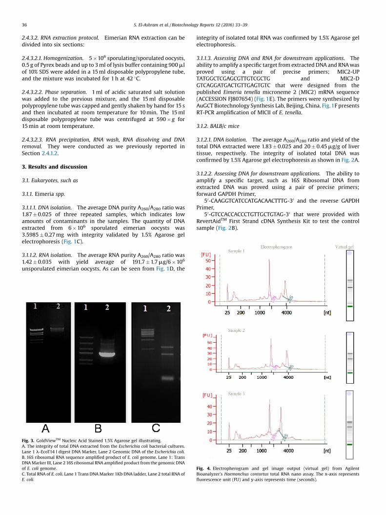
3.1.1.1. DNA isolation. The average DNA purity A260/A280 ratio was1.87 � 0.025 of three repeated samples, which indicates lowamounts of contaminants in the samples. The quantity of DNAextracted from 6 � 106 sporulated eimerian oocysts was3.5985 � 0.27 mg with integrity validated by 1.5% Agarose gelelectrophoresis (Fig. 1C).
3.1.1.2. RNA isolation. The average RNA purity A260/A280 ratio was1.42 � 0.035 with yield average of 191.7 � 1.7 mg/6 � 106
unsporulated eimerian oocysts. As can be seen from Fig. 1D, the
Fig. 3. GoldViewTM Nucleic Acid Stained 1.5% Agarose gel illustrating.A. The integrity of total DNA extracted from the Escherichia coli bacterial cultures.Lane 1 l-EcoT14 I digest DNA Marker, Lane 2 Genomic DNA of the Escherichia coli.B. 16S ribosomal RNA sequence amplified product of E. coli genome. Lane 1: TransDNA Marker III, Lane 2 16S ribosomal RNA amplified product from the genomic DNAof E. coli genome.C. Total RNA of E. coli. Lane 1 Trans DNA Marker 1Kb DNA ladder, Lane 2 total RNA ofE. coli.
integrity of isolated total RNA was confirmed by 1.5% Agarose gelelectrophoresis.
3.1.1.3. Assessing DNA and RNA for downstream applications. Theability to amplify a specific target from extracted DNA and RNA wasproved using a pair of precise primers; MIC2-UPTATGGCTCGAGCGTTGTCGCTG and MIC2-DGTCAGGATGACTGTTGAGTGTC that were designed from thepublished Eimeria tenella microneme 2 (MIC2) mRNA sequence(ACCESSION FJ807654) (Fig. 1E). The primers were synthesized byAuGCT Biotechnology Synthesis Lab, Beijing, China. Fig.1F presentsRT-PCR amplification of MICII of E. tenella.
3.1.2. BALB/c mice
3.1.2.1. DNA isolation. The average A260/A280 ratio and yield of thetotal DNA extracted were 1.83 � 0.025 and 20 � 0.45 mg/g of livertissue, respectively. The integrity of isolated total DNA wasconfirmed by 1.5% Agarose gel electrophoresis as shown in Fig. 2A.
3.1.2.2. Assessing DNA for downstream applications. The ability toamplify a specific target, such as 16S Ribosomal DNA fromextracted DNA was proved using a pair of precise primers;forward GAPDH Primer,
50-CAAGGTCATCCATGACAACTTTG-30 and the reverse GAPDHPrimer,
50-GTCCACCACCCTGTTGCTGTAG-30 that were provided withRevertAidTM First Strand cDNA Synthesis Kit to test the controlsample (Fig. 2B).
Fig. 4. Electropherogram and gel image output (virtual gel) from AgilentBioanalyzer's Haemonchus contortus total RNA nano assay. The x-axis representsfluorescence unit (FU) and y-axis represents time (seconds).
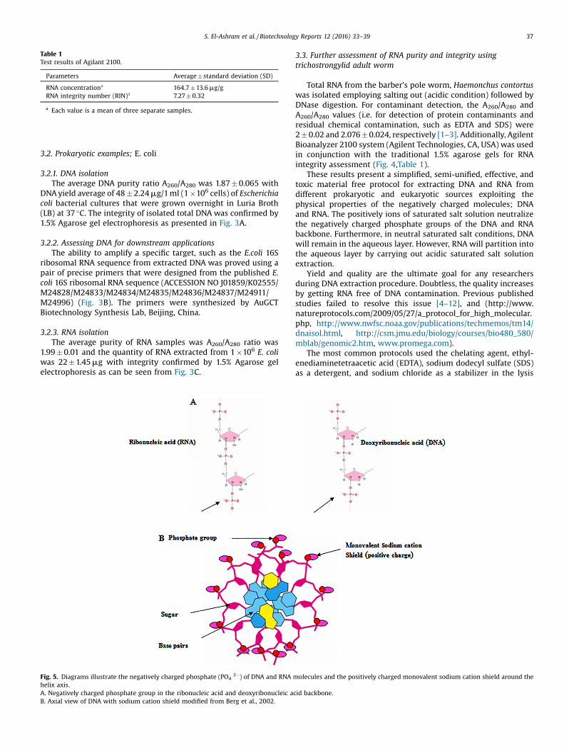
Table 1Test results of Agilant 2100.
Parameters Average � standard deviation (SD)
RNA concentrationa 164.7 � 13.6 mg/gRNA integrity number (RIN)a 7.27 � 0.32
a Each value is a mean of three separate samples.
S. El-Ashram et al. / Biotechnology Reports 12 (2016) 33–39 37
3.2. Prokaryotic examples; E. coli
3.2.1. DNA isolationThe average DNA purity ratio A260/A280 was 1.87 � 0.065 with
DNA yield average of 48 � 2.24 mg/1 ml (1 �106 cells) of Escherichiacoli bacterial cultures that were grown overnight in Luria Broth(LB) at 37 �C. The integrity of isolated total DNA was confirmed by1.5% Agarose gel electrophoresis as presented in Fig. 3A.
3.2.2. Assessing DNA for downstream applicationsThe ability to amplify a specific target, such as the E.coli 16S
ribosomal RNA sequence from extracted DNA was proved using apair of precise primers that were designed from the published E.coli 16S ribosomal RNA sequence (ACCESSION NO J01859/K02555/M24828/M24833/M24834/M24835/M24836/M24837/M24911/M24996) (Fig. 3B). The primers were synthesized by AuGCTBiotechnology Synthesis Lab, Beijing, China.
3.2.3. RNA isolationThe average purity of RNA samples was A260/A280 ratio was
1.99 � 0.01 and the quantity of RNA extracted from 1 �106 E. coliwas 22 � 1.45 mg with integrity confirmed by 1.5% Agarose gelelectrophoresis as can be seen from Fig. 3C.
Fig. 5. Diagrams illustrate the negatively charged phosphate (PO43�) of DNA and RNA m
helix axis.A. Negatively charged phosphate group in the ribonucleic acid and deoxyribonucleic aB. Axial view of DNA with sodium cation shield modified from Berg et al., 2002.
3.3. Further assessment of RNA purity and integrity usingtrichostrongylid adult worm
Total RNA from the barber’s pole worm, Haemonchus contortuswas isolated employing salting out (acidic condition) followed byDNase digestion. For contaminant detection, the A260/A280 andA260/A280 values (i.e. for detection of protein contaminants andresidual chemical contamination, such as EDTA and SDS) were2 � 0.02 and 2.076 � 0.024, respectively [1–3]. Additionally, AgilentBioanalyzer 2100 system (Agilent Technologies, CA, USA) was usedin conjunction with the traditional 1.5% agarose gels for RNAintegrity assessment (Fig. 4,Table 1).
These results present a simplified, semi-unified, effective, andtoxic material free protocol for extracting DNA and RNA fromdifferent prokaryotic and eukaryotic sources exploiting thephysical properties of the negatively charged molecules; DNAand RNA. The positively ions of saturated salt solution neutralizethe negatively charged phosphate groups of the DNA and RNAbackbone. Furthermore, in neutral saturated salt conditions, DNAwill remain in the aqueous layer. However, RNA will partition intothe aqueous layer by carrying out acidic saturated salt solutionextraction.
Yield and quality are the ultimate goal for any researchersduring DNA extraction procedure. Doubtless, the quality increasesby getting RNA free of DNA contamination. Previous publishedstudies failed to resolve this issue [4–12], and (http://www.natureprotocols.com/2009/05/27/a_protocol_for_high_molecular.php, http://www.nwfsc.noaa.gov/publications/techmemos/tm14/dnaisol.html, http://csm.jmu.edu/biology/courses/bio480_580/mblab/genomic2.htm, www.promega.com).
The most common protocols used the chelating agent, ethyl-enediaminetetraacetic acid (EDTA), sodium dodecyl sulfate (SDS)as a detergent, and sodium chloride as a stabilizer in the lysis
olecules and the positively charged monovalent sodium cation shield around the
cid backbone.
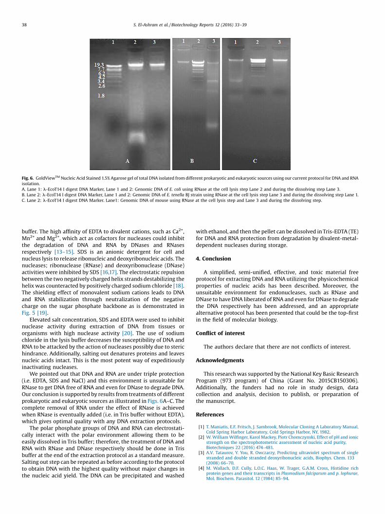
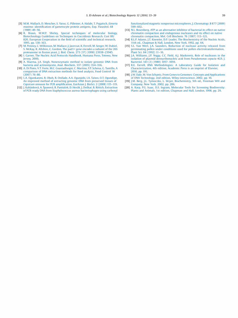
Fig. 6. GoldViewTM Nucleic Acid Stained 1.5% Agarose gel of total DNA isolated from different prokaryotic and eukaryotic sources using our current protocol for DNA and RNAisolation.A. Lane 1: l-EcoT14 I digest DNA Marker, Lane 1 and 2: Genomic DNA of E. coli using RNase at the cell lysis step Lane 2 and during the dissolving step Lane 3.B. Lane 2: l-EcoT14 I digest DNA Marker, Lane 1 and 2: Genomic DNA of E. tenella BJ strain using RNase at the cell lysis step Lane 3 and during the dissolving step Lane 1.C. Lane 2: l-EcoT14 I digest DNA Marker. Lane1: Genomic DNA of mouse using RNase at the cell lysis step and Lane 3 and during the dissolving step.
38 S. El-Ashram et al. / Biotechnology Reports 12 (2016) 33–39
buffer. The high affinity of EDTA to divalent cations, such as Ca2+,Mn2+ and Mg2+, which act as cofactors for nucleases could inhibitthe degradation of DNA and RNA by DNases and RNasesrespectively [13–15]. SDS is an anionic detergent for cell andnucleus lysis to release ribonucleic and deoxyribonucleic acids. Thenucleases; ribonuclease (RNase) and deoxyribonuclease (DNase)activities were inhibited by SDS [16,17]. The electrostatic repulsionbetween the two negatively charged helix strands destabilizing thehelix was counteracted by positively charged sodium chloride [18].The shielding effect of monovalent sodium cations leads to DNAand RNA stabilization through neutralization of the negativecharge on the sugar phosphate backbone as is demonstrated inFig. 5 [19].
Elevated salt concentration, SDS and EDTA were used to inhibitnuclease activity during extraction of DNA from tissues ororganisms with high nuclease activity [20]. The use of sodiumchloride in the lysis buffer decreases the susceptibility of DNA andRNA to be attacked by the action of nucleases possibly due to sterichindrance. Additionally, salting out denatures proteins and leavesnucleic acids intact. This is the most potent way of expeditiouslyinactivating nucleases.
We pointed out that DNA and RNA are under triple protection(i.e. EDTA, SDS and NaCl) and this environment is unsuitable forRNase to get DNA free of RNA and even for DNase to degrade DNA.Our conclusion is supported by results from treatments of differentprokaryotic and eukaryotic sources as illustrated in Figs. 6A–C. Thecomplete removal of RNA under the effect of RNase is achievedwhen RNase is eventually added (i.e. in Tris buffer without EDTA),which gives optimal quality with any DNA extraction protocols.
The polar phosphate groups of DNA and RNA can electrostati-cally interact with the polar environment allowing them to beeasily dissolved in Tris buffer; therefore, the treatment of DNA andRNA with RNase and DNase respectively should be done in Trisbuffer at the end of the extraction protocol as a standard measure.Salting out step can be repeated as before according to the protocolto obtain DNA with the highest quality without major changes inthe nucleic acid yield. The DNA can be precipitated and washed
with ethanol, and then the pellet can be dissolved in Tris-EDTA (TE)for DNA and RNA protection from degradation by divalent-metal-dependent nucleases during storage.
4. Conclusion
A simplified, semi-unified, effective, and toxic material freeprotocol for extracting DNA and RNA utilizing the physicochemicalproperties of nucleic acids has been described. Moreover, theunsuitable environment for endonucleases, such as RNase andDNase to have DNA liberated of RNA and even for DNase to degradethe DNA respectively has been addressed, and an appropriatealternative protocol has been presented that could be the top-firstin the field of molecular biology.
Conflict of interest
The authors declare that there are not conflicts of interest.
Acknowledgments
This research was supported by the National Key Basic ResearchProgram (973 program) of China (Grant No. 2015CB150306).Additionally, the funders had no role in study design, datacollection and analysis, decision to publish, or preparation ofthe manuscript.
References
[1] T. Maniatis, E.F. Fritsch, J. Sambrook, Molecular Cloning A Laboratory Manual,Cold Spring Harbor Laboratory, Cold Springs Harbor, NY, 1982.
[2] W. William Wilfinger, Karol Mackey, Piotr Chomczynski, Effect of pH and ionicstrength on the spectrophotometric assessment of nucleic acid purity,Biotechniques 22 (2016) 474–481.
[3] A.V. Tataurov, Y. You, R. Owczarzy, Predicting ultraviolet spectrum of singlestranded and double stranded deoxyribonucleic acids, Biophys. Chem. 133(2008) 66–70.
[4] M. Wallach, D.F. Cully, L.O.C. Haas, W. Trager, G.A.M. Cross, Histidine richprotein genes and their transcripts in Plasmodium falciparum and p. lophurae,Mol. Biochem. Parasitol. 12 (1984) 85–94.
S. El-Ashram et al. / Biotechnology Reports 12 (2016) 33–39 39
[5] M.M. Wallach, D. Mencher, S. Yarus, G. Pillemer, A. Halabi, T. Pugatsch, Eimeriamaxima: identification of gametocyte protein antigens, Exp. Parasitol. 68(1989) 49–56.
[6] R. Braun, M.W.P. Shirley, Special techniques of molecular biology,Biotechnology Guidelines on Techniques in Coccidiosis Research. Cost 89/820, European Cooperation in the field of scientific and technical research,1995, pp. 138–163.
[7] M. Penney, C. Wilkinson, M. Wallace, J. Javerzat, K. Ferrell, M. Seeger, W. Dubiel,S. McKay, R. Allshire, C. Gordon, The pad1+ gene encodes a subunit of the 26Sproteasome in fission yeast, J. Biol. Chem. 273 (37) (1998) 23938–23945.
[8] I. Garner, The Nucleic Acid Protocols Handbook, Humana Press, Totowa, NewJersey, 2000.
[9] A. Sharma, J.A. Singh, Nonenzymatic method to isolate genomic DNA frombacteria and actinomycete, Anal. Biochem. 337 (2005) 354–356.
[10] A. Di Pinto, V.T. Forte, M.C. Guastadisegni, C. Martino, F.P. Schena, G. Tantillo, Acomparison of DNA extraction methods for food analysis, Food Control 18(2007) 76–80.
[11] L.A. Ogunkanmi, B. Oboh, B. Onifade, A.A. Ogunjobi, I.A. Taiwo, O.T. Ogundipe,An improved method of extracting genomic DNA from preserved tissues ofCapsicum annuum for PCR amplification, EurAsian J. BioSci. 2 (2008) 115–119.
[12] J. Kahánková, A. Španová, R. Pantu� 9cek, D. Horák, J. Doška�r, B. Rittich, Extractionof PCR-ready DNA from Staphylococcus aureus bacteriophages using carboxyl
functionalized magnetic nonporous microspheres, J. Chromatogr. B 877 (2009)599–602.
[13] N.L. Rosenberg, ATP as an alternative inhibitor of bacterial its effect on nativechromatin compaction and endogenous nucleases and its effect on nativechromatin compaction, Mol. Cell Biochem. 76 (1987) 113–121.
[14] R.L.P. Adams, J.T. Knowler, D.P. Leader, The Biochemistry of the Nucleic Acids,11th ed., Chapman & Hall, London, New York, 1992, pp. 64.
[15] S.L. Van Wert, J.A. Saunders, Reduction of nuclease activity released fromgerminating pollen under conditions used for pollen electrotransformation,Plant Sci. 84 (1992) 11–16.
[16] J.A. Williams, J.P. Yeggy, C.C. Field, A.J. Markovetz, Role of nucleases in theisolation of plasmid deoxyribonucleic acid from Pseudomonas cepacia 4G9, J.Bacteriol. 143 (2) (1980) 1057–1059.
[17] R.E. Farrell, RNA Methodologies: A Laboratory Guide for Isolation andCharacterization, 4th edition, Academic Press is an imprint of Elsevier,2010, pp. 191.
[18] J.W. Dale, M. Von Schantz, From Genes to Genomes: Concepts and Applicationsof DNA Technology, 2nd edition, Wiley-interscience, 2002, pp. 10.
[19] J.M. Berg, J.L. Tymoczko, L. Stryer, Biochemistry, 5th ed., Freeman WH andCompany, New York, 2002, pp. 206.
[20] A. Karp, P.G. Isaac, D.S. Ingram, Molecular Tools for Screening Biodiversity:Plants and Animals, 1st edition, Chapman and Hall, London, 1998, pp. 29.


![Nucleic Acid Extraction echniques T€¦ · nucleic acid without ampli cation inhibitors or contaminants such as protein, car-bohydrate, and other nucleic acids [ 8 ] . There are](https://img.pdfslide.us/doc/110x75/601f05c49bc97203e65e2d57/nucleic-acid-extraction-echniques-t-nucleic-acid-without-ampli-cation-inhibitors.jpg)