Embed Size (px)
Citation preview

1956/57, No. 8 • 229
NUCLEAR X-RAY SOURCES
.by S. FINE *) and C. F. HENDEE *).
With the rapidly expanding availability of artificially produced radioisotopes (radioactiuenuclides} it has becomepossible to use many of these as radiation sources for routine work.Nuclides directly emitting soft X-rays have been put to such use in recent years. The authorshave made a survey of X-ray emitting nuclides and have prepared a number of X-ray sourcesof this type ("nuclear X-ray sources") which have proved very valuable ~n work on and with,X-ray counter tubes.
3) See e.g, W. Parrish-and T. R. Kohler, Rev. sci. Instr. 27,795-808,1956;also P. H.Dowling,C.F. Hendee, T.R.Kohlerand'W. Parrish, Counter tubes for X-ray analysis, Philipstech. Rev., to appear shortly.
4) Part of the materialof this article was presented by S. Fineand C.F.Hendee at thePittsburgh ConferenceonAnalyticalChemistry and Applied Spectroscopy, Feb. 1956.
. 5) .X-rays can also be produced indirectly, by a, (J or y-rays*) Philips Laboratories, Irvington-on-Hudson, N.Y., U.S.A.. emitted from a radionuclide and impinging on a suitable1) See e.g. 1. Kaplan, Nuclear physics, Addison-Wesley, . target material. These methods have been known and ap-
Cambridge (Mass.), 1955. See also Philips techno Rev.4, plied for a long time. 1. Curie and F. Joliot discussed this153-161, 1939; 15, 1-26, 1953/54; 17, 31-33, 1955/56. possibility with reference to a-rays (J. Phys. Rad. 2, 20-28,
2) G. M. Insch, PhiI. Mag. 7,41, 857-862,1950. 1931); for {J-ray excitation, see L. Reiffel, Nucleouics 13,P. Rothwell and D. West, Proc. Phys. Soc. A63, 541-543, No. 3, March 1955, pp. 22-24. Although these methods1950. • certainly should not be overlooked, we shall not considerW~V. Mayneord, Brit. J. Radiology 25, 517-525, 1952. them in this article.
Radioactive substances, either naturalor artificial,have been employed for a 'long time as sources ofa- and fJ-particles and of high energy photons (y_rays). A classical example is natural r~dioactivepolonium, specimens of which are often used fordemonstrating the effects of a-radiation or for
. checking apparatus sensitive to it. A youngermember of this family is the artificial radioisotope ofcobalt (Co 60) emitting 1.17 MeV and 1.33 MeVy-rays. Radiation of similar energy, which is used,inter alia, for industrial radiography and fortherapeutic irradiation, mayalso be obtained fromX-ray tubes run at voltages up to 2 MeV, or frombetatrons or linear accelerators 1). Nevertheless theradioactive y-ray source has proved a very valuableextension of the array of high-energy X-ray ma-chines.In addition to the energetic particles and high
energy photons, resulting from the decay of radio-active nuclei, photons of lower energy, e.g. less than100 keY (soft X-rays), are in some cases emitted.Because the investigation of nuclear processes usu-ally involves an interpretation of high energy events,there has heen little emphasis on the low energyphenomena. In fact the latter ofter appear only asperturbations or corrections in nuclear theory(background or noise). .Only in recent years has it been realized that the
emission of low energy X-rays by some rádioactivenuclides can be put to good use 2), just as usefulapplications have been found for y-ray emitterssuch as cobalt 60. Interest in this possibility wasprompted by the development of X-ray quantumcounting techniques. For checking and calibrating
539.167.3:537.531:621.387.4
proportional and scintillation counters developedfor X-ray detection 3), very stable monochromatieX-ray sources.: were required. X-ray emittingnuclides ("nuclear X -ray sources"), while lacking'the easy controllability and the very large radiationoutput of modern "Xvray generating equipment,usually greatly surpass the latter in stability andtherefore promised to be useful in work with X-raycounters.Unfortunately, nuclear X-ray sources have not
yet become a commercially available produc!,probably owing to the tendency among physiciststo be more preoccupied with high energy phenomena.It seemed desirable, therefore, to survey the present-ly known artificially produced radioisotopes fromthe viewpoint of X-ray production. This survey isoffered in this article, together with a discuesion ofthe underlying phenomena in the nuclear' X-raysource and a description of some sources which havebeen developed and are currently used in thePhilips Laboratorles at Irvington 4).
Mechanisms of X-ray emission by radioactive nuclides
There are two decay processes of radioactivenuclei which lead directly to the emission ofX-rays5):1) Internal conversion of emitted gamma rays.2) Capture of orbital electrons.
230 • PHILIPS TECHNICAL REVIEW VOLUME 18
Both these mechanisms can be readily describedwhen it is remembered how the characteristic X-rayemission of an atom, say the K-lines, is produced:an electron is removed from an orbit in the K-shell,and the vacancy so produced is filled byan electronfrom one of the outer shells, each of the possibletransitions being accompanied by the emission ofthe energy difference as an X-ray quantum ofcorresponding wavelength. In the target of an X-raytube, the removal of a K-electron (or of an L-elec-tron for the emission of L-lines; etc.) is effected byelectron impact. This is different from the above-mentioned mechanisms occurring in radioactivenuclides.
In the first mechanism, a nucleus being in anexcited state decays to its normal state and emits agamma quantum; th~ latter has a finite probability.of ejecting an orbital electron of the atom. Theefficiency of this process (coefficient, of "internalconversion", i.e." average number of electronsejected per gamma quantum) increases with theatomic number Z and with the lifetime of theexcited state, and rather rapidly with decreasinggamma quantum energy. The efficiency will be highand may even approach unity for nuclei in a meta-stable ("isomeric") excited state, since such statesare long-lived and generally have low excitationenergies (small gamma quanta). Nuclei of this typecan therefore be effective X-ray sources (the effec-tiveness, however, depending also on other factors,see hereafter).
In the second mechanism, the orbital electron isnot ejected but swallowed (captured) by the un-stable nucleus itself. In this process the nucleus ofatomic number Z loses one positive charge, and theX-ray photons subsequently emitted by the fillingof the orbital vacancy are characteristic of the newatom, with atomic number Z -1. This should becontrasted with the first mechanism, in which nochange of .nuclear charge takes place and thecharacteristic X-ray· sp~ctrum of the atom ofatomic number Z is 'emitted.It should be noted that in both cases a pure
characteristic X-ray spectrum is emitted, notcontaining a Bremsstrahlung continuum which isunavoidable in X-ray tubes. This is an importantfeature for X-ray sources whose emissions ar~ usedas wavelength standards, e.g. in high resolutionspectroscopy.The K-electrons are most frequently ejected or
captured by the nuclear processes described above,because of their close proximity to the nucleus.However, there is a finite probability for the ejec-tion or capture of L-, M- etc. electrons.
List of X-ray emitting nuclidesAbout 100 radioactive nuclides decaying by the
above processes and producingXvrays are known,with half-lives varying from hours to thousands ofyears and X-ray wavelengths varying from 2.5 to0.15 Á (energies' 5 to 80 keY). Table I is a partiallisting of these nuclides together with some of theirimportant characteristics. The information wasassembled from literature and official publicationson isotopes available from atomic piles or cyclo-trons 6). In assembling the list, an attempt wasmade to select nuclides with K emissions in a some-what continuous series-up to 75 keY. Nuclides with
.' a half-life of less than ten days are not listed, sincetheir radiation intensity will 'decrease too rapidlyfor most practical purposes.
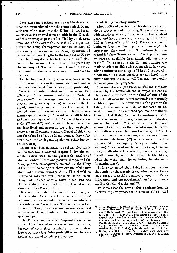
The nuclides are produced in nuclear reactionscaused by the bombardment of target substances .The reactions are listed in the third column of thetable. In all cases the target materials are naturallystable isotopes, whose abundance is also given in thetable; the increased abundance indicated in thenext column refers to enriched specimens obtainablefrom the Oak Ridge National Laboratories, U.S.A.The mechanism of. X-ray emission is indicatedunder the heading "Decay mode". In the nextcolumn the table gives the element whose character-istic K-lines are emitted, and the energy of Kal 7).In most cases other emissions, such as y-radiation,conversion electrons (e-) or electrons from thenucleus ({3-) accompany X-ray ermssron (lastcolumn). These need not be an interfering factor inmany applications. If necessary, the electrons maybe eliminated by metal foil or plastic film filters,while the y-rays may be minimized by electronicdiscrimination 3). .It is to be noted that Table I includes nuclides
that emit the characteristic radiations of the X-raytube target materials commonly used for X-raydiffraction and spectrochemical analysis, namelyCr, Fe, Co, Cu, Mo, Ag and W.In some cases the new nucleus resulting from an
electron capture process is in a metastable excited
G) J. M. Hollander J. Perlman and G. T. Seaborg, Table ofisotopes, Rev. mod. Phys. 25, 469-651, 1953. A. H. W.Atenand J. Halberstadt, The production of radioisotopes, Philips
'. tech. Rev. 16, 1-12,1954';55. This article also gives a briefexposition of a number of nuclear reactions and of chemicalmethods used in the separation of the isotopes. J. R.Bradford, Chart of the isotopes, publ, Harshaw Scientific.G. Friedländer and M. Perlman, Chart of the nuclides(revised by J. R. Stehn), publ, General Electric, U.S.A.
7) S. Fine and C.F. Hendee, X-ray critical-absorption andemission energies in keV, Nucleonics J.3, No. 3, March1955, pp. 36-37. '
..
1956/57; No. 8 NUCLEAR X-RAY SOURCES 231
Table I. Partial list of X-ray emitting radioactive nuclides, with their half-life, nuclear reactions by which they are produc-ed, decay mode, and emitted radiation. Under decay mode, EC stands for electron capture process, IT for internal conver-sion or transition and m for metastable nucleus; competing processes by which some nuclei can decay (p+ -emission) arealso indicated; in some cases decay involves successive processes.
Radioactive nuclide Target Radiation
Symbol Natural Increased X-rayand mass Half-life Reaction. abundance abundance Decay mode
Kal (keV)Other
number % 0'/0
Be7 53 d LP (d,2n )Be7 92.6 - EC Li 0.052 l'Cau 1.1 X 10⁵ Y Ca4O(n,y)Ca41 96.97 - EC K 3.31 -V49 1 Y Ti4S(d,n)V4U 73.45 99.2 EC Ti 4.51 -Cr51 27 d Cr5°(n,y)Cr51 4.31 88.3 EC V 4.95 l'
Mn54 300 d ~Cr53(d,n )Mn54 9.55 92.1 f EC Cr 5.41Fé6(d,a)Mn54 91.68 99.9 l'
Fé5 2.94 y Fé4(n,y)Fé5 . 5.84 93.3 EC Mn 5.90 -C057 270 d Fé6(d;n)Co57 91.68 99.9 P+---')-IT(Fe57m) Fe 6.40 1', «:C05S 72 d Fé7(d,n)Co5S 2.17 87.3 EC,p+ Fe 6.40 l'Ni59 7.5XI05y Ni5S(n,y)Ni5U 67.76 99.3 EC Co 6.93 -Zn65 250 d ~ Zn64(n,y)Zn65 48.89 93.4
~ EC,p+ Cu 8.05Cu65(d,2n)Zn65 30.9 98.2 l'
Ge7l 11.4 d Ge7O(n,y)Ge7l· 20.55 88.1 EC Ga 9.25 -As73 76 d Ge72(d,n)As73 27.37 89.2 EC Ge 9.89 1', e"Se75 127 d Se74(n,y)Se75 0.87 33.1 EC As 10.54. 1', «:yss 105 d SrBS(d,2n)Yss 82.56 99.7 EC,P+ Sr 14.16 l'Moo3 >2 y Mo02(n,y)MoU3 15.86 95.5 EC Nb 16.61 -Tcu5 60 d Mo04(d,n)Tc05m 9.12 79.1 EC Mo 17.48 l'
PdlO3 17 d ~ RhI03(d,2n)PdI03 100.00 -~ EC --+ IT(RhI03m) Rh 20.21 1', e:PdI02(n,y)PdI03 0.8 -
CdlO9 330 d ~ CdIOS(n,y)CdIOO 0.89 24.8~ EC ---')-IT(AgIOOm) Ag 22.16 1', «:AgIOO(d,2n)CdIOO 48.65 99.6
Sn113 112d Snl12(n,y)Sn113 0.95 72.5 EC In 24.21 l'
In114 49 d ~ In113(n,y )ln114m 4.23 65.4~ IT In 24.21 1', p-, e:Cd1l4( d,2n)ln114m 28.86 94.2
Bal33 9.5 y CsI33(d,2n)BaI33m 100.00 -- IT ---')-EC(Bal33) Cs 30.97 1', e"CSl37 33 y Fission product p- ---')-IT(Bal37m) Ba 32.19 1', e-
Sml45 410 d Sm144(n,y)Sml45 3.16 ~EC Pm 38.65
- 30y EC (Pml45) Nd 37.36 1', e-,
Gdl53 236 d ~Gd152(n,y)Gd153 0.2 - I EC Eu 41.53 1', «:Eu153(d,2n)Gd153 52.23 - ç
Dy159 140 d Tb159(d,2n )Dy15o 100.00 - EC Tb 44.47 -Yb160 33 d Tm160(d,2n)Yb160 100.00 - EC Tm 50.73 1', «:Tml70 127 d Tm169(n,y)Tm170 100.00 - p- ---')-IT(Yb17om) Yb 52.36 p-, y, «:Hf175 70 d Lu175(d,2n)Hfl75 97.40 - EC Lu 54.06 1', e:WISI 140 d TalSl(d,2n)WlSl 100.00 - EC Ta 57.52 1', «:Rels3 120 d TalSl(a,2n)RelS3 100.00 - EC W 59.31 y, e-Rels4 50 d ReI85(n,2n)Re184 37.07 85.4 EC W 59.31 y,e -OS185 97 d Re185(d,2n)Os185 37.07 85.4 EC Re 61.13 1', e-AulO5 185 d Pt194(d,n)Aul05 32.8 - EC Pt 66.82 y"e-Tl202 11.5 d Hg20l(d,n )TI202 13.22 71.8 EC Hg 70.82 y, «:Tl204 2.7 Y TI203(n,y)TI2o4 29.50 96.0 EC, p- Ug 70.82 p-Bi207 50 Y Pb206(d,n )Bi207 23.6 81.0 EC Pb 74.96 1', e-
state and decays to the normal state with internalconversion. Thus, two X-ray photons, character-istic of the same element (Z-l), may be succes-sively emitted. Cd109 and Pd103 are examples ofthese "double emitters".
Photon Hû density of nuclear X-ray sourcesT.heusefulness of a nuclear X-ray source generally
will depend on the photon flux density of the radia-tion obtained, i.e. the number of X-ray photonsemitted per sec and per cm2• Given a sample of

232 PHILIPS TECHNICAL .REVIEW . VOLUME 18
infinite thickness (see below) 'of a pure nuclide.the flux density is a characteristic of the material.For iron Fe55 e.g. it is 25 xl010 photons /sec cm''.
Some factors governing this characteristic may beindicated. The first factor is the specific activity,i.e. the numher of disintegrations per second'occurring in one gram of the pure nuclide. It ismeasured in curie/grain (c/g), one curie correspond-ing to 3.7 Xl010 disintegrationa/sec. For isotopicallyÎmreFe55, the specific activitywould be 2.2 Xl03 cJg.Other factors are: the branching ratio, i.e. the frac-tion of the total number of disintegrations due toanyone of the X-ray producing mechanismsdescribed; the internal conversion c~efficient men-tioned above, when internal conversion is the X-rayproducing mechanism; the fluorescence yield, i.e.the fraction of the created K (or L, ) vacanciesresulting in the emission of K (or L, ) photons;and the absorption of the X-ray producing elementfor the characteristic radiation emitted ("self-ahsorption" coefficient PK or ilL' ... ). Owing to thelatter factor an increase in thickness of a nuclidelayer does not provide a proportional increase inradiated flux density but a saturation is approachedso that a source of a certain minimum thickness,will have practically the same flux density as theinfinite source:. The preparatien of a nuclear Xvray source inusable from starts from a powdered, solid or dissolvedspecimen material, supplied by an atomic pile ora cyclotron 6). This material, in general, containsthe desired nuclide not in a pure form hut mixedwith other isotopes of the same element, whichcannot be separated by normal laboratory means.The useful specific activity, i.e. the activity pergram of the element, is usually greatly lowered bythese admixtures. Moreover, the other isotopes.may give rise to corrtamination of the X-rays byundesired radiations, In order to minimize theseadverse effects, the target material in the pileor cyclotron should contain the useful nuclide- from which the radionuclide in question isgenerated - in the highest possible concentration(target of increased ahundancé, cf. Tahle I).Obviously a high flux of the hombarding particlesis also favourable for attaining high specificactivities, since it will increase the theoreticallyattainable level 'of specific activity, at whichequilibrium exists between the formation of newnuclei and 'the disintegration of nuclei already form-ed. The latter advantage is particularly important;when the desired nuclide is contaminated hy radio-active isotopes having a longer half-life: in thatcase, 'it may he desirable to keep the actual specific
activity well below the theoretically attainable level(by reducing the irradiation period of the target) inorder not to get an X-ray source unduly contaminat- .ed hy unwanted radiations.The specific activities 'normally commercially
availahle are given in Table 11 for a number of
Table n. Specific activity of normally available specimens ofX-ray emitting nuclides, and X-ray photon flux density ofinfinite thickness samples.
Specific activity Photon flux densityNuclide (millicurie per (107 photons per
gram of element) sec.cm")
Cr51 400 2.85Fe55 1500 16.7Ni59 0.005 0.000087.Zn65 5000 122Ge7l 700 20.5Se75 100 3.25Sn1l3 1.6 0.283In1l4 720 129Tm170 10000 330Tl204 300 6.1
nuclides important as X-ray sources. Higher specificoctivities may be ohtained in many cases at highercost and longer delay in delivery. The last columnof the table gives the calculated Xvray photonflux densities of infinitely thick samples of thesespecimens.
For an appreciation of these values consider thecase of Fe55, on which most of the nuclear X-raywork in this laboratory was done. The Fe55-sourcesare usually used in a configuration shown in fig. 1.
O,5cm2
1-25cm 89929
Fig. 1. Usual configuration of nuclear X-ray source (left) andcounter tube under test (right). '
The X-rays are emitted from a source of 0.5 cm2 areaand enter the window of the detector (counter tuhe)to he checked or calibrated.' The window area inmost cases was 1 cmê, the dist~nce 2.5 cm. Underthese conditions, the counter window encompassesahout 1% of the radiation emitted hy the source in.all directions. The counting rate (at 100% countingefficiency of the detector) to be expected from a 0.5cm2 source of infinite thickness of Fe55 having a fluxdensity of16.7Xl07 photons/sec.cmê would therefore

1956/57, No. 8 NUCLEAR X-RAY SOURCES 233
be 13.4.x105 counts/sec. With a "self-absorption"coefficient # R:: 700 cm"? of the source, i.e. theabsorption coefficient of iron for the emittedMnKá radiation (2.1 A, 5.90 keV), it is calculatedthat even a source 0.03 mm (0.0012") thick wouldgive 90% of the maximum rate. Several of the Fe55sources prepared in this laboratory from a batch ofFe55 .of the above-mentioned specific -activityapproached a rate of 8 X10⁴ counts/sec, i.e. 10% ofthe maximum rate (the corresponding thickness of.the layer is calculated to be about 0.002 mm 'or0.0001"). This counting rate is amply sufficient forpractical purposes, since larger counting rates wouldin any case have to be attenuated in order not tochoke the counting systems at present available 3):For the calculation of the absorption effect use is made of
Dixon's calculations for large gamma-ray sources 8). Theconfiguration of fig: 1 approximately meets the conditions thatall detected photons traverse thesource in a direction perpen-dicular to its surface and that the source has equal thicknessin: all parts of the beam cross-section. Under these conditionsand with the assumptions of a homogeneous source, no scatterin the source and no absorption in the source holder window,the self-absorption in a source of thickness D is expressed bythe equation D):
I and Io are the flux densities with and without self-absorptionrespectively. Io evidently will be proportional to the thieknessof the source:
Io = GxDand hence
For D --+00, the flux density approaches the value G/ft,which in fact is the value listed in Table II. From the lastequation the flux density of a souree of any thickness can becalculated.An obvious question is whether the flux density of the Fe65
source or 'other nuclear X-ray sources would render thempossible substitutes for X-ray tubes in various applications,such as diffractometry and radiography. In diffractometry,however, the utilizdtion of the X-rays emitted by the source isenormously poorer than in the configuration of fig. 1, so thatmuch larger flux densities of the source are needed to getadequate counting rates in the diffraction lines. To illustratethis point, it should be mentioned that a normal diffraetionX-ray tube with a loading of ouly 0.1 mW would emit approx-imately the same total number of Ksphotons/sec (,__,107) asthe best practical 0.5 cm2 Fe66 souree described here 10). Since.the actual loading of diffraction tubes may amount to 1 kW
8) W. R. Dixon, Nucleonics 8, No. 4, April 1951, pp. 68-72.D) Cf. e.g. D. Taylor, The measurement of radioisotopes,
Methuen and Co., London, 1951.10) This calculation and the following ones are due to W. J.
Oosterkamp of the Philips Laboratory at Eindhoven.Cf. also:W. J. Oosterkamp, The radiography of the human'body with radioactive isotopes, Brit. J. Radiel. 26, 111,1953. .
and their effective source size is e.g, 0.015 cm'', the Fé6 sourceis evidently inferior by a factor of more than 108• In radiog-raphy, the advantage of the X-ray tube as a source is evenmore pronounced since the continuous spectrum which thetube emits and which is useless in diffraction analysis,' is fullyeffective photographically. In this case the Fe55 source wouldbe equivalent to an X-ray tube with a specificloading of onlyappr. 0.5 '!LW/mm2. This should be compared to the actualloading applied in normal radiography, which may be 4X10⁸times higher (200 W/mm2) for a stationary anode and even 1010times higher (8000 W/mm2) for a rotating anode (shortexposures). .Obviously, then, the Fe55 source even at its theoretical
maximum (i.e. with the radionuclide in a pure, undiluted state)cannot competewith theX-ray t:Ubein fluxdensity. Other radio-active nuclides may have higher theoretical flux density limits;however, because of the fundamental relationship between'specific activity Sand half-life T of a radioactive element,
S = 4;.16 X 1023AxT
(T being measured in se~ond; and A being the atomic weight),flux densities comparable to those of X-ray tubes will beattainable only with nuclides far too short-lived for practicalpurposes.
Preparatien of the sources.P]
A number of factors led us to select Fe55 as themain nuclide for our experimental X-ray sources.Fe55 was found suitable for the envisaged applica-tions not only because of the wavelength of theemitted MnKa radiation but also by virrue 01 its3 year half-life and its decay by pure electroncapture unaccompanied by processes generatingother radiations. It has furthermore been shown inthe above that the specific activity of the availableFe55 specimens permits the fabrication. of sourcesof sufficient X-ray intensity for work on counter.tubes.
The radioactive Fe55 is obtained from the suppliers. in the form of an FeCl3 solution; from which Fe55
may be laid down in some solid, homogeneous formby means of a number of chemical and physicaltechniques. Three methods were successfully used.(Similar techniques are applicable to most otherX-ray emitting nuclides.)1) The filter paper method. Several drops of theactive solution are transferred to a 1 cm2 piece offilter paper which has been positioned in a previouslyprepared holder. The assembly is then sealed With12 fL polyester film, which is 98% transparent to theMnKa radiation and which prevents the FeClásolution giving offH;CI-vapor. This simple procedureproduces a stable source, which, however, is rather,
. weak owing to the diluted state of the radioactivenuclide and the absorption of X-rays by Cl atoms
11) The authors gratefully acknowledge the assistance of~. B-.Brown ofthis laboratory in this phase of the work.
-,

. ,234 PHILlPS TECHNICAL REVIEW VOLUME 18
(mass absorption coefficient for 2 A appr, 300) andby the filter paper. L,
2) The ammonia method. From a drop ofthe activeFeCl3 solution placed on a small glass or metal plate,Fe(OH)a is precipitated by means of an ammoniaatmosphere. Heating the plate then converts theiron hydroxide into the non-volatile iron oxide.which strongly adheres to the base material. Nowindow is' necessary and the absorption is sub-stantially lower than in the previous case (the massabsorption coefficient ~f the oxygen atoms for 2 Ais only: about 30). Even higher intensities can beobtained when the .oxide is subsequently reduced inan H2-atmosphere and, for protection, the metallicFe fused dnto a 'platinum backing by heating.This method was developed at Oak Ridge NationalLaboratory 12).3) Electroplating. This method, whereby pure Fe55can be deposited in successive thin layers, resultsin maximum concentration of the radioactivematerial and in the highest photon flux densitiesattained. Conventional procedures for electro-plating iron proved not amenable to the micro-techniques involved in the preparation of radio-active sources. A procedure was worked out aftera suggestion by Platzer and Lewis 13), the main,problem being to avoid etching away of the deposit-ed iron by acid formed in the electrolysis of theFeCI3-solution. Fig. 2 shows the equipment used.A small amount of the active solution, to which no."carrier" substance is added, is pipetted onto a filterpaper placed on a graphite anode. A copper plateforming the cathode, on which the Fe is to beplated, is held on a movable fixture and is loweredinto position on the filter paper, while a -potentialof 3 V is applied to the electrodes. After the deposi-tion process, the whole structure is flooded witha spray of water to was haway the formed acid 14).The' process can be repeated with a fresh FeCl3portion, and in this way as,~any as ten successivelayers have been plated onto one backing piece,building up.a total thickness. which produces 10%of-the infinite-thickness flux density (see above).
Similar techniques have been evolved to depositZn65• This radioactive nuclide is particularly
. .12) R. E. Mcffenry (Oak Ridge National Laboratory),' unpuh-, lished report, Dec. 29, 1954.13) G. E. Platzer and C. R. Lewis (Chrysler Corp., Detroit,
Mich.), private communicatiou.'14) Of course, the radioactively contaminated washing water
must be collected and safely disposed of. We do not dwell onthese and similar protective measures that have to be taken .in order to safeguard the operators and surroundings in allprocedures for preparing nuclear X-ray sources. It may,however,be mentioned that suchmeasures are relatively fewand easy in the electroplating method, since intermediatemanipulation and processing of the material -isnot required.
+
89930
Fig. 2. Equipment for electroplating Fe65• K copper plate onwhich Fé6 is to be deposited, held by the movable :fixture M inglass beaker B. P :filter paper placed on graphite anode A andsaturated with radioactive FeCl3 solution. A 3V potential isapplied between K and A and the cathode K is then loweredonto the filter paper. After the plating process, wash water issprayed on the electrodes from a perforated ring W beforethe electrodes are separated.
important since it emits CuKa rays (À = 1.54 Á,energy 8.05 keV). From the tables it is seen that thehalf-life of this source is 250 days and that theinfinite-thickness flux density of the best specimensat present obtainable is 122 X107 photons/sec cm'',The design of the holder for the nuclear X-ray
source depends on the intended application, since itshould provide means for proper positioning in the
89931
Fig. 3. Holder for nuclear X-ray source designed for locationin the position of the scatter slit of the "Norelco" X-raygoniometer. To the left, part' of a protective cover for thesource when not in use.

1956/57, No. 8 NUCLEAR X-RAY SOURCES 235
equipment III which the source is to be used.General requirements are easy portability, adequateshielding during use and storage and a design suchas to prevent the dispersal of the active materialwithin or outside the holder. For many laboratoryapplications a long-handled holder with a remotecontrol shutter mechanism has proved very useful.For users of the "Norelco" equipment for X-raydiffractometry and X-ray spectrochemical analysis15) the holder shown in fig. 3 will be of foremostinterest. In this holder, the source is mounted on abrass strip that can be located in the position of thescatter slit of the "Norelco" goniometer, i.e. imme-diately in front of and at an accurately reproducibledistance from the X-ray detector (Geiger counter,proportional counter, scintillation counter), asshown infig. 4.
Fig. 4. Detector carrying arm of "Norelco" X-ray goniometershowing the detector to the right and the source of fog. 3 inposition in the scatter-slit holder (arrow).
Applications
Sources as shown in fig. 3 have been extensivelyused in our laboratories for checking the stabilityof the complete detecting system of the above-mentioned "Norelco" equipment. The nuclear sourceby virtue of its ideal stability is a very effectiveauxiliary for testing and singling out causes ofinstability in such equipment. Owing to the 3 yearhalf-life of Fe55, its radiation output shows afractional decrease of only 0.07% per day, whichmoreover can be easily corrected for, since thefractional disintegration rate IS constant and
15) Philips tech. Rev. 16, 123-133, 1954/55; 17, 269-286,1955/56.
accurately known. Output fluctuations due to thestatistical nature of the photon emission processesare low enough, with the high intensity sourcedescribed, to preclude difficulties in this application.
10
5
Fig. 5. Calibration of proportional counter. The photon energy(in keY) is plotted against the mean pulse amplitude (in volts),using four points corresponding to the radiation of the nuclidesFe55, ZuG5, Pd'03 and Ag,07.
The monochromatic emissions of nuclear X-raysources, such as Fé5 and Zn65, have made themvery useful for checking the proportionality betweenpulse amplitude and photon energy in the pro-portional and scintillation counter. At the sametime a calibration of the counter enabling theidentification of quanta from measured pulseamplitudes is obtained (fig. 5), and recordings canbe made of the pulse amplitude distribution curve(fig. tia, b) on which depends the discriminationability of the counter 3). The degree of linearity ofthe relation between counting rate and irradiationintensity, and the "plateau" characteristic of Geigerand proportional counters are other characteristicsof counters which can be easily tested with the aidof a nuclear X-ray source.All these tests, using nuclear sources, are perform-
ed during the actual manufacture of "Norelco"radiation detectors 3). Thus proportional countersare checked before being sealed off, and the totalgas pressure is adjusted so as to give a specified pulseamplitude at a given operating voltage on irradia-tion with a Fe55 source. The components of scintilla-tion detectors, i.e. the crystal and the photomulti-plier tube, are tested separatelyon the work bench.Finally, life-tests of groups of detectors at highcounting rates are effected by means of a numberof Fe55 sources, thus avoiding the tying-up of costlyX-ray tube equipment for long periods.The applications of nuclear X-ray sources so far
mentioned are concerned with the testing of detec-tors. Applications in other fields, especially thosein which monochromatic radiations are required,have been considered or tried out already. X-rays

236 PHILlPS TECHNICAL REVIEW VOLUME 18
Fig. 6. a) Pulse amplitude distribution of a proportional counter irradiated with Mn Kilrays from an Fe5s source.Il) The same for a scintillation counter.The curves were obtained with a pulse height analyzer channel width (,,1V) of l/lO
of the half-width of the peak (cf. ") ): the numbcr of pulses transmitted via the differentchannels were directly reeorded over the corresponding channel posi tion (pulse amplitude).
from radioactive nuclides have been used as wave-length standards!"); absorption measurements of theemission of two adjacent nuclides have been pro-posed for quantitative analysis of an element havingan absorption edgein the samewavelength region17);
absorption analysis of sulfur in hydrocarbons hasbeen accomplished with MnKa radiation from aFe55 source 18). Application of radionuclides such asTm170 (thulium) emitting photons of energy 50 and80 keV have been described, e.g. for inspection inindustry and for emergency medical radiography2) 19). Thickness gauging is another possible applica-tion in industry. Further work in the developmentof nuclear X-ray sources is warranted in order tobroaden the field of application of X-rays by simple,
16) Ph. Snelgrove, J. EI-Hussaini and J. W. M. DuMond, Phys.Rev.95, 1203-1204, 1954.
17) Suggested by H. A. Liebhafsky, in paper presented atPittsburgh Conf. on Anal. Chem. and App!. Spectroscopy,March 1955.
18) H. K. Hughes and J. W. Wilczewski, Anal. Chem. 26, 1889-1893 1954.
19) S. Untermyer et al., Nucleonics 12, No. 5, May 1954, pp.35-37; R. Halmshaw, Brit. J. appl. Phys. 6, 8-10, 1955.
inexpensive techniques for cases where the very highphoton flux densities of X -ray tubes are notrequired.
Summary. A number of ar tifici al radioactive nuclides emitphotons of energy less than 100 ke V, i.e. of wavelength longerthan 0.15 Á (relatively soft X-rays). Such nuclear X-rayemittersowing to their high stability, monochromatic emissionand low cost provide excellent means for testing X-ray countertubes (especially proportional and scintillation counters), bothduring mannfacture and in operat.ion. After a discussion of theunderlying mechan isms of the X-ray emissions, a survey ofX-ray emitting nuclides with their main characteristics is given,and some techniques for preparing practically usable sourcesfrom these nuclides are described. The theoretically attainablephoton flux density of Fe5S would provide a counting rate ofnearly lOG counts/sec in the envisaged application; the bestpractical Fe55-sources prepared in the Philips Laboratoriesattain about 100/0 of this value, which is amply sufficient.Fe55, ZnG5 and other X-ray sources made in tbese laboratorieshave for some time been used for testing the stability of thedetection system of "Norelco" X-ray goniometers, for checkingand calibrating the photon energy proportionality, the pulseamplitude distribution, the counting-rate linearity and theplateau characteristics of "Norelco " propor-tional and scintilla-tion counters, for life-testing these counters etc.
Nuclear X-ray sources cannot compete on equal terms withX-ray tubes, whose useful photon flux densities may be 108or in some cases 1010 times higher; nevertheless, they may beimportant for a broad and diverse field of applications in whichmonochromatic emissions are desired and high flux densitiesare not necessary.

1956/57, No. 8 237
ELECTRON MICROSCOPE PHOTOGRAPH OF FERROXDURE
Surface of a block of ferroxdure contauung a slight excess of BaO, sintered at 1200 oe, sawn through and then reheatedto a somewhat higher temperature. Photographs such as this can provide information on dilIusion and grain growth in theprocess of recrystallization.The photograph was made with a carbon replica (shadowed with palladium), i.e. a carbon skin evaporated on to a poly-
styrene impression prepared from the unpolished surface of the ferroxdure. The photograph was taken with an electronmicroscope EM 75 kV with an anastigmat lens 1). Magnification 23000 X .
1) Philips tech. Rev. 17, 47-59, 1955/56. The anastigmat developed for this microscope, which improves the resolving powerto about 25 A, will be described later in this Review.


![X-ray emission from very high energy gamma-ray sources [Horns]](https://img.pdfslide.us/doc/110x75/55986aa61a28ab2e0b8b468a/x-ray-emission-from-very-high-energy-gamma-ray-sources-horns.jpg)