Embed Size (px)
Citation preview

EUROPEAN COMMISSION
nuclear science and technology
Safety and Efficacy of Computed Tomography: A broad perspective
(CT Safety and Efficacy)
Authors: Jacob (Koos) Geleijns
Willi Kalender Wendy Krispijn K. Schneider P. Shrimpton
Contract FI6R-CT2004-002388
Final report
Project co-funded by the European Commission under the Euratom research and training programme on nuclear energy within the Sixth Framework Programme (2002-2006)
Area: Radiation protection
Directorate-General for Research 2008 Euratom EUR 23463

II
Project coordinator Jacob (Koos) Geleijns, Leiden University Medical Centre (NL) Project partners Georg Bongartz, University Hospital Basel (CH)
Alfonso Calzado Cantera, Complutense University (ES)
John Damilakis, University of Crete (EL)
Stephen Golding, University of Oxford (UK)
Anne Grethe Jurik, Århus University Hospital (DK)
Willi Kalender, Institut für Medizinische Physik (DE)
Karl Schneider, Dr. von Haunersches Kinderspital (DE)
Paul Shrimpton, Health Protection Agency (UK)
Maria Zankl, GSF (DE)

III
Table of contents
1. Introduction....................................................................................................................... 1
2. Major achievements.......................................................................................................... 3 2.1. Justification of multislice CT .................................................................................. 3 2.2. Optimisation of CT.................................................................................................. 7 2.3. Paediatric CT......................................................................................................... 10 2.4. CT dosimetry......................................................................................................... 13
3. Impact and potential applications ................................................................................... 17
4. The CT Safety and Efficacy consortium......................................................................... 19

IV

1
1. Introduction Radiologists are able to visualise any part of the human body in three dimensions with excep-tional quality by using the latest generation of computed tomography (CT) scanners. How-ever, the increasing use of CT scanners also causes growing concern about the associated exposure of patients to X-rays. Are all CT scans really necessary? Are up-to-date protocols used to keep the radiation exposure of patients to X-rays as low as possible? Are further improvements in CT scanner design and engineering possible? How can CT scans of children be improved? What is the radiation exposure from the latest CT scanners and modern acquisi-tion protocols? Such questions are being raised in the medical community but also in the popular press.
Figure 1: The impressive imaging performance and clinical value of computed tomography is obvious and leads to rapid growth of its clinical application. From left to right: examples of chest, cardiac and whole-body CT imaging. CT is associated with a relatively high patient dose and its frequency still grows considerably This EU research project aimed at answering all these questions. To achieve it, a scientific consortium consisting of well-known radiologists and physicists of 10 leading organisations was established (see Section 4, ‘The CT Safety and Efficacy consortium’). The individual know-how of the scientists covered many scientific sub-specialties, such as paediatric radiol-ogy, radiation measurements, and advanced computer simulations. The project was designed around four major scientific challenges in computed tomography: justification of CT scans, optimisation of CT technology, the proper application of CT scans of children, and assessment of patient exposure to X-rays during CT scans. The study on the justification of the application of CT was designed in a joint co-operation between the LUMC Radiology and Medical Decision-making Departments and five other European hospitals participated in the research. The research on optimisation of CT technol-ogy was performed by a group of researchers at the world’s most prominent research institute on CT, the Institute of Medical Physics (IMP) in Erlangen, Germany. Researchers at the paediatric hospital Dr. von Haunersches Kinderspital in Munich (DVKH), in cooperation with a European expert group, contributed to the optimisation of CT scans of children. This is particularly relevant since children are more susceptible to radiation effects than adults. A researcher who pioneered in patient dosimetry already 25 years ago at the former British National Radiation Protection Broad (NRPB, now Health Protection Agency) coordinated

2
research that led to the development of new techniques for patient dosimetry in the latest generation of CT scanners.

3
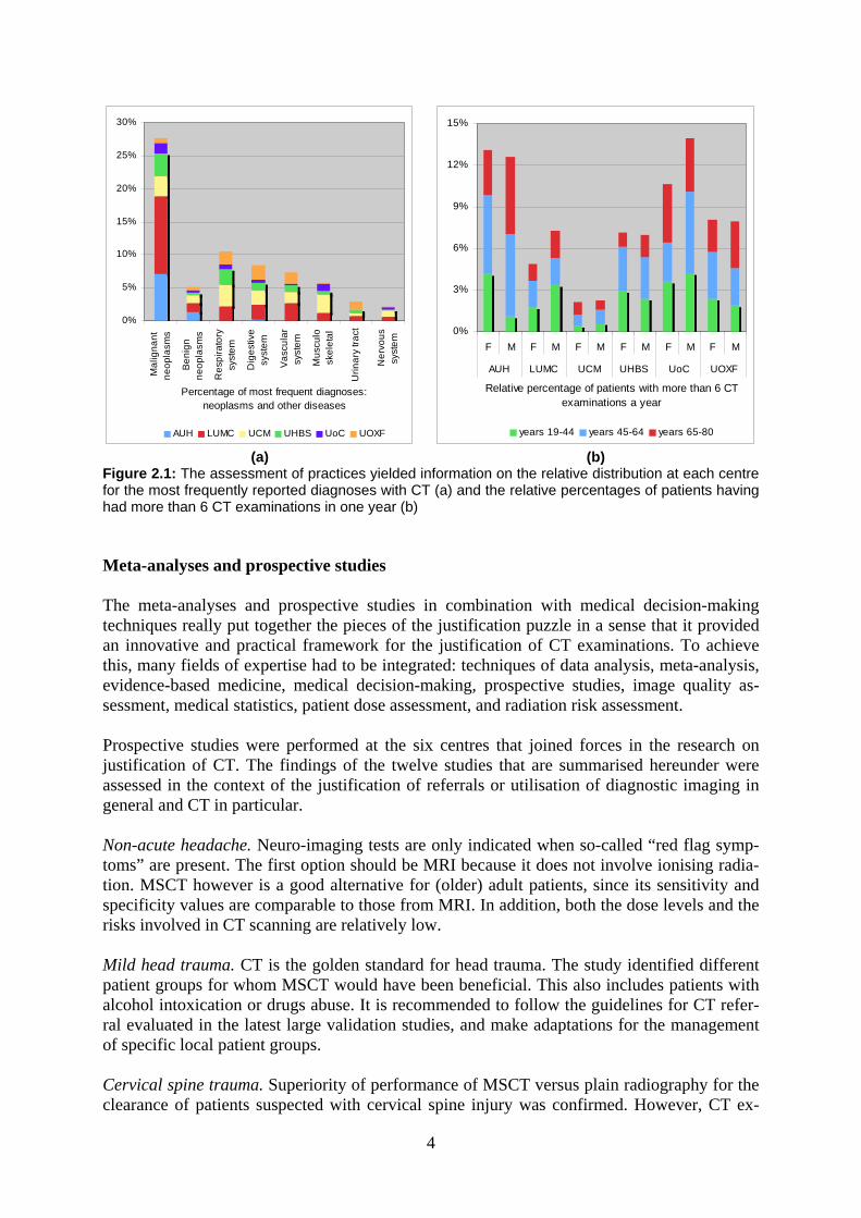
2. Major achievements 2.1. Justification of multislice CT The increasing use of computed tomography (CT) in diagnostic radiology and the associated radiation exposure of patients give rise to concern about the safety of this X-ray imaging technology. The number of CT scans conducted in the USA has grown from 18.3 million in 1993 to 62 million in 2006. CT now accounts for 45 % of the US population’s collective medical radiation dose, even though it makes up only 12 % of all medical radiation proce-dures done in the United States. The same process of growing frequency of CT examinations also takes place in Europe. Concerns about radiation exposure from CT not only exist within professional groups, like radiologists, referring physicians, and medical physicists. In 2007 the general public was alerted repeatedly about high radiation doses from CT by publications in the popular press. Newspapers appeared with headlines like “With Rise in Radiation Expo-sure, Experts Urge Caution on Tests” (The New York Times, 19 June 2007) and “Germany Worried about Excess Use of CT Scans” (Reuters, 12 July 2007). This stressed even more the high importance of better justification of CT referrals. The research on “Justification of CT” was divided into three consecutive phases: starting with a study on “Assessment of practices”, next “Meta-analyses” were performed, and finally specific clinical problems were addressed in the “Prospective studies”. The research thus started with a general overview of practices and gradually focused on specific cases of dis-eases or symptoms. The main objective was to prevent inappropriate and unnecessary application of medical imaging with ionising radiation, with a focus on state-of-the-art computed tomography. This objective was achieved by developing evidence-based guidelines for the utilisation of com-puted tomography. In the research it was essential to include also diagnostic information that is obtained before referral for medical imaging, such as medical history, application of diag-nostic scoring systems, and results from clinical tests (laboratory tests, organ function tests). Conclusions on proper referral for medical imaging also take into account all available diag-nostic imaging modalities (radiography, CT, nuclear medicine, MRI, and ultrasound). Assessment of practices An inventory was carried out concerning the use and yield of CT for different clinical prob-lems in relation to three anatomic regions: abdomen, thorax, and head and neck. Annual administrative data on patients undergoing CT studies at six participating centres were col-lected and analysed. In addition, specific reports of all CT examinations during one particular month were investigated, and the indications and findings were analysed. Figure 2.1 summa-rises some of the findings, knowingly the percentage distributions observed at each centre for the most frequent diagnoses with CT and the relative percentages of patients having more than 6 CT examinations in one year. Clearly, most CT examinations yield a diagnosis that is associated with serious diseases. The high number of patients receiving multiple CT examina-tions in one year raised concern about the appropriateness of the repeated CT examinations.
4
(a) (b) Figure 2.1: The assessment of practices yielded information on the relative distribution at each centre for the most frequently reported diagnoses with CT (a) and the relative percentages of patients having had more than 6 CT examinations in one year (b) Meta-analyses and prospective studies The meta-analyses and prospective studies in combination with medical decision-making techniques really put together the pieces of the justification puzzle in a sense that it provided an innovative and practical framework for the justification of CT examinations. To achieve this, many fields of expertise had to be integrated: techniques of data analysis, meta-analysis, evidence-based medicine, medical decision-making, prospective studies, image quality as-sessment, medical statistics, patient dose assessment, and radiation risk assessment. Prospective studies were performed at the six centres that joined forces in the research on justification of CT. The findings of the twelve studies that are summarised hereunder were assessed in the context of the justification of referrals or utilisation of diagnostic imaging in general and CT in particular. Non-acute headache. Neuro-imaging tests are only indicated when so-called “red flag symp-toms” are present. The first option should be MRI because it does not involve ionising radia-tion. MSCT however is a good alternative for (older) adult patients, since its sensitivity and specificity values are comparable to those from MRI. In addition, both the dose levels and the risks involved in CT scanning are relatively low. Mild head trauma. CT is the golden standard for head trauma. The study identified different patient groups for whom MSCT would have been beneficial. This also includes patients with alcohol intoxication or drugs abuse. It is recommended to follow the guidelines for CT refer-ral evaluated in the latest large validation studies, and make adaptations for the management of specific local patient groups. Cervical spine trauma. Superiority of performance of MSCT versus plain radiography for the clearance of patients suspected with cervical spine injury was confirmed. However, CT ex-
0%
3%
6%
9%
12%
15%
F M F M F M F M F M F M
AUH LUMC UCM UHBS UoC UOXF
Relative percentage of patients with more than 6 CT examinations a year
years 19-44 years 45-64 years 65-80
0%
5%
10%
15%
20%
25%
30%
Mal
igna
ntne
opla
sms
Ben
ign
neop
lasm
s
Res
pira
tory
syst
em
Dig
estiv
esy
stem
Vas
cula
rsy
stem
Mus
culo
skel
etal
Urin
ary
tract
Ner
vous
syst
emPercentage of most frequent diagnoses:
neoplasms and other diseases
AUH LUMC UCM UHBS UoC UOXF

5
aminations are associated with relative high dose to the patient compared to conventional radiography and therefore bare the potential of late radiation effects. But higher diagnostic accuracy obtained by the MSCT examination counterbalances the increase in dose compared to plain film evaluation, and renders CT utilisation dose effective and justified, since a missed Cervical Spine fracture may lead to significant deterioration of the patient’s quality of life (paralysis). Acute chest pain: coronary artery disease. Very good accuracy of 64-slice CT coronary an-giography for the detection of coronary artery stenoses was found. Irregular and fast heart rates remain a problem as this often renders an examination non-diagnostic. A model-based cost-effectiveness study showed that age has very little effect on the range of clinical prob-ability where application of CTCA is optimal. The current practice of the use of 64-slice CT coronary angiography in suspected unstable angina seems to be justified in patients with a low to intermediate probability after clinical evaluation and monitoring. Acute chest pain: pulmonary embolism. A tendency to abstain from any treatment in patients with isolated sub-segmental pulmonary embolism was observed. If the depiction of isolated sub-segmental pulmonary embolism hence proves to be without significant clinical impact the spatial resolution can be reduced during image reading by the radiologist (i.e. reading thicker slices and thick multiplanar reformats (MPRs)) with the inherent potential for dose reduction at the stage of image acquisition by modifying e.g. tube current and tube voltage. This pre-serves visualisation of the segmental pulmonary arteries and the majority of sub-segmental ones. CT remains the golden standard in imaging suspected pulmonary embolism. Testis cancer: retroperitoneal spread. CT is the recommended standard for detecting spread of testicular cancers. However, testicular seminoma primarily spread to the retroperitoneal glands and if chest X-ray is normal there is no need for chest CT. In these patients MRI could be advantageous giving the possibility of avoiding a series of follow-up CT examinations of the retroperitoneum. In non-seminomatous tumour there can be spread to other locations which demands CT examination. The use of MRI of the retro-peritoneum in these patients will therefore often be in combinations with CT of other regions which maybe inconvenient to the patient. Acute abdominal pain: appendicitis. In Europe, ultrasound is the preferred imaging modality for suspected appendicitis in particular and acute abdomen in general. In the USA, CT is the preferred imaging modality because of its high accuracy. This study found that almost all patients with acute lower abdominal pain had ultrasound. False positive cases where CT would have been beneficial have been identified. Ultrasound is the primary modality of imag-ing in acute abdominal pain. CT should be performed in clinically equivocal cases when there is a reasonable suspicion of other diseases, when the inflammatory markers are not elevated or when results of US are uncertain or the appendix is not visible. Specific indications for CT based on signs and laboratory results in appendicitis scoring, and indications for other acute abdomen diseases should be determined more precisely. Abdominal sepsis. Diagnostic accuracy for MSCT scans using 16 slices confirm that CT remains a suitable modality for imaging causes of abdominal sepsis. There is no distinct group of clinical indications that are suggestive of abdominal sepsis. The clinical role of CT has not changed with the development of new technology, using MSCT with 16 or more slices. Patients presenting for initial diagnosis should receive a full scan of the peritoneal cavity. Repeated use of MSCT during abdominal sepsis drainage can lead to rapid increases

6
in cumulative dose, particularly for obese patients. Patients who have been treated success-fully may not need a follow-up scan. A low dose and region specific scan can be used for removal of catheter and confirmation that the collection has resolved fully. If a patient is not responding or getting worse after catheterisation, a repeat full diagnostic scan is required to check catheter location and search for further sites of infection. Haematuria: urothelial cancer. Irrespectively of patient sex and age at exposure, the use of MSCT for the evaluation of haematuria is always associated with lower total risk of detri-ment, resulting from missed or false diagnosis and exposure to radiation, compared to other imaging modalities. Despite the increased radiation burden, the low false negative and false positive rates achieved with MSCT make MSCT urography the method of choice for adult patients older than 40 years screened positive for haematuria, when medical renal disease and bladder cancer are excluded. Although upper urinary tract transitional cell carcinoma is an aggressive disease, the post surgery (radical nephroureterectomy) survival rates are high. Therefore accurate diagnosis is essential even at the cost of increased radiation burden. In younger patients, where aggressive imaging investigation is justified, further dose reduction can be possibly achieved by single-phase examinations, split-bolus contrast administration and combinations of scanning phases. Acute flank pain: urolithiasis Diagnostic accuracy of CT for the detection of urolithiasis is reported higher than of intravenous urogram (IVU). Using a CT protocol with automatic tube current modulation for detection of urolithiasis is associated with a weight-independent ‘stan-dard’ dose of 4.5 mSv. IVU is associated with an effective dose of 2.6 mSv. Decreasing the radiation dose of the CT study down to 50 % of the standard dose still results in excellent sensitivities/specificities of 0.98/0.99. Therefore, a low-dose CT protocol in the work-up of suspected urolithiasis can be used as first-line imaging modality. A further decrease down to 25 % of standard dose protocols results in still acceptable sensitivity/specificity of 0.92/0.98 and can be recommended for follow-up studies. Detection of high-density structures such as concrements allows significant dose reduction without significant loss of sensitiv-ity/specificity. Automatic tube current modulation enables dose reduction even in obese pa-tients. Fixed tube current protocols should be avoided whenever possible. Radiologists should be encouraged to further evaluate the yield of lower radiation studies at increased noise level. Sterno-clavicular joint. CT is the modality of choice for imaging the sterno-clavicular joint region until there is evidence proving that the diagnostic value of MRI is comparable to that of CT. It may sometimes be necessary to perform supplementary MRI since it particularly well visualises the soft tissue. Based on the knowledge gained by patient examinations in our prospective study it seems that MRI is not able to visualise osseous structures as clearly as MSCT, but it is superior with regard to soft tissue inflammation and lesion of the intra-articular disc. CT will therefore probably still be the modality of choice in the imaging of the sterno-clavicular joint region unless the clinical findings predominantly give suspicion of soft tissue abnormalities. Low-back pain. Whenever an imaging test is warranted the first option should be MRI, due to its high diagnostic accuracy and because it does not involve ionising radiation. When MRI is not available or contraindicated, MSCT is a good alternative since it can reach a similar diag-nostic accuracy. In this case the examination must be fully justified by the presence of so-called “red-flag symptoms”, and the involved dose values should be optimised.

7
Conclusions Avoidance of excessive radiation was pursued in our project by scientific efforts to better justify the referral for CT examinations. A better scientific base for proper justification of CT scans was achieved by conducting research on the current practice of CT with the aim to yield an inventory of the use and clinical value of CT by applying medical decision-making tech-niques. This resulted in evidence-based optimal decision algorithms for a large range of appli-cations of CT. The work provides a scientific basis for the proper justification of CT scans, such that the expected benefits of the scan, including accurate and rapid diagnosis and, there-fore, proper treatment of the patient, outweigh the risks of exposure to X-rays. The accomplishments of the research group were realised by tackling the scientific challenges in a constructive and synergetic multidisciplinary cooperation. The research focused on twelve relevant clinical problems and it yielded clear and balanced answers on aspects of justification associated with these specific problems. More importantly, the research showed that justification of diagnostic medical imaging with X-rays can be achieved through scien-tific research that takes into account all aspects that play a role in the complex process of medical diagnosis. Our research results provide guidance on the appropriateness of medical imaging with X-rays taking into account the medical history of the patient, laboratory tests, tests of organ function, benefits of diagnosis from medical imaging with X-rays, assessment of the value of alternative techniques for medical imaging that do not use X-rays (MRI, ultra-sound), and patient safety (including the risk of missing a diagnosis, the risk of false positive findings, and the imaging procedure-related risks such as the short-term risk of catheterisation and the long-term risk of radiation-induced cancers). The achievements of the group are unique since its research for the first time provides a com-prehensive framework for justification. So far, research supporting the evidence-based appli-cation of diagnostic medical imaging with X-rays only focused on specific aspects such as patient dose or the risks of radiation exposure or the sensitivity and specificity of X-ray imag-ing. Such research does not provide the needed comprehensive evidence base for the justifica-tion of medical imaging with X-rays. Particularly the reports on radiation risks that are asso-ciated with medical X-ray imaging that appeared recently in the scientific journals are not well balanced since they do not take into account the associated benefits.
2.2. Optimisation of CT The objective of the research on optimisation of CT was to evaluate options for performing a CT scan for a given diagnostic question with minimal radiation dose while ensuring the re-quired image quality level. Both the minimisation of the radiation dose and the determination of the required image quality were topics of research. Determination of the required image quality The question ”Which exposure level is adequate?” is the first question asked when consider-ing noise levels. When answering this question, the spatial resolution has to be specified as well. Soft-tissue imaging is done adequately with smooth reconstruction filters which limit spatial resolution but also reduce noise. Bone, lung and high-resolution imaging in general make use of filters which enhance both structure detail and noise.
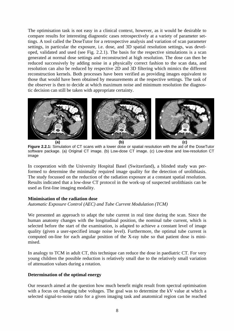
8
The optimisation task is not easy in a clinical context, however, as it would be desirable to compare results for interesting diagnostic cases retrospectively at a variety of parameter set-tings. A tool called the DoseTutor for a retrospective analysis and variation of scan parameter settings, in particular the exposure, i.e. dose, and 3D spatial resolution settings, was devel-oped, validated and used (see Fig. 2.2.1). The basis for the respective simulations is a scan generated at normal dose settings and reconstructed at high resolution. The dose can then be reduced successively by adding noise in a physically correct fashion to the scan data, and resolution can also be reduced by respective 2D and 3D filtering which mimics the different reconstruction kernels. Both processes have been verified as providing images equivalent to those that would have been obtained by measurements at the respective settings. The task of the observer is then to decide at which maximum noise and minimum resolution the diagnos-tic decision can still be taken with appropriate certainty.
(a) (b) (c)
Figure 2.2.1: Simulation of CT scans with a lower dose or spatial resolution with the aid of the DoseTutor software package. (a) Original CT image. (b) Low-dose CT image. (c) Low-dose and low-resolution CT image In cooperation with the University Hospital Basel (Switzerland), a blinded study was per-formed to determine the minimally required image quality for the detection of urolithiasis. The study focussed on the reduction of the radiation exposure at a constant spatial resolution. Results indicated that a low-dose CT protocol in the work-up of suspected urolithiasis can be used as first-line imaging modality. Minimisation of the radiation dose Automatic Exposure Control (AEC) and Tube Current Modulation (TCM) We presented an approach to adapt the tube current in real time during the scan. Since the human anatomy changes with the longitudinal position, the nominal tube current, which is selected before the start of the examination, is adapted to achieve a constant level of image quality (given a user-specified image noise level). Furthermore, the optimal tube current is computed on-line for each angular position of the X-ray tube so that patient dose is mini-mised. In analogy to TCM in adult CT, this technique can reduce the dose in paediatric CT. For very young children the possible reduction is relatively small due to the relatively small variation of attenuation values during a rotation. Determination of the optimal energy Our research aimed at the question how much benefit might result from spectral optimisation with a focus on changing tube voltages. The goal was to determine the kV value at which a selected signal-to-noise ratio for a given imaging task and anatomical region can be reached

9
with the lowest dose. The investigations were carried out both by simulations and by meas-urements using standard and semi-anthropomorphic phantoms. It was of particular importance and also novel that the investigation was carried out for various contrast materials and that complete 3D dose distributions were generated (see section on CT dosimetry). As contrast materials we chose a soft-tissue contrast resulting from a pure electron density difference, i.e. without any dependence on energy, a calcium hydroxyapatite contrast which represents bone mineral to mimic skeletal imaging, and an iodine contrast corresponding to a typical contrast medium examination such as CT angiography.
We here describe briefly the case for paediatric CT to indicate the high potential of using optimised X-ray spectra. Both phantoms and cadavers were examined at a range of voltages by simulation and measured on a CT scanner (Somatom Definition, Siemens Medical Solu-tions, Forchheim, Germany). Figure 2.2.2 shows the results for a paediatric thorax phantom which was examined with kV settings from 40 to 140 kV examining density difference, cal-cium and iodine contrasts. Going from 120 kV to 80 kV in this case would mean a dose re-duction by a factor of 2 or more with uncompromised signal-to-noise ratio for skeletal and contrast medium studies. For soft-tissue imaging there is no significant change associated with the low voltage. These results are new and have not been validated yet in extended clini-cal studies. Nevertheless, we consider them as valid due to the excellent agreement between simulations and measurements. They indicate that a wider range of kV settings should be used and be made available than is common practice today. Special applications Three special optimisation cases were studied in detail: cardiac CT, interventional CT and dose to the female breast. For the optimisation of cardiac CT, dose and image quality meas-urements were made with a cardiac motion phantom and for the optimisation of interventional CT the number of scans was minimized by using image-guided navigation. With respect to the female breast, two dose reduction techniques were evaluated: bismuth shielding and tube current modulation. The Tube Current Modulation (TCM) concept was adapted to include so-called partial scan techniques at which the X-ray tube has virtually no output in the anterior-posterior direction. The evaluation addressed both the dose to the patient (see Fig. 2.2.3) and the image quality. TCM appeared to be the superior concept.
(a) (b)
Figure 2.2.2: Determination of optimal voltage settings for paediatric thoracic CT. (a) For soft-tissue imaging, a range of 80 to 120 kV appears adequate. (b) For contrast medium and for skeletal studies, however, 80 kV or lower allows cutting the patient dose in half. Measurements and simulations are in good agreement

10
(a) (b) (c)
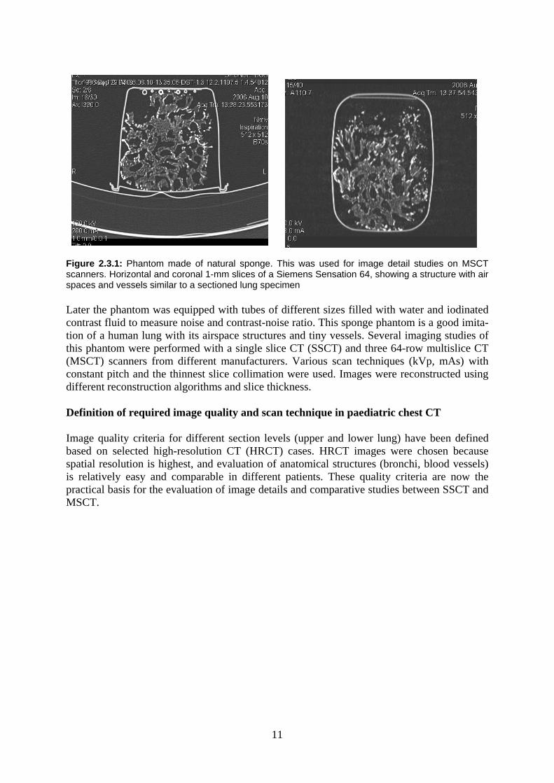
Figure 2.2.3: Dose reduction to the female breast in chest CT. (a) Simulated CT scan of an anthro-pomorphic chest phantom. (b) Dose distribution in case of bismuth shielding. (c) Dose distribution in case of partial scanning. In both cases the dose to the breasts is reduced. Image quality, however, is deteriorated significantly by bismuth shielding (not shown) Conclusions We here conclude that there are many means to reduce the dose per exam further on. This should be based on two measures: (1) enforcing the dissemination of new technologies into standard practice, e.g. encourage the use of AEC concepts; (2) continuing the optimisation efforts, e.g. the evaluation and recommendation of optimal X-ray spectra for the different diagnostic tasks. Support by the manufacturers for setting up respective scan protocols and the evaluation by experienced radiological practices are necessary and in progress. This can be done relatively easily for most settings as respective modes are available on most clinical scanners. Work with other settings like the lower voltages is not commonly possible at present but the results presented in this project will help to convince users to perform further investigation and manufacturers to work on the necessary implementation. 2.3. Paediatric CT Image quality and scan technique by phantom studies To investigate image quality and scan techniques in paediatric CT, phantoms were prepared from fresh human lung specimens inflated during CT scanning. However, even when con-tinuously inflated, the air volume of the lung specimens did not remain stable for more than 1 hour. Therefore, it was decided to develop a phantom made of a natural sponge, which was prepared using different concentrations of iodinated contrast medium solutions and was dried in the air. The first CT images of this phantom are shown in Figure 2.3.1.
11
Figure 2.3.1: Phantom made of natural sponge. This was used for image detail studies on MSCT scanners. Horizontal and coronal 1-mm slices of a Siemens Sensation 64, showing a structure with air spaces and vessels similar to a sectioned lung specimen Later the phantom was equipped with tubes of different sizes filled with water and iodinated contrast fluid to measure noise and contrast-noise ratio. This sponge phantom is a good imita-tion of a human lung with its airspace structures and tiny vessels. Several imaging studies of this phantom were performed with a single slice CT (SSCT) and three 64-row multislice CT (MSCT) scanners from different manufacturers. Various scan techniques (kVp, mAs) with constant pitch and the thinnest slice collimation were used. Images were reconstructed using different reconstruction algorithms and slice thickness. Definition of required image quality and scan technique in paediatric chest CT Image quality criteria for different section levels (upper and lower lung) have been defined based on selected high-resolution CT (HRCT) cases. HRCT images were chosen because spatial resolution is highest, and evaluation of anatomical structures (bronchi, blood vessels) is relatively easy and comparable in different patients. These quality criteria are now the practical basis for the evaluation of image details and comparative studies between SSCT and MSCT.

12
MSCT:SSCT (apical bronchi) MSCT:SSCT (basal bronchi)
MSCT:SSCT (apical vessels) MSCT:SSCT (basal vessels)
MSCT:better results
MSCT:equal results
MSCT:significantly better results
MSCT:equal results
p = 0,06 P = 0,67
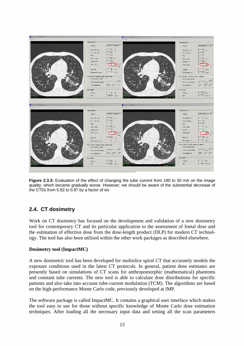
p = 0,001 p = 0,61 Figure 2.3.2: Comparison of anatomical structures of high-resolution CT of the lungs obtained with SSCT or MSCT. The upper lobe bronchi and vessels are significantly better visible in MSCT A study on the most appropriate window settings (window width and window level) was performed based on HRCT images of the lungs from patients with different lung diseases who were examined both with SSCT and MSCT within a period of 18 months. Selected images were sent to members of the CT Study Group of the ESPR with a deliberately false window width (WW) and window level (WL) preset. The task for each member of the group was to choose two different WW and WL values for each case. The goal was to find out the optimal visualisation of interstitial lung structures and peripheral airspaces, especially when a so-called mosaic pattern was present. The upper lobe bronchi and vessels were significantly better visible in MSCT (Figure 2.3.3). Patient dose and radiation risks in paediatric chest CT The biological effects of radiation were measured, i.e. chromosome aberrations, in a small group of paediatric patients who needed a CT. We were able to demonstrate a greater biologi-cal effect on patients who were younger than 10 years. This is the first time that direct radio-biological effects could be demonstrated in vivo after routine CT scanning in paediatrics. Raw scan data of a patient scanned with a 64-slice MSCT scanner was processed for use in combination with the DoseTutor (see Section 2.2). Evaluation of the effect of changing the tube current from 180 to 30 mA on image quality showed that image quality gradually be-came worse. However, there was a substantial decrease of the CTDIvol. from 5.82 to 0.97 by a factor of six.
13
Figure 2.3.3: Evaluation of the effect of changing the tube current from 180 to 30 mA on the image quality, which became gradually worse. However, we should be aware of the substantial decrease of the CTDIl from 5.82 to 0.97 by a factor of six
2.4. CT dosimetry Work on CT dosimetry has focused on the development and validation of a new dosimetry tool for contemporary CT and its particular application to the assessment of foetal dose and the estimation of effective dose from the dose-length product (DLP) for modern CT technol-ogy. The tool has also been utilised within the other work packages as described elsewhere. Dosimetry tool (ImpactMC) A new dosimetric tool has been developed for multislice spiral CT that accurately models the exposure conditions used in the latest CT protocols. In general, patient dose estimates are presently based on simulations of CT scans for anthropomorphic (mathematical) phantoms and constant tube currents. The new tool is able to calculate dose distributions for specific patients and also take into account tube-current modulation (TCM). The algorithms are based on the high-performance Monte Carlo code, previously developed at IMP. The software package is called ImpactMC. It contains a graphical user interface which makes the tool easy to use for those without specific knowledge of Monte Carlo dose estimation techniques. After loading all the necessary input data and setting all the scan parameters

14
required by the tool (see Figure 2.4.1.a), the resultant dose distribution is calculated for sub-sequent evaluation, for example, by determining relative dose values for individual voxels, or dose distributions in specified planes or regions of interest (see Figures 2.4.1.b and 2.4.1.c).
Validation of the tool was performed for a modern multislice CT scanner, including options such as TCM and automatic exposure control (AEC), by comparing simulated and measured results under a variety of conditions for homogeneous and heterogeneous phantoms of various forms and sizes. Quantitative CT Dose Index (CTDI) measurements were performed in cylin-drical CTDI phantoms and in anthropomorphic thorax phantoms of various sizes; dose pro-files were measured with thermo-luminescent dosimeters (TLDs) in the CTDI phantoms and compared with the computed dose profiles. The in-plane dose distributions were simulated and compared with TLD measurements in an Alderson-Rando phantom (see Fig. 2.4.2). The calculated dose values were generally within 10 % of measurements for all phantoms and all investigated conditions. In addition, benchmark calculations against two other Monte Carlo dosimetry tools demonstrated satisfactory agreement. Three-dimensional dose distributions can thus be calculated accurately with the ImpactMC tool for arbitrary scanners and protocols including schemes for TCM.
(a) (b) (c)
Figure 2.4.2: Validation measurements with the Alderson-Rando phantom. (a) Positions of the TLDs in an axial slice of the phantom. (b) Calculated dose distribution. (c) Comparison of calculated (red dots) and measured (green dots with error bars) dose profiles along the horizontal line in the phantom with 18 TLDs
Dose to the foetus The software was used to provide estimates of the dose to the foetus from CT scans on preg-nant patients. Mathematically described phantoms were implemented and used to model the pregnant woman at various stages (0, 3, 6 and 9 months) of the pregnancy. Calculations were performed for a range of typical scanning conditions using a total beam collimation of 19.2
(a) (b) (c)
Figure 2.4.1: The graphical user interface for ImpactMC. (a) Scanner- and scan-specific parameters can be entered. (b) The three-dimensional dose distribution is visualised interac-tively with colour maps. (c) Information on the (absolute) dose values in regions of interest is directly available to the user

15
mm. The dose to the foetus was calculated both for abdominal-pelvis CT scans, where the foetus is irradiated in the primary beam (and typical doses were between 51 and 67 mGy), and for shoulder-chest CT scans, where the foetus receives scattered radiation only (doses be-tween 0.1 and 0.7 mGy). The study confirms the need for particular care in selecting scan protocols (e.g. tube voltage, current-time product, and pitch value) for essential examinations involving the abdomen or pelvis of pregnant patients. Estimating effective dose from the dose-length product Effective dose estimates are generally based on anatomy-specific coefficients normalised to values of CTDI or DLP. These coefficients are usually generated for a constant tube current. In order to investigate the influence of TCM on these coefficients, a whole-body anthropo-morphic phantom (Alderson-Rando) was scanned with a multislice CT scanner (Somatom Definition, Siemens, Forchheim, Germany) utilising TCM (CareDose4D). Beam collimation was 19.2 mm and pitch 1. A voxelised patient model was used to define the tissues and organs in the phantom. Tube current values as a function of tube-angle were obtained from the raw data for each individual tube rotation of the scan. These values were used together with the dosimetry tool ImpactMC to calculate organ dose values both with and without account of TCM. Finally, effective dose conversion coefficients were determined for both cases accord-ing to the recently approved (2007) recommendations of the ICRP. TCM amplitude was greatest in the shoulder region. Consequently, dose distributions and organ dose values for particular cross-sections changed considerably when taking angular modulation into account (see Fig. 2.4.3).
x (mm)
z (m
m)
-200 -100 0 100 2000
100
200
300
400
500
600
x (mm)
z (m
m)
-200 -100 0 100 200
0
100
200
300
400
500
600
0.4
0.5
0.6
0.7
0.8
0.9
1
0.4
0.5
0.6
0.7
0.8
0.9
1
(a) (b)
Figure 2.4.3: Absorbed dose distributions. The absorbed dose in the phantom is averaged in the y-direction. (a) Distribution for longitudinal modulation only. (b) Distribution for combined longitudinal and angular modulation
For longitudinal modulation only, the effective dose conversion coefficients were up to 10 % higher for cross-sections in the shoulder region and 15 % higher in the pelvis compared with corresponding data also including angular modulation. In the head, neck, thorax and upper abdominal regions, conversion coefficients changed only by 5 % or less. We can conclude that for accurate (effective) dose estimates in individual cross-sections in the shoulder or pelvic regions, angular TCM should be taken into account. Conversion coeffi-

16
cients for estimating effective doses for scans of complete regions, e.g. chest or abdomen, are approximately 8 % lower when taking angular and longitudinal TCM into account. Conclusions This project has resulted in a validated, user-friendly CT dosimetry tool that can estimate three-dimensional dose distributions under a wide range of exposure conditions. It has pro-vided valuable estimates of dose in circumstances where these could not easily be measured. Knowledge about dose is essential in the optimisation of CT and the flexibility of the package has allowed dose estimates in the case of new scanning techniques, such as Tube Current Modulation (TCM). Undoubtedly, further scanning techniques will be introduced in the future. The outcome of our work, in the form of a flexible, easily accessible and modular dosimetry tool, will con-tinue to play a key role in estimating the effects of these new techniques on patient dose.

17
3. Impact and potential applications The result of the CT Safety and Efficacy project is an improved scientific basis for the appli-cation of modern CT scanners for medical diagnosis. This was required since the fast devel-opment of CT technology and the fast increase of the application of CT scans raised concern about the risks of the associated radiation exposure of patients. The ultimate impact of the project will be avoiding unnecessary CT examinations and improving the acquisition of nec-essary CT examinations. The project addressed both CT scans of adults and paediatric CT. New techniques for patient dosimetry had to be developed since established methodology was not capable to deal with the new generation of multislice scanners and tube current modula-tion. Are all CT scans really necessary? A better scientific framework for proper justification of CT scans was achieved. This resulted in evidence-based optimal decision algorithms for applications of CT. The work provides a scientific basis for proper justification of CT scans, so that the expected benefits of the scan, including accurate and rapid diagnosis and, therefore, proper treatment of the patient, out-weigh the risks of exposure to X-rays. For some clinical indications the researchers concluded that MRI and not CT should be the first option (e.g. non-acute headache and low-back pain). For other clinical indications the justification of CT was confirmed, and in some cases the need for even more CTs was expressed (appendicitis). Are up-to-date protocols used to keep the radiation exposure of patients as low as possible? For assessment of the interaction of radiation exposure and image quality a tool called the DoseTutor was developed and validated. It allows for retrospective analysis and variation of scan parameter settings, in particular the exposure, i.e. dose, and 3D spatial resolution set-tings. A blinded study was performed to determine the minimally required image quality for the detection of urolithiasis. Results indicated that a low-dose CT protocol in the work-up of suspected urolithiasis can be used as first-line imaging modality. Are further improvements in CT scanner design and engineering possible? Minimisation of radiation dose can be achieved by enforcing dissemination of new technolo-gies such as Automatic Exposure Control and Tube Current Modulation. There is a high potential for using optimised X-ray spectra. Work with lower voltages is not commonly pos-sible at present, but results presented will help convincing users to perform further investiga-tion and manufacturers to work on the necessary implementation. How can CT scans of children be improved? For a paediatric thorax phantom it was found that going from 120 kV to 80 kV would mean a dose reduction by a factor of 2 or more with uncompromised signal-to-noise ratio for skeletal and contrast medium studies. A wider range of kV settings should therefore be used and be made available than is common practice today.

18
What is the radiation exposure from the latest CT scanners and modern acquisition proto-cols? For Monte Carlo dose calculations, the ImpactMC software package (version 1.0, VAMP, Erlangen, Germany) was developed. ImpactMC runs a Monte Carlo (MC) simulation to ob-tain a three-dimensional dose distribution resulting from CT acquisitions. The algorithm is based on the high-performance Monte Carlo code, previously developed at IMP. The do-simetry tool allows for calculating patient exposures for any CT scanner, provided that infor-mation on X-ray spectra and design of the bow-tie filter are made available. Modern acquisi-tion techniques involving Tube Current Modulation (TCM) can be integrated in the dose calculations with ImPACT MC.

19
4. The CT Safety and Efficacy consortium The consortium consisted of a group of senior scientists that offered a well-balanced represen-tation of experienced radiologists, physicists and experts in radiation protection. The scientific partners in the consortium represented complementary expertises, skills, infrastructures and networks. The consortium represented seven European countries involving ten institutes for medical research, medical physics, and radiation protection: Leiden University Medical Centre, Leiden The Netherlands Jacob Geleijns Århus University Hospital, Århus Denmark Anne Grethe Jurik Complutense University, Madrid Spain Alfonso Calzado Cantera Dr. von Haunersches Kinderspital, Munich Germany Karl Schneider GSF, Neuherberg Germany Maria Zankl Institut für Medizinische Physik, Erlangen Germany Willi Kalender University Hospital Basel, Basel Switzerland Georg Bongartz HPA, Chilton United Kingdom Paul Shrimpton University of Crete, Iraklion, Crete Greece John Damilakis University of Oxford, Oxford United Kingdom Stephen Golding Justification of CT Cooperation has been established during the course of the project between Complutense University Madrid (UCM) and Príncipe de Asturias University Hospital (PdAUH) in Alcalá de Henares; principal contacts at this hospital are Eduardo Fraile Moreno and Isabel Salmerón Béliz (senior radiologists). At the LUMC, the department of Medical Decision-making con-tributed substantially to the project (Job Kievit and Jaap Sont). This created a multidiscipli-nary platform for the scientific research on justification of computed tomography. The contri-butions of 30 scientists from six centres (LUMC, AUH, UCM, UHBS, UoC, UOXF HJ) to the research on justification of CT was coordinated by Ying-Lie O. Optimisation of CT At IMP, the research on optimisation of CT was coordinated by Willi Kalender and was embedded in a dynamic research group with a long history in technical CT research. IMP has contributed in the past to major steps in the improvement of CT technology. The experience and expertise of this group was of great value for the research on optimisation. Paediatric CT At DVHK, Karl Schneider led the research on paediatric CT, with support from the European Expert Group (ESPR CT-Study Group). CT dosimetry Paul Shrimpton from the Radiation Protection Division of the HPA, who has a reputation in Europe in the field of CT dosimetry, coordinated the research on CT dosimetry. IMP acted as a partner in this research by providing access to and experience with sophisticated software tools. GSF has a long history in innovative computational dosimetry for medical exposures and provided support to the dosimetric research. Details of the project are available on the project website www.msct.eu.