Embed Size (px)
Citation preview
Histochemistry (1992) 98:155-164
Histochemistry © Springer-Verlag 1992
Nuclear receptor sites for vitamin D-soltriol in midbrain and hindbrain of Siberian hamster (Phodopus sungorus) assessed by autoradiography W.E. Stumpf 1, H.-J. Bidmon 1, L. Li i, C. Pilgrim 2, A. Bartke 3, A. Mayerhofer 2, and C. Heiss 2
1 Department of Cell Biology and Anatomy, University of North Carolina, Chapel Hill, NC 27599-7090, USA 2 Abteilung Anatomic und Zellbiologie, Universit/it Ulm, W-7900 Ulm, Federal Republic of Germany 3 Department of Physiology, Southern Illinois University, Carbondale, IL 62901-6512, USA
Accepted May 12, 1992
Summary. Autoradiograms were prepared from mid- brains and hindbrains of male and female Siberian ham- sters (Phodopus sungorus), kept under short-day or long- day illumination, after injection of tritium-labeled 1,25- dihydroxycholecalciferol (vitamin D, soltriol). Concen- tration and retention of radioactivity was noted in nuclei of certain neurons, glial cells, and ependymal cells, and in choroid epithelium. Labeled neurons of varying inten- sity were found throughout the brainstem in distinct populations at characteristic topographical sites, which include cranial nerve motor nuclei, the nucleus (n.) reti- cularis tegmenti pontis, the caudoventral region of the n. raphe dorsalis, the n. trapezoides, the n. vestibularis lateralis and n. vestibularis superior, neurons in the var- ious nuclei of the sensory trigeminus, accessory optic nuclei, scattered neurons in nuclei of the reticular forma- tion, the n. ambiguus, certain cells in the area postrema, and many others. Glial cells with nuclear labeling, prob- ably microglia, were scattered predominantly in or near myelinated nerve fascicles. The choroid epithelium showed strong nuclear labeling throughout the ventricle. Nuclear labeling of ependyma was variable and weak, mainly at ventral and lateral extensions (recesses) of the ventricle. The extensive presence of nuclear binding in select neural structures indicates that vitamin D exerts specific genomic effects on cell populations that are known to be involved in the regulation of motor, senso- ry, autonomic, neuroendocrine, metabolic, and immune functions. The results of these studies, in conjunction with those from other brain and peripheral tissues, re- cognize vitamin D-soltriol as a steroid hormone with a wide scope of hormone-specific target cells, similar to estrogen, androgen, and adrenal steroids, and which are topographically distinct and characteristic for its functions as the steroid hormone of sunlight.
Correspondence to: W.E. Stumpf
Introduction
Evidence for vitamin D-specific nuclear binding in brain and spinal cord (Stumpf et al. 1979, 1980) has been ob- tained in the same experiments that provided the first evidence for the existence of target cells in endocrine organs (pituitary, pancreatic B-cells, thyroid, adrenal, parathyroid chief cells, gonads, thymus), in heart, in skin, and in other tissues (for review see Stumpf 1988). These discoveries were based on the use of our autora- diographic technique (Stumpf 1976), which is highly sen- sitive and permits cellular and subcellular resolution of receptor sites in non-disrupted tissues within a main- tained topographical context. This extensive new infor- mation on vitamin D target sites and the related follow- up led to the recognition of a "vitamin D endocrine system" (Stumpf et al. 1979, /980, 1981 ; Norman et al. 1982; Bell 1985; Merke et al. 1986) and to the develop- ment of the concept of vitamin D-soltriol as a heliogenic seasonal activator and regulator. In contrast to the tradi- tional concept of vitamin D as "the calcium homeostatic steroid hormone", and a vitamin D endocrine system that subserves mainly calcium regulation, we propose that regulation of calcium levels is but one of the many effects of vitamin D. This skin-derived solar zeitgeber- steroid hormone serves primarily the need for biological systems to adapt to solstitial changes and the related maintenance and procreation of life (Stumpf 1988; Stumpf and Privette 1991).
Among the tissues with highest affinities to this helio- genic steroid are thyrotropes in the pituitary (Sar et al. 1980; Stumpf et al. 1987) and neurons in the stria ter- minalis brain circuit with the central amygdaloid nucleus and the lateral bed nucleus of the stria terminalis (Stumpf and O'Brien 1987) in all vertebrate phyla stud- ied (Stumpf and Bidmon 1990; Bidmon and Stumpf 1991). Motor neurons of cranial nerves and spinal cord, and sensory neurons in spinal ganglia (Stumpf and O'Brien 1987; Stumpf et al. 1988a), as well as epithelial cells of the choroid plexus (Bidmon et al. 1991) have also been noted as species-specific sites with high nuclear
156
b i n d i n g o f v i t a m i n D. T h e p r e s e n t e x p e r i m e n t s were a i m e d at i n v e s t i g a t i n g fu r t he r the cen t r a l n e r v o u s sys t em o f a species w i th h igh s ea sona l a d a p t a t i o n , i n o rde r to c o m p l e m e n t the d a t a for c e r t a i n f o r e b r a i n r eg ions ob- t a i n e d in the s a m e species ( M u s i o l et al. 1991, 1992a) a n d to c o m p a r e t h e m w i t h those o b t a i n e d for l a b o r a t o r y ra ts a n d mice.
Materials and methods
Fourteen 6-month-old Siberian hamsters (Phodopus sungorus), sev- en males and seven females, weighing 29-35 g were raised on a normal diet. Animals were kept under long-day conditions [light/ dark (L/D) 14:10 h, four males and three females] or transferred to short-day conditions for 2 months (LID 10:14 h, three males and four females). The short-day cycle causes testis regression and in both sexes weight loss and changes in hair color.
During daylight the animals were injected subcutaneously with 1,25-[26,27-3H]dihydroxycholecalciferol (3H-1,25D3, DuPont, Boston, Mass., USA, sp. act. 160 Ci/mmol), dissolved in 20% etha- nol-isotonic saline, two pulses each of 0.2 gg/100 g body wt. at an interval of i h. One female (long-day) and one male (short-day) were injected with a 1000-fold excess of unlabeled 1,25-D3 (Hoff- mann-LaRoche Nutley, NJ, USA), 50% at 30 min before and 50% simultaneously with the first tracer injection. The animals were decapitated 4 h after the first tracer injection. Mid- and hindbrains were mounted on tissue holders, frozen in liquified Freon 22 cooled by liquid nitrogen, and the freeze-mounted samples stored in liquid nitrogen.
Serial section autoradiograms were prepared after the thaw- mount technique (Stumpf 1976). Frozen sections 4 gm thick were cut in a cryostat (Frigocut 2800, Jung, Heidelberg, FRG) and under safe-light were thaw-mounted onto nuclear emulsion (Kodak NTB 3)-coated slides. The section-mounted slides were stored in light- proof desiccator boxes at - 1 5 ° C. Exposure times between 10 and 26 months were utilized in order to be able to detect target tissues with strong and weak nuclear concentrations of radiolabeled com- pound. At the end of the exposure, the slides were photographically processed, stained with methylgreen-pyronin, and coverslipped with Entellan (Hoechst).
The autoradiograms were evaluated qualitatively and quantita- tively by assessing microscopically the silver grain concentration over selected compartments and cell nuclei. A cell was considered labeled if the density of nuclear silver grains exceeded at least twice the extracellular silver grain density. The relative intensity of nucle- ar labeling was assessed as weak, intermediate, or strong, as de- picted in the schematic drawings in Fig. 1 A-K.
Controls against chemographic artefacts included development of autoradiograms immediately after thaw-mounting or short expo- sure for a few hours or days, at which time positive chemography would be apparent (Stumpf and Pilgrim 1992).
The topographic designation of labeled cells was aided by infor- mation from several rat and mouse atlases (K6nig and Klippel 1963; Wfinscher et al. 1965; Stumpf and Sar 1975a; Paxinos and Watson 1986). Frequently, the latin nomenclature was found more precise and convenient and has been preferred in most instances. The abbreviations utilize small letters for nuclei and capital letters for proper names and nerve fiber tracts; thus, the designation n for nucleus can be omitted and a fiber tract with identical name can be recognized (e.g., s t=bed nucleus of the stria terminalis, ST = stria terminalis).
Results
Resu l t s are dep ic t ed s c h e m a t i c a l l y in Fig. 1 A - M a n d as r e l a t ed a u t o r a d i o g r a m s in Figs. 2 -27 . Af t e r the in jec-
t i o n o f t r i t i u m - l a b e l e d 1,25D3 a n d e x p o s u r e t imes be- tween 11 a n d 26 m o n t h s , l ow levels o f r a d i o a c t i v i t y were f o u n d t h r o u g h o u t the t issue, w i th a c c u m u l a t i o n o f ra- d ioac t iv i t y in the l u m i n a o f b l o o d vessels a n d in nuc le i o f ce r t a in cells w i t h i n a n a t o m i c a l l y d i s t inc t r eg ions (to- p o g r a p h i c nuc le i ) o r d ispersed . N u c l e a r l abe l ing was re-
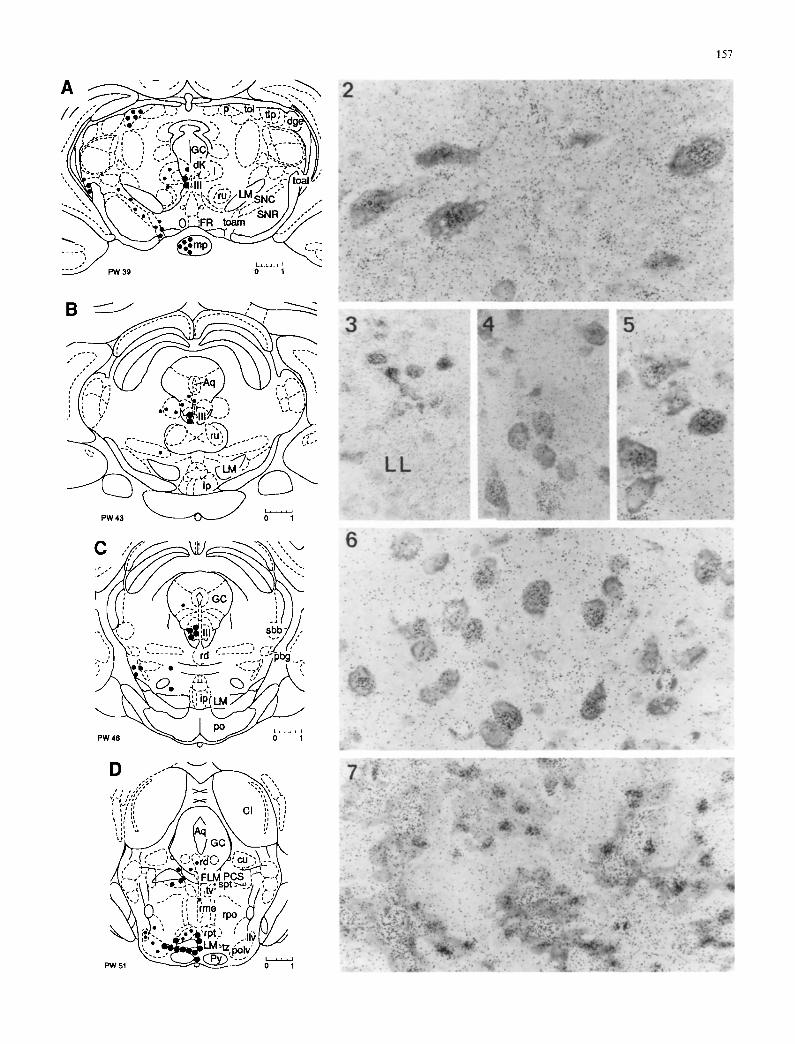
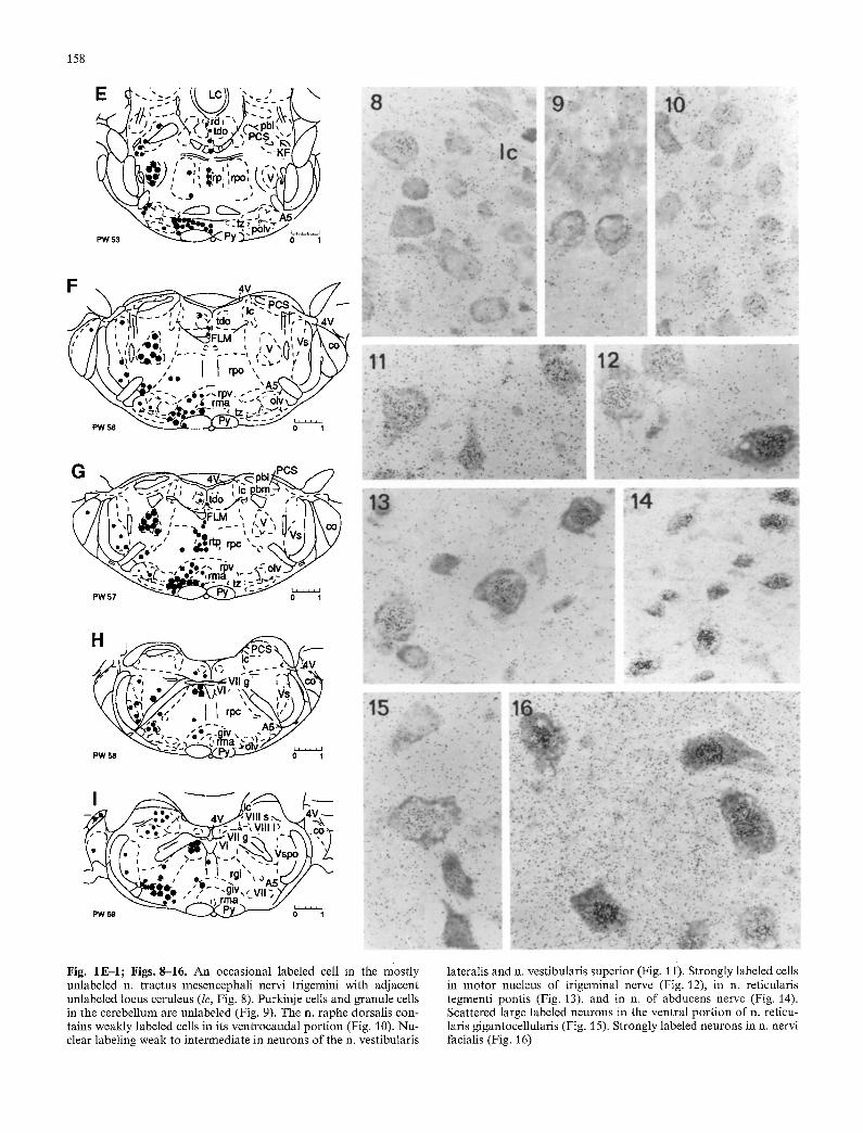
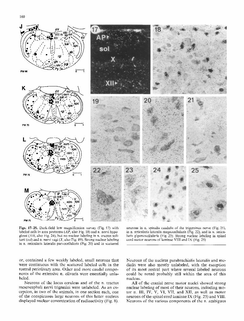
Fig. 1 A-M. Schematic representation of rodent mid- and hindbrain in rostrocaudal sequence, showing the topographical distribution of neurons with nuclear concentration and retention of radioactivi- ty after injection of 3H-1,25-dihydroxycholecalciferol (3H-I,25D3) in Siberian hamster (Phodopus sungorus). Transposed from thaw- mounted autoradiograms. Left side: Size and number o f dots indi- cate intensity of nuclear labeling and frequency of occurrence of target neurons respectively. Right side." Designations of structures. The schematic outlines were adjusted from atlases by Wfinscher et al. (1965), Stumpf and Sar (1975a, b), and Paxinos and Watson (1986). The frontal planes correspond with those of the atlas of Paxinos and Watson, indicated as PW number. Terminology and abbreviations are based on traditional Latin usage
Figs. 2-7. Autoradiograms (Figs. 2-27) selected from Siberian hamster brains frozen 4 h after injection of 3H 1,25D3. Varying concentrations of nuclear radioactivity exist in different target cell populations throughout the midbrain and hindbrain. Nucleus (n.) of the oculomotorius nerve (Fig. 2), n. paralemniscalis dorsomedial to the lateral lemniscus (LL, Fig. 3), n. subtrigeminalis (Fig. 4), ventrolateral cell group in n. principalis sensibilis of nervus trige- minus (Fig. 5), n. trapezoides (Fig. 6), choroid plexus of fourth ventricle with nuclear labeling of choroid epithelium and radioac- tivity in lumina of capillaries.(Fig. 7)
Abbreviations: (Lower case for nuclei; upper case for pathways and special structures; n = nucleus) A5 noradrenalin cell group; amb n. ambiguus; ap area postrema; Aq aqueduct; cc central canal; CI colliculus inferior; co n. cochlear- is; c u n . cuneiformis; cul n. cuneatus lateralis; cure n. cuneatus medialis; d n. Darkschewitsch; dgl n. dorsalis (corporis) geniculati lateralis; F L M fasciculus longitudinalis medialis; FR fasciculus re- troflexus; GC griseum centrate; gin. gigantocellularis; giv n. gigan- tocellularis ventralis; gr n. gracilis; i n. interstitialis (Cajal); ip n. interpeduncularis; KF n. K611iker-Fuse; lc locus ceruleus; LC lingula cerebelli; L M lemniscus medialis; llv n. lemnisci lateralis ventralis; mp n. mammillaris posterior; olv n. olivaris;p n. pretecta- lis; pbg n. parabigeminalis; pbl n. parabrachialis lateralis; pbm n. parabrachialis medialis; P C S pednnculus cerebelli superior; p o n . pontis; polv n. paraolivaris; prh n. prepositus hypoglossi; Py trac- tus corticospinalis; rd n. raphe dorsalis; rdm n. reticularis dorsalis medullae oblongatae; rgi n. reticularis gigantocellularis; rlm n. reti- cularis lateralis magnocellularis; rlp n. reticularis lateralis parvocel- lularis; rma n. raphe magnus; rme n. raphe medianus; rp n. raphe pontis; rpa n. reticularis parvocellularis; rpc n. reticularis pontis caudalis; rpo n. reticularis pontis oralis; rpt n. reticularis pontis tegementalis; rpv n. reticularis pontis ventralis; rtp n. reticularis tegmenti pontis; run . ruber; rvm n. reticularis ventralis medulae oblongatae; sbb n. subbrachialis; S N C substantia nigra, zona com- pacta; S N R substantia nigra, zona reticulata; sol n. tractus solitar- ii; spt n. subpeduncularis tegmenti; tdo n. tegmentalis dorsalis; tlp n. lateralis thalami, pars posterior; toal n. tractus optici acces- sorii lateralis; toam n. tractus optici accessorii medialis; tol n. trac- tus optici, pars lateralis; tv n. tegmentalis ventralis; tz n. trape- zoides; vt n. ventralis tegmenti; x n. x; I I I n. nervi oculomotorii; 4 V fourth ventricle; V n. motorius nervi trigemi; Vs n. sensibilis nervi trigemini; Vspc n. caudalis tractus spinalis nervi trigemi; Vspo n. oralis tractus spinalis nervi trigemini; VI n. nervi abducentis; VII n. nervi facialis; VIIg genu nervi facialis; VIIIrn n. vestibularis medialis; VIIIl n. vestibularis lateralis; VIIIs n. vestibularis superi- or; VIIIsp n. vestibularis spinalis; X n. dorsalis motorius nervi vagi
ZII #1
157
, , , 4 : :° ',;',
!,il, :! ,, , , , , , :
I P W 51 1
158
E d . . ,, .," i i LC~\ '..., , Y
t,( I tk.-,,'i),' .',f :~ ,,', ,~,:-,cM-"7/ ~(k\ \ k~ i ) / " i irPl ,,rpo;,t~j,(ll) j )7
"-.V'.-~ i ,~.._---- .---~-. " - ~ . A 5 / - , ~ .%. .~ , , . ~ - . = , ; - ~ PW~ ~ P Y ~ . . . .
47
l , , , l l
G - - , o, PCS %pc~
IO0 t - ,
( " / / [ L n , . i~ i X~_FLM , , . . , rl , I ~ \ t /1 I*',11 ~ '--. ._." . . . . ~.v ~il," 1 I \col \ / I I , ~ ~ / 'i,,,,_ ,. v - ~Vst I t / \ k ~ . ~ . ' , " • ~ , ' ~ " ' , , ~ 2,4 Y
" ~ . ~ - , l i t i ~ : " - " ~ =, tz; =%~>>"
H
_ ' : ~ "Vllg " C a
\* ~" ," I I rai"
\ x . L ~;e_ , , .u ,, v l ; ;/,
PW59 " : 0 1
Fig. 1E,-I; Figs. 8-16. An occasional labeled cell in the mostly unlabeled n. tractus mesencephali nervi trigemini with adjacent unlabeled locus ceruleus (lc, Fig. 8). Purkinje cells and granule cells in the cerebellum are unlabeled (Fig. 9). The n. raphe dorsalis con- tains weakly labeled cells in its ventrocaudal portion (Fig. 10). Nu- clear labeling weak to intermediate in neurons of the n. vestibularis
tateralis and n. vestibularis superior (Fig. 1 i). Strongly labeled cells in motor nucleus of trigeminal nerve (Fig. 12), in n. reticularis tegmenti pontis (Fig. 13), and in n. of abducens nerve (Fig. 14). Scattered large labeled neurons in the ventral portion of n. reticu- laris gigantocellularis (Fig. 15). Strongly labeled neurons in n. nervi facialis (Fig. 16)
159
cognized by a concentration of radioactivity, that is, of silver grains that underly the nuclear compartment. This concentration varied among labeled cells and was desig- nated as strong, intermediate, or weak, A cell was con- sidered labeled if the nuclear density of silver grains ex- ceeded at least twice the perinuclear cytoplasmic and neuropil density of silver grains.
Nuclear labeling was recognizable in strongly labeled cells after relatively short exposure times of 2 months, at which time weakly labeled cells were undetectable. At about 10 months of exposure, under the conditions of the experiment, most of the pattern of labeled cells was recognizable and nearly "comple te" .
Longer exposure times were utilized in order to assure a complete or near-complete topographical pattern of target cells. The longest exposure times were favorable for low-magnification surveys, although quantification would then be impaired because of non-linearity of im- age formation due to coincidence quenching. After long exposure times, the whole nuclear area may have been covered with silver grains, except for the region of the nucleolus which remained free. Also, variations in the distribution of radioactivity in different regions of the neuropil became apparent. For instance, the silver grain density was low in the neuropil surrounding the unla- beled neurons of the nucleus of the solitari tract and of the vagus nerve, but high in the adjacent ventral neu- ropil surrounding the strongly labeled neurons of the nucleus nervi hypoglossi (Fig. 17).
In competition studies with excess unlabeled hor- mone, the nuclear concentration of radioactivity was abolished. Evidence has been published concerning com- petition studies in identical animals (Bidmon et al. 1991a, b; Musiol et al. 1992a, b) and in other animals (Stumpf and O'Brien 1987). Among the most strongly labeled cells in the midbrain and hindbrain were motor neurons of cranial nerve nuclei (Figs. 2, 12, 14, 16) and cells of the choroid epithelium (Fig. 7). While most of the labeled cells appeared to be neurons of different sizes, as judged by size, presence of a conspicuous nucle- olus, or a relatively large pyronin-positive perikaryon, there were also small labeled cells with only a small rim of pale cytoplasm.
Topography of neurons with nuclear concentration of hormone
In most of the regions and nuclei described, the identifi- cation of labeled cells and their topographical attribu- tion appeared with a sufficient degree of certainty. There were, however, several regions in which the delineation and attribution of labeled cells was uncertain and fo- cused follow-up studies were indicated. Also, for several of the regions studied, available brain atlases provided unclear or conflicting information. In the present experi- ment, for instance, the boundaries were uncertain be- tween the n. raphe magnus and the n. reticularis pontis ventralis, or between the n. vestibularis lateralis and n. vestibularis superior. These and other uncertainties were in part due to the use of thin sections, the 100 gm spac-
ing of sections for the serial autoradiograms, variations in the plane of cutting and in the quality of sections, possible species differences in the topography gleaned from available rodent atlases, and unavailability of a specific atlas for the Siberian hamster brain. In general, a topographic correspondence of structures among rat, mouse, and Phodopus was noted.
In the midbrain, at the most rostral and transitional level to the forebrain, labeled neurons accumulated dor- sally in the region of the n. tractus optici pars lateralis. In the ventral griseum centrale, scattered labeled neurons were seen ventrolaterally, especially in the n. interstitialis Cajal and also in the Edinger-Westphal nucleus, and with particularly strong labeling in the n. of the nervus oculomotorius (Fig. 2). In the ventrolateral rostral mid- brain, a small group of labeled neurons was seen close to the surface dorsolateral to the substantia nigra. Whether or not this group is identical with the lateral nucleus of the accessory optic tract, as we suspect, re- mains to be studied further. In the pars compacta of the substantia nigra a few weakly labeled neurons could be identified. Ventrally, between the substantia nigra and the interpeduncular nucleus, a small group of neurons, probably belonging to the medial nucleus of the accesso- ry optic tract, showed weak nuclear labeling. At this level and further caudally, the n. ruber and the n. inter- peduncularis were unlabeled. A cut through the most caudal portion of the mamillary body revealed interme- diate intensity labeling of most of the neurons of the n. mammillaris posterior. Both the colliculus superior and inferior appeared unlabeled, except for a few scat- tered, labeled cells in the ventral and ventrolateral annu- lus.
At the level of the rostral pons, scattered labeled cells were present in the n. reticularis pontis. A small group of strongly labeled cells was seen ventrolaterally immedi- ately dorsolateral to the crus cerebri, ventral to the n. parabigeminalis, which we tentatively identified as n. paralemniscalis (Fig. 3). Cells of the n. pontis were unla- beled. Neurons in the rostral n. raphe dorsalis were es- sentially unlabeled; only in its more caudal pontine part were a variable number of mostly weakly labeled cells apparent (Fig. 10). In other raphe nuclei, which include the n. raphe medianus, pontis, pallidus, and obscurus, only an occasional labeled cell was present. In the region of the n. raphe magnus, labeled cells were more numer- ous and continuous with the labeled cells in the dorsal n. pontis ventralis and in the ventral n. trapezoides.
There were a few scattered labeled neurons in the griseum pontis, and one or two labeled neurons in the n. of Gudden, while the n. tegmenti ventralis remained free of labeled cells. Intermediate to strong nuclear label- ing was conspicuous in the near-midline n. reticularis tegmenti pontis (Fig. 13). Scattered large labeled neu- rons appeared in the n. reticularis pontis oralis and cau- dalis, while a more dense labeling was seen of the medi- um-sized neurons in the n. reticularis pontis ventralis, immediately dorsal to the strongly labeled, small- to me- dium-sized cells of the n. trapezoides (Fig. 6). Lateral to the relatively extensive n. trapezoides, a small group of neurons, probably belonging to the n. olivaris superi-
160
/ " / 4 x,: 4v II(. : ' - / 71 I l l .' / , I , I , , , . s ~ , l ~ .,,.., , , , ,
rgi r " , "(_.,//
~ ' ~ " " , , ' o ' , , g i v , ," ~ , /
- " : ~ 2 ~ py ~S->" . . . . . . PW~ 0 1
" ' ~ . ~ ~ V ~ r lm ,~ . / " ' - - . 2 . ~ t Z Py ~ . . . . . .
PW 70 o 1
\ , " ~ . % - ' . "J, ,~' i .~.. .~ - ; " r l n ' "~ : . " ~/t.~'~ .'-2"~---~2 °g' 'C , rim ;,2~'
PW 75
Figs. 17-25. Dark-field low magnification survey (Fig. 17) with labeled cells in area postrema (AP, also Fig. 18) and n. nervi hypo- glossi (XII, also Fig. 24), but no nuclear labeling in n. tractus soli- tarii (sol) and n. nervi vagi (X, also Fig. 19). Strong nuclear labeling in n. reticularis lateralis parvocellularis (Fig. 20) and in scattered
' ~ !~!i~/''¸ " '~!!!~i~!
neurons in n. spinalis caudalis of the trlgeminus nerve (Fig. 21), in n. reticularis lateralis magnocellularis (Fig. 22), and in n. reticu- laris gigantocellularis (Fig. 23). Strong nuclear labeling in spinal cord motor neurons of laminae VIII and IX (Fig. 25)
or, conta ined a few weakly labeled, small neurons that were con t inuous with the scattered labeled cells in the rostral periol ivary area. Other and more caudal compo- nents o f the extensive n. olivaris were essentially unla- beled.
Neurons o f the locus ceruleus and of the n. t ractus mesencephali nervi trigemini were unlabeled. As an ex- ception, in two o f the animals, in one section each, one o f the conspicuous large neurons o f this latter nucleus displayed nuclear concent ra t ion o f radioact ivi ty (Fig. 8).
Neurons o f the nucleus parabrachial is lateralis and me- dialis were also most ly unlabeled, with the exception o f its m o s t rostral par t where several labeled neurons could be noted p robab ly still within the area o f this nucleus.
All o f the cranial nerve m o t o r nuclei showed s t rong nuclear labeling o f mos t o f their neurons, including mo- tor n. I I I , IV, V, VI, VII , and XII , as well as m o t o r neurons o f the spinal cord laminae IX (Fig. 25) and VIII . Neurons o f the various componen t s o f the n. ambiguus
161
showed weak to intermediate nuclear labeling. The dor- sal n. vagus was unlabeled (Fig. 19) as was the n. of the solitary tract, except for a small cell in its lateral and caudal portion, which was very occasionally labeled. The sensory components of the n. trigeminus contained medium intensity-labeled, small- to medium-sized neu- rons especially in its ventral aspects, throughout its oral, principal, and caudal parts (Fig. 21). The representation of labeled neurons in this nucleus varied in its rostro- caudal extent, with the highest density in its caudal levels and in a ventral group of the n. principalis sensibilis of the trigeminus. At the levels of the n. cuneatus, a group of intermediate to strongly labeled cells was con- spicuous at the cranial extent of the n. spinalis caudalis V immediately ventral to the n. cuneatus lateralis, corre- sponding in part to n. x in the atlas of Paxinos and Watson (1986). In the n. vestibularis lateralis and superi- or, weakly labeled neurons were present in all of the animals studied. Neuronal groups of weak to intermedi- ate intensity of labeling existed in the region of the n. Koelliker-Fuse and of A5 ventral to the motor n. of the nervus trigeminus. Also, more caudally in the medul- la oblongata, labeled neurons were present in the A1 region of the n. reticularis lateralis, partes parvocellular- is, and magnocellularis (Figs. 20 and 22).
Within the area postrema (Figs. 17 and 18), a subpo- pulation of strongly labeled cells could be noted, which were mostly small in size and therefore probably mostly of glial origin; also a few somewhat larger labeled cells, possibly small neurons, could be seen. In the cervical spinal cord, in regions of laminae IX and VIII, strongly labeled, large neurons could be identified. In lamina X, ventrally and dorsally to the central canal, also in lamin- ae II and III, on occasion a single labeled small neuron was visible.
Ependyma, choroid plexus, pia mater
Ependyma that lines the aqueduct and fourth ventricle was mostly unlabeled. Occasionally ependymal cells dis- played weak nuclear labeling, especially at the floor of the fourth ventricle at the recess of the locus ceruleus and laterally to the medial eminence of the floor of the fourth ventricle at the level of the pontine sulcus median- us. The choroid epithelium was labeled throughout with a' strong nuclear concentration of radioactivity in more than 90% of all epithelial cells (Fig. 7). Cells of the stro- ma of the choroid plexus, which include fibroblasts and endothelial cells, as well as cells of the pia mater, ap- peared generally unlabeled.
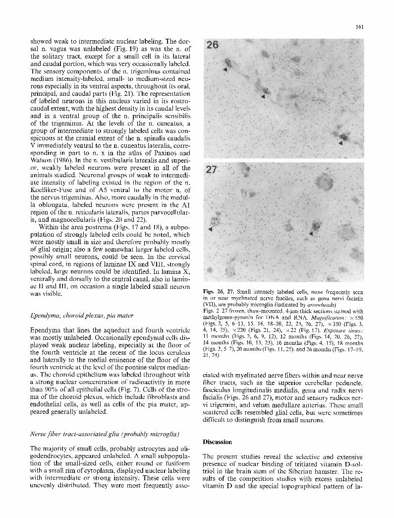
Figs. 26, 27. Small intensely labeled celIs, most frequently seen in or near myelinated nerve fasciles, such as genu nervi facialis (VII), are probably microglia (indicated by arrowheads) Figs. 2-27 frozen, thaw-mounted, 4-gm-thick sections stained with methylgreen-pyronin for DNA and RNA. Magnification: x 550 (Figs. 2, 5, 6-13, 15, 16, 18 20, 22, 23, 26, 27), x350 (Figs. 3, 4, 14, 25), x220 (Figs. 21, 24), x22 (Fig. 17). Exposure times: 11 months (Figs. 2, 6, 9, 12), 12 months (Figs. 14, 20, 26, 27), 14 months (Figs. 10, 13, 23), 16 months (Figs. 4, 15), 18 months (Figs. 3, 5-7), 20 months (Figs. 11, 25), and 26 months (Figs. 17-19, 21, 24)
ciated with myelinated nerve fibers within and near nerve fiber tracts, such as the superior cerebellar peduncle, fasciculus longitudinalis medialis, genu and radix nervi facialis (Figs. 26 and 27), motor and sensory radices ner- vi trigemini, and velum medullare anterius. These small scattered cells resembled glial cells, but were sometimes difficult to distinguish from small neurons.
Nerve fiber tract-associated glia (probably microglia)
The majority of small cells, probably astrocytes and oli- godendrocytes, appeared unlabeled. A small subpopula- tion of the small-sized cells, either round or fusiform with a small rim of cytoplasm, displayed nuclear labeling with intermediate or strong intensity. These cells were unevenly distributed. They were most frequently asso-
Discussion
The present studies reveal the selective and extensive presence of nuclear binding of tritiated vitamin D-sol- triol in the brain stem of the Siberian hamster. The re- sults of the competition studies with excess unlabeled vitamin D and the special topographical pattern of la-
162
beled neurons in the various brain stem levels indicate that the observed binding represents nuclear receptors specific for vitamin D. The pattern of distribution of the vitamin D target neurons with nuclear labeling is characteristic for that hormone, when compared to the patterns of target neurons in the midbrain and hindbrain reported previously for other hormonal ligands of the receptor family, which include estradiol, glucocorticoid, progestin, androgen, thyroid hormone, and retinoic acid (Stumpf and Grant 1975; Stumpf et al. 1991).
A near-comprehensive mapping of vitamin D nuclear - binding sites has been achieved in Phodopus under defined experimental conditions. This is due to the high resolution and high sensitivity of our thaw-mounting technique in order to record high affinity-binding and also low-affinity cells. The use of thin sections accounts for the cellular and subcellular resolution, and the long exposure times allowed storage and accumulation of weak signals derived from only a few labeled molecules, which would remain undetected with thick sections and with short exposure times. Whether or not the presence of low levels of endogenous unlabeled ligand decreases or increases the amount of nuclear receptor remains to be studied, since any change in the level of tissue ligands is likely to affect the expression and detection of the receptor. This study reveals different target cells and target cell groups with strong or intense binding and others with intermediate or low binding. This diversity of nuclear binding, also observed in the pituitary (Stumpf et al. 1987) and peripheral tissues with this and other steroid hormones, is probably of functional signifi- cance and deserves further investigation. With the infor- mation provided, further studies can now be conducted and need to focus on individual brain regions and nuclei for both anatomical and functional follow-up.
The present results indicate a qualitative similarity of the topographical pattern of vitamin D target neurons between male and female, and between short-day and long-day animals. Quantitative differences among these groups are possible or even likely. More extensive and detailed quantitative studies, which include monitoring of blood and tissue hormone levels, would be required for further clarification. The emphasis in the present ex- periments was on the identification of neural targets; gross differences between long- and short-day animals have not been detected. A higher amount of nuclear binding in long-day animals, compared to short-day ani- mals, was apparent, however, for choroid epithelium in the same animals (Bidmon et al. 1991).
The extent of vitamin D nuclear binding at all levels in rat, mouse, and hamster forebrain, midbrain, and hindbrain (Stumpf and O'Brien 1987; Musiol et al. 1991, 1992), suggests that vitamin D circulating in the blood has ready access to all sites within central nervous tissue and, nevertheless, exerts extensive effects on specific components of diverse neural systems. The precise iden- tification and characterization of the steroid hormone- specific neural systems, as in the present experiments, is a prerequisite for the study and understanding of the mechanisms of action of a hormonal messenger and for clarification to the question of hormone-specific func-
tions. Such considerations have been made with the in- formation from our previous studies, which provided the basis for the action concept for the three major groups of mammalian steroids as chief regulators and adaptors to the most vital tenets for survival: control of reproduction (sex steroids); securing of food, territo- ry, and partner (adrenal steroids); and seasonal adjuste- ment (solar-integumental steroid vitamin D-soltriol).
The nuclear target cell types for vitamin D include choroid epithelium, regions of ependymal cells, certain glial cells, and different types of neurons present in dis- tinct brain nuclei. The autoradiographic data indicate direct effects of vitamin D on neurons of all cranial nerve and spinal cord motor nuclei, which suggests neu- rotrophic effects on skeletal muscles. Muscle weakness associated with clinical conditions of vitamin D defi- ciency, therefore, may be due predominantly to altered neurotrophic input, rather than to a direct genomic ac- tion of vitamin D on muscle cells. In the experimental rodents studied by autoradiography, nuclear uptake and retention of 3H-I,25DHCC has not been observed in skeletal muscle nuclei under conditions when strong nu- clear labeling existed in motor neurons, intestinal ab- sorptive epithelium, pituitary thyrotropes, and other tar- get cells (Stumpf 1988). Evidence for nuclear receptor binding to cranial and spinal motor neurons has been furnished earlier in rat and mouse for vitamin D (Stumpf et al. 1988b) and for other steroid hormones, including dihydrotestosterone (Sar and Stumpf 1977) and cortico- sterone (Duncan and Stumpf 1984), but not for estradiol (Keefer et al. 1973). The presence of nuclear receptors for androgen, glucocorticoid, and soltriol in identical cells point to interactive mechanisms of steroid hormone effects that may be supportive and potentiating, or possi- bly antagonistic.
The present data indicate direct effects of soltriol not only on somatomotor neurons, but also on neurons of somatosensory systems. Accordingly, labeled cells in la- mina II of the spinal cord and in the various components of the sensory nuclei of the trigem!nus suggest modula- tory effects on touch and pain perception. Similarly, the strong nuclear binding in the trapezoid body suggests a modulation of acoustic input and processing, selective- ly of that component of acoustic relays, while no or little direct effects are indicated at the levels of the nuclei of the inferior colliculus and the geniculate body. Nucle- ar binding in accessory optic nuclei suggests modulatory effects of vitamin D on autonomic transduction of reti- nal light input.
Effects on autonomic centers in the medulla and pons are indicated by the presence of target neurons in the nucleus ambiguus and in various regions of the reticular formation. Nuclear labeling in the caudal nucleus raphe dorsalis suggests some regulatory influence on certain serotoninergic-peptidergic neurons. Labeling with vita- min D of neurons in regions of areas A5 and A1 suggests modulation of catecholamine manufacture and secretion in these cell populations. Since neurons in area A2 ap- pear to be free of nuclear vitamin D receptors, this cate- cholamine cell population is most likely not to be direct- ly addressed by vitamin D. The latter contrasts with
163
localized estradiol and co-localized catecholamine (Heri- tage et al. 1980) in the nucleus of the solitary tract. In the area postrema, a subpopulation of cells is strongly labeled with vitamin D, similar to estradiol and dihydro- testosterone. The functional significance of this circum- ventricular organ and the effects of vitamin D on this conspicuous region remain to be studied.
The general population of small glia cells, probably mostly astrocytes and oligodendrocytes, does not show nuclear labeling. However, a subpopulation of the small cells is strongly labeled. These rounded or fusiform, small, labeled cells are scattered mainly in or near re- gions of myelinated fasciles. Estimations of their size, their topographical distribution, and their negative staining with ED1 macrophage-specific antibodies (un- published data), the latter in agreement with similar find- ings published in the literature (Sminia et al. 1987), strongly suggest that these small labeled cells are micro- glia and that vitamin D is involved in modulating neu- roimmunological processes and metabolism of myelin (Lamper and Kies 1967; Polman et al. 1986). While the results of the present studies provide important anatomi- cal information and related functional clues, follow-up studies are required to clarify further the functions of the hormone in the identified target regions and also further to define the anatomical target sites, focusing on individual nuclei and regions.
In the Siberian hamster, compared to the laboratory rats and mice, a generally more extensive and, in some brain regions, anatomically different representation of vitamin D target cell populations is observed. Such dif- ferences have been reported for the choroid plexus (Bid- mort et al. 1991a) and for certain septal and thalamic nuclei (Musiol et al. 1991, 1992). Species-specific target cell distribution, that is, expression of receptors for vita- min D, may be linked to the species-specific habitat and related differing kinds and degrees of seasonal adapta- tion and response.
A comparison was made of our autoradiographic data with sites of expression of 28 kDa-calcium binding protein (Clark et al. 1986; Musiol et al. 1992); it has become increasingly apparent that there is limited corre- spondence between vitamin D nuclear binding and ex- pression of the cytoplasmic 28 kDa-calcium binding pro- tein. There is, however, correspondence between nuclear vitamin D-soltriol binding and other cellular products such as insulin in pancreatic island B-cells (Clark et al. 1980), thyroid-stimulating hormone in the pituitary and blood (Sar et al. 1980, 1981), gastrin in the pyloric stom- ach (Stumpf et al. 1988), ANF (atrial natriuretic factor) in the heart and blood (Bidmon et al. 1991 b), and meth- yl transferase and tyrosine hydroxylase mRNA expres- sion in adrenal medulla (Puchacz et al. 1991). Vitamin D has been reported to be a potent inducer of nerve growth factor synthesis in murine L-929 fibroblasts in vitro (Wion et al. 1991) which raises the possibility that vitamin D plays a role in the in vivo regulation of the nerve growth factor gene.
The results of our present experiments together with those from our previous studies with vitamin D in ro- dents and in representatives of other vertebrate phyla
(Stumpf and Bidmon 1990; Bidmon and Stumpf 1991), indicate a wide scope of effects of the hormone of sun- light and support our concept of vitamin D-soltriol as a seasonal regulator of vital functions.
References
Bell NH (1985) Vitamin-D-endocrine system. J Clin Invest 76:1 6 Bidmon H-J, Stumpf WE (1991) Phylogeny of receptors for 1,25-
dihydroxyvitamin D3 (1,25-D3) in vertebrates and inverte- brates. In: Norman A, Bouillon R, Thomasset M (eds) Vitamin D gene regulation, structure-function, analogies, and clinical application. Walter de Gruyter, Berlin, pp 673-674
Bidmon H-J, Bartke A, Mayerhofer A, Heiss C, Stumpf WE (1991 a) Vitamin D-soltriol receptors in the choroid plexus and ependyma: their species-specific presence. Mol Cell Neurosci 2:145 156
Bidmon H-J, Gutkowska J, Murakami R, Stumpf WE (1991b) Vitamin D receptors in the heart: effects on atrial natriuretic factor. Experientia 47:958-962
Clark SA, Stumpf WE, Sat M, DeLuca HF, Tanaka Y (1980) Target cells for 1,25 dihydroxyvitamin D3 in the pancreas. Cell Tissue Res 209 : 515-520
Clark SA, Stumpf WE, Bishop CW, DeLuca HF, Park DH, Joh TH (1986) The adrenal: a new target organ of the calciotropic hormone 1,25-dihydroxyvitamin D3. Cell Tissue Res 243:299- 302
Duncan GE, Stumpf WE (1984) Target neurons for aH-corticoste- rone in the rat spinal cord. Brain Res 307: 321-326
Heritage AS, Stumpf WE, Sar M, Grant LD (1980) Brainstem catecholamine neurons are target sites for sex steroid hormones. Science 207:1377-1379
Keefer DA, Stumpf WE, Sat M (1973) Topographical localization of estrogen-concentrating cells in the rat spinal cord following 3H-estradiol administration. Proc Soc Expt Biol Med 143:414- 417
K6nig JFR, Klippel RA (1963) The rat brain. Williams and Wil- kins, Baltimore
Lamper PW, Kies MW (1967) Mechanism of demyelination in allergic encephalomyelitis of guinea pigs. An electron micro- scopic study. Exp Neurol 18:210-223
Merke J, Ritz E, Schettler G (1986) Neuere Gesichtspunkte zur Rolle von Vitamin D. Dtsch Med Wochenschr 111 : 345-349
Musiol IM, Stumpf WE, Bidmon H, Pilgrim Ch (1991) [3H]-l,25- dihydroxyvitamin D3 (soltrioi) binding in the midline and intra- laminar thalamus of the Siberian hamster. Abstr Soc Neurosci 17:418
Mnsiol IM, Perez-Delgado MM, Bidmon H-J, Bartke A, Stumpf WE (1992a) Comparison of the location of vitamin D receptor sites and calbindin 28 kD-immunoreactivity in basal forebrain regions of the Siberian hamster. Abstr Soc Neurosci (in press)
Musiol IM, Stumpf WE, Bidmon H-J, Heiss C, Mayerhofer A, Bartke A (1992b) Vitamin D nuclear binding to neurons of the septal, substriatal and amygdaloid area in the Siberian ham- ster (Phodopus sungurus) brain. Neuroscience (in press)
Norman AW, Roth J, Orci L (1982) The vitamin D endocrine system: steroid metabolism, hormone receptors, and biological response (calcium binding protein). Endocrine Rev 3:331-366
Paxinos G, Watson C (1986) The rat brain, 2nd edn. Academic Press, San Diego
Polman CH, Dijkstra CD, Sminia T, Koetsier JC (1986) Immuno- logical analysis of macrophages in the central nervous system of Lewis rats with acute experimental allergic encephalomyeli- tis. J Neuroimmunol 11:215 222
Puchacz E, Goc A, Stumpf WE, Bidmon H-J, Stachowiak EK, Stachowiak MK (1991) Vitamin D regulates expression of tyro- sine hydroxylase gene in adrenal chromaffin cells. Abstr Soc Neurosci 17: 981
164
Sar M, Stumpf WE (1975) Distribution of androgen-concentrating neurons in rat brain. In: Stumpf WE, Grant LD (eds) Anatomi- cal neuroendocrinology. Karger, Basel, pp 120 133
Sar M, Stumpf WE (1977) Androgen concentration in motor neu- rons of cranial nerves and spinal cord. Science 196:319-321
Sar M, Stumpf WE, DeLuca HF (1980) Thyrotropes in the pitui- tary are target cells for 1,25(OH)2 vitamin D3. Cell Tissue Res 209:161 166
Sar M, Miller WL, StumpfWE (1981) Effects of 1,25(OH)2 vitamin D3 on thyrotropin secretion in vitamin D deficient male rats. Physiologist 24: 70
Sminia T, DeGroot CJA, Dijkstra CD, Koetsier JC, Polman CH (1987) Macrophages in the central nervous system of the rat. Immunobiology 174: 43-50
Stumpf WE (1976) Techniques for the autoradiography of diffusi- ble compounds. Methods Cell Biol 13:171-193
Stumpf WE (1988) Vitamin D-soltriol. The heliogenic steroid hor- mone: Somatotrophic activator and modulator. Discoveries from histochemical studies lead to new concepts. Histochem- istry 89:209 219
Stumpf WE, Bidmon H-J (1990) Lokalisation von 1,25-(OH)2-vit- amin D3 Rezeptoren und ihre organspezifische Verteilung in niederen Vertebraten (Pisces, Amphibia, Reptilia). Verh Dtsch Zool Ges 83 : 591-592
Stumpf WE, Grant LD (eds) (1975) Anatomical neuroendocrino- logy. Karger, Basel
Stumpf WE, O'Bricn LP (1987) 1,25(OH)1 Vitamin D3 sites of action in the brain. Histochemistry 87 : 393-406
Stumpf WE, Pilgrim Ch (1992) Artifacts in autoradiography. In: Stumpf WE, Solomon HF (eds) Autoradiography and correla- tive imaging. Academic Press, San Diego, in press
Stumpf WE, Privette TH (1991) The steroid hormone of sunlight soltriol (vitamin D) as a seasonal regulator of biological activi- ties and photoperiodic rhythms. J Steroid Biochem Mol Biol 39:283-289
Stumpf WE, Sar M (1975a) Hormone-architecture of the mouse brain with 3H-estradiol. In: Stumpf WE, Grant LD (eds) Ana- tomical neuroendocrinology. Karger, Basel, pp 82-103
Stumpf WE, Sar M (1975 b) Anatomical distribution of corticoste- rone concentrating neurons in rat brain. In: Stumpf WE, Grant LD (eds) Anatomical neuroendocrinology. Karger, Basel, pp 83-103
StumpfWE, Sar M, Reid FA, Tanaka Y, DeLuca HF (1979) Target cells for 1,25-dihydroxyvitamin D3 in intestinal tract, stomach, kidney, skin, pituitary and parathyroid. Science 206 : 1188-1190
Stumpf WE, Sar M, Clark SA, Lieth E, DeLuca HF (1980) Target neurons for 1,25 (OH)2 vitamin D3 in brain and spinal cord. Neuroendocrinol Lett 2:297 301
Stumpf WE, Sar M, DeLuca HF (1981) Sites of action of 1,25(OH)2 vitamin D3 identified by thaw-mount autoradiogra- phy. In: Cohn DV, Talmage RV, Matthew JL Jr (eds) Hormon- al control of calcium metabolism. Exerpta Medica, Amsterdam, pp 22~229
Stumpf WE, Sar M, Clark SA, DeLuca HF (1982) Brain target sites for 1,25-dihydroxyvitamin D3. Science 215:309-312
Stumpf WE, Sar M, O'Brien LP (1987) Vitamin D sites of action in the pituitary studied by combined autoradiography and im- munohistochemistry. Histochemistry 88:11 16
Stumpf WE, Clark SA, O'Brien LP, Reid FA (1988a) 1,25(OH)2 vitamin D3 sites of action in spinal cord and sensory ganglion. Anat Embryol 177:307-310
Stumpf WE, Sar M, O'Brien LP, Morin J (1988 b) Pyloric gastrin- producing cells and pyloric sphincter muscle cells are nuclear targets for 3H 1,25(OH)2 vitamin D3 studied by autoradiogra- phy and immunohistochemistry. Histochemistry 89:447-450
Stumpf WE, Bidmon H-J, Murakami R (1991) Retinoic acid bind- ing sites in adult brain, pituitary, and retina. Naturwissenschaf- ten 78:561-562
Wion D, MacGrogan D, Neveu I, Jehan F, Houlgatte R, Brachet P (1991) 1,25-Dihydroxyvitamin D3 is a potent inducer of nerve growth factor synthesis. J Neurosci Res 28:110-114
Wood GW, Gollahon KA, Tilzer SA, Vats T, Morantz RA (1979) The failure of microglia in normal brain to exhibit mononuclear phagocyte markers. J Neuropathol Exp Neurol 38:369-376
W/inscher W, Schober W, Werner L (1965) Architektonischer Atlas vom Hirnstamm der Ratte. Hirzel Verlag, Leipzig


![Vitamin D in PdPregnancy and Infancy · 1,25-dihydroxy vitamin D = calcitriol [1,25(OH)2D] assessment (hormonal form) ↓ Calcitriol binds to vitamin D receptor (VDR) to regulate](https://img.pdfslide.us/doc/110x75/5ffb3ce26517d830b10f5d09/vitamin-d-in-pdpregnancy-and-125-dihydroxy-vitamin-d-calcitriol-125oh2d.jpg)