Embed Size (px)
Citation preview
Nuclear Medicine in the Evaluation
of Trauma
Helena Balon, MDHelena Balon, MD
Wm. Beaumont HospitalWm. Beaumont Hospital
Royal Oak, MI, USARoyal Oak, MI, USA
Charles UniversityCharles University
3rd 3rd SchoolSchoolofof MedicineMedicine
DeptDeptNuclNucl Med, Med, PraguePrague
MaterialsMaterials for for medicalmedical studentsstudents
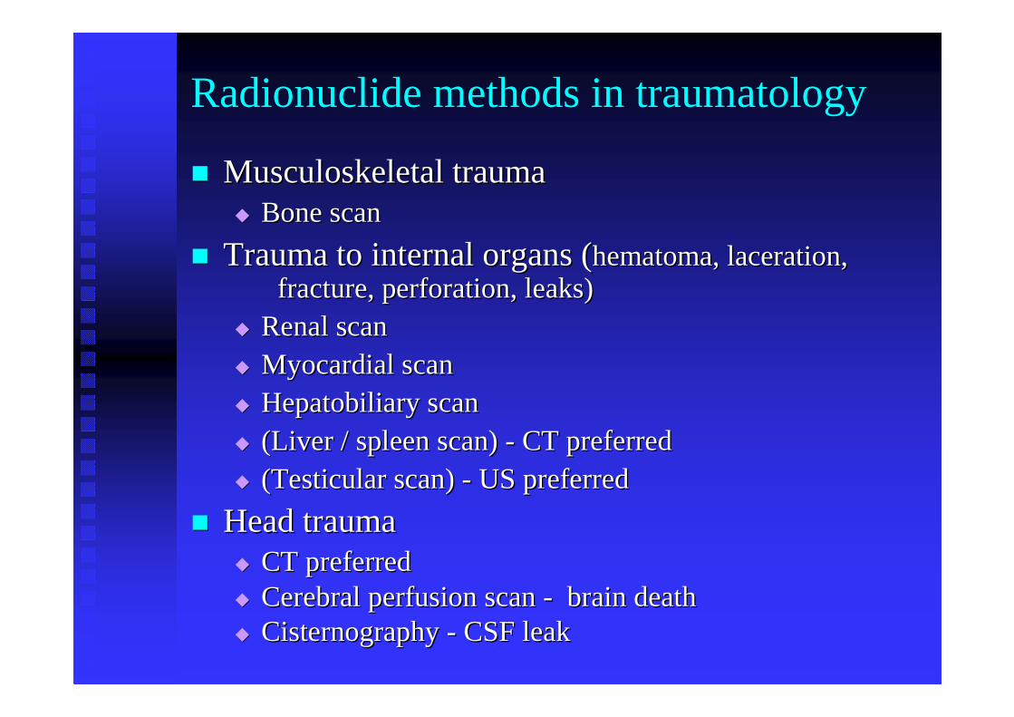
Radionuclide methods in traumatology
�� Musculoskeletal trauma Musculoskeletal trauma �� Bone scanBone scan
�� Trauma to internal organs (Trauma to internal organs (hematomahematoma, laceration, , laceration, fracture, perforation, leaks)fracture, perforation, leaks)
�� Renal scan Renal scan �� Myocardial scan Myocardial scan �� Hepatobiliary scan Hepatobiliary scan �� (Liver / spleen scan) (Liver / spleen scan) -- CT preferredCT preferred�� (Testicular scan) (Testicular scan) -- US preferredUS preferred
�� Head traumaHead trauma�� CT preferredCT preferred�� Cerebral perfusion scan Cerebral perfusion scan -- brain deathbrain death�� Cisternography Cisternography -- CSF leakCSF leak
Bone scan in trauma
�� Very sensitiveVery sensitive
�� Detects areas of abnormal bone turnoverDetects areas of abnormal bone turnover
�� Shows areas that need further Shows areas that need further radiol.evaluationradiol.evaluation
�� Provides objective evidence of disorder Provides objective evidence of disorder when X ray negativewhen X ray negative
Bone scan
�� Tracers:Tracers: diphosphonatesdiphosphonates((TcTc--99m MDP, HDP)99m MDP, HDP)
�� Dose:Dose: 500500--900MBq 900MBq
�� Tracer localization (chemisorption onto surface Tracer localization (chemisorption onto surface of bone of bone trabeculaetrabeculae) depends on: ) depends on:
�� blood flowblood flow
�� capillary permeabilitycapillary permeability
�� bone metabolism (activity of osteoblasts, bone metabolism (activity of osteoblasts, osteoclastsosteoclasts, new bone formation), new bone formation)
Bone scan
�� Patient preparationPatient preparation�� PrePre--test: nonetest: none�� PostPost--injection: good oral hydration injection: good oral hydration �� Frequent voidingFrequent voiding�� Perchlorate p.o. Perchlorate p.o. preinjpreinj. to decrease rad. . to decrease rad.
dose to thyroiddose to thyroid
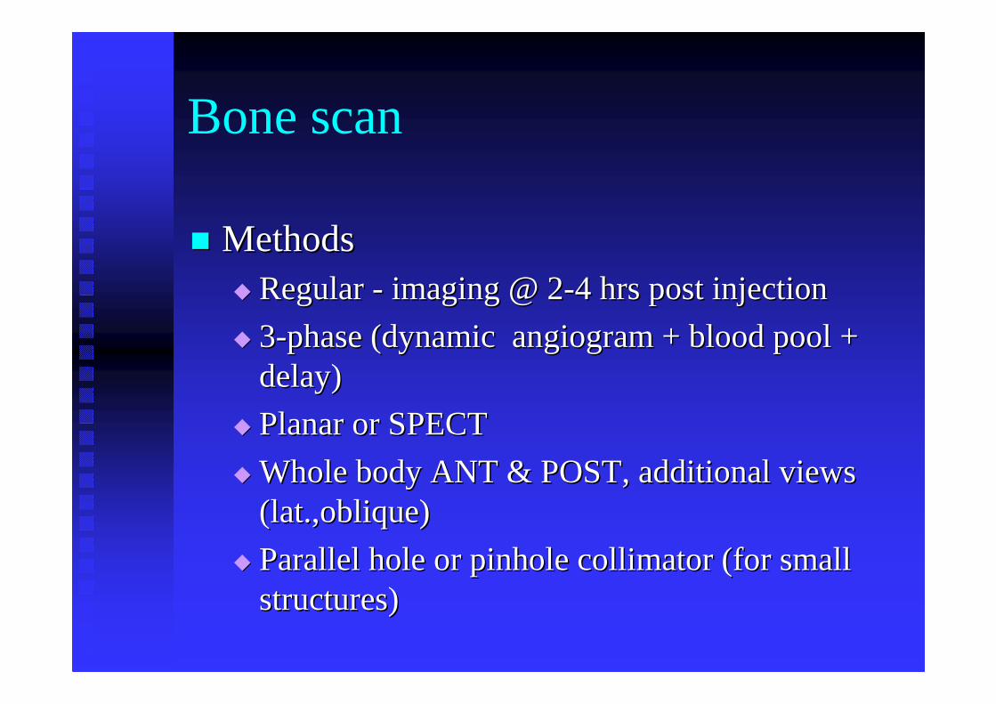
�� MethodsMethods�� Regular Regular -- imaging @ 2imaging @ 2--4 hrs post injection4 hrs post injection
�� 33--phase (dynamic angiogram + blood pool + phase (dynamic angiogram + blood pool + delay)delay)
�� Planar or SPECTPlanar or SPECT
�� Whole body ANT & POST, additional views Whole body ANT & POST, additional views (lat.,oblique)(lat.,oblique)
�� Parallel hole or pinhole collimator (for small Parallel hole or pinhole collimator (for small structures)structures)
Bone scan
Bone Scan in Trauma
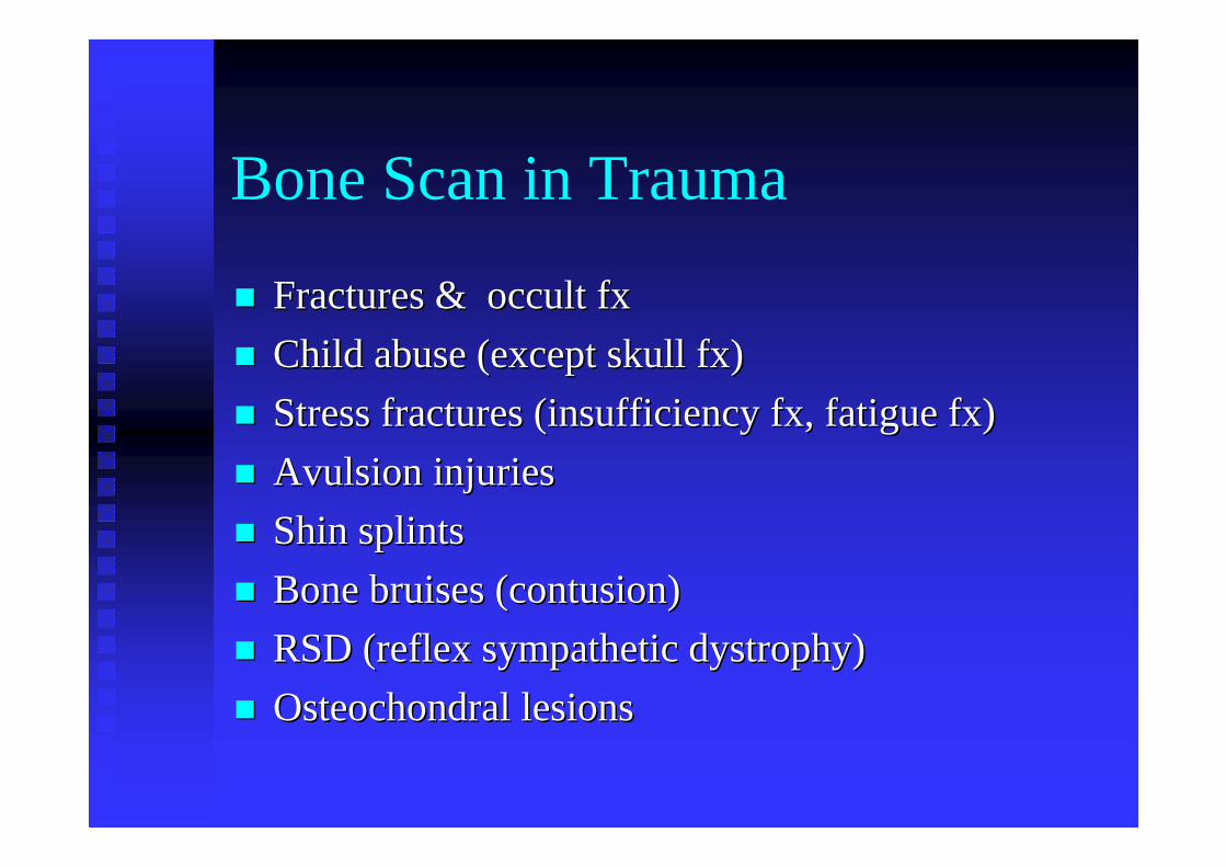
�� Fractures & occult Fractures & occult fxfx
�� Child abuse (except skull fx)Child abuse (except skull fx)
�� Stress fractures (insufficiency fx, fatigue fx)Stress fractures (insufficiency fx, fatigue fx)
�� Avulsion injuriesAvulsion injuries
�� Shin splintsShin splints
�� Bone bruises (contusion)Bone bruises (contusion)
�� RSD (reflex sympathetic dystrophy)RSD (reflex sympathetic dystrophy)
�� OsteochondralOsteochondrallesionslesions
Diagnosis of Fractures
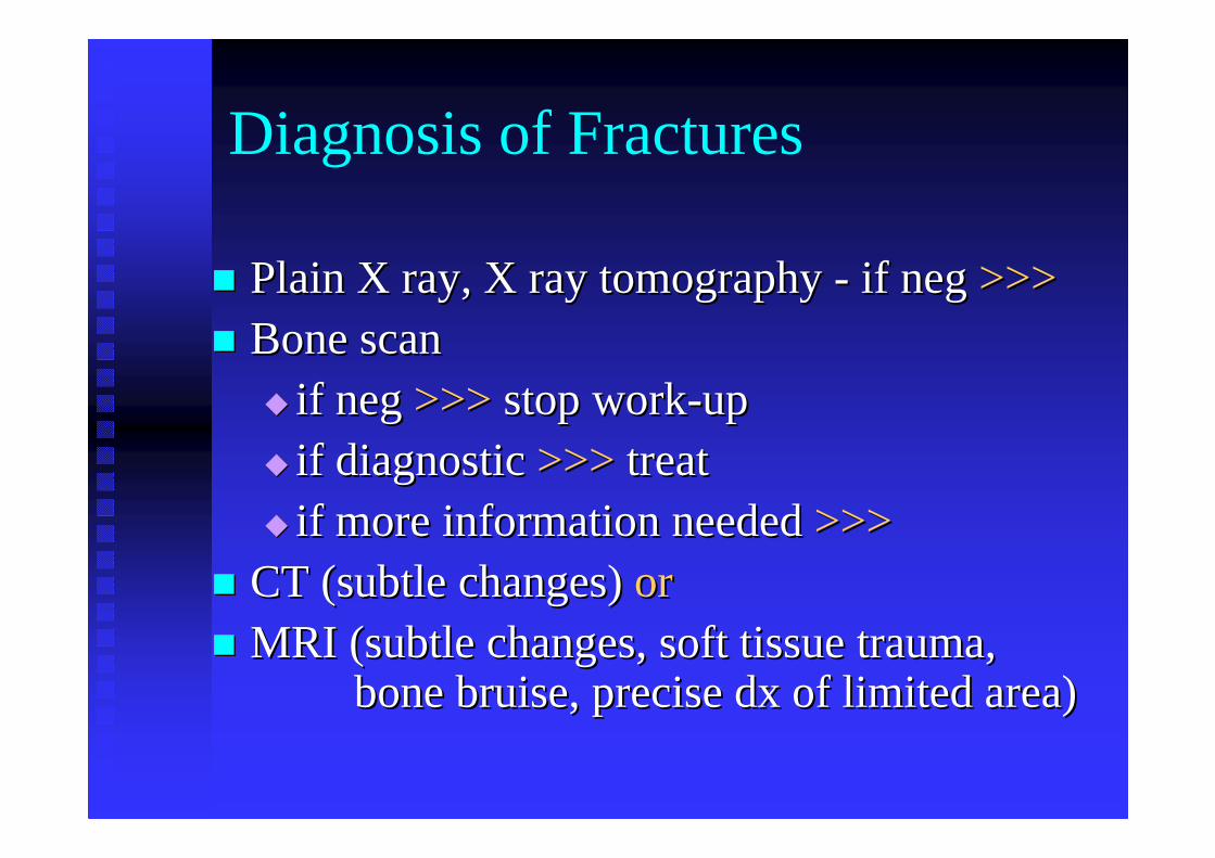
�� Plain X ray, X ray tomography Plain X ray, X ray tomography -- if neg if neg >>>>>>�� Bone scanBone scan
�� if neg if neg >>>>>> stop workstop work--upup�� if diagnostic if diagnostic >>>>>> treattreat�� if more information needed if more information needed >>>>>>
�� CT (subtle changes) CT (subtle changes) oror�� MRI (subtle changes, soft tissue trauma, MRI (subtle changes, soft tissue trauma,
bone bruise, precise dx of limited area)bone bruise, precise dx of limited area)
Fractures on Bone scan
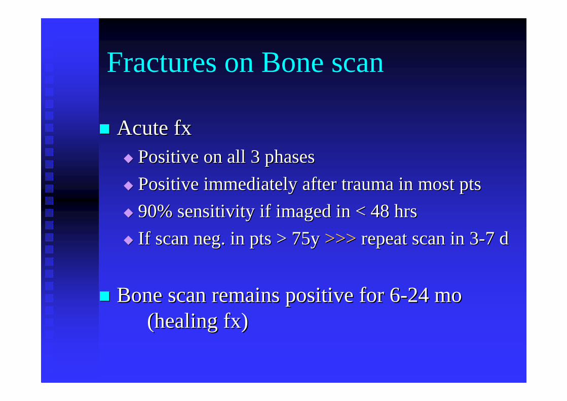
�� Acute fxAcute fx�� Positive on all 3 phasesPositive on all 3 phases
�� Positive immediately after trauma in most ptsPositive immediately after trauma in most pts
�� 90% sensitivity if imaged in < 48 hrs90% sensitivity if imaged in < 48 hrs
�� If scan neg. in pts > 75y If scan neg. in pts > 75y >>>>>> repeat scan in 3repeat scan in 3--7 d7 d
�� Bone scan remains positive for 6Bone scan remains positive for 6--24 mo 24 mo (healing fx) (healing fx)
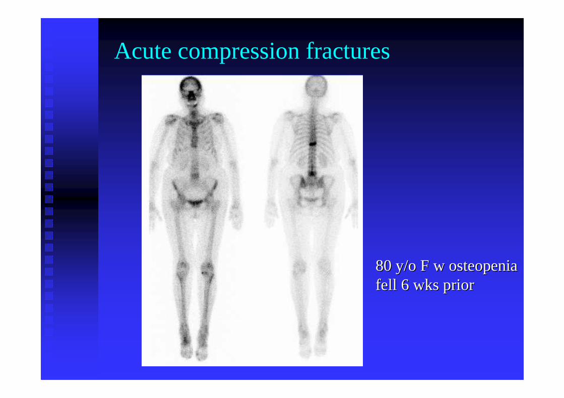
Acute compression fractures
80 y/o F w 80 y/o F w osteopeniaosteopeniafell 6 wks priorfell 6 wks prior
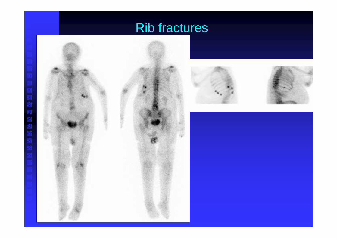
Rib fractures
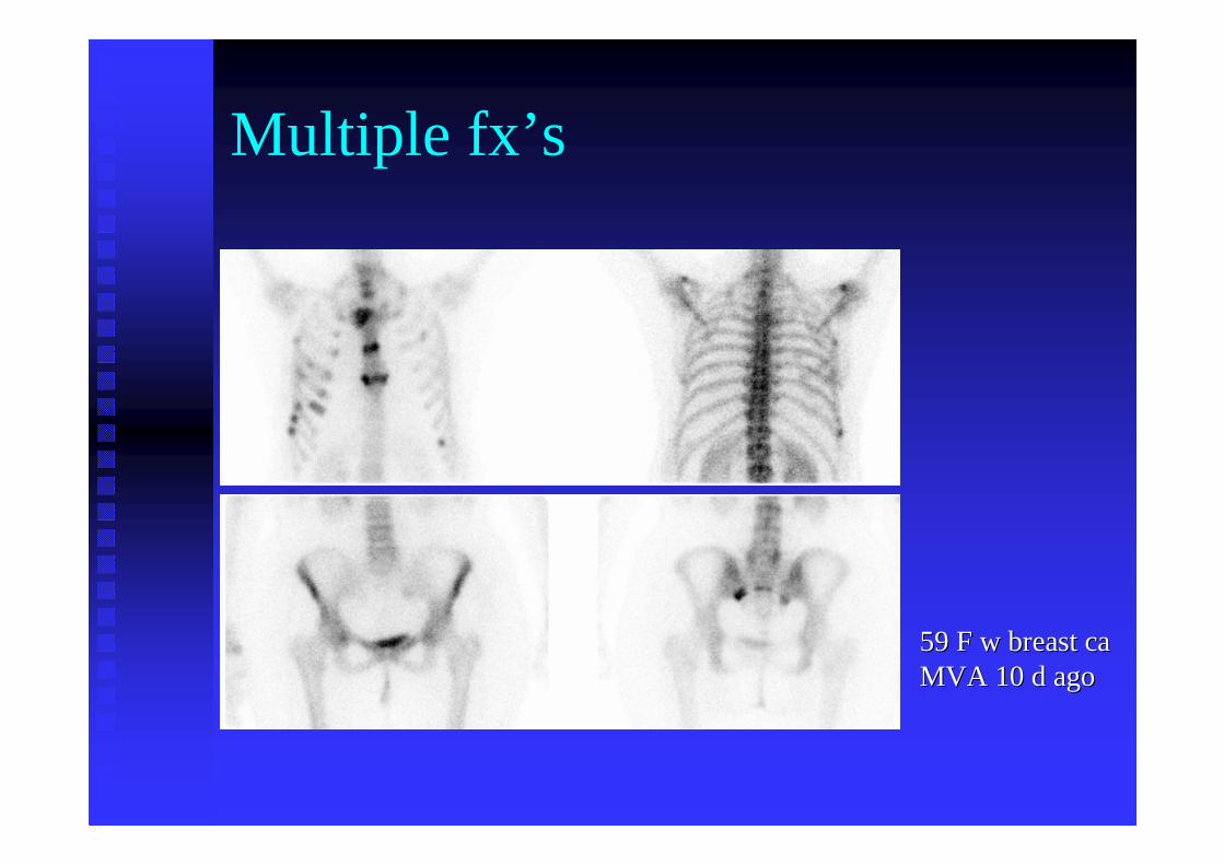
Multiple fx’s
59 F w breast ca59 F w breast caMVA 10 d agoMVA 10 d ago
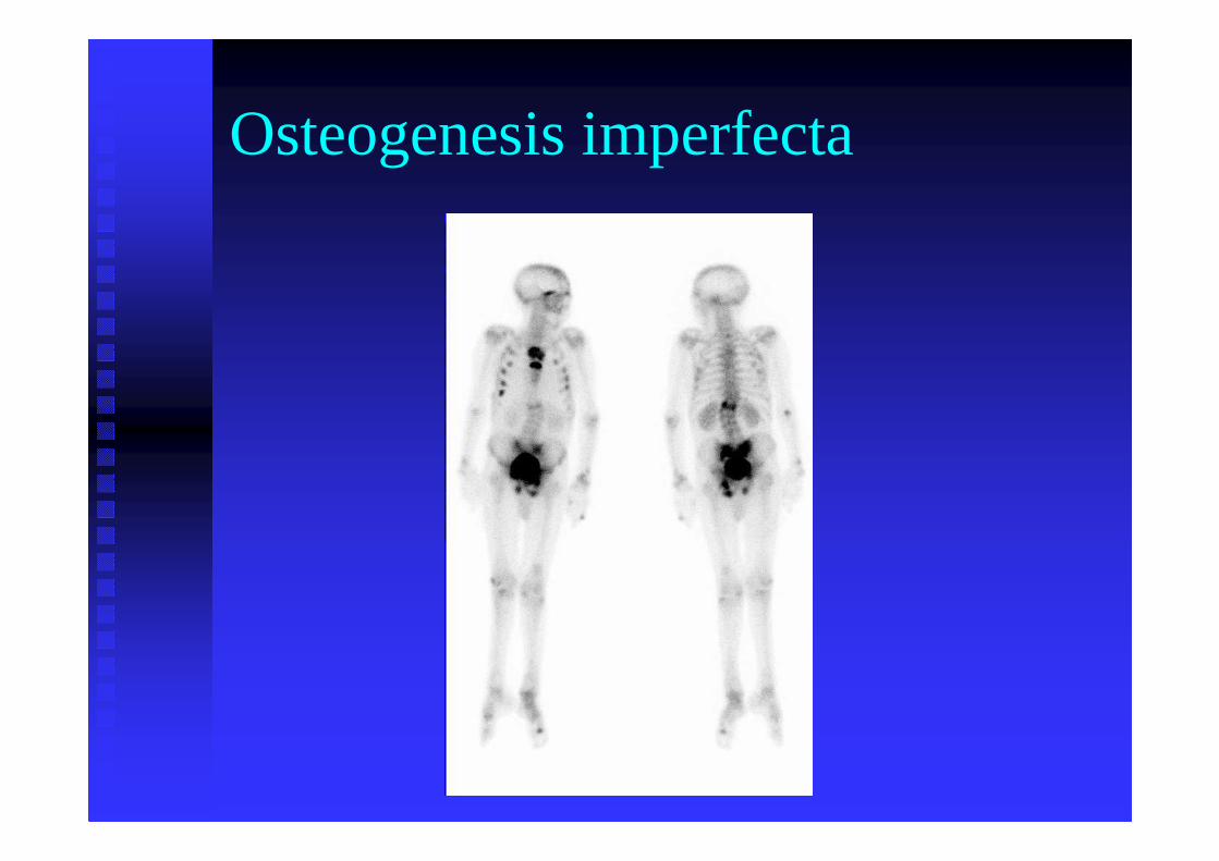
Osteogenesis imperfecta
Bone Bruise
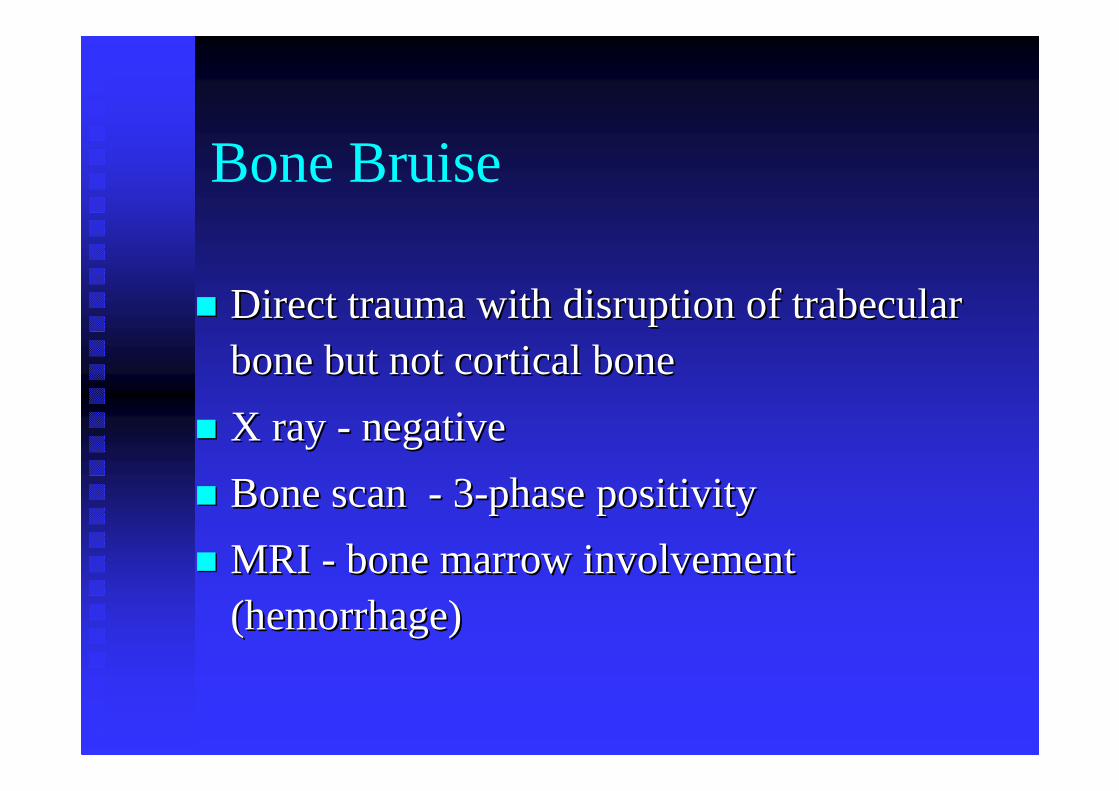
�� Direct trauma with disruption of Direct trauma with disruption of trabeculartrabecularbone but not cortical bone bone but not cortical bone
�� X ray X ray -- negativenegative
�� Bone scan Bone scan -- 33--phase phase positivitypositivity
�� MRI MRI -- bone marrow involvement bone marrow involvement (hemorrhage)(hemorrhage)
Leg & Foot Leg & Foot TraumaTrauma
Shin / thigh splints
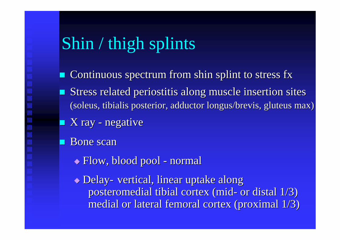
�� Continuous spectrum from shin splint to stress fxContinuous spectrum from shin splint to stress fx
�� Stress related periostitis along muscle insertion sites Stress related periostitis along muscle insertion sites (soleus, (soleus, tibialistibialis posterior, adductor posterior, adductor longus/brevislongus/brevis, gluteus max), gluteus max)
�� X ray X ray -- negativenegative
�� Bone scanBone scan
�� Flow, blood pool Flow, blood pool -- normalnormal
�� DelayDelay-- vertical, linear uptake alongvertical, linear uptake alongposteromedial tibial cortex (midposteromedial tibial cortex (mid-- or distal 1/3) or distal 1/3) medial or lateral femoral cortex (proximal 1/3)medial or lateral femoral cortex (proximal 1/3)
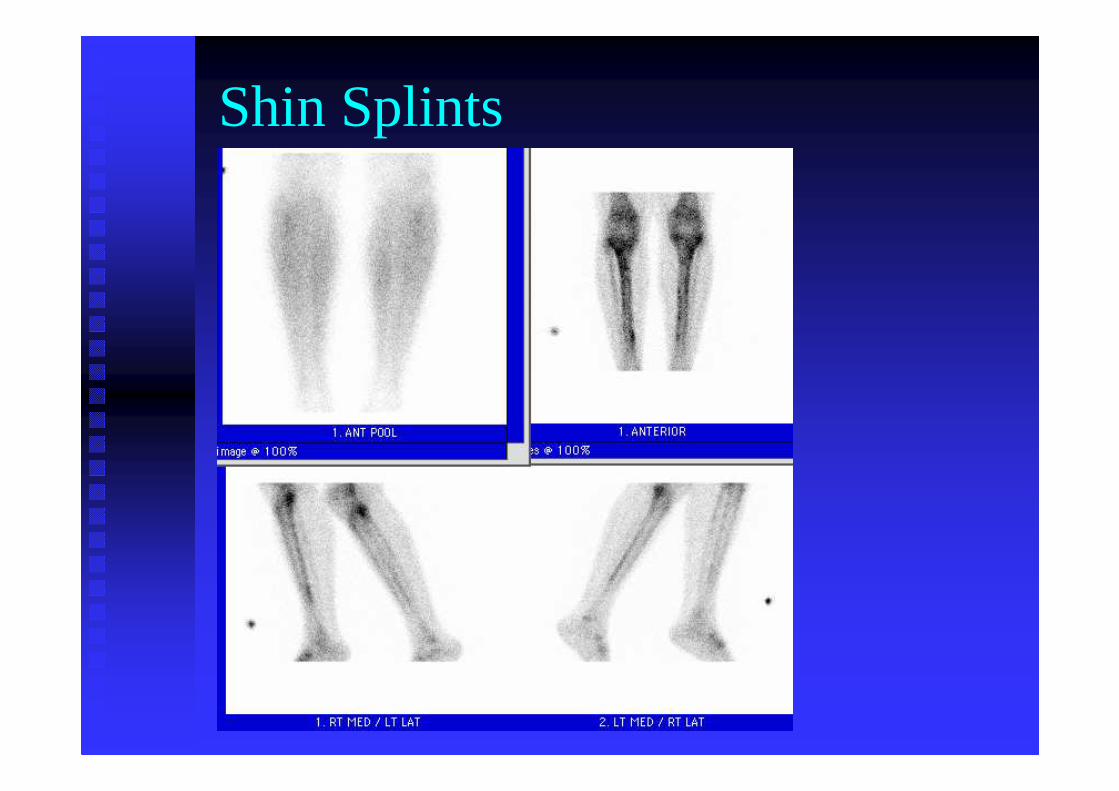
Shin Splints
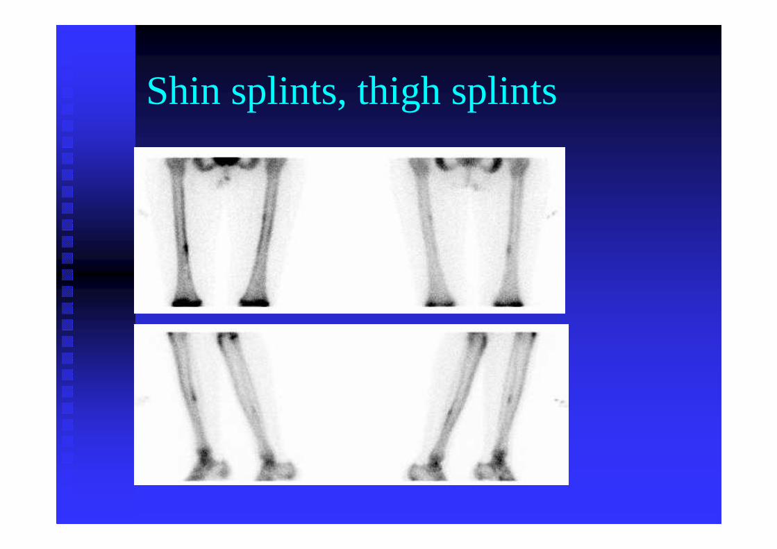
Shin splints, thigh splints
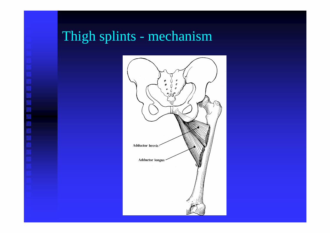
Thigh splints - mechanism
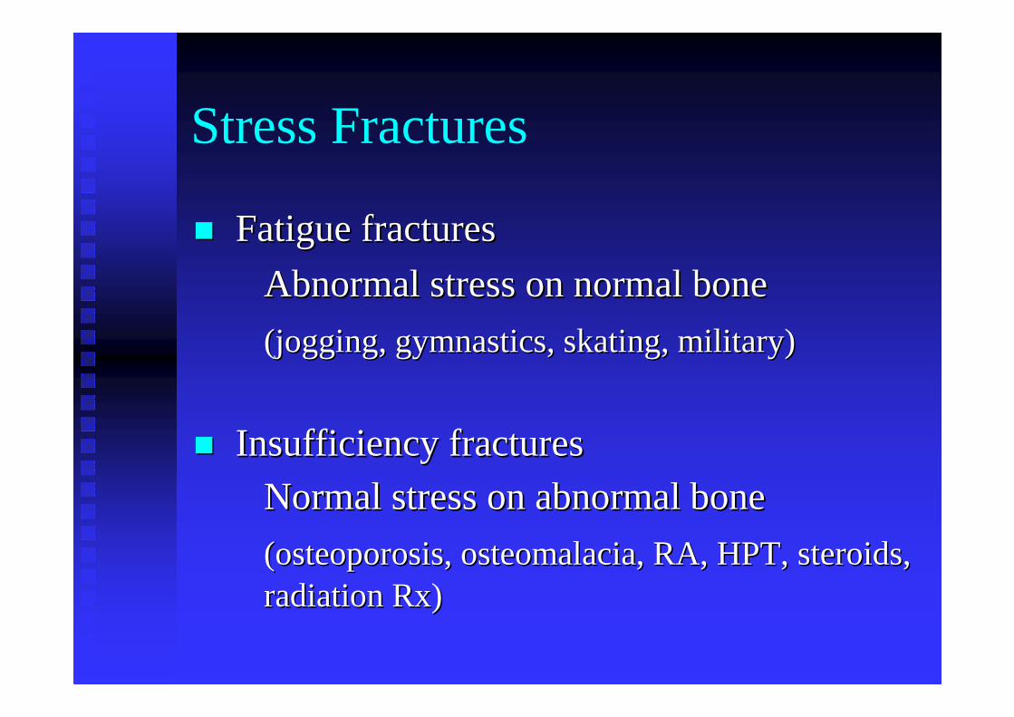
Stress Fractures
�� Fatigue fracturesFatigue fractures
Abnormal stress on normal boneAbnormal stress on normal bone
(jogging, gymnastics, skating, military)(jogging, gymnastics, skating, military)
�� Insufficiency fracturesInsufficiency fractures
Normal stress on abnormal boneNormal stress on abnormal bone
(osteoporosis, osteomalacia, RA, HPT, steroids, (osteoporosis, osteomalacia, RA, HPT, steroids, radiation Rx)radiation Rx)
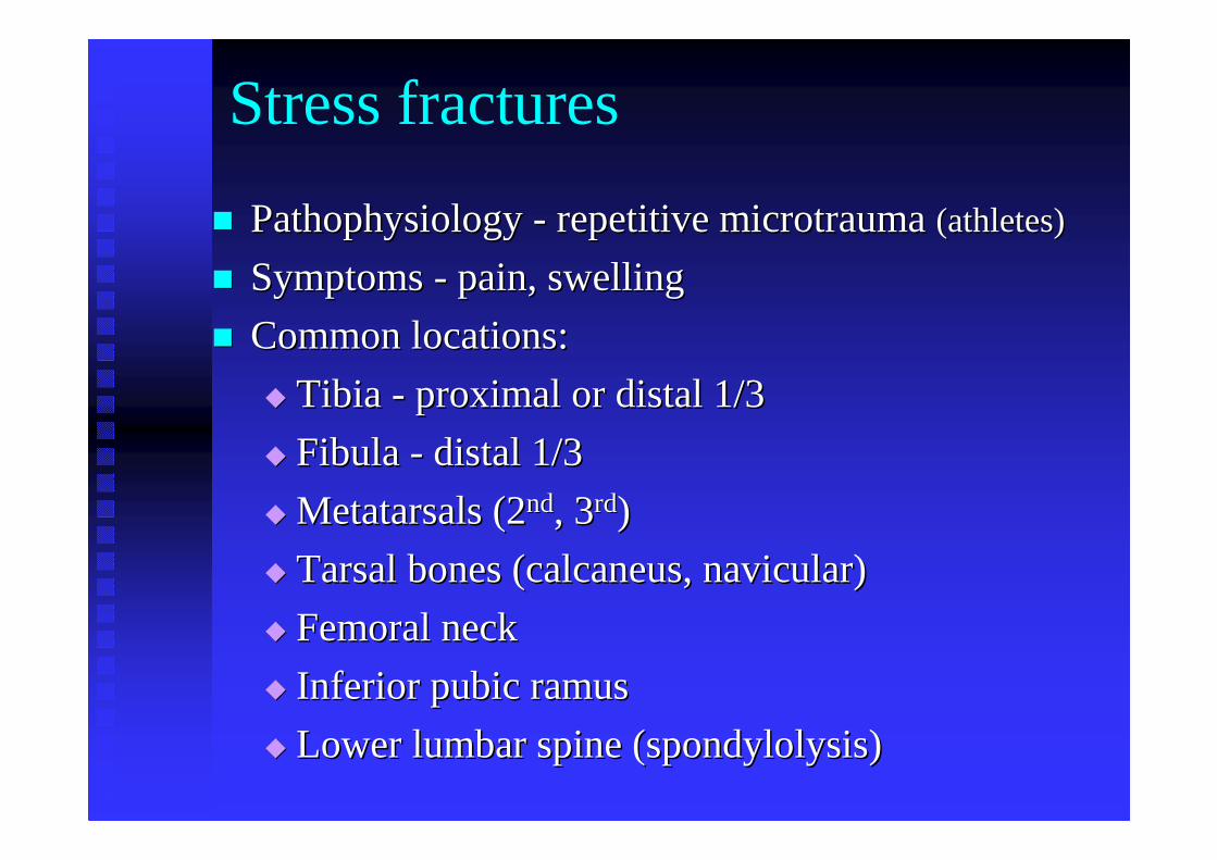
Stress fractures
�� Pathophysiology Pathophysiology -- repetitive microtrauma repetitive microtrauma (athletes) (athletes)
�� Symptoms Symptoms -- pain, swellingpain, swelling
�� Common locations:Common locations:
�� Tibia Tibia -- proximal or distal 1/3 proximal or distal 1/3
�� Fibula Fibula -- distal 1/3distal 1/3
�� Metatarsals (2Metatarsals (2ndnd, 3, 3rdrd))
�� Tarsal bones (calcaneus, navicular)Tarsal bones (calcaneus, navicular)
�� Femoral neck Femoral neck
�� Inferior pubic ramusInferior pubic ramus
�� Lower lumbar spine (spondylolysis)Lower lumbar spine (spondylolysis)
Stress fractures
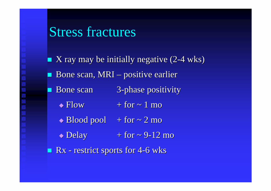
�� X ray may be initially negative (2X ray may be initially negative (2--4 wks)4 wks)
�� Bone scan, MRI Bone scan, MRI –– positive earlier positive earlier
�� Bone scan Bone scan 33--phase positivityphase positivity
�� FlowFlow + for ~ 1 mo+ for ~ 1 mo
�� Blood poolBlood pool + for ~ 2 mo+ for ~ 2 mo
�� DelayDelay + for ~ 9+ for ~ 9--12 mo12 mo
�� Rx Rx -- restrict sports for 4restrict sports for 4--6 wks6 wks
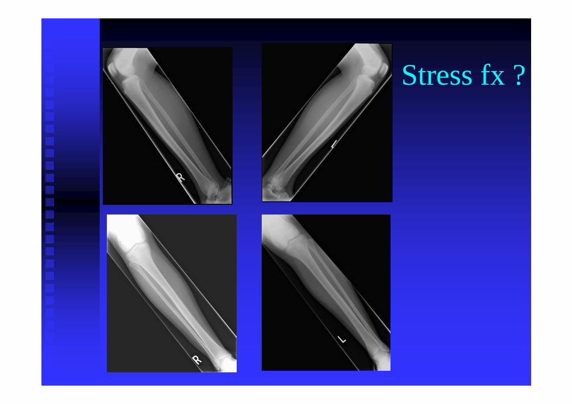
Stress fx ?
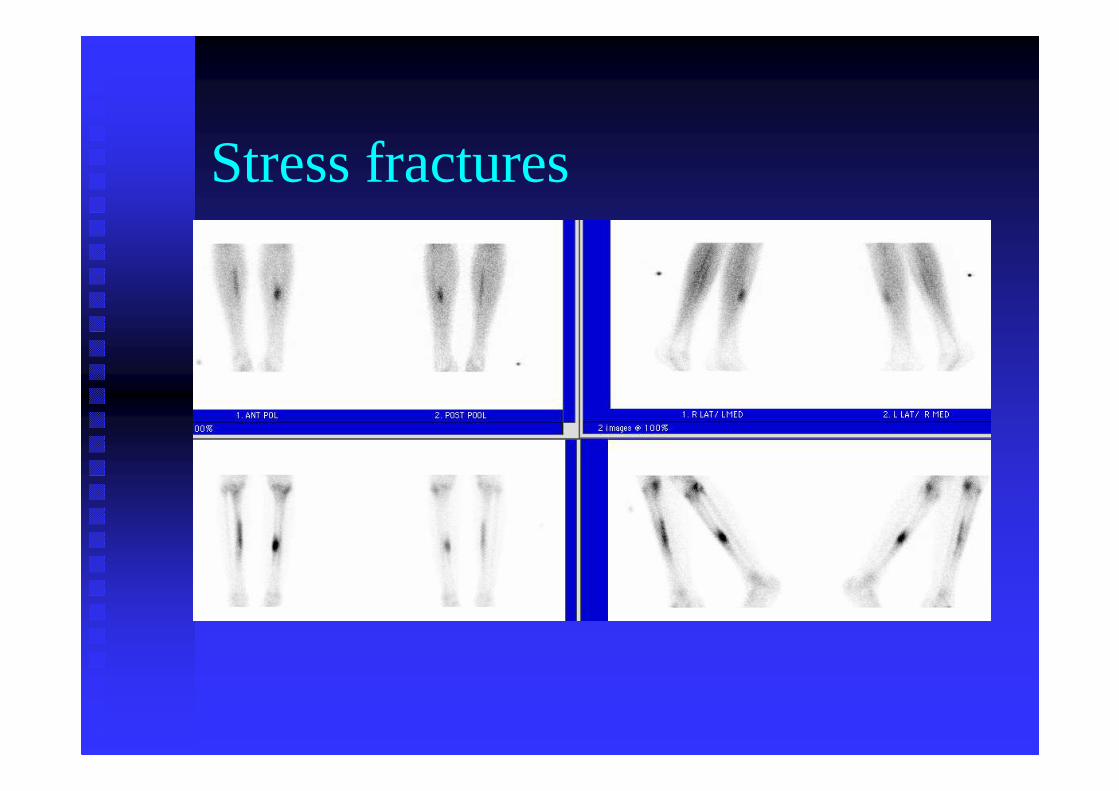
Stress fractures
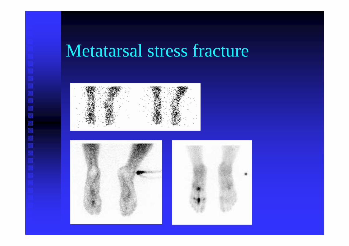
Metatarsal stress fracture
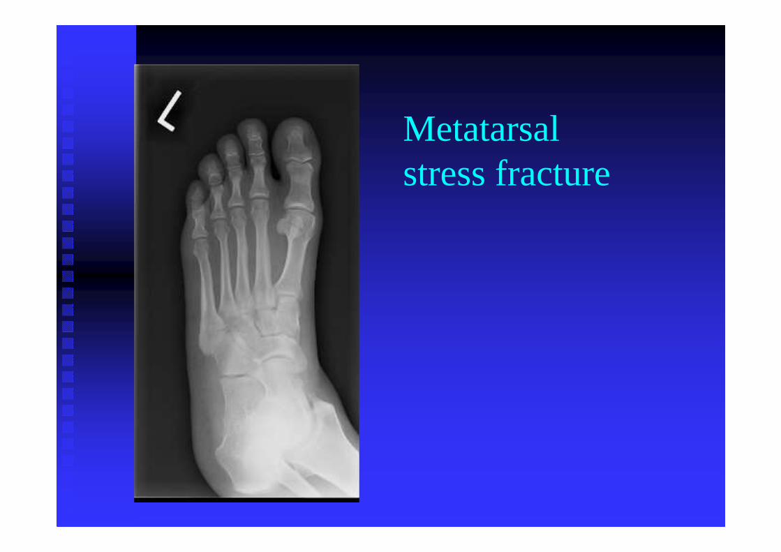
Metatarsal stress fracture

Metatarsal stress fx
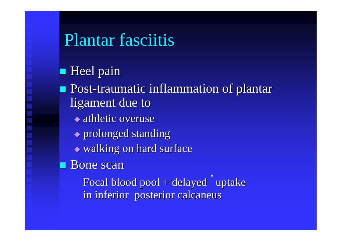
Plantar fasciitis
�� Heel painHeel pain�� PostPost--traumatic inflammation of plantar traumatic inflammation of plantar
ligament due to ligament due to �� athletic overuse athletic overuse �� prolonged standingprolonged standing�� walking on hard surfacewalking on hard surface
�� Bone scanBone scanFocal blood pool + delayed uptake Focal blood pool + delayed uptake in inferior posterior calcaneusin inferior posterior calcaneus
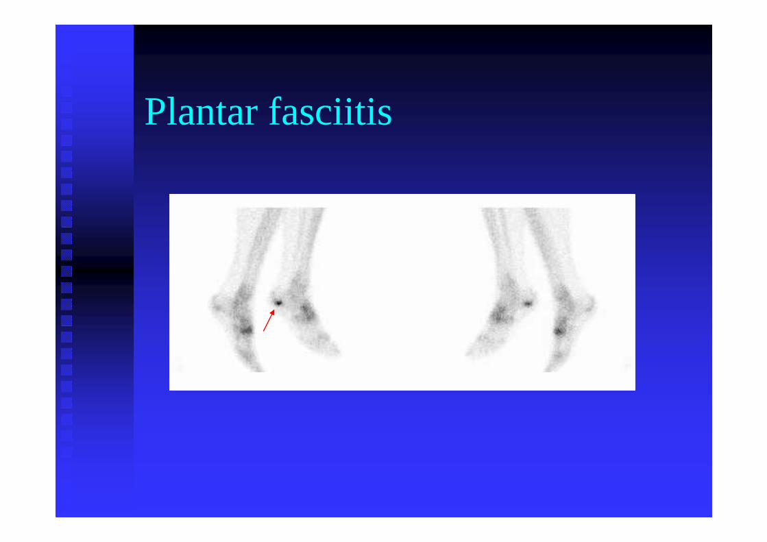
Plantar fasciitis
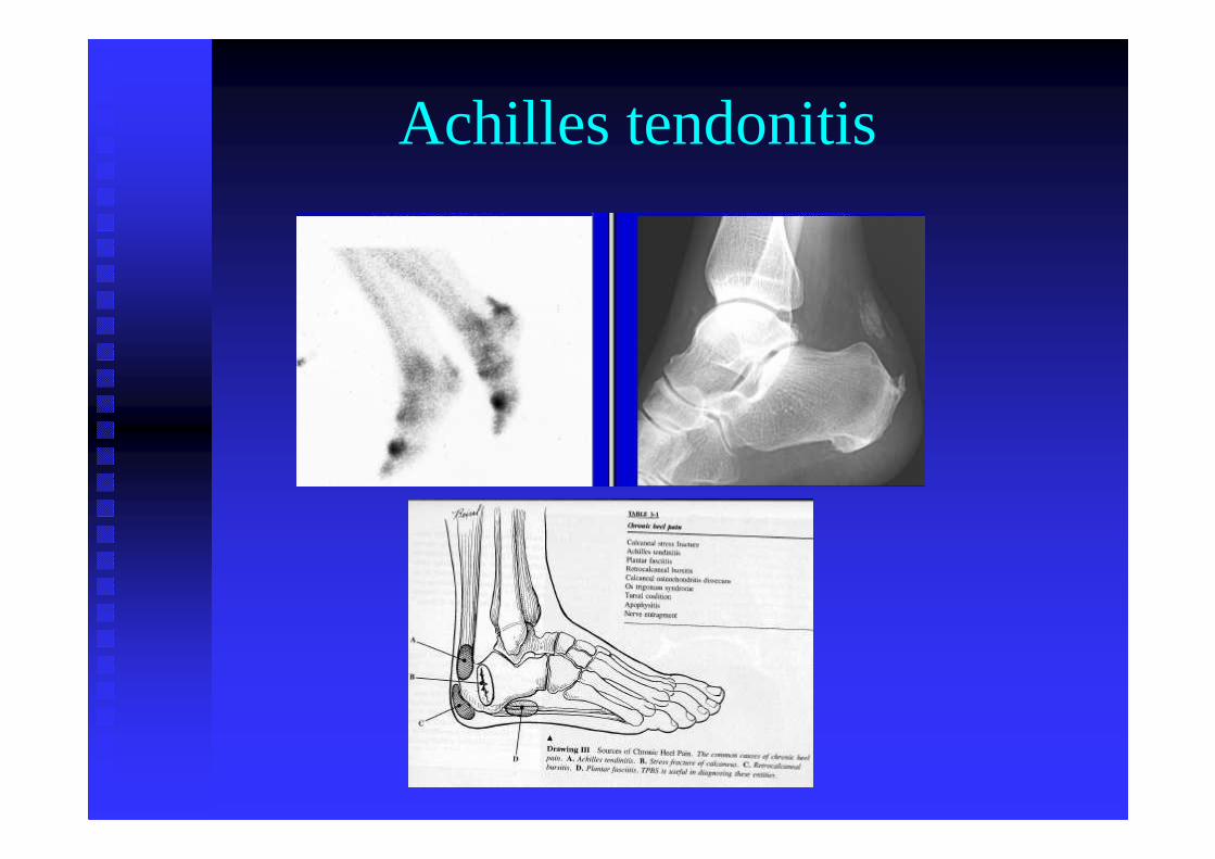
Achilles tendonitis
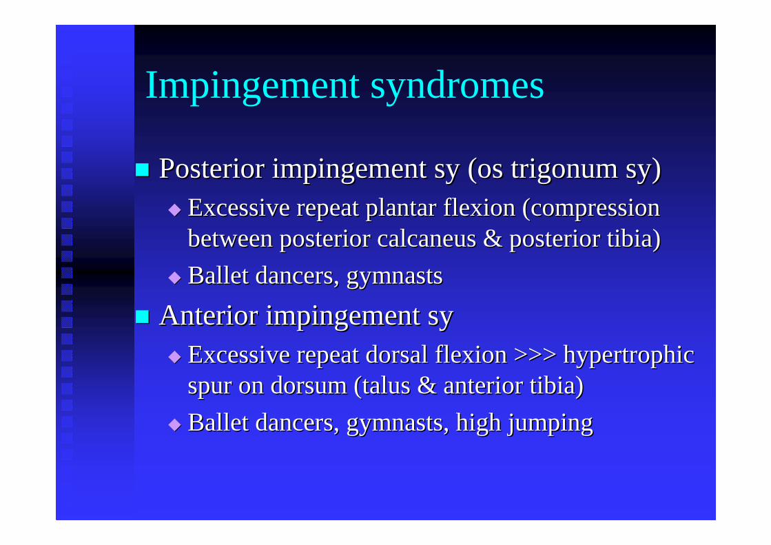
Impingement syndromes
�� Posterior impingement sy (os trigonum sy)Posterior impingement sy (os trigonum sy)�� Excessive repeat plantar flexion (compression Excessive repeat plantar flexion (compression
between posterior calcaneus & posterior tibia)between posterior calcaneus & posterior tibia)
�� Ballet dancers, gymnastsBallet dancers, gymnasts
�� Anterior impingement sy Anterior impingement sy �� Excessive repeat dorsal flexion >>> hypertrophic Excessive repeat dorsal flexion >>> hypertrophic
spur on dorsum (talus & anterior tibia)spur on dorsum (talus & anterior tibia)
�� Ballet dancers, gymnasts, high jumpingBallet dancers, gymnasts, high jumping
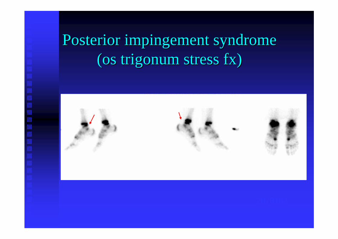
Posterior impingement syndromePosterior impingement syndrome((osos trigonumtrigonumstress stress fxfx))
2078102
Hip & PelvisHip & PelvisTraumaTrauma
Femoral neck stress fracture
�� Thigh or groin pain in athletesThigh or groin pain in athletes
�� Must distinguish femoral neck stress fx Must distinguish femoral neck stress fx from pubic ramus stress fxfrom pubic ramus stress fx
�� Must treat / immobilize early to prevent Must treat / immobilize early to prevent complete fx, AVNcomplete fx, AVN
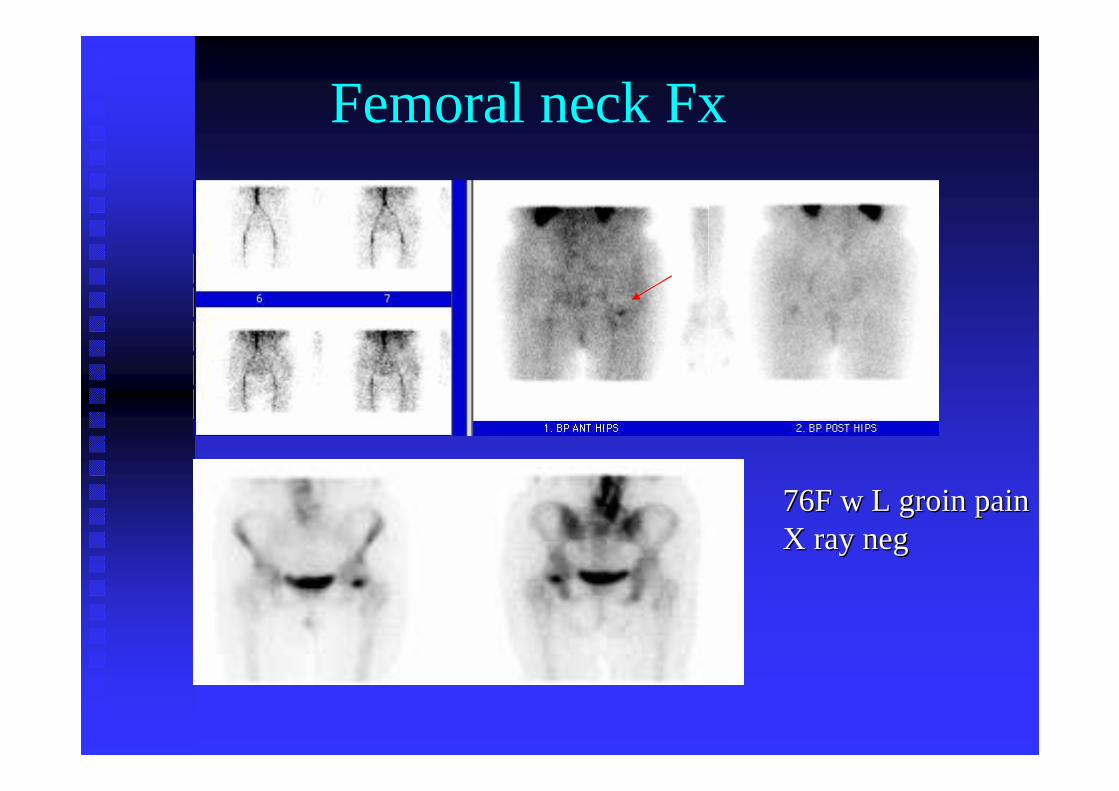
Femoral neck Fx
76F w L groin pain76F w L groin painX ray X ray negneg

X ray X ray 2 weeks later2 weeks later
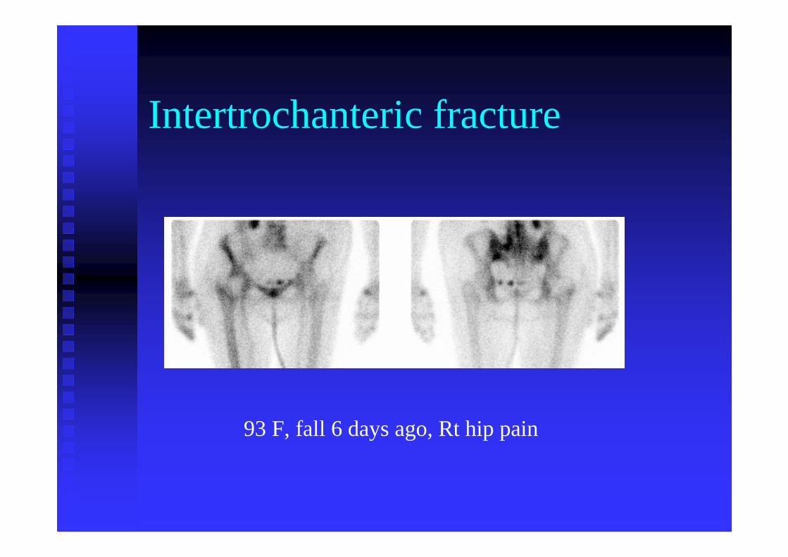
Intertrochanteric fracture
93 F, fall 6 days ago, Rt hip pain
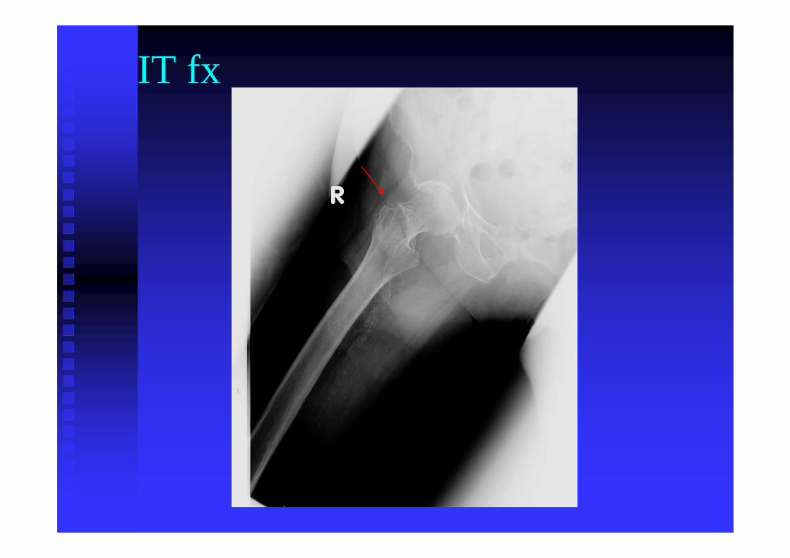
IT fx
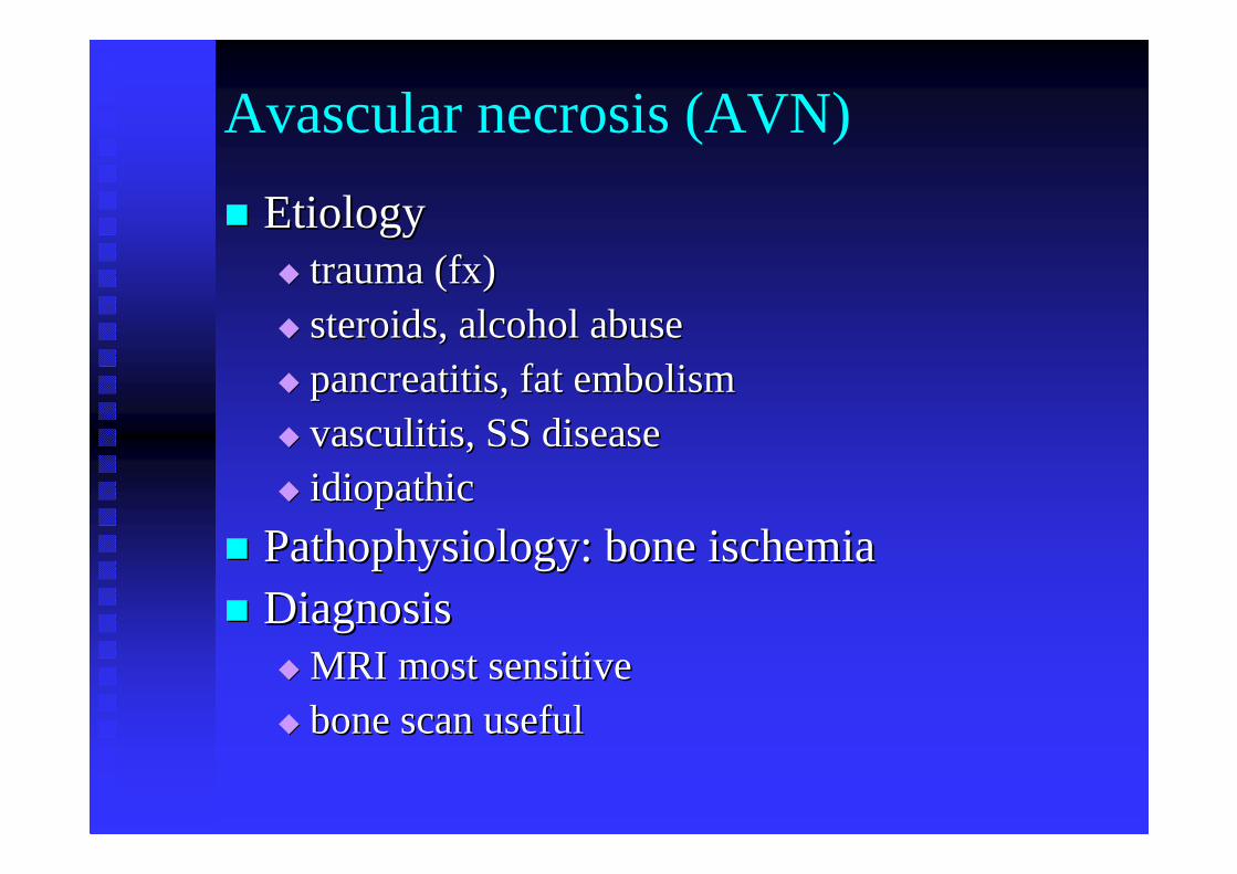
Avascular necrosis (AVN)
�� EtiologyEtiology�� trauma (fx)trauma (fx)�� steroids, alcohol abusesteroids, alcohol abuse�� pancreatitispancreatitis, fat embolism, fat embolism�� vasculitis, SS diseasevasculitis, SS disease�� idiopathicidiopathic
�� Pathophysiology: bone ischemiaPathophysiology: bone ischemia�� DiagnosisDiagnosis
�� MRI most sensitiveMRI most sensitive�� bone scan useful bone scan useful
AVN
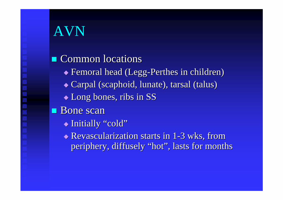
�� Common locationsCommon locations�� Femoral head (LeggFemoral head (Legg--Perthes in children)Perthes in children)�� Carpal (scaphoid, lunate), tarsal (talus) Carpal (scaphoid, lunate), tarsal (talus) �� Long bones, ribs in SSLong bones, ribs in SS
�� Bone scanBone scan�� Initially Initially ““ coldcold””�� Revascularization starts in 1Revascularization starts in 1--3 wks, from 3 wks, from
periphery, diffusely periphery, diffusely ““ hothot”” , lasts for months, lasts for months
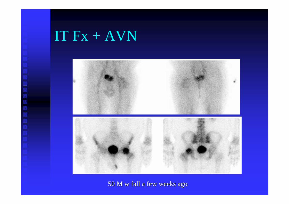
IT Fx + AVN
50 M w fall a few weeks ago50 M w fall a few weeks ago
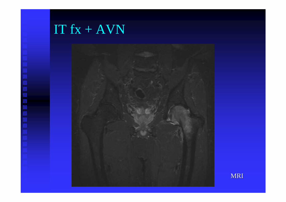
MRIMRI
IT fx + AVN
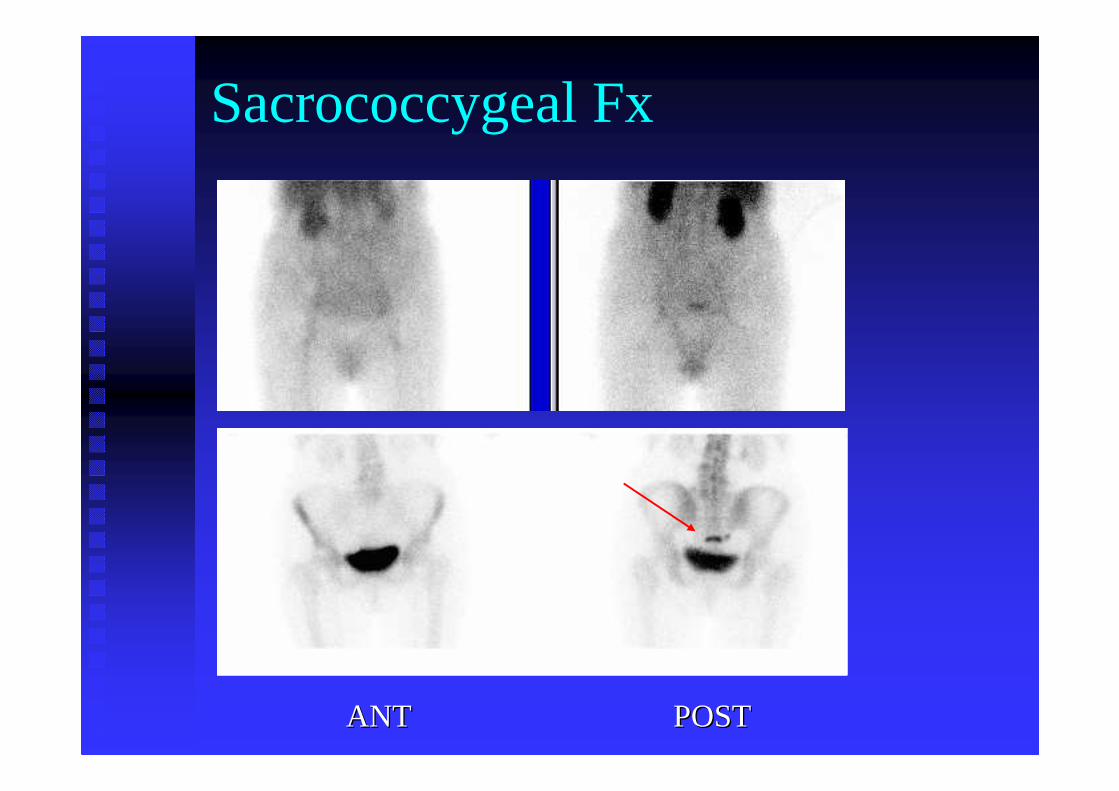
Sacrococcygeal Fx
ANTANT POST POST
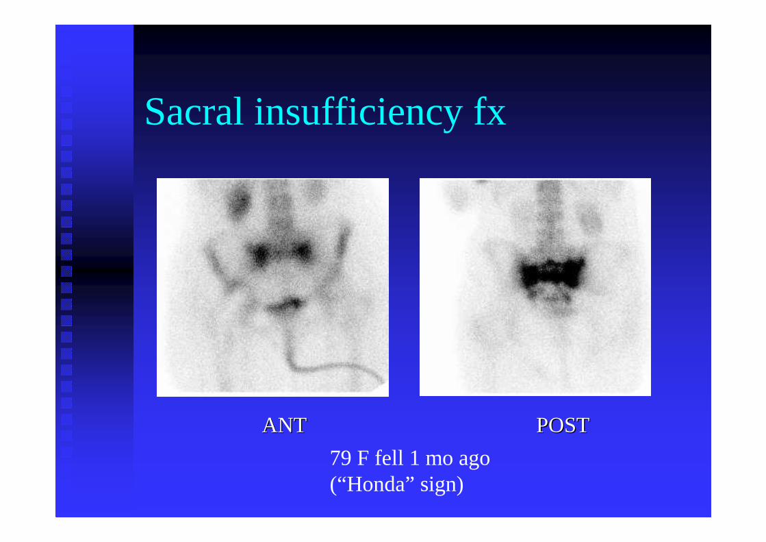
Sacral insufficiency fx
ANT POSTANT POST
79 F fell 1 mo ago(“Honda” sign)
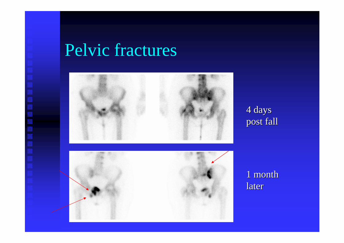
Pelvic fractures
4 days 4 days post fallpost fall
1 month 1 month laterlater
Spine traumaSpine trauma
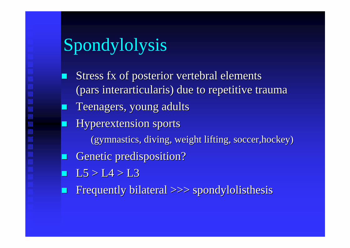
Spondylolysis
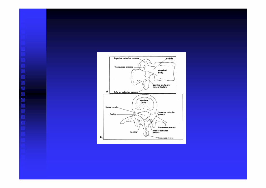
�� Stress fx of posterior vertebral elements Stress fx of posterior vertebral elements (pars interarticularis) due to(pars interarticularis) due torepetitive traumarepetitive trauma
�� Teenagers, young adultsTeenagers, young adults
�� Hyperextension sports Hyperextension sports (gymnastics, diving, weight lifting, soccer,hockey)(gymnastics, diving, weight lifting, soccer,hockey)
�� Genetic predisposition?Genetic predisposition?
�� L5 > L4 > L3L5 > L4 > L3
�� Frequently bilateral >>> spondylolisthesisFrequently bilateral >>> spondylolisthesis
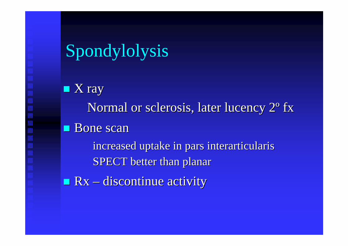
Spondylolysis
�� X ray X ray
Normal or sclerosis, later lucency 2Normal or sclerosis, later lucency 2ºº fxfx
�� Bone scanBone scanincreased uptake in pars interarticularisincreased uptake in pars interarticularis
SPECT better than planarSPECT better than planar
�� Rx Rx –– discontinue activitydiscontinue activity
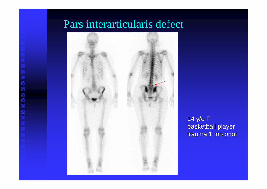
Pars interarticularis defect
14 y/o F14 y/o Fbasketball playerbasketball playertrauma 1 mo priortrauma 1 mo prior
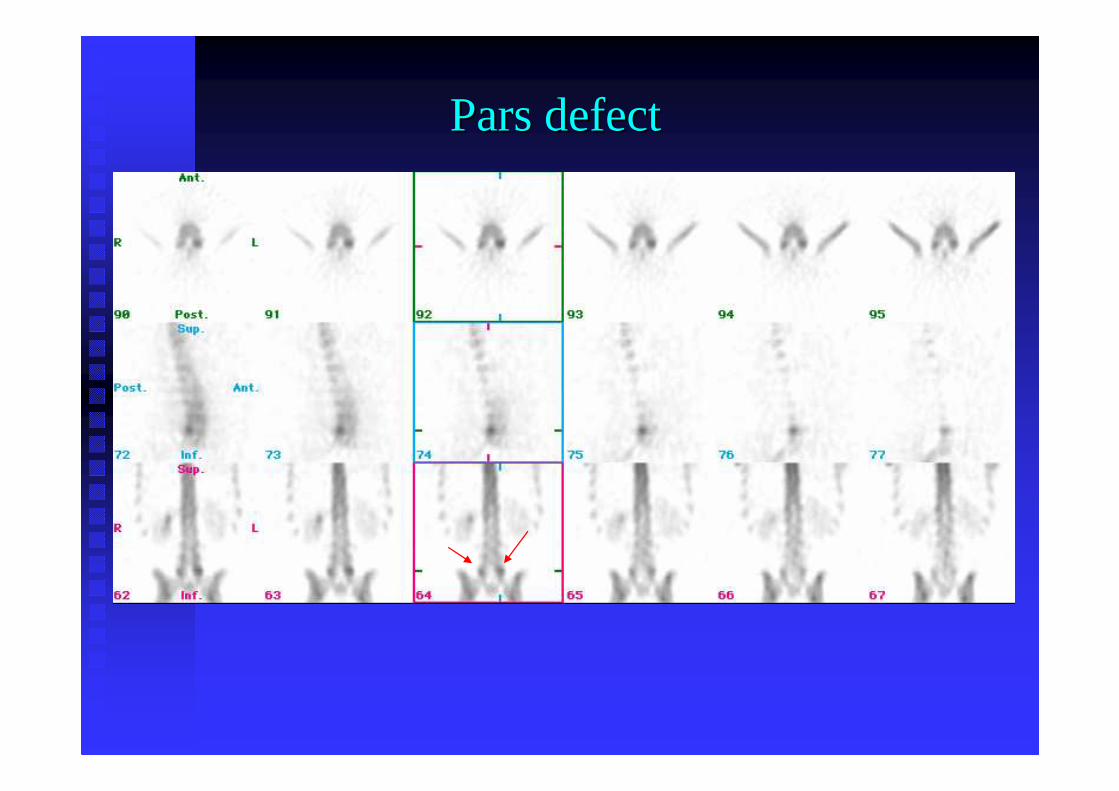
Pars defectPars defect
CNM 2001:863
planar SPECT
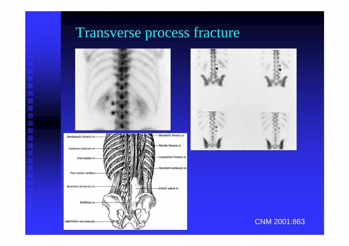
Transverse process fracture
Hand & Wrist Trauma
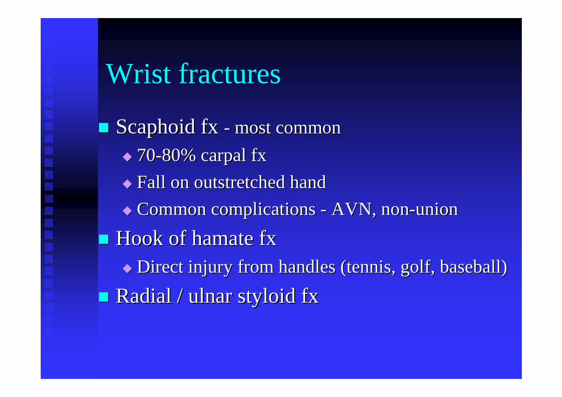
Wrist fractures
�� Scaphoid fx Scaphoid fx -- most common most common
�� 7070--80% carpal fx80% carpal fx
�� Fall on outstretched handFall on outstretched hand
�� Common complications Common complications -- AVN, nonAVN, non--unionunion
�� Hook of hamate fxHook of hamate fx�� Direct injury from handles (tennis, golf, baseball)Direct injury from handles (tennis, golf, baseball)
�� Radial / ulnar styloid Radial / ulnar styloid fxfx
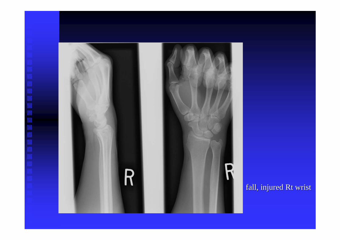
fall, injured fall, injured RtRt wristwrist
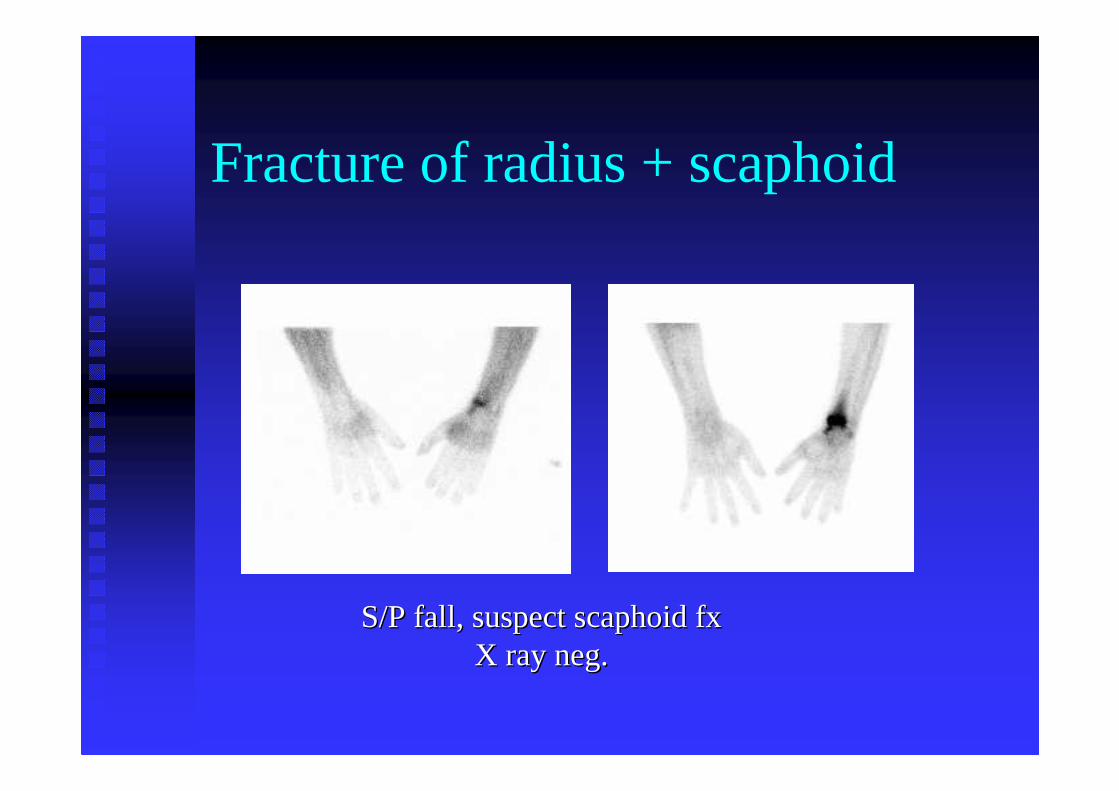
Fracture of radius + scaphoid
S/P fall, suspect S/P fall, suspect scaphoidscaphoidfxfxX ray neg.X ray neg.
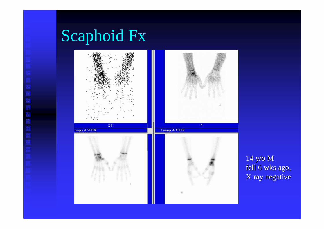
Scaphoid Fx
14 y/o M 14 y/o M fell 6 wks ago, fell 6 wks ago, X ray negativeX ray negative
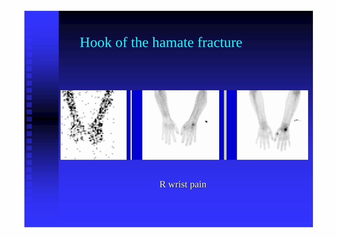
Hook of the hamate fracture
R wrist painR wrist pain
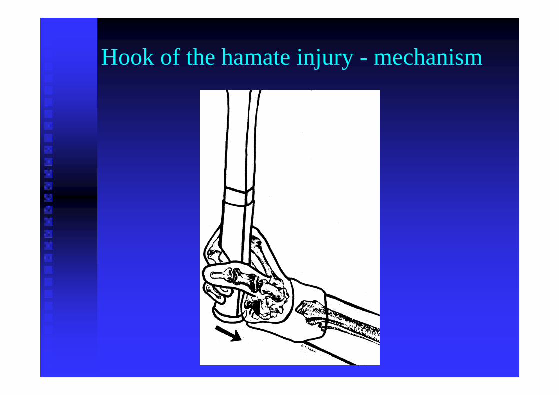
Hook of the hamate injury - mechanism
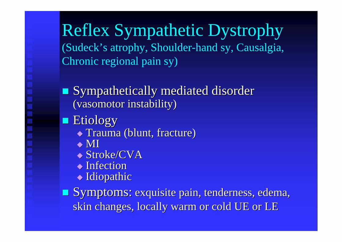
Reflex Sympathetic Dystrophy (Sudeck’s atrophy, Shoulder-hand sy, Causalgia, Chronic regional pain sy)
�� Sympathetically mediated disorder Sympathetically mediated disorder (vasomotor instability)(vasomotor instability)
�� EtiologyEtiology�� Trauma (blunt, fracture)Trauma (blunt, fracture)�� MIMI�� Stroke/CVAStroke/CVA�� InfectionInfection�� IdiopathicIdiopathic
�� Symptoms:Symptoms:exquisite pain, tenderness, edema, exquisite pain, tenderness, edema, skin changes, locally warm or cold UE or LEskin changes, locally warm or cold UE or LE
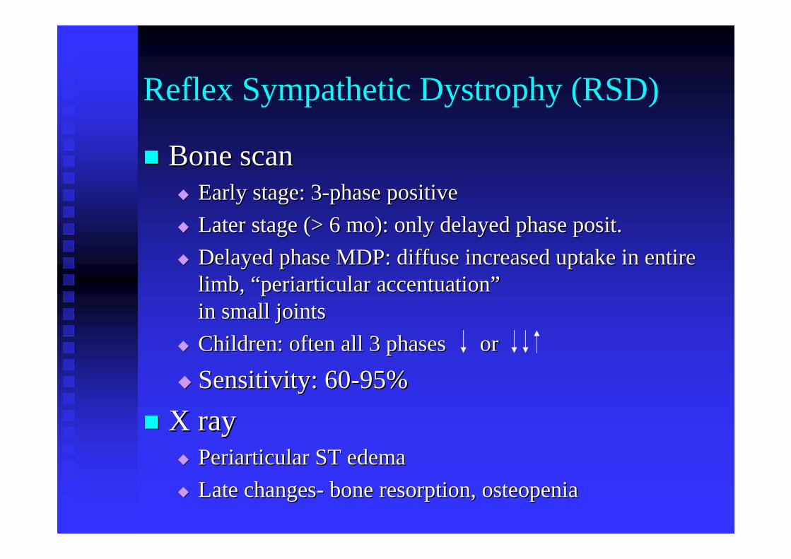
Reflex Sympathetic Dystrophy (RSD)
�� Bone scanBone scan�� Early stage: 3Early stage: 3--phase positivephase positive
�� Later stage (> 6 mo): only delayed phase posit.Later stage (> 6 mo): only delayed phase posit.
�� Delayed phase MDP: diffuse increased uptake in entire Delayed phase MDP: diffuse increased uptake in entire limb, limb, ““ periarticular accentuationperiarticular accentuation””in small jointsin small joints
�� Children: often all 3 phases or Children: often all 3 phases or
�� Sensitivity: 60Sensitivity: 60--95%95%
�� X rayX ray�� Periarticular ST edemaPeriarticular ST edema
�� Late changesLate changes-- bone resorption, osteopeniabone resorption, osteopenia
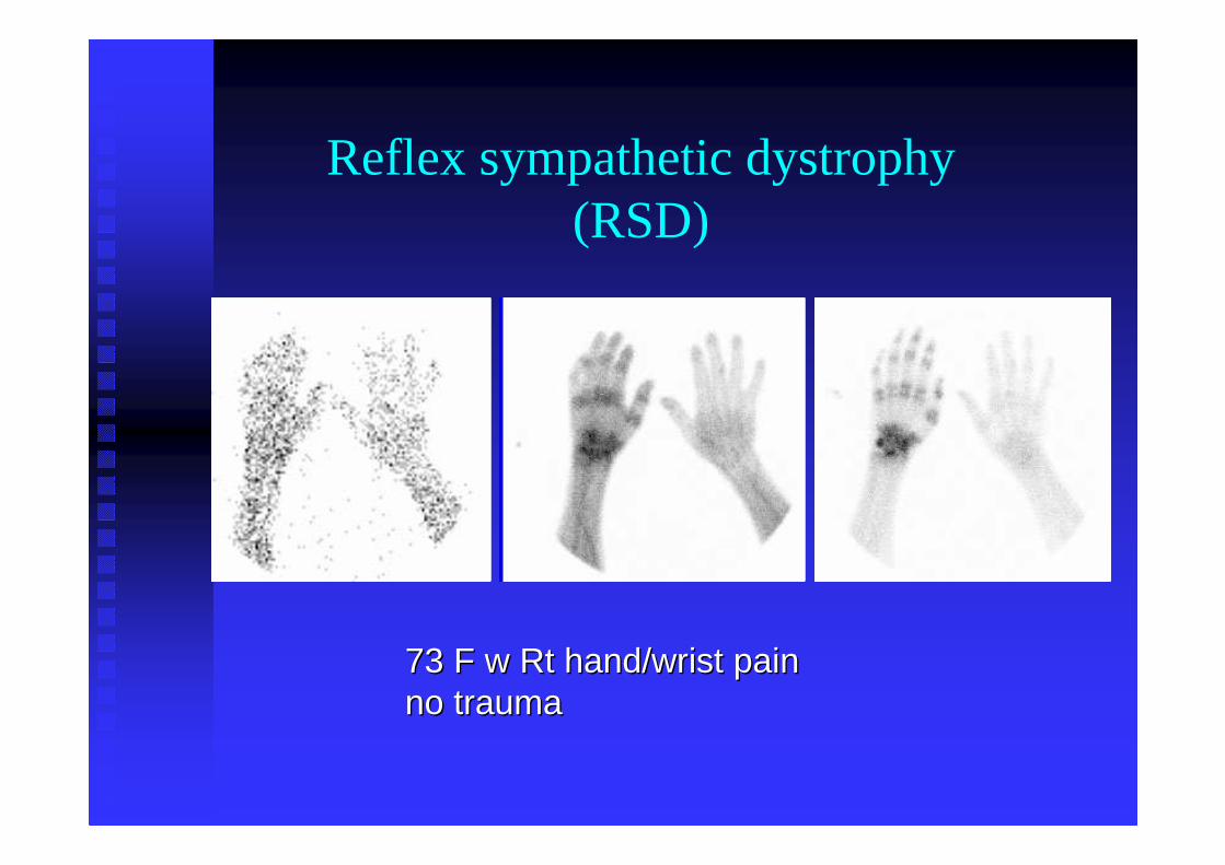
73 F w 73 F w RtRt hand/wrist painhand/wrist painno traumano trauma
Reflex sympathetic dystrophy(RSD)
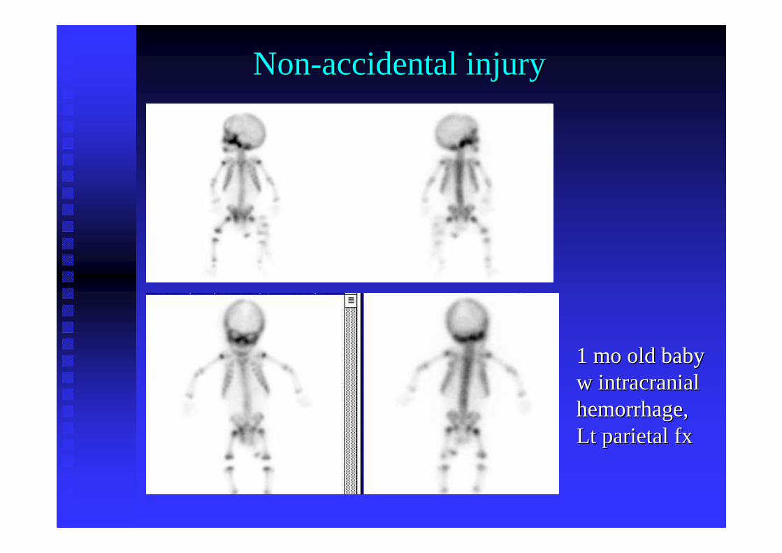
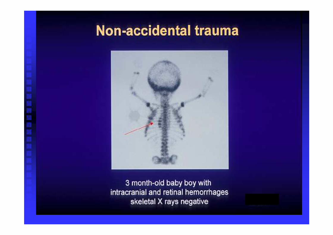
NonNon--accidental injuryaccidental injury
1 mo old baby1 mo old babyw intracranial w intracranial hemorrhage, hemorrhage, Lt parietal Lt parietal fxfx
MDP
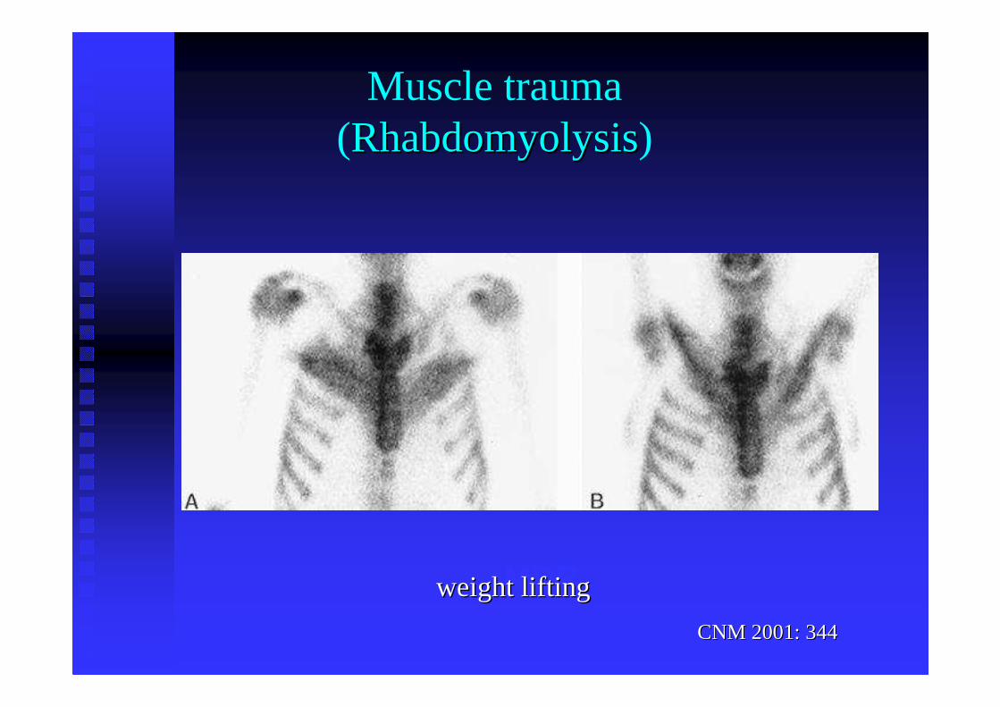
CNM 2001: 344CNM 2001: 344
Muscle trauma(RhabdomyolysisRhabdomyolysis)
weight liftingweight lifting
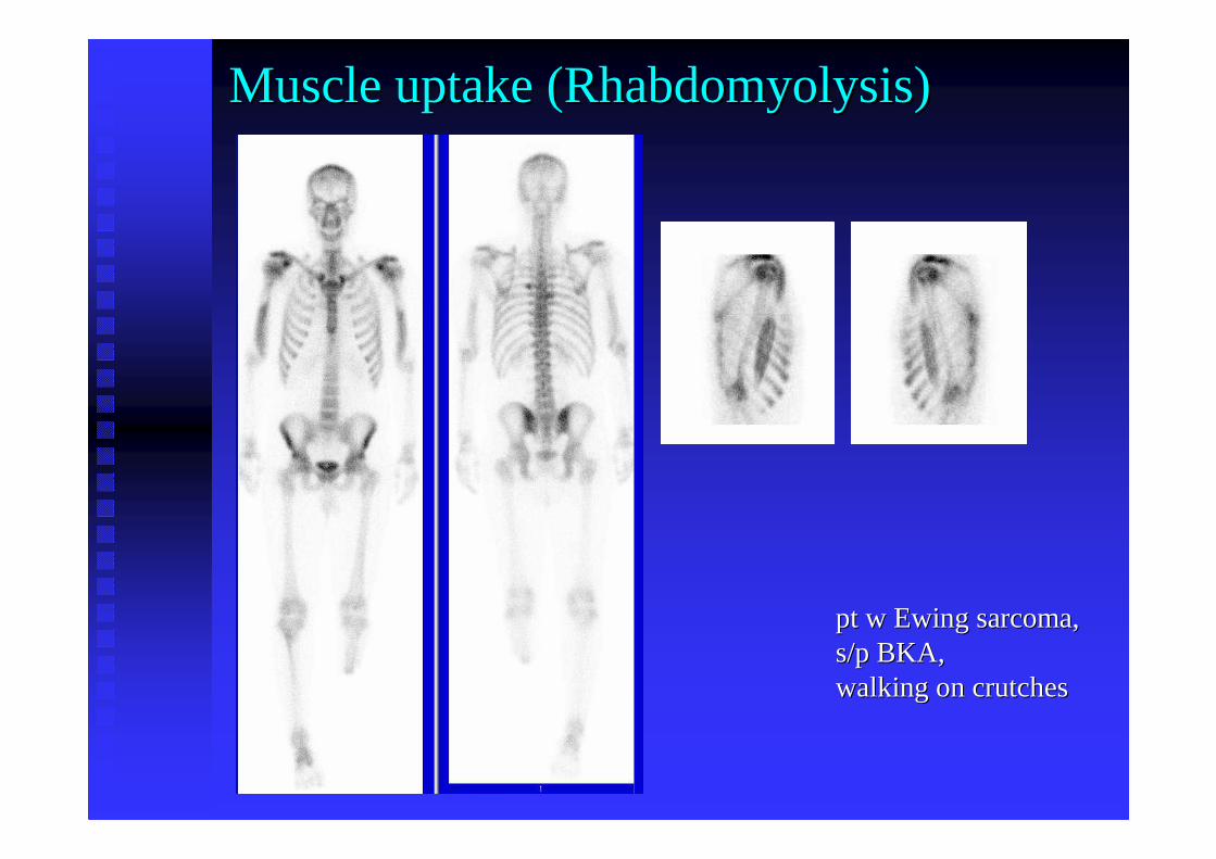
Muscle uptake (Muscle uptake (RhabdomyolysisRhabdomyolysis))
pt w Ewing sarcoma, pt w Ewing sarcoma, s/p BKA, s/p BKA, walking on crutcheswalking on crutches
Trauma to internal organs
Hepatobiliary Scan
�� TcTc--99m IDA (disofenin, mebrofenin)99m IDA (disofenin, mebrofenin)�� dose ~ 150dose ~ 150--250 MBq i.v.250 MBq i.v.
�� imaging of liver, abdomen, pelvis over 1 hrimaging of liver, abdomen, pelvis over 1 hr
�� delayed images if 1delayed images if 1stst hr negativehr negative
�� Bile leak Bile leak -- activity anywhere in peritoneal cavity activity anywhere in peritoneal cavity
�� Common after laparoscopic cholecystectomyCommon after laparoscopic cholecystectomy
�� Usually seals off spontaneouslyUsually seals off spontaneously
�� Leak clin. more significant if no transit into bowel Leak clin. more significant if no transit into bowel
seen (needs surgical intervention)seen (needs surgical intervention)
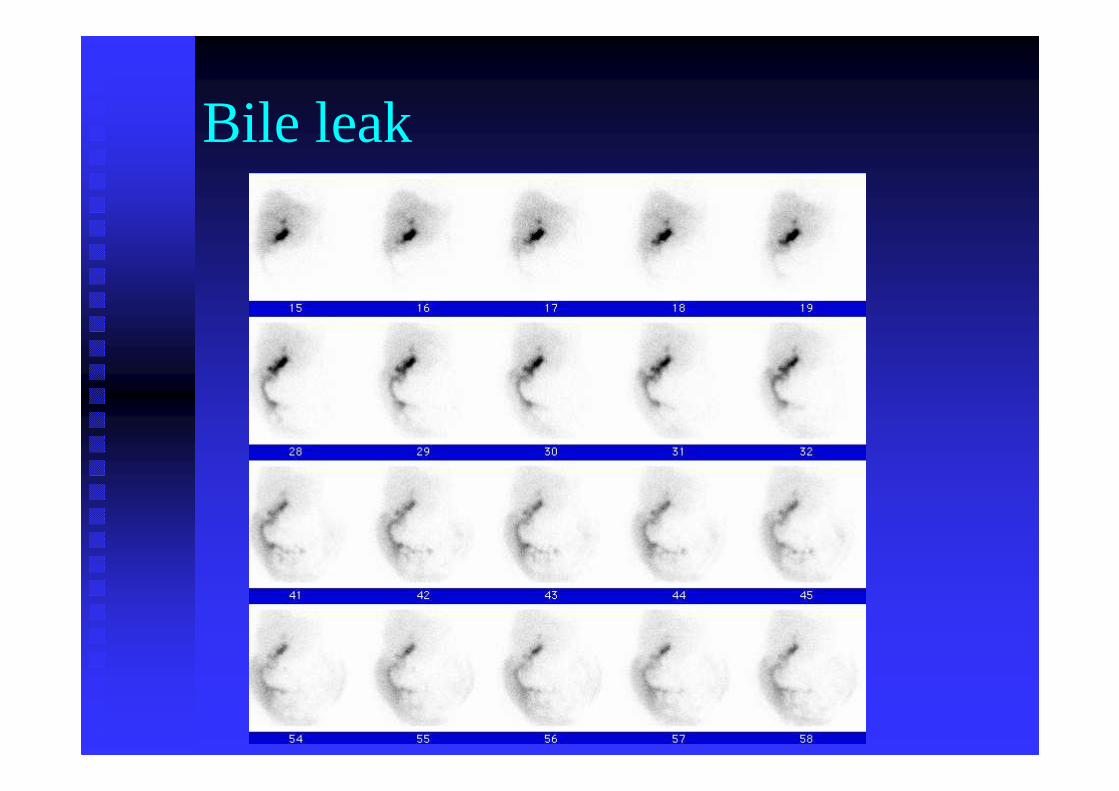
Bile leak
Liver - Spleen Scan
�� TcTc--99m sulfur colloid99m sulfur colloid
�� dose ~ 150dose ~ 150--250 MBq i.v.250 MBq i.v.
�� SPECT imaging better than planar SPECT imaging better than planar
�� Parenchymal defectsParenchymal defects
�� laceration, rupture, laceration, rupture, hematomahematoma
�� Splenosis Splenosis
�� splenicsplenicimplants on peritoneum following spleen ruptureimplants on peritoneum following spleen rupture
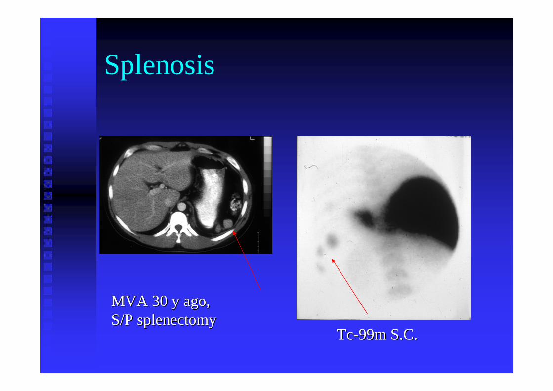
Splenosis
MVA 30 y ago, MVA 30 y ago, S/P S/P splenectomysplenectomy
TcTc--99m S.C.99m S.C.
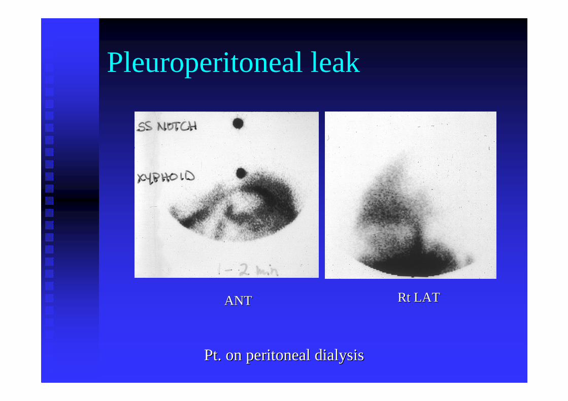
Pleuroperitoneal leak
Pt. on peritoneal dialysisPt. on peritoneal dialysis
RtRt LATLATANTANT
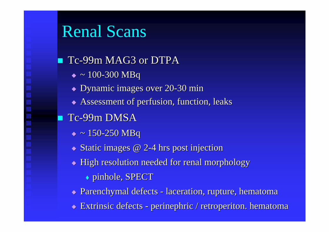
Renal Scans
�� TcTc--99m MAG3 or DTPA99m MAG3 or DTPA�� ~ 100~ 100--300 MBq300 MBq
�� Dynamic images over 20Dynamic images over 20--30 min30 min
�� Assessment of perfusion, function, leaksAssessment of perfusion, function, leaks
�� TcTc--99m DMSA 99m DMSA �� ~ 150~ 150--250 MBq250 MBq
�� Static images @ 2Static images @ 2--4 hrs post injection4 hrs post injection
�� High resolution needed for renal morphology High resolution needed for renal morphology
�� pinhole, SPECTpinhole, SPECT
�� ParenchymalParenchymaldefects defects -- laceration, rupture, hematomalaceration, rupture, hematoma
�� Extrinsic defects Extrinsic defects -- perinephricperinephric/ / retroperitonretroperiton. hematoma. hematoma
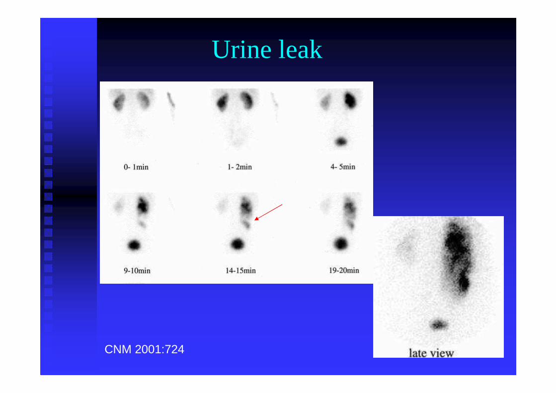
CNM 2001:724
Urine leak
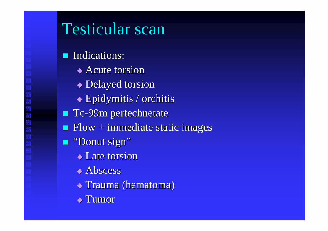
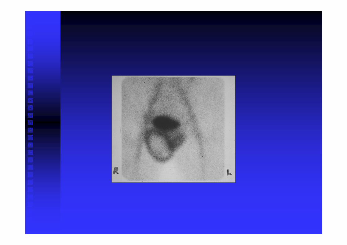
Testicular scan
�� Indications:Indications:�� Acute torsionAcute torsion�� Delayed torsionDelayed torsion�� EpidymitisEpidymitis / / orchitisorchitis
�� TcTc--99m 99m pertechnetatepertechnetate�� Flow + immediate static imagesFlow + immediate static images�� ““ Donut signDonut sign””
�� Late torsionLate torsion�� AbscessAbscess�� Trauma (Trauma (hematomahematoma))�� TumorTumor
Cisternography
�� InIn--111 DTPA intrathecally111 DTPA intrathecally
�� CSF leak CSF leak -- paraspinal (meningeal tears)paraspinal (meningeal tears)
�� CSF CSF rhinorrhearhinorrhea, , otorrheaotorrhea
�� imagingimaging
�� counting nasal pledgets for radioactivitycounting nasal pledgets for radioactivity
�� pledgetpledget/ plasma ratio/ plasma ratio
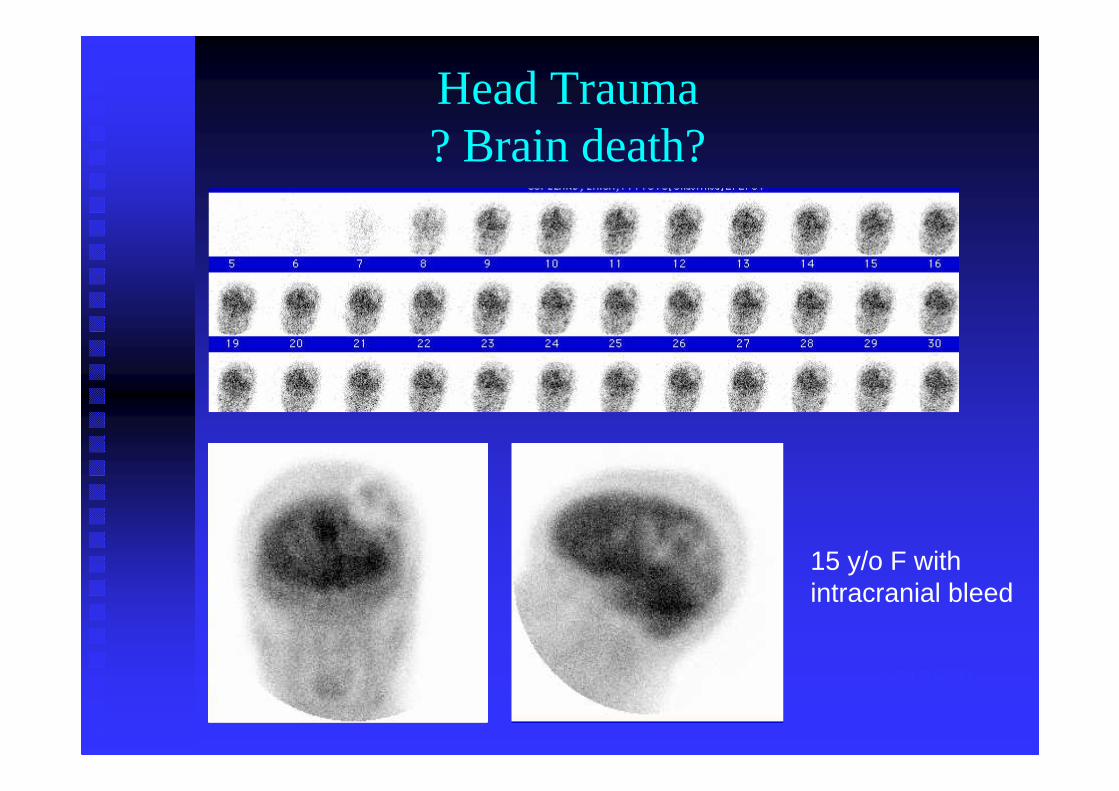
Cerebral perfusion
�� TcTc--99m HMPAO or ECD99m HMPAO or ECD
�� dose ~ 800 MBqdose ~ 800 MBq
�� PostPost--traumatic perfusion defects traumatic perfusion defects
�� Assessment of brain death Assessment of brain death -- role of NM role of NM complementarycomplementary
�� no flowno flow
�� no parenchymal uptakeno parenchymal uptake
1717870
Head Trauma? Brain death?
15 y/o F withintracranial bleed
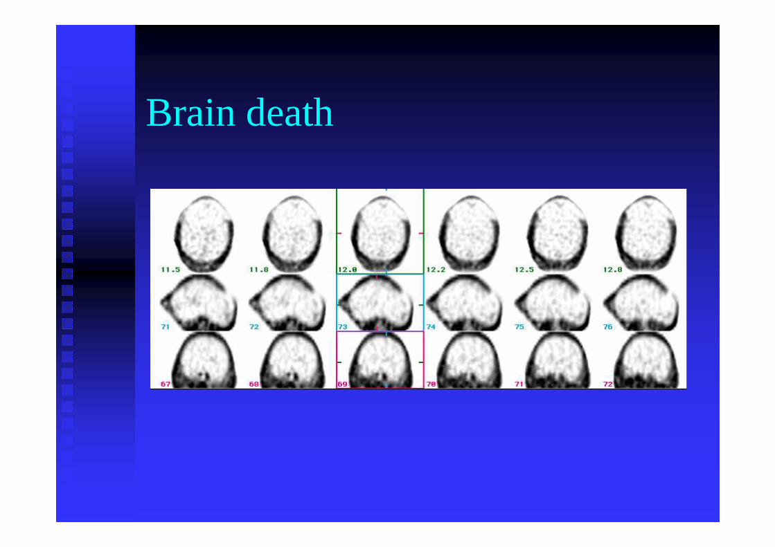
Brain death