Embed Size (px)
Citation preview
66
NUCLEAR AND INFRANUCLEAR LESIONS OF 3,4,6
CRANIAL NERVE LESIONS AND THEIR
CLINICORADIOLOGICAL CORRELATION
Dissertation Submitted to
THE TAMILNADU DR. M.G.R. MEDICAL UNIVERISTY
CHENNAI – 600 032
In partial fulfillment of the regulations
for the Award of the Degree of
BRANCH -1: D.M (NEUROLOGY)
INSTITUTE OF NEUROLOGY
MADRAS MEDICAL COLLEGE
RAJIV GANDHI GOVERNMENT GENERAL HOSPITAL
CHENNAI- 600 003
AUGUST 2013
67
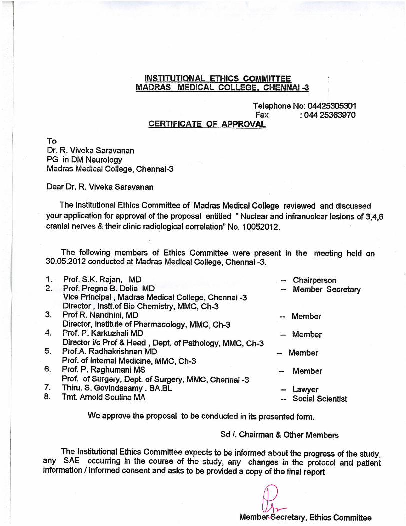
CERTIFICATE
This is to certify that the dissertation entitled “NUCLEAR
AND INFRANUCLEAR LESIONS OF 3,4,6 CRANIAL NERVE
LESIONS AND THEIR CLINICORADIOLOGICAL
CORRELATION” is a bonafide original work of DR.R.VIVEKA
SARAVANAN, in partial fulfillment of the requirements for D.M.
Branch– I (NEUROLOGY) Examination of the Tamil Nadu
Dr.M.G.R Medical University to be held in August 2013, under our
guidance and supervision.
Dr.R.Lakshmi Narasimhan, D.M., Professor of Neurology,
Institute of Neurology,
Madras Medical College,
Chennai – 3.
Dr.C.Mutharasu, DM,
Professor of Neurology,
Institute of Neurology,
Madras Medical College,
Chennai – 3.
Dr.K.Deiveegan, M.Ch.,
Professor and Head,
Institute of Neurology,
Madras Medical College,
Chennai – 3.
Dr.V. Kanagasabai, MD.,
Dean,
Madras Medical College,
Chennai – 3.
68
DECLARATION
I hereby solemnly declare that this dissertation titled
“NUCLEAR AND INFRANUCLEAR LESIONS OF 3, 4, 6
CRANIAL NERVE LESIONS AND THEIR
CLINICORADIOLOGICAL CORRELATION” was done by me
in Institute of Neurology, Madras Medical college and Rajiv
Gandhi Government General Hospital, Chennai -3, under the
guidance and supervision of Prof.R.LAKSHMINARASIMHAN,
MD, D.M, Professor of Neurology, Institute of Neurology, Madras
Medical College & Rajiv Gandhi Government General Hospital,
Chennai. This dissertation is submitted to the Tamil Nadu Dr.
M.G.R. Medical University towards the partial fulfillment of
requirement for the award of D.M Degree Branch I (Neurology).
Place: Chennai
Date: Dr.R.Viveka saravanan, DM, Post Graduate,
Institute of Neurology,
Madras Medical College,
Chennai – 3.
69
ACKNOWLEDGEMENT
It gives me great pleasure to acknowledge all those who
guided, encouraged and supported me in the successful completion
of my dissertation.
First and foremost, I express my gratitude to, the Dean
Dr.V.Kanagasabai M.D. for having permitted me to carry out this
dissertation work at Rajiv Gandhi Government General Hospital,
Madras Medical College, Chennai.
I am extremely thankful to Prof. Dr. K.Deiveegan, M.ch.,
Professor of Neurosurgery, Head of the department, Institute of
Neurology, Rajiv Gandhi Government General Hospital Chennai
for his constant encouragement, valuable guidance and support.
I express my deep sense of gratitude and sincere thanks to our
respected and beloved Chief Dr.R.LakshmiNarasimhan, D.M,
Professor of Neurology, Institute of Neurology, Rajiv Gandhi
Government General Hospital, Chennai for his valuable
suggestions, constant motivation, kind guidance and moral support
without which this study would not have been possible.
70
I express my sincere thanks and gratitude to our Professors
Dr.C.Mutharasu, D.M., Dr. K. Bhanu, D.N.B, D.M., and Dr. S.
Balasubramanian, D.M., Dr.V.Kamaraj, D.M., for their valuable
suggestions and support.
I am extremely thankful to our Assistant Professors
Dr.Ramakrishnan, D.M.,Dr.V.Kannan,D.M, Dr.P.Muthukumar,
D.M. Dr.S.Shanmuga Sundaram. DM., and Dr.N.Shanmuga
sundaram. DM.., for their valuable guidance and support.
I owe my sincere thanks to all the patients and the technical
staff who participated in the study for their cooperation which made
this study possible.
71
CONTENTS
Sl.
No Title
Page
No
1 INTRODUCTION 1
2 AIM OF THE STUDY 3
3 MATERIALS AND METHODS 4
4 REVIEW OF LITERATURE 5
5 RESULTS 32
6 DISCUSSION 47
7 CONCLUSION 55
8 BIBLIOGRAPHY
9 ABBREVIATION
10 ANNEXURES
MASTER CHART
PROFORMA
CONSENT FORM
ETHICAL CLEARANCE
PLAGIARISM
1
INTRODUCTION
Palsies of any of the three cranial nerves supplying the extra
ocular muscles have their presentations, disturbing ocular motility.
Abnormalities of ocular motility help in the localization of lesions
of the cerebral hemispheres, brain stem, cranial nerves (CNs), and
even the striated muscle. Only one nerve may be involved or there
may be a combination of the three nerves. The palsies are usually
acquired.
Sometimes palsies can be congenital due to the
developmental defect of the nucleus or motor nerve fibers. Oculo
motor fibers can be interrupted intraaxially or extraaxially.
Lesions can be in the foramens or extra cranial e.g. Intraorbital.
All these Oculomotor nerves can be affected in the brainstem
(nucleus or fascicular portion)1, in the subarachnoid space, in the
cavernous sinus, at the superior orbital fissure, or in the orbit.
MRI has become the most useful diagnostic tool in the
localization, diagnosis and management of the nuclear and
infranuclear lesions of 3, 4, 6 cranial nerves.
2
In conditions like vasculitis and prothrombotic states,
laboratory investigations add on to radiological findings and
histopathological evaluation is useful when there are therapeutic
difficulties.
Cranial nerves 3, 4, and 6th palsies can be due to head
injury2,26
. In many studies head injury is very well correlated with
imaging. In this study, we have excluded the head injury patients
presenting with 3, 4&6 cranial nerve palsies.
This study is about the clinicoradiological correlation in
nuclear, infranuclear lesions of 3, 4, 6th
cranial nerves.
3
AIM OF THE STUDY
1) To study the demography in patients with nuclear and
infranuclear lesions of 3, 4, 6 cranial nerves
2) To study the clinical localizations in patients with nuclear and
infranuclear lesions of 3, 4, 6 cranial nerves
3) To analyze the radiological findings in patients with nuclear
and infranuclear lesions of 3, 4, 6 cranial nerves.
4) To analyze the various etiological factors of nuclear and
infranuclear lesions of 3, 4, 6 cranial nerves
5) To study clinicoradiological correlation in patients with
nuclear and infranuclear lesions of 3, 4, 6 cranial nerves
4
REVIEW OF LITERATURE
The oculomotor nerve emerges from the supero-ventral aspect
of the pons near the midline and courses in the interpeduncular
cisterns. It passes in between the posterior cerebral arteries and the
superior cerebellar arteries with a slight angulation in the distal
course. Then it passes through the basilar cisterns inferior to the
posterior-communicating arteries and the uncus of the temporal
lobes, after which it enters the cavernous sinuses superiorolaterlly.
The pore for these nerves is lateral, posterior, and superior to the
internal carotid arteries. A small sheath of cerebrospinal fluid may
be present around these nerves on entering the cavernous sinuses.
The third cranial nerve comes out through the superior orbital
fissures. Then it divides into inferior and superior divisions and
innervates the superior, inferior, and medical recti, the inferior
oblique and levator palpebrae muscles, and the pupillary sphincters.
The component controlling the extra ocular muscles is called
somatic component. It‟s nucleus is located in the midbrain at the
level of the superior colliculi, anterior to the sylvian aqueduct near
the midline. These oblong shaped nuclei have three components:
lateral (subdivided into dorsal, intermediate, and ventral parts),
medial, and central sub nuclei. The Edinger-Westphal (EW)
5
nucleus, which controls visceral innervation (mainly constriction of
the pupillary muscles), is behind these nuclei.
The third cranial nerve is formed when the somatic portion
joins the parasympathetic portion from the EW nucleus. Cisternal
portions of the third cranial nerve are better visualized on sagittal,
axial, and coronal MR images. Within the cavernous sinuses, the
third nerves lie supero lateral to the upper portion of the ICA
siphon3. It does not enhance after contrast administration.
The fissures can be easily identified but the structures in the
fissure cannot be differentiated. The 3,4,5,6 cranial nerves are seen
as a group inferior to the anterior clinoid process before exiting
through the superior orbital fissures. Micro vascular disease caused
by diabetes mellitus is the most common cause of third cranial
nerve palsy33
. Other causes like age-related micro vascular changes
and syphilis may also cause third cranial nerve palsy7. Here the end
arteries are affected and the involvement of the nerve is global,
which causes a complete palsy (muscular and pupillary).
Posterior-communicating artery aneurysms compress the
outer portion of the nerve. Pupillary constrictor fibers travel in this
portion, and hence these patients present with an incomplete palsy
6
(blown pupil, same as in herniation of the temporal lobe). Tumors
and trauma are other causes of cranial nerve III palsy8.
Approximately, one third of cranial nerve III palsies are idiopathic9.
Midbrain infarctions may result in complex syndromes with
accompanying third cranial nerve palsies. When the nuclei of the
oculomotor nerves and the ipsilateral cortical spinal tract are
affected, it results in Weber syndrome10
(ipsilateral
ophthalmoplegia and contralateral hemiplegia). If an infarct affects
the nuclei of the oculomotor nerve and adjacent red nucleus, it
results in Benedikt syndrome (Ipsilateral ophthalmoplegia and
contralateral intention tremor). Characteristics of a third cranial
nerve ophthalmoplegia are a wrinkled forehead, raised eyebrow,
eyelid droop, dilated pupil, and a downward and laterally displaced
eye. Infections such as meningitis, particularly tuberculosis, may
result in a third cranial nerve palsy33
. Herniation of a temporal lobe
displaces the brainstem towards the opposite side and this results in
stretching of the oculomotor nerve and results in palsy.
The third cranial nerves are affected by
1) Aneurysm
2) Vascular disease (infarction in the brainstem and/or the nerve)
7
3) Tumor
4) Trauma
Diseases affecting the cavernous sinus may also result in third
cranial nerve palsy (Foix syndrome). Third cranial nerve palsy in
combination with a superior orbital fissure syndrome is called
Rochon-Duvigneaud syndrome11
.
A lesion at the level of the orbital apex results in Rollet
syndrome12
. A lesion at the petrosphenoidal junction affecting also
the second, fourth, and sixth cranial nerves is called Garcin
syndrome13
. Osteitis of the petrous apex (Gradenigo syndrome)14
results in palsies of the third, fifth, and sixth cranial nerves.
Fractures affecting the sphenoid bone can occasionally result in
palsies of the oculomotor nerves.
Third nerve palsy
Third nerve palsy can be diagnosed by abnormalities in the
function of the superior rectus, inferior rectus, inferior oblique and
medial rectus muscles. As the third nerve innervates levator
palpebrae superioris and pupil, involvement of the eyelid with
induced ptosis is seen and the eye is deviated out and slightly
down. Isolated extrtraocular muscle palsies are unlikely to
8
represent a third nerve palsy, especially in the following cases :an
apparent superior rectus weakness without ptosis; an inferior
oblique weakness without medial and inferior rectus weakness,
which is possibly Brown syndrome; or an isolated medal rectus
weakness, most likely an INO. Myasthenia can involve the extra
ocular muscles and levator but it spares the pupil and is not
associated with pain.
Abnormalities of the pupil such as mydriasis and failure to
react may be signs of third nerve involvement. However, because of
the peripheral location of the pupillary fibers in the extra axial
portion of the third nerve, it is possible for sparing of the pupil to
coexist with even complete involvement of the extra ocular muscles
and levator. Rarely an acute third nerve palsy to be caused by a
mass lesion when the pupil is completely spared, which is not true
for progressive oculomotor dysfunction. 50% of slowly progressive
third nerve palsies resulting from a slowly expanding parasellar
lesion (carotid cavernous aneurysm, meningioma, pituitary
adenoma, etc. may be unassociated with pupillary compromise. In
addition, sympathetic dysfunction, which is common with
cavernous sinus lesions, may partially mask pupillary involvement
by reducing mydriasis.
9
Acute onset of third nerve palsy associated with a dilated, and
reactive pupil is presumed to represent an aneurysm, usually of the
posterior communicating artery. When associated with headache
and stiff neck, it may represent an aneurysmal subarachnoid
hemorrhage.
Neoplastic, inflammatory and even micro vascular causes
may result in a pupil- involving third nerve palsy. Pain is almost a
constant finding with aneurysmal third nerve palsy, but it is also
frequent with micro vascular paresis15
. Third nerve dysfunction
associated with opthalmoplegic migraine can mimic that caused by
an aneurysm. This rare syndrome is usually preceded by a headache
and initial onset essentially always occurs in childhood.
Congenital third nerve palsies often represent perinatal
trauma, involvement of the nerve is variable39
, aberrant
regeneration is common and divisional palsies may occur. Slowly
progressive oculomotor palsies are an indication for repeat MRI in
spite of previous negative studies, as neurlemmomas can remain
cryptic. One rare congenital syndrome is cyclic oculomotor
palsy16
.an eye with an underlying palsy develops intermittent
spasms during which the paretic eye turns from its exotropic
position towards midline, the ptotic eyelid elevates and the dilated
10
pupil constricts.
Acute isolated pupil-sparing third nerve palsy in a
vasculopathic patient over 40 does not require work-up other than
blood pressure and fasting blood sugar tests. It is important to
follow these patients as the expected recovery is within 3 months33
.
In a third nerve palsy associated with trauma a careful check for
carotid cavernous fistula is essential.
When the pupil is enlarged or the third nerve involvement is
progress, a work up is indicated. MRI with gadolinium is the
primary diagnostic study. When combined with MRA, it can show
aneurysms larger than4mm. Inflammatory lesions and even some
infiltrates (lymphoma) may not show up on MRI.CSF analysis for
cells and protein may be helpful. Often serial MRIs over a period of
years are required to uncover the etiology in cryptogenic third
nerve palsies.
Following viral infections 33
or vaccination, children may have
transient opthalmoplegia. In older patients, an erythrocyte
sedimentation rate may be used to screen for giant cell arteritis.
Since giant cell arteritis is an important cause of opthalmoplegia17
in elderly, it is worthwhile to consider giant cell arteritis as a cause
11
even though diplopia that results from giant cell arteritis is a result
of orbital hypoperfusion that leads to extraocular muscle ischemia.
Diagnosis is more difficult in cases of incomplete third nerve
palsy. It was previously thought that divisional palsies (superior
rectus and levator, or medial rectus, inferior rectus and inferior
oblique) indicate pathology within the cavernous sinus or orbital
apex. Now it is known that divisional palsies can also occur with
pathology more proximally situated including within the midbrain.
Although microvascular diseases can cause divisional involvement,
these patients should be carefully worked up with MRI with contrast.
Ophthalmic artery aneurysms can cause superior division palsies.
Relative pupil sparing also presents diagnostic difficulties. In
the presence of significant mobility impairment, slight dilatation of
the pupil may occur with microvascular disease. Progressive
pupillary enlargement is always an indication for work –up.
As a damaged third nerve recovers misdirection of fibre
growth can result in aberrant regeneration. The classical findings in
aberrant regeneration are
1) Eyelid elevation or hang up with adduction and depress
2) Miosis with elevation
12
The causes of acquired third nerve palsy are
1) Brain stem lesions
2) Inflammatory conditions like meningitis, encephalitis, toxin
exposure causing polyneuritis, Echo virus infection, Herpes
infection
3) Vascular causes like ICA dissection, aneurysms and carotico
cavernous fistula
4) Tumors like Glioblastoma multiforme
5) Demyelinating disorders
6) Trauma
7) Miscellaneous causes are
a. Anterior communicating artery aneurysm
b. Bilateral chronic subdural hematoma
c. Congenital toxoplasmosis
d. “Crack” cocaine
e. Diagnostic angiography
f. Eosinophilic granuloma of the optic nerve
13
g. Frontal sinus mucocele
h. Infectious mononucleosis
i. Leukemia
j. Measles immunization
k. Myasthenia gravis
l. Ophthalmoplegic migraine
m. Polyarteritis nodosa
n. Porphyria
o. Sarcoidosis
p. Schwannoma
q. Temporal arteritis
r. Viagra therapy (sildenafil citrate)
s. HIV20
TROCHLEAR NERVE (IV)
The trochlear nerve is the only cranial nerve arising from the
dorsal brainstem and has the longest intracranial course (especially
in their cisternal portion). The fourth cranial nerve is 7.5 cm long
14
approximately. At the level of the inferior colliculi these nerves
decussate before emerging from the dorsal midbrain and they are
the most slender of all cranial nerves coursing parallel to the free
edge of the tentorium between the posterior and superior cerebellar
arteries. They enter the cavernous sinuses inferior to the
oculomotor nerves and usually cannot be individually discriminated
within the lateral wall of the cavernous sinus.
It travels in the outer dural wall of the cavernous sinuses.
They innervate only the superior oblique muscles after exiting
through the superior orbital fissures.
These nerves cannot be seen by routine MR imaging
generally because of their small size, but it is possible to identify
their cisternal portions in approximately 20% of all coronal
examinations4. They are never affected in an isolated fashion.
Fourth cranial nerve palsies are commonly caused by trauma
(because of their long course), tumor, brainstem infarction, and
aneurysm19,22
. About 50% of fourth cranial nerve palsies are
idiopathic. Diseases involving the superior orbital fissure and the
orbital apex can affect fourth cranial nerves. Patients with fourth
cranial nerve palsy typically have difficulty going downstairs.
15
Patients turn their head and may present with torticollis to
compensate for the weakness of downward and lateral gaze.
FOURTH NERVE PALSY
A fourth nerve palsy is the presumptive diagnosis in a patient
presenting with vertical double vision and when there is no history
s/o restrictive phenomena. Finding a hyperdeviation also raises
suspicion of a fourth nerve palsy. Hyperdeviation increases on
contralateral gaze and with ipsilateral head tilt.
The lines of the maddox rod are tilted with the patient and the
testing must be done with the patient upright. Subtle restrictive
problems or a skew deviation must be reconsidered if the deviation
does not follow the expected pattern. Double maddox rod testing
should show evidence of excyclotorsion between 3 –10 degrees.
Results that show greater than 10 degrees of excyclotorsion raise
the possibility of a bilateral fourth nerve palsy.
Fourth nerve palsies are either congenital or acquired. Head
tilt in old photographs or vertical fusional range of greater than 3 D
is strongly suggestive of a long standing or congenital fourth nerve
paresis. Often patients are unaware of a previous head tilt until o ld
photographs including driver's licenses are examined. Head trauma
16
is the most common cause of an acquired fourth nerve palsy which
may have been so trivial the patient had forgotten it. Microvascular
causes are the leading diagnosis of fourth nerve palsies in older
patients.
It is extremely uncommon for nuclear involvement of the
fourth nerve although it has been reported with vascular disease
(including arteriovenous malformation), trauma and demyelination.
The fourth nerve may be involved as it exits the brain stem,
crossing in the superior medullary velum. Pineal gland lesions
(pinealoma, pineoblastoma, teratoma, dysgerminoma or
choriocarcinoma) may all affect the fourth nerve, usually bilaterally
and frequently with other signs of the dorsal mid brain syndrome.
Other inflammatory pathology (including meningitis) and vascular
phenomenon (including carotico cavernous fistula) can impair
trochlear function within the area of cavernous sinus. In the
subarachnoid space the fourth nerve may be affected by
microvascular abnormalities quite often24
. Following a
neurosurgical procedure iatrogenic fourth nerve paresis is not rare,
occurring most frequently when the tentorial edge is cut.
To make sure, the fourth nerve palsy is clearing older patients
should be followed up. Restrictive syndromes such as thyroid
17
ophthalmopathy or old trauma and myasthenia gravis should be
considered.
ABDUCENT NERVE
The sixth cranial nerves emerge at the Ponto medullary
junction. The cisternal portions are inconsistently identified on
sagittal and coronal MR images. They appear as thin lines of
intermediate signal intensity coursing parallel to the clivus5. They
can be easily mistaken for the anterior wall of the basilar artery or
for pulsation artifacts within the prepontine cistern. The sixth
cranial nerves enter the medial petrous bones via the Dorello‟s
canal6. Then they enter cavernous sinuses medial to the gasserian
ganglia. They are prone to injuries as they are the only cranial
nerves coursing inside of the cavernous sinuses.
These cranial nerves travel inferior to the ophthalmic division
of cranial nerve V. Via the superior orbital fissure they exit the
skull. They innervate the lateral recti muscles. Common pathologies
affecting them are tumors, trauma, infarction, demyelinating
diseases, and vascular pathology of the cavernous sinuses (e.g.,
ICA– cavernous sinus fistula). More than 40% of cranial nerve VI
palsies are idiopathic.
18
The nuclei for the sixth cranial nerves are located close to the
midline (most motor cranial nerve nuclei are nearly midline in
position) at the level of the floor of the fourth ventricle. The nuclei
are located medially because the fibers for facial nerves wrap
around them creating slight bulges on the floor of the fourth
ventricle (facial colliculi).
The nuclei for the abducens nerves communicate via the
medial longitudinal fasiculi, with the third cranial nerve nuclei and
coordinate eye movements.
SIXTH NERVE PALSY
In the finding of an isolated abduction deficit a sixth nerve
palsy is suspected. The deficit need not be complete and may, be
indicated only as an esodeviation increasing on ipsilateral gaze.
Slowed ipsilateral saccades strongly suggest a sixth nerve etiology.
Inflammatory lesions (post viral, demyelinating etc.) may
affect the sixth nerve within the brainstem, particularly in the
young patient, and by vascular lesions, especially in the older
patients. Metabolic disease such as vitamin B Wernicke-Korasakoff
syndrome can also affect the sixth nerve. A pontine glioma may
present as unilateral or bilateral sixth nerve palsy in children.
19
Intra axial lesions usually result in other neurologic findings,
including involvement of the seventh cranial nerve and facial
sensation. If the ventral brain stem is involved, associated
abnormalities of the corticospinal tract lead to contralateral
hemiparesis (Millard Gubler syndrome)18
. Nuclear involvement of
the sixth nerve means the patient will show gaze palsy, usually
without associated diplopia. When the gaze palsy begins to clear,
additional involvement of the fascicle will lead to more rapid
improvement in the adducting eye, causing the appearance of a
relative ipsilateral sixth nerve palsy and possibly diplopia.
Following types of pathology can affect the sixth nerve within the
subarachnoid space:
Inflammatory (sarcoid)
Infiltrative(including lymphoproliferative abnormalities)
Infectious (especially basilar meningitis such as tuberculosis
or fungus)
Compressive (usually arising from the clivus, meningioma,
chordoma,chondrosarcoma, metastatic disease)
In addition, lesions of the cerebellar pontine angle may get
large enough to affect the sixth nerve (neurlemmomas of eight
20
nerve, the auditory nerve or meningiomas). These lesions are
generally associated with decreased hearing and vestibular findings
as well as seventh nerve dysfunction and facial sensory loss).
Finally the subarachnoid sixth nerve may be affected by shifts in
the positions of the brain stem that cause the tugging on the nerve
itself. Changes in the intracranial pressure as with acute
hydrocephalous, following lumbar puncture, and associated with
pseudotumor cerebri are particularly likely to affect the sixth
nerve21
.
As the sixth cranial nerve passes over the petrous pyramid, it
may be affected by trauma, enlargement of the inferior petrous
pyramid (Gradenigo syndrome), or neoplastic processes.
Meningiomas or neurlemmomas may affect the sixth nerve within
the posterior cavernous sinus or as it crosses the petrous pyramid.
In addition, nasopharyngeal carcinoma invading the skull base
through the foramen lacerum may produce sixth nerve palsy as well
as pain and decreased hearing. Aneurysms of the intracavernous
carotid artery often present with sixth nerve palsy but may also
have sensory changes and a Horner's syndrome.
In isolation congenital sixth nerve palsies almost never
occur.25
Congenital absence of the sixth nerve is seen in Duane
21
syndrome. The lateral rectus muscle in these patients is innervated
by various branches from the third nerve, which results in
narrowing of the palpebral fissure with attempted adduction. Co-
contraction commonly causes upshoot and downshoot. Patients are
frequently unaware of the abnormality and usually do not have
trouble with double vision. In Mobius syndrome, bilateral
involvement of the sixth and seventh nerves is seen. Other lower
cranial nerves may also be involved. Using their intact adduction
patients with both Duane and Mobius syndromes cross fixate,.
It is important to determine whether the non-traumatic sixth
nerve palsy is isolated. Evidence of associated dysfunction of the
third, fourth, fifth, seventh or eighth cranial nerves or of
sympathetic abnormalities is an indication for a work up including
at least an MRI with and without gadolinium. Increased intracranial
pressure such as history of headaches or findings of papilledema
and obesity might indicate that the sixth nerve palsy is non-
localizing. An acute inflammatory or microvascular sixth nerve
palsy may cause pain and significant pain is an indication for a
work up. In the older patients, the diagnosis of a microvascular
sixth nerve palsy anticipates that it should clear within a period of
2-3 months. Any evidence of progression or failure to resolve is an
22
indication for MRI. In children post viral inflammatory lesions may
be seen.
In adolescents and young adults demyelinating disease may
be an under-recognized cause of sixth nerve palsy. Yield of
neuroimaging (MRI) is low in the absence of other findings. Failure
to resolve or development of any other long tract signs of brain
stem findings suggests the presence of pontine or cerebellar glioma,
ependymoma or medulloblastoma. Patients must be followed up
and persistence of sixth nerve dysfunction is always an indication
for MRI. Metabolic syndromes causing sixth nerve palsies are rare.
In the setting of negative MRI cerebrospinal fluid analysis may be
indicated.
Persistent limitation of vertical gaze can be caused by co-
contraction of the superior and inferior recti.
MULTIPLE CRANIAL NERVE PALSIES
Diagnosis of involvement of more than a single ocular motor
nerve paresis is critical. It is difficult to detect involvement of the
fourth nerve in the presence of a third nerve palsy. In this condition
there is failure of intorsion with attempted downgaze with the eye
in abduction. Microvascular diseases rarely cause simultaneous
23
involvement of more than one cranial nerve. Paresis of
extraocularmuscles innervated by two or more ocular nerve requires
work up. Pain or facial sensory loss suggests pathology in the area
of cavernous sinus that may be neoplastic (meningioma,
neurilemoma, pituitary adenoma, metastatic disease), vascular
(caraticocavernous fistula or aneurysm) or inflammatory (sarcoid,
Tolosa Hunt syndrome). In MRI we can visualize the pathology in
majority of cases. Evidence of neoplastic and inflammatory cells in
cerebrospinal fluid should be considered if the MRI is negative.
Elevated proteins and pleocytosis may appear in the CSF in
the setting of post infectious polyradiculoneuropathy (Fisher
syndrome). Botulism can cause multiple cranial nerve palsies
associated with diffuse weakness, particularly respiratory. Painful
ophthalmoplegia of Tolosa Hunt syndrome responds to
corticosteroid therapy. This response has been suggested as a
diagnostic criterion. Mass lesions (particularly lymphoproliferative)
may also respond to a course of corticosteroids and Tolosa Hunt
syndrome should be considered a diagnosis of exclusion following
MRI with gadolinium. Failure to fit the pattern of single cranial
nerve palsy should always stimulate reconsideration of myasthenia,
skew deviation or restrictive strabismus.
24
Bilateral cranial nerve involvement is always an indication
for work up. Bilateral sixth nerve palsies rarely caused by
microvascular disease. In this setting MRI should be the first step
in approaching diagnosis.
CAVERNOUS SINUS
Cavernous sinuses are
1) Paired venous structures located lateral and inferior to
sphenoid sinus/hypophysis, anterior and medial to the petrous
apex, and posterior to the optic canal and inferior and
superior orbital fissures.
2) These are endothelial-lined structures containing multiple
septations.
3) They are the only veins in the body to be traversed by arteries
(ICAs).
4) They form the lateral sellar compartment of the extradural
neuroaxis, which is a system of veins extending from the
orbits via the spinal epidural space to the coccyx.
25
The dural walls of the cavernous sinuses are characterized as
follows:
1) The outer layer extends to the clinoids and laterally to the
middle cranial fossae where it continues as the dura matter.
The outer dural layer may contain a separate venous
compartment called the lateral sinus.
2) The inner layer follows the bone margins of the sinus and has
secondary septations, including those forming the Meckels
cave.
3) The petroclinoid ligaments separate the cavernous sinuses
into an oculomotor trigone (containing most cranial nerves)
and a vascular trigone (located medially).
There are vascular communications between the cavernous
sinuses located as follows:
1) Basilar venous sinus or plexus communications are located
dorsal to the clivus.
2) Anterior intercavernous communications are located ventral
to the hypophysis.
3) Posterior intercavernous communications are located dorsal
to the hypophysis.
26
4) Intrasellar intercavernous communications are located
between the anterior and posterior lobes of the pituitary
gland.
5) All of these communications may be normally seen in
children (particularly in babies) but are not easy to see in
adults. They are enlarged in cavernous sinus-carotid artery
fistulas (direct and indirect) and unilateral processes
obstructing the drainage of one cavernous sinus.
The porous trigeminus is the dorsal opening of the cavernous
sinuses through which the main trunk of the fifth cranial nerves
courses. The Meckel cave is a dural reflection containing the
semilunar ganglion, which is surrounded by the trigeminal cistern.
The mandibular branch of the third division of the trigeminal
nerve exits from the Meckel cave via the foramen ovale. It does not
enter the venous portion of the cavernous sinus. The walls of the
cavernous sinus enhance normally.
The features of gasserian (semilunar) ganglion are:
1. Its trunk separates in three bundles immediately.
2. It is not a true ganglion.
27
3. The inferolateral aspect of a gasserian ganglion
may enhance normally after gadolinium is given, possibly
because of a combination of perineural vascular plexus and
presence of ganglion cells
The oculomotor trigone comprises the cranial nerves III, IV,
V1, and V2 located between the inner and the outer dural layers
forming the lateral walls of the cavernous sinuses, in which the ICA
is located between the lateral wall of the sphenoid sinus and inner
dural layer of the cavernous sinus. The carotid artery lies mostly
outside the sinus but becomes intracavernous in some portions. Th is
region comprises the vascular trigone. Here the cavernous sinus
contains venous blood and multiple septations.
Sixth cranial nerves may be surrounded by the inner dural
layer or be within the cavernous sinus.
Before exiting the superior orbital fissure the Cranial nerves
III, IV, VI and V1 are grouped inferolateral to anterior clinoid. The
ophthalmic vein and artery are contained in the middle of the
anterior region of the fissure. The anteroinferior portion of the
fissure contains the maxillary division of the trigeminal nerve
before it exits via foramen rotundum.
28
The superior dural ring surrounding the ICA marks the
superior-most aspect of the cavernous sinuses. It comprises the
outer dural layer. It is inseparable from the arterial adventia. The
inner dural layer forms the inner dural ring. The space between
both rings is called the carotid cave. The ophthalmic and superior
hypophyseal arteries lie outside the cavernous sinus in most
individuals.
Structures that drain into and out of the cavernous sinus are
1) Into the cavernous sinuses: the superior and inferior
ophthalmic veins, the pterygoid plexus, the superficial
sylvian vein, and the uncal (temporal) vein.
The pterygoid plexus drains into the cavernous sinus via the
inferior ophthalmic vein, the vein of the foramen ovale, the vein of
the foramen spinosum, the vein of foramen lacerum, and the veins
of the carotid canal.
2) Out of the cavernous sinus: the inferior and superior petrosal
sinuses
In patients with cranial nerve palsies MRI is the imaging
method of choice. The oculomotor cranial nerves can be visualized
by standard MR sequences. In MR images of normal subjects, it is
29
demonstrated that the oculomotor nerve, the trochlear nerve and the
abducens nerve can be identified not only in the subarachnoid space
and cavernous sinus, but also in the orbit. However, a precondition
is the use of appropriate imaging sequences and planes (e.g.,
subarachnoid cisterns: T2-weighted fast spin-echo or T2-weighted
three-dimensional sequences in oblique-axial and sagittal planes;
cavernous sinus: contrast-enhanced T1-weighted coronal images;
orbit: T1-weighted images without contrast agent in the coronal
plane obtained using surface coils). Imaging the cranial nerves is
clinically important not only for diagnostic purposes in eye muscle
palsies but also for planning surgical procedures at the cranio-
orbital junction.
Discussing about one of the etiologies, opthalmoplegic
migraine is a rare condition that almost always has its onset in
childhood. Recurrent attacks are stereotypical. Every episode
begins with a unilateral orbital and retro-orbital headache,
accompanied by vomiting, that may last for 1 to 4 days. During the
migrainous attack, ipsilateral ptosis occurs and within a few hours
progresses to a complete paralysis of cranial nerve III. Cranial
nerves IV or VI may be rarely involved. The neurological deficit
can last from hours to several months. In the past, the focal nature
30
of the deficit often led to major investigations including
angiography to rule out the presence of an internal carotid or
posterior communicating artery aneurysm. Usually, no abnormality
is found. MRI scans have shown thickening and contrast
enhancement of the nerve as it exits the midbrain, which may
persist after the third nerve palsy has disappeared and this
represents a recurrent demyelinating/inflammatory neuropathy. It is
to be determined whether cases with no identifiable cause (or
cranial nerve enhancement on MRI) and spontaneously remitting
episodes represent a form of migraine. The prognosis is favorable
for recovery unless attacks occur very frequently.
31
MATERIALS AND METHODS
In this study, we analyzed data of patients with nuclear and
infranuclear lesions of 3, 4, 6 cranial nerves, who were inpatients in
Rajiv Gandhi Government General Hospital, Chennai between
February 2011 to February 2013.Data pertaining to patient
demographics,signs,investigations and radiological findings were
analyzed . Informed consent was taken for enrolment in the study
and for the investigations. X-ray skull, CT brain plain and contrast
(if necessary) and MRI brain plain and contrast study (if necessary)
were done in all the patients, in the department of radiology in
Rajiv Gandhi Government General Hospital, Chennai.
INCLUSION CRITERIA
Patients with Nuclear and infranuclear lesions of 3, 4, 6
cranial nerves, who were inpatients in Rajiv Gandhi Government
General Hospital, Chennai, between February 2011 to February
2013.
EXCLUSION CRITERIA
Patients with lesions of 3, 4, 6 cranial nerves following head
trauma were excluded from this study
32
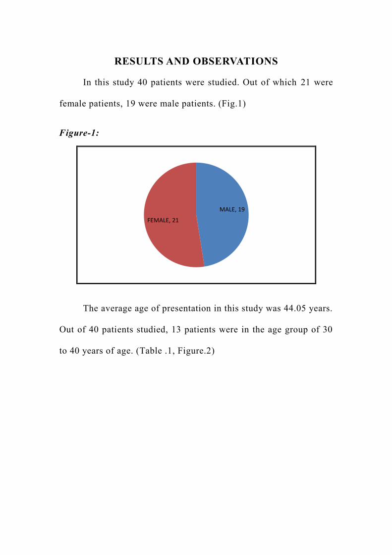
RESULTS AND OBSERVATIONS
In this study 40 patients were studied. Out of which 21 were
female patients, 19 were male patients. (Fig.1)
Figure-1:
MALE, 19
FEMALE, 21
The average age of presentation in this study was 44.05 years.
Out of 40 patients studied, 13 patients were in the age group of 30
to 40 years of age. (Table .1, Figure.2)
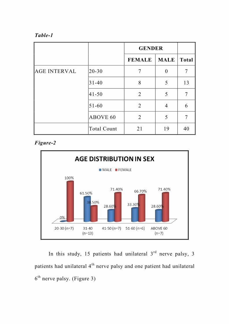
33
Table-1
GENDER
FEMALE MALE Total
AGE INTERVAL 20-30 7 0 7
31-40 8 5 13
41-50 2 5 7
51-60 2 4 6
ABOVE 60 2 5 7
Total Count 21 19 40
Figure-2
In this study, 15 patients had unilateral 3rd
nerve palsy, 3
patients had unilateral 4th
nerve palsy and one patient had unilateral
6th
nerve palsy. (Figure 3)
34
Figure-3
15
3
1
unillateral nerve palsy
unilateral nerve palsies
3rd 4th 6th
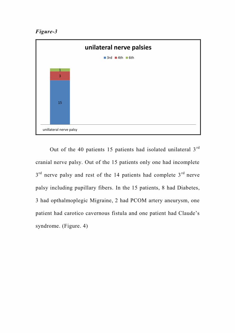
Out of the 40 patients 15 patients had isolated unilateral 3rd
cranial nerve palsy. Out of the 15 patients only one had incomplete
3rd
nerve palsy and rest of the 14 patients had complete 3rd
nerve
palsy including pupillary fibers. In the 15 patients, 8 had Diabetes,
3 had opthalmoplegic Migraine, 2 had PCOM artery aneurysm, one
patient had carotico cavernous fistula and one patient had Claude‟s
syndrome. (Figure. 4)
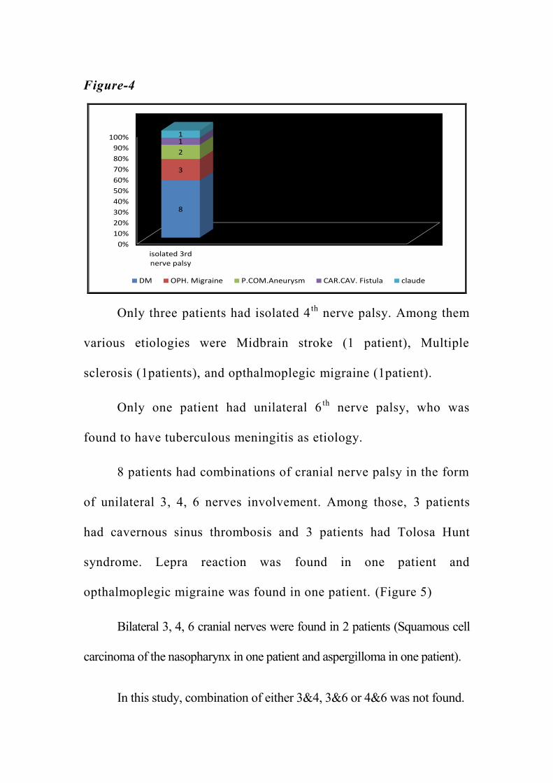
35
Figure-4
0%
10%
20%
30%
40%
50%
60%
70%
80%
90%
100%
isolated 3rdnerve palsy
8
3
2
1 1
DM OPH. Migraine P.COM.Aneurysm CAR.CAV. Fistula claude
Only three patients had isolated 4th
nerve palsy. Among them
various etiologies were Midbrain stroke (1 patient), Multiple
sclerosis (1patients), and opthalmoplegic migraine (1patient).
Only one patient had unilateral 6th
nerve palsy, who was
found to have tuberculous meningitis as etiology.
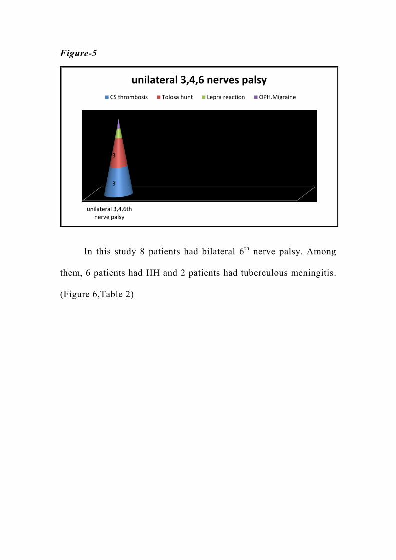
8 patients had combinations of cranial nerve palsy in the form
of unilateral 3, 4, 6 nerves involvement. Among those, 3 patients
had cavernous sinus thrombosis and 3 patients had Tolosa Hunt
syndrome. Lepra reaction was found in one patient and
opthalmoplegic migraine was found in one patient. (Figure 5)
Bilateral 3, 4, 6 cranial nerves were found in 2 patients (Squamous cell
carcinoma of the nasopharynx in one patient and aspergilloma in one patient).
In this study, combination of either 3&4, 3&6 or 4&6 was not found.
36
Figure-5
unilateral 3,4,6thnerve palsy
3
3
1
1
unilateral 3,4,6 nerves palsy
CS thrombosis Tolosa hunt Lepra reaction OPH.Migraine
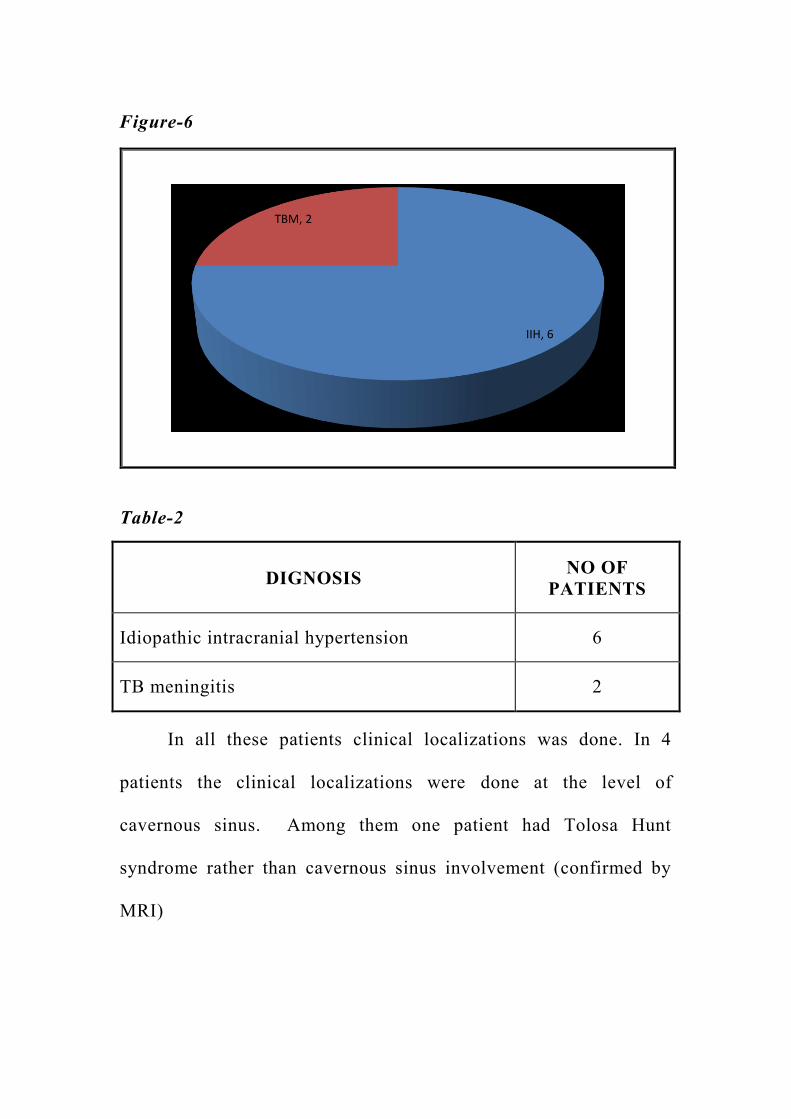
In this study 8 patients had bilateral 6th
nerve palsy. Among
them, 6 patients had IIH and 2 patients had tuberculous meningitis.
(Figure 6,Table 2)
37
Figure-6
IIH, 6
TBM, 2
Table-2
DIGNOSIS NO OF
PATIENTS
Idiopathic intracranial hypertension 6
TB meningitis 2
In all these patients clinical localizations was done. In 4
patients the clinical localizations were done at the level of
cavernous sinus. Among them one patient had Tolosa Hunt
syndrome rather than cavernous sinus involvement (confirmed by
MRI)
38
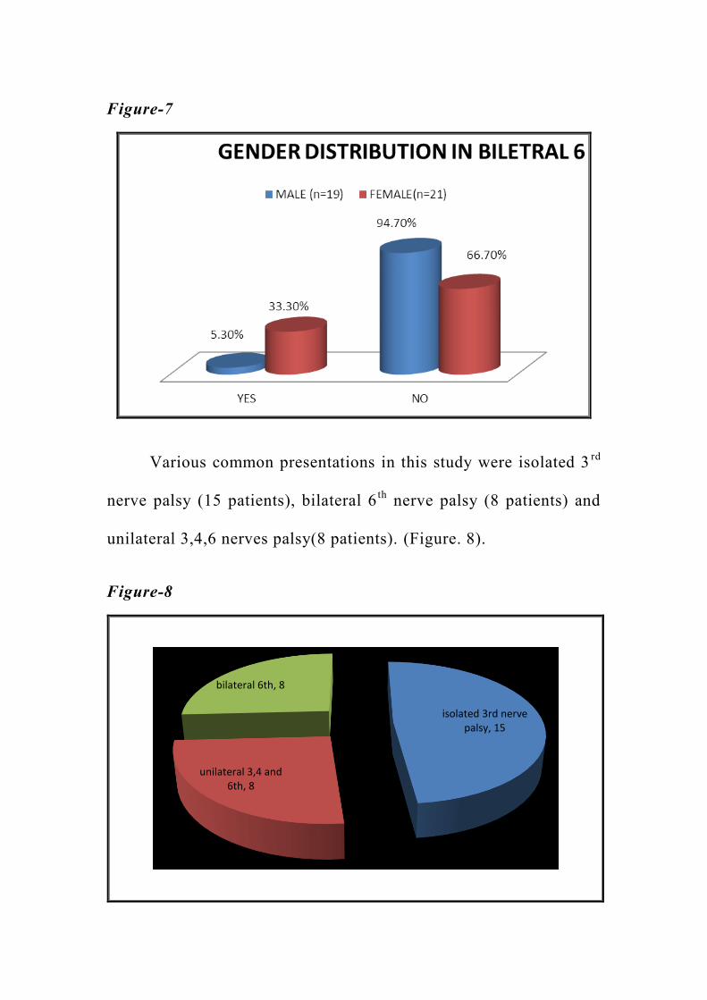
Figure-7
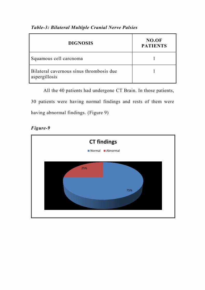
Various common presentations in this study were isolated 3rd
nerve palsy (15 patients), bilateral 6th
nerve palsy (8 patients) and
unilateral 3,4,6 nerves palsy(8 patients). (Figure. 8).
Figure-8
isolated 3rd nerve palsy, 15
unilateral 3,4 and 6th, 8
bilateral 6th, 8
39
Table-3: Bilateral Multiple Cranial Nerve Palsies
DIGNOSIS NO.OF
PATIENTS
Squamous cell carcnoma 1
Bilateral cavernous sinus thrombosis due
aspergillosis
1
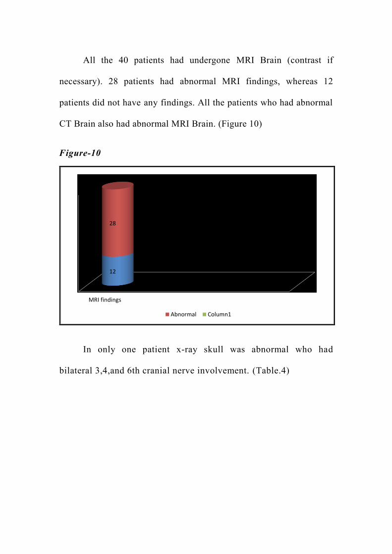
All the 40 patients had undergone CT Brain. In those patients,
30 patients were having normal findings and rests of them were
having abnormal findings. (Figure 9)
Figure-9
75%
25%
CT findings
Normal Abnormal
40
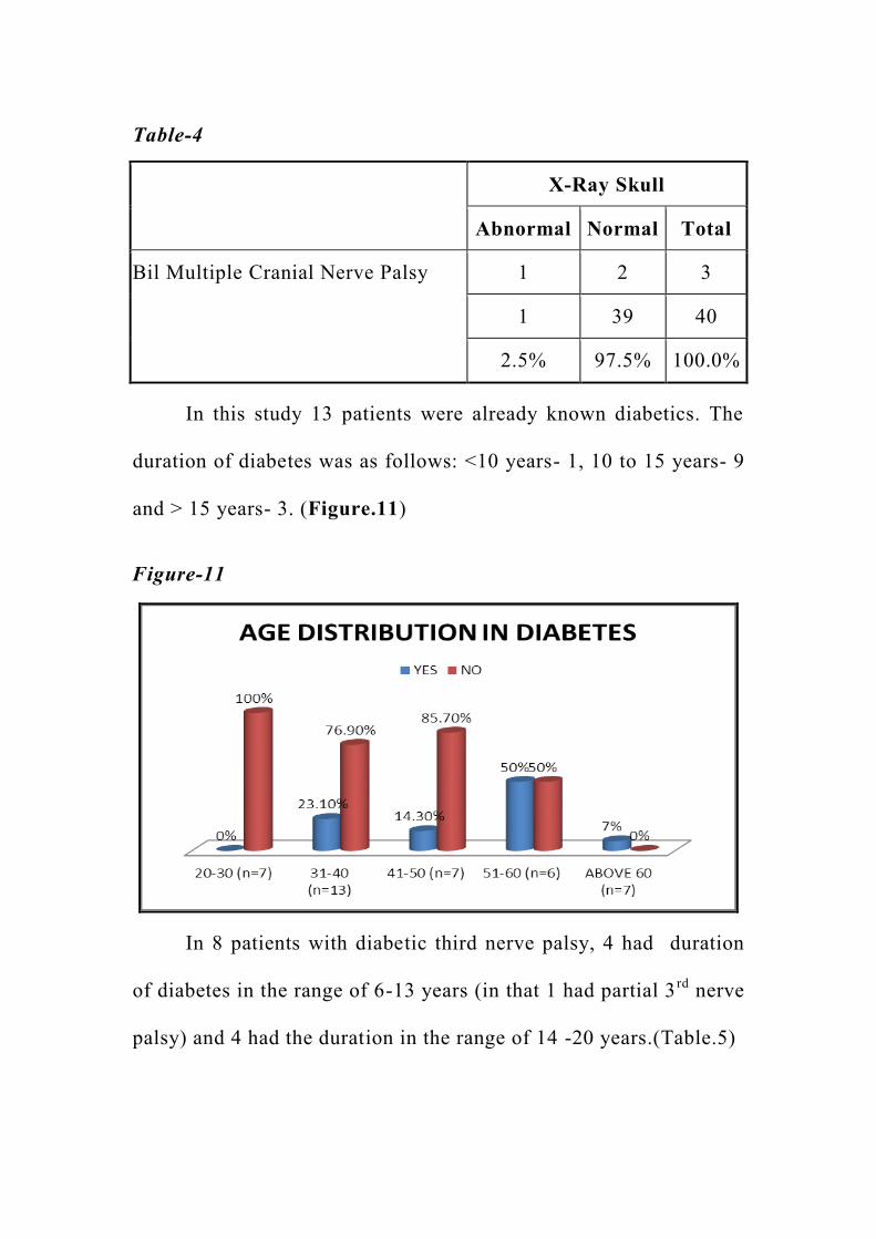
All the 40 patients had undergone MRI Brain (contrast if
necessary). 28 patients had abnormal MRI findings, whereas 12
patients did not have any findings. All the patients who had abnormal
CT Brain also had abnormal MRI Brain. (Figure 10)
Figure-10
MRI findings
12
28
Abnormal Column1
In only one patient x-ray skull was abnormal who had
bilateral 3,4,and 6th cranial nerve involvement. (Table.4)
41
Table-4
X-Ray Skull
Abnormal Normal Total
Bil Multiple Cranial Nerve Palsy 1 2 3
1 39 40
2.5% 97.5% 100.0%
In this study 13 patients were already known diabetics. The
duration of diabetes was as follows: <10 years- 1, 10 to 15 years- 9
and > 15 years- 3. (Figure.11)
Figure-11
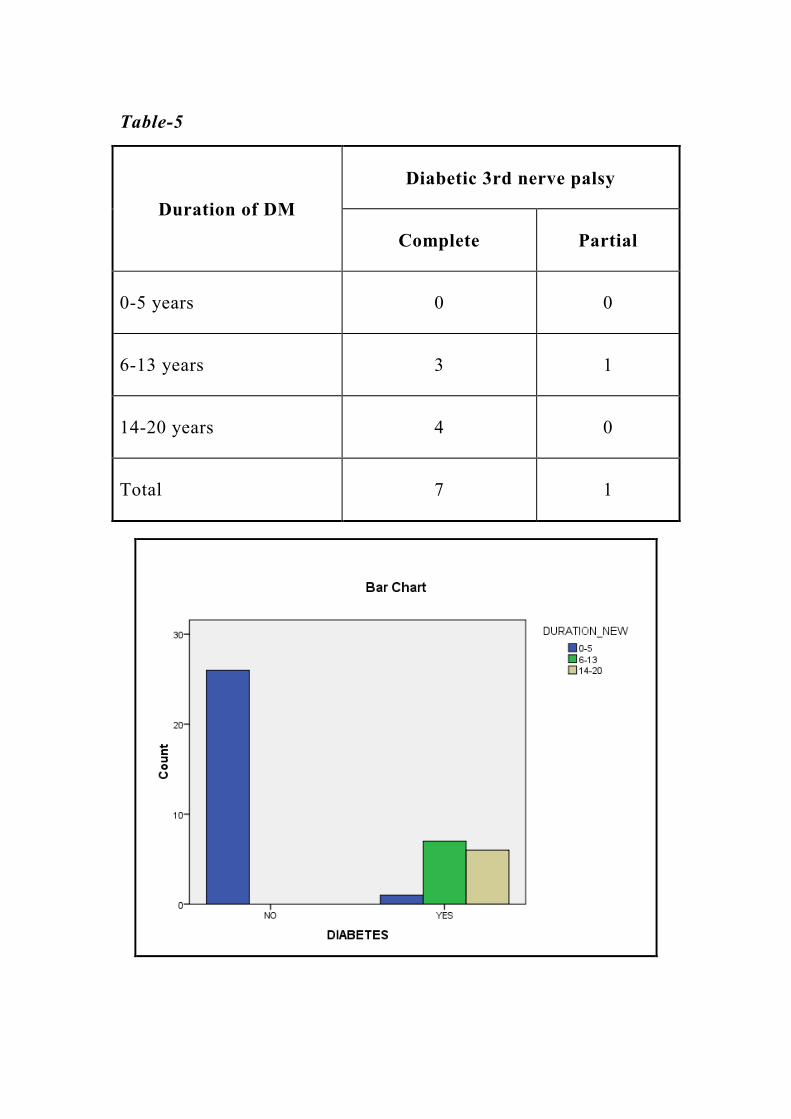
In 8 patients with diabetic third nerve palsy, 4 had duration
of diabetes in the range of 6-13 years (in that 1 had partial 3rd
nerve
palsy) and 4 had the duration in the range of 14 -20 years.(Table.5)
42
Table-5
Duration of DM
Diabetic 3rd nerve palsy
Complete Partial
0-5 years 0 0
6-13 years 3 1
14-20 years 4 0
Total 7 1
43
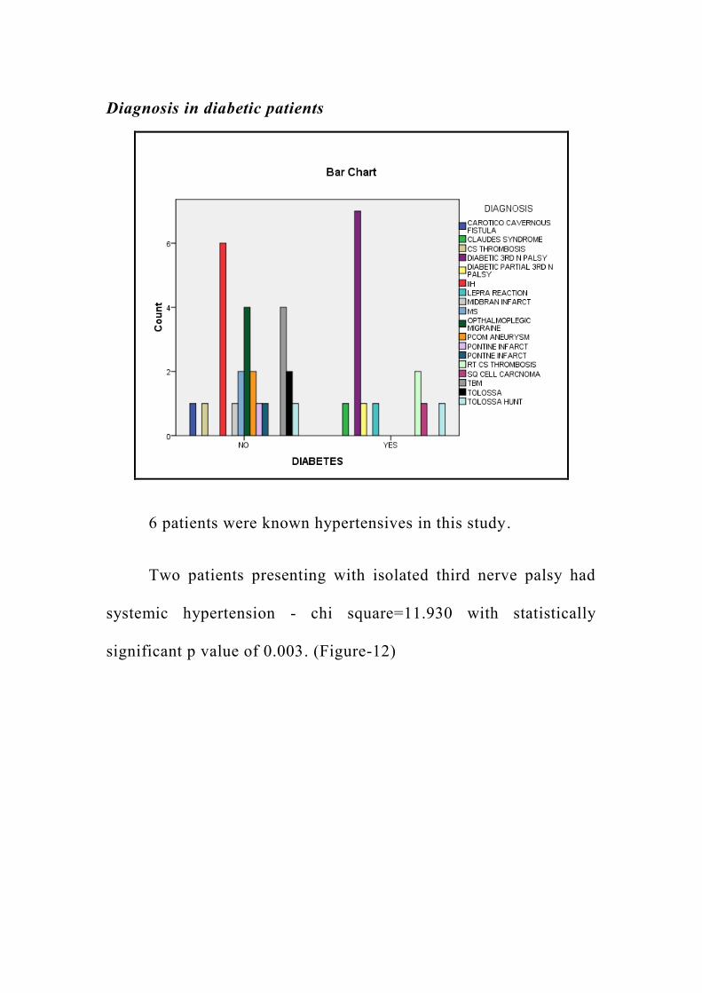
Diagnosis in diabetic patients
6 patients were known hypertensives in this study.
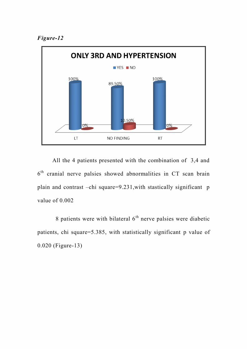
Two patients presenting with isolated third nerve palsy had
systemic hypertension - chi square=11.930 with statistically
significant p value of 0.003. (Figure-12)
44
Figure-12
All the 4 patients presented with the combination of 3,4 and
6th
cranial nerve palsies showed abnormalities in CT scan brain
plain and contrast –chi square=9.231,with stastically significant p
value of 0.002
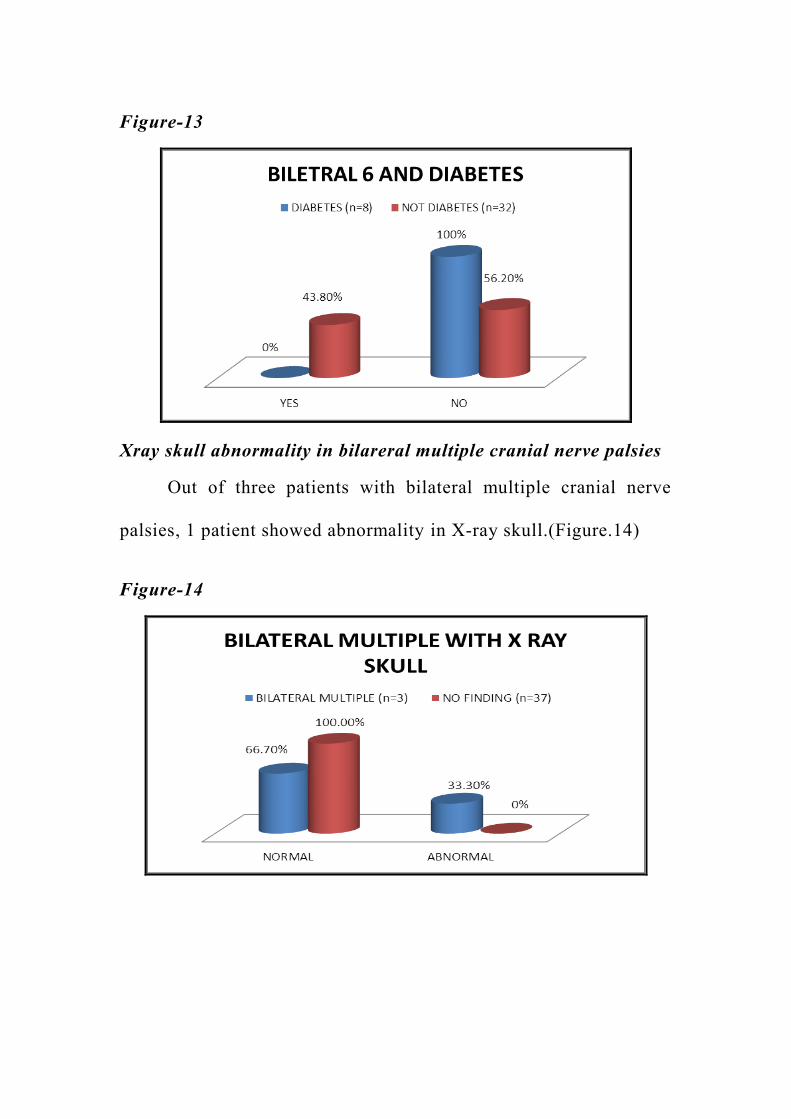
8 patients were with bilateral 6th
nerve palsies were diabetic
patients, chi square=5.385, with statistically significant p value of
0.020 (Figure-13)
45
Figure-13
Xray skull abnormality in bilareral multiple cranial nerve palsies
Out of three patients with bilateral multiple cranial nerve
palsies, 1 patient showed abnormality in X-ray skull.(Figure.14)
Figure-14
46
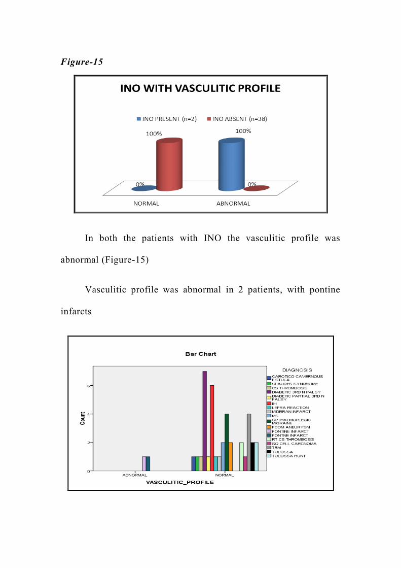
Figure-15
In both the patients with INO the vasculitic profile was
abnormal (Figure-15)
Vasculitic profile was abnormal in 2 patients, with pontine
infarcts
47
DISCUSSION
In this study 40 patients were included.21 patients were
females (52.5%) and 19 patients were males (47.5%) (Figure-1)
Hence in this study of 3,4,6 cranial nerves, we had slight
female preponderance.
Patients were in the age group ranging from 23 to 65 years.
The average age is 44.05 years. 7 patients were in the age group of
20 to 30 years, 13 patients were in the age group of 31-40 years, 7
patients were in the age group of 41-50 years, 6 patients were in the
age group of 51-60 years and 7 patients were in the age group of
above 60 years. So in this study, majority of patients are in the age
group of 31-40 years. Out of 13 patients in this age group 8 patients
were females. (Table 1).
In this study 15 patients were found to have isolated third
nerve palsy (37.5 %).Compared with the study by Rucker et al27
,
which showed 30% of patients with third nerve palsy, this study has
a higher incidence (37.5%). The commonest etiology of isolated
third nerve palsies is diabetes mellitus.8 patients had diabetic third
nerve palsy, followed by opthalmoplegic migraine in 3 patients. 2
patients were having PCOM aneurysm, one patient had carotico
48
cavernous fistula and one had Claude‟s syndrome31
. (Figure 3)
Among these, one patient of diabetic third nerve palsy had
presented with partial 3rd
nerve involvement (pupillary sparing).
None of these patients had a nuclear lesion.
In patients with diabetic third nerve palsy 9 patients were
males and six patients were females.
The study conducted by al Saleh et al in Arabic populations30
,
showed a male preponderance in patients with diabetic third nerve
palsies and our study also has a similar picture.
In 8 patients with diabetic third nerve palsy, the duration of
diabetes in 8 patients was within the range of 6-13 years and in the
remaining 4 patients the duration of diabetes was within the range
of 14-20 years.(Table.5)
In this study, 3 patients had isolated fourth nerve palsy
(7.5%) Compared with the study by Rucker et al which showed
11% of patients with isolated 4th nerve palsy, this study has a lower
incidence (7.5%). None of this patient had diabetes as risk factor.
One patient had a midbrain infarct40
, one was a known case of
relapsing remitting form of multiple sclerosis (In the previous
49
episodes, she had only optic nerve involvement) and one was a case
of opthalmoplegic migraine.
In a study conducted by Amy Gelfand et al32,28
in patients
with opthalmoplegic migraine, the involvement of isolated third
cranial nerve was common (83%) and involvement of isolated
fourth cranial nerve was rare (2%). In our study, out of 4 patients
with opthalmoplegic migraine, 3 had isolated 3rd
nerve palsy (75%)
and 1 had isolated 4th
nerve palsy (25%)
In this study, 9 patients had isolated sixth nerve palsy
(22.5%). Compared with the study by Rucker et al which showed
45% of patients with isolated 6th nerve palsy, this study has a lower
incidence. Out of these one patient had unilateral sixth nerve palsy
with TB meningitis as the etiology.
In the remaining 8 patients with bilateral 6th
nerve palsy, 7
patients were females and 1 patient was male, with a statistically
significant p value of 0.027.
In the 8 patients with bilateral sixth nerve palsy, 6 patients
had idiopathic intracranial hypertension and 2 patients had TB
meningitis. (Figure 6). The diagnosis of IIH was made with the
clinical signs and after measurement of the CSF opening pressure.
50
In this study, 27.5% patients had multiple cranial nerve palsy.
Compared with the study by Rucker et al, which showed 14% of
patients in this group, this study has a higher incidence. In this
group 8 patients, had unilateral 3,4&6 th cranial nerve involvement.
3 patients had cavernous sinus thrombosis, 3 patients had
Tolosa Hunt syndrome, 1 patient had a lepra reaction and 1 patient
had opthalmoplegic migraine.
Diabetes was the commonest etiological factor in these
patients (62.5%)27
with unilateral 3,4,&6th
cranial nerve lesions.
2 patients presented with bilateral 3,4& 6th
cranial nerve
palsies .In those patients, one had squamous cell carcinoma of the
naso pharynx with extension into the bilateral cavernous sinus 33
and
one patient with aspergillosis causing cavernous sinus thrombosis,
,who was diabetic.(Table .3)
2 patients presented with internuclear opthalmoplegia.In both
these patients vasculitic profiles were abnormal.
Summarizing, the common presentations in this study were
1) Isolated third nervepalsy in 15 patients
2) Bilateral sixth nerve palsy in 8 patients
51
3) Unilateral 3,4and 6th
nerve palsy in 8 patients.
In this study we did not have any patients with combination
of 3&4, 4&6 or 3&6
In this study, 13 patients were diabetics. The duration of
diabetes was as follows: < 10 years -1 patient, 10-15 years-9
patients and > 15 years -3 patients. (Figure.11)
6 patients had systemic hypertension.
Vasculitic profile was abnormal in 2 patients.
X-ray skull was abnormal in 1 patient with squamous cell
carcinoma.
All the 40 patients were evaluated with CT scan brain plain
and contrast. CT brain was abnormal in only 10 patients. 4 patients
with cavernous sinus thrombosis, 2 patients with Tolosa hunt
syndrome, 3 patients with TB meningitis and 1 patient with lepra
reaction had abnormal CT findings.
52
All the 40 patients were evaluated with MRI brain plain and
contrast (if necessary). 28 patients had abnormal MRI findings,
whereas 12 patients did not have any findings.
All the patients with abnormal CT brain also had abnormal
MRI brain.
CT brain was normal in 6 patients with IIH, 4 cases with
opthalmoplegic migraine, 2 cases with Tolosa Hunt syndrome, 2
patients with pontine infarct, 2 patients with multiple sclerosis, 2
patients with INO37
and in 1 patient with Claude‟s syndrome, but
their MRI showed abnormalities
In this study, MRI was found to be superior to CT scan brain
in all patients with nuclear, brain stem, fascicular lesions and some
patients with lesions in cavernous sinus and nerve lesions36
.
In all the 8 patients with diabetic third nerve palsy, in 2
patients with PCOM aneurysm and in 2 patients with IIH, MRI was
normal.
There was a correlation between the clinical findings, clinical
localization and imaging in 28 patients with MRI findings and in 2
patients with PCOM aneurysm, with CT angiogram findings.
53
In a study by Pamela y.blake et al 34
with 50 patients of
isolated third nerve palsy , 18 patients were with diabetic third
nerve palsy and none of them showed MR changes and in among
those who showed MRI changes , 2 had brainstem infarct,2 had
carotico cavernous aneurysm,1 had opthalmoplegic migraine and 1
had aneurysm.
CT angiogram was done for 2 patients, in whom PCOM
aneurysm was suspected, based on the clinical signs (head ache
with pupillary involvement) 38
, and both of them had aneurysm of
PCOM.
Histopathological examination was done in 3 patients who
had extensive lesion in MRI and not responding to therapy. 1
patient had mucormycosis, 1 patient had aspergillosis and 1 patient
had squamous cell carcinoma. Patients with mucormycosis and
aspergillosis were diabetics35
.
Though INO is a common presentation in multiple
sclerosis29,23
, in the two patients included in our study one had only
isolated 3rd
and the other had isolated 4th
nerve palsy.
Chi SL et al 38
suggested that diabetes mellitus or a
combination of diabetes and hypertension, but not hypertension
54
alone, is a risk factor for micro vascular ischemic ocular motor
cranial neuropathies. In this study we had 2 patients with both
hypertension and diabetes as risk factor who developed 3rd
nerve
palsies.
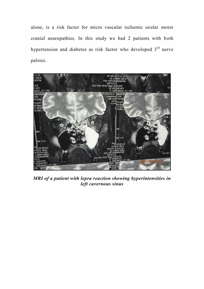
MRI of a patient with lepra reaction showing hyperintensities in
left cavernous sinus
55
CONCLUSION
1) In this study, there was no sex preponderance.
2) Commonest age group of presentation was 30-40 years.
3) Among the 3,4 and 6 cranial nerves,commonly affected was
the 3rd
cranial nerve followed by 4th
cranial nerve and
6th
cranial nerve
4) Among the combinations of cranial nerves, Bilateral 6th
nerve
involvement and unilateral 3,4,6 cranial nerve involvement
were the commonest presentations, followed by bilateral
3,4,6 th cranial nerve involvement.
5) The combinations of 3&4, 3&6 and 4&6 cranial nerve
involvement were not found in our series.
6) MRI showed abnormal findings in 70% of the patients,
whereas CT showed abnormal findings only in 25% of
patients.
7) The commonest cause of nuclear and infranuclear 3.4.6
cranial nerve lesion was Diabetes Mellitus in this study.
8) The commonest cause of imaging negative etiology in this
study was Diabetes Mellitus.
56
BIBLIOGRAPHY
1) Meienberg O, Müri R. Nuclear and infranuclear disorders.
Baillieres Clin Neurol. 1992 Aug;1(2):417-34.
2) Dhaliwal, Avninder MD; West, Adrienne L MD; Trobe,
Jonathan D MD; Musch, David C PhD Third, Fourth, and
Sixth Cranial Nerve Palsies Following Closed Head Injury
Journal of Neuro-Ophthalmology: March 2006 - Volume 26 -
Issue 1 - pp 4-10
3) Marinkovic S, Gibo H. The neurovascular relationships and
the blood supply of the oculomotor nerve: the microsurgical
anatomy of its cisternal segment. Surg Neurol. 1994;42:505–516
4) Yousry I, Moriggl B, Dieterich M, Naidich TP, Schmid UD,
Yousry T. MR anatomy of the proximal cisternal segment of
the trochlear nerve: neurovascular relationships and
landmarks. Radiology. 2002;223:31–38.
5) Marinkovic SV, Gibo H, Stimec B. The neurovascular
relationships and the blood supply of the abducent nerve:
surgical anatomy of its cisternal segment. Neurosurgery.
1994;34:1017–1026
57
6) 6.Yousry I, Camelio S, Wiesmann M, Schmid UD, Moriggl B,
Brückmann H, et al. Detailed magnetic resonance imaging
anatomy of the cisternal segment of the abducent nerve:
Dorello's canal and neurovascular relationships and
landmarks. J Neurosurg. 1999;91 (2:276–283.
7) Vogl T, Dresel S, Lochmüller H, Bergman C, Reimers C,
Lissner J. Third cranial nerve palsy caused by gummatous
neurosyphilis: MR findings. AJNR Am J Neuroradiol. 1993
Nov-Dec;14(6):13
8) S. R. SoniAneurysms of the posterior communicating artery
and oculomotor paresis.J Neurol Neurosurg Psychiatry. 1974
April; 37(4): 475–484.
9) Q. Rush JA, Younge BR. Paralysis of cranial nerves III, IV
and VI. Cause and prognosis in1000 cases. Arch Ophthalmol.
1981;99:76–79.
10) Klopfer J, Umlauf PC. Weber's syndrome. J Am Optom
Assoc. 1988 Nov;59(11):889-92
11) A Ayzam1, M Irfan2 Orbital Apex Syndrome secondary to
odontogenic sinusitis Bangladesh Journal of Medical Science
Vol.10 No.2 Apr‟11
58
12) The Journal of Laryngology & Otology / Volume 88 /
Issue 06 / June 1974, pp 551-557
13) Fujii M, Kiura K, Takigawa N, Yumoto T, Sehara Y, Tabata
M, Tanimoto M Presentation of Garcin syndrome due to lung
cancer J Thorac Oncol. 2007 Sep;2(9):877-8.
14) M Motamed, A Kalan Gradenigo's syndrome Postgrad Med J
2000;76:559-560 doi:10.1136/pmj.76.899.559
15) Shawn C. Wilker, MD, Janet C. Rucker, MD, Nancy J.
Newman, MD, Valerie Biousse, MD, and Robert L. Tomsak,
MD, PhD Pain in Ischemic Ocular Motor Cranial Nerve
Palsies Br J Ophthalmol. 2009 December; 93(12): 1657–1659.
16) Fells P, Collin JR. Cyclic oculomotor palsy Trans Ophthalmol
Soc U K. 1979 Apr;99(1):192-6
17) R Foroozan, L M Buono, P J Savino, and R C Sergott Tonic
pupils from giant cell arteritis J Ophthalmol. 2003 April;
87(4): 510–512
18) Omer Onbas, Mecit Kantarci, Fatih Alper, Leyla Karaca,
Adnan OkurMillard–Gubler syndrome: MR findingsJanuary
2005, Volume 47, Issue 1, pp 35-37
59
19) Agostinis C, Caverni L, Moschini L, et al. Paralysis of fourth
cranial nerve due to superior cerebellar artery. Neurology
1992;42:457–458.
20) Andreo LK, Gardner TA, Enzenauer RW. Third nerve palsy
in an AIDS patient. Presented at the North American Neuro-
Ophthalmology Society Meeting. Durango, CO. February 27-
March 3, 1994
21) Apte RS, Bartek W, Mello A, et al. Spontaneous intracranial
hypotension. Am J Ophthalmol 1999;127:482-485.
22) Arruga J, De Rivas P, Espinet HL, et al. Chronic isolated
trochlear nerve palsy produced by intracavernous internal
carotid artery aneurysm: report of a case. J Clin
Neuroophthalmol 1991;11:104-108.
23) Aschoff JC, Conrad B, Kornhuber HH. Acquired pendular
nystagmus with oscillopsia in multiple sclerosis: a sign of
cerebellar nuclear disease. J Neurol Neurosurg Psychiatry
1974;37:570-577.
24) L.Chou KL, Galetta SL, Liu GT, Volpe NJ, Bennett JL,
Asbury AK, Balcer LJ.Acute ocular motor mononeuropathies:
60
prospective study of the roles of neuroimaging and clinical
assessment. J Neurol Sci. 2004 Apr 15;219(1-2):35-9.
25) 25.Ophthalmoplegic Cranial Neuropathy:
26) New Cases and a Systematic Review J Child Neurol
published online 12 January 2012
27) 26. Baker RS, Epstein AD. Ocular motor abnormalities from
head trauma. Surv Ophthalmol 1991;35:245-267.
28) 27. Rucker CW. Paralysis of the third, fourth and sixth
cranial nerves. Am J Ophthalmol. 1958;46:787–794.
29) 28.Miglio L, Feraco P, Tani G, Ambrosetto P Computed
tomography and magnetic resonance imaging findings in
ophthalmoplegic migrainePediatr Neurol. 2010
Jun;42(6):434-6. doi: 10.1016/j.pediatrneurol.2010.02.005.
30) 29. de Seze J, Vukusic S, Viallet-Marcel M, Tilikete C,
Zéphir H, Delalande S, Stojkovic T, Defoort-Dhellemmes S,
Confavreux C, Vermersch P. Unusual ocular motor findings
in multiple sclerosis. J Neurol Sci. 2006 Apr 15;243(1-2):91-
5. Epub 2006 Feb 8 al Saleh M,bosley TM.Microvascular
61
cranial nerve palsies in an Arabic population.J Neuroopthalmol.
1999 Dec;19(4):252-6
31) Asakawa H, Yanaka K, Nose T. MRI of Claude's syndrome.
Neurology 2003;61:575
32) Amy A. Gelfand, MD1,2, Jeffrey M. Gelfand, MD3, Prab
Prabakhar, MD4, and Peter J. Goadsby, MD, PhD2,4
Ophthalmoplegic „„Migraine‟‟ or Recurrent
33) X. P Siva Reddy, R Chandrasekar Reddy, M Satapathy
Aetiological study of the third, fourth and sixth cranial nerve
paralysis. Sarojini Devi Eye Hospital and Institute of
Ophthalmology, Hyderabad, India
34) Pamela Y. Blake, Alexander S. Mark, Jorge Kattah, and
Martin Kolsky MR of Oculomotor Nerve Palsy .Am J
Neuroradiol 16:1665–1672, September 1995
35) Duke-Elder, W. S.: Text book of ophthalmology, Mosby,
London 1941.
36) Ettl ASalomonowitz E Visualization of the oculomotor
cranial nerves by magnetic resonance imagingStrabismus.
2004 Jun;12(2):85-96
62
37) Atlas SW, Grossman RI, Savino PJ, Schatz NJ, Sergott RC,
Bosley TM, Hackney DB, Goldberg HI, Bilaniuk LT,
Zimmerman RA. Internuclear ophthalmoplegia: MR-anatomic
correlation
38) M. Chi SL, Bhatti MT. The diagnostic dilemma of neuro-
imaging in acute isolated sixth nerve palsy. Curr Opin
Ophthalmol. 2009 Nov;20(6):423-9.
39) Barbas NR, Hedges TR, Schwenn M. Isolated oculomotor
nerve palsy due to neoplasm in infancy. Neuroophthalmology
1995;15:157-160
40) Barr DB, McFadzean RM, Hadley D, et al. Acquired bilateral
superior oblique palsy: a localizing sign in the dorsal
midbrain syndrome. Eur J Ophthalmol 1997;7:271-276.
63
ABBREVIATIONS
CSF : Cerebro spinal fluid
ICA : Internal carotid artery
IIH : Idiopathic intracranial hypertension.
INO : Internuclear opthalmoplegia
MS : Multiple sclerosis.
PCOM : Posterior communicating artery
TBM : Tuberculous meningitis
64
PROFORMA
Name : Serial No :
Age : IP No :
Sex : Address :
DOA :
DOD: Phone No :
Diagnosis :
History :
Clinical Findings :
Investigations ; Basic investigations
CSF Analysis and opening pressure(in selected cases)
65
OTHERS
1) Diabetic profile
2) Hypertensive profile
3) Thyroid profile
4) Vasculitic profile
RADIOLOGY
1) X-ray skull with foraminal views
2) CT Brain plain and contrast (if necessary)
3) CT angiogram- in selected cases
4) MRI Brain plain with MRA and MRV and contrast
study(if necessary)
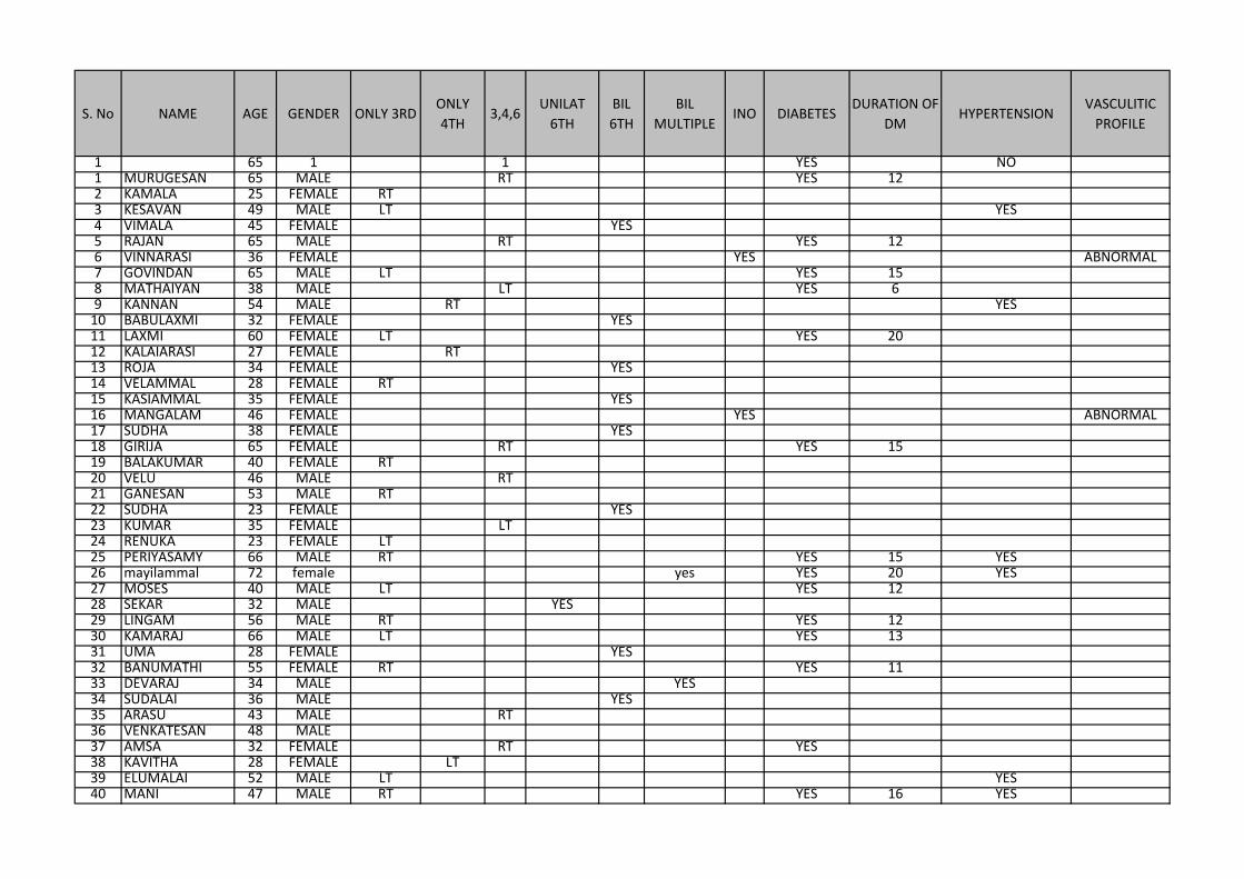
S. No NAME AGE GENDER ONLY 3RDONLY
4TH3,4,6
UNILAT
6TH
BIL
6TH
BIL
MULTIPLEINO DIABETES
DURATION OF
DMHYPERTENSION
VASCULITIC
PROFILE
1 65 1 1 YES NO1 MURUGESAN 65 MALE RT YES 122 KAMALA 25 FEMALE RT3 KESAVAN 49 MALE LT YES4 VIMALA 45 FEMALE YES5 RAJAN 65 MALE RT YES 126 VINNARASI 36 FEMALE YES ABNORMAL7 GOVINDAN 65 MALE LT YES 158 MATHAIYAN 38 MALE LT YES 69 KANNAN 54 MALE RT YES
10 BABULAXMI 32 FEMALE YES11 LAXMI 60 FEMALE LT YES 2012 KALAIARASI 27 FEMALE RT13 ROJA 34 FEMALE YES14 VELAMMAL 28 FEMALE RT15 KASIAMMAL 35 FEMALE YES16 MANGALAM 46 FEMALE YES ABNORMAL17 SUDHA 38 FEMALE YES18 GIRIJA 65 FEMALE RT YES 1519 BALAKUMAR 40 FEMALE RT20 VELU 46 MALE RT21 GANESAN 53 MALE RT22 SUDHA 23 FEMALE YES23 KUMAR 35 FEMALE LT24 RENUKA 23 FEMALE LT25 PERIYASAMY 66 MALE RT YES 15 YES26 mayilammal 72 female yes YES 20 YES27 MOSES 40 MALE LT YES 1228 SEKAR 32 MALE YES29 LINGAM 56 MALE RT YES 1230 KAMARAJ 66 MALE LT YES 1331 UMA 28 FEMALE YES32 BANUMATHI 55 FEMALE RT YES 1133 DEVARAJ 34 MALE YES34 SUDALAI 36 MALE YES35 ARASU 43 MALE RT36 VENKATESAN 48 MALE37 AMSA 32 FEMALE RT YES38 KAVITHA 28 FEMALE LT39 ELUMALAI 52 MALE LT YES40 MANI 47 MALE RT YES 16 YES
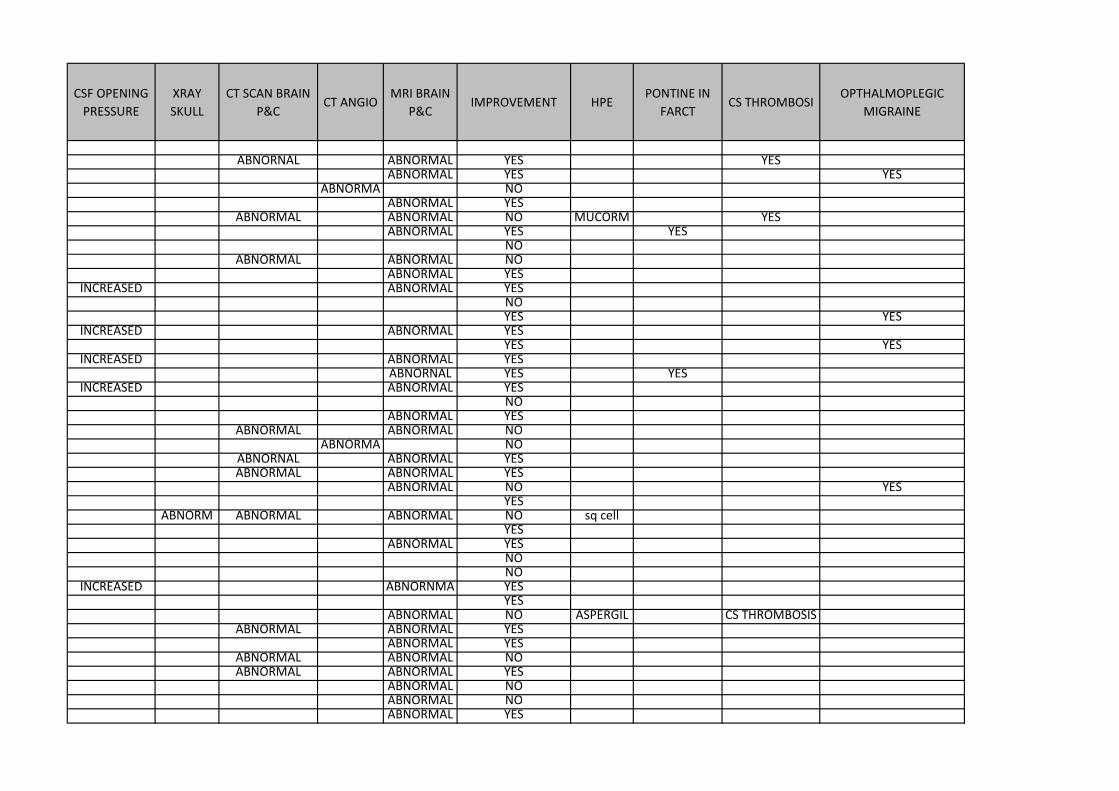
CSF OPENING
PRESSURE
XRAY
SKULL
CT SCAN BRAIN
P&CCT ANGIO
MRI BRAIN
P&CIMPROVEMENT HPE
PONTINE IN
FARCTCS THROMBOSI
OPTHALMOPLEGIC
MIGRAINE
ABNORNAL ABNORMAL YES YESABNORMAL YES YES
ABNORMA NOABNORMAL YES
ABNORMAL ABNORMAL NO MUCORM YESABNORMAL YES YES
NOABNORMAL ABNORMAL NO
ABNORMAL YESINCREASED ABNORMAL YES
NOYES YES
INCREASED ABNORMAL YESYES YES
INCREASED ABNORMAL YESABNORNAL YES YES
INCREASED ABNORMAL YESNO
ABNORMAL YESABNORMAL ABNORMAL NO
ABNORMA NOABNORNAL ABNORMAL YESABNORMAL ABNORMAL YES
ABNORMAL NO YESYES
ABNORM ABNORMAL ABNORMAL NO sq cell YES
ABNORMAL YESNONO
INCREASED ABNORNMA YESYES
ABNORMAL NO ASPERGIL CS THROMBOSISABNORMAL ABNORMAL YES
ABNORMAL YESABNORMAL ABNORMAL NOABNORMAL ABNORMAL YES
ABNORMAL NOABNORMAL NOABNORMAL YES
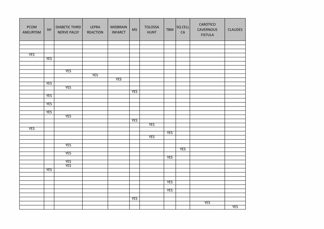
PCOM
ANEURYSMIIH
DIABETIC THIRD
NERVE PALSY
LEPRA
REACTION
MIDBRAIN
INFARCTMS
TOLOSSA
HUNTTBM
SQ CELL
CA
CAROTICO
CAVERNOUS
FISTULA
CLAUDES
YESYES
YESYES
YESYES
YESYES
YES
YES
YESYES
YESYES
YESYES
YES
YESYES
YESYES
YESYES
YES
YES
YES
YESYES
YES
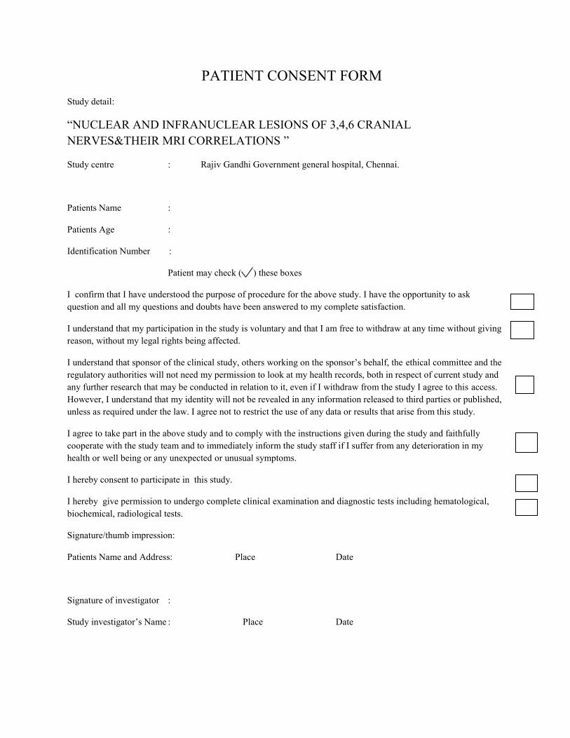
PATIENT CONSENT FORM
Study detail:
“NUCLEAR AND INFRANUCLEAR LESIONS OF 3,4,6 CRANIAL
NERVES&THEIR MRI CORRELATIONS ”
Study centre : Rajiv Gandhi Government general hospital, Chennai.
Patients Name :
Patients Age :
Identification Number :
Patient may check ( ) these boxes
I confirm that I have understood the purpose of procedure for the above study. I have the opportunity to ask
question and all my questions and doubts have been answered to my complete satisfaction.
I understand that my participation in the study is voluntary and that I am free to withdraw at any time without giving
reason, without my legal rights being affected.
I understand that sponsor of the clinical study, others working on the sponsor’s behalf, the ethical committee and the
regulatory authorities will not need my permission to look at my health records, both in respect of current study and
any further research that may be conducted in relation to it, even if I withdraw from the study I agree to this access.
However, I understand that my identity will not be revealed in any information released to third parties or published,
unless as required under the law. I agree not to restrict the use of any data or results that arise from this study.
I agree to take part in the above study and to comply with the instructions given during the study and faithfully
cooperate with the study team and to immediately inform the study staff if I suffer from any deterioration in my
health or well being or any unexpected or unusual symptoms.
I hereby consent to participate in this study.
I hereby give permission to undergo complete clinical examination and diagnostic tests including hematological,
biochemical, radiological tests.
Signature/thumb impression:
Patients Name and Address: Place Date
Signature of investigator :
Study investigator’s Name : Place Date
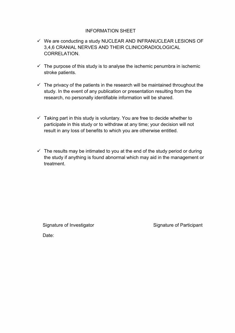
INFORMATION SHEET
We are conducting a study NUCLEAR AND INFRANUCLEAR LESIONS OF
3,4,6 CRANIAL NERVES AND THEIR CLINICORADIOLOGICAL
CORRELATION.
The purpose of this study is to analyse the ischemic penumbra in ischemic
stroke patients.
The privacy of the patients in the research will be maintained throughout the
study. In the event of any publication or presentation resulting from the
research, no personally identifiable information will be shared.
Taking part in this study is voluntary. You are free to decide whether to
participate in this study or to withdraw at any time; your decision will not
result in any loss of benefits to which you are otherwise entitled.
The results may be intimated to you at the end of the study period or during
the study if anything is found abnormal which may aid in the management or
treatment.
Signature of Investigator Signature of Participant
Date:
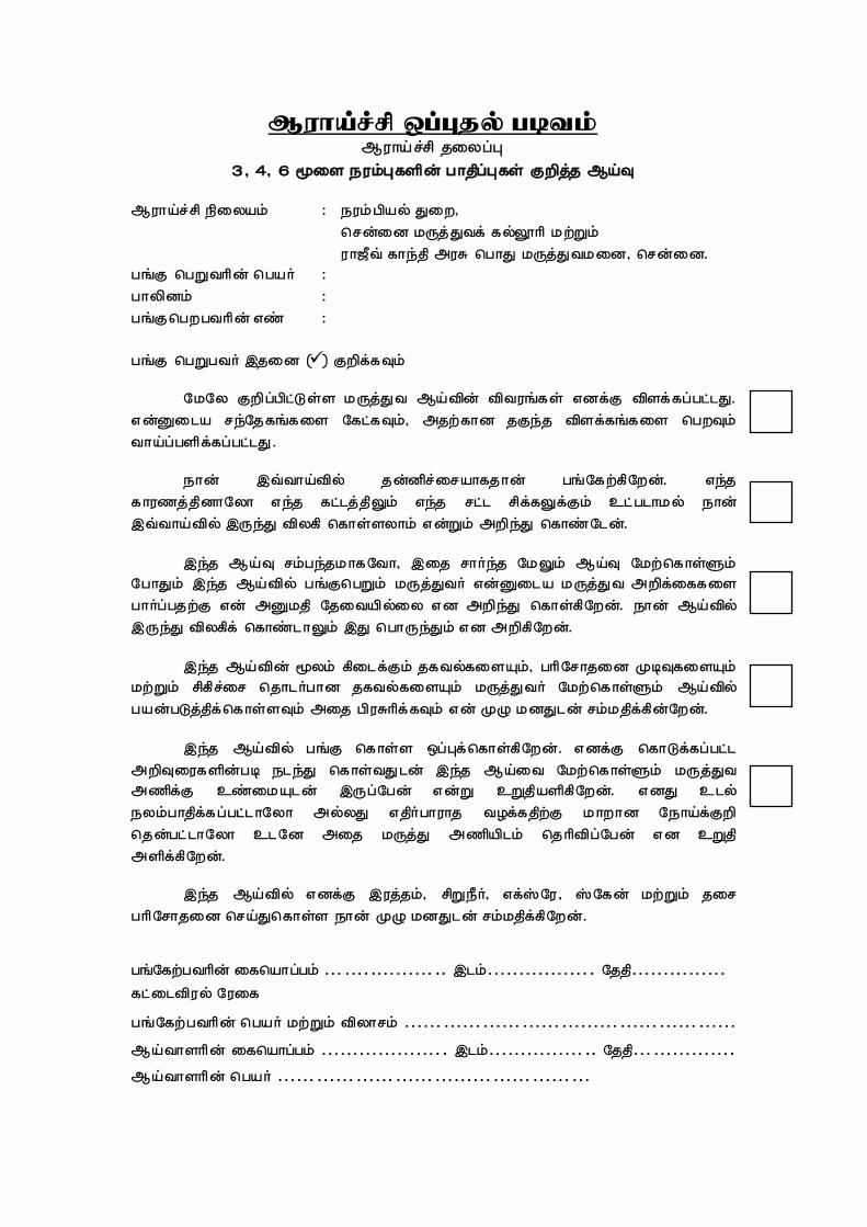
MuhŒ¢á x¥òjš got« MuhŒ¢á jiy¥ò
3 , 4, 6 _is eu«òfË‹ ghâ¥òfŸ F¿¤j MŒî
MuhŒ¢á Ãiya« : eu«ãaš Jiw, br‹id kU¤Jt¡ fšÿÇ k‰W« uhé› fhªâ muR bghJ kU¤Jtkid, br‹id.
g§F bgWtÇ‹ bga® : ghÈd« : g§FbgwgtÇ‹ v© :
g§F bgWgt® ïjid ( ) F¿¡fî«
nkny F¿¥ã£LŸs kU¤Jt MŒÉ‹ Étu§fŸ vd¡F És¡f¥g£lJ. v‹Dila rªnjf§fis nf£fî«, mj‰fhd jFªj És¡f§fis bgwî« thŒ¥gË¡f¥g£lJ.
eh‹ ï›thŒÉš j‹Å¢irahfjh‹ g§nf‰»nw‹. vªj fhuz¤âdhnyh vªj f£l¤âY« vªj r£l á¡fY¡F« c£glhkš eh‹ ï›thŒÉš ïUªJ Éy» bfhŸsyh« v‹W« m¿ªJ bfh©nl‹.
ïªj MŒî r«gªjkhfnth, ïij rh®ªj nkY« MŒî nk‰bfhŸS« nghJ« ïªj MŒÉš g§FbgW« kU¤Jt® v‹Dila kU¤Jt m¿¡iffis gh®¥gj‰F v‹ mDkâ njitÆšiy vd m¿ªJ bfhŸ»nw‹. eh‹ MŒÉš ïUªJ Éy»¡ bfh©lhY« ïJ bghUªJ« vd m¿»nw‹.
ïªj MŒÉ‹ _y« »il¡F« jftšfisí«, gÇnrhjid Koîfisí« k‰W« Ợir bjhl®ghd jftšfisí« kU¤Jt® nk‰bfhŸS« MŒÉš ga‹gL¤â¡bfhŸsî« mij ãuRÇ¡fî« v‹ KG kdJl‹ r«kâ¡»‹nw‹.
ïªj MŒÉš g§F bfhŸs x¥ò¡bfhŸ»nw‹. vd¡F bfhL¡f¥g£l m¿îiufË‹go elªJ bfhŸtJl‹ ïªj MŒit nk‰bfhŸS« kU¤Jt m¡F c©ikíl‹ ïU¥ng‹ v‹W cWâaË»nw‹. vdJ clš ey«ghâ¡f¥g£lhnyh mšyJ vâ®ghuhj tH¡fâ‰F khwhd nehŒ¡F¿ bj‹g£lhnyh clnd mij kU¤J mÂÆl« bjÇÉ¥ng‹ vd cWâ mË¡»nw‹.
ïªj MŒÉš vd¡F ïu¤j«, áWÚ®, v¡Þnu, Þnf‹ k‰W« jir gÇnrhjid brŒJbfhŸs eh‹ KG kdJl‹ r«kâ¡»nw‹.
g§nf‰gtÇ‹ ifbah¥g« ……..……….. ïl«…………….. njâ…………… f£ilÉuš nuif
g§nf‰gtÇ‹ bga® k‰W« Éyhr« ……………………………………………
MŒthsÇ‹ ifbah¥g« ……………….. ïl«…………….. njâ……………. MŒthsÇ‹ bga® …………………………………………
jftš m¿¡if
br‹id kU¤Jt¡ fšÿÇ k‰W« kU¤JtkidÆš “3 , 4, 6 _is
eu«òfË‹ ghâ¥òfŸ F¿¤j MŒî” brŒJ tU»nwh«. mj‰fhf
nehahËfis¤ nj®î brŒ»nwh«.
ïªj MuhŒ¢áÆš g§nf‰F« nehahËfË‹ Égu§fŸ MŒî
Koí« tiu ïufáakhf it¡f¥gL«. MuhŒ¢áÆ‹ Koî g‰¿a gâ¥òfŸ
mšyJ btËpLfËš ahUila jÅ¥g£l Étu§fS« g»®ªJ
bfhŸs¥glkh£lhJ.
ïªj MuhŒ¢áÆš g§nf‰F« c§fŸ Koî j‹Å¢irahdJ, ïªj
MuhŒ¢áÆš g§nf‰F« vªj neu¤âY« Éy¡»¡ bfhŸtj‰F« c§fS¡F
thŒ¥ò cŸsJ. c§fË‹ ïªj Ô®khd¤âdhš c§fS¡F
ï«kU¤JtkidÆš tH§f¥gL« ga‹fËš v›Éj kh‰wK« ïU¡fhJ.
ïªj áw¥ò MŒÉ‹ KoîfŸ, ïªj MŒÉ‹ KoÉš mšyJ
MŒÉ‹nghJ V‰gL« vâ®kiwahd Éisîfis mªnehahËÆ‹ ey‹
fUânah mšyJ ỢiraË¡F« bghU£nlh nehahË¡F bjÇÉ¡f¥gL«.
MŒthsÇ‹ ifbah¥g« nehahËÆ‹ ifbah¥g«
njâ

Your digital receiptThis receipt acknowledges that Turnitin received your paper. Below you will find the receipt informationregarding your submission.
Paper ID 313095697Paper title thesis
Assignment title MedicalAuthor Viveka Saravanan Raju 16101014 D.M. NeurologyE-mail [email protected]
Submission time 22-Mar-2013 08:05AMTotal words 7182
First 100 words of your submission
INTRODUCTION Palsies of any of the three cranial nerves supplying the extra ocular muscles havetheir presentations, disturbing ocular motility. Abnormalities of ocular motility help in the localization oflesions of the cerebral hemispheres, brain stem, cranial nerves (CNs), and even the striated muscle.Only one nerve may be involved or there may be a combination of the three nerves. The palsies areusually acquired. Sometimes palsies can be congenital due to the developmental defect of the nucleusor motor nerve fibers. Oculo motor fibers can be interrupted extraaxially or extraaxially. Lesions canbe in the foramens or extra cranial e.g. Intraorbital. All these Oculomotor nerves can be...
Copyright 2012 Turnitin. All rights reserved.