Embed Size (px)
Citation preview
NRAS mutation causes a human autoimmunelymphoproliferative syndromeJoao B. Oliveira*†, Nicolas Bidere‡, Julie E. Niemela*, Lixin Zheng‡, Keiko Sakai‡, Cynthia P. Nix‡§, Robert L. Danner¶,Jennifer Barb�, Peter J. Munson�, Jennifer M. Puck**, Janet Dale††, Stephen E. Straus††, Thomas A. Fleisher*‡‡,and Michael J. Lenardo‡§§
*Department of Laboratory Medicine, Clinical Center, ‡Molecular Development Section, Laboratory of Immunology, National Institute of Allergyand Infectious Diseases, ¶Functional Genomics and Proteomics Facility, Critical Care Medicine Department, Clinical Center, �Mathematical andStatistical Computing Laboratory, Center for Information Technology, **Genetics and Molecular Biology Branch, National Human GenomeResearch Institute, and ††Laboratory of Clinical Infectious Diseases, National Institute of Allergy and Infectious Diseases, National Institutesof Health, Bethesda, MD 20892
Communicated by Jacques F. A. P. Miller, The Walter and Eliza Hall Institute of Medical Research, Parkville, Victoria, Australia, April 3, 2007(received for review January 5, 2007)
The p21 RAS subfamily of small GTPases, including KRAS, HRAS,and NRAS, regulates cell proliferation, cytoskeletal organization,and other signaling networks, and is the most frequent target ofactivating mutations in cancer. Activating germline mutations ofKRAS and HRAS cause severe developmental abnormalities leadingto Noonan, cardio-facial-cutaneous, and Costello syndrome, butactivating germline mutations of NRAS have not been reported.Autoimmune lymphoproliferative syndrome (ALPS) is the mostcommon genetic disease of lymphocyte apoptosis and causesautoimmunity as well as excessive lymphocyte accumulation, par-ticularly of CD4�, CD8� �� T cells. Mutations in ALPS typicallyaffect CD95 (Fas/APO-1)-mediated apoptosis, one of the extrinsicdeath pathways involving TNF receptor superfamily proteins, butcertain ALPS individuals have no such mutations. We show herethat the salient features of ALPS as well as a predisposition tohematological malignancies can be caused by a heterozygousgermline Gly13Asp activating mutation of the NRAS oncogene thatdoes not impair CD95-mediated apoptosis. The increase in active,GTP-bound NRAS augments RAF/MEK/ERK signaling, which mark-edly decreases the proapoptotic protein BIM and attenuates in-trinsic, nonreceptor-mediated mitochondrial apoptosis. Thus,germline activating mutations in NRAS differ from other p21 Rasoncoproteins by causing selective immune abnormalities withoutgeneral developmental defects. Our observations on the effects ofNRAS activation indicate that RAS-inactivating drugs, such asfarnesyltransferase inhibitors should be examined in human au-toimmune and lymphocyte homeostasis disorders.
autoimmunity � B cell lymphoma 2-interacting mediator of cell death �intrinsic apoptosis � lymphoma � lymphoproliferation
The RAS genes (NRAS, KRAS, and HRAS) encode 21-kDaproteins that are members of the superfamily of small
GTP-binding proteins, which have diverse intracellular signalingfunctions including control of cell proliferation, growth, andapoptosis (1). Somatic activating mutations in RAS are presentin up to 30% of all human cancers (2). Germline RAS pathwaymutations have only recently been described as causing therelated Costello (HRAS), Noonan (PTPN11, KRAS, SOS1), andcardiofaciocutaneous syndromes (KRAS, BRAF, MEK1, andMEK) (3–7). Individuals with these syndromes typically presentwith severe developmental anomalies in various combinations offacial abnormalities, heart defects, short stature, skin and genitalabnormalities, and mental retardation (8). Defects in the im-mune system have not been reported. Patients with Costello andNoonan syndromes have an increased propensity to solid andhematopoietic tumors, respectively (3, 8). Germline mutations inNRAS have not yet been described.
The autoimmune lymphoproliferative syndrome (ALPS)(OMIM 601859/603909) is the most common genetic disorder oflymphocyte apoptosis and is characterized by chronic accumu-
lation of nonmalignant lymphocytes, defective lymphocyte apo-ptosis, and an increased risk for the development of hemato-logical malignancies (9). A signature of the disease is theaccumulation of �� T cells lacking the CD4 and CD8 corecep-tors that are termed ‘‘double-negative’’ T cells (DNTs: CD4�,CD8� TCR��� cells). These cells bear no known relationship tothymic DNTs, a stage that occurs before �� TCR gene rear-rangements in ontogeny (10). According to genotype, ALPS canbe classified as types Ia, Ib, and II, which are due to germlinemutations in CD95 (TNFRSF6), CD95 ligand (TNFSF6), andcaspase 10 (CASP10), respectively (10–13). Additionally, so-matic mutations of CD95 in �� DNTs can also cause ALPS oftype Im (mosaic) (14). All of these mutations impair extrinsic,Fas receptor-mediated apoptosis (10). An enigma has been theALPS individuals who have no defects in CD95 pathway apo-ptosis (some ALPS type III patients) (9, 10). This groupencompasses a large number of individuals and is probablygenetically heterogeneous. In an attempt to unveil new geneticdefects, we investigated alternative apoptosis pathways in ALPStype III and identified one ALPS patient with a unique defect incytokine withdrawal-induced apoptosis due to an activatingNRAS mutation.
ResultsDefective IL-2 Withdrawal-Induced Apoptosis in a Patient with Clinicaland Laboratory Hallmarks of ALPS. The intrinsic mitochondrialpathway of apoptosis can be triggered by developmental cues inthe thymus or bone marrow, cytokine deprivation, DNA dam-age, or treatment with cytotoxic drugs (15, 16). To screen for
Author contributions: J.B.O., T.A.F., and M.J.L. designed research; J.B.O., N.B., J.E.N., L.Z.,K.S., and C.P.N. performed research; R.L.D. and J.M.P. contributed new reagents/analytictools; J.B.O., N.B., J.E.N., R.L.D., J.B., P.J.M., T.A.F., and M.J.L. analyzed data; J.B.O., T.A.F.,and M.J.L. wrote the paper; J.D. coordinated patient information and clinical samplehandling; and S.E.S. coordinated the clinical aspects of the research.
The authors declare no conflict of interest.
Abbreviations: ALPS, autoimmune lymphoproliferative syndrome; DNT, double-negative Tcell; BCL-2, B cell lymphoma 2; BIM, BCL-2-interacting mediator of cell death; siRNA, smallinterfering RNA; FTI, farnesyltransferase inhibitor; PBL, peripheral blood lymphocyte.
Data deposition: The data reported in this paper have been deposited in the GeneExpression Omnibus (GEO) database, www.ncbi.nlm.nih.gov/geo (accession no. GSE7345).
†Present address: Laboratory of Medical Investigation (LIM-56), University of Sao Paulo,05630-020, Sao Paulo, Brazil.
§Present address: Bascom Palmer Eye Institute, University of Miami Miller School ofMedicine, Miami, FL 33136.
‡‡To whom correspondence may be addressed at: Clinical Center, National Institutes ofHealth, MSC 1508, Bethesda, MD 20892. E-mail: [email protected].
§§To whom correspondence may be addressed at: Laboratory of Immunology, NationalInstitute of Allergy and Infectious Diseases, National Institutes of Health, MSC 1892,Bethesda, MD 20892. E-mail: [email protected].
This article contains supporting information online at www.pnas.org/cgi/content/full/0702975104/DC1.
www.pnas.org�cgi�doi�10.1073�pnas.0702975104 PNAS � May 22, 2007 � vol. 104 � no. 21 � 8953–8958
IMM
UN
OLO
GY
Dow
nloa
ded
by g
uest
on
Janu
ary
13, 2
020
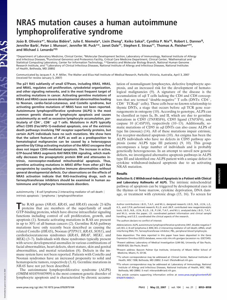
defects in this pathway, we exposed activated lymphocytes fromindividuals with salient features of ALPS (lymphadenopathy andincreased �� DNTs) but normal CD95-mediated apoptosis, toinducers of intrinsic apoptosis including staurosporine, �-radi-ation, and cytokine withdrawal. We identified an individualwhose lymphocytes clearly resisted death induced by IL-2 with-drawal (Fig. 1A).
The affected individual [National Institutes of Health (NIH)cohort patient 58, P58] is a 49-year-old male with lifelongoverexpansion of lymphocytes, and an unusual history of twomalignancies: childhood leukemia and early adulthood lym-phoma, both successfully treated [supporting information (SI)Table 1]. Peripheral blood immunophenotyping revealed a sus-tained elevation in �� DNT cells over several years and otherfindings frequently seen in ALPS, including an elevated per-centage of CD5� B cells and low numbers of CD27� B cells (17).However, other features such as low CD25/HLA-DR ratio andhigh numbers of CD3�CD57� were not seen (SI Table 1).Lymph node biopsy performed elsewhere and reviewed at theNIH revealed reactive follicular hyperplasia and sinus histiocy-tosis, but DNT cells were not prominent. Several serum auto-antibodies were detected and elevations of several T helper 2cytokines including IL-5, -6, -8, -10, and -13 were observed (SITables 1 and 2). Based on the published NIH ALPS diagnosticcriteria of elevated DNT on peripheral blood, chronic lifelongnonmalignant hyperplasia and defective lymphocyte apoptosis,P58 received a provisional clinical diagnosis of ALPS withrecognition that this is not a typical clinical presentation of thisdisease.
Despite defective IL-2 withdrawal death, we found no abnor-malities in apoptosis induced by an agonistic anti-APO-1 (CD95)antibody (Apo1.3), staurosporine, or �-radiation (Fig. 1 B–D).Moreover, activated T cells did not spontaneously proliferate orsecrete cytokines, but did show persistent proliferation after IL-2withdrawal compared with normal cells (SI Fig. 6 and data notshown). Thus, lymphocytes from P58 demonstrated a specificdefect in the intrinsic pathway of apoptosis.
During cytokine withdrawal, the proapoptotic B cell lym-phoma 2 (BCL-2) family members BAX and BAK are activatedand oligomerize at mitochondrial surfaces, causing permeabili-
zation of the outer membrane, which releases cytochrome c andtriggers apoptosis (15, 16). After IL-2 withdrawal, we found thatP58 cells were markedly defective in BAX activation and mito-chondrial cytochrome c release, correlating with a decreasedpercentage of apoptotic nuclei compared with control cells aftercytokine withdrawal, but not staurosporine treatment, clearlyindicating a selective mitochondrial apoptosis defect (Fig. 1 Eand F, and SI Fig. 7 A and B).
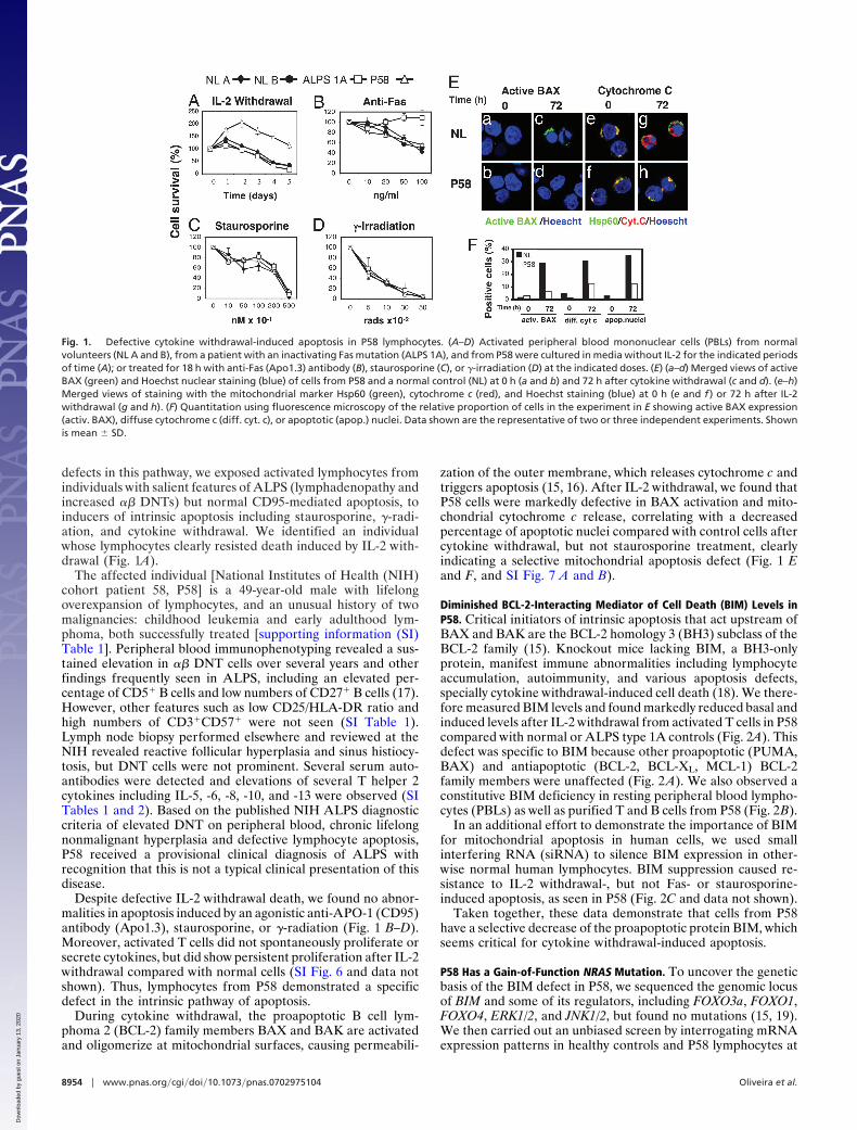
Diminished BCL-2-Interacting Mediator of Cell Death (BIM) Levels inP58. Critical initiators of intrinsic apoptosis that act upstream ofBAX and BAK are the BCL-2 homology 3 (BH3) subclass of theBCL-2 family (15). Knockout mice lacking BIM, a BH3-onlyprotein, manifest immune abnormalities including lymphocyteaccumulation, autoimmunity, and various apoptosis defects,specially cytokine withdrawal-induced cell death (18). We there-fore measured BIM levels and found markedly reduced basal andinduced levels after IL-2 withdrawal from activated T cells in P58compared with normal or ALPS type 1A controls (Fig. 2A). Thisdefect was specific to BIM because other proapoptotic (PUMA,BAX) and antiapoptotic (BCL-2, BCL-XL, MCL-1) BCL-2family members were unaffected (Fig. 2 A). We also observed aconstitutive BIM deficiency in resting peripheral blood lympho-cytes (PBLs) as well as purified T and B cells from P58 (Fig. 2B).
In an additional effort to demonstrate the importance of BIMfor mitochondrial apoptosis in human cells, we used smallinterfering RNA (siRNA) to silence BIM expression in other-wise normal human lymphocytes. BIM suppression caused re-sistance to IL-2 withdrawal-, but not Fas- or staurosporine-induced apoptosis, as seen in P58 (Fig. 2C and data not shown).
Taken together, these data demonstrate that cells from P58have a selective decrease of the proapoptotic protein BIM, whichseems critical for cytokine withdrawal-induced apoptosis.
P58 Has a Gain-of-Function NRAS Mutation. To uncover the geneticbasis of the BIM defect in P58, we sequenced the genomic locusof BIM and some of its regulators, including FOXO3a, FOXO1,FOXO4, ERK1/2, and JNK1/2, but found no mutations (15, 19).We then carried out an unbiased screen by interrogating mRNAexpression patterns in healthy controls and P58 lymphocytes at
Fig. 1. Defective cytokine withdrawal-induced apoptosis in P58 lymphocytes. (A–D) Activated peripheral blood mononuclear cells (PBLs) from normalvolunteers (NL A and B), from a patient with an inactivating Fas mutation (ALPS 1A), and from P58 were cultured in media without IL-2 for the indicated periodsof time (A); or treated for 18 h with anti-Fas (Apo1.3) antibody (B), staurosporine (C), or �-irradiation (D) at the indicated doses. (E) (a–d) Merged views of activeBAX (green) and Hoechst nuclear staining (blue) of cells from P58 and a normal control (NL) at 0 h (a and b) and 72 h after cytokine withdrawal (c and d). (e–h)Merged views of staining with the mitochondrial marker Hsp60 (green), cytochrome c (red), and Hoechst staining (blue) at 0 h (e and f ) or 72 h after IL-2withdrawal (g and h). (F) Quantitation using fluorescence microscopy of the relative proportion of cells in the experiment in E showing active BAX expression(activ. BAX), diffuse cytochrome c (diff. cyt. c), or apoptotic (apop.) nuclei. Data shown are the representative of two or three independent experiments. Shownis mean � SD.
8954 � www.pnas.org�cgi�doi�10.1073�pnas.0702975104 Oliveira et al.
Dow
nloa
ded
by g
uest
on
Janu
ary
13, 2
020
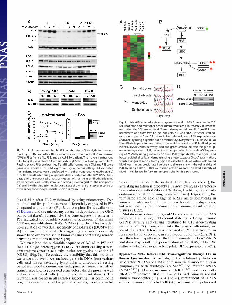
0 and 24 h after IL-2 withdrawal by using microarrays. Twohundred and five probe sets were differentially expressed in P58compared with controls (Fig. 3A; a complete list is available inSI Dataset, and the microarray dataset is deposited in the GEOpublic database). Surprisingly, the gene expression pattern inP58 indicated the possible constitutive activation of the smallGTPase, neuroblastoma RAS (NRAS) (Fig. 3B). This includedup-regulation of two dual-specificity phosphatases (DUSP4 and-6) that are inhibitors of ERK signaling and were previouslyshown to be overexpressed in tumor cell lines containing somaticactivating mutations in NRAS (20).
We examined the nucleotide sequence of NRAS in P58 andfound a single heterozygous G-to-A transition causing a non-conservative aspartic acid substitution for glycine at codon 13(G13D) (Fig. 3C). To exclude the possibility that this mutationwas a somatic event, we analyzed genomic DNA from variouscells and tissues including lymphoblasts, unseparated restingperipheral blood mononuclear cells, purified monocytes, EBV-transformed B cells generated years before the diagnosis, as wellas buccal epithelial cells (Fig. 3C and data not shown). Themutation was found in all samples suggesting it is germline inorigin. Because neither of the patient’s parents, his sibling, or his
two children harbored the mutant allele (data not shown), theactivating mutation is probably a de novo event, as characteris-tically observed with KRAS and HRAS or, less likely, a very earlyembryonic mutation causing mosaicism (3–6). Importantly, thevery same amino acid change in NRAS arises somatically inhuman pediatric and adult myeloid and lymphoid malignancies,but was never before documented in nonmalignant cells ortissues (21, 22).
Mutations in codons 12, 13, and 61 are known to stabilize RASproteins in an active, GTP-bound state by reducing intrinsicGTPase activity and causing resistance to GTPase-activatingproteins (23, 24). Consistent with the genetic alteration, wefound that active NRAS was increased in P58 lymphocytes inserum-rich and, especially, in serum-poor conditions (Fig. 3D).We therefore hypothesized that the ‘‘gain-of-function’’ NRASmutation may result in hyperactivation of the RAS/RAF/ERKpathway, which can negatively regulate BIM expression (25–27).
Hyperactive NRAS Induces BIM Down-Regulation Through ERK inHuman Lymphocytes. To investigate the relationship betweenhyperactive NRAS and BIM suppression, we transfected humanlymphocytes with wild-type (NRASWT) or mutant NRAS(NRASG13D). Overexpression of NRASWT and especiallyNRASG13D reduced BIM in H-9 cells and primary normalhuman lymphocytes (Fig. 4 A and B), reminiscent of HRASoverexpression in epithelial cells (28). We consistently observed
Fig. 2. BIM down-regulation in P58 lymphocytes. (A) Analysis by immuno-blotting of BIM and other BCL-2 members expression after IL-2 withdrawal(CW) in PBLs from a NL, P58, and an ALPS 1A patient. The isoforms extra-long(EL), long (L), and short (S) are indicated. �-Actin is a loading control. (B)Resting ex vivo PBLs and purified T and B cells from normals (NL) and P58 werelysed and assessed for BIM expression by immunoblotting. (C) Activatedhuman lymphocytes were transfected with either nonsilencing RNAi (nsRNAi)or with a small interfering oligonucleotide directed at BIM (BIM RNAi) for 3days, and then deprived of IL-2 or treated with anti-Fas antibody. Silencingefficiency was assessed by immunoblotting (Lower Right) for the nonspecific(ns) and the silencing (si) transfections. Data shown are the representative ofthree independent experiments. Shown is mean � SD.
Fig. 3. Identification of a de novo gain-of-function NRAS mutation in P58.(A) Heat map and relational dendrogram results of a microarray study dem-onstrating the 205 probe sets differentially expressed by cells from P58 com-pared with cells from two normal subjects, NL1 and NL2. Activated lympho-cytes were lysed at 0 and 24 h after IL-2 withdrawal, and mRNA expression wasanalyzed by using oligonucleotide microarrays (Affymetrix U133Plus2.0). (B)Simplified diagram demonstrating differential expression in P58 cells of genesin the NRAS/RAF/ERK pathway. Red and green arrows indicate the genes up-or down-regulated in P58, respectively, compared with controls. (C) Sequenc-ing of NRAS by using genomic DNA from P58 lymphoblasts, monocytes, andbuccal epithelial cells, all demonstrating a heterozygous G-to-A substitution,which changes codon 13 from glycine to aspartic acid. (D) Active GTP-boundNRAS was immunoprecipitated before and after serum withdrawal in a NL andP58, by using a Raf-1 (RBD)-GST fusion protein as bait. The total quantity ofNRAS in cell lysates before immunoprecipitation is also shown.
Oliveira et al. PNAS � May 22, 2007 � vol. 104 � no. 21 � 8955
IMM
UN
OLO
GY
Dow
nloa
ded
by g
uest
on
Janu
ary
13, 2
020
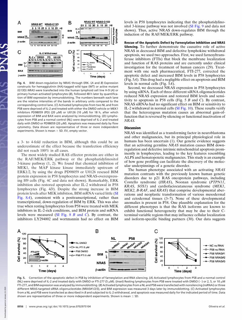
a 3- to 4-fold reduction in BIM, although this could be anunderestimate of the effect because the transfection efficiencydid not reach 100% in all cases.
The most widely studied RAS effector proteins are either inthe RAF/MEK/ERK pathway or the phosphatidylinositol3-kinase pathway (1, 2). We found that chemical inhibition ofMEK1, the MAP kinase kinase immediately upstream ofERK1/2, by using the drugs PD98059 or U0126 rescued BIMprotein expression in P58 lymphocytes and NRAS-overexpress-ing H9 cells (Fig. 4C and data not shown). Remarkably, ERKinhibition also restored apoptosis after IL-2 withdrawal in P58lymphocytes (Fig. 4D). Despite the strong increase in BIMprotein levels after MEK inhibition, BIM mRNA varied little (SIFig. 8A), consistent with a posttranscriptional, rather thantranscriptional, down-regulation of BIM by ERK. This was alsotrue when resting lymphocytes from P58 were treated with MEKinhibitors in IL-2-rich conditions, and BIM protein and mRNAlevels were measured (SI Fig. 8 B and C). By contrast, theinhibitors LY294002 and wortmannin had no effect on BIM
levels in P58 lymphocytes indicating that the phosphatidylino-sitol 3-kinase pathway was not involved (SI Fig. 9 and data notshown). Thus, active NRAS down-regulates BIM through theinduction of the RAF/MEK/ERK pathway.
Rescue of the Apoptotic Defect by Farnesylation Inhibition and NRASSilencing. To further demonstrate the causative role of activeNRAS in decreased BIM and defective lymphokine withdrawalapoptosis, we used two approaches. First, we used farnesyltrans-ferase inhibitors (FTIs) that block the membrane localizationand function of RAS proteins and are currently under clinicalinvestigation for the treatment of human cancers (29). Treat-ment with one such pharmaceutical, FTI-277, corrected theapoptotic defect and increased BIM levels in P58 lymphocytes(Fig. 5A). This drug had a negligible effect on apoptosis and BIMlevels in normal cells (Fig. 5A).
Second, we decreased NRAS expression in P58 lymphocytesby using siRNA. Each of three different siRNA oligonucleotidesreduced NRAS expression and restored BIM levels and sensi-tivity to apoptosis in P58 cells (Fig. 5 B and C). By contrast,NRAS siRNAs had no significant effect on BIM or sensitivity toIL-2 withdrawal in normal cells (SI Fig. 10). These results verifythat the heterozygous mutation causes an abnormal gain-of-function that is reversed by silencing or functional inactivation ofNRAS.
DiscussionNRAS was identified as a transforming factor in neuroblastomaand other malignancies, but its principal physiological role inhumans has been uncertain (1). Our genetic evidence suggeststhat an activating germline NRAS mutation causes BIM down-regulation and defective intrinsic mitochondrial apoptosis prom-inently in lymphocytes, leading to the key features resemblingALPS and hematopoietic malignancies. This study is an exampleof how gene profiling can facilitate the discovery of the molec-ular underpinnings of a genetic disorder.
The human phenotype associated with an activating NRASmutation contrasts with the previously known human geneticdisorders due to p21 RAS oncoprotein pathways, includingCostello syndrome (HRAS), Noonan syndrome (PTPN11,KRAS, SOS1) and cardiofaciocutaneous syndrome (MEK1,MEK2, B-RAF, and KRAS) that comprise developmental aber-rations and neoplastic transformation of various mesodermaland ectodermal tissues (3–7). None of these developmentalanomalies is present in P58. One plausible explanation for thedifferent phenotypes is that the RAS isoforms are known toexhibit functional heterogeneity that may be due to their C-terminal variable regions that may influence cellular localizationand isoform-specific binding partners (30). Our data suggests
Fig. 4. BIM down-regulation by NRAS through ERK. (A and B) Expressionconstructs for hemagglutinin (HA)-tagged wild type (WT) or active mutant(G13D) NRAS were transfected into the human lymphoid cell line H-9 (A) orprimary human activated lymphocytes (B), followed 48 h later by quantifica-tion of BIM expression by immunoblotting. The numbers beneath each laneare the relative intensities of the bands in arbitrary units compared to thecorresponding control lane. (C) Activated lymphocytes from two NL and fromP58 were deprived of IL-2 and treated with either the DMSO vehicle or MEK1inhibitors PD98059 (PD) (20 �M) or U0126 (10 �M) for 18 h, after whichexpression of BIM and BAX were analyzed by immunoblotting. (D) Lympho-cytes from P58 and a normal control (NL) were deprived of IL-2 and treateddaily with DMSO or PD98059 (20 �M). Apoptosis was measured daily by flowcytometry. Data shown are representative of three or more independentexperiments. Shown is mean � SD. EV, empty vector.
Fig. 5. Correction of the apoptotic defect in P58 by inhibition of farnesylation and RNA silencing. (A) Activated lymphocytes from P58 and a normal control(NL) were deprived of IL-2 and treated daily with DMSO or FTI-277 (5 �M). (Inset) Resting lymphocytes from P58 were treated with DMSO (�) or 2, 5, or 10 �MFTI-277, and BIM expression was analyzed by immunoblotting. (B) Activated lymphocytes from a NL and P58 were transfected with nonsilencing (nsRNAi) or threedifferent NRAS-targeted siRNA oligonucleotides (NRAS#1/2/3), and BIM expression was measured 3 days later by immunoblotting. (C) Activated lymphocytesfrom a NL and P58 were transfected as described in B and subjected to IL-2 withdrawal, and apoptosis was measured daily for the indicated period of time. Datashown are representative of three or more independent experiments. Shown is mean � SD.
8956 � www.pnas.org�cgi�doi�10.1073�pnas.0702975104 Oliveira et al.
Dow
nloa
ded
by g
uest
on
Janu
ary
13, 2
020
that ‘‘neuroblastoma’’ or NRAS is an historical misnomer whenconsidering the physiological function in humans of this genesuggested here, and other data in the literature supports thisconclusion. In rodents, an NRAS gene knockout causes subtleimmune deficiencies, despite the fact that the mice are overallhealthy (31). By contrast, KRAS deficiency causes embryoniclethality, whereas HRAS knockout mice are completely normal(32, 33). Transgenic mice bearing activating NRAS mutationsdevelop several hematological tumors, such as leukemia, lym-phoma, mastocytosis, and rare mammary carcinomas (32, 33).The absence of developmental features and the completelydifferent phenotype of a human germline NRAS mutation mightexplain the fact that no mutation in NRAS was ever detected inany of the developmental syndromes cited above, despite effortsto find one (3–7). Taken together, these data indicate that NRAShas an immune regulatory function and its absence or gain-of-function affects primarily hematopoietic cells.
Our observations reveal a vital role of intrinsic mitochondrialapoptosis in peripheral lymphocyte homeostasis and tolerance inhumans. Although gene manipulation in rodents suggested thatthis pathway was important for peripheral immune homeostasis,our human data validate the hypothesis with some surprisingtwists. The NRAS mutation caused a clinical phenotype that, inseveral important aspects, is similar to other ALPS patients, withmodestly elevated TCR-���CD4�CD8� T cells, chronic lym-phoid accumulation, and a clear propensity to hematologicaltumors. However, certain other immunophenotypic and histo-logical features of ALPS were not seen such as marked expan-sions of DNTs in the lymph nodes and elevated HLA-DR� Tcells and CD57� T cells in the periphery (17). The apoptosisdefect is also clearly different from all previous ALPS cases andunderscores a confusing point in the literature. Originally, theALPS phenotype was based on defects in TCR-induced apo-ptosis indicating a deficiency of the propriocidal regulatorymechanism. Because many cases over the years were identifiedwith mutations in the Fas receptor, it was generally assumed thatALPS could be defined by a defect in Fas-induced apoptosis. Ifwe adopt a broader view of apoptosis defects that could disturbperipheral lymphocyte homeostasis, then P58 reveals anotherderangement of this regulatory process. Because we favor thisview, it is reasonable to consider P58’s lymphocyte apoptosisdefect to fulfill one of the diagnostic criteria of ALPS. Becausehe also exhibits increased ��DNTs and expanded secondarylymphoid tissue, he would manifest all of the required featuresfor a diagnosis of ALPS. Because his overall phenotype andgenotype are distinctive, and clearly different from ALPS typesI and II, we provisionally name this condition ALPS, type IV(type III represents undefined molecular pathogenesis).
The active NRAS phenotype we observed is different fromhomozygous deficiency of BIM in rodents, because the latterdoes not cause an increase in CD4�CD8� �� T cells, and inducesthe expansion of other cell types, such as granulocytes (18).These differences may reflect either the residual expression ofBIM or the stimulatory effects of NRAS on ERK and otherdownstream RAS effectors, which could have direct mitogeniceffects that are not triggered by BIM modulation. Moreover,there are several regulatory levels between NRAS and BIMwhere other factors could account for the difference between theBIM knockout mice and P58.
The mechanism of BIM suppression under conditions ofhyperactive NRAS is not clear. In other cellular models ofhyperactive KRAS, BIM is down-regulated through phosphor-ylation, ubiquitination, and degradation by the proteasomalmachinery (26). Here, we show that BIM protein levels areseverely depressed in resting and activated T cells from P58compared with normals, despite equivalent basal mRNA levels.Additionally, the usual up-regulation of BIM mRNA after IL-2withdrawal was impaired in cells from P58. Also, BIM protein
returned to almost normal levels on MEK/ERK blockage, butmRNA remained the same. In our preliminary experiments(data not shown), there was no change in BIM levels onproteasomal blockade in lymphocytes from P58. Taken together,these data suggest a double mechanism for BIM suppression viahyperactive NRAS: inhibition of up-regulation of mRNA andtranslational inhibition, but this remains to be formally shown.Our new understanding of NRAS suggests that RAS antagonistssuch as FTIs could have beneficial effects on disorders oflymphocyte homeostasis and autoimmunity in addition to cancer.
Materials and MethodsCells and Treatments. Patients were studied under an NIH Institu-tional Review Board-approved ALPS research protocol after ob-taining informed consent. PBLs were isolated, activated, andcultivated as previously described (34). Activated T cells werecultured in 100 units/ml recombinant human IL-2 (Roche AppliedScience, Indianapolis, IN) for at least 6 days, and for withdrawalstudies, the cells were then washed three times with PBS andresuspended at 1 � 106 cells/ml in complete media without IL-2 andcultured for different periods of time. Other apoptosis assays usedstaurosporine (Calbiochem, EMD Biosciences, San Diego, CA), anagonistic anti-Fas antibody Apo1.3 (Alexis, San Diego, CA) or�-irradiation. The level of apoptosis was determined by stainingwith 50 ng/ml propidium iodide, and 40 nM 3,3�-dihexyloxacarbo-cyanine iodide (DiOC6) (Calbiochem, EMD Biosciences) and flowcytometry using a constant time acquisition as described (34).Chemical inhibitors PD98059, U0126, LY294002, and FTI-277were obtained from Calbiochem.
DNA and Protein Analyses. Immunoblotting and confocal micro-scopic analysis used the following antibodies: anti-BIM, anti-BAK (Stressgen, Ann Arbor, MI); anti-BCL-2, anti-p27kip1,anti-MCL-1, anti-�-actin, anti-BAX, anti-cytochrome c (clone7H8.2C12), anti-cytochrome c oxidase IV subunit II, anti-BCL-XL (BD Biosciences, San Jose, CA); anti-PUMA (Axxora,San Diego, CA); anti-AIF, anti-NRAS (clone F155) (SantaCruz, Santa Cruz, CA), anti-cytochrome c (6H2.B4; BD Pharm-ingen, San Diego, CA), anti-HSP60 (E-1; Santa Cruz), andanti-BAX (NT; Upstate, Charlottesville, VA). Active GTP-bound NRAS was immunoprecipitated by using the EZ detec-tion Ras activation kit (Pierce, Rockford, IL), according to themanufacturer’s protocol. The immunoprecipitated proteins wereseparated by SDS/PAGE and probed with an anti-NRAS anti-body (F-155; Santa Cruz). Quantitative PCR was done on a 7700ABI PRISM instrument with all of the probes and primerspurchased from the same company (Applied Biosystems, FosterCity, CA) by using established procedures. The reactions werecarried out and normalized to the 18S rRNA signal as described(35). The values were again normalized against a normal controlvalue in each experiment, thus to calculate the fold changes incomparison with the normal. DNA sequencing was carried outon purified PCR products by using puReTaq Ready-To-Go PCRbeads (Amersham Biosciences, Piscataway, NJ), and then di-rectly sequenced by using ABI Prism BigDye (version 1.1)terminators and analyzed on an ABI 3100 Sequencer (AppliedBiosystems).
siRNA and Transient Transfections. Activated PBLs were trans-fected with either a small interfering oligonucleotide RNA(siRNA) or a scrambled nonsilencing control oligo (nsRNA) byusing the Nucleofection system (Amaxa, Koln, Germany). siR-NAs were designed online by using the software BLOCK-iTRNAi designer and purchased from Invitrogen (San Diego, CA).Assessment of knockdown efficiency and experiments wereperformed 3 days later by immunoblotting. The sequences of therelevant oligoribonucleotides are described in SI Methods. Plas-mids expressing human wild-type NRAS (kindly provided by
Oliveira et al. PNAS � May 22, 2007 � vol. 104 � no. 21 � 8957
IMM
UN
OLO
GY
Dow
nloa
ded
by g
uest
on
Janu
ary
13, 2
020
Silvio Gutkind, National Institute of Dental and CraniofacialResearch, NIH, Bethesda, MD) and G13D mutant and weretransiently transfected into appropriate cells.
Microarray Analysis. RNA expression in P58 was compared withthat in two normal controls (NL1, NL2) at 0 and 24 h after IL-2withdrawal from activated cycling T lymphocytes. Total RNAwas hybridized to human Affymetrix (Santa Clara, CA)U133Plus2.0 microarrays following standard Affymetrix proce-dures (Affymetrix) and samples were stained and scanned in theFunctional Genomics and Proteomics Facility by using the
Affymetrix GeneChip Scanner. More detailed methods areavailable in SI Methods.
We thank C. Logun for technical assistance; T. Inaba (HiroshimaUniversity, Hiroshima, Japan), J. Gavard (National Institutes of Health),and S. Gutkind for reagents; R. Germain, R. Siegel, and A. Snow forcritical reading of the manuscript; and S. Patel for help with cytokinemeasurement. Cynthia P. Nix was supported by the Howard HughesMedical Institute–NIH Research Scholar Program. This work wassupported by the National Institute of Allergy and Infectious DiseasesNIH Intramural Research Program.
1. Malumbres M, Barbacid M (2003) Nat Rev Cancer 3:459–465.2. Barbacid M (1987) Annu Rev Biochem 56:779–827.3. Aoki Y, Niihori T, Kawame H, Kurosawa K, Ohashi H, Tanaka Y, Filocamo
M, Kato K, Suzuki Y, Kure S, et al. (2005) Nat Genet 37:1038–1040.4. Rodriguez-Viciana P, Tetsu O, Tidyman WE, Step AL, Conger BA, Cruz MS,
McCormick F, Paven KA (2006) Science 3:1287–1290.5. Schubbert S, Zenker M, Rowe SL, Boll S, Klein C, Bollag G, van der Burgt I,
Musante L, Kalscheuer V, Wehner LE, et al. (2006) Nat Genet 38:331–336.6. Niihori T, Aoki Y, Narumi Y, Neri G, Cave H, Verloes A, Okamoto N,
Hennekam RC, Gillessen-Kaesbach G, Wieczorek D, et al. (2006) Nat Genet38:294–296.
7. Tartaglia M, Pennacchio LA, Zhao C, Yadav KK, Fodale V, Sarkozy A, PanditB, Oishi K, Martinelli S, Schackwitz W, et al. (2007) Nat Genet 39:75–79.
8. Gelb BD, Tartaglia M (2006) Hum Mol Genet 15:R220–R226.9. Oliveira JB, Fleisher T (2004) Curr Opin Allergy Clin Immunol 4:497–503.
10. Bidere N, Su HC, Lenardo MJ (2006) Annu Rev Immunol 24:321–352.11. Fisher GH, Rosenberg FJ, Straus SE, Dale JK, Middleton LA, Lin AY, Strober
W, Lenardo MJ, Puck JM (1995) Cell 81:935–946.12. Wang J, Zheng L, Lobito A, Chan FK, Dale J, Sneller M, Yao X, Puck JM,
Straus SE, Lenardo MJ (1999) Cell 98:47–58.13. Wu J, Wilson J, He J, Xiang L, Schur PH, Mountz JD (1996) J Clin Invest
98:1107–1113.14. Holzelova E, Vonarbourg C, Stolzenberg MC, Arkwright PD, Selz F, Prieur
AM, Blanche S, Bartunkova J, Vilmer E, Fischer A, et al. (2004) N Engl J Med351:1409–1418.
15. Strasser A (2005) Nat Rev Immunol 5:189–200.16. Danial NN, Korsmeyer SJ (2004) Cell 116:205–219.17. Bleesing JJ, Brown MR, Straus SE, Dale JK, Siegel RM, Johnson M, Lenardo
MJ, Puck JM, Fleisher TA (2001) Blood 98:2466–2473.18. Bouillet P, Metcalf D, Huang DC, Tarlinton DM, Kay TW, Kontgen F, Adams
JM, Strasser A (1999) Science 286:1735–1738.19. Strasser A, Pellegrini M (2004) Trends Immunol 25:610–615.
20. Bloethner S, Chen B, Hemminki K, Muller-Berghaus J, Ugurel S, SchadendorfD, Kumar R (2005) Carcinogenesis 26:1224–1232.
21. Bos JL, Toksoz D, Marshall CJ, Verlaan-de Vries M, Veeneman GH, van derEb AJ, van Boom JH, Janssen JW, Steenvoorden AC (1985) Nature 315:726–730.
22. Lubbert M, Mirro J, Jr., Miller CW, Kahan J, Isaac G, Kitchingman G,Mertelsmann R, Herrmann F, McCormick F, Koeffler HP (1990) Blood75:1163–1169.
23. Rodriguez-Viciana P, Tetsu O, Oda K, Okada J, Rauen K, McCormick F(2005) Cold Spring Harb Symp Quant Biol 70:461–467.
24. Vetter IR, Wittinghofer A (2001) Science 294:1299–1304.25. Ley R, Balmanno K, Hadfield K, Weston C, Cook SJ (2003) J Biol Chem
278:18811–18816.26. Ley R, Ewings KE, Hadfield K, Cook SJ (2005) Cell Death Differ 12:1008–1014.27. Ley R, Ewings KE, Hadfield K, Howes E, Balmanno K, Cook SJ (2004) J Biol
Chem 279:8837–8847.28. Tan TT, Degenhardt K, Nelson DA, Beaudoin B, Nieves-Neira W, Bouillet P,
Villunger A, Adams JM, White E (2005) Cancer Cell 7:227–238.29. Karp JE, Lancet JE (2005) Curr Mol Med 5:643–652.30. Mor A, Philips MR (2006) Annu Rev Immunol 24:771–800.31. de Castro IP, Diaz R, Malumbres M, Hernandez MI, Jagirdar J, Jimenez M,
Ahn D, Pellicer A (2003) Cancer Res 63:1615–1622.32. Johnson L, Greenbaum D, Cichowski K, Mercer K, Murphy E, Schmitt E,
Bronson RT, Umanoff H, Edelmann W, Kucherlapati R, et al. (1997) GenesDev 11:2468–2481.
33. Esteban LM, Vicario-Abejon C, Fernandez-Salguero P, Fernandez-MedardeA, Swaminathan N, Yienger K, Lopez E, Malumbres M, McKay R, Ward JM,et al. (2001) Mol Cell Biol 21:1444–1452.
34. Bidere N, Snow AL, Sakai K, Zheng L, Lenardo MJ (2006) Curr Biol16:1666–1671.
35. Livak KJ, Schmittgen TD (2001) Methods 25:402–408.
8958 � www.pnas.org�cgi�doi�10.1073�pnas.0702975104 Oliveira et al.
Dow
nloa
ded
by g
uest
on
Janu
ary
13, 2
020