Embed Size (px)
Citation preview
Research ArticleNovel Technique to Overcome the Nonavailability of aLong Needle 9-0 Polypropylene Suture for Sutured ScleralFixation of the Posterior Chamber Intraocular Lens Using aSingle Fisherman’s Knot
Yong Un Shin,1,2 Mincheol Seong,1,2 Hee Yoon Cho,1,2 and Min Ho Kang1,2
1Department of Ophthalmology, Hanyang University College of Medicine, Seoul, Republic of Korea2Department of Ophthalmology, Hanyang University Guri Hospital, Guri, Republic of Korea
Correspondence should be addressed to Min Ho Kang; [email protected]
Received 21 March 2017; Accepted 27 June 2017; Published 31 July 2017
Academic Editor: Lisa Toto
Copyright © 2017 Yong Un Shin et al. This is an open access article distributed under the Creative Commons AttributionLicense, which permits unrestricted use, distribution, and reproduction in any medium, provided the original work isproperly cited.
Purpose. To describe a method to overcome the nonavailability of a long needle 9-0 polypropylene suture for sutured scleral fixationof the posterior chamber intraocular lens (PC-IOL) using a single fisherman’s knot (SFK). Methods. First, a 10-0 polypropylenesuture was passed from the sclera to the ciliary sulcus using a long needle. A 9-0 suture was tied to the unpassed portion of the10-0 suture with an SFK. We pulled the 10-0 suture to pass the SFK through the sclera, and then we cut the knot and removedthe 10-0 suture. IOL fixation with 9-0 sutures used the conventional techniques used for sutured scleral-fixated IOL.Preoperative and postoperative visual acuity, corneal astigmatism, and endothelial cell count and intraoperative/postoperativecomplications were evaluated. Results. An SFK joining the two sutures was passed through the sclera without breakage orslippage. A total of 35 eyes from 35 patients who underwent sutured scleral fixation of the IOL. An intraoperative complication(minor intraocular hemorrhage) was recorded in four cases. Knot exposure, IOL dislocation, subluxation, and retinaldetachment were not observed. Conclusions. The SFK offers the opportunity to use 9-0 sutures for the long-term safety and maynot require the surgeon to learn any new technique.
1. Introduction
Long-term safety of sutured scleral fixation of the posteriorchamber intraocular lens (PC-IOL) implantation is animportant issue for surgeons. IOL dislocations by spontane-ous breakage of 10-0 polypropylene sutures have beenreported [1–3]. To avoid material problems that arise fromthese sutures, the 9-0 polypropylene suture or Gore-Tex hasbeen recommended as an alternative for sutured scleral fixa-tion of PC IOL [3–5]. Curved long needles (CIF-4, Ethicon)or straight long needles (STC-6, Ethicon) are commonly usedfor sutured scleral fixation. However, the types of needles thatcan attach to a 9-0 polypropylene suture are in limited supply
in Asia and many other regions; CIF-4 and STC-6 needles arenot available. In addition to Ethicon products, a 9-0 polypro-pylene suture is available on long curved or straight needlesfrom Visionary Medical Supplies (Madison, USA), but it isnot available either in Asia or in other regions. As an alterna-tive, a simple needle-and-hook apparatus was proposed toachieve the same effect as the STC6 needle with the 9-0 poly-propylene suture [5]. Sutureless scleral fixation of the IOLcan be another solution, but this technique requires vitrec-tomy instruments and additional surgical experience [6].
We previously suggested a simple method to restore afractured 10-0 polypropylene suture using a single fisher-man’s knot (SFK) [4]. The SFK can be used to join two lines
HindawiJournal of OphthalmologyVolume 2017, Article ID 2683415, 4 pageshttps://doi.org/10.1155/2017/2683415
with a symmetrical structure consisting of two overhandknots, each tied around the standing part of the other; ithas sufficient knot security and is simple to perform.
We introduce this clinically useful and simple SFKmethod to be used with 9-0 polypropylene sutures toovercome the nonavailability of a long needle 9-0 poly-propylene suture.
2. Patients and Methods
Patients who needed the scleral fixation of IOL because ofthe absence of sufficient capsular structure to support theintraocular lens were included. Surgical results are retrospec-tively reviewed via chart review. The preoperative and post-operative visual acuity, corneal astigmatism, and cornealendothelial cell count and intraoperative and postoperativecomplications were evaluated. The study was approved bythe Hanyang University Guri Hospital’s ethics committee.
3. Surgical Techniques
After peribulbar or retrobulbar anesthesia, localized perit-omy was performed at the appropriate site. A single-arm,straight (STC-6, Ethicon), or curved needle (CIF-4, Ethicon)with a 10-0 polypropylene suture was passed through thesclera 1–1.5mm behind the limbus using the ab externo tech-nique, and the suture was pulled from the opposite corneawound. The ends of the 10-0 and 9-0 polypropylene sutureswere overlapped, where they were joined. The unpassedend of the 10-0 polypropylene suture was tied to the 9-0 poly-propylene suture using an SFK (Figures 1(a), 1(b), and 1(c)).Next, the 10-0 polypropylene suture was pulled to allow theSFK to pass through the sclera (Figure 1(d)). The SFK wasremoved from the anterior chamber through the cornealor scleral wound for IOL insertion (Figure 1(e)), the 10-0 polypropylene suture was cut off at the SFK, and the9-0 polypropylene suture was used to close the knot(Figure 1(f), see Supplementary Video available online athttps://doi.org/10.1155/2017/2683415, which shows the sur-gical procedure for Figures 1(a), 1(b), 1(c), 1(d), 1(e), and1(f)). Repeating the samemethod, another 9-0 polypropylenesuture was tied to the haptic of the IOL (Figure 1(g)). Thesurgeon can choose any IOL type that is clinically available.To avoid knot exposure, we used a Z-suture technique tofix the haptic to the sclera [7].
4. Results
An SFK joining 10-0 and 9-0 polypropylene sutures waspassed through the sclera without breakage or slippage.A total of 35 eyes from 35 patients who underwentsutured scleral fixation of the IOL were included in thisstudy. Seven patients were female (20%), and 28 were male(80%); the average patient age at the time of surgery was63.2± 8.6 years (2 SD) (range, 51–83 years). The preoperativemean uncorrected visual acuity was 20/300. The preoperativemean corneal astigmatism was −1.30± 1.27 diopters, and thepreoperative endothelial cell density ranged from 1266–3265cells per square millimeter (mean, 2217.3± 626.2 cells/mm2).
Postoperative mean uncorrected visual acuity, postopera-tive mean corneal astigmatism, and postoperative meanendothelial density were 20/60, −1.34± 0.86 diopters, and1944.8± 683.2 cells/mm2, respectively (p < 0 001, p = 0 853,and p = 0 02).
An intraoperative complication (minor intraocularhemorrhage during needle passage) was recorded in fourcases. Postoperative complications, such as knot exposure,IOL dislocation, subluxation, and retinal detachment, werenot observed.
5. Discussion
Sutureless scleral fixation of the IOL is a new technique forovercoming the common weaknesses of sutured scleral fixa-tion, which include spontaneous breakage, knot exposure,and IOL position problems [6, 8].
However, sutureless scleral fixation of the IOL requiresposterior segment instruments and a machine for pars planavitrectomy. Manipulation of the haptic to pull it through thesclerotomy can cause physical damage to the haptic and cancompromise the long-term stability of the IOL. Intraopera-tive breakage or haptic deformity requires removal and rein-sertion of the IOL [9]. Additionally, the risk of exposure ofthe haptic through the sclera cannot be ruled out. Manyauthors have reported that no complications related tothe use of the sutureless technique arose in the shortterm, but long-term outcomes were not reported [10].Even the scleral-sutured knot, which should be buried bythe scleral flap, can be exposed through the scleral flapand the conjunctiva [11]. An intrastromal fixated haptic hassustained tension to the sclera, and the thickness of the scleraflap is similar to that of the flap of a sutured scleral-fixatedIOL. Thus, an intrastromal haptic can erode over the scleraand conjunctiva.
Sutured scleral-fixated IOLs will continue to be a widelyused option for managing IOL dislocation without IOLexchange, because dislocated IOLs are not always availablein three pieces. In such cases, IOL exchanges are necessaryin order to perform a sutureless scleral fixation. And it isdifficult to handle the IOL haptics in the anterior chamberof severely damaged eyes. Furthermore, each surgeon has adifferent set of surgical skills and instruments, and wecannot ignore that operation results are affected bysurgeon proficiency.
Sutured scleral-fixated IOLs with 10-0 polypropylenesutures can break spontaneously, so a thicker 9-0 polypropyl-ene suture is more suitable for long-term stability. But theavailable needles are not varied enough (e.g., straight,curved, short, and long) to meet the needs of these newtechniques. By using an SFK, a surgeon can use a familiarneedle type, and except for making a knot, surgeons donot need additional surgical instruments or techniques.Also, ciliary sulcus fixation can be advantageous for hapticadhesion by the fibrous membrane to the ciliary body forlong-term stability [12–14].
However, the larger knots made by 9-0 polypropylenesutures could erode externally through the scleral flap, expos-ing the patient to a higher risk of endophthalmitis. To
2 Journal of Ophthalmology
prevent knot erosion, a “Z-suture” technique is a good solu-tion that does not involve scleral flaps or grooves [7].
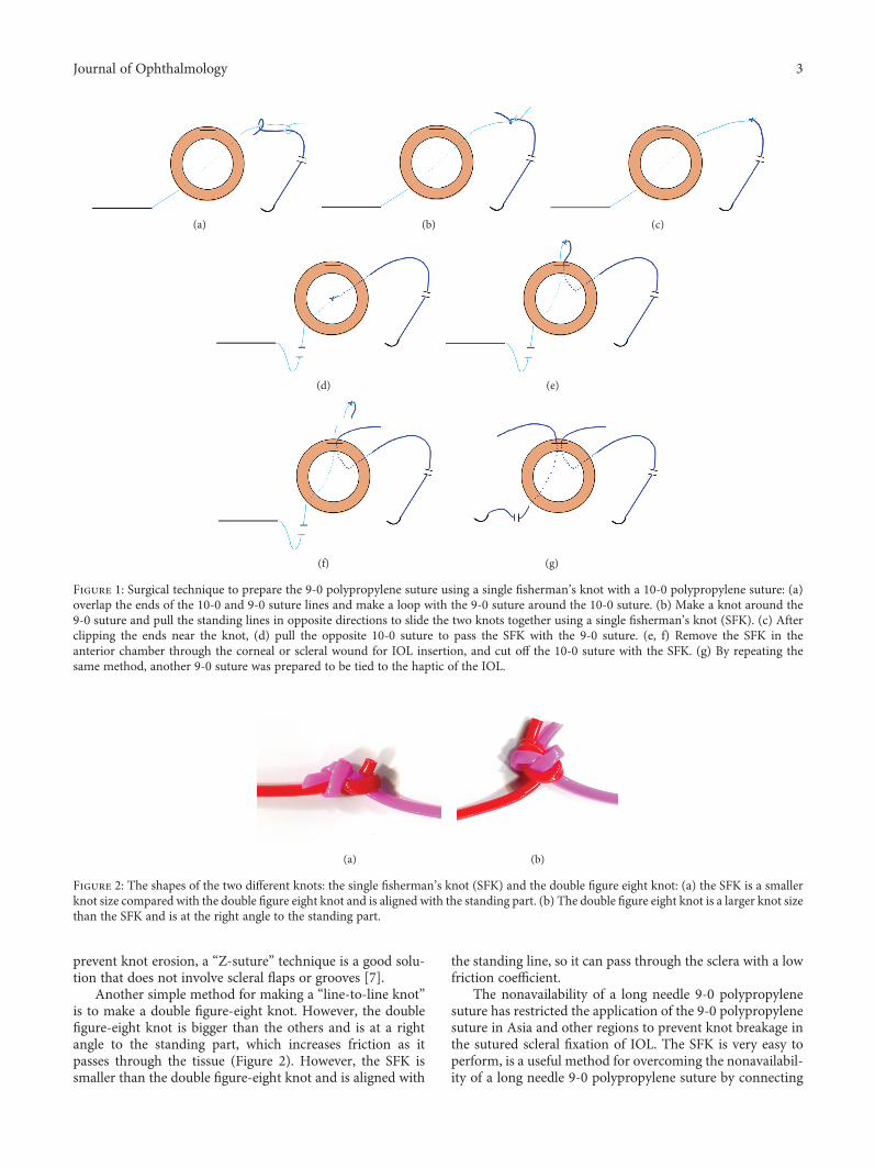
Another simple method for making a “line-to-line knot”is to make a double figure-eight knot. However, the doublefigure-eight knot is bigger than the others and is at a rightangle to the standing part, which increases friction as itpasses through the tissue (Figure 2). However, the SFK issmaller than the double figure-eight knot and is aligned with
the standing line, so it can pass through the sclera with a lowfriction coefficient.
The nonavailability of a long needle 9-0 polypropylenesuture has restricted the application of the 9-0 polypropylenesuture in Asia and other regions to prevent knot breakage inthe sutured scleral fixation of IOL. The SFK is very easy toperform, is a useful method for overcoming the nonavailabil-ity of a long needle 9-0 polypropylene suture by connecting
(a) (b) (c)
(d) (e)
(f) (g)
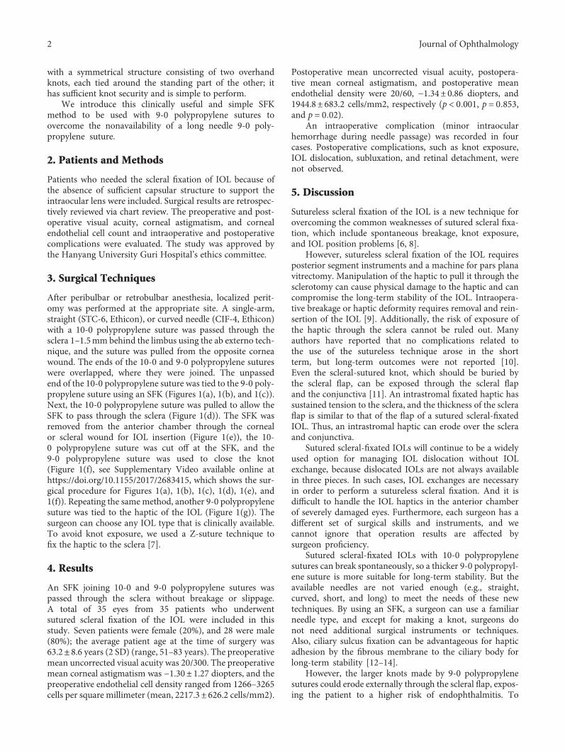
Figure 1: Surgical technique to prepare the 9-0 polypropylene suture using a single fisherman’s knot with a 10-0 polypropylene suture: (a)overlap the ends of the 10-0 and 9-0 suture lines and make a loop with the 9-0 suture around the 10-0 suture. (b) Make a knot around the9-0 suture and pull the standing lines in opposite directions to slide the two knots together using a single fisherman’s knot (SFK). (c) Afterclipping the ends near the knot, (d) pull the opposite 10-0 suture to pass the SFK with the 9-0 suture. (e, f) Remove the SFK in theanterior chamber through the corneal or scleral wound for IOL insertion, and cut off the 10-0 suture with the SFK. (g) By repeating thesame method, another 9-0 suture was prepared to be tied to the haptic of the IOL.
(a) (b)
Figure 2: The shapes of the two different knots: the single fisherman’s knot (SFK) and the double figure eight knot: (a) the SFK is a smallerknot size compared with the double figure eight knot and is aligned with the standing part. (b) The double figure eight knot is a larger knot sizethan the SFK and is at the right angle to the standing part.
3Journal of Ophthalmology
the two lines from the 10-0 and 9-0 polypropylene sutures,and is small enough to pass through the sclera without break-age or slippage.
Conflicts of Interest
No conflicting relationships exist for any of the authors.
Acknowledgments
This work was supported by the Research Fund of HanyangUniversity (HY-2015).
References
[1] R. Asadi and A. Kheirkhah, “Long-term results of scleralfixation of posterior chamber intraocular lenses in children,”Ophthalmology, vol. 1, no. 1, pp. 67–72, 2008.
[2] E. G. Buckley, “Safety of transscleral-sutured intraocular lensesin children,” Journal of American Association for PediatricOphthalmology and Strabismus, vol. 12, no. 5, pp. 431–439,2008.
[3] M. O. Price, F. W. Price Jr., L. Werner, C. Berlie, andN. Mamalis, “Late dislocation of scleral-sutured posteriorchamber intraocular lenses,” Journal of Cataract RefractiveSurgery, vol. 31, no. 7, pp. 1320–1326, 2005.
[4] M. H. Kang, M. Seong, H. W. Lim, and H. Y. Cho, “Simplemethod to restore a fractured 10-0 polypropylene suture usinga single fisherman's knot,” Journal of Cataract RefractiveSurgery, vol. 40, no. 9, pp. 1568–1570, 2014.
[5] M. W. Stewart and M. B. Landers 3rd, “Transscleral intraocu-lar lens fixation with a "homemade" needle and hook,” Journalof Cataract Refractive Surgery, vol. 32, no. 2, pp. 200–202,2006.
[6] A. Agarwal, D. A. Kumar, S. Jacob, C. Baid, A. Agarwal,and S. Srinivasan, “Fibrin glue-assisted sutureless posteriorchamber intraocular lens implantation in eyes with deficientposterior capsules,” Journal of Cataract Refractive Surgery,vol. 34, no. 9, pp. 1433–1438, 2008.
[7] P. Szurman, K. Petermeier, S. Aisenbrey, M. S. Spitzer, andG. B. Jaissle, “Z-suture: a new knotless technique for transscle-ral suture fixation of intraocular implants,” British Journal ofOphthalmology, vol. 94, no. 2, pp. 167–169, 2010.
[8] S. G. Gabor and M. M. Pavlidis, “Sutureless intrascleral poste-rior chamber intraocular lens fixation,” Journal of CataractRefractive Surgery, vol. 33, no. 11, pp. 1851–1854, 2007.
[9] Y. Benayoun, S. Petitpas, K. Turki, J. P. Adenis, and P. Y.Robert, “Sutureless scleral intraocular lens fixation: reportof nine cases and literature review,” Journal of Franch Oph-thalmology, vol. 36, no. 8, pp. 658–668, 2013.
[10] D. A. Kumar, A. Agarwal, S. Packiyalakshmi, S. Jacob, andA. Agarwal, “Complications and visual outcomes after gluedfoldable intraocular lens implantation in eyes with inadequatecapsules,” Journal of Cataract Refractive Surgery, vol. 39, no. 8,pp. 1211–1218, 2013.
[11] K. Solomon, J. R. Gussler, C. Gussler, and W. S. Van Meter,“Incidence and management of complications of transsclerallysutured posterior chamber lenses,” Journal of Cataract Refrac-tive Surgery, vol. 19, no. 4, pp. 488–493, 1993.
[12] E. J. Holland, A. R. Djalilian, and J. Pederson, “Gonioscopicevaluation of haptic position in transsclerally sutured posterior
chamber lenses,” American Journal of Ophthalmololgy,vol. 123, no. 3, pp. 411–413, 1997.
[13] M. L. McDermott and J. E. Puklin, “Pars plana cicatrization ofsewn-in posterior chamber intraocular lens haptics,” Ophthal-mic Surgery and Lasers, vol. 28, no. 3, pp. 239-240, 1997.
[14] M. Kuchle, B. Seitz, C. Hofmann-Rummelt, and G. O.Naumann, “Histopathologic findings in a transsclerallysutured posterior chamber intraocular lens,” Journal of Cata-ract Refractive Surgery, vol. 27, no. 11, pp. 1884–1888, 2001.
4 Journal of Ophthalmology

Submit your manuscripts athttps://www.hindawi.com
Stem CellsInternational
Hindawi Publishing Corporationhttp://www.hindawi.com Volume 2014
Hindawi Publishing Corporationhttp://www.hindawi.com Volume 2014
MEDIATORSINFLAMMATION
of
Hindawi Publishing Corporationhttp://www.hindawi.com Volume 2014
Behavioural Neurology
EndocrinologyInternational Journal of
Hindawi Publishing Corporationhttp://www.hindawi.com Volume 2014
Hindawi Publishing Corporationhttp://www.hindawi.com Volume 2014
Disease Markers
Hindawi Publishing Corporationhttp://www.hindawi.com Volume 2014
BioMed Research International
OncologyJournal of
Hindawi Publishing Corporationhttp://www.hindawi.com Volume 2014
Hindawi Publishing Corporationhttp://www.hindawi.com Volume 2014
Oxidative Medicine and Cellular Longevity
Hindawi Publishing Corporationhttp://www.hindawi.com Volume 2014
PPAR Research
The Scientific World JournalHindawi Publishing Corporation http://www.hindawi.com Volume 2014
Immunology ResearchHindawi Publishing Corporationhttp://www.hindawi.com Volume 2014
Journal of
ObesityJournal of
Hindawi Publishing Corporationhttp://www.hindawi.com Volume 2014
Hindawi Publishing Corporationhttp://www.hindawi.com Volume 2014
Computational and Mathematical Methods in Medicine
OphthalmologyJournal of
Hindawi Publishing Corporationhttp://www.hindawi.com Volume 2014
Diabetes ResearchJournal of
Hindawi Publishing Corporationhttp://www.hindawi.com Volume 2014
Hindawi Publishing Corporationhttp://www.hindawi.com Volume 2014
Research and TreatmentAIDS
Hindawi Publishing Corporationhttp://www.hindawi.com Volume 2014
Gastroenterology Research and Practice
Hindawi Publishing Corporationhttp://www.hindawi.com Volume 2014
Parkinson’s Disease
Evidence-Based Complementary and Alternative Medicine
Volume 2014Hindawi Publishing Corporationhttp://www.hindawi.com