Embed Size (px)
Citation preview

296 | wileyonlinelibrary.com/journal/myc Mycoses 2017;60:296–309© 2017 Blackwell Verlag GmbH
Received: 3 November 2016 | Revised: 30 December 2016 | Accepted: 30 December 2016
DOI: 10.1111/myc.12601
O R I G I N A L A R T I C L E
Novel taxa of thermally dimorphic systemic pathogens in the Ajellomycetaceae (Onygenales)
Karolina Dukik1,2† | Jose F. Muñoz3,4,5† | Yanping Jiang1,6 | Peiying Feng1,7 | Lynne Sigler8 | J. Benjamin Stielow1,9 | Joanna Freeke1,9 | Azadeh Jamalian1,9 | Bert Gerrits van den Ende1 | Juan G. McEwen4,10 | Oliver K. Clay4,11 | Ilan S. Schwartz12,13 | Nelesh P. Govender14,15 | Tsidiso G. Maphanga15 | Christina A. Cuomo3 | Leandro F. Moreno1,2,16 | Chris Kenyon14,17 | Andrew M. Borman18 | Sybren de Hoog1,2
1CBS-KNAW Fungal Biodiversity Centre, Utrecht, The Netherlands2Institute for Biodiversity and Ecosystem Dynamics, University of Amsterdam, Amsterdam, The Netherlands3Broad Institute of MIT and Harvard, Cambridge, MA, USA4Cellular and Molecular Biology Unit, Corporación para Investigaciones Biológicas (CIB), Medellín, Colombia5Institute of Biology, Universidad de Antioquia, Medellín, Colombia6Department of Dermatology, The Affiliated Hospital, Guizhou Medical University, Guiyang, China7Third Affiliated Hospital, Sun Yat-sen University, Guangzhou, China8University of Alberta Microfungus Collection and Herbarium and Biological Sciences, Edmonton, AB, Canada9Thermo Fisher Scientific, Landsmeer, The Netherlands10School of Medicine, Universidad de Antioquia, Medellín, Colombia11School of Medicine and Health Sciences, Universidad del Rosario, Bogotá, Colombia12Faculty of Medicine and Health Sciences, Epidemiology for Global Health Institute, University of Antwerp, Antwerp, Belgium13Rady Faculty of Health Sciences, Max Rady College of Medicine, University of Manitoba, Winnipeg, MB, Canada14University of Cape Town, Cape Town, South Africa15National Institute for Communicable Diseases, Johannesburg, South Africa16Basic Pathology Department, Federal University of Paraná, Curitiba, Brazil17Sexually Transmitted Infection Unit, Institute of Tropical Medicine, Antwerp, Belgium18PHE UK Mycology Reference Laboratory, Bristol, UK
†Contributed equally to this work.
CorrespondenceYanping Jiang and Sybren de Hoog, CBS-KNAW Fungal Biodiversity Centre, Utrecht, The Netherlands.Emails: [email protected] and [email protected]
Funding InformationThis project has been funded in whole or in part with Federal funds from the National Institute of Allergy and Infectious Diseases, National Institutes of Health, Department of Health and Human Services, under Grant Number U19AI110818 to the Broad Institute. This work was partly supported by Colciencias via the grants “A Gene Atlas for Human Pathogenic Fungi,” 122256934875. CBS- KNAW dedicated a grant from the
SummaryRecent discoveries of novel systemic fungal pathogens with thermally dimorphic yeast- like phases have challenged the current taxonomy of the Ajellomycetaceae, a family currently comprising the genera Blastomyces, Emmonsia, Emmonsiellopsis, Helicocarpus, Histoplasma, Lacazia and Paracoccidioides. Our morphological, phylogenetic and phy-logenomic analyses demonstrated species relationships and their specific phenotypes, clarified generic boundaries and provided the first annotated genome assemblies to support the description of two new species. A new genus, Emergomyces, accommo-dates Emmonsia pasteuriana as type species, and the new species Emergomyces afri-canus, the aetiological agent of case series of disseminated infections in South Africa. Both species produce small yeast cells that bud at a narrow base at 37°C and lack
| 297DUKIK et al.
1 | INTRODUCTION
Recent discoveries of novel systemic human pathogens with a ther-mally dimorphic pathogenic phase that consist of budding yeast cells have challenged the current taxonomy of the family Ajellomycetaceae (order Onygenales).1 For nearly 100 years, only four genera of clas-sical systemic pathogens, each containing just one or two species, were recognised in the order Onygenales, ie Coccidioides, Blastomyces, Histoplasma and Paracoccidioides. All these fungi reside in their fila-mentous forms in soil or guano, and upon inhalation by the host, they morphologically shift to an invasive spherule (Coccidioides) or a yeast- like form in the host’s pulmonary system. While phylogenetic anal-yses have placed the genus Coccidioides in the family Onygenaceae, Blastomyces, Histoplasma and Paracoccidioides proved to be members of the family Ajellomycetaceae.2
Another documented genus within the Ajellomycetaceae is Emmonsia, until recently known mainly for species that cause pulmo-nary infections in small mammals. Until the description of Emmonsia pasteuriana in 1998, the genus Emmonsia contained two species: the genetically homogeneous Emmonsia crescens and a more diverse spe-cies, Emmonsia parva.3 These are the aetiological agents of adiaspiro-mycosis, a pulmonary disease of terrestrial mammals and occasionally of humans.4,5 They differ from classical dimorphic pathogenic fungi by their pathogenic phase consisting of large, thick- walled adiaspores in-stead of budding yeast cells. Emmonsia crescens has adiaspores often over 100 μm in diameter and a maximum growth temperature of 37°C, while the adiaspores of Emmonsia parva are mostly 15- 25 μm in diam-eter and the fungus grows up to 40°C. Multilocus phylogenetic analy-sis suggested these species to be less closely related than anticipated. Emmonsia parva clustered with Blastomyces dermatitidis/B. gilchris-tii, while Emmonsia crescens took a rather isolated position.1 Recent phylogenomic analysis supported Emmonsia crescens as a sister group to the clade including Histoplasma, Emmonsia parva and B. dermatiti-dis/B. gilchristii.6 Taken together, the genetic evidence suggests that, in spite of striking morphological, ecological and pathophysiological similarities, aetiological agents of adiaspiromycosis are polyphyletic.
Since the 1970s, novel pathogens have emerged with phylo-genetic, morphological and clinical similarities to known members of the Ajellomycetaceae. Schwartz et al. [1] summarised reports of numerous additional human cases due to novel species in the fam-ily Ajellomycetaceae, most of which remained undescribed. Recently, Wang et al. [7] reported on another novel species from China. Several
of these novel taxa are opportunistic pathogens of immunocom-promised hosts, primarily persons infected with HIV.1 An important emerging species associated with disease in advanced HIV infection was found in South Africa with at least 56 cases reported since being correctly identified in 2008.8,9 The agent causing this mycosis was closely related to Emmonsia pasteuriana, which is also known to infect patients with AIDS and patients with other immune disorders.10–12
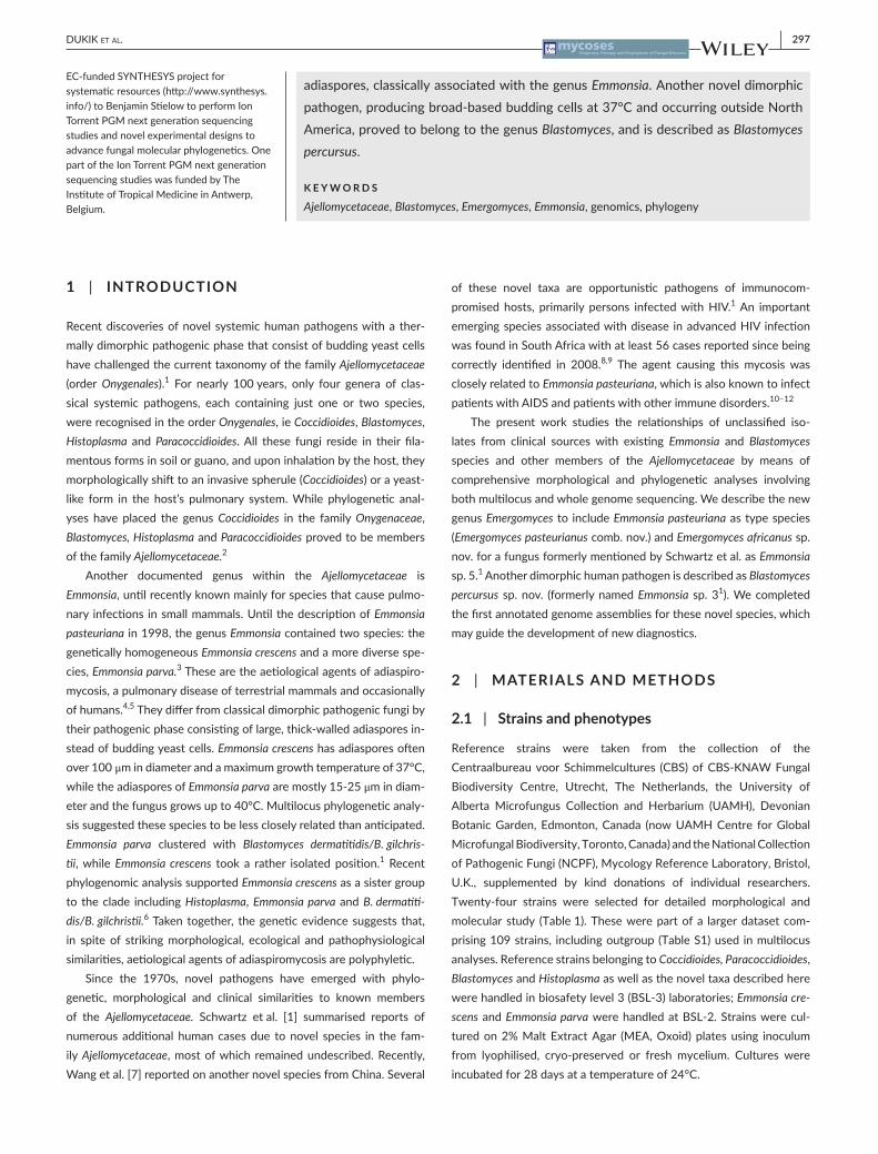
The present work studies the relationships of unclassified iso-lates from clinical sources with existing Emmonsia and Blastomyces species and other members of the Ajellomycetaceae by means of comprehensive morphological and phylogenetic analyses involving both multilocus and whole genome sequencing. We describe the new genus Emergomyces to include Emmonsia pasteuriana as type species (Emergomyces pasteurianus comb. nov.) and Emergomyces africanus sp. nov. for a fungus formerly mentioned by Schwartz et al. as Emmonsia sp. 5.1 Another dimorphic human pathogen is described as Blastomyces percursus sp. nov. (formerly named Emmonsia sp. 31). We completed the first annotated genome assemblies for these novel species, which may guide the development of new diagnostics.
2 | MATERIALS AND METHODS
2.1 | Strains and phenotypes
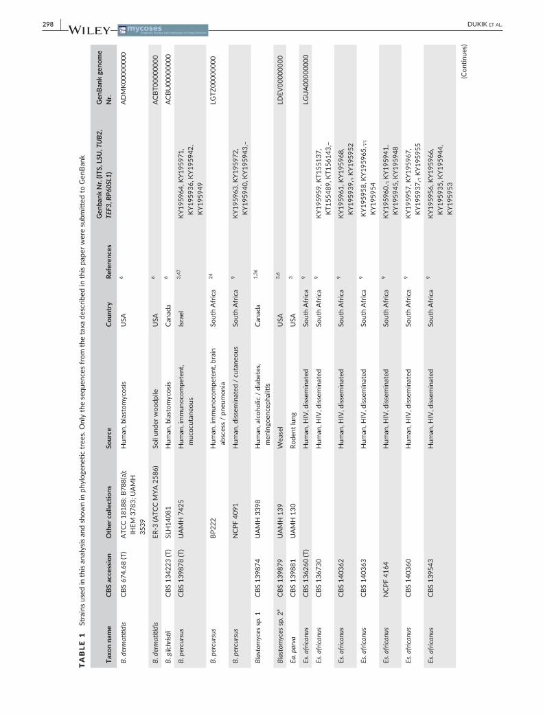
Reference strains were taken from the collection of the Centraalbureau voor Schimmelcultures (CBS) of CBS- KNAW Fungal Biodiversity Centre, Utrecht, The Netherlands, the University of Alberta Microfungus Collection and Herbarium (UAMH), Devonian Botanic Garden, Edmonton, Canada (now UAMH Centre for Global Microfungal Biodiversity, Toronto, Canada) and the National Collection of Pathogenic Fungi (NCPF), Mycology Reference Laboratory, Bristol, U.K., supplemented by kind donations of individual researchers. Twenty- four strains were selected for detailed morphological and molecular study (Table 1). These were part of a larger dataset com-prising 109 strains, including outgroup (Table S1) used in multilocus analyses. Reference strains belonging to Coccidioides, Paracoccidioides, Blastomyces and Histoplasma as well as the novel taxa described here were handled in biosafety level 3 (BSL- 3) laboratories; Emmonsia cre-scens and Emmonsia parva were handled at BSL- 2. Strains were cul-tured on 2% Malt Extract Agar (MEA, Oxoid) plates using inoculum from lyophilised, cryo- preserved or fresh mycelium. Cultures were incubated for 28 days at a temperature of 24°C.
EC- funded SYNTHESYS project for systematic resources (http://www.synthesys.info/) to Benjamin Stielow to perform Ion Torrent PGM next generation sequencing studies and novel experimental designs to advance fungal molecular phylogenetics. One part of the Ion Torrent PGM next generation sequencing studies was funded by The Institute of Tropical Medicine in Antwerp, Belgium.
adiaspores, classically associated with the genus Emmonsia. Another novel dimorphic pathogen, producing broad- based budding cells at 37°C and occurring outside North America, proved to belong to the genus Blastomyces, and is described as Blastomyces percursus.
K E Y W O R D S
Ajellomycetaceae, Blastomyces, Emergomyces, Emmonsia, genomics, phylogeny
298 | DUKIK et al.
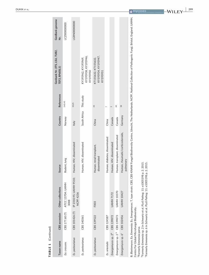
TABLE 1
Stra
ins
used
in th
is an
alys
is an
d sh
own
in p
hylo
gene
tic tr
ees.
Onl
y th
e se
quen
ces
from
the
taxa
des
crib
ed in
this
pape
r wer
e su
bmitt
ed to
Gen
Bank
Taxo
n na
me
CBS
acce
ssio
nO
ther
col
lecti
ons
Sour
ceCo
untr
yRe
fere
nces
Gen
bank
Nr.
(ITS,
LSU
, TU
B2,
TEF3
, RP6
0SL1
)G
enBa
nk g
enom
e N
r.
B. d
erm
atitid
isCB
S 67
4.68
(T)
ATC
C 18
188;
B78
8(a)
; IH
EM 3
783;
UA
MH
35
39
Hum
an, b
last
omyc
osis
USA
6A
DM
K000
0000
0
B. d
erm
atitid
isER
- 3 (A
TCC
MYA
258
6)So
il un
der w
oodp
ileU
SA6
ACB
T000
0000
0
B. g
ilchr
istii
CBS
1342
23 (T
)SL
H14
081
Hum
an, b
last
omyc
osis
Cana
da6
ACB
U00
0000
00
B. p
ercu
rsus
CBS
1398
78 (T
)U
AM
H 7
425
Hum
an, i
mm
unoc
ompe
tent
, m
ucoc
utan
eous
Isra
el3,
47KY
1959
64, K
Y195
971,
KY
1959
36, K
Y195
942,
KY
1959
49
B. p
ercu
rsus
BP22
2H
uman
, im
mun
ocom
pete
nt, b
rain
ab
sces
s /
pneu
mon
iaSo
uth
Afr
ica
24LG
TZ00
0000
00
B. p
ercu
rsus
NCP
F 40
91H
uman
, diss
emin
ated
/ c
utan
eous
Sout
h A
fric
a9
KY19
5963
, KY1
9597
2,
KY19
5940
, KY1
9594
3,
Blas
tom
yces
sp.
1CB
S 13
9874
UA
MH
339
8H
uman
, alc
ohol
ic /
dia
bete
s, m
enin
goen
ceph
aliti
sCa
nada
1,36
Blas
tom
yces
sp.
2a
CBS
1398
79U
AM
H 1
39W
ease
lU
SA3,
6LD
EV00
0000
00
Ea. p
arva
CBS
1398
81U
AM
H 1
30Ro
dent
lung
USA
3
Es. a
frica
nus
CBS
1362
60 (T
)H
uman
, HIV
, diss
emin
ated
Sout
h A
fric
a9
LGU
A00
0000
00
Es. a
frica
nus
CBS
1367
30H
uman
, HIV
, diss
emin
ated
Sout
h A
fric
a9
KY19
5959
, KT1
5513
7,
KT15
5489
, KT1
5614
3,
Es. a
frica
nus
CBS
1403
62H
uman
, HIV
, diss
emin
ated
Sout
h A
fric
a9
KY19
5961
, KY1
9596
8,
KY19
5939
, , K
Y195
952
Es. a
frica
nus
CBS
1403
63H
uman
, HIV
, diss
emin
ated
Sout
h A
fric
a9
KY1
9595
8, K
Y195
965,
, ,
KY19
5954
Es. a
frica
nus
NCP
F 41
64H
uman
, HIV
, diss
emin
ated
Sout
h A
fric
a9
KY19
5960
, , K
Y195
941,
KY
1959
45, K
Y195
948
Es. a
frica
nus
CBS
1403
60H
uman
, HIV
, diss
emin
ated
Sout
h A
fric
a9
KY19
5957
, KY1
9596
7,
KY19
5937
, , K
Y195
955
Es. a
frica
nus
CBS
1395
43H
uman
, HIV
, diss
emin
ated
Sout
h A
fric
a9
KY19
5956
, KY1
9596
6,
KY19
5935
, KY1
9594
4,
KY19
5953
(Con
tinue
s)
| 299DUKIK et al.
Taxo
n na
me
CBS
acce
ssio
nO
ther
col
lecti
ons
Sour
ceCo
untr
yRe
fere
nces
Gen
bank
Nr.
(ITS,
LSU
, TU
B2,
TEF3
, RP6
0SL1
)G
enBa
nk g
enom
e N
r.
Ea. c
resc
ens
CBS
177.
60 (T
)A
TCC
1370
4; U
AM
H
3008
Rode
nt, l
ung
Nor
way
3,42
,43
LCZI
0000
0000
Es. p
aste
uria
nus
CBS
1014
26 (T
)IP
231
0.95
; UA
MH
951
0;
NCP
F 42
36H
uman
, HIV
, diss
emin
ated
Italy
10,2
5LG
RN00
0000
00
Es. p
aste
uria
nus
CBS
1403
61H
uman
, HIV
, diss
emin
ated
Sout
h A
fric
aTh
is st
udy
KY19
5962
, KY1
9596
9,
KY19
5938
, KY1
9594
6,
KY19
5950
Es. p
aste
uria
nus
CBS
1395
22F0
03H
uman
, ren
al tr
ansp
lant
, di
ssem
inat
edCh
ina
45KT
1556
32, K
T155
632,
KY
1959
34, K
Y195
947,
KY
1959
51
Es. o
rient
alis
CBS
1245
87H
uman
, dia
bete
s, di
ssem
inat
edCh
ina
7
Emer
gom
yces
sp.
2b
CBS
1398
72U
AM
H 7
172
Hum
an, H
IV, d
issem
inat
edCa
nada
3
Emer
gom
yces
sp.
2 b
CBS
1398
73U
AM
H 1
0370
Hum
an, t
rans
plan
t, di
ssem
inat
edCa
nada
40
Emer
gom
yces
sp.
6c
CBS
1024
56U
AM
H 1
0427
Hum
an, r
heum
atic/
corti
cost
eroi
ds,
pulm
onar
yG
erm
any
38
B, B
last
omyc
es; E
a, E
mm
onsia
; Es,
Emer
gom
yces
; T, t
ype
stra
in; C
BS, C
BS- K
NA
W F
unga
l Bio
dive
rsity
Cen
tre,
Utr
echt
, The
Net
herla
nds;
NCP
F, N
ation
al C
olle
ction
of P
atho
geni
c Fu
ngi,
Brist
ol, E
ngla
nd; U
AM
H,
Cent
re F
or G
loba
l Mic
rofu
ngal
Bio
dive
rsity
.a Fo
rmer
ly E
mm
onsia
par
va.
b Form
erly
Em
mon
sia s
p. 2
in S
chw
artz
et a
l. PL
oS P
atho
g. 1
1: e
1005
198
p. 2
. 201
5.c Fo
rmer
ly E
mm
onsia
sp.
6 in
Sch
war
tz e
t al.
PLoS
Pat
hog.
11:
e10
0519
8 p.
2. 2
015.
TABLE 1
(Con
tinue
d)
300 | DUKIK et al.
Microscopic observations were done with slide cultures using MEA, as an optimal medium for conidium formation. Agar blocks of ~0.5 cm2 were placed on agar plates and inoculated at the four sides. The block was subsequently covered with a sterile cover slip (~2 cm2). Plates were incubated at 24°C for 7, 14, 21 and 28 days in a closed plastic box with sterile gauze soaked with 5 ml sterile water to avoid drying of the culture. Slides were made by Shear’s mounting medium without pigments. Micrographs were taken using a Nikon Eclipse 80i microscope and DS Camera Head DS- Fi1/DS- 5m/DS- 2Mv/DS- 2MBW using nIs-element freeware package (Nikon Europe, Badhoevedorp, The Netherlands). Dimensions were determined with the Nikon Eclipse 80i measurement module and the mean and stan-dard deviation were calculated from measurements of 50 conidia.
Cardinal temperatures were determined on MEA at 5, 15, 21, 24, 27, 30, 33, 36, 37, 40 and 42°C. Growth rates were determined in triplicate after 4 weeks incubation. Thermal dimorphism was evalu-ated by incubation on MEA and Brain Heart Infusion agar plates (BHI, BD Difco) for 1 to 4 weeks, using temperature switch from 24 to 37°C as sole stimulus for transition.13
2.2 | DNA extraction, PCR and sequencing
Fungal material was harvested for DNA extraction using MasterPure™ Yeast DNA Purification Kit from Epicentre (Madison, WI, USA). Five gene regions were amplified. The first two loci were ITS and LSU of the rDNA operon.14 The universal fungal locus ITS1- 5.8- ITS2 of the rDNA was amplified with primers ITS515 and ITS4 operated under standard PCR conditions14. Partial LSU of the rDNA operon was amplified using LR0R and LR5 primer set16 under the same PCR conditions but with cycle extension of 90 s. Partial β- tubulin (TUB2) covering the variable 5’- end containing four small introns, was amplified with TUB2Fd and TUB4Fd primer set,17 partial gene encoding elongation factor 3 (TEF3) with Al50+51_EF3_2900_F and Al50+51_EF3_3300_R primer set, and 60S ribosomal protein L10 (RP 60S L1) with AlGr52_412- 433_F1 and Algr52_1102_1084_R1 primer set.18 Primers and PCR protocols were designed and tested for the development of potential secondary fungal barcodes.18 PCR products were visualised on 1% agarose gels. Positive PCR products were sequenced in cycle- sequencing reaction using ABI big dye terminator v.3.1 with a modified manufacturer’s protocol.18 Following the cycle- sequencing reaction, a capillary elec-trophoresis system (DNA analyser, Thermo Fisher Scientific, Waltham, USA) was used for performing bidirectional sequencing. Sequences obtained were manually edited and consensus sequences stored in a Biolomics database.19
2.3 | Sequence alignment and phylogenetic analysis
Obtained sequences were aligned with mafft v. 6.850b with default settings except for the ‘genafpair’ option.20 Datasets for the five loci were assembled in a single multilocus dataset using seqUence matrIx software.21 For both the ITS and multilocus datasets, a maximum like-lihood phylogeny was inferred using raxml v.8.0.0 employing GTRCAT model and 1000 bootstrap replicates.22 Bootstrap branch support
above 70% was considered as significant. Multiple sequences of spe-cies and genera outside the focus of our analysis were collapsed or represented by one or two strains. The interspecific variation (%) was estimated and included in Table S2. The multilocus dataset was ad-ditionally analysed by Markov chain Monte Carlo (MCMC) algorithm with mrbayes v. 3.2.623 on the CIPRES portal (http://www.phylo.org) with four simultaneous runs for 10 million generations, with a sam-pling frequency of 1000 trees. A burn- in tree sample of 25% was discarded. Bayesian posterior probabilities from 50% majority- rule consensus trees with a probability value higher than 0.80 were con-sidered as significant.
2.4 | Genome sequencing and de novo assembly
Three strains were selected for genome sequencing, including Ea. pasteuriana CBS 101426, and BP222 and CBS 136260 initially men-tioned as Emmonsia sp.3 and Emmonsia sp.5 respectively1 (Table 1). Genomic DNA of strain BP222, isolated from a brain abscess in an im-munocompetent person in South Africa24 was extracted and a library with insert sizes ranging from 500 to 1500 bp was sequenced on the Illumina MiSeq platform to obtain paired- end reads of 300 bp. Strain CBS 136260, isolated from a skin biopsy in an HIV- infected patient9 was sequenced using IonTorrent, generating unpaired reads from 8 to 361 bp. For strain CBS 101426 (=UAMH 9510=NCPF 4236) of Ea. pasteuriana, isolated from cutaneous lesions in an Italian woman with advanced HIV infection10,25 100 ng of genomic DNA was sheared to approximately 250 bp for library construction, using a Covaris LE in-strument and prepared for sequencing as previously described.26 A library with 180- base inserts was constructed and sequenced on an Illumina HiSeq 2000 platform to generate 101 bp paired- end reads, producing average genome coverage of 191X.
Both Illumina and IonTorrent reads of BP222 and CBS 136260 were assembled using the spaDes assembler v3.1.1.27 Next, Pilon v1.1628 was used to correct the best assembly from each species, resolving single nucleotide errors (SNPs), artifactual indels and local mis- assemblies, as previously described for Paracoccidioides species.29 The 101- bp Illumina reads of Ea. pasteuriana were assembled using ALLPATHS- LG30 with default parameters. All three de novo assem-blies were evaluated using gaemr package (http://www.broadinstitute.org/software/gaemr/), which revealed no aberrant regions of cover-age, GC content or unexpected sequence similarity suggestive of con-tamination. Scaffolds representing the mitochondrial genome were separated out from the nuclear assembly.
2.5 | Gene prediction and annotation
Genes were predicted and annotated by combining calls from mul-tiple methods to obtain the best consensus model for a given locus. These included ab initio predictions (GlimmerHMM, Augustus, Snap, GeneMark- ES), homologous inference (Genewise, TBlastN) and gene model consolidation programs (evIDencemoDeler).31 For protein- coding gene name assignment, we combined HMMER PFAM/TIGRFAM, Swissprot and Kegg products. Kinannote was used to annotate

| 301DUKIK et al.
protein kinases.32 To evaluate the completeness of predicted gene sets, the representation of highly conserved genes in a wide range of eukaryotic taxa (core eukaryotic genes; CEGs) were analysed using CEGMA genes33 with the CoreAlyze tool (http://sourceforge.net/projects/corealyze/).
2.6 | Identification of orthologs and phylogenomic analysis
To examine the phylogenetic relationship of novel sequenced species relative to other dimorphic fungi, single- copy orthologs of species from the family Ajellomycetaceae were determined and clus-tered using orthomcl (version 1.4)34 with a Markov inflation index of 1.5 and a maximum e- value of 1e- 5. A total of 19 genomes from the Onygenales order and three Aspergillus genomes were chosen for comparative analyses. These include the following genomes: Blastomyces gilchristii SLH 14081 (ACBU00000000), Blastomyces der-matitidis ATCC 26199 (AEII00000000), B. dermatitidis ATCC 18188 (ADMK00000000), B. dermatitidis ER- 3 (ACBT00000000), Emmonsia parva UAMH 139 (LDEV00000000) and Ea. crescens UAMH 3008 (LCZI00000000), Histoplasma capsulatum WU24 (AAJI01000000), H. capsulatum G186AR (ABBS01000000), Paracoccidioides lutzii Pb01 (ABKH02000000), P. brasiliensis Pb03 (ABHV02000000), P. brasilien-sis Pb18 (ABKI02000000), Coccidioides immitis RS (AAEC00000000), C. posadasii CBS 113859=Silveira (ABAI02000000), Uncinocarpus reesii UAMH 1704 (AAIW00000000), Microsporum gypseum CBS 118893 (ABQE00000000), Trichophyton rubrum CBS 118892 (ACPH01000000), Aspergillus nidulans FGSC A4 (AACD00000000), A. flavus NRRL3357 (AAIH00000000), A. fumigatus Af293 (AAHF01000000). To estimate the species phylogeny, orthologs present in a single copy (1:1) in all of 22 genomes were identified. Multiple protein sequence alignment was performed for each single- copy ortholog cluster using MUSCLE to generate sequence align-ments of the same length. Then, the cluster multiple alignments were concatenated, and a phylogeny was estimated using raxml v7.7.822 with model PROTCATWAG with a total of 1000 bootstrap replicates.
2.7 | Data availability statement
The assemblies and annotations of the described species genomes were deposited at DDBJ/ENA/GenBank under the following acces-sion numbers: strain BP222 (PRJNA284520), strain CBS 101426 (PRJNA234734) and strain CBS 136260 (PRJNA284519).
3 | RESULTS
3.1 | Multilocus phylogeny
To examine the phylogenetic relationships of the novel species, we used a panel of 107 strains from the family Ajellomycetaceae and multilocus phylogenetic analysis of concatenated sequences of ITS, LSU, TUB2, TEF3 and RP 60S L1. Using Coccidioides species (fam. Onygenaceae) as outgroup, five monophyletic clades were clearly
recognisable and highly supported within the family Ajellomycetaceae, representing the new genus, the systemic pathogens in the genera Paracoccidioides, Histoplasma and Blastomyces, and the recently estab-lished environmental genus Emmonsiellopsis35 (Figure 1). Within the Ajellomycetaceae, the genus Emmonsiellopsis is located in an ancestral basal position, followed by Paracoccidioides. Bootstrap support (BS) and posterior probabilities (PP) of the genera Emmonsiellopsis and Paracoccidioides were both high (BS/PP 100/1).
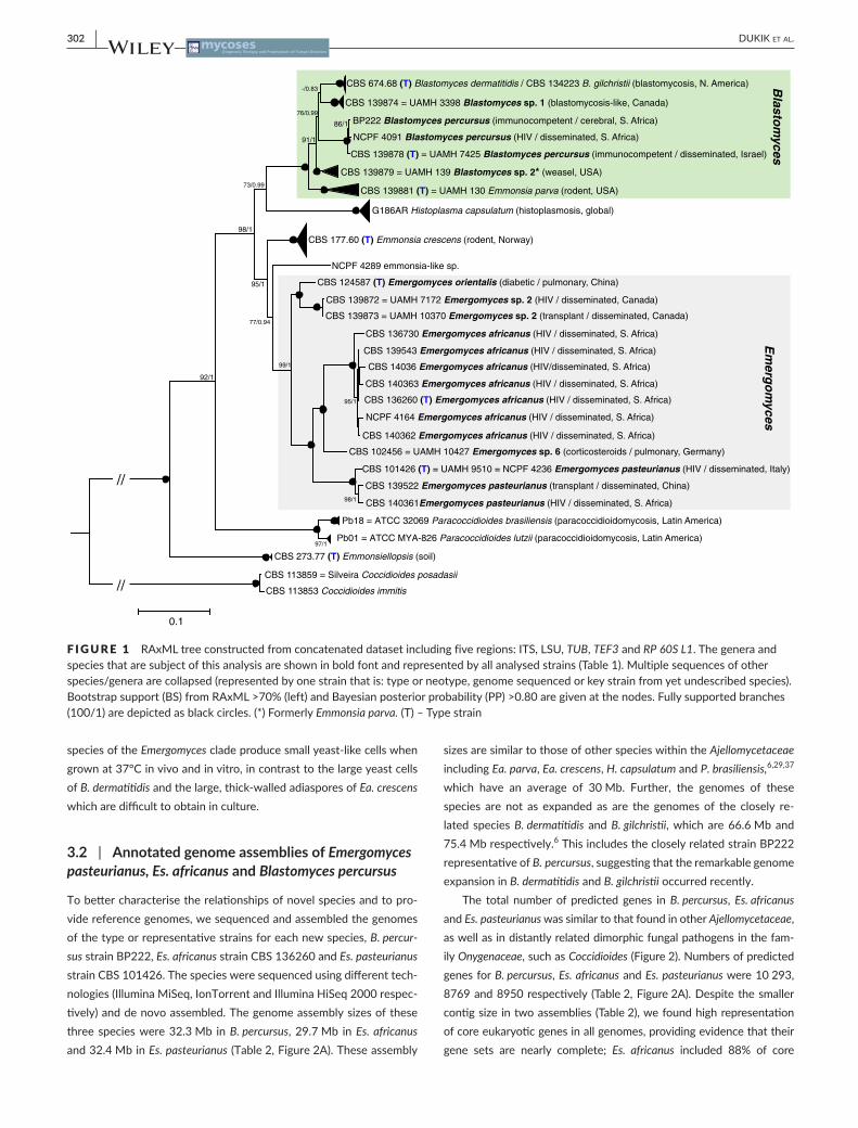
Two clades (green and grey boxes; Figure 1) were analysed in de-tail. The upper clade (green box) contains Blastomyces in its current sense and in total is interpreted to represent the genus Blastomyces. The Blastomyces clade has five monophyletic subgroups, all supported by highest BS/PP values of 100/1 (black dots). The uppermost clade contains the aetiological agents of blastomycosis in North America, B. dermatitidis and B. gilchristii, which could not be separated with the chosen set of loci. The closest clade to B. dermatitidis/B. gilchristii is indicated as Blastomyces sp. 1 and includes the strain CBS 139874 (=UAMH 3398) originating from Canada, which was reported as an unusual case of blastomycosis.36 The nearest clade to the group B. dermatitidis/B. gilchristii/Blastomyces sp. 1 (BS/PP 76/0.99) in-cludes strains of a novel species causing systemic mycosis in healthy human hosts, described here as Blastomyces percursus. Two of the three B. percursus strains, BP222 and NCPF 4091 originating from South Africa are grouped together (BS/PP 86/1). A third strain, CBS 139878=UAMH 7425 originating from Israel, is slightly divergent. Two further clades (BS/PP 91/1, 100/1) include strains previously de-scribed as Emmonsia parva. The clade denoted as Blastomyces sp. 2 includes the genome- sequenced Ea. parva strain CBS 139879=UAMH 1396 isolated by W. L. Jellison from a weasel in the USA. This is clearly separated from the clade including another classical Ea. parva strain, CBS 139881 (=UAMH 130) isolated by C.W. Emmons, and confirms its basal position and the generic distinctions among Ea. parva strains noted previously.3 The Emmonsia- like strains producing small adias-pores phylogenetically cluster in Blastomyces, while Ea. crescens re-mains outside Blastomyces (Figure 1).
The lower clade in Figure 1 (grey box) is clearly separated from Blastomyces and Histoplasma, and has a large diversity of isolates, includ-ing emerging dimorphic species, and is here described as Emergomyces. The clade is monophyletic with BS/PP 99/1 support and includes three well- defined subclades. The monophyletic Es. pasteurianus clus-ter (BS/PP 100/1) comprises the type strain CBS 101426=UAMH 9510 and two additional strains. A second large monophyletic cluster (BS/PP 100/1) comprises seven strains obtained from HIV- infected in-dividuals with disseminated mycoses in South Africa.9 The new taxon Emergomyces africanus has the genome- sequenced strain CBS 136260 as type. The group is separated from Es. pasteurianus by a single strain from a human infection in Germany, CBS 102456 (=UAMH 10427), which is significantly different from Es. africanus (eg by 19/508 ITS alignment difference). Thus, it is denoted as Emergomyces sp. 6 (for-merly Emmonsia sp. 6 in Schwartz et al. [1]). A sister clade to the Es. pasteurianus- group comprises the recently described species Es. orien-talis7 and another undescribed taxon, Emergomyces sp. 2 (as Emmonsia sp. 2 in Schwartz et al. [1]) represented by two strains from Canada. All
302 | DUKIK et al.
species of the Emergomyces clade produce small yeast- like cells when grown at 37°C in vivo and in vitro, in contrast to the large yeast cells of B. dermatitidis and the large, thick- walled adiaspores of Ea. crescens which are difficult to obtain in culture.
3.2 | Annotated genome assemblies of Emergomyces pasteurianus, Es. africanus and Blastomyces percursus
To better characterise the relationships of novel species and to pro-vide reference genomes, we sequenced and assembled the genomes of the type or representative strains for each new species, B. percur-sus strain BP222, Es. africanus strain CBS 136260 and Es. pasteurianus strain CBS 101426. The species were sequenced using different tech-nologies (Illumina MiSeq, IonTorrent and Illumina HiSeq 2000 respec-tively) and de novo assembled. The genome assembly sizes of these three species were 32.3 Mb in B. percursus, 29.7 Mb in Es. africanus and 32.4 Mb in Es. pasteurianus (Table 2, Figure 2A). These assembly
sizes are similar to those of other species within the Ajellomycetaceae including Ea. parva, Ea. crescens, H. capsulatum and P. brasiliensis,6,29,37 which have an average of 30 Mb. Further, the genomes of these species are not as expanded as are the genomes of the closely re-lated species B. dermatitidis and B. gilchristii, which are 66.6 Mb and 75.4 Mb respectively.6 This includes the closely related strain BP222 representative of B. percursus, suggesting that the remarkable genome expansion in B. dermatitidis and B. gilchristii occurred recently.
The total number of predicted genes in B. percursus, Es. africanus and Es. pasteurianus was similar to that found in other Ajellomycetaceae, as well as in distantly related dimorphic fungal pathogens in the fam-ily Onygenaceae, such as Coccidioides (Figure 2). Numbers of predicted genes for B. percursus, Es. africanus and Es. pasteurianus were 10 293, 8769 and 8950 respectively (Table 2, Figure 2A). Despite the smaller contig size in two assemblies (Table 2), we found high representation of core eukaryotic genes in all genomes, providing evidence that their gene sets are nearly complete; Es. africanus included 88% of core
F IGURE 1 RAxML tree constructed from concatenated dataset including five regions: ITS, LSU, TUB, TEF3 and RP 60S L1. The genera and species that are subject of this analysis are shown in bold font and represented by all analysed strains (Table 1). Multiple sequences of other species/genera are collapsed (represented by one strain that is: type or neotype, genome sequenced or key strain from yet undescribed species). Bootstrap support (BS) from RAxML >70% (left) and Bayesian posterior probability (PP) >0.80 are given at the nodes. Fully supported branches (100/1) are depicted as black circles. (*) Formerly Emmonsia parva. (T) – Type strain
86/1
76/0.99
91/1
73/0.99
95/1
99/1
77/0.94
95/1
98/1
97/1
92/1
0.1
//
//
CBS 674.68 (T) Blastomyces dermatitidis / CBS 134223 B. gilchristii (blastomycosis, N. America)
CBS 139874 = UAMH 3398 Blastomyces sp. 1 (blastomycosis-like, Canada)
BP222 Blastomyces percursus (immunocompetent / cerebral, S. Africa)
NCPF 4091 Blastomyces percursus (HIV / disseminated, S. Africa)
CBS 139878 (T) = UAMH 7425 Blastomyces percursus (immunocompetent / disseminated, Israel)
CBS 139879 = UAMH 139 Blastomyces sp. 2* (weasel, USA)
CBS 139881 (T) = UAMH 130 Emmonsia parva (rodent, USA)
G186AR Histoplasma capsulatum (histoplasmosis, global)
CBS 177.60 (T) Emmonsia crescens (rodent, Norway)
NCPF 4289 emmonsia-like sp.
CBS 124587 (T) Emergomyces orientalis (diabetic / pulmonary, China)
CBS 139872 = UAMH 7172 Emergomyces sp. 2 (HIV / disseminated, Canada)
CBS 139873 = UAMH 10370 Emergomyces sp. 2 (transplant / disseminated, Canada)
CBS 136730 Emergomyces africanus (HIV / disseminated, S. Africa)
CBS 139543 Emergomyces africanus (HIV / disseminated, S. Africa)
CBS 14036 Emergomyces africanus (HIV/disseminated, S. Africa)
CBS 140363 Emergomyces africanus (HIV / disseminated, S. Africa)
CBS 136260 (T) Emergomyces africanus (HIV / disseminated, S. Africa)
NCPF 4164 Emergomyces africanus (HIV / disseminated, S. Africa)
CBS 140362 Emergomyces africanus (HIV / disseminated, S. Africa)
CBS 102456 = UAMH 10427 Emergomyces sp. 6 (corticosteroids / pulmonary, Germany)
CBS 101426 (T) = UAMH 9510 = NCPF 4236 Emergomyces pasteurianus (HIV / disseminated, Italy)
CBS 139522 Emergomyces pasteurianus (transplant / disseminated, China)
CBS 140361Emergomyces pasteurianus (HIV / disseminated, S. Africa)
Pb18 = ATCC 32069 Paracoccidioides brasiliensis (paracoccidioidomycosis, Latin America)
Pb01 = ATCC MYA-826 Paracoccidioides lutzii (paracoccidioidomycosis, Latin America)
CBS 273.77 (T) Emmonsiellopsis (soil)
CBS 113859 = Silveira Coccidioides posadasii
CBS 113853 Coccidioides immitis
Blasto
myces
Em
ergo
myces
-/0.83
98/1

| 303DUKIK et al.
eukaryotic genes, while B. percursus and Es. pasteurianus gene sets in-cluded 96‒98% (Figure 1B). Based on their completeness, the B. percur-sus, Es. africanus and Es. pasteurianus reference genome assemblies can define a wide set of genes that is shared across the dimorphic patho-genic fungi. We classified these references according to the mating- type locus. Es. africanus and Es. pasteurianus contained mating- type alpha (MAT1-1; locus ID ACJ72_07256 and AI78_01298 respectively), while B. percursus contained the mating- type HMG (MAT1-2; locus ID ACJ73_00817).
3.3 | Phylogenomics of Emergomyces pasteurianus, Es. africanus and Blastomyces percursus
To compare gene content and conservation, we identified ortholo-gous gene clusters in the three genomes sequenced here, Onygenales
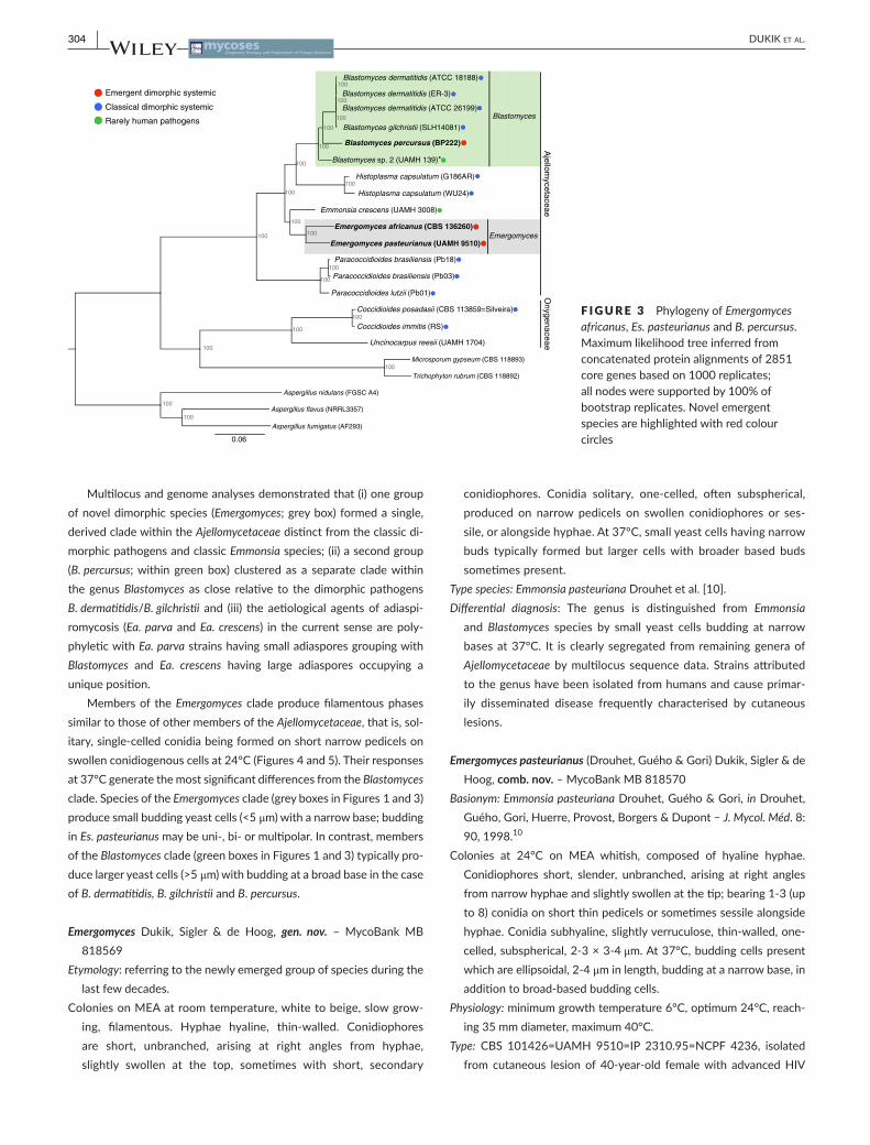
genomes of other dimorphic pathogens (Blastomyces, Histoplasma, Paracoccidioides and Coccidioides) and two dimorphic non- human path-ogenic species, Ea. parva and Ea. crescens, the aetiological agents of adiaspiromycosis in small mammals. As outgroups, three Aspergillus ge-nomes were also included. Using 2851 single- copy core genes present in all strains, we estimated a strongly supported phylogeny of these organ-isms using RAxML (Figure 3). Blastomyces percursus clustered with the primary pathogen B. dermatitidis/B. gilchristii (100% bootstrap support). Blastomyces sp. 2 (strain UAMH 139; formerly Ea. parva) was also closely related, branching earlier as a sister species within the Blastomyces clade (Figure 3; green box). Es. africanus and Es. pasteurianus clustered in a sin-gle, strongly supported (100% bootstrap replicates) clade as sister spe-cies, and this clade was sister to Ea. crescens (Figure 3; grey box). The Ea. crescens – Emergomyces clade is a sister group of the clade including Histoplasma and Blastomyces, with Paracoccidioides in a basal position.
F IGURE 2 (A) Genome sizes in megabases (Mb) and total number of protein- coding genes of the sequenced and assembled strains of Emergomyces africanus, Es. pasteurianus and Blastomyces percursus, and other previously sequenced and assembled species from the Ajellomycetaceae. * formerly Emmonsia parva (B) Conservation of core eukaryotic genes (CEGs) across Es. africanus, Es. pasteurianus, B. percursus and other compared genomes
0
50
100
150
200
250
Emer
gomyc
es p
aste
urianus
Emer
gomyc
es af
rican
us
Blasto
myc
es p
ercu
rsus
Blasto
myc
es d
erm
atitid
is
Histop
lasm
a ca
psula
tum
Parac
occid
ioide
s bra
silien
sis
Nu
mb
er o
f C
EG
s
90-100%
80-90%
70-80%
60-70%
50-60%
<50%
Missing
0 10 20 30 40 50 60 70 80
0 2000 4000 6000 8000 10000
Microsporum gypseum (CBS 118893)
Trichophyton rubrum (CBS 118892)
Uncinocarpus reesii (UAMH 1704)
Coccidioides posadasii (Silveira)
Coccidioides immitis (RS)
Paracoccidioides brasiliensis (Pb18)
Paracoccidioides lutzii (Pb01)
Emergomyces pasteurianus (UAMH 9510)
Emergomyces africanus (CBS 136260)
Emmonsia crescens (UAMH 3008)
Histoplasma capsulatum (G186AR)
Blastomyces sp. 2 (UAMH 139)*
Blastomyces percursus (BP222)
Blastomyces gilchristii (SLH14081)
Blastomyces dermatitidis (ER-3)
Genome size (Mb)
Number of Protein-Coding Genes
Protein-coding Genes Genome Size
Aje
llom
ycet
acea
e
(A) (B)
TABLE 2 Assembly and annotation statistics for Emergomyces pasteurianus, Es. africanus, Blastomyces percursus. For comparison, GC content percentage, genome size and number of genes were included for reference genomes of B. dermatitidis (ER- 3) and Blastomyces sp. 2 (UAMH 139)
Genus Emergomyces Blastomyces
Species Es. africanus Es. pasteurianus B. percursus B. dermatitidis B. sp. 2a
Strain CBS 136260 UAMH 9510 BP222 ER- 3 UAMH 139
Scaffolds 4444 1643 3868 25 2682
Scaffold N50 (kb) 13.8 45.9 12.8 5550 31.17
Scaffold N90 (kb) 2.5 8.8 4.0 2312 3.4
Max Scaffold (kb) 79.7 267.9 71.4 10 302.04 265.3
Assembly GC 43.5 44.5 47.3 37.1 44.7
Total Length (Mb) 29.7 32.4 32.3 66.6 30.3
Protein-coding genes 8769 8950 10 293 9755 8563
aFormerly Emmonsia parva.
304 | DUKIK et al.
Multilocus and genome analyses demonstrated that (i) one group of novel dimorphic species (Emergomyces; grey box) formed a single, derived clade within the Ajellomycetaceae distinct from the classic di-morphic pathogens and classic Emmonsia species; (ii) a second group (B. percursus; within green box) clustered as a separate clade within the genus Blastomyces as close relative to the dimorphic pathogens B. dermatitidis/B. gilchristii and (iii) the aetiological agents of adiaspi-romycosis (Ea. parva and Ea. crescens) in the current sense are poly-phyletic with Ea. parva strains having small adiaspores grouping with Blastomyces and Ea. crescens having large adiaspores occupying a unique position.
Members of the Emergomyces clade produce filamentous phases similar to those of other members of the Ajellomycetaceae, that is, sol-itary, single- celled conidia being formed on short narrow pedicels on swollen conidiogenous cells at 24°C (Figures 4 and 5). Their responses at 37°C generate the most significant differences from the Blastomyces clade. Species of the Emergomyces clade (grey boxes in Figures 1 and 3) produce small budding yeast cells (<5 μm) with a narrow base; budding in Es. pasteurianus may be uni- , bi- or multipolar. In contrast, members of the Blastomyces clade (green boxes in Figures 1 and 3) typically pro-duce larger yeast cells (>5 μm) with budding at a broad base in the case of B. dermatitidis, B. gilchristii and B. percursus.
Emergomyces Dukik, Sigler & de Hoog, gen. nov. – MycoBank MB 818569
Etymology: referring to the newly emerged group of species during the last few decades.
Colonies on MEA at room temperature, white to beige, slow grow-ing, filamentous. Hyphae hyaline, thin-walled. Conidiophores are short, unbranched, arising at right angles from hyphae, slightly swollen at the top, sometimes with short, secondary
conidiophores. Conidia solitary, one-celled, often subspherical, produced on narrow pedicels on swollen conidiophores or ses-sile, or alongside hyphae. At 37°C, small yeast cells having narrow buds typically formed but larger cells with broader based buds sometimes present.
Type species: Emmonsia pasteuriana Drouhet et al. [10].Differential diagnosis: The genus is distinguished from Emmonsia
and Blastomyces species by small yeast cells budding at narrow bases at 37°C. It is clearly segregated from remaining genera of Ajellomycetaceae by multilocus sequence data. Strains attributed to the genus have been isolated from humans and cause primar-ily disseminated disease frequently characterised by cutaneous lesions.
Emergomyces pasteurianus (Drouhet, Guého & Gori) Dukik, Sigler & de Hoog, comb. nov. – MycoBank MB 818570
Basionym: Emmonsia pasteuriana Drouhet, Guého & Gori, in Drouhet, Guého, Gori, Huerre, Provost, Borgers & Dupont − J. Mycol. Méd. 8: 90, 1998.10
Colonies at 24°C on MEA whitish, composed of hyaline hyphae. Conidiophores short, slender, unbranched, arising at right angles from narrow hyphae and slightly swollen at the tip; bearing 1-3 (up to 8) conidia on short thin pedicels or sometimes sessile alongside hyphae. Conidia subhyaline, slightly verruculose, thin-walled, one-celled, subspherical, 2-3 × 3-4 μm. At 37°C, budding cells present which are ellipsoidal, 2-4 μm in length, budding at a narrow base, in addition to broad-based budding cells.
Physiology: minimum growth temperature 6°C, optimum 24°C, reach-ing 35 mm diameter, maximum 40°C.
Type: CBS 101426=UAMH 9510=IP 2310.95=NCPF 4236, isolated from cutaneous lesion of 40-year-old female with advanced HIV
F IGURE 3 Phylogeny of Emergomyces africanus, Es. pasteurianus and B. percursus. Maximum likelihood tree inferred from concatenated protein alignments of 2851 core genes based on 1000 replicates; all nodes were supported by 100% of bootstrap replicates. Novel emergent species are highlighted with red colour circles
100
Emergent dimorphic systemic
Classical dimorphic systemic
Rarely human pathogens
Histoplasma capsulatum (G186AR)
Histoplasma capsulatum (WU24)
Emmonsia crescens (UAMH 3008)
Paracoccidioides brasiliensis (Pb18)
Paracoccidioides brasiliensis (Pb03)
Paracoccidioides lutzii (Pb01)
Coccidioides posadasii (CBS 113859=Silveira)
Coccidioides immitis (RS)
Uncinocarpus reesii (UAMH 1704)
Microsporum gypseum (CBS 118893)
Trichophyton rubrum (CBS 118892)
Aspergillus nidulans (FGSC A4)
Aspergillus flavus (NRRL3357)
Aspergillus fumigatus (AF293)
Ajellom
ycetaceaeO
nygenaceae100
100 100
100
100
100
100
100
100
100
100
100
100
100
100
100
100
100
0.06
Blastomyces percursus (BP222)
Blastomyces dermatitidis (ATCC 18188)
Blastomyces dermatitidis (ER-3)
Blastomyces dermatitidis (ATCC 26199)
Blastomyces gilchristii (SLH14081)
Blastomyces sp. 2 (UAMH 139)*
Emergomyces pasteurianus (UAMH 9510)
Blastomyces
Emergomyces africanus (CBS 136260)Emergomyces

| 305DUKIK et al.
disease and a history of injection drug use with disseminated fungal infection, Italy, 1984, reported in 1998.25
Differential diagnosis: This species is characterised by formation of small, ellipsoidal yeast-like cells (2-4 μm) at 37°C, showing nar-row-based, mostly unipolar budding, with rare bi- or multipolar scars, intermingled with broad-based budding cells. In vivo, along with small thin-walled yeast cells, few larger thick-walled yeast cells of 8-10 μm were observed.10
Emergomyces africanus Dukik, Kenyon, Govender, Schwartz & de Hoog, sp. nov. – Figure 4, MycoBank MB 818571
= Emmonsia sp., Kenyon et al. ‒ N. Engl. J. Med. 369: 1416. 2013.9
= Emmonsia sp. 5, Schwartz et al. ‒ PLoS Pathog. 11: e1005198 p. 2. 2015.1
Etymology: referring to the species causing outbreaks in South Africa.Holotype: New Somerset Hospital, Cape Town, South Africa, specimen
of culture CBS 136260 (preserved in metabolically inactive condi-tion in liquid nitrogen) from skin biopsy of an HIV-infected male, collected by N.P. Govender, 11 June 2010.
Colonies on MEA at 24°C, 4 week moist, circular, flat or slightly raised towards the centre, reaching 21 mm diameter, often with central hyphal tufts but otherwise lacking aerial mycelium. The firm hyphal mat is almost concolorous with agar. Colony reverse warm-buff in the centre, light buff around, radially sulcate. Mycelium delicate, hyphae 1.4‒2.5 μm in diameter, hyaline, septate, branched, with few spirally twisted hyphae. Conidiophores mostly one-celled, solitary, arising at right angles from vegetative hyphae, 0.6‒1.5 μm in diameter, with a septum
F IGURE 4 Emergomyces africanus sp. nov., CBS 126360 (type strain). (A, B) Colony on MEA after 4 weeks at 24°C, obverse and reverse. (C- I) Conidia of mycelial phase. (J- L) Small yeast cells at 37°C. Scale bars=10 μm
(A) (B) (C)
(D) (E) (F)
(G) (H) (I)
(J) (K) (L)

306 | DUKIK et al.
at the base and mostly swollen at the tip; usually forming short, secondary conidiophores. Conidia emerging from swollen tips on narrow pedicels, each forming a terminal conidium, estab-lishing a grouping or ‘floret’ of four to eight conidia. Conidia sol-itary, occasionally in chains of two or four, subspherical, slightly shortened along the vertical axis, 1.2-3.2 × 1.7-3.8 μm (2.2±0.5 × 2.7±0.5, n=45), smooth-walled to finely roughened; rhexo-lytic, sometimes adherent to the conidiophore; rarely sessile.
Colonies on MEA at 37°C, 4 week smooth, glistening, cream-coloured to greyish-brown, reaching 5 mm diameter. Yeast cells abundant, ovoidal to subspherical, 1.7-5.3 × 0.9-2.2 μm (2.9±0.73 × 1.6±0.31, n=45), mostly single, occasionally multiple. Budding unilateral from a narrow base. Some swollen and short hyphae also present
Physiology: minimum growth temperature 6°C, optimum 24-27°C reaching 21 mm diameter, maximum 40°C.
Differential diagnosis: Es. africanus is differentiated by having small, ovoidal to subspherical yeast cells below 5 × 3 μm at 37°C, budding at narrow bases at the poles. At 24°C, conidia are borne in a com-plex cluster or ‘floret’ of four to eight conidia formed individually at the ends of slender stalks.
Blastomyces percursus Dukik, Muñoz, Sigler & de Hoog, sp. nov. – Figure 5, MycoBank MB 817662 = Emmonsia sp. 3, Schwartz et al. ‒ PLoS Pathog. 11: e1005198 p. 2. 2015.1
Etymology: referring to the ability of the fungus to infect multiple sites of human patients.
F IGURE 5 Blastomyces percursus sp. nov., CBS 139878 (type strain). (A, B) Colony on MEA after 4 weeks at 24°C, obverse and reverse. (C- H) Conidia of mycelial phase. (I–K) Hyphal elements in transition to yeast- like growth at 37°C. (L) Large yeast with unipolar or bipolar budding at a broad base at 37°C. Scale bars=10 μm
(A) (B) (C)
(D) (E) (F)
(G) (H)
(I) (J)
(K)
(L)

| 307DUKIK et al.
Holotype: Israel, specimen of culture CBS 139878 (preserved in met-abolically inactive condition in liquid nitrogen) from granulomatous lesion on lip of otherwise healthy patient with disseminated in-fection, isolated by I. Polachek, November 1993; living strain CBS 139878=Kemna 408-93=UAMH 7425=UAMH 7426.
Colonies on MEA 24°C, 4 week flat, reaching 42-43 mm diameter, with a loose, whitish felt of aerial mycelium and often with cen-tral hyphal tufts. Margin flat, with reptant hyphae. Colony reverse pale buff, warm-buff at the centre, radially sulcate from the centre. Exudate absent. Hyphae 1.1-2.8 μm in diameter, hyaline, septate, irregularly branched, locally swollen, with some spirally twisted hy-phae. Conidiophores solitary, arising at right angles from vegetative hyphae, mostly swollen around the middle or near the end, 1.6-4.1 (2.2±0.59) μm wide, with a septum at the base and directly below the conidium, sometimes bearing 2-4 secondary conidiophores; solitary conidia produced on short and narrow pedicels of <1 μm long. Conidia solitary, subspherical, 1.5-4.4 × 1.7-4.6 μm (2.7±0.6 × 2.6±0.5, n=45), smooth to slightly roughened, rhexolytic, some-times adherent to the conidiophore. Chlamydospore-like cells oc-casionally present on short lateral branches, having thickened cell walls and often a median septum.
Colonies on MEA at 37°C, 4 week smooth, shiny, cream to grey-ish-brown, reaching about 6 mm diameter. Commonly short and swollen hyphal elements present with shorter intercalary cells and disarticulating into smaller fragments, intermingled with large yeast-like cells with uni- or bipolar budding at a broad base, 5.2-12.2 × 2.4-6.5 μm (8.1±1.7 × 4.8±0.90, n=45).
Physiology: minimum growth temperature 9-15°C, optimum 27°C reaching 48 mm, maximum 40°C.
Differential diagnosis: This species differs in having more elaborate conidiophores bearing conidia on stalks, in contrast to the simple conidiogenous cells bearing single conidia that are typical of B. der-matitidis/B. gilchristii. Yeast cells produced at 37°C are subspherical, over 5 μm long, and bud at a broad base as in B. dermatitidis.
4 | DISCUSSION
Recent reports document the emergence of infections in humans caused by new types of systemic thermo- dimorphic fungi over the past few decades.1,7,9,10,12,24,25,38–40,47 Initially, these fungi were con-sidered Emmonsia- like, based on the microscopic morphology of conidia, which cluster in florets on conidiophores.1 However, their yeast- like appearance in tissue and in culture at 37°C differed from classical species of Emmonsia. The disease caused by Emmonsia spe-cies is adiaspiromycosis, a pulmonary disease occurring in rodents and other small burrowing animals in which the fungus resides as spherical structures in the lungs, known as adiaspores which enlarge but do not multiply, in contrast to the endosporulating spherules of Coccidioides species.5 Human infections are uncommon.4,5 The dis-covery of adiaspiromycosis occurred in 1942 when Emmons and Ashburn41 observed a fungus producing spherule- like structures up to 20 μm in the lungs of rodents trapped in Arizona, U.S.A. The
fungus was described originally as Haplosporangium parvum, but was reclassified as the type species of a new genus Emmonsia as Emmonsia parva. The second described species, Ea. crescens differed in producing giant adiaspores in tissue up to 200 μm in diameter, which could be reproduced in vitro at 37°C.5,42 Peterson and Sigler3 found that Ea. parva, the type species of Emmonsia, is phylogeneti-cally closely related to B. dermatitidis. This was confirmed in our mul-tilocus and phylogenomic data. With the type species of the genus Emmonsia clustering in Blastomyces, the former genus becomes a synonym of Blastomyces. Peterson and Sigler3 showed Ea. crescens to belong to a single, rather invariable clade and our data indicate that the species is separated from Blastomyces by Histoplasma. Its exact phylogenetic position and its relationship with other taxa within the Ajellomycetaceae remain unresolved. Ea. crescens is one of only three taxa within the Ajellomycetaceae including B. dermatitidis and H. capsulatum that are heterothallic and have proven Ajellomyces- like sexual stages.43
The first human- associated species classified in Emmonsia was Ea. pasteuriana10 but it was fundamentally different, particularly from Ea. crescens in the type of infection caused (disseminated cutaneous mycosis rather than limited pulmonary disease); in production of yeast- like cells rather than adiaspores in tissue; and in its occurrence in humans. Emergomyces pasteurianus was first isolated in 1994 from an Italian woman with advanced HIV- infection presenting with dis-seminated skin lesions in which small budding yeast cells of 2- 4 μm were observed in tissue and in culture at 37°C.10,25 Additional reports can be attributed to Es. pasteurianus. A case of disseminated infec-tion occurred in Spain in an HIV- infected person who was also a liver transplant recipient and presented with pulmonary and skin lesions44. Malik et al. [11] reported an Es. pasteurianus disseminated infection in an Indian woman with advanced HIV who presented with multiple non- tender skin lesions and pulmonary disease. Two further cases in-volved disseminated skin infection in non- HIV- infected patients from Guangzhou, China; one occurred in a renal transplant recipient45 and another in a male receiving high dose corticosteroids.46 Additionally, we report that Es. pasteurianus has been isolated from a patient in South Africa (Figure 1, Table 1). The clinical, morphological and phylo-genetic analyses described here determined that Ea. pasteuriana and several other Emmonsia- like fungi with yeast stages formed a single, derived clade in the Ajellomycetaceae, described here as Emergomyces with type species Es. pasteurianus.
The emergence of Es. africanus as the cause of disease among HIV- infected persons from South Africa and the Kingdom of Lesotho has been remarkable with respect to the numbers of cases reported.1,8,9,39 Thirteen cases were discovered during an initial surveillance pro-gramme in South Africa using broad- range fungal PCR assay of all deep fungal infection clinical isolates9; the number of reported cases was soon expanded to 52.8 Fifty- one patients had advanced HIV dis-ease, and one was a renal transplant recipient. Ninety- five per cent of patients had widespread skin lesions, which were protean and often misdiagnosed. Isolates were primarily cultured from skin and bone- marrow biopsies or blood culture. Another species, Es. orientalis has been described separately for a strain from Beijing, China causing
308 | DUKIK et al.
disseminated infection in an individual with diabetes.7 Other fungi appear to warrant placement in Emergomyces. These include a strain recovered from the lung tissue of a male with rheumatoid arthritis treated with low doses of corticosteroids in Germany38 (Figure 1 as Emergomyces sp. 6). Molecular analysis of two isolates from immuno-compromised patients in Canada40 placed them as sister clade to Es. orientalis (Figure 1 as Emergomyces sp. 2). Taken together, these cases underline the potential of Emergomyces species as new cosmopoli-tan opportunistic pathogens in the immunocompromised host. Most persons infected with Emergomyces species have impaired cellular immunity. Some cases now attributed to Es. africanus infection were misclassified because they were incorrectly diagnosed by histopatho-logical examination as H. capsulatum.1,9 Based on the sequencing data, none of the Histoplasma strains retained in the CBS collection rep-resents any of the novel dimorphic fungi described here. The environ-mental reservoir is unproven although early evidence has implicated soil (I. Schwartz, unpublished data). Infection of animals has not been reported.
Strains identified as Blastomyces percursus came from immuno-competent and immunocompromised hosts. The type strain (CBS 139878=UAMH 7425) was found to cause granulomatous oral le-sions in an immunocompetent patient in Israel.47 Molecular analyses by Peterson and Sigler3 and Schwartz et al. [1] placed this isolate in the Blastomyces clade. Our analysis of this strain and two additional isolates confirmed this, and showed a clear separation between this clade as compared with B. dermatitidis/B. gilchristii. Two other strains are from cases in South Africa.9,24 The first case was isolated from ul-cerated skin of an HIV- infected person in Johannesburg in 1986 and it was originally identified as B. dermatitidis (NCPF 4091). The second strain (BP222) came from a 52- year- old previously healthy male with a cerebellar abscess. The diagnosis was based on a brain tissue bi-opsy which showed budding yeast cells suggestive of B. dermatitidis. However, sequencing of the ITS locus showed partial alignment with the ITS sequences of the South African Emmonsia- like strains. The pa-tient received amphotericin B followed by oral itraconazole therapy with good clinical response.
The polyphyletic nature of the analysed dimorphic human patho-gens and the aetiological agents of adiaspiromycosis (Ea. parva and Ea. crescens) which are separated from each other by species with other types of pathogenic phases suggest that members of the Ajellomycetaceae have undergone multiple evolutionary transitions al-lowing infection of humans and other mammals. In addition, it shows how, in spite of phylogenetic transitions, they have retained their mesophilic morphology including sporulation with solitary, slightly to moderately rough- walled conidia; Histoplasma is exceptional in its production of conidia of two sizes, the larger being coarsely or-namented (tuberculate). Major differences between species and gen-era (Blastomyces, Emergomyces, Histoplasma, Paracoccidioides) are in their invasive forms, whereby Histoplasma is again exceptional in its intracellular growth of small yeast cells in host macrophages. It may be noted that the biological coherence of taxa in Ajellomycetaceae is not only underlined by monomorphic filamentous stages occurring throughout the family, but also by Ajellomyces teleomorphs having
elaborate morphology of gymnothecia, asci and ascospores. From a point of view of ambient morphology at room temperature, sexual as well as clonal, all members of Ajellomycetaceae show a number of highly conserved traits, which likely are linked to their alternating life cycle with mammal hosts.
ACKNOWLEDGMENTS
The authors acknowledge the sequencing work done by Ali Mushal and Ismail Arshad at NICD National Institute for Communicable Diseases, Johannesburg, South Africa.
CONFLICT OF INTEREST
None to declare.
REFERENCES
1. Schwartz IS, Kenyon C, Feng P, et al. 50 Years of Emmonsia disease in humans: the dramatic emergence of a cluster of novel fungal patho-gens. PLoS Pathog. 2015;11:e1005198.
2. Untereiner WA, Scott JA, Naveau FA, Sigler L, Bachewich J, Angus A. The Ajellomycetaceae, a new family of vertebrate- associated Onygenales. Mycologia. 2004;96:812–821.
3. Peterson SW, Sigler L. Molecular genetic variation in Emmonsia crescens and Emmonsia parva, etiologic agents of adiaspiromyco-sis, and their phylogenetic relationship to Blastomyces dermatitidis (Ajellomyces dermatitidis) and other systemic fungal pathogens. J Clin Microbiol. 1998;36:2918–2925.
4. England DM, Hochholzer L. Adiaspiromycosis: an unusual fun-gal infection of the lung. Report of 11 cases. Am J Surg Pathol. 1993;17:876–886.
5. Sigler L. Adiaspiromycosis and other infections caused by Emmonsia Species. In: Hodder A, ed. Topley & Wilson’s Microbiology and Microbial Infections, 10th edn. London, UK: John Wiley & Sons, Ltd; 2005:809–824.
6. Muñoz JF, Gauthier GM, Desjardins CA, et al. The dynamic genome and transcriptome of the human fungal pathogen Blastomyces and close relative Emmonsia. PLoS Genet. 2015;11:e1005493.
7. Wang O, Kenyon C, de HG, et al. A novel dimorphic pathogen, Emergomyces orientalis (Onygenales), agent of disseminated infection. Mycoses. 2017. (in press).
8. Schwartz IS, Govender NP, Corcoran C, et al. Clinical characteristics, diagnosis, management, and outcomes of disseminated emmonsiosis: a retrospective case series. Clin Infect Dis. 2015;61:1004–1012.
9. Kenyon C, Bonorchis K, Corcoran C, et al. A dimorphic fungus causing disseminated infection in South Africa. N Engl J Med. 2013;369:1416–1424.
10. Drouhet E, Guého E, Gori S, et al. Mycological, ultrastructural and ex-perimental aspects of a new dimorphic fungus Emmonsia pasteuriana sp. nov. isolated from a cutaneous disseminated mycosis in AIDS. J Mycol Med. 1998;8:64–77.
11. Malik R, Capoor MR, Vanidassane I, et al. Disseminated Emmonsia pasteuriana infection in India: a case report and a review. Mycoses. 2016;59:127–132.
12. Pelegrin I, Ayats J, Xiol X, et al. Disseminated adiaspiromycosis: case report of a liver transplant patient with human immunodefi-ciency infection, and literature review. Transpl Infect Dis. 2011;13: 507–514.
13. Gauthier GM. Dimorphism in fungal pathogens of mammals, plants, and insects. PLoS Pathog. 2015;11:e1004608.

| 309DUKIK et al.
14. Schoch CL, Seifert KA, Huhndorf S, et al. Nuclear ribosomal internal transcribed spacer (ITS) region as a universal DNA barcode marker for Fungi. Proc Natl Acad Sci USA. 2012;109:6241–6246.
15. Ward E, Adams MJ. Analysis of ribosomal DNA sequences of Polymyxa species and related fungi and the development of genus- and species- specific PCR primers. Mycol Res. 1998;102:965–974.
16. Vilgalys R, Hester M. Rapid genetic identification and mapping of en-zymatically amplified ribosomal DNA from several Cryptococcus spe-cies. J Bacteriol. 1990;172:4238–4246.
17. Woudenberg JH, Aveskamp MM, de Gruyter J, Spiers AG, Crous PW. Multiple Didymella teleomorphs are linked to the Phoma clematidina morphotype. Persoonia. 2009;22:56–62.
18. Stielow JB, Levesque CA, Seifert KA, et al. One fungus, which genes? Development and assessment of universal primers for potential sec-ondary fungal DNA barcodes. Persoonia. 2015;35:242–263.
19. Vu TD, Eberhardt U, Szoke S, Groenewald M, Robert V. A laboratory information management system for DNA barcoding workflows. Integr Biol (Camb). 2012;4:744–755.
20. Katoh K, Kuma K, Toh H, Miyata T. MAFFT version 5: improve-ment in accuracy of multiple sequence alignment. Nucleic Acids Res. 2005;33:511–518.
21. Vaidya G, Lohman DJ, Meier R. SequenceMatrix: concatenation soft-ware for the fast assembly of multi- gene datasets with character set and codon information. Cladistics. 2011;27:171.80.
22. Stamatakis A. RAxML- VI- HPC: maximum likelihood- based phy-logenetic analyses with thousands of taxa and mixed models. Bioinformatics. 2006;22:2688–2690.
23. Huelsenbeck JP, Ronquist F. MRBAYES: Bayesian inference of phylo-genetic trees. Bioinformatics. 2001;17:754–755.
24. Heys I, Taljaard J, Orth H. An Emmonsia species causing disseminated infection in South Africa. N Engl J Med. 2014;370:283–284.
25. Gori S, Drouhet E, Guého E, et al. Cutaneous disseminated mycosis in a patient with AIDS due to a new dimorphic fungus. J Mycol Med. 1998;8:57–63.
26. Fisher S, Barry A, Abreu J, et al. A scalable, fully automated process for construction of sequence- ready human exome targeted capture libraries. Genome Biol. 2011;12:R1.
27. Bankevich A, Nurk S, Antipov D, et al. SPAdes: a new genome assem-bly algorithm and its applications to single- cell sequencing. J Comput Biol. 2012;19:455–477.
28. Walker BJ, Abeel T, Shea T, et al. Pilon: an integrated tool for compre-hensive microbial variant detection and genome assembly improve-ment. PLoS ONE. 2014;9:e112963.
29. Munoz JF, Gallo JE, Misas E, et al. Genome update of the dimorphic human pathogenic fungi causing paracoccidioidomycosis. PLoS Negl Trop Dis. 2014;8:e3348.
30. Gnerre S, Maccallum I, Przybylski D, et al. High- quality draft assem-blies of mammalian genomes from massively parallel sequence data. Proc Natl Acad Sci USA. 2011;108:1513–1518.
31. Haas BJ, Salzberg SL, Zhu W, et al. Automated eukaryotic gene structure annotation using EVidenceModeler and the Program to Assemble Spliced Alignments. Genome Biol. 2008;9:R7.
32. Goldberg JM, Griggs AD, Smith JL, Haas BJ, Wortman JR, Zeng Q. Kinannote, a computer program to identify and classify mem-bers of the eukaryotic protein kinase superfamily. Bioinformatics. 2013;29:2387–2394.
33. Parra G, Bradnam K, Korf I. CEGMA: a pipeline to accurately annotate core genes in eukaryotic genomes. Bioinformatics. 2007;23:1061–1067.
34. Li L, Stoeckert CJ Jr, Roos DS. OrthoMCL: identification of ortholog groups for eukaryotic genomes. Genome Res. 2003;13:2178–2189.
35. Marin-Felix Y, Stchigel AM, Cano-Lira JF, Sanchis M, Mayayo E, Guarro J. Emmonsiellopsis, a new genus related to the thermally dimorphic fungi of the family Ajellomycetaceae. Mycoses. 2015;58:451–460.
36. Sekhon AS, Jackson FL, Jacobs HJ. Blastomycosis: report of the first case from Alberta Canada. Mycopathologia. 1982;79:65–69.
37. Desjardins CA, Champion MD, Holder JW, et al. Comparative genomic analysis of human fungal pathogens causing paracoccidioidomycosis. PLoS Genet. 2011;7:e1002345.
38. Wellinghausen N, Kern WV, Haase G, et al. Chronic granulomatous lung infection caused by the dimorphic fungus Emmonsia sp. Int J Med Microbiol. 2003;293:441–445.
39. van Hougenhouck-Tulleken WG, Papavarnavas NS, Nel JS, et al. HIV- associated disseminated emmonsiosis, Johannesburg, South Africa. Emerg Infect Dis. 2014;20:2164–2166.
40. Sanche S, Wong A, Sigler L, Angel S, Peterson SW. Invasive infection caused by a novel Emmonsia species in a renal transplant patient. Focus on Fungal Infections Miami. 2005;Abstract 87.
41. Emmons CW, Ashburn LL. The isolation of Haplosporangium parvum n. sp. and Coccidioides immitis from wild rodents. Their relationship to coccidioidomycosis. Public Health Rep. 1942;57:1715–1727.
42. Emmons CW, Jellison WL. Emmonsia crescens sp. n. and adiaspiromy-cosis (haplomycosis) in mammals. Ann N Y Acad Sci. 1960;89:91–101.
43. Sigler L. Ajellomyces crescens sp. nov., taxonomy of Emmonsia spp., and relatedness with Blastomyces dermatitidis (teleomorph Ajellomyces dermatitidis). J Med Vet Mycol. 1996;34:303–314.
44. Pelegrin I, Alastruey-Izquierdo A, Ayats J, et al. A second look at Emmonsia infection can make the difference. Transpl Infect Dis. 2014;16:519–520.
45. Feng P, Yin S, Zhu G, et al. Disseminated infection caused by Emmonsia pasteuriana in a renal transplant recipient. J Dermatol. 2015;42:1179–1182.
46. Tang XH, Zhou H, Zhang XQ, Han JD, Gao Q. Cutaneous disseminated emmonsiosis due to Emmonsia pasteuriana in a patient with cytomeg-alovirus enteritis. JAMA Dermatol. 2015;151:1263–1264.
47. Kemna ME, Weinberger M, Sigler L, Zeltser R, Polachek I, Salkin IF. A primary oral blastomycosis-like infection in Israel. 94th General Meeting of the American Society for Microbiology Washington, DC; 1994;Abstract F-75, p. 601.
SUPPORTING INFORMATION
Additional Supporting Information may be found online in the support-ing information tab for this article.
How to cite this article: Dukik K, Muñoz JF, Jiang Y, et al. Novel taxa of thermally dimorphic systemic pathogens in the Ajellomycetaceae (Onygenales). Mycoses. 2017;60:296–309. https://doi.org/10.1111/myc.12601