Embed Size (px)
Citation preview
Novel Responses of Human Skin to Intradermal Recombinant Granulocyte/Macrophage-Colony- stimulating Factor: Langerham Cell Recruitment, Keratinocyte Growth, and Enhanced Wound Healing By Gilla Kaplan,* Gerald Walsh,I Laarni S. Guido, I Peter Meyn,* Rochel A. Burkhardt,* Rodolfo M. Abalos, I Jeanne Barker,* Paula A. Frindt,* Tranquilino T. Fajardo, t Roland Celona, I and Zanvil A. Cohn*
From the *Laboratory of Cellular Physiology and Immunology, The Rockefeller University, New York, New York 10021; and the *Leonard Wood Memorial Center for Leprosy Research, Eversley Childs Sanitarium, Cebu City, Philippines
Stlmll r lary
Recombinant granulocyte/macrophage-colony-stimulating factor (rGM-CSF), prepared from Chinese hamster ovary (CHO) cells and Escherichia coli, was administered to 35 patients with the borderline and polar lepromatous forms of leprosy by the intradermal and subcutaneous routes at doses of 7.5-45.0/zg/d for 10 d. With each of these doses and routes, increases in the number of circulating eosinophils were noted. After the intradermal injection, the local skin sites demonstrated zones of roughening and micronod~ari ty that appeared within 2 A. AS h and persisted for more than 6 d. Reinjection of sites led to enhanced areas of epidermal reaction. GM-CSF prepared from CHO cells was a more potent inducer of this effect. GM-CSF given by the subcutaneous route, at higher doses, failed to initiate these changes. At the microscopic level, the epidermis became thickened (+ 75 %) with increased numbers and layers of enlarged keratinocyteg These contained increased numbers of ribosomes and prominent nucleoli, and were imbedded in a looser meshwork of the zona Pellucida. The modified keratinocytes remained MHC class II antigen negative throughout the course of the response. A major change in the dermis was the progressive accumulation of CDI+, Birbeck granule-positive cells. These Langerhans were recognizable at 48 h after intradermal injection and reached maximum numbers by 4 d. During this period the number of epidermal Langerhans cells remained relatively constant. No increment in dermal Langerhans cells occurred when GLM-CSF was injected by the subcutaneous route. No appreciable increase in the numbers of T cells and monocytes was noted, and granulocytes and eosinophils were largely present within the dermal microvasculature. 4-mm punch biopsies taken from injected sites and adjacent controls were compared in terms of the rapidity of wound healing. 22 of 26 sites demonstrated more rapid filling and hemostasis, whereas four were equivalent to controls. We conclude that rGM-CSF, when introduced into the skin, leads to enhanced keratinocyte growth, the selective recruitment of Langerhans cells into the dermis, and enhanced wound healing of the prepared site. There was no evidence of an enhanced ceU-mediated response to Mycobacterium leprae, and bacillary numbers remained unchanged.
O ur previous studies have dissected some of the cellular and molecular events associated with cell-mediated im-
munity in humans (1). These have focussed largely on delayed- type cutaneous responses, after the introduction of antigens or recombinant lymphokines (2). One of the prominent changes noted in the epidermis, overlying an antigen-generated response, was an increase in epidermal thickness as well as keratinocyte size and numbers (3). The effector molecules as- sociated with this event were then explored using cultured keratinocytes (4). These studies indicated that II.-3 and GM-
CSF possessed stimulatory effects on keratinocyte growth in vitro. GM-CSF also has other effects on cutaneous cells and is a major factor in potentiating the APC capacity of Lange- rhans cells and inducing their cell division (5, 6). In addi- tion, GM-CSF is a well-known stimulant of myeloid cells in the marrow and a modest activator of circulating mono- cytes (7). These studies prompted us to examine the effects of rGM-CSF when introduced into the skin of patients with severe leprosy. We now report on the novel effects of GM- CSF on keratinocytes and Langerhans cells in situ.
1717 J. Exp. Med. �9 The Rockefeller University Press �9 0022-1007/92/06/1717/12 $2.00 Volume 175 June 1992 1717-1728
Dow
nloaded from http://rupress.org/jem
/article-pdf/175/6/1717/1102673/1717.pdf by guest on 27 Novem
ber 2021
Materiah and Methods
Patient Population. 35 leprosy patients, 7 females and 28 males, (13-55 yr old, 29 with polar lepromatous leprosy, and 6 with bor- derline lepromatous leprosy (8), were selected for GM-CSF adminis- tration. Patients were seen at the Clinical Branch of the Leonard Wood Memorial Center for Leprosy Research (Cebu City, Philip- pines). Patients in reaction were excluded from the study. Written consent was obtained from all patients.
Patients had been treated from <1 to 21 mo with multidrug therapy with rifampin (600 mg/d) clofazimine (300 mg/mo and 50 mg/d), and dapsone (100 mg/d). All chemotherapy was con- tinued during the study. Six sites (two ear lobes, two on the arms, and two lesions) were evaluated by slit smear for bacterial index (B.I.) 1 (9, 10) determinations at the start of the study and 21 d (20 patients) or 11 d (15 patients) after the first injection. The B.I. of patients tested ranged from 2.0 to 5.8 at the start of the study (mean _+ SD at the start, 4.5 _+ 0.85).
Human GM-CSE Two preparations of recombinant human (rhu)GM-CSF were tested (Sandoz Pharma Ltd./Schering-Plough Ltd., Basel, Switzerland). The first was rhuGM-CSF protein pro- duced in CHO cells (glycoprotein) (GMC 89-107) (batch y 026 0188). Vials contained 0.150 mg of lyophilized protein as calcu- lated by amino acid analysis, or the corresponding 0.216-0217 mg of glycoprotein. Vials were reconstituted with 2 ml of preservative- free sterile water, to give a stock solution of 75/~g/ml (isotonic, pli 6.7-6.8) with a specific activity of 8 x l& chronic myelocytic leukemia (CML) U/mg protein. The second was rhuGM-CSF pro- tein produced in Escherichia coli (CSF 39-300) (batch U028 0689) with a specific activity of 2 x 10 s U/mg acute myelocytic leukemia (AML). Vials contained 0.150 mg of lyophilized protein were reconstituted in 0.75 ml as above to give a stock solution of 200/~g/ml. Reconstituted GM-CSF was used within 2 h. In- tradermal injections were given in 100-/~1 volume while subcuta- neous injections were given in a volume of 200 #1.
Experimental Design. Collaborative arrangements were made with Sandoz Pharma Ltd./Schering-Plough Ltd. (Basel, Switzer- land) to supply rhuGM-CSF. The material used for the first clin- ical trial was the recombinant protein prepared from ClIO cells whereas the E. coli product was used in the second trial. Both clin- ical trials were carried out in conjunction with physicians and in- vestigators working in The Leonard Wood Memorial Hospital (Cebu City, Philippines), under the direction of Dr. Gerald Walsh. In the first trial carried out in April 1990, intradermal injections of either 7.5/~g (10 patients) or 15.0 #g (10 patients) of the CHO product were administered daily for 10 d to a total of 20 patients. A second trial in March 1991 used the E. coli material at a dose of 45/~g given subcutaneously for 10 d to 15 patients. During the course of this systemic trial, intradermal injections of 10 and 20 #g were also used. Nine patients received 10-/~g intradermal injec- tions on day 1 of the study and all 15 patients received two 20-#g intradermal injections into the same site on day 6 and 7 of the study. All injections were given into the skin of the back of the patients. Before, during, and after the administration of GM-CSF, blood samples were collected for the analysis of circulating cells and rou- tine blood chemistries. Injected sites were evaluated for macroscopic changes and sampled by 4-mm punch biopsies 24-144 h after cytokine administration, prepared for immunocytochemistry, trans- mission electron microscopy, and histopathology. The biopsy site was scored for wound healing.
Histopathology. A part of each biopsy specimen was fixed in
1 Abbreviation used in this paper: B.I., bacterial index.
neutral buffered 10% formalin overnight, embedded in paraffin, sectioned, and stained with hematoxylin and eosin for histological diagnosis and acid-fast staining of enumeration of Mycobacterium leprae .
Immunocytochemistry. Biopsy specimens were fixed in PBS con- taining 3% paraformaldehyde, 75 mM lysine, and 10 taM'sodium metaperiodate (11), and were processed as described (12). Frozen sections were stained with mouse mAbs as described (12) to iden- tify cell types and phenotypes.
mAbs. Mouse mAbs were used for the identification of specific cell types. Antibodies Leu-4, Leu-2a, and Leu-3a (CD3, CD4, and CD8 anti-T cells) (13, 14) and Leu-M5 (CDllc, anti-monocyte/mac- rophage) (15) were obtained from Becton Dickinson and Co. (Moun- tain View, CA). Antibody OKT6 (CD1, anti-Langerhans cells) (16) was obtained from Ortho Pharmaceutical Corp. (Raritan, NJ) and antibody 9.3F10 (anti-MHC class II antigen) was produced in this laboratory (17).
Electron Microsco1~. A part of each biopsy was processed for trans- mission EM studies. Biopsies were fixed in 2.5% glutaraldehyde in 0.1 M cacodylate buffer with 0.1 M sucrose, pH 7.4, for 16 h at 4~ postfixed in 2% OsO4 for 1 h at 4~ stained in block for 1 h with uranyl acetate in 70% ethanol, dehydrated in incre- ments with ethanol, and embedded in epon blocks. Sections were stained with lead citrate and examined with a transmission elec- tron microscope (JEM 100CX; Jeol USA, Peabody, MA).
PBMC Phenotyping. Cells were analyzed by flow cytometry. The buffer used throughout consisted of PBS containing 2% FCS, 0.1% NaN3. PE-conjugated mAbs reactive with CD3, CD4, CD8, CD56, and CD26 (NKH1) CDllc (monocytes), CD1 (I_anger- hans cells), 3'/~ T cells, and CD20 (B cells) were obtained from Coulter Immunology (Hialeah, FL). Ceils were stained at 4~ for I h. After incubation, the cells were centrifuged, the supernatant removed, and 1 ml of PBS with 0.4% paraformaldehyde was added. The cells were stored at 4~ until analyzed by flow cytometry (Ortho Diagnostic Systems, Inc.). At least 2,000 ceils were ana- lyzed in each preparation, and the percentage of positively stained cells was calculated.
Determination of Epidermal Thickness. Epidermal thickness was evaluated by direct examination of hematoxylin-stained sections using a computer-based image digitizing system (Southern Micro Instruments, Inc., Atlanta, GA). The distance from the epidermal- dermal junction to the outer surface of the epidermis was mea- sured at equal intervals along the length of the sections. At least 25 measurements were made per section. Results were expressed as the ratio of mean thickness of the GM-CSF-injected site to the mean thickness of the control site for each patient.
Evaluation of Langerhans Cell Numbers in the Epidermis and Dermis. Sections were evaluated for specific cell staining with a Nikon Microphot-FX light microscope. The numbers of CD1 + ceUs of the epidermis and the dermis were evaluated by direct counting of the stained cell bodies (with nuclei) per x40 micro- scopic field. The total area of epidermis and upper dermis was ex- amined on each section, CD1 + ceils counted, and the mean and standard deviation of the mean calculated. At least four different sections, with 10 adjacent x40 microscopic fields from every bi- opsy, were counted.
Cytokine Determinations. Plasma samples were collected before and after GM-CSF injection and stored at - 700C until used. GM- CSF and TNF-c~ concentrations were determined by spedfic ELISA (Endogen, Boston, MA). ELISAs were performed exactly as specified by the manufacturers. The limits of detection were 4-6 pg/ml.
PCR-assisted R N A Amplification. Total KNA preparation and cDNA were prepared as described (18). Lymphokine-speciflc primers
1718 Effects of Recombinant GM-CSF on Lepromatous Leprosy Patients
Dow
nloaded from http://rupress.org/jem
/article-pdf/175/6/1717/1102673/1717.pdf by guest on 27 Novem
ber 2021
for GM-CSF, IL-1B, II.-2, -3, -4, -5, -6, -8, TNF-c~, and TNF-/3, as well as ~-actin (3'), were prepared and provided by S. Ehlers as described (19).
Wound Healing. Biopsy sites were evaluated daily by three in- dependent investigators, and the extent of healing (drying, rounding of the wound edge, formation of granulation tissue, and keratino- cyte growth) was evaluated.
R e s u l t s
Clinical and Laboratory Results. The administration of GM- CSF from mammalian or prokaryotic cell origin was not as- sociated with systemic symptoms in any of the 35 patients injected, except for a slight elevation in fever (a maximum of 39~ in seven patients. No alterations in blood chemis- tries or urinalysis were noted during and after the trial. The levels of circulating WB C are presented in Table 1 and 2. After the intradermal administration of 7.5 or 15 #g of CHO GM-CSF to patients (10 per group), a small increment in the total W B C count was noted. About half the increment was due to a threefold increase in the number of eosinophils
(Table 2) and the rest was accounted for by enhanced number (~20% increase) of neutrophils. The numbers of eosinophils returned to preinjection levels by 21 d while those of neutro- phils remained sightly elevated. No differences were observed between the patients injected with 7.5 or 15/~g of C H O GM-CSF (Table 2). After the subcutaneous administration of 45/zg of E. coli GM-CSF to 15 patients, a smaller incre- ment in total WBC counts was noted, the majority of which was also accounted for by the increase in eosinophils. The response of eosinophils to the intradermal and subcutaneous injection of GM-CSF is seen in Table 2. In each case there was a 2-2.5-fold increase in the percentage of eosinophils in the absence of other significant changes.
Phenotypic analysis of PBMC with mAbs and FACS | anal- ysis showed no differences in the distribution of cell types before and immediately after the administration of GM-CSF. ELISAs of serum GM-CSF and TNF-ol were uniformly nega- tive during the experiment. Finally, PCR amplification of PBMC mRNAs failed to show the induction of GM-CSF message and revealed no significant changes in messages for II.-1, II.,6, or II.-8.
T a b l e 1. Influence of rGM-CSF on Levels of Circulating Leukocytes
Total no. of leukocytes per mm 3 of blood
Day 0 Day 11 Day 11-0 Day 21 Day 21-0
A. WBC* 9,577' Neutrophils 5,966 Lymphocytes/monocytes 2,978 Eosinophils 589
B. WBC 11,720 Neutrophils 7,700 Lymphocytes/monocytes 2,930 Eosinophils 809
12,550 2,973 11,292 1,715 7,128 1,162 7,413 1,447 3,287 309 3,274 296 1,995 1,406 575 - 14
12,650 930 NT NT 7,754 54 NT NT 2,972 42 NT NT 1,632 823 NT NT
A, 7.5 or 15/zg recombinant CHO GM-CSF given daffy for 10 d. *. = 20.
* Means for all patients. S . - 15.
intradermally daffy for 10 d; or B, 45/~g recombinant E. coli GM-CSF given subcutaneously
T a b l e 2. Influence of rGM-CSF on the Levels of Circulating Eosinophils
Type of cytokine Dose Day 1
Eosinophils as percent of total cells
Day 11 Day 21
A. CHO GM-CSF
B. E. coli GM-CSF
7.5 /zg i.d.*/d for 10 d 15.0/~g i.d.*/d for 10 d
45/zg s.c.*/d for 10 d
5.4 6.7
6.9
13.8 17.1
12.9
6.4 5.3
N.T.
* . g 10. ~ . = 15.
1719 Kaplan et al.
Dow
nloaded from http://rupress.org/jem
/article-pdf/175/6/1717/1102673/1717.pdf by guest on 27 Novem
ber 2021
700t[~' ,,"I 600 i
/ /
"~" 4 0 0 - I t l
i 300 -
"~ 200 -
100 ~
0
2.x 20 ,ug E.coli G M - C S F i.d.
L l
0 I 2 3 4
T i m e A f t e r G M - C S F ( d )
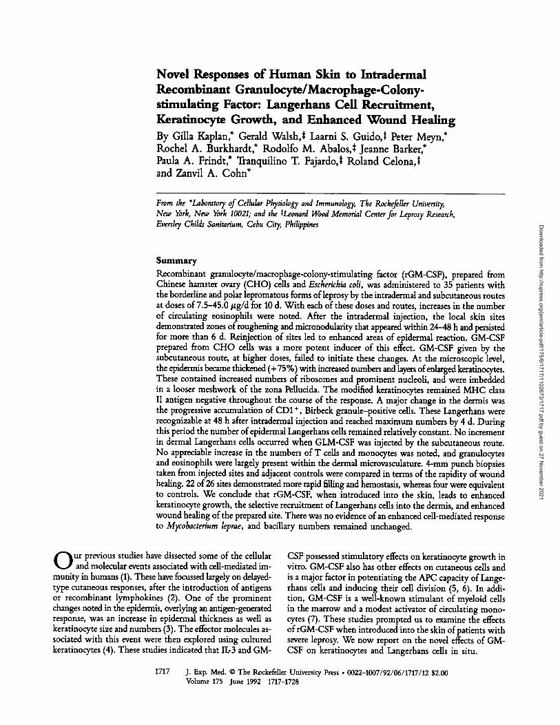
Figure 1. The local cutaneous response to intradermal GM-CSF injec- tion. The area (in mm 2) of modified epidermis in response to cytokine injection is shown for CHO GM CSF (left) (20 patients) and E. col GM- CSF (right) (15 patients). An early (24-h) response to a single 7.5-#g (O) and 15-/zg (A) CHO GM-CSF injection peaked at 3 d and then subsided slowly. Keinjection of the site at 2 d (7.5 #g) and 3 d (15 #g) led to a consistent increase in the area of epidermis modified by the cytokine (filled symbols). The first 20-#g intradermal dose of E. cell GM-CSF ([[]) did not induce epidermal changes. Keinjection of the site (20 #g; filled syrabols) induced a local response that peaked 2 d later and then subsided slowly. Results are expressed as the mean area of response for all patients injected + 1SD.
The Local Cutaneous Response to Intradermal Injection of rGM- CSF: Macroscopic Changes. The intradermal injection of GM- CSF at doses of 7.5-20.0 #g/0.1 ml of diluent was followed by a consistent series of events, the extent and kinetics of which depended upon the type of the GM-CSF and the dose used. In all cases injection sites exhibited slight erythema and no significant swelling or induration. In this sense the re- sponse differed markedly from the results with the intradermal injection of both IFN-~/and II.-2 (12, 20). Within 24 h, the surface of the skin of the responsive site showed roughening in a circular zone up to 200 mm in diameter. Within the next 24 h, examination of the area with a hand lens illus- trated the presence of many sites of micro-papillation con- tained within the roughened area and not associated with hair follicles. At 6 d this zone was scaly, as the keratinized layers were desquamated, leaving normal appearing skin.
The areas of the epidermal response are demonstrated in Fig. 1, which compares the responses to rGM-CSF obtained from CHO cells and E. cot. It appears that on a weight basis, the CHO preparation (Fig. 1, left) is more potent and leads to a prompter (24-h) modification of the epidermis. The rein- jection of the site at 48 h (7.5 ~g) or 72 h (15/zg) after the first injection led to a consistent increase in the area of the responsive site with accentuation of the changes noted above.
With the E. cot material little response occurred at 24 h with 10/~g (not shown) or 20/xg (Fig. 1, right), but upon reinjection of the 20-/~g sites, the typical cutaneous modifica- tions noted with the CHO-derived GM-CSF ensued. It was of interest that little or no epidermal change occurred when 45/.r of the E. coli GM-CSF was injected subcutaneously
for a 10-d period. In only 4 of 170 sites examined was there a slight roughening of the overlying epidermis after subcuta- neous injection.
We conclude that rGM-CSF has the ability of modifying the epidermis only when introduced in close proximity to this compartment.
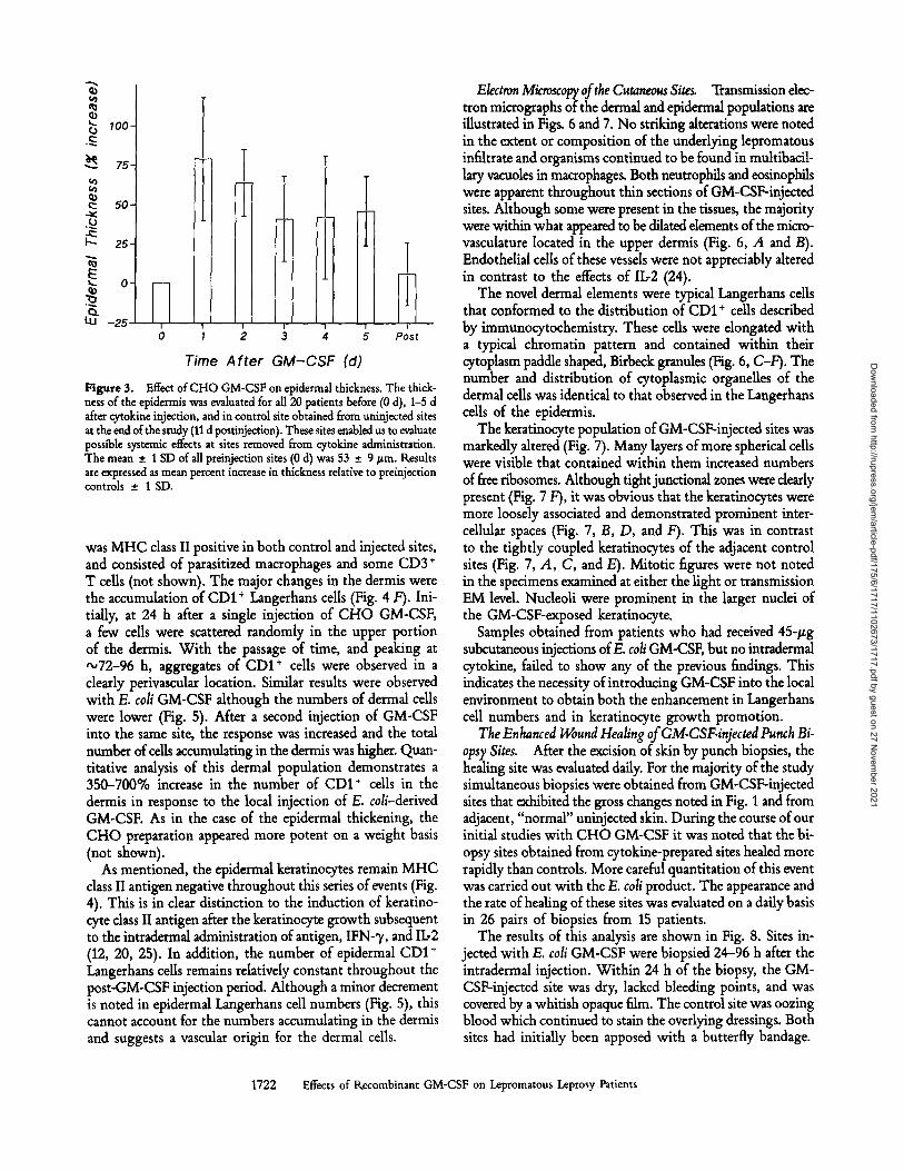
Microscopic Changes in the Epidermis. Striking changes were noted in the epidermis of responsive sites when biopsies were examined histologically. Within 24 h after CHO GM-CSF injection, enlargement of keratinocytes was apparent and the thickness of the epidermis was increased (Fig. 2). Quantita- tive analysis of the biopsies indicated that the epidermis had increased in thickness by a mean of 75% by 24 h and re- mained thickened for -120 h (Fig. 3). These effects were not observed as clearly after E. cot GM-CSF injection into the skin. The fine structure of the epidermis will be discussed in a later section.
Histopatholog~ and Bacillary Indices of Skin Biopsies and Slit Smears. Biopsies obtained from control and injected skin sites during the course of the intradermal and subcutaneous administration of rGM-CSFs were used to evaluate the histopathology and B.I. of the skin in these patients. Con- trol specimens showed the usual sparse mononudear cell infiltrate of lepromatous leprosy, the majority of cells being macrophages with varying numbers of acid-fast bacilli (21). The epidermis was thin and the fetes were often shallow with only one to two layers of rounded, basal cells. Only a small amount of additional cellular infiltrate was noted during the course of the GM-CSF injections (Fig. 2). This consisted of scattered eosinophils and neutrophils and small accumulations of perivascular mononudear cells; in some patients these sites resembled the lesions of erythema nodosum leprosum without the vascular changes. Little else was noted in the dermis. The B.I.s of the patients evaluated before and after completion of a 10-d course of cutaneous injections of GM-CSF are shown in Table 3. No significant changes in the bacterial load were noted during this period in contrast to results obtained with PPD antigen, rlFN-% and rib2 (22-24). We conclude that locally administered GM-CSF fails to modify the mononudear phagocyte population of the dermis and the number of in- tracelular leprosy bacilli.
Iramunocytocheraistry of the Cutaneous Site. Immunohisto- logical staining of the dermal and epidermal cellular compo- nents are illustrated in Fig. 4. The basal, lepromatous infiltrate
Table 3. B.I. in Response to GM-CSF
B.I.*
Type of cytokine No. of patients Pre Post
CHO GM-CSF 20 E. cot GM-CSF 15
4.47 _+ 1.06 4.43 _+ 1.16
4.54 _+ 0.69 4.30 +_ 0.83
* Mean B.I. _+ 1 SD of the mean.
1720 Effects of Recombinant GM-CSF on Lepromatous Leprosy Patients
Dow
nloaded from http://rupress.org/jem
/article-pdf/175/6/1717/1102673/1717.pdf by guest on 27 Novem
ber 2021

Figure 2. Effect of CHO GM- CSF on the histological appearance of the skin. Light micrographs of sections of biopsies obtained from two representative patients are shown. A comparison ofa preincu- bation control site (A and C) to a 24-h postcytokine administration site (B and D), a 96-h postcytokine (E), and an 11-d control (F) are shown for patient no. 5. A prein- jection control site (G and/), 24-h post- (H and .]), and 48-h post- cytokine (K), and an 11-d control (L) are shown for patient no. 8. A thickening of the epidermis (arrow- heads) is observed already by 24 h (D and.J). No systemic enhanced thickness is observed (F and L com- pared with C and I, respectively) after subcutaneous injection. GM- CSF injection did not significantly enhance the dermal infiltration by leukocytes (small arrows). A, B, G, and H, x25; C-F and I-L, x200 (H+E staining).
1721 Kaplan et al.
Dow
nloaded from http://rupress.org/jem
/article-pdf/175/6/1717/1102673/1717.pdf by guest on 27 Novem
ber 2021
100-
75-
cb 50-
~--~ 25-
~ O-
t~l -25
Figure 3.
0 3 I I
1 2 4 5
Time After
Post
GM-CSF (d) Effect of CHO GM-CSF on epidermal thickness. The thick-
ness of the epidermis was evaluated for all 20 patients before (0 d), 1-5 d after cytokine injection, and in control site obtained from uninjected sites at the end of the study (11 d postinjection). These sites enabled us to evaluate possible systemic effects at sites removed from cytokine administration. The mean • 1 SD of all preinjection sites (0 d) was 53 • 9 ~m. Results are expressed as mean percent increase in thickness relative to preinjection controls +_ I SD.
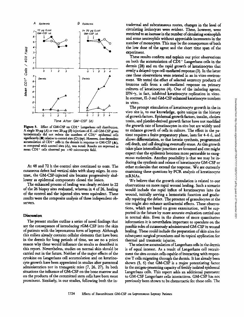
was MHC class II positive in both control and injected sites, and consisted of parasitized macrophages and some CD3 + T cells (not shown). The major changes in the dermis were the accumulation of CD1 + Langerhans cells (Fig. 4 F). Ini- tially, at 24 h after a tingle injection of CHO GM-CSF, a few cells were scattered randomly in the upper portion of the dermis. With the passage of time, and peaking at ,'~72-96 h, aggregates of CD1 + cells were observed in a dearly perivascular location. Similar results were observed with E. coli GM-CSF although the numbers of dermal cells were lower (Fig. 5). After a second injection of GM-CSF into the same site, the response was increased and the total number of cells accumulating in the dermis was higher. Quan- titative analysis of this dermal population demonstrates a 350-700% increase in the number of CD1 + cells in the dermis in response to the local injection of E. coli-derived GM-CSF. As in the case of the epidermal thickening, the CHO preparation appeared more potent on a weight basis (not shown).
As mentioned, the epidermal keratinocytes remain MHC class II antigen negative throughout this series of events (Fig. 4). This is in dear distinction to the induction of keratino- cyte class II antigen after the keratinocyte growth subsequent to the intradermal administration of antigen, IFN-% and IL-2 (12, 20, 25). In addition, the number of epidermal CD1 + Langerhans cells remains relatively constant throughout the post-GM-CSF injection period. Although a minor decrement is noted in epidermal Langerhans cell numbers (Fig. 5), this cannot account for the numbers accumulating in the dermis and suggests a vascular origin for the dermal cells.
Electron Microsco~ of the Cutaneous Sites. Transmission elec- tron micrographs of the dermal and epidermal populations are illustrated in Figs. 6 and 7. No striking alterations were noted in the extent or composition of the underlying lepromatous infiltrate and organisms continued to be found in muhibacil- lary vacuoles in macrophages. Both neutrophils and eosinophils were apparent throughout thin sections of GM-CSF-injected sites. Although some were present in the tissues, the majority were within what appeared to be dilated elements of the micro- vasculature located in the upper dermis (Fig. 6, A and B). Endothelial cells of these vessels were not appreciably altered in contrast to the effects of II~2 (24).
The novel dermal elements were typical Langerhans cells that conformed to the distribution of CD1 + cells described by immunocytochemistry. These cells were elongated with a typical chromatin pattern and contained within their cytoplasm paddle shaped, Birbeck granules (Fig. 6, C-F). The number and distribution of cytoplasmic organelles of the dermal cells was identical to that observed in the Langerhans cells of the epidermis.
The keratinocyte population of GM-CSF-injected sites was markedly altered (Fig. 7). Many layers of more spherical cells were visible that contained within them increased numbers of free ribosomes. Although tight junctional zones were clearly present (Fig. 7 F), it was obvious that the keratinocytes were more loosely associated and demonstrated prominent inter- cellular spaces (Fig. 7, B, D, and F). This was in contrast to the tightly coupled keratinocytes of the adjacent control sites (Fig. 7, A, C, and E). Mitotic figures were not noted in the specimens examined at either the light or transmission EM level. Nudeoli were prominent in the larger nuclei of the GM-CSF-exposed keratinocyte.
Samples obtained from patients who had received 45-/~g subcutaneous injections orE. coli GM-CSF, but no intradermal cytokine, failed to show any of the previous findings. This indicates the necessity of introducing GM-CSF into the local environment to obtain both the enhancement in Langerhans cell numbers and in keratinocyte growth promotion.
The Enhanced Wound Healing of GM-CSF-injected Punch Bi- opsy Sites. After the excision of skin by punch biopsies, the healing site was evaluated daily. For the majority of the study simultaneous biopsies were obtained from GM-CSF-injected sites that exhibited the gross changes noted in Fig. I and from adjacent, "normal" uninjected skin. During the course of our initial studies with CHO GM-CSF it was noted that the bi- opsy rites obtained from cytokine-prepared sites healed more rapidly than controls. More careful quantitation of this event was carried out with the E. coli product. The appearance and the rate of healing of these sites was evaluated on a daily basis in 26 pairs of biopsies from 15 patients.
The results of this analysis are shown in Fig. 8. Sites in- jected with E. coli GM-CSF were biopsied 24-96 h after the intradermal injection. Within 24 h of the biopsy, the GM- CSF-injected site was dry, lacked bleeding points, and was covered by a whitish opaque film. The control site was oozing blood which continued to stain the overlying dressings. Both sites had initially been apposed with a butterfly bandage.
1722 Effects of Recombinant GM-CSF on Lepromatous Leprosy Patients
Dow
nloaded from http://rupress.org/jem
/article-pdf/175/6/1717/1102673/1717.pdf by guest on 27 Novem
ber 2021
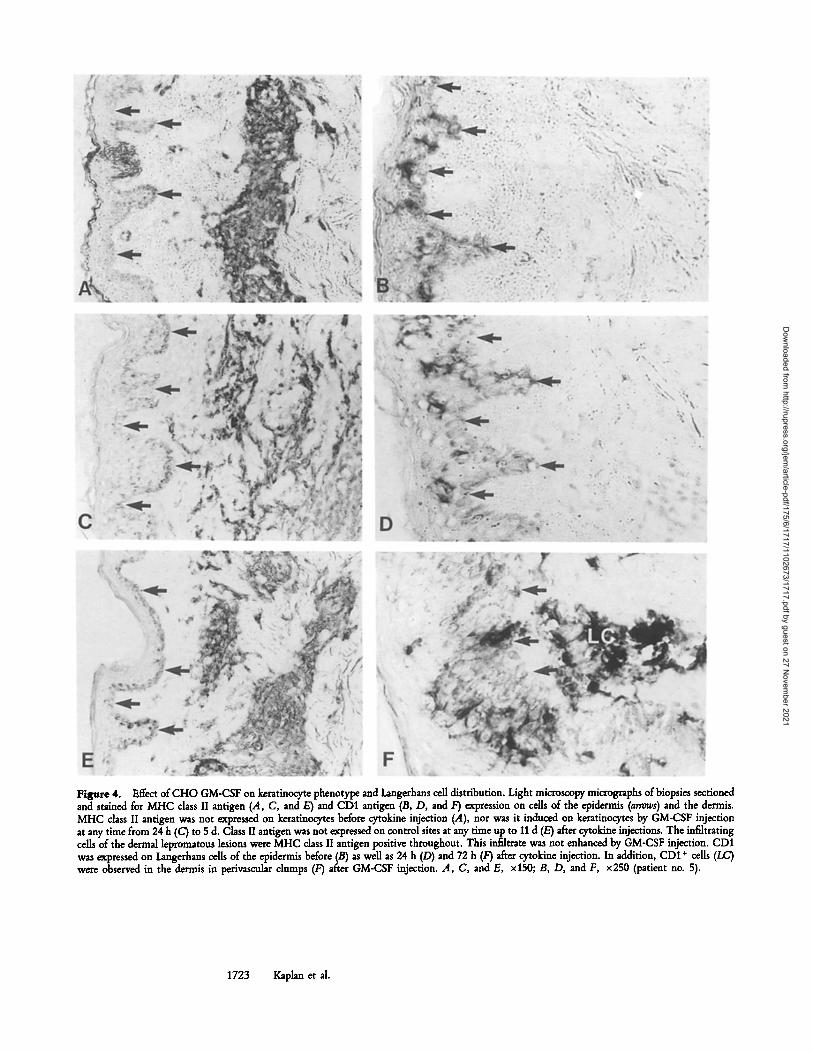
Figure 4. Effect of CHO GM-CSF on keratinocyte phenotype and Langerhans cell distribution. Light microscopy micrographs of biopsies sectioned and stained for MHC class II antigen (A, C, and E) and CD1 antigen (B, D, and F) expression on cells of the epidermis (arrows) and the dermis. MHC class II antigen was not expressed on keratinocytes before cytokine injection (A), nor was it induced on keratinocytes by GM-CSF injection at any time from 24 h (C) to 5 d. Class II antigen was not expressed on control sites at any time up to 11 d (E) after cytokine injections. The infiltrating cells of the dermal lepromatous lesions were MHC class II antigen positive throughout. This infiltrate was not enhanced by GM-CSF injection. CD1 was expressed on Langerhans cells of the epidermis before (B) as well as 24 h (D) and 72 h (F) after cytokine injection. In addition, CD1 + cells (LC) were observed in the dermis in perivascular dumps (F) after GM-CSF injection. A, C, and E, x150; B, D, and F, x250 (patient no. 5).
1723 Kaplan et ai.
Dow
nloaded from http://rupress.org/jem
/article-pdf/175/6/1717/1102673/1717.pdf by guest on 27 Novem
ber 2021
4-
2-
l -
Ib
7-
5-
3 -
A Epidermis
7- 10 IJg E.coli GM-CSF i.d.
5"
2-
Dermis
e Epidermis
2x 20 IJg E.coli GM-CSF Ld.
L I
[ i l l i
Derm~ A ~ /
I 1 ~ I I 0 1 3 4
Time A f t e r G M - C S F ( d )
' l ) i
0 I 2, ,3
Figure S. Effect of GM-CSF on CD1 + Langerhans cell distribution. A single 10-~g (.4) or two 20-~g (B) injections orE. coil GM-CSF given intradermally did not reduce the numbers of CD1 + epidermal cells significantly (I) relative to control sites (O) (9)" However, dose -~-~ t accumulation ofCD1 + cells in the dermis in response to GM-CSF (A), as compared with control sites (A), was noted. Results are expressed as mean CD1 + cells observed per • microscopic field.
At 48 and 72 h the control sites continued to ooze. The cutaneous defect had vertical sides with sharp edges. In con- trast, the GM-CSF-injected site became progressively shal- lower as epidermal components closed the lesion.
The enhanced process of healing was clearly evident in 22 of the 26 biopsy sites evaluated, whereas in 4 of 26, healing of the control and the injected sites were equivalent. These results were the composite analysis of three independent ob- servers.
Diseuuion
The present studies outline a series of novel findings that are the consequence of introducing rGM-CSF into the skin of patients with the lepromatous form of leprosy. Although this milieu already contains cellular elements that have been in the dermis for long periods of time, we see no a priori reason why these would influence the results as described in this report. Nevertheless, studies on normal skin should be carried out in the future. Neither of the major effects of the cytokine on Langerhans cell accumulation and on keratino- cyte growth have been appreciated in studies after parenteral administration nor in transgenic mice (7, 26, 27). In both situations the influence of GM-CSF on the bone marrow and on the products of the committed stem cells have been most prominent. Similarly, in our studies, following both the in-
tradermal and subcutaneous routes, changes in the level of circulating leukocytes were evident. These, however, were restricted to an increase in the number of circulating eosinophils and some neutrophils without appreciable increments in the number of monocytes. This may be the consequences of both the low dose of the agent and the short time span of the experiment.
These results confirm and explain our prior observations on both the accumulation of CD1 + Langerhans cells in the dermis (28) and on the rapid growth of keratinocytes that overly a delayed-type cell-mediated response (3). In the latter case these observations were retested in an in vitro environ- ment. We tested the effect of selected secretory products of immune cells from a cell-mediated response on primary cultures of keratinocytes (4). One of the inducing agents, IFN-% in fact, inhibited keratinocyte replication in vitro. In contrast, IL-3 and GM-CSF enhanced keratinocyte numbers in vitro.
The prompt stimulation of keratinocyte growth in the in vivo site is, to our knowledge, quite unique in the history of growth factors. Epidermal growth factors, insulin, cholera toxin, and platdet-derived growth factor have not modified the growth rate of keratinocytes in situ but are widely used to enhance growth of cells in culture. The effect in the pa- tient requires a finite preparatory phase, lasts for 4-6 d, and allows differentiation, so that keratin synthesis, programmed cell death, and cell sloughing eventually ensue. As this growth takes place interceUularjunctions are loosened and one might expect that the epide~nfis becomes more permeable to exog- enous molecules. Another possibility is that we may be in- ducing the synthesis and release of keratinocyte GM-CSF or other molecules that extend the response. We are currently examining these questions by PCR analysis of keratinocyte mRNAs.
We believe that the growth stimulation is related to our observations on more rapid wound healing. Such a scenario would include the rapid influx of keratinocytes into the wound, initially serving a hemostatic function and gradu- ally repairing the defect. The presence of granulocytes at the site might also enhance antibacterial effects. These observa- tions, which are based on gross examination, will be sup- ported in the future by more accurate evaluation carried out in normal skin. Even in the absence of more quantitative information it is nevertheless important to speculate on the possible roles of cutaneously administrated GM-CSF in wound healing. These could include the preparation of skin sites for subsequent surgical procedures and its topical application for thermal and traumatic injuries.
The selective accumulation of Langerhans cells in the dermis is of equal interest. As a result of Langerhans cell recruit- ment the sites contain cells capable of interacting with respon- sive T cells migrating through the dermis. It has already been shown (5, 6) that GM-CSF is a major potentiating factor in the antigen-presenting capacity of freshly isolated epidermal Langerhans cells. This report adds an additional parameter to GM-CSF Langerhans cells interactions. GM-CSF has not previously been shown to be chemotactic for these cells. The
1724 Effects of Recombinant GM-CSF on Lepromatous Leprosy Patients
Dow
nloaded from http://rupress.org/jem
/article-pdf/175/6/1717/1102673/1717.pdf by guest on 27 Novem
ber 2021
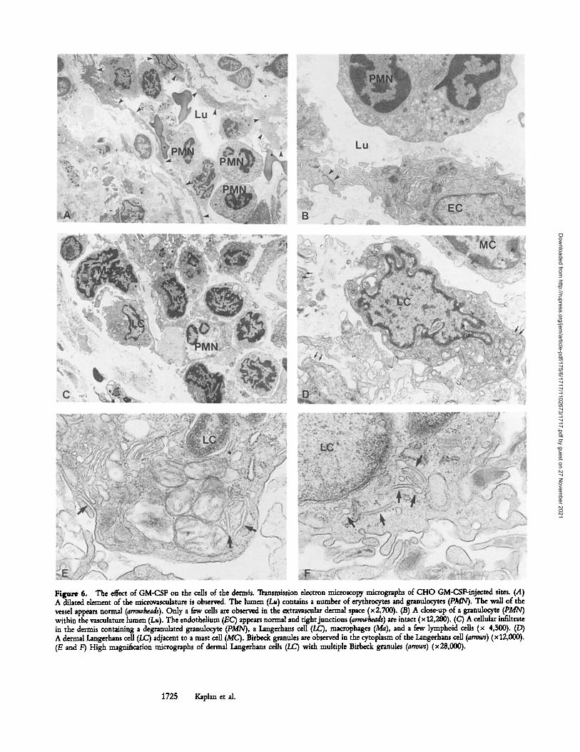
Figure 6. The effect of GM-CSF on the cells of the dermis. Transmission electron microscopy micrographs of CHO GM-CS~injected sites. (A) A dilated element of the microvasculature is observed. The lumen (La) contains a number of erythrocytes and granulocytes (PMN). The wall of the vessel appears normal (arrowheads). Ouly a few cells are observed in the e~travascular dermal space (x2,700). (B) A dose-up of a granulocyte (PMN) within the vasculature lumen (Lu). The endothelium (EC) appears normal and tight junctiom (arrowheads) are intact (x 12,200). (C) A cellular inf~trate in the dermis cont~inlng a degranulated granulocyte (PMN), a Langerhans cell (LC), macrophages (Ma), and a few lymphoid cells (x 4,500). (D) A dermal I.angerhans cell (LC) adjacent to a mast cell (MC). Birbeck granules are observed in the cytoplasm of the Langerhans cell (arrows) (x12,000). (E and F) High magnification micrographs of dermal Langerhans cells (LC) with multiple Birbeck granules (arrows) (x 28,000).
1725 Kaplan et al.
Dow
nloaded from http://rupress.org/jem
/article-pdf/175/6/1717/1102673/1717.pdf by guest on 27 Novem
ber 2021
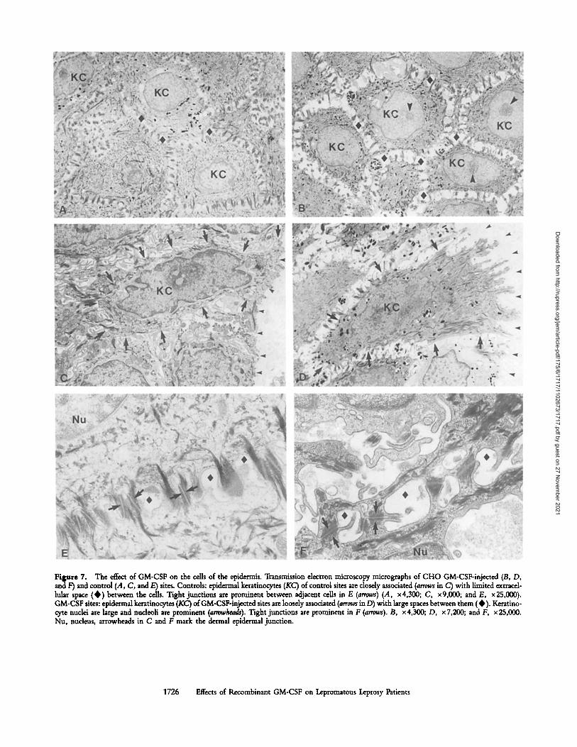
Figure 7, The effect of GM-CSF on the cells of the epidermis. Transmission electron microscopy micrographs of CHO GM-CSF-injected (B, D, and F) and control (A, C, and E) sites. Controls: epidermal keratinocytes (KC) of control sites are closely associated (arrows in C) with limited extracei- lular space (0 ) between the cells. Tight junctions are prominent between adjacent cells in E (arrows) (A, x4,300; C, xg,000; and E, x25,000). GM-CSF sites: epidermal keratinocytes (KC) of GM-CSF-injected sites are loosely associated (arrotvs in D) with large spaces between them ( �9 Keratino- cyte nuclei are large and nucleoli are prominent (arrowhea&). Tight junctions are prominent in F (arrows). B, • D, x7,200; and F, x25,000. Nu, nucleus, arrowheads in C and F mark the dermal epidermal junction.
1726 Effects of Recombinant GM-CSF on Lepromarous Leprosy Patients
Dow
nloaded from http://rupress.org/jem
/article-pdf/175/6/1717/1102673/1717.pdf by guest on 27 Novem
ber 2021
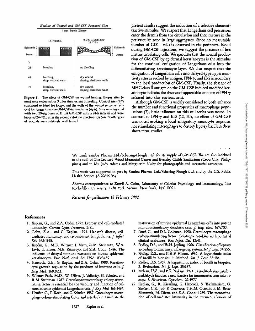
Figure 8. The effect of GM-CSF on wound healing. Biopsy sites (4 mm) were evaluated for 3 d for their extent of healing. Control sites (left) continued to bleed for longer and the walls of the wound remained ver- tical for longer than the GM-CSF-injected sites (right). Sites were injected with two 20-#g doses of//. coli GM-CSF with a 24-h interval and were biopsied 24-72 h after the second cytokine injection. By 5-6 d both types of wounds were relativdy well healed.
present results suggest the induction of a selective chemoat- tractive stimulus. We suspect that Langerhans cell precursors enter the dermis from the circulation and then mature in the perivascular zone in large aggregates. Since no measurable number of CD1 + cells is observed in the peripheral blood during GM-CSF injections, we suggest the presence of less mature circulating cells. We speculate that the normal produc- tion of GM-CSF by epidermal keratinocytes is the stimulus for the continual emigration of Langerhans cells into the differentiating keratinocyte layer. We also suspect that the emigration of Langerhans cells into ddayed-type hypersensi- tivity sites as evoked by antigen, IFN-% and IL2 is secondary to the local production of GM-CSE Finally, the absence of MHC class II antigen on the GM-CSF-induced modified ker- atinocyte indicates the absence of appreciable amounts of IFN-3r released into this environment.
Although GM-CSF is widely considered to both enhance the number and functional properties of macrophage popu- lations (7), little influence on this cell series was noted. In contrast to IFN-~ and IL-2 (12, 20), no effect of GM-CSF was noted evoking a local emigratory monocyte response, nor stimulating macrophages to destroy leprosy bacilli in these short-term studies.
We thank Sandoz Pharma Ltd./Schering-Plough Ltd. for its supply of GM-CSE We are also indebted to the staff of The Leonard Wood Memorial Center and Eversley Childs Sanitarium (Cebu City, Philip- pines) and to Ms. Judy Adams and Marguerite Nulty for photographic and secretarial assistance.
This work was supported in part by Sandoz Pharma Ltd./Schering-Plough Ltd. and by the U.S. Public Health Service (A-22616-06).
Address correspondence to Zanvil A. Cohn, Laboratory of Cellular Physiology and Immunology, The Rockefeller University, 1230 York Avenue, New York, NY 10021.
Received for publication 18 February 1992.
1. Kaplan, G., and Z.A. Cohn. 1991. Leprosy and ceil-mediated immunity. Current Opin. Imraunol. 3:91.
2. Cohn, Z.A., and G. Kaplan. 1991. Hansen's disease, ceil- mediated immunity, and recombinant lymphokines. J. Infect. D/s. 163:1195.
3. Kaplan, G., M.D. Witmer, I. Nath, R.M. Steinman, W.A. Levis, U. Elvers, M.E. Patarroyo, and Z.A. Cohn. 1986. The influence of delayed immune reactions on human epidermal keratinocytes. Proa Natl./had. Sci. USA. 83:3469.
4. Hancock, G.E., G. Kaplan, and Z.A. Cohn. 1988. Keratino- cyte growth regulation by the products of immune cells, f Exlx Med. 168:1811.
5. Witmer-Pack, M.D., W. Oliver, J. Valinsky, G. Schuler, and K.M. Steinman. 1987. Granulocyte/macrophage colony-stimu- lating factor is essential for the viability and function of cul- tured routine epidermal Langerhans cells.f Exp MecL 166:1484.
6. Heufler, C., E Koch, and G. Schuler. 1987. Granulocyte-macro- phage colony-stimulating factor and interleukin I mediate the
maturation of routine epidermal Langerhans ceils into potent immunostimulatory dendritic cells, f Exi~ Med. 167:700.
7. Kuef, C., and D.L. Coleman. 1990. Granulocyte-macrophage colony-stimulating factor: pleiotropic cytokine with potential clinical usefulness. Rev. Infect. Dis. 12:41.
8. Ridley, D.S., and W.H. Jopling. 1966. Classification of leprosy according to immunity: a five group system, lnt.J. Letg. 34:255.
9. Kidley, D.S., and G.K.F. Hilson. 1967. A logarithmic index of bacilli in biopsies. 1. Method. Int. J. Lepr. 35:184.
10. Ridley, D.S. 1967. A logarithmic index of bacilli in biopsies. 2. Evaluation. Int. f Lepr. 35:187.
11. Mclean, I.W., and P.K. Nakane. 1974. Periodateqysine-parafor- maldehyde fixative: a new fixative for immunoelectron micros- copy. f Histochem. CTtochem. 22:1977.
12. Kaplan, G., R. Kiessling, G. Hancock, S. Teklemariam, G. Sheftel, C.K. Job, P. Converse, T.H.M. Ottenhoff, M. Becx- Bleumink, M. Dietz, and Z.A. Cohn. 1989. The reconstitu- tion of ceil-mediated immunity in the cutaneous lesions of
1727 Kaplan et al.
Dow
nloaded from http://rupress.org/jem
/article-pdf/175/6/1717/1102673/1717.pdf by guest on 27 Novem
ber 2021
lepromatous leprosy by recombinant interhukin 2.J. Exla Med. 169:893.
13. Engelman, E.G., R. Warnke, R.I. Fox, and R. Levy. 1981. Studies of human T lymphocyte antigen recognized by a mono- donal antibody. Proa Natl. Acad. Sci. USA. 78:1791.
14. Evans, K.K., D.W. Wall, C.D. Platsoucas, E.P. Siegal, S.M. Fikrig, C.M. Testa, and R.A. Good. 1981. Thymus-dependent membrane antigens in man: inhibition of cell mediated lym- pholysis by monoclonal antibodies to the TH2 antigen. Proc. Natl. Acad. Sci. USA. 78:544.
15. Schwarting, R,, H. Stein, and C.Y. Wang. 1985. The mono- clonal antibodies anti S-HCI-1 and anti-S-HC1-3 allow the di- agnosis of hairy cell leukemia. Blood. 65:974.
16. Fithian, E., P. King, G. Goldstein, M. Rubenfeld, C. Fenoglio, and E. Edelson. 1981. Reactivity of Langerhans' cells with hy- bridoma antibody. Proc Natl./,cad. Sci. USA. 78:2541.
17. Van Voorhis, W.C., lL.M. Steinman, L. Hair, J. Luban, M.D. Witmer, and Z.A. Cohn. 1983. Specific mononuclear phago- cyte monoclonal antibodies. Application to the purification of dendritic cells and tissue localization of macrophages. J. Exl~ Med. 158:126.
18. Chomczynski, P., and N. Sacchi. 1987. Single-step method of RNA isolation by acid guanidinium thiocyanate-phenol- chloroform extraction. Anal. Biochem. 162:156.
19. Ehlers, S., and K.A. Smith. 1991. Differentiation o f t cell lym- phokine gene expression: the in vitro acquisition of T cell memory..J. ExI~ Med. 173:25.
20. Nathan, C.F., G. Kaplan, W.R. Levis, A. Nusrat, M.D. Witmer, S.A. Shervcin, C.K. Job, C.R. Horowitz, lL.M. Steinman, and Z.A. Cohn. 1986. Local and systemic effects of low doses of recombinant interferon 3' after intraderrnal in-
jection in patients with lepromatous leprosy. N. Engl.J. Med. 315:6.
21. Kaplan, G., and Z.A. Cohn. 1986. The immunobiology of leprosy. Int. R~. Exp Pathol. 28:45.
22. Kaphn, G., G. Sheftel, C.K. Job, N.K. Nathur, I. Nath, and Z.A. Cohn. 1988. The efficacy of a cell mediated reaction to PPD in the disposal ofM. leff'ae from human skin. Pro~ Natl. Acad. Sci. USA. 85:5210.
23. Kaplan, G., N.K. Mathur, C.K. Job, I. Nath, and Z.A. Cohn. 1989. The effect of mukiple IFN-~/injections on the disposal of mycobacterium leprae, Pro~ Natl. AcacL Sci. USA. 86:8073.
24. Kaplan, G., W.J. Britton, G,E. Hancock, W.J. Theuvenet, K.A. Smith, C.K. Job, P.W. Roche, A. Molloy, K. Burkhardt, J. Barker, H.M. Pradhan, and Z.A. Cohn. 1991. The systemic influence of recombinant interleukin 2 on the manifestations of lepromatous leprosy. J. Exig Med. 173:993.
25. Kaplan, G., S. Laal, G. Sheftel, A. Nusrat, I. Nath, and Z.A. Cohn. 1988. The nature and kinetics of a delayed immune re- sponse to PPD in skin of lepromatous leprosy patients.J. Extx Med. 168:1811.
26. Steinman, K.M. 1991. The dendritic cell system and its role in immunogenicity. Annu. Rev. Imraunol. 9:271.
27. Lung, K.A., D. Metcalf, K.A. Cuthbertson, I. Lyons, E. Stanley, A, Kelso, G. Kannourakis, D.J. Williamson, G.K. Klintworth, T.J. Gonda, and A.K. Dunn. 1987. Transgenic mice expressing a hemopoietic growth factor gene (GM-CSF) develop accumulations of macrophages, blindness, and a fatal syndrome of tissue damage. Cell. 51:675.
28. Kaphn, G., A. Nusrat, M.D. Witmer, I. Nath, and Z.A. Cohn. 1987. The distribution and turnover of Langerhans cells during delayed immuno responses in human skin.J. Ex F Med. 165:763.
1728 Effects of Recombinant GM-CSF on Lepromatous I.eprosy Patients
Dow
nloaded from http://rupress.org/jem
/article-pdf/175/6/1717/1102673/1717.pdf by guest on 27 Novem
ber 2021


![Paroxetine suppresses recombinant human P2X7 responses · KN-62 [19], calmidazolium [20], chelerythrine [21], a tyrosine kinase inhibitor [22], SB203580 [23], DIDS [24], clemastine](https://img.pdfslide.us/doc/110x75/5f3666be62cca735e15dfa9e/paroxetine-suppresses-recombinant-human-p2x7-responses-kn-62-19-calmidazolium.jpg)


![Glyco-Dendrimers as Intradermal Anti-Tumor Vaccine ...multiple subsets simultaneously may induce superior immune responses [9, 10]. In vivo vaccines are often applied in the human](https://img.pdfslide.us/doc/110x75/5f49aadb00276b62e7206dc0/glyco-dendrimers-as-intradermal-anti-tumor-vaccine-multiple-subsets-simultaneously.jpg)