Embed Size (px)
Citation preview
Novel Method for Enumeration of Viable Lactobacillus plantarumWCFS1 Cells after Single-Droplet Drying
Jimmy Perdana,a Ludmila Bereschenko,b,c* Mark Roghair,a Martijn B. Fox,c Remko M. Boom,a Michiel Kleerebezem,b,c,d andMaarten A. I. Schutysera
Food Process Engineering Group, Wageningen UR, Wageningen, The Netherlandsa; Laboratory of Microbiology, Wageningen UR, Wageningen, The Netherlandsb; NIZOFood Research, Ede, The Netherlandsc; and Host-Microbe Interactomics Group, Wageningen UR, Wageningen, The Netherlandsd
Survival of probiotic bacteria during drying is not trivial. Survival percentages are very specific for each probiotic strain and canbe improved by careful selection of drying conditions and proper drying carrier formulation. An experimental approach is pre-sented, comprising a single-droplet drying method and a subsequent novel screening methodology, to assess the microbial via-bility within single particles. The drying method involves the drying of a single droplet deposited on a flat, hydrophobic surfaceunder well-defined drying conditions and carrier formulations. Semidried or dried particles were subjected to rehydration, fluo-rescence staining, and live/dead enumeration using fluorescence microscopy. The novel screening methodology provided accu-rate survival percentages in line with conventional plating enumeration and was evaluated in single-droplet drying experimentswith Lactobacillus plantarum WCFS1 as a model probiotic strain. Parameters such as bulk air temperatures and the carrier ma-trices (glucose, trehalose, and maltodextrin DE 6) were varied. Following the experimental approach, the influence on the viabil-ity as a function of the drying history could be monitored. Finally, the applicability of the novel viability assessment was demon-strated for samples obtained from drying experiments at a larger scale.
Probiotics are defined as “live microorganisms which when ad-ministered in adequate amounts confer a health benefit on the
host” (11). Health benefits are usually related to the influence ofprobiotic bacteria on the microbial balance in the hosts’ intestineor via modulation of the gut-associated immune system (6, 12, 19,21, 25). Probiotics are delivered to the gastrointestinal tract asfood products or dietary supplements and supplied on the marketas fermented food commodities, freeze-dried cultures, or frozencultures, which enhance their stability and facilitate their imple-mentation in appropriate product formulations (1, 16, 30). Com-pared to freeze drying and freezing, spray drying could be aninteresting alternative for providing shelf life to probiotic ingredi-ents. Spray drying is more energy and cost efficient and can beoperated continuously at higher production capacities (2, 26, 34).The major drawback of spray drying is the limited survival ofprobiotics (31, 33). Several studies have successfully explored ap-proaches to increase the survival percentages after spray drying (8,10, 13, 23). However, most of the results so far are very specific anddifficult to translate between different species. It was, for example,found that a high variability exists between different strains of thesame species (14, 24). In addition, the process conditions appliedduring spray drying (2, 28) and the protective carrier materialsadded (10, 31) have strong influence on the final viability. Sincethese parameters need to be optimized for each specific case (dif-ferent process parameters, different strains or species, and differ-ent carrier formulations used during drying), many cost-, time-,and labor-intensive experiments are required.
Here we present an experimental approach to assess probioticsurvival during drying, starting with the drying of small singledroplets deposited on a flat hydrophobic surface (29), followed bycell rehydration on Anopore chips (17), and subsequent evalua-tion of the microbial viability by fluorescence microscopy.
A recently developed single-droplet drying method is used toproduce powder particles dried under well-defined conditions toinvestigate the influence of drying process conditions and carrier
formulations (29). Specifically, drying parameters, such as dropletsize, temperature, relative humidity, and flow rates of the air, canbe varied effectively and systematically over wide ranges.
The availability of a rapid and reliable live/dead assay is criticalto the assessment of the viability after drying. The assay developedhere employs a microporous aluminum oxide chip (Anopore)(17, 18). Following rehydration of the droplets, fluorescenceprobes are used for live/dead enumeration using fluorescence mi-croscopy. This assay is compatible with medium- to high-throughput techniques, especially compared to more conven-tional enumeration by plating. Moreover, the proposed methodallows direct visualization of live and dead populations after dry-ing without the requirement for growth. It has the potential tomeasure the viability in small sample volumes, i.e., the viability ofbacteria present in a single powder particle. Subsequently, themethod is applied just as well to assess the microbial viability in theparticles obtained from single-droplet drying experiments andfrom stabilization experiments at a larger scale. In our experi-ments, Lactobacillus plantarum WCFS1 was selected as the modelbacterium. This selection is based on the fact that this Lactobacillusstrain has been the topic of extensive studies before (20), withoutper se L. plantarum being a probiotic bacterium.
Received 6 July 2012 Accepted 6 September 2012
Published ahead of print 14 September 2012
Address correspondence to Maarten A. I. Schutyser, [email protected].
* Present address: Ludmila Bereschenko, KWR Watercycle Research Institute,Nieuwegein, The Netherlands.
J.P. and L.B. contributed equally to this article.
Copyright © 2012, American Society for Microbiology. All Rights Reserved.
doi:10.1128/AEM.02063-12
8082 aem.asm.org Applied and Environmental Microbiology p. 8082–8088 November 2012 Volume 78 Number 22
on June 22, 2018 by guesthttp://aem
.asm.org/
Dow
nloaded from
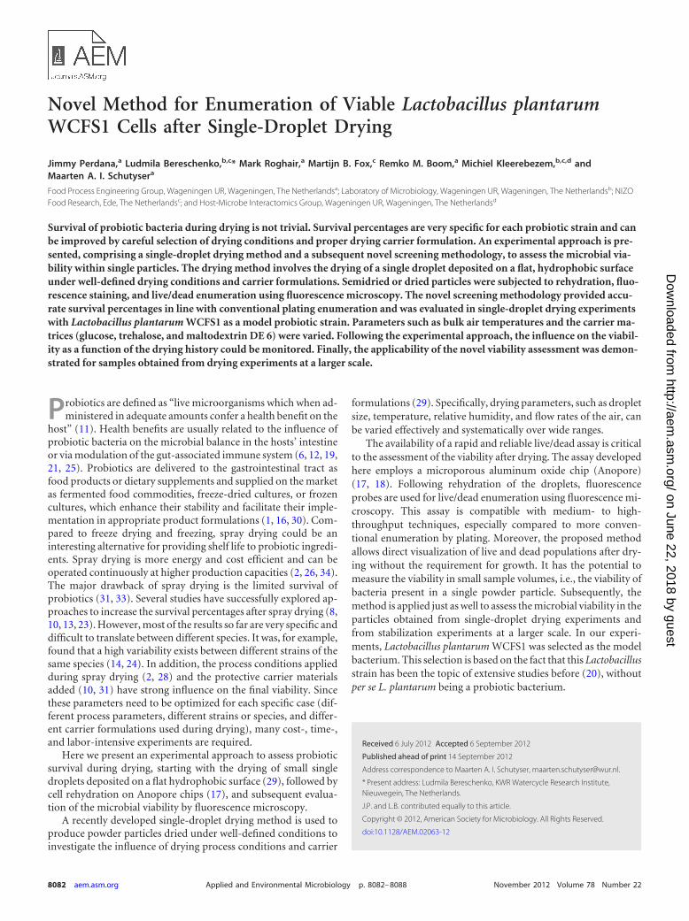
MATERIALS AND METHODSThe entire experimental procedure includes the following steps: (i) mi-crobial culture preparation, (ii) drying of single droplets, (iii) particlerehydration and fluorescence probe staining, and (iv) fluorescence mi-croscopy analysis and automated relative viable cell enumeration (Fig. 1).
Culture preparation and washing. A culture with high viability(�99%) was acquired by growing L. plantarum WCFS1 in 10 ml sterilizedlactobacillus culture medium (MRS; BD Difco) at 30°C for 16 h. The finalcell density approximated 109 CFU per ml.
The overnight culture was centrifuged using Eppendorf Centrifuge5804R with an F-34-6-38 rotor at 13.5 � g at 4°C for 10 min. The resultingpellet was washed twice with phosphate-buffered saline (PBS) solution(BD Difco). To minimize washing stress, the pH of the PBS solution wasadjusted to a pH of 4, which is similar to the pH of the culture at the endof growth.
Single-droplet drying experiments. Directly before the drying exper-iment was initiated, the washed bacterial cells were suspended in a carriermatrix, consisting of 20% (wt/wt) maltodextrin DE 6 (Glucidex 6,Roquette, France), trehalose (Sigma-Aldrich, Germany), or glucose(Sigma-Aldrich, Germany). These carbohydrates are known to provideprotection to bacterial cells (4, 7).
The drying experiments were performed using the same equipment asdescribed by Perdana et al. (27). The droplets were generated using apneumatic dispenser of the Microdot 741 MD-SS series (Engineered FluidDispensing; Nordson). The droplet deposition on the flat surface (5 by 20by 0.2 mm) was automated, using an XYZ positioning platform Ultra 525TT automation series (Engineered Fluid Dispensing; Nordson). The dis-pensing needle AKA740TK precision tips (Engineered Fluid Dispensing;Nordson) were coated with DOW Corning 340 heat sink compound(Dow Chemical) to prevent the droplets from creeping up along the out-side of the needle.
Prior to the deposition of the droplets, the microdispensing systemwas flushed with sterilized MilliQ water (Millipore), followed by 70%(vol/vol) ethanol solution (VWR International, France), and then rinsedagain with MilliQ water. The droplets were deposited on a flat hydropho-bic membrane, Accurel type PP 2E HF (Akzo Nobel Faser AG, The Neth-erlands). To prevent microbial contamination, the membrane was steril-ized with 70% (vol/vol) ethanol solution and dried under asepticconditions at room temperature.
The droplets were dried using preconditioned, filtered (dust/microbesand oil-free) drying air, which was heated to the desired temperature byleading it through a coil that was submerged in an oil bath (Julabo EH-5,Germany). The heated air was then fed to an insulated tunnel which actedas the drying chamber. The air temperature in the tunnel was monitoredusing a thermocouple type K (NiCr-NiAl) (RS Component, United King-dom), with a probe diameter of 500 �m. The airflow velocity was moni-tored (type 1355; Brooks Instruments, The Netherlands). The tunnel wasfilled with a highly porous medium to develop airflow with a uniformvelocity. Meanwhile, the temperature was maintained by insulating thetunnel with heating oil. After the deposition of the droplets, the sampleswere placed in the drying chamber and dried using various drying timesand regimes. The setup was equipped with a �Eye 1480ME charge-cou-pled-device (CCD) camera with a lens magnification of �9 (ImagingDevelopment Systems GMBH, Germany) to monitor the droplet geome-try evolution during drying.
The single-droplet drying experiments were performed using dry air(rH � 0.0%), preheated to a temperature between 25 and 70°C and a bulkair velocity of 0.12 to 0.52 m/s. For each experiment, three identical drop-lets were dispensed and dried simultaneously. Biological duplicates wereperformed by repeating the experiments twice.
Rehydration and staining chip. The substrate for cell rehydration andstaining was prepared by applying an Anopore chip (17) on a low-meltingpoint (LMP) agarose gel (Sigma, The Netherlands). The gel was preparedby dissolving 1 g of agarose in 100 ml of MilliQ water (Millipore) andautoclaved (121°C) for 20 min. The agarose solution was allowed to cool;after the temperature reached approximately 40°C, 2 �l of a fluorescencestaining probe, Live/Dead BacLight bacterial viability kit (Invitrogen),was added to 10 ml of the agarose solution. The agarose solution was thenspread on microscope slides (76 by 26 mm) and allowed to solidify for 30min in a dark environment. Afterwards, a sterilized Anopore chip (8 by35.6 mm; thickness, 60 �m; pore size, 0.2 �m; 3 � 109 pores per cm2)(Microdish BV, The Netherlands) was carefully placed on the agarose gel.The Anopore chips were sterilized by submerging them into 70% (vol/vol)ethanol solution for 2 h in Falcon tubes. Subsequently, ethanol was de-canted, the chips were dried in a sterile flow chamber for 12 h, and then thetubes were tightly closed for storage. The Anopore chips were positionedon the agarose gel for at least 30 min prior to the rehydration of the driedparticles. All preparations were carried out under aseptic conditions.
FIG 1 Schematic overview of the experimental procedure, including microbial culture preparation (1), drying of single droplets (2), particle rehydration andfluorescence probe staining (3), and fluorescence microscopy analysis and automated relative viable cell enumeration (4).
A Novel Method for Viability Enumeration of Probiotics
November 2012 Volume 78 Number 22 aem.asm.org 8083
on June 22, 2018 by guesthttp://aem
.asm.org/
Dow
nloaded from
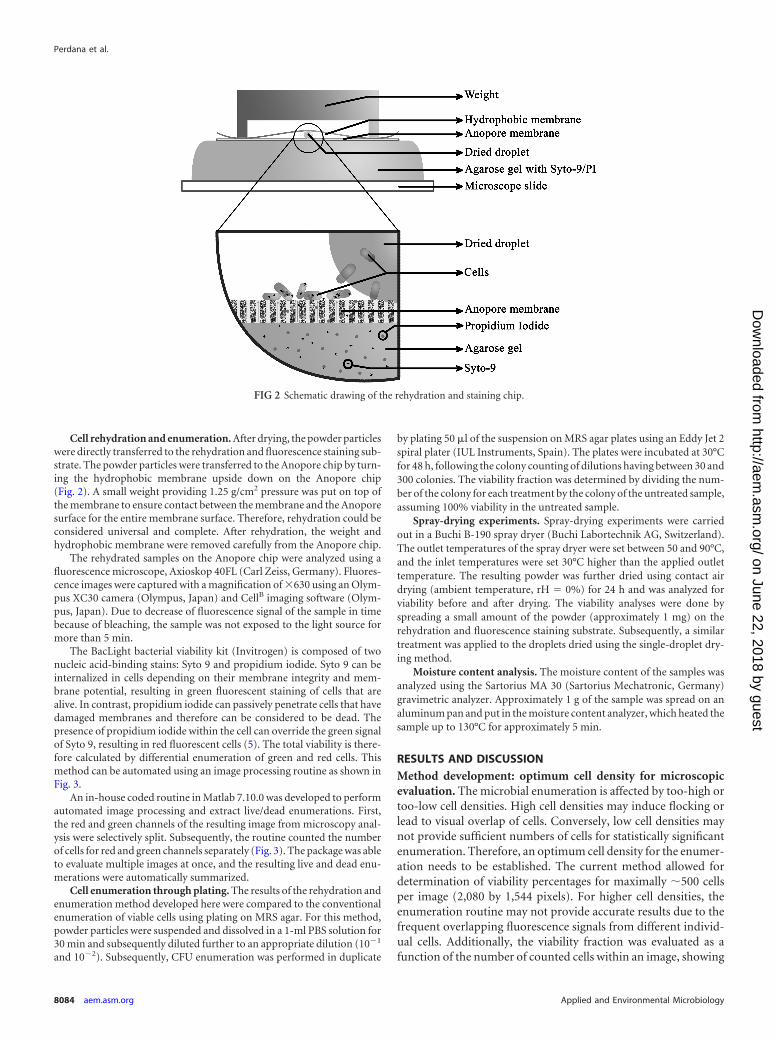
Cell rehydration and enumeration. After drying, the powder particleswere directly transferred to the rehydration and fluorescence staining sub-strate. The powder particles were transferred to the Anopore chip by turn-ing the hydrophobic membrane upside down on the Anopore chip(Fig. 2). A small weight providing 1.25 g/cm2 pressure was put on top ofthe membrane to ensure contact between the membrane and the Anoporesurface for the entire membrane surface. Therefore, rehydration could beconsidered universal and complete. After rehydration, the weight andhydrophobic membrane were removed carefully from the Anopore chip.
The rehydrated samples on the Anopore chip were analyzed using afluorescence microscope, Axioskop 40FL (Carl Zeiss, Germany). Fluores-cence images were captured with a magnification of �630 using an Olym-pus XC30 camera (Olympus, Japan) and CellB imaging software (Olym-pus, Japan). Due to decrease of fluorescence signal of the sample in timebecause of bleaching, the sample was not exposed to the light source formore than 5 min.
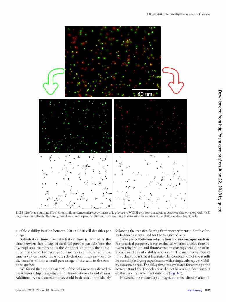
The BacLight bacterial viability kit (Invitrogen) is composed of twonucleic acid-binding stains: Syto 9 and propidium iodide. Syto 9 can beinternalized in cells depending on their membrane integrity and mem-brane potential, resulting in green fluorescent staining of cells that arealive. In contrast, propidium iodide can passively penetrate cells that havedamaged membranes and therefore can be considered to be dead. Thepresence of propidium iodide within the cell can override the green signalof Syto 9, resulting in red fluorescent cells (5). The total viability is there-fore calculated by differential enumeration of green and red cells. Thismethod can be automated using an image processing routine as shown inFig. 3.
An in-house coded routine in Matlab 7.10.0 was developed to performautomated image processing and extract live/dead enumerations. First,the red and green channels of the resulting image from microscopy anal-ysis were selectively split. Subsequently, the routine counted the numberof cells for red and green channels separately (Fig. 3). The package was ableto evaluate multiple images at once, and the resulting live and dead enu-merations were automatically summarized.
Cell enumeration through plating. The results of the rehydration andenumeration method developed here were compared to the conventionalenumeration of viable cells using plating on MRS agar. For this method,powder particles were suspended and dissolved in a 1-ml PBS solution for30 min and subsequently diluted further to an appropriate dilution (10�1
and 10�2). Subsequently, CFU enumeration was performed in duplicate
by plating 50 �l of the suspension on MRS agar plates using an Eddy Jet 2spiral plater (IUL Instruments, Spain). The plates were incubated at 30°Cfor 48 h, following the colony counting of dilutions having between 30 and300 colonies. The viability fraction was determined by dividing the num-ber of the colony for each treatment by the colony of the untreated sample,assuming 100% viability in the untreated sample.
Spray-drying experiments. Spray-drying experiments were carriedout in a Buchi B-190 spray dryer (Buchi Labortechnik AG, Switzerland).The outlet temperatures of the spray dryer were set between 50 and 90°C,and the inlet temperatures were set 30°C higher than the applied outlettemperature. The resulting powder was further dried using contact airdrying (ambient temperature, rH � 0%) for 24 h and was analyzed forviability before and after drying. The viability analyses were done byspreading a small amount of the powder (approximately 1 mg) on therehydration and fluorescence staining substrate. Subsequently, a similartreatment was applied to the droplets dried using the single-droplet dry-ing method.
Moisture content analysis. The moisture content of the samples wasanalyzed using the Sartorius MA 30 (Sartorius Mechatronic, Germany)gravimetric analyzer. Approximately 1 g of the sample was spread on analuminum pan and put in the moisture content analyzer, which heated thesample up to 130°C for approximately 5 min.
RESULTS AND DISCUSSIONMethod development: optimum cell density for microscopicevaluation. The microbial enumeration is affected by too-high ortoo-low cell densities. High cell densities may induce flocking orlead to visual overlap of cells. Conversely, low cell densities maynot provide sufficient numbers of cells for statistically significantenumeration. Therefore, an optimum cell density for the enumer-ation needs to be established. The current method allowed fordetermination of viability percentages for maximally �500 cellsper image (2,080 by 1,544 pixels). For higher cell densities, theenumeration routine may not provide accurate results due to thefrequent overlapping fluorescence signals from different individ-ual cells. Additionally, the viability fraction was evaluated as afunction of the number of counted cells within an image, showing
FIG 2 Schematic drawing of the rehydration and staining chip.
Perdana et al.
8084 aem.asm.org Applied and Environmental Microbiology
on June 22, 2018 by guesthttp://aem
.asm.org/
Dow
nloaded from
a stable viability fraction between 200 and 500 cell densities perimage.
Rehydration time. The rehydration time is defined as thetime between the transfer of the dried powder particle from thehydrophobic membrane to the Anopore chip and the subse-quent removal of the hydrophobic membrane. The rehydrationtime is critical, since too-short rehydration times may lead tothe transfer of only a small percentage of the cells to the Ano-pore surface.
We found that more than 90% of the cells were transferred tothe Anopore chip using rehydration times between 15 and 90 min.Additionally, the fluorescent dyes could be detected immediately
following the transfer. During further experiments, 15 min of re-hydration time was used for the transfer of cells.
Time period between rehydration and microscopic analysis.For practical purposes, it was evaluated whether a delay time be-tween rehydration and fluorescence microscopy would be of in-fluence on the final viability assessment. The major advantage ofthis delay time is that it facilitates the combination of the resultsfrom multiple drying experiments with a single subsequent viabil-ity assessment run. The delay time was evaluated for a time periodbetween 0 and 3 h. The delay time did not have a significant impacton the viability assessment outcome (Fig. 4C).
However, the microscopic images obtained directly after re-
FIG 3 Live/dead counting. (Top) Original fluorescence microscopy image of L. plantarum WCFS1 cells rehydrated on an Anopore chip observed with �630magnification. (Middle) Red and green channels are separated. (Bottom) Cell counting to determine the number of live (left) and dead (right) cells.
A Novel Method for Viability Enumeration of Probiotics
November 2012 Volume 78 Number 22 aem.asm.org 8085
on June 22, 2018 by guesthttp://aem
.asm.org/
Dow
nloaded from
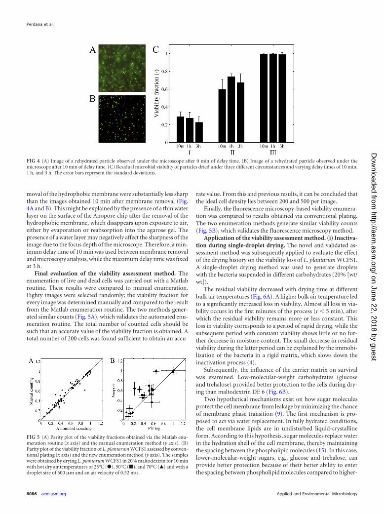
moval of the hydrophobic membrane were substantially less sharpthan the images obtained 10 min after membrane removal (Fig.4A and B). This might be explained by the presence of a thin waterlayer on the surface of the Anopore chip after the removal of thehydrophobic membrane, which disappears upon exposure to air,either by evaporation or reabsorption into the agarose gel. Thepresence of a water layer may negatively affect the sharpness of theimage due to the focus depth of the microscope. Therefore, a min-imum delay time of 10 min was used between membrane removaland microscopy analysis, while the maximum delay time was fixedat 3 h.
Final evaluation of the viability assessment method. Theenumeration of live and dead cells was carried out with a Matlabroutine. These results were compared to manual enumeration.Eighty images were selected randomly; the viability fraction forevery image was determined manually and compared to the resultfrom the Matlab enumeration routine. The two methods gener-ated similar counts (Fig. 5A), which validates the automated enu-meration routine. The total number of counted cells should besuch that an accurate value of the viability fraction is obtained. Atotal number of 200 cells was found sufficient to obtain an accu-
rate value. From this and previous results, it can be concluded thatthe ideal cell density lies between 200 and 500 per image.
Finally, the fluorescence microscopy-based viability enumera-tion was compared to results obtained via conventional plating.The two enumeration methods generate similar viability counts(Fig. 5B), which validates the fluorescence microscopy method.
Application of the viability assessment method. (i) Inactiva-tion during single-droplet drying. The novel and validated as-sessment method was subsequently applied to evaluate the effectof the drying history on the viability loss of L. plantarum WCFS1.A single-droplet drying method was used to generate dropletswith the bacteria suspended in different carbohydrates (20% [wt/wt]).
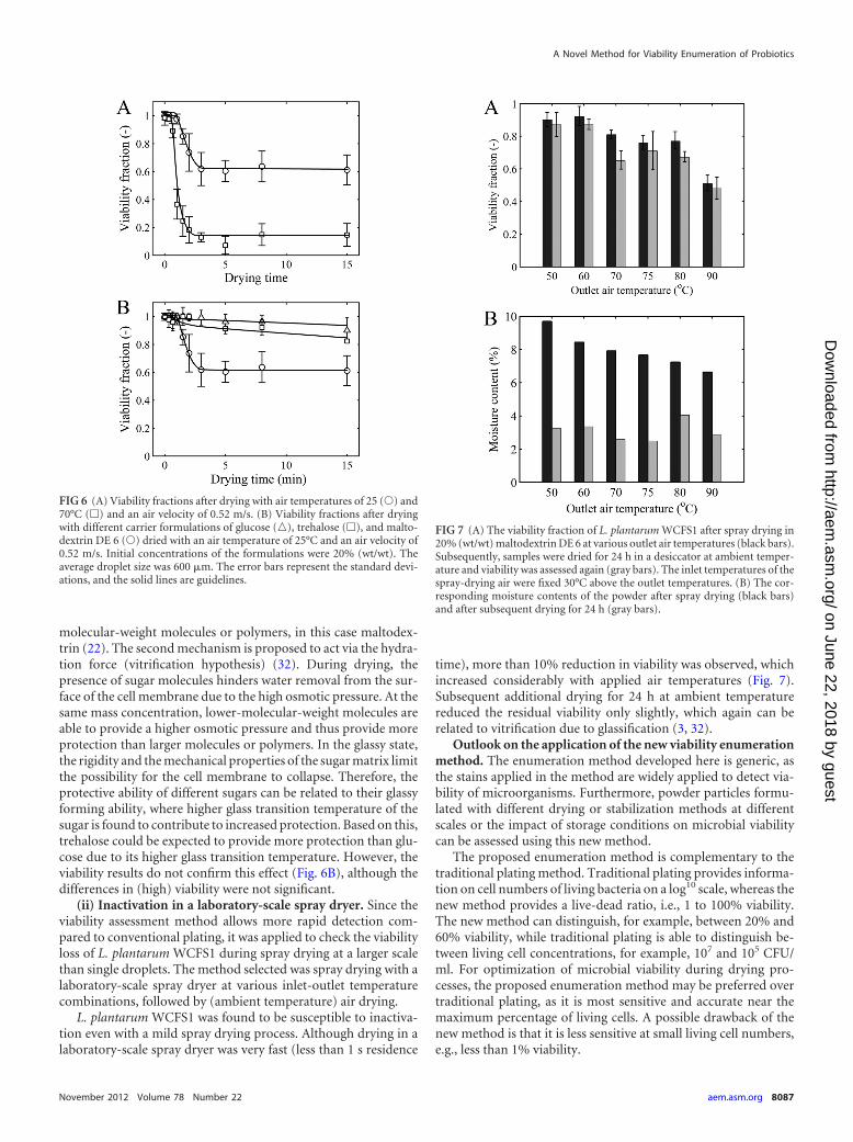
The residual viability decreased with drying time at differentbulk air temperatures (Fig. 6A). A higher bulk air temperature ledto a significantly increased loss in viability. Almost all loss in via-bility occurs in the first minutes of the process (t � 5 min), afterwhich the residual viability remains more or less constant. Thisloss in viability corresponds to a period of rapid drying, while thesubsequent period with constant viability shows little or no fur-ther decrease in moisture content. The small decrease in residualviability during the latter period can be explained by the immobi-lization of the bacteria in a rigid matrix, which slows down theinactivation process (4).
Subsequently, the influence of the carrier matrix on survivalwas examined. Low-molecular-weight carbohydrates (glucoseand trehalose) provided better protection to the cells during dry-ing than maltodextrin DE 6 (Fig. 6B).
Two hypothetical mechanisms exist on how sugar moleculesprotect the cell membrane from leakage by minimizing the chanceof membrane phase transition (9). The first mechanism is pro-posed to act via water replacement. In fully hydrated conditions,the cell membrane lipids are in undisturbed liquid-crystallineform. According to this hypothesis, sugar molecules replace waterin the hydration shell of the cell membrane, thereby maintainingthe spacing between the phospholipid molecules (15). In this case,lower-molecular-weight sugars, e.g., glucose and trehalose, canprovide better protection because of their better ability to enterthe spacing between phospholipid molecules compared to higher-
FIG 4 (A) Image of a rehydrated particle observed under the microscope after 0 min of delay time. (B) Image of a rehydrated particle observed under themicroscope after 10 min of delay time. (C) Residual microbial viability of particles dried under three different circumstances and varying delay times of 10 min,1 h, and 3 h. The error bars represent the standard deviations.
FIG 5 (A) Parity plot of the viability fractions obtained via the Matlab enu-meration routine (x axis) and the manual enumeration method (y axis). (B)Parity plot of the viability fraction of L. plantarum WCFS1 assessed by conven-tional plating (x axis) and the new enumeration method (y axis). The sampleswere obtained by drying L. plantarum WCFS1 in 20% maltodextrin for 10 minwith hot dry air temperatures of 25°C (�), 50°C (�), and 70°C (Œ) and with adroplet size of 600 �m and an air velocity of 0.52 m/s.
Perdana et al.
8086 aem.asm.org Applied and Environmental Microbiology
on June 22, 2018 by guesthttp://aem
.asm.org/
Dow
nloaded from
molecular-weight molecules or polymers, in this case maltodex-trin (22). The second mechanism is proposed to act via the hydra-tion force (vitrification hypothesis) (32). During drying, thepresence of sugar molecules hinders water removal from the sur-face of the cell membrane due to the high osmotic pressure. At thesame mass concentration, lower-molecular-weight molecules areable to provide a higher osmotic pressure and thus provide moreprotection than larger molecules or polymers. In the glassy state,the rigidity and the mechanical properties of the sugar matrix limitthe possibility for the cell membrane to collapse. Therefore, theprotective ability of different sugars can be related to their glassyforming ability, where higher glass transition temperature of thesugar is found to contribute to increased protection. Based on this,trehalose could be expected to provide more protection than glu-cose due to its higher glass transition temperature. However, theviability results do not confirm this effect (Fig. 6B), although thedifferences in (high) viability were not significant.
(ii) Inactivation in a laboratory-scale spray dryer. Since theviability assessment method allows more rapid detection com-pared to conventional plating, it was applied to check the viabilityloss of L. plantarum WCFS1 during spray drying at a larger scalethan single droplets. The method selected was spray drying with alaboratory-scale spray dryer at various inlet-outlet temperaturecombinations, followed by (ambient temperature) air drying.
L. plantarum WCFS1 was found to be susceptible to inactiva-tion even with a mild spray drying process. Although drying in alaboratory-scale spray dryer was very fast (less than 1 s residence
time), more than 10% reduction in viability was observed, whichincreased considerably with applied air temperatures (Fig. 7).Subsequent additional drying for 24 h at ambient temperaturereduced the residual viability only slightly, which again can berelated to vitrification due to glassification (3, 32).
Outlook on the application of the new viability enumerationmethod. The enumeration method developed here is generic, asthe stains applied in the method are widely applied to detect via-bility of microorganisms. Furthermore, powder particles formu-lated with different drying or stabilization methods at differentscales or the impact of storage conditions on microbial viabilitycan be assessed using this new method.
The proposed enumeration method is complementary to thetraditional plating method. Traditional plating provides informa-tion on cell numbers of living bacteria on a log10 scale, whereas thenew method provides a live-dead ratio, i.e., 1 to 100% viability.The new method can distinguish, for example, between 20% and60% viability, while traditional plating is able to distinguish be-tween living cell concentrations, for example, 107 and 105 CFU/ml. For optimization of microbial viability during drying pro-cesses, the proposed enumeration method may be preferred overtraditional plating, as it is most sensitive and accurate near themaximum percentage of living cells. A possible drawback of thenew method is that it is less sensitive at small living cell numbers,e.g., less than 1% viability.
FIG 6 (A) Viability fractions after drying with air temperatures of 25 (Œ) and70°C (�) and an air velocity of 0.52 m/s. (B) Viability fractions after dryingwith different carrier formulations of glucose (o), trehalose (�), and malto-dextrin DE 6 (Œ) dried with an air temperature of 25°C and an air velocity of0.52 m/s. Initial concentrations of the formulations were 20% (wt/wt). Theaverage droplet size was 600 �m. The error bars represent the standard devi-ations, and the solid lines are guidelines.
FIG 7 (A) The viability fraction of L. plantarum WCFS1 after spray drying in20% (wt/wt) maltodextrin DE 6 at various outlet air temperatures (black bars).Subsequently, samples were dried for 24 h in a desiccator at ambient temper-ature and viability was assessed again (gray bars). The inlet temperatures of thespray-drying air were fixed 30°C above the outlet temperatures. (B) The cor-responding moisture contents of the powder after spray drying (black bars)and after subsequent drying for 24 h (gray bars).
A Novel Method for Viability Enumeration of Probiotics
November 2012 Volume 78 Number 22 aem.asm.org 8087
on June 22, 2018 by guesthttp://aem
.asm.org/
Dow
nloaded from
Conclusion. A novel method was developed to determine themicrobial viability of probiotics in small samples, such as singlepowder particles. The method employs Anopore carrier chips forrehydration of semidried particles followed by live/dead stainingusing fluorescence probe methodology. The enumeration wascarried out by microscopy and automated image analysis using aMatlab routine. The robustness of the method was evaluated andvalidated with conventional plating. In combination with a single-droplet drying approach, the influence of the drying air tempera-ture and carrier formulation on viability loss of L. plantarumWCFS1 was mapped as a function of drying time. The latter ap-proach can also be of major support in determining optimal dry-ing conditions and formulations for spray drying of probiotics.
ACKNOWLEDGMENTS
We thank the Advanced Chemical Technologies for Sustainability(ACTS), Netherlands Organization for Scientific Research (NWO), forfinancial aid through a grant from the Process on a Chip (PoaC) program.
We also thank Clint van Melis for his advice and support during mi-croscopy analysis and Ingrid Maas and Diah Chandra Aryani for theirsupport in the microbiology lab.
REFERENCES1. Anal AK, Singh H. 2007. Recent advances in microencapsulation of
probiotics for industrial applications and targeted delivery. Trends FoodSci. Technol. 18:240 –251.
2. Ananta E. 2005. Impact of environmental factors on viability and stabilityand high pressure pretreatment on stress tolerance of Lactobacillus rham-nosus GG (ATCC 53103) during spray drying. Technischen UniversitätBerlin, Berlin, Germany. http://opus.kobv.de/tuberlin/volltexte/2005/1119/.
3. Ananta E, Volkert M, Knorr D. 2005. Cellular injuries and storagestability of spray-dried Lactobacillus rhamnosus GG. Int. Dairy J. 15:399 –409.
4. Augustin MA, Hemar Y. 2009. Nano- and micro-structured assembliesfor encapsulation of food ingredients. Chem. Soc. Rev. 38:902–912.
5. Boulos L, Prévost M, Barbeau B, Coallier J, Desjardins R. 1999. Live/Dead BacLight: application of a new rapid staining method for directenumeration of viable and total bacteria in drinking water. J. Microbiol.Methods 37:77– 86.
6. Bron PA, van Baarlen P, Kleerebezem M. 2012. Emerging molecularinsights into the interaction between probiotics and the host intestinalmucosa. Nat. Rev. Microbiol. 10:66 –78.
7. Chávez BE, Ledeboer AM. 2007. Drying of probiotics: optimization offormulation and process to enhance storage survival. Dry Technol. 25:1193–1201.
8. Corcoran BM, Ross RP, Fitzgerald GF, Stanton C. 2004. Comparativesurvival of probiotic lactobacilli spray-dried in the presence of prebioticsubstances. J. Appl. Microbiol. 96:1024 –1039.
9. Crowe JH, Crowe LM, Hoekstra FA. 1989. Phase transitions and perme-ability changes in dry membranes during rehydration. J. Bioenerg.Biomembr. 21:77–91.
10. Desmond C, Ross RP, O’Callaghan E, Fitzgerald G, Stanton C. 2002.Improved survival of Lactobacillus paracasei NFBC 338 in spray-driedpowders containing gum acacia. J. Appl. Microbiol. 93:1003–1011.
11. FAO/WHO. 2002. Report of a joint FAO/WHO expert consultation onguidelines for the evaluation of probiotics in food. Food and AgricultureOrganization of the United Nations and World Health Organization, Lon-don, Ontario, Canada.
12. Fuller R. 1991. Probiotics in human medicine. Gut 32:439 – 442.
13. Gardiner GE, et al. 2000. Comparative survival rates of human-derivedprobiotic Lactobacillus paracasei and L. salivarius strains during heat treat-ment and spray drying. Appl. Environ. Microbiol. 66:2605–2612.
14. Guilbaud M, Zagorec M, Chaillou S, Champomier-Vergès M-C. 2012.Intraspecies diversity of Lactobacillus sakei response to oxidative stress andvariability of strain performance in mixed strains challenges. Food Micro-biol. 29:197–204.
15. Hoekstra FA, Golovina EA, Buitink J. 2001. Mechanisms of plant desic-cation tolerance. Trends Plant Sci. 6:431– 438.
16. Holzapfel WH, Haberer P, Geisen R, Björkroth J, Schillinger U. 2001.Taxonomy and important features of probiotic microorganisms in foodand nutrition. Am. J. Clin. Nutr. 73:365S–373S.
17. Ingham CJ, et al. 2007. The micro-petri dish, a million-well growth chipfor the culture and high-throughput screening of microorganisms. Proc.Natl. Acad. Sci. U. S. A. 104:18217–18222.
18. Ingham CJ, van den Ende M, Pijnenburg D, Wever PC, SchneebergerPM. 2005. Growth and multiplexed analysis of microorganisms on a sub-divided, highly porous, inorganic chip manufactured from Anopore.Appl. Environ. Microbiol. 71:8978 – 8981.
19. Jankovic I, Sybesma W, Phothirath P, Ananta E, Mercenier A. 2010.Application of probiotics in food products— challenges and new ap-proaches. Curr. Opin. Biotechnol. 21:175–181.
20. Kleerebezem M, et al. 2003. Complete genome sequence of Lactobacillusplantarum WCFS1. Proc. Natl. Acad. Sci. U. S. A. 100:1990 –1995.
21. Kleerebezem M, Vaughan EE. 2009. Probiotic and gut lactobacilli andbifidobacteria: molecular approaches to study diversity and activity.Annu. Rev. Microbiol. 63:269 –290.
22. Koster KL, Lei YP, Anderson M, Martin S, Bryant G. 2000. Effects ofvitrified and nonvitrified sugars on phosphatidylcholine fluid-to-gelphase transitions. Biophys. J. 78:1932–1946.
23. Mauriello G, Aponte M, Andolfi R, Moschetti G, Villani F. 1999.Spray-drying of bacteriocin-producing lactic acid bacteria. J. Food Prot.62:773–777.
24. Molenaar D, et al. 2005. Exploring Lactobacillus plantarum genome di-versity by using microarrays. J. Bacteriol. 187:6119 – 6127.
25. Parvez S, Malik KA, Ah Kang S, Kim HY. 2006. Probiotics and theirfermented food products are beneficial for health. J. Appl. Microbiol. 100:1171–1185.
26. Peighambardoust SH, Tafti AG, Hejazi MA, Hesari J. 2011. Applicationof spray drying for preservation of lactic acid starter cultures: a review.Trends Food Sci. Technol. 22:215–224.
27. Perdana J, Fox M, Schutyser M, Boom R. Mimicking spray drying bydrying of single droplets deposited on a flat surface. Food BioprocessTechnol. doi:10.1007/s11947-011-0767-4.
28. Perdana J, Fox MB, Schutyser MAI, Boom RM. 2012. Enzyme inacti-vation kinetics: coupled effects of temperature and moisture content.Food Chem. 133:116 –123.
29. Perdana J, Fox MB, Schutyser MAI, Boom RM. 2011. Single-dropletexperimentation on spray drying: evaporation of a sessile droplet. Chem.Eng. Technol. 34:1151–1158.
30. Ranadheera RDCS, Baines SK, Adams MC. 2010. Importance of food inprobiotic efficacy. Food Res. Int. 43:1–7.
31. Rokka S, Rantamäki P. 2010. Protecting probiotic bacteria by microen-capsulation: challenges for industrial applications. Eur. Food Res. Tech-nol. 231:1–12.
32. Santivarangkna C, Higl B, Foerst P. 2008. Protection mechanisms ofsugars during different stages of preparation process of dried lactic acidstarter cultures. Food Microbiol. 25:429 – 441.
33. Santivarangkna C, Kulozik U, Foerst P. 2008. Inactivation mechanismsof lactic acid starter cultures preserved by drying processes. J. Appl. Mi-crobiol. 105:1–13.
34. Schutyser MAI, Perdana J, Boom RM. 2012. Single droplet drying foroptimal spray drying of enzymes and probiotics. Trends Food Sci. Tech-nol. [Epub ahead of print.] http://dx.doi.org/10.1016/j.tifs.2012.05.006.
Perdana et al.
8088 aem.asm.org Applied and Environmental Microbiology
on June 22, 2018 by guesthttp://aem
.asm.org/
Dow
nloaded from


![The Role of Trehalose 6-Phosphate in Crop Yield and … · 2020. 5. 18. · Update on Trehalose 6-Phosphate Signaling The Role of Trehalose 6-Phosphate in Crop Yield and Resilience1[OPEN]](https://img.pdfslide.us/doc/110x75/60a94aac2e9d0b10d12c4d11/the-role-of-trehalose-6-phosphate-in-crop-yield-and-2020-5-18-update-on-trehalose.jpg)


![Trehalose 6-Phosphate Regulates Photosynthesis and · Trehalose 6-Phosphate Regulates Photosynthesis and Assimilate Partitioning in Reproductive Tissue1[OPEN] Maria Oszvald,a Lucia](https://img.pdfslide.us/doc/110x75/611f5cf3b1f8956e674d52f9/trehalose-6-phosphate-regulates-photosynthesis-trehalose-6-phosphate-regulates-photosynthesis.jpg)