Embed Size (px)
Citation preview
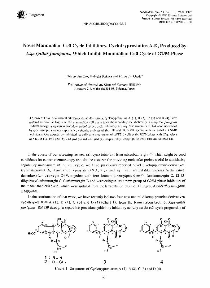
Pergamon Terrahedron. Vol. 53, No. I, pp. 59-72. 1997
Copyright 0 1996 Elsevier Science Ltd Prmted I” Great Bntatn. All rights reserved
0040.4020/97 $17.00 + 0.00 PII: SOO40-4020(96)00978-7
Novel Mammalian Cell Cycle Inhibitors, Cyclotryprostatins A-D, Produced by
Aspergillus fumigatus, Which Inhibit Mammalian Cell Cycle at G2/M Phase
Cheng-Bin Cui, Hideaki Kakeya and Hiroyuki Osada*
The Instilutc ot Physical and Chemical Research (RIKEN), Hirosawa 2-1, Wako-shi 351-01. Saitama, Japan
Absfracl: Four new natural dikctopipcrazine derivatives, cyclouyprostatins A (1). B (2), C (3) and D (4). were isolarcd as new inhibilors of the mammalian cell cycle from lhc secondary mclabolites of Aspergiflus fumigalus BM939 through a separation prc~cdnre guided by ccl1 cycle inhibitory activity. The suucturcs of 1-4 were tirmined by spectroscopic methods cspccially by dctailcd analysts of their lH and l3C NMR spcclra with the aid of 2D NMR techniques. Compounds 1-4 Inhibited the cell cycle progrcwon of tsFl210 cells at the G2/M phase with IC50 values
of 5.6 FM (1). 19.5 FM (2). 23.4 FM (3) and 25.3 KM (4), rcspcctivcly. Copyright 0 I996 Elsevier Science Ltd
In the course of our screening for new cell cycle inhibitors from microbial origini-?, which might be good
candidates for cancer chemotherapy and also be a source for providing molecular probes useful in elucidating
regulatory mechanism of the cell cycle, we have previously reported novel diketopiperazine derivatives,
tryprostatins2.4.5) A, B and spirotryprostatins 6.71 A, B as well as a new natural diketopiperazine derivative,
demethoxyfumitremorgin C4.5), together with four known diketopiperazines’J), fumitremorgin C, 12,13-
dihydroxyfumitremorgin C, fumitremorgin B and vertuculogen, as a new group of G2/M-phase inhibitors of
the mammalian cell cycle, which were isolated from the fermentation broth of a fungus, Aspergillusfumigaru
BM9392-7).
In the continuation of that work, we have recently isolated four new natural diketopiperazine derivatives,
cyclottyprostatins A (l), B (2), C (3) and D (4) (Chart l), from the fermentation broth of Aspergillus
futnigatus BM939 through a separation procedure guided by inhibitory activity on the cell cycle progression of
1: R=H 2: R=CHs
Chart 1 Structures of Cyclotryprostatins A (I), B (2), C
3 4
(3) and D (4).
59
60 C.-B. GUI et al.
mouse tsFT210 cells. Cyclotryprostatins A (1)-D (4) inhibited the cell cycle progression of tsFT210 cells at the
G2/M phase and were named according to their structural and biological relations to tryprostatins. We describe
herein the isolation, structure determination and biological activities of 1-4.
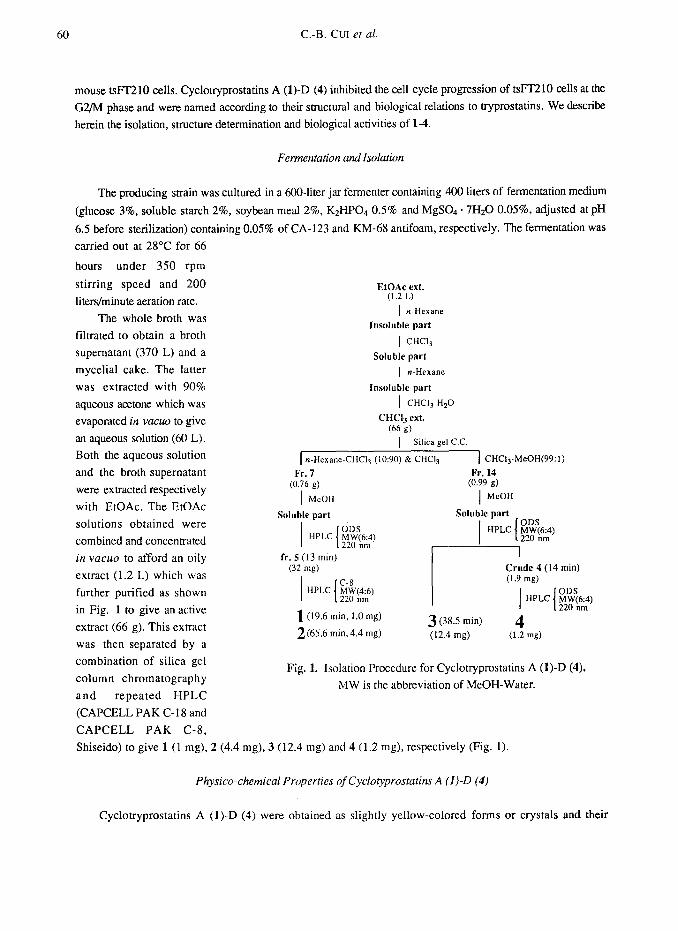
Fermentation and Isolation
The producing strain was cultured in a 6OO-liter jar fermenter containing 400 liters of fermentation medium
(glucose 3%, soluble starch 2%, soybean meal 2%, KaHP04 0.5% and MgS04 * 7HaO 0.05%, adjusted at pH
6.5 before sterilization) containing 0.05% of CA-123 and KM-68 antifoam, respectively. The fermentation was
carried out at 28°C for 66
hours under 350 rpm
stirring speed and 200
liters/minute aeration rate.
The whole broth was
filtrated to obtain a broth
supernatant (370 L) and a
mycelial cake. The latter
was extracted with 90%
aqueous acetone which was
evaporated in vacua to give
an aqueous solution (60 L).
Both the aqueous solution
and the broth supernatant
were extracted respectively
with EtOAc. The EtOAc
solutions obtained were
combined and concentrated
in vacua to afford an oily
extract (1.2 L) which was
further purified as shown
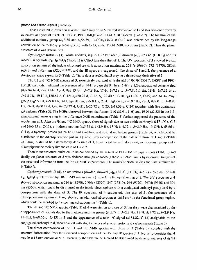
in Fig. 1 to give an active
extract (66 g). This extract
was then separated by a
combination of silica gel
column chromatography
EtOAc ext. (1.2 L)
1 n-Hexane
Insoluble part
1 CHCI,
Soluble part
1 n-Hexane
Insoluble part
1 CHC13-H20
CHQ ext. (66 9)
1 Silica gel CC.
1 n-Hexane-CHCI, (10:90) & CHQ
Fr. 7 (0.76 g)
McOH
Soluble part
1 CHC13-MeOH(99: 1)
Fr. 14 (0.99 g)
MeOH
fr. 5 (13 min) (32 mg)
I I C-8 HPLC y2yt‘i”’
l(19.6 min, 1.0 mg)
z(6.5.6 min, 4.4 mg) 3 (38.5 min)
(12.4 mg)
I Crude 4 (14 min) (1.9 w)
Fig. 1. Isolation Procedure for Cyclotryprostatins A (1)-D (4).
MW is the abbreviation of MeOH-Water. and repeated HPLC
(CAPCELL PAK C-l 8 and
CAPCELL PAK C-8,
Shiseido) to give 1 (1 mg), 2 (4.4 mg), 3 (12.4 mg) and 4 (1.2 mg), respectively (Fig. 1).
Physico-chemical Properties of Cyclotyprostatins A (1)-D (4)
Cyclotryprostatins A (1)-D (4) were obtained as slightly yellow-colored forms or crystals and their
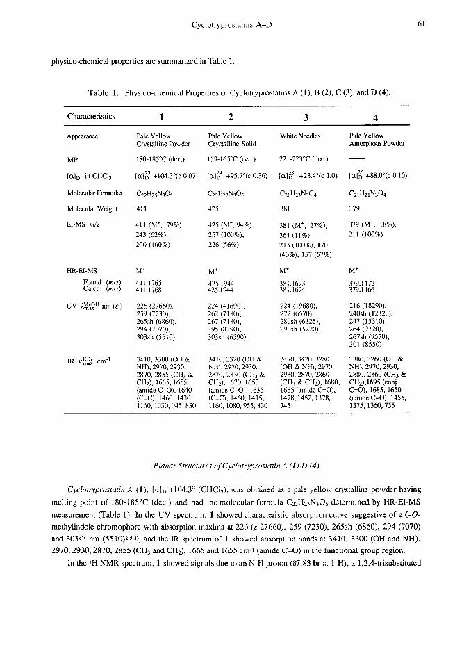
Cyclotryprostatins A-D 61
physico-chemical properties are summarized in Table 1.
Table 1. Physico-chemical Properties of Cyclotryprostatins A (1). B (2), C (3), and D (4).
Characteristics 1 2 3 4
MP
[aID in CHCI,
Molecular Formular
Molecular Weight
Et-MS m/z
IR v!$ cm-’
Pale Yellow Crystalline Powder
180-185°C (dcc.)
[al;3 +104.3”(c 0.07)
Cz2H2sNGs
411
411 (M+, 79%), 425 (M+, 94%),
243 (62%). 257 (lOO%),
200 (100%) 226 (56%)
M+ M+ Mf
411.1765 425.1944 381.1693 411.1768 425.1944 381.1694
226 (27660), 259 (7230). 265sh (6860). 294 (7070). 303sh (5510)
224 (41690), 262 (7 180). 267 (7180). 295 (8290). 303sh (6590)
224 (19680), 272 (6570). 280sh (6325), 290sh (5220)
3410,330O (OH & 3410,332O (OH & 3470, 3420.3280 3380.3260 (OH & NH), 2970,2930, NH), 2970,2930, (OH & NH), 2970, NH), 2970,2930, 2870,2855 (CH3 & 2870,283O (CH3 & 2930,2870,2860 2880,286O (CH3 & CH2), 1665,1655 CH2), 1670, 1650 (CH3 & CHz), 1680, CH2),1695 (conj. (amide C=O), 1640 (amide C=O), 1635 1665 (amide C=O), C=O), 1685, 1650 (C=C), 1460, 1430, (C=C), 1460, 1415, 1478.1452, 1378, (amide GO), 1455, 1160, 1030,945,830 1160, 1080,955, 830 745 1375,1360,755
Pale Yellow Crystalline Solid
159-165°C (dcc.)
[a]k4 +95.7”(c 0.36)
425
White Needles Pale Yellow Amorphous Powder
221-223°C (dcc.)
[a# +23.4”(c 1.0)
381
381 (M+, 27%).
364 (II%),
213 (loo%), 170
(40%). 157 (57%)
[al: +88.O”(c 0.10)
CZIW’W~
379
379 (M+, 18%),
211 (100%)
M+
%:E 216 (18290), 240sh (12320), 247 (15310). 264 (9720). 267sh (9570). 301 (SSSO)
Planar Structures of Cyclotryprostatin A (Ii-D (4)
Cyclotryprostatin A (I), [aID +104.3’ (CHC13), was obtained as a pale yellow crystalline powder having
melting point of 180-185°C (dec.) and had the molecular formula C2ZH2sN30s determined by HR-EI-MS
measurement (Table 1). In the UV spectrum, 1 showed characteristic absorption curve suggestive of a 6-0-
methylindole chromophore with absorption maxima at 226 (E 27660), 259 (7230), 265sh (6860), 294 (7070)
and 303sh nm (5510)2.s.s), and the IR spectrum of 1 showed absorption bands at 3410, 3300 (OH and NH),
2970,2930,2870,2855 (CH3 and CH2), 1665 and 1655 cm-i (amide C=O) in the functional group region.
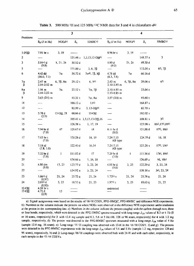
In the tH NMR spectrum, 1 showed signals due to an N-H proton (67.83 br s, 1-H). a 1,2,4-trisubstituted
62 C.-B. CIJI et nl.
benzene ring (67.45 d, J=8.5 Hz, 16-H; 66.81 dd, J= 8.5, 2.1 Hz, 17-H; and 66.86 d, J=2.1 Hz, 19-H) and a
methoxy (63.82 s, OC&) and two methyl (61.79 s, 23-b and 62.04 s, 24-b) groups along with signals due
to several methine and methylene groups (Table 2). The i3C NMR spectrum of 1, analyzed by the DEPT
method, indicated the presence of two amide carbonyls (S165.55 s, C-5 and 6166.87 s, C-l l), an oxygen-
bearing sp2 carbon (6156.72 s, C-18) and a methoxy (655.73 q, 0cH3) and two methyl (626.05 q. C-23 and
618.29 q, C-24) groups together with four sp2 and three sp3 methines, five spz and a sps quaternaty carbons,
and three methylene groups (Table 2).
The amide carbonyl absorption at 1665 and 1655 cm-i together with the absence of the amide II band near
1550 cm-t in the IR spectrum suggested the presence of a diketopiperazine systemV~9-it) in 1. This was further
supported by the amide carbonyl carbon signals at 6165.55 and 6166.87 (C-5 and C-11) in the t3C NMR
spectrum, which were assignable to the carbonyls in the diketopiperazine partz.5-7).
Detailed analyses of the tH and W NMR spectra (Table 2) of 1 with the aid of IH-iH COSY and pulse
field gradient heteronuclear multiple quantum coherence (PFG-HMQC) spectroscopy, coupled with the results
of difference NOE experiments (Table 2) and the above structural information, led us to consider that the
structure of 1 is composed from a 60methylindole moiety, a diketopiperazine part and an isoprenyl residue,
forming a pentacyclic ring skeleton.
The above structural units associated with the proton spin systems and the through-space interactions in 1
were confirmed by the structural information from the PFG heteronuclear multiple bond correlation (PFG-
HMBC) spectrum (Table 2). For instance, the oxygen-bearing sp2 quaternary carbon C-18 (6156.72) and the
spz quatemary carbons C-20 (6136.76) and C-15 (6120.57) in the O-methylindole moiety in 1 could be
assigned according to the long-range correlations between C-18 and the protons 16-H, 17-H, and 19-H,
between C-20 and the protons I-H, 16-H and 19-H, and between C-15 and the protons I-H, 17-H and 19-H,
respectively, and the methoxy (6tr3.82 and k55.73) was linked to C- 18 according to the long-range correlation
of the methoxy protons (63.82 ) with C-18 in the PFG-HMBC spectrum. Similarly, the quaternary carbons C-
14 (S107.41) and C-2 (6133.34) in the same structural unit could be assigned on the basis of the long-range
correlations, the former C-14 with 1-H and 16-H and the latter C-2 with l-H, respectively, in the PFG-HMBC
spectrum. Thus the methoxyindole moiety in 1 was confirmed. Also, the other long-range correlations
confirming the isoprenyl residue and the diketopiperazine part in 1 are given in Table 2.
The connectivities between those structural units were determined by detailed analysis of the PFG-HMBC
spectrum (Table 2). Both 3-H in the isoprenyl residue and 13-H neighbored to C-12 to form the
diketopiperazine part in 1, for example, showed long-range correlations with the carbons C-2, C-14 (in the
methoxyindole moiety) and C-12 (in the diketopiperazine part). The former 3-H and the latter 13-H correlated
further with C-5 in the diketopiperazine part and with C-15 in the methoxyindole moiety, respectively, in the
PFG-HMBC spectrum. Those data evidenced the connectivity between the above structural units to form a
central heterocyclic ring in 1. Some other long-range correlations detected in the PFG-HMBC spectrum am
summarized in Table 2, which evidenced also the connectivities between the structural units in 1 and thus the
planar structure of 1 was deduced.
Cyclotryprostutin B (2) was obtained as a pale yellow crystalline solid having melting point of 159-165°C
(dec.) and showed [a]n +95.7’ (CHCls). The molecular formula of 2 was determined to be C~sHa7Ns05 by HR-
ELMS measurement (Table l), which is CH2 more than that of 1. The UV and IR spectra (Table 1) of 2
respectively suggested, like those of 1, the presence of a 6-U-methylindole chromophore and a diketopiperazine
system in 2. The 1H and W NMR spectra of 2 closely resembled those of 1, except for the appearance of
additional methoxy signals (&3.36 s and 6c56.59 q) and the slight changes in the chemical shifts of several
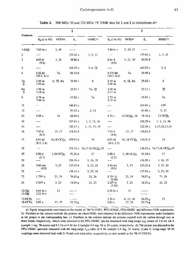
Cyclotryprostatins A-D 63
Table 2. 500 MHz tH and 125 MHz 13C NMR data for 1 and 2 in chloroform-da)
1 2 Positions
6~ (J in Hz) NOEshI 8, HMBW St, (-I in Hz) NOES”) 6, HMBCc)
7.83 br s 3, 19
5
6
6.65 d (9.8)
1. 24
________
133.34 s
48.86 d
4.42 dd (10.5, 6.1)
7a
165.55 s
60.10d
la P
8a P
9
2.49 m 6,78,8a 1.99 m
1.99 m 2.10 m
3.76 m 3.66 m
29.86 t 6
22.07 t 7a. 7p
45.85 t 7a
11
12
13
14
15
16
5.09 s 16
166.87 s
85.51 s
68.88 d
107.41 s
120.57 s
118.42 d
17
1.45 d 13, 17 (8.5)
6.81 dd l6,l8-OCH3 (8.5, 2.1)
109.97 d 19
18
19
20
21
22
23
6.86 d (2.1)
-
5.60 dm (9.8)
24
_____
1.79c) s
2.04r) s
156.72 s
1 ,18-OCLi3 95.26 d
136.76 s
3, 23 123.35 d
138.12 s
21.24 26.05 q
3, 23 18.29 q
12-OH 4.44 br s 13 13-of! 2.21 br s _____ __..
13-QCH3 - 18-oCH3 3.82 s 17, 19 55.73 q
7.86 br s 3, 19, 21 ______
133.65 s 1.3, 13
6.64 d 1, 21, 24 49.06 d (9.8)
____.
4.315 dd (10.7, 6.4)
la
167.02 s
59.98 d
3, 6
1, 3, 13
3.6, 7p
3, 13
1, 3, 13, 16
1. 13, 17, 19
2.49 m 2.00 m
2.00 m 2.11 m
3.75 m 3.70 m
_____
___.-
4.74 s
_____
7.44 d (8.6)
6.82 dd (8.6, 2.2)
6, 7l3,8a 29.68 t 6
22.11 t 7P
45.81 t la
165.84 s
84.68 s
13-OC&, 16 76.16 d
105.29 s
122.59 s
13, 17 118.63 d
13d)
3, 13
13-c!CH3
1, 3, 13, 16
1,13,16,17.19
16, IS-OC& 110.01 d 19
16,17,18-octJ3.19 -----
17 6.88 d
156.45 s 16.17.18~QC&,l9
1,18-OCH3 95.18 d 17
136.59 s 1, 16, 19
3, 23 123.52 d 3, 23,24
137.91 s 3, 23, 24
21.24 26.07 q 21.24
3, 23 18.23 q 21,23
(2.2) 1. 16, 19 _____
3, 23. 24 5.56 dm (9.8)
3, 23, 24 _____
21. 24
21,23
1.79~) d (0.98)
2.05r) d (1.46)
4.38 br s ---
3.36 s 3.83 s
13 __ ______ _
6, 13. 16 17, 19
56.59 q 13 55.73 q
a): Signal assignments were based on the results of t H-tH COSY, PFG-HMQC, PFG-HMBC and difference NOE experiments. b): Numbers in the column indicate the protons on where NOES were observed in the difCerence NOE experiments under irradiation at the proton in the corresponding line. c): Numbers in the column indicate the protons coupled with the carbon through two or three bonds, respectively, which were detected in the PFG-HMBC spccua measured with long-range Jctt values of 2.8 Hz for 1
(sample 1 mg, 96 scans) and 8.3 Hz or 4 Hz for 2 (sample 4.4 mg, 48 or 24 scans), respectively. d): The proton was detected in the PFG-HMBC spectrum measured with the long-range Jr-tt value of 4 Hz (sample 4.4 mg, 24 scans). e) and f): Long-range tH-tH
couplings were observedboth with 21-H and with each other, respcctivcly, in each sample in the tH-tH COSYs.
64 C.-B. GUI et al.
proton and carbon signals (Table 2).
Those structural information revealed that 2 may be an O-methyl derivative of 1 and this was confirmed by
extensive analyses of its tH-iH COSY, PFG-HMQC and PFG-HMBC spectra (Table 2). The location of the
additional methoxy group (Su3.36 and Sc56.59, 13-OCHs) in 2 at C-13 was determined by the long-range
correlation of the methoxy protons (63.36) with C- 13, in the PFG-HMBC spectrum (Table 2). Thus the planar
structure of 2 was determined.
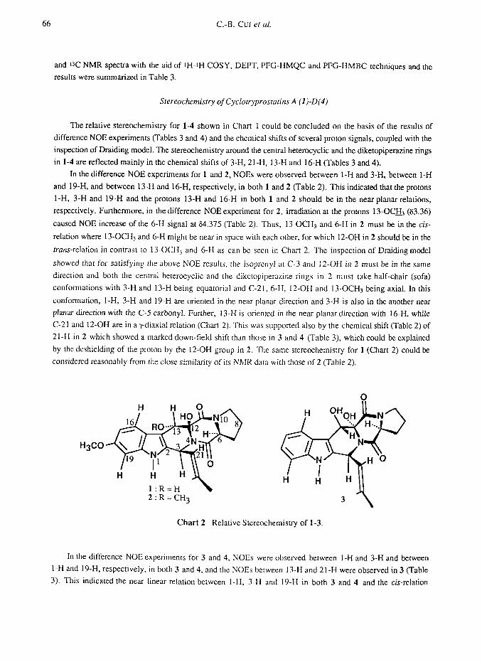
Cyclotryprosratin C (3), white needles, mp 221-223’C (dec.), showed [a]o +23.4’ (CHCls) and its
molecular formula CaLHz3N304 (Table 1) is CH20 less than that of 1. The UV spectrum of 3 showed typical
absorption pattern of the indole chromophore with absorption maxima at 224 (E 19680), 272 (6570), 280sh
(6325) and 290sh nm (522O)zX%ta), and the IR spectrum suggested, like those of 1 and 2, the presence of a
diketopiperazine system in 3 (Table 1). Those data revealed that 3 may be a demethoxy derivative of 1.
The tH and t3C NMR spectra of 3, extensively analyzed with the aid of tH-iH COSY, DEPT and PFG-
HMQC methods, indicated the presence of an N-H proton (67.91 br s, l-H), a 1,2-disubstituted benzene ring
(Su7.94 br d, J=7.8 Hz, 16-H; Sit7.13 br t, J=7.8 Hz, 17-H; Stt7.18 td, J=7.8, 1.0 Hz, 18-H; 6~7.32 br d,
J=7.8 Hz, 19-H; Scl20.67 d, C-16; 6cl20.28 d, C-17; Scl22.40 d, C-18; Sclll.02 d, C-19) and an isoprenyl
group (Str5.91 d, J=9.8 Hz, 3-H; Su4.80 dm, J=9.8 Hz, 21-H; Sitl.66 d, J=O.97 Hz, 23-H; Su2.01 d, J=O.95
Hz, 24-H, Sc50.12 d, C-3; Sc123.77 d, C-21; Sc25.72 q, C-23; Sc18.33 q, C-24) together with five quatemary
spz carbons (Table 3). The NOES observed between the former N-H (67.91, 1-H) and 19-H (67.32) in the 1,2-
disubstituted benzene ring in the difference NOE experiments (Table 3) further supported the presence of the
indole unit in 3. Also the tH and W NMR spectra showed signals due to two amide carbonyls (6171.00 s, C-5
and 6166.13 s, C-l 1), a hydroxymethine (&5.78 d, J=2.9 Hz, 13-H; Stt4.72 d, J=2.9 Hz, 13-Ofl; Sc68.66 d,
C-13). a hydroxyl proton (64.24 br s) and a methine and several methylene groups (Table 3), which could be
distributed to the diketopiperazine part in 3 (Table 3) by a comparison of the data with those of 1 and 2 (Table
2). Thus, 3 should be a demethoxy derivative of 1, constructed by an indole unit, an isoprenyl group and a
diketopiperazine moiety like the case of 1 and 2.
Then those structural units could be confirmed by the results of PFG-HMBC experiments (Table 3) and
finally the planar structure of 3 was deduced through connecting those structural units by extensive analysis of
the structural information from the PFG-HMBC experiments. The results of NMR studies for 3 are summarized
in Table 3.
Cyclotryprostatin D (4), an amorphous powder, showed [a]” +88.0° (CHCls) and its molecular formula
C2iHztNs04 determined by HR-EI-MS measurement (Table 1) is HZ less than that of 3. The UV spectrum of 4
showed absorption maxima at 216 (E 18290) 240sh (12320), 247 (15310), 264 (9720), 267sh (9570) and 301
nm (SSSO), which could be distributed to the indole chromophore with a conjugated carbonyl group in 4 by a
comparison with the data of 3. The IR spectrum of 4 suggested, like that of 3, the presence of a
diketopipetazine system in 4 and showed an additional absorption at 1695 cm-i in the functional group region,
which could be ascribed to the conjugated carbonyl in 4 (Table 1).
The tH and 13C NMR spectra (Table 3) of 4 were similar to those of 3, but they were characterized by the
disappearance of signals due to the hydroxymethine group (Stt5.78 d, J=2.9 Hz, 13-H; Sn4.72 d, J=2.9 Hz,
13-O!& 6~68.66 d, C-13) in 3 and the appearance of a new i3C signal (6182.02, C-13) assignable to the
conjugated carbonyl in 4, accompanied with slight changes of several proton and carbon signals (Table 3).
The direct comparison of the iH and 13C NMR spectra with those of 3 (Table 3), coupled with the
structural information from the elemental composition and the UV and IR spectra of 4, led us to consider that 4
may be a 13-0~0 derivative of 3. Eventually the structure of 4 could be determined by detailed analyses of its iH
Cyclotryprostatins A-D 65
Table 3. 500 MHz 1H and 125 MHz 13C NMR data for 3 and 4 in chloroform-da)
3 4 Positions
8B (J in Hz) NOEsb) 6, HMBCc) 6~ (J in Hz) NOEsb) 8, HMBW
1OW 2
3
5
6
la B
8a
P
9
11
12
13
14
15
16
17
18
19
20
21
22
23
24
12-oH 13_oH
7.91 br s 3, 19
-
5.91c) d 1, 21, 24 (9.8)
-
4.42 dd 7a (10.0. 7.1)
2.47 m 6,7P, 8a 2.14-2.02 m
1.94 m 7a 2.14-2.02 m
3.63 (2H) m
- -
5.78 d (2.9)
-
__-
7.94 br d (7.8)
7.13 br t (7.8)
7.18 td (7.8, 1.0)
7.32 br d (7.8)
--__
4.80 dm (9.8)
___-
1.6@) d (0.97)
2.011) d (0.95)
4.24 br s 4.72 d
(2.9)
13-OH, 21
17
1
13.23
21, 24
3, 23
13
-______
131.46 s
50.12 d
171.00 s
58.72 d
1,3,13,13-OH4
396, 7!3
7ad), 7B, 8B
29.12 t 6, 9d)
22.52 t 7a, 78
45.31 1 7a, 8a
166.13 s 13d)
82.99 s 3, 13-O@)
68.66 d 13-OH
105.41 s 1,3,13,13-OH.16
126.38 s 1, 17, 19
120.67 d 18
120.28 d 18, 19
122.40 d 16,19
111.02d 17
136.66 s 1, 16, 18
123.77 d 3, 23, 24
134.92 s 3, 23.24
25.72 q 21. 24
18.33 q 21, 23
____-_ _____-_
8.96 br s 3. 19
_____
5.90 d (9.0)
21,24
4.71 dd (8.3, 7.8)
2.42 m 2.10-1.85 m
2.10-1.85 m 2.10-1.85 m
3.57 (2H) m
7a
6,7P. 8a 29.06 t 6’)
_____
_____
_____
8.11 brd (8.0)
7.24-7.15 AB type
7.24-7.15 AB type
7.29 br d (7.0)
_____
1
4.95 br d (9.0)
_____
3, 23
1.72h) s
1.97i) s
UIKiCtCClCd _.._.
21.24
3, 23
____ ____
148.37 s
49.30 d
173.05 s
60.16 d
3
6% 7B
23.04 t
45.60 t
164.87 s
82.70 s
182.02 s
108.81 s
123.98 s
121.98 d
30
160,17f),190
170, 1893
124.79 d 16, 18
123.28 s 17’). 19s)
111.56d 17s). 18s)
136.69 s 16, 18s)
122.29 d 3, 23, 24
138.18 s 3s), 23, 24
25.78 q 21.24
18.63 q 21.23
___ _____ ___ _____
a): Signal assignmems were based on tic results of ‘H-rH COSY, PFG-HMQC, PFG-HMBC and differenceNOE expcrimems. b): Numbers in the column indicate the protons on where NOES were obscrvcd in the difference NOE experiments under irradiation at the proton in the corresponding line. c): Numbers in the column indicate me protons coupled with the carbon through two, three or four bonds, respectively, which were delected in the PFG-HMBC spectra measured with long-range JCH values of 8.3 or 4 Hz (8
or 16 scans, respectively) for 3 with 12.4 mg sample and 8.3. 5.6 or 5 Hz (48, 128 or 96 scans, respectively) for 4 with 1.2 mg sample, respectively. d): The proton was dekctcd in the PFG-HMBC spectrum measured with a long-range JCH value of 4 Hz
(sample 12.4 mg. 16 scans). e): Long-range ‘H-IH coupling was obscrvcd with 13-H in the IH-IH COSY. f) and g): The protons were detected in the PFG-HMBC experiments with the long-range J,-H values of 5.6 and 5 Hz (sample 1.2 mg, respective 128 and
96 scans), respectively. h) and i): Long-range IH-IH couplings were observed both with 21-H and with each other, respectively, in each sample in the IH-IH COSYs.
66 C.-B. Cur et al.
and W NMR spectra with the aid of IH-IH COSY, DEPT, PFG-HMQC and PFG-HMBC techniques and the
results were summarized in Table 3.
Stereochemistry of Cyclotryprostarins A (1)-D(4)
The relative stereochemistry for l-4 shown in Chart 1 could be concluded on the basis of the results of
difference NOE experiments (Tables 3 and 4) and the chemical shifts of several proton signals, coupled with the
inspection of Draiding model. The stereochemistry around the central heterocyclic and the diketopiperazine rings
in 1-4 are reflected mainly in the chemical shifts of 3-H, 21-H. 13-H and 16-H (Tables 3 and4).
In the difference NOE experiments for 1 and 2, NOES were observed between 1-H and 3-H, between 1-H
and 19-H, and between 13-H and 16-H, respectively, in both 1 and 2 (Table 2). This indicated that the protons
1-H. 3-H and 19-H and the protons 13-H and 16-H in both 1 and 2 should be in the near planar relations,
respectively. Furthermore, in the difference NOE experiment for 2, irradiation at the protons 13-OC& (63.36)
caused NOE increase of the 6-H signal at 64.375 (Table 2). Thus, 13.OCHs and 6-H in 2 must he in the cis-
relation where 13-OCHa and 6-H might be near in space with each other, for which 12.OH in 2 should be in the
rrans-relation in contrast to 1 3-0CH3 and 6-H as can be seen in Chart 2. The inspection of Draiding model
showed that for satisfying the above NOE results, the isoprenyl ;I[ C-3 and 12-OH in 2 must be in the same
direction and both the central heterocyclic and the diketopiperazine rings in 2 must take half-chair (sofa)
conformations with 3-H and 13-H being equatorial and C-21, h-13, 12.OH and 13-OCHa being axial. In this
conformation, I-H, 3-H and 19-H are oriented in the near planar direction and 3-H is also in the another near
planar direction with the C-5 carbonyl. Further, 13-H is oriented in the near planar direction with 16-H, while
C-21 and 12-OH are in a y-diaxial relation (Chart 2). This was supported also by the chemical shift (Table 2) of
21-H in 2 which showed a marked down-field shift than those in 3 and 4 (Table 3). which could be explained
by the deshielding of the proton by the 12-OH group in 2. The same stereochemistry for 1 (Chart 2) could be
considered reasonably from the close similarity of its NMK data with those of 2 (Table 2).
H3CO
l:R=H \ 2:R=CH3 3
Chart 2 Relative Stereochemistry of 1-3.
In the difference NOE experiments for 3 and 4, NOES were observed between 1-H and 3-H and between
1-H and 19-H, respectively, in both 3 and 4, and the NOES between 13-H and 21-H were observed in 3 (Table
3). This indicated the near linear relation between l-H, 3-H and 19-H in both 3 and 4 and the c&-relation
Cyclotryprostatins A-D 67
between 13-H and C-21 in 3, respectively. The inspection of Draiding model showed that for satisfying the
above NOE results, the central heterocyclic and the diketopiperazine rings in 3 should respectively take the near
boat and boat forms in which 3-H and 13-OH are quasi-equatorial and C-21,6-H, 12-OH and 13-H are quasi-
axial or axial (Chart 2). In this conformation, 3-H in 3 is in the near planar direction with the protons 1-H and
19-H but not with the C-5 carbonyl, and 13-OH is oriented in the near planar direction with 16-H. Furthermore,
the cis-1,4-diaxial relations between C-21 and 13-H and between 6-H and 12-OH are required respectively in
the opposite side of the pentacyclic ring in 3 (Chart 2), coinciding with the NOES between 13-H and 21-H. The
same stereochemistry at C-3, C-6 and C-12 for 4 could be considered reasonably based on the above NOES and
from the similarity of the chemical shifts of 3-H and 21-H with those of 3 (Table 3), like in the case of 2 and 1.
The relative stereochemistry for l-4 mentioned above were also supported by the chemical shifts of 3-H,
13-H, 16-H and 21-H (Tables 2 and 3). The protons 3-H (about 66.65) and 21-H (about 65.60) in 1 and 2
were deshielded markedly by the C-5 carbonyl and by the 12-OH group, respectively, compared with those in 3
and 4 (about 65.90 for 3-H in 3 and 4 and 64.80 in 3 and 64.95 in 4 both for 21-H). In contrast, the protons
13-H (65.78) in 3 and 16-H in both 3 (67.94) and 4 (68.11) were deshielded by the hydroxyl group at C-13 in
3 and the carbonyl group at C-13 in 4, respectively, compared with those (Table 2) in 1 and 2. On the other
hand, the down-field shifts of 16-H (68.11) and 21-H (64.95) in 4 than those in 3 (67.94 for 16-H and 64.80
for 16-H) could be explained in terms of the strong deshielding by the carbonyl group at C-13 for 16-H and the
lack of the deshilding by 13-H for 21-H in 4, respectively, which reflected well the functional change at the C-
13 position.
Biological Activities of Cyclotryprostatins A (I) - D (4)
Cell cycle inhibitory activities for l-4 were measured by using mouse tsFT210 cells and the bioassay was
carried out by the synchronously cultured assay as we have previously reportedt.4). The tsFT210 cell line is a
temperature-sensitive p34ck2 mutant and the cells grew normally at 32’C, but were arrested in the G2 phase at
39°C. The G2-arrested cells by high temperature synchronously passed through M phase to enter into Gl phase
when they were transferred to 32°C. Cyclotryprostatins A (1)-D(4) inhibited the cell cycle progression of
tsFT210 cells at the G2/M phase in the bioassay condition that the G2arrested cells allowed to pass through M
phase to enter into Gl phase by releasing from the temperature-arrest.
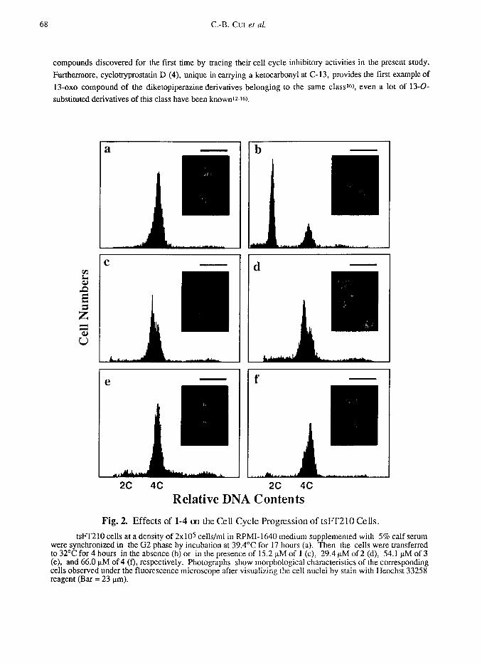
Typical flow cytometric histograms for l-4 are given in Fig. 2 and morphological observations of the
corresponding cells are also given. Cyclotryprostatin A (1)-D (4) completely inhibited the cell cycle progression
of tsFT210 cells in the G2/M phase at concentrations over 15.2 uM for 1, 29.4 pM for 2, 54.1 FM for 3 and
66.0 uM for 4, respectively, like as shown in Fig. 2. Half maximal inhibitory concentrations (ICsa’s) of l-4
were 5.6 uM for 1, 19.5 uM for 2, 23.4 uM for 3 and 25.3 uM for 4, respectively.
DISCUSSIONS
The present work has provided four new natural diketopiperazine derivatives, cyclotryprostatins A (1)-D
(4), as new G2/M phase inhibitors of the mammalian cell cycle.
Cyclotryprostatins A (1)-D (4) have a pentacyclic ring skeleton involved a 2,5_diketopiperazine ring,
which is composed from a tryptophan unit, a proline residue and an isoprenyl group. Up to date, it is the first
time to isolate l-4 from a natural source even many natural diketopiperazine derivatives belonging to the same
class, such as fumitremorgins, verruculogen and its derivatives, have been reportedtz-Is), and 2 and 4 are new
68 C.-B. GUI et al.
compounds discovered for the first time by tracing their cell cycle inhibitory activities in the present study.
Furthermore, cyclotryprostatin D (4), unique in carrying a ketocarbonyl at C-13, provides the first example of
13-0~0 compound of the diketopiperazine derivatives belonging to the same classtQ, even a lot of 13-o-
substituted derivatives of this class have been knownia-16).
a
e
b
Relative DNA Contents
Fig. 2. Effects of l-4 on the Cell Cycle Progression of tslT210 Cells.
tsPT210 cells at a density of 2x10⁵ cells/ml in RPMI-1640 medium supplemented with 5% calf serum were synchronized in the G2 phase by incubation at 39.4”C for 17 hours (a). Then the cells were transferred to 32°C for 4 hours in the absence (b) or in the presence of 15.2 nM of 1 (c), 29.4 nM of 2 (d), 54.1 uM of 3 (e), and 66.0 uM of4 (f), respectively. Photographs show morphological characteristics of the corresponding cells observed under the fluorescence microscope after visualizing the cell nuclei by stain with Heochst 33258 reagent (Bar = 23 urn).
Cyclotryprostatins A-D 69
On the other hand, 1 and its cis- and [runs-12,13-isomers as well as 3 and its two rrans-12,13-isomers
were prepared by Kodato er nl during the synthesis of fumitremorgin Bt6). But the data reported for the title
compounds (UV, EEMS and iH NMR for 1; UV, IR and tH NMR for 3) and their isomers are not enough to
simply identify 1 and 3 by a direct comparison with those of natural compounds. However, the limited data in
the literatureW, especially the tH NMR data, are well agreed with those reported in this paper, evidencing the
stereochemistry deduced for 1 and 3 and thus also for 2 and 4 in the present study.
To the cell cycle progression of tsFT210 cells, l-4 showed an inhibitory activity with the ICsu values in the
same micromolar order as that for tryprostatins A (78.8 uM)4) and B (18.8 uM)4), fumitremorgin C (14.0 PM)+,
demethoxyfumitremorgin C (1.78 uM)4) and spirotryprostatin B (14.0 uM)7). Previously we have reported4.7)
that the methoxy group on the benzene ring in tryprostatin A, spirotryprostatin A and fumitremorgin C
negatively dominated the cell cycle inhibitory activity for those compounds, compared with their demethoxy
derivatives, tryprostatin B, spirotryprostatin B and demethoxyfumitremorgin C, respectively. The same is true
that cyclotryprostatin C (3) showed stronger inhibitory activity (I&=23.4 PM) than that (I& > 243 t~M)4) of
its 18-methoxy derivative 12a,13a-dihydroxyfumitremorgin C (DHFT-C). Cyclotryprostatin A (1) is a 12-
epimer of DHFT-C, but its inhibitory effect (5.6 uM) was dramatically enhanced than that of DHFT-C (I&a >
243 p.M) and was also stronger than that (ICsa=23.4 t&l) of 3 which is less the methoxy at C-18 and different in
the stereochemistry at C-12 than those of 1 (Chart 1). On the other hand, a little change was observed in the
inhibitory effects between 3 (23.4 uM) and 4 (25.3 KM), the structures of which were different only at the C-13
position (Chart 1). Those observations suggested an important role of the stereochemistry at C-12 in l-4, which
resulted in the conformational changes of the central and the diketopiperazine rings in l-4, in the inhibitory
effects of those compounds on the cell cycle progression of mammalian cells at the G2/M phase. Also, the
above results showed that the negative effect of methoxy group on the benzene ring in the related compounds
having the pentacyclic ring skeleton should be limited in those with the 12a-proton or 12a-hydroxyl group.
In morphological observations, most portion of the tsFT210 cells treated by 1 and 2 showed
morphological characteristics of the condensed chromosomes (Fig. 2, c and d), which are typical of the M phase
cells, and a little portion appeared with morphological characteristics of the G2 phase cells (Data could not
shown on the photographs in Fig. 2 for 1 and 2). In contrast, the tsFT210 cells treated by 3 and 4 showed both
M and G2 phase morphological characteristics (Fig 2, e and l), but the G2 phase cells occupied the most portion
in those cells. This means that the tsFI’210 cells treated by 1-4 are trapped both in G2 and M phases, but the
main inhibitory points for 1-4 are little different, the M phase for 1 and 2 and the G2 phase for 3 and 4,
respectively. Incidently, the portion of G2 cells are increased dose-dependently in the tsFT210 cells treated by
l-4, especially in the cells treated by 3 and 4 (data not shown).
Detailed studies on their action mechanisms are currently being undertaken,
EXPERIMENTAL SECTION
General Instrumental Analyses
Melting point was measured using a Yanagimoto micro melting point apparatus and were uncorrected.
Optical rotations were determined in CHCla solutions on a JASCO DIP-370 polarimeter. UV spectra were taken
with a Hitachi 220A spectrophotometer in MeOH solutions and IR spectra were recorded on a Shimadzu FTIR-
8100M Fourier transform infrared spectrophotometer in KBr discs. EI-MS (ionization voltage, 70 eV,
70 C-B. GUI et al.
accelerating voltage, 3kV) and HR-EI-MS were measured respectively on Hitachi M-80A and Hitachi M-80
mass spectrometers using a direct inlet system. 1H and 13C NMR spectra were taken on a JEOL GSX-500 or a-
400 spectrometer with tetramethylsilane as an internal standard and chemical shifts are recorded in 6 values.
Multiplicities of 13C NMR signals were determined by the DEPT method and are indicated as s (singlet), d
(doublet), t (triplet) and q (quartet). 2D NMR spectra (IH-IH or PFG IH-IH COSY, PFG-HMQC and PFG-
HMBC spectra) were measured on a JEOL GSX-500 or a-400 spectrometer by the use of JEOL standard pulse
sequences and collected data were processed by JEOL standard software. Difference NOE spectra were obtained
by the use of a JEOL standard pulse sequence with irradiation for 5 seconds.
Conditions for Isolation of the Cyclotryprostatins
TLC was done on pre-coated silica gel 60 FZs4 plates (0.25 mm thick, 20 x 20 cm, Merck) and the spots
were detected under UV lights (254 and 365 nm) or by the use of 10% aqueous sulfuric acid reagent. Silica gel
60 (230-400 mesh, Merck) was used for column chromatography.
Analytical HPLC was carried out on a reversed phase column (CAPCELL PAK C-18 or C-8,4.6 x 250
mm, Shiseido Co., Japan) by the use of a HPLC equipment with a Hitachi L-6000 pump and a Waters 991J
photodiode array detector system under a flow rate of 1 ml/min. Preparative HPLC was performed on a HPLC
system equipped with a Hitachi L-6000 pump and a SSC UV detector under a flow rate of 10 ml/min.
CAPCELL PAK C-18 and C-8 columns (20 x 250 mm, Shiseido) were used in the preparative HPLC.
Fermentation of the Producing Fungal Strain
The producing fungal strain, Aspergillus fumigatus BM939, on a potato dextrose agar slant was inoculated
into a 500-ml cylindrical flask containing 100 ml of the seed medium consisting of glucose 3%, soluble starch
2%, soybean meal 2%, KzHP040.5%, MgS04 - 7H20 0.05% (adjusted to pH 6.5 prior to sterilization) and
cultured at 28’C for 27 hours on a rotary shaker at 300 rpm. The culture was transferred into 18 liters of the
seed medium with 0.05% of antifoam reagents CA- 123 and KM-68, respectively, in a 30-liter jar fermenter.
Further fermentation for seed culture was carried out at 28°C for 24 hours with an agitation rate of 350 rpm and
an aeration rate of 9 liters/minute.
Then the producing fermentation was carried out in a 600.liter jar fermenter containing 400 liters of the
production medium having the same composition of the seed medium with 0.05% of antifoam reagents CA-123
and KM-68, respectively. The seed culture (18 L) was transferred into the 600-liter fermenter and the
fermentation was performed at 28’C for 66 hours with an agitation rate of 350 ‘pm and an aeration rate of 200
liters/min.
Separation of the Fermentation Broth
The whole broth was filtrated to separate into a broth supernatant (370 L) and a mycelial cake. The former
was extracted once with 400 L of EtOAc to give an EtOAc solution and the latter was extracted with 90%
aqueous acetone (400 L x 1 time) which was evaporated in vacua to remove acetone to give an aqueous solution
(60 L). The aqueous solution was extracted with EtOAc (120 L x 2 times) to afford 240 L of EtOAc solution.
Both the EtOAc solutions obtained were combined and concentrated in vacua to give an oily extract (1.2 L). To
the oily exmact, 1.2 L of n-hexane was added, and the extract was suspended by stirring followed by sonication
for 10 minutes and then the n-hexane-insoluble part was obtained by centrifugation at 6000 ‘pm for 20 minutes.
The n-hexane-insoluble part was treated two more times with n-hexane (1.2 L x 2) by the same manner to
remove nonpolar oil fraction. To the n-hexane-insoluble part obtained, CHC13 (1.2 L) was added, sonicated for
Cyclotryprostatins A-D 71
10 minutes and then centrifuged for 20 minutes at 6000 ‘pm to obtain a CHCls solution. The CHCls-insoluble
part was extracted one more time with CHCls (1.2 L) by the same manner, and both the CHCls-solutions
obtained were combined and concentrated under reduced pressure to afford a CHCls-soluble syrup. This was
treated once more with n-hexane (1.2 L) as described above to give a n-hexane-insoluble part which was
partitioned between CHCls-H20. The CHCls solution obtained was concentrated under reduced pressure to give
an active CHCls extract (66 g) as powders.
The CHCla extract (66 g) was dissolved in 250 ml of n-hexane-CHCla (2O:SO) solution and subjected to a
silica gel column packed in n-hexane (silica gel, 1500 g; bed volume, 7.5 x 75 cm; retention volume, 3 L). The
column was then eluted successively with n-hexane-CHCls 15050 (7.5 L)- 25:75 (13.7 L) + lo:90 (9.5 L)]
solution, CHCls (1.2 L) and CHCls-MeOH [99.5:0.5 (9 L)+ 99:l (1.2 L)- 98:2 (6 L)- 96:4 (9 L)+ 9O:lO (9
L)- SO:20 (10 L) - 70:30 (6 L)] solution, respectively. After elution with 7.5 L of n-hexane-CHCls (50:50)
solution which was collected in a portion, the eluate was collected in each 3 L portion, monitored by TLC,
combined and concentrated in vacua to give twenty five fractions [Fr.l, n-hexane-CHCls (50:50) eluate; Fr.2-
Fr.4, n-hexane-CHCls (25:75) eluate; Fr.5.Fr.6, n-hexane-CHCIs (10:90) eluate; Fr.7, n-hexane-CHCl,
(10:90) & CHCla eluate; Fr.8.Fr.10, CHC& eluate; Fr.11, CHC& & CHCla-MeOH (99.5:0.5) eluate;
Fr.12-Fr.13, CHCls-MeOH (99.5:0.5) eluate; Fr.14, CHCI,-MeOH (99.5:0.5 & 99:l) eluate; Fr.15
Fr.17, CHCls-MeOH (99:l) eluate; Fr.18.Fr.20, CHQ-MeOH (98:2) eluate; Fr.21-Fr.22, CHCls-MeOH
(96:4) eluate; Fr.23, CHCis-MeOH (96:4 & 9O:lO) eluate; Fr.24 CHCls-MeOH (9O:lO) eluate; Fr.25 CHCls-
MeOH (SO:20 &70:30) eluate].
Isolation of Cyclotryprostatins A (1)-D (4)
The fraction Fr.7 (0.76 g) eluted by n-hexane-CHCl:, (10:90) and CHCls solutions from the silica gel
column was treated with MeOH to give a MeOH soluble part which was separated by preparative HPLC on a
CAPCELL PAK C-18 column using MeOH-H20 (60:40) as eluting solvent (detector wave length, 210 nm) to
give an active fraction fr.5 (Rt=13 mitt, 32 mg). The fraction fr.5 was further subjected to a HPLC separation
on a CAPCELL PAK C-8 column using MeOH-Hz0 (40:60) eluting solution (detector wave length, 210 nm) to
give cyclotryprostatin A (1, Rt=19.6 min, 1.0 mg) and cyclotryprostatin B (2, Rt=65.6 min, 4.4 mg),
respectively, both as pale yellow crystalline forms from MeOH solutions.
The fraction Fr.14 (0.99 g) eluted by CHCls-MeOH (99.5:0.5 and 99:l) solvents from the silica gel
column was separated by preparative HPLC on a CAPELL PAK C-18 column using MeOH-Hz0 (60:40) as
eluting solvent (detector wave length, 220 nm) to give cyclotryprostatin C (3, Rt=38.5 min; 12.4 mg) as
white needles from MeOH and crude cyclotryprostatin D (4, Rt=14.2 min) which was further purified by
repeated HPLC under the same condition to give 1.2 mg of pure 4 as a pale yellow amorphous powder from a
MeOH solution.
Bioassay for Cell Cycle Inhibitory Activity
The tsFT210 cells were routinely maintained at 32°C in RPMI-1640 medium supplemented with 5% calf
serum (HyClone Inc., Logan, UT, USA) in the presence of 30 &ml of penicillin and 42 &ml of streptomycin
under a humidified atmosphere of 5% CO2 and 95% air. The cells were arrested in the G2 phase by incubation
at 39.4“C for 17 hours and the cells were allowed to pass through M phase to enter into Cl phase by incubation
at 32°C for 4 hours after treatment with the samples. Then distribution of the cells within cell cycle were
determined by flow cytometry as we have previously reportedt.4).
72 C.-B. GUI et al.
ACKNOWLEDGEMENTS
We thank Ms. Tamiko Chijimatsu (RIKEN) for the measurements of PFG-HMBC spectra. This research
work was supported in part by a Grant for “Biodesign Research Program” from RIKEN (C.-B. Cui and H.
Osada) and a Grant from Ministry of Education, Japan.
1.
2.
3.
4.
5.
6.
7.
8.
9
10.
11.
12.
13.
14.
15.
16.
REFERENCES AND NOTES
Osada, H.; Cui, C.-B.; Onose, R.; Hanaoka, F. Bioorg. Med. Chem., 1996, in press.
Cui, C.-B.; Kakeya, H.; Okada, G.; Onose, R.; Ubukata, M.; Takahashi, I.; Isono, K.; Osada, H. J.
Antibiot., 1995,48, 1382-1384.
Cui, C.-B.; Ubukata, M.; Kakeya, H.; Onose, R.; Okada, G.; Takahashi, I.; Isono, K.; Osada, H. J.
Antibiof., 1996,49, 216-219.
Cui, C.-B.; Kakeya, H.; Okada, G.; Onose, R.; Osada, H. J. Antibiot., 1996,49, 527-533.
Cui, C.-B.; Kakeya, H.; Osada, H. J. Antibiot., 1996,49, 534-540.
Cui, C.-B.; Kakeya, H.; Osada, H. J. Antibiot., 1996,49, 832-835.
Cui, C.-B.; Kakeya, H.; Osada, H. Tetrahedron, 1996,52, 12651-12666.
In the UV spectra, both the 6-O-methylindole and the indole chromophores show a strong absorption in
the 220-230 nm region. While in the 250-320 nm region, two major absorption bands like the humps of a
camel are typical of the 6-O-methylindole chromophore and the appearance of a major one in the same
region is characteristic for the indole chromophore, respectively (cfi reference 2 and see Cole, R. J.; Cox,
R. H. Handbook ofToxic FungalMeraholites, Academic Press Inc.: New York, 1981; pp. 357-383 for
the 60methylindole chromophore, pp. 390 and pp. 453-462 for the indole chromophore).
Fayos, J.; Lokensgard, D.; Clardy, J.; Cole, R. J.; Kirksey, J. W. J. Am. Chem. Sot., 1974, 96,
6785-6787.
Steyn. P. S. Tetrahedron, 1973,29, 107-120.
Steyn, P. S. Tetrahedron Lett., 1971, 3331-3334.
Steyn, P. S.; Vleggaar, R. Progress in the Chemistry of Organic Natural Products (Founded by
Zechmiester, L.; Edited by Herz, W.; Grisebach, H.; Kirby, G. W.), 1985,48, l-80.
Abaraham, W.-R.; Arfmann, H.-A. Phytochem., 1990.29, 1025-1026.
Yamazaki, M.; Fujimoto, H.; kawasaki, T. Chem. Pharm. Bull., 1980,28, 245-254.
Cole, R. J.; Cox, R. H. Handbook of Toxic Fungal Metabolites, Academic Press Inc.: New York, 1981;
pp. 355-385.
Kodato et al have recorded that in a mixture of the synthetic mediates for fumitremorgin B, the presence of
a 13-ketocarbonyl compound having the same structural skeleton as that in 4 was revealed by the UV
absorption at 294.5, 269.5 and 308 nm, corresponding to the 3-acylindole chromophore, in the UV
spectrum of the mixture in an EtOH solution. But they did not isolate the compound in a pure form.
Kodato, S.; Nakagawa, M.; Hongu, M.; Kawata, T.; Hino, T. Tetrahedron, 1988,44, 359-377.
(Received in Japan 26 August 1996; accepted 2 I October 1996)