Embed Size (px)
Citation preview
Experimental Parasitology 126 (2010) 69–72
Contents lists available at ScienceDirect
Experimental Parasitology
journal homepage: www.elsevier .com/locate /yexpr
Novel in vitro and in vivo models to study central nervous system infectionsdue to Acanthamoeba spp.
Naveed Ahmed Khan *
School of Veterinary Medicine and Science, University of Nottingham, Sutton Bonington, LE12 5RD England, UK
a r t i c l e i n f o a b s t r a c t
Article history:Received 20 August 2009Accepted 27 August 2009Available online 31 August 2009
Keywords:AcanthamoebaBlood–brain barrierIn vivo modelsPathogenesis
0014-4894/$ - see front matter � 2009 Elsevier Inc. Adoi:10.1016/j.exppara.2009.08.018
* Fax: +44 1159516440.E-mail address: [email protected]
Acanthamoeba granulomatous encephalitis is a serious human infection with fatal consequences. Themost distressing aspect of Acanthamoeba granulomatous encephalitis is the limited improvement in mor-tality. The underlying neurobiology is at present not well understood and treatment options are often oflimited efficacy. There is therefore a real need to obtain more knowledge regarding the pathogenesis andpathophysiology of Acanthamoeba granulomatous encephalitis and to develop new chemotherapeuticapproaches. However, the difficulties in using mammalian models to study this infection have hinderedour search for therapeutic interventions. Recent availability of the blood–brain barrier, in vitro and use oflocust as an in vivo model will undoubtedly allow us to investigate disease pathogenesis, mechanisms ofparasite traversal across the blood–brain barrier and new drug therapies. It is argued that the modelsdescribed here can offer several advantages in terms of speed, cost, technical convenience, and ethicalacceptance. Furthermore, they are extremely valuable tools to discriminate molecules participating fromboth sides of the host–parasite interaction and will generate potentially useful leads in the identificationof new potential drugs, as well as testing drug toxicity.
� 2009 Elsevier Inc. All rights reserved.
1. Introduction
Central nervous system infection due to Acanthamoeba almostalways proves fatal. It is thought to be more deadly than other para-sitic infections such as malaria because death is inevitable if a patientis untreated. The terminal stages of Acanthamoeba encephalitis arecharacterized by hemiparesis, cranial nerve palsies, increased intra-cranial pressure, focal neurological symptoms, and seizures, due tohemorrhagic necrotizing lesions with severe meningeal irritationand encephalitis (Carter et al., 1981; Martinez et al., 1980; Martinez,1985; Martinez and Visvesvara, 1991). Even with advances in anti-microbial chemotherapy and supportive care, the prognosis forAcanthamoeba encephalitis is dismal, with a mortality rate of morethan 95%. In part, this is due to our incomplete understanding ofthe pathogenesis and pathophysiology of Acanthamoeba encephali-tis, as well as difficulties in using mammalian models to study thisinfection. A key step in Acanthamoeba encephalitis is parasite inva-sion of the central nervous system (CNS), followed by neurologicdegeneration with psychiatric disorders and death. Despite theimportance of this step, the underlying pathogenic mechanisms bywhich Acanthamoeba crosses the blood–brain barrier to invade theCNS are not clearly understood. Here, I describe novel in vitro and
ll rights reserved.
in vivo models that are available to study mechanisms of Acantha-moeba traversal of the blood–brain barrier.
2. In vitro model of the blood–brain barrier
In vitro models of the blood–brain barrier have clearly becomeimportant tools for identifying the cellular and molecular elementsthat may be possible targets for interventions for the transmigra-tion of many pathogens into the CNS (Kim, 2008). The blood–brainbarrier is composed of tightly apposed brain microvascular endo-thelial cells held together by tight junction proteins, which makesit a highly selective barrier compared with the peripheral endothe-lium (Miller, 1999). The spaces that occur between endothelialcells elsewhere (except testes) in the body are not found at theblood–brain barrier sites. The tight junction proteins are primarilyresponsible for the barrier function, preventing entry of blood con-tents into the CNS (Miller, 1999). Notably, endothelial cells of theblood–brain barrier exhibit high trans-endothelial electrical resis-tance, TEER (�2000 ohm/cm2) (Pardridge, 1999), while endothelialcells from human placenta exhibit less than 50 ohm/cm2 (Jingaet al., 2000). The high selectivity ensures that even small moleculessuch as dyes and antibiotics are prevented from entry into the CNS,while non-brain endothelium is more permissive. The availabilityof in vitro cultures of primary human brain microvascular endothe-lial cells has been of tremendous value in the study of host–para-site interactions (Stins et al., 1997; Alsam et al., 2003). Primary
70 N.A. Khan / Experimental Parasitology 126 (2010) 69–72
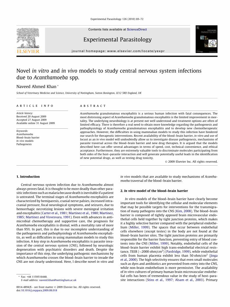
brain microvascular endothelial cells (BMEC) are isolated, charac-terized, and purified from the cerebral cortex (Stins et al., 1997).The human BMEC used in our laboratory are positive for brainendothelial markers such as factor VIII, carbonic anhydrase, UlexEuropaeus Agglutinin I, gamma-glutamyl transpeptidase, and tookup acetylated low-density lipoprotein. In addition, these cells dem-onstrated the expression of the tight junction proteins and the for-mation of a polar monolayer. The human BMEC are routinelygrown on rat-tail collagen-coated dishes in complete medium[RPMI 1640 containing 10% heat-inactivated fetal bovine serum,10% Nu-serum, 2 mM glutamine, 1 mM pyruvate, penicillin(100 U/ml), streptomycin (100 lg/ml), non-essential amino acidsand vitamins] (Invitrogen, Paisley, England) (Stins et al., 1997;Alsam et al., 2003). The in vitro model is created by cultivating hu-man BMEC in the upper chamber of collagen-coated Transwellspolycarbonate tissue culture inserts (Corning Costar, Corning Ltd.,Hemel Hempstead, England) (Fig. 1), and grown for at least 2 daysin BMEC culture media. Next, the media are replaced with freshculture media containing 500 nM hydrocortisone and cultured foran additional 3–6 days. Culture medium containing hydrocortisoneis changed every 2 days and confluency determined by lightmicroscopy. Under these conditions, the BMEC monolayer exhibitstight junction formation and polarization and develops TEER ofmore than 200 ohm/cm2 using a tissue-resistant measurementchamber and a voltohmmeter (EVOM, World Precision Instru-ments). To determine the effects of amoebae or conditioned med-ium on BMEC monolayer integrity, parasites can be added in theupper chamber and TEER changes can be monitored. In addition,to investigate the ability of amoebae to cross the human BMECmonolayer, parasites can be added to the upper chamber and al-lowed to cross the monolayer for various intervals of time. Subse-quently, samples are taken from the bottom chamber and thenumber of amoebae can simply be determined by haemocytometercounting. This model can be used to study molecular mechanismsof (i) cytoadhesion, (ii) cytotoxicity, (iii) junctional protein degra-dation leading to paracellular transmigration, and (iv) traversalacross the blood–brain barrier as live amoebae. As well as studyingdisease pathogenesis, this is a potentially useful model to test no-vel drug therapies, in vitro. In addition, purified human BMEC mon-olayers can be used to identify molecular determinants involved inparasite–host cell interactions leading to the blood–brain barriertraversal.
3. In vivo models of Acanthamoeba encephalitis
A whole-organism approach to the study of disease is essentialin gaining a full understanding of the interrelationships betweeninfectious agents and their hosts. Animal models that have beenused for investigating Acanthamoeba encephalitis include mice,rats and non-human primates such as monkeys (El-Dein et al.,1998; Cabral and Marciano-Cabral, 2004; Cerva, 1967; Culbertson
Upper chamber
Lower chamber
Fig. 1. In vitro model of the blood–brain barrier. The polarized in vitro model is formedtranswell inserts. Amoebae can be added in the upper chamber (blood-side). After variousdetermine the number of amoebae that traverse the BMEC monolayer.
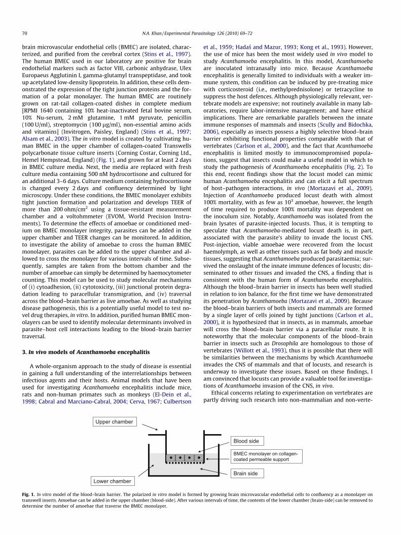
et al., 1959; Hadas and Mazur, 1993; Kong et al., 1993). However,the use of mice has been the most widely used in vivo model tostudy Acanthamoeba encephalitis. In this model, Acanthamoebaare inoculated intranasally into mice. Because Acanthamoebaencephalitis is generally limited to individuals with a weaker im-mune system, this condition can be induced by pre-treating micewith corticosteroid (i.e., methylprednisolone) or tetracycline tosuppress the host defences. Although physiologically relevant, ver-tebrate models are expensive; not routinely available in many lab-oratories, require labor-intensive management; and have ethicalimplications. There are remarkable parallels between the innateimmune responses of mammals and insects (Scully and Bidochka,2006), especially as insects possess a highly selective blood–brainbarrier exhibiting functional properties comparable with that ofvertebrates (Carlson et al., 2000), and the fact that Acanthamoebaencephalitis is limited mostly to immunocompromised popula-tions, suggest that insects could make a useful model in which tostudy the pathogenesis of Acanthamoeba encephalitis (Fig. 2). Tothis end, recent findings show that the locust model can mimichuman Acanthamoeba encephalitis and can elicit a full spectrumof host–pathogen interactions, in vivo (Mortazavi et al., 2009).Injection of Acanthamoeba produced locust death with almost100% mortality, with as few as 103 amoebae, however, the lengthof time required to produce 100% mortality was dependent onthe inoculum size. Notably, Acanthamoeba was isolated from thebrain lysates of parasite-injected locusts. Thus, it is tempting tospeculate that Acanthamoeba-mediated locust death is, in part,associated with the parasite’s ability to invade the locust CNS.Post-injection, viable amoebae were recovered from the locusthaemolymph, as well as other tissues such as fat body and muscletissues, suggesting that Acanthamoeba produced parasitaemia; sur-vived the onslaught of the innate immune defences of locusts; dis-seminated to other tissues and invaded the CNS, a finding that isconsistent with the human form of Acanthamoeba encephalitis.Although the blood–brain barrier in insects has been well studiedin relation to ion balance, for the first time we have demonstratedits penetration by Acanthamoeba (Mortazavi et al., 2009). Becausethe blood–brain barriers of both insects and mammals are formedby a single layer of cells joined by tight junctions (Carlson et al.,2000), it is hypothesized that in insects, as in mammals, amoebaewill cross the blood–brain barrier via a paracellular route. It isnoteworthy that the molecular components of the blood–brainbarrier in insects such as Drosophila are homologous to those ofvertebrates (Willott et al., 1993), thus it is possible that there willbe similarities between the mechanisms by which Acanthamoebainvades the CNS of mammals and that of locusts, and research isunderway to investigate these issues. Based on these findings, Iam convinced that locusts can provide a valuable tool for investiga-tions of Acanthamoeba invasion of the CNS, in vivo.
Ethical concerns relating to experimentation on vertebrates arepartly driving such research into non-mammalian and non-verte-
BMEC monolayer on collagen-coated permeable support
Blood side
Brain side
by growing brain microvascular endothelial cells to confluency as a monolayer onintervals of time, the contents of the lower chamber (brain-side) can be removed to
Amoebae can be injectedinto the abdomen (up to20µl can be injected) A puncture can be made
using a capillary tube to lookat the presence of pathogenin blood (haemolymph)
Locusta migratoria (up to50 locusts can behoused a single cage)
Parasitemia
InjectionLocust as anin vivo model
Brain dissection
Locust brain shown by black arrowcan be removed using a fine forceps
Isolated brains can betested for the presenceof pathogens
Brain invasionB
A
Fig. 2. Use of locusts as an in vivo model to study Acanthamoeba granulomatous encephalitis. (A) Infection can be studied in the blood (i.e., haemolymph) and (B) brains can beisolated to study the involvement of the central nervous system.
N.A. Khan / Experimental Parasitology 126 (2010) 69–72 71
brate models, but such models also offer significant advantages ofspeed and lower cost (especially in view of the current economicclimate), infrastructure, and legislative restrictions. The proposedin vitro model of the blood–brain barrier and in vivo locust modelis a timely response to the expressed desires of the public and gov-ernments to replace and reduce the use of animals in research,especially of mammals. In addition, locust models will encourageother scientists to test insect models for studying the early stagesof disease pathogenesis in vivo.
In summary, we believe that such technically convenient mod-els can be used for the initial screening and identification of novelvirulence factors, providing useful leads for the rational develop-ment and evaluation of therapeutic interventions, and strengthenthe move away from a total dependency on vertebrate models. Thispaper should encourage other scientists to consider using com-monly available insects such as locusts as tractable models forstudying disease pathogenesis, in vivo.
Acknowledgments
The authors are grateful to Mary Lightfoot, Graham Goldswor-thy and Parasia Mortazavi (Birkbeck, University of London) fortheir sincere help and kind support in developing locust model,and to Monique Stins (Johns Hopkins University) for her help indeveloping the blood–brain barrier model.
References
Alsam, S., Kim, K.S., Stins, M., Rivas, A.O., Sissons, J., Khan, N.A., 2003. Acanthamoebainteractions with human brain microvascular endothelial cells. MicrobialPathogenesis 35, 235–241.
Cabral, G.A., Marciano-Cabral, F., 2004. Cannabinoid-mediated exacerbation of braininfection by opportunistic amebae. Journal of Neuroimmunology 147, 127–130.
Carlson, S.D., Juang, J.L., Hilgers, S.L., Garment, M.B., 2000. Blood barriers of theinsect. Annual Reviews Entomology 45, 151–174.
72 N.A. Khan / Experimental Parasitology 126 (2010) 69–72
Carter, R.F., Cullity, G.J., Ojeda, V.J., Silberstein, P., Willaert, E., 1981. A fata case ofmeningoencephalitis due to free-living amoeba of uncertain identity-probablyAcanthamoeba sp. Pathology 13, 51–68.
Cerva, L., 1967. Intranasal, intrapulmonary and intracardial inoculation of experimentalanimals with Hartmanella castellanii. Folia Parasitologica 14, 207–215.
Culbertson, C.G., Smith, J.W., Cohen, H.K., Minner, J.R., 1959. Experimental infection ofmice and monkeys by Acanthamoeba. American Journal of Pathology 35, 185–197.
El-Dein, S.Z., Khalifa, A.M., Sadaka, H.A., Hegazy, I.H., Ibrahim, H.S., 1998.Electroencephalographic changes in rats received antigens of differentparasites. Journal of Egyptian Society of Parasitology 28, 797–805.
Hadas, E., Mazur, T., 1993. Biochemical markers of pathogenicity and virulence ofAcanthamoeba sp. strains. Parasitology Research 79, 696–698.
Jinga, V.V., Gafencu, A., Antohe, F., Constantinescu, E., Heltianu, C., Raicu, M.,Manolescu, I., Hunziker, W., Simionescu, M., 2000. Establishment of a purevascular endothelial cell line from human placenta. Placenta 21, 325–336.
Kim, K.S., 2008. Mechanisms of microbial traversal of the blood–brain barrier.Nature Reviews Microbiology 6, 625–634.
Kong, H.H., Seo, S.A., Shin, C.O., Im, K.I., 1993. The effect of active immunization withAcanthamoeba culbertsoni in mice born to immune mother. Korean Journal ofParasitology 31, 157–163.
Martinez, A.J., Garcia, C.A., Halks-Miller, M., Arce-Vela, R., 1980. Granulomatousamebic encephalitis presenting as a cerebral mass lesion. Acta Neuropathol-ogica 51, 85–91.
Martinez, A.J., 1985. Free-Living Amebas: Natural History, Prevention,Diagnosis, Pathology and Treatment of Disease. CRC Press, Boca Raton,Florida, p. 156.
Martinez, A.J., Visvesvara, G.S., 1991. Laboratory diagnosis of pathogenic free-livingamoebas: Naegleria, Acanthamoeba, and Leptomyxid. Clinical LaboratoryMedicine 11, 861–872.
Miller, D.W., 1999. Immunobiology of the blood–brain barrier. Journal ofNeurovirology 5, 570–578.
Mortazavi, P.N., Goldsworthy, G., Kirk, R., Khan, N.A., 2009. Novel model for thein vivo study of central nervous system infection due to Acanthamoeba spp. (T4genotype). Journal of Medical Microbiology 58, 503–508.
Pardridge, W.M., 1999. Blood–brain barrier: biology and methodology. Journal ofNeurovirology 5, 556–569.
Scully, L.R., Bidochka, M.J., 2006. Developing insect models for the study ofcurrent and emerging human pathogens. FEMS Microbiology Letters 263,1–9.
Stins, M.F., Gilles, F., Kim, K.S., 1997. Selective expression of adhesion molecules onhuman brain microvascular endothelial cells. Journal of Neuroimmunology 76,81–90.
Willott, E., Balda, M.S., Fanning, A.S., Jameson, B., Van Itallie, C., Anderson, J.M., 1993.The tight junction protein ZO-1 is homologous to the Drosophila discs-largetumor suppressor protein of septate junctions. Proceedings of the NationalAcademy of Sciences USA 90, 7834–7838.