Embed Size (px)
Citation preview
Schizophrenia Research 118 (2010) 248–255
Contents lists available at ScienceDirect
Schizophrenia Research
j ourna l homepage: www.e lsev ie r.com/ locate /schres
Novel immune response to gluten in individuals with schizophrenia
Diana Samaroo a, Faith Dickerson b, Donald D. Kasarda c, Peter H.R. Green d, Chiara Briani e,Robert H. Yolken f, Armin Alaedini a,⁎a Department of Neurology and Neuroscience, Weill Medical College of Cornell University, New York, NY, United Statesb Sheppard Pratt Health System, Baltimore, MD, United Statesc Western Regional Research Center, U.S. Department of Agriculture, Albany, CA, United Statesd Department of Medicine, College of Physicians and Surgeons, Columbia University, New York, NY, United Statese Department of Neurosciences, University of Padova, Padova, Italyf The Stanley Laboratory of Developmental Neurovirology, Department of Pediatrics, Johns Hopkins University Medical Center, Baltimore, MD, United States
a r t i c l e i n f o
⁎ Corresponding author. 1300 York Ave., LC-819,United States.
E-mail address: [email protected] (A. Alae
0920-9964/$ – see front matter © 2009 Elsevier B.V.doi:10.1016/j.schres.2009.08.009
a b s t r a c t
Article history:Received 8 July 2009Received in revised form 7 August 2009Accepted 11 August 2009Available online 11 September 2009
A link between celiac disease and schizophrenia has been postulated for several years, basedprimarily on reports of elevated levels of antibody to gliadin in patients. We sought to examinethe proposed connection between schizophrenia and celiac disease by characterizing themolecular specificity and mechanism of the anti-gliadin immune response in a subset ofindividuals with schizophrenia. Blood samples from individuals with schizophrenia andelevated anti-gliadin antibody titer were examined for celiac disease-associated biomarkers,including antibodies to transglutaminase 2 (TG2) enzyme and deamidated gliadin peptides, aswell as the HLA-DQ2 and -DQ8 MHC genes. The anti-gliadin antibody response was furthercharacterized through examination of reactivity towards chromatographically separatedgluten proteins. Target proteins of interest were identified by peptide mass mapping. Incontrast to celiac disease patients, an association between the anti-gliadin immune responseand anti-TG2 antibody or HLA-DQ2 and -DQ8 markers was not found in individuals withschizophrenia. In addition, the majority of individuals with schizophrenia and anti-gliadinantibody did not exhibit antibody reactivity to deamidated gliadin peptides. Furthercharacterization of the antibody specificity revealed preferential reactivity towards differentgluten proteins in the schizophrenia and celiac disease groups. These findings indicate that theanti-gliadin immune response in schizophrenia has a different antigenic specificity from that inceliac disease and is independent of the action of transglutaminase enzyme and HLA-DQ2/DQ8.Meanwhile, the presence of elevated levels of antibodies to specific gluten proteins points toshared immunologic abnormalities in a subset of schizophrenia patients. Furthercharacterization and understanding of the immune response to gluten in schizophrenia mayprovide novel insights into the etiopathogenesis of specific disease phenotypes.
© 2009 Elsevier B.V. All rights reserved.
Keywords:GlutenGliadinSchizophreniaGluten sensitivityCeliac diseaseAntibody
1. Introduction
There is increasing evidence for the involvement of immu-nologic factors in schizophrenia (Patterson, 2008; Torrey andYolken, 2001). Among the various reported immunologicabnormalities, increased immune sensitivity to gluten and
New York, NY 10065
dini).
All rights reserved.
,
association with celiac disease have been described in severalreports (Bender, 1953; Cascella et al., 2009; Dohan et al., 1972;Graff andHandford, 1961; Jin et al., 2008;Reichelt andLandmark,1995). Glutens, made up of two main fractions, gliadins andglutenins, are the main storage proteins of wheat and arecomprised of about 100 different proteins in a given wheatcultivar (variety). When the various wheat cultivars areconsidered, the number of different gluten proteins is evengreater, although almost all cultivars are made up of the samemain types, α-type (also known as α/β type), γ-type, ω-type,
249D. Samaroo et al. / Schizophrenia Research 118 (2010) 248–255
low-molecular-weight glutenin subunits, and high-molecular-weight glutenin subunits (Jabri et al., 2005). Gluten sensitivitymay be defined as a state of heightened immune response toingested gluten, the most common clinical manifestation ofwhich is celiac disease, an inflammatory enteropathy that ischaracterized by villous atrophy and lymphocytic infiltration inthe small intestine in genetically predisposed individuals(Alaedini and Green, 2005). Genetic susceptibility for celiacdisease is associated with close genetic linkage to class II humanleukocyte antigens (HLA) DQ2 (DQA1 *05/DQB1 02) and DQ8(DQA1 *0301/DQB1 *0302) that are involved in the presentationof gluten peptides to T cells (Louka and Sollid, 2003). Presence ofantibodies to gluten and to the enzyme transglutaminase 2(TG2) is a hallmark of celiac disease. The TG2 protein is animportant element of the disease, not only as a targetautoantigen, but also as an enzyme that enhances the immu-nostimulatory effect of gluten peptides through the deamidationand conversion of glutamines to glutamate residues (Briani et al.,2008). In fact, antibodies to selectively deamidated gluten arenowknown to bemore specific for celiac disease than antibodiesagainst the native species (Mothes, 2007; Naiyer et al., 2009).
The clinical presentation of celiac disease is highly variable,ranging from asymptomatic to severe malnutrition. In the nowcommonly diagnosed atypical form, gastrointestinal symptomsmay be absent or less pronounced, while patients present withextra-intestinal features such as anemia, osteoporosis, shortstature, and infertility (Alaedini and Green, 2005). Neurologicand psychiatric abnormalities have also been reported to beassociatedwith celiac disease in various studies (Bushara, 2005).In addition, elevated levels of anti-gliadin antibody have beenassociated with some of these deficits, even in the apparentabsence of the characteristic mucosal pathology (Burk et al.,2001; Bushara et al., 2001; Hadjivassiliou et al., 1998). Thecurrent literature indicates shared immunologic abnormalitiesand genetic associations between schizophrenia and celiacdisease and is suggestive of an elevated relative risk of non-affective psychosis in celiac patients (Jungerius et al., 2008;Kalaydjian et al., 2006; Wei and Hemmings, 2005). A number ofcase studies and small trials indicate significantpositive responseto gluten-free diet in some individuals with schizophrenia(Kalaydjianet al., 2006).However,methodological shortcomingsin some of these studies and contradictory findings by othergroups have led to a lack of consensus on the relevance of glutenin schizophrenia (Kalaydjian et al., 2006). Specifically, theprevalence of true celiac disease versus anti-gliadin antibodiesin patients with schizophrenia is a subject of contention. Inaddition, little is known about the specificity and mechanism ofthe immune response to gluten in these patients, or its role indisease pathogenesis. The aimof this studywas to investigate thelink between gluten sensitivity and schizophrenia by character-izing the immune response in patients and control subjects. Ourresults reveal an immunologic response to gluten in individualswith schizophrenia that is clearly different from that in celiacdisease.
2. Materials and methods
2.1. Subjects
Serum samples from 17 individuals with schizophrenia (9female, 8 male; mean age 37.6±3.0y), selected because of
previously documented elevated IgG and IgA antibodies togliadin (hereon referred to as gluten-sensitive individualswith schizophrenia), were studied. The initial screening ofpatients for antibodies to gliadin had been done using acommercially available ELISA kit (IBL International). Thecharacteristics of the population fromwhich these individualswere selected have been described previously (Dickersonet al., 2003). In addition, serum specimens from 25 biopsy-proven celiac disease patients with elevated IgG and IgAantibody to gliadin and 20 healthy subjects without elevatedlevels of anti-gliadin antibody were included in the study ascontrols. Diagnosis of celiac disease was made according topreviously described criteria (Alaedini and Green, 2005).
2.2. Animal immunization
New Zealand White rabbits (n=2) were immunizedusing a commercial gliadin extract (Sigma-Aldrich). Immu-nizations were carried out with injection of 200 μg of gliadinprotein in Complete Freund's Adjuvant on day 0, and 100 μgon days 14, 28, and 42. Serum was collected on day 77.
2.3. Anti-gliadin antibody detection
All serum samples were tested for anti-gliadin antibody byELISA as previously described (Alaedini et al., 2007). Briefly,96-well round-bottom polystyrene plates (BD Biosciences)were coated with 50 μL/well of a 0.01 mg/mL solution ofgliadin (Sigma-Aldrich) in 0.1 M carbonate buffer (pH 9.6).Control wells were coated only with buffer. After overnightincubation at 4 °C, all wells were washed and blocked byincubation with 1% BSA in PBS-T for 1.5 h at roomtemperature. Serum samples were diluted at 1:200 for IgAmeasurement and 1:800 for IgG measurement and added at50 μL/well in duplicates. After washing the wells, they wereincubated with peroxidase-conjugated goat anti-human IgG(Amersham Biosciences) or IgA (MP Biomedicals) secondaryantibody for 1 h. After washing thewells, developing solution,comprising 27 mM citric acid, 50 mM Na2HPO4, 5.5 mM o-phenylenediamine, and 0.01% H2O2 (pH 5), was added.Absorbance was measured at 450 nm after incubating theplates for 30 min. Absorbance values were corrected for non-specific binding by subtraction of the absorbance of thecorresponding non-coated wells. Cutoff values were assignedas two standard deviations above the mean for the healthycontrol group results.
2.4. Anti-TG2 antibody detection
IgA antibodies to recombinant human TG2weremeasuredin all serum samples that were positive for IgG and/or IgAanti-gliadin antibodies using an ELISA kit, according to themanufacturer's protocol (Euroimmun AG).
2.5. HLA typing
The HLA-DQ2 and -DQ8 haplotypes were measured byreal time polymerase chain reaction as previously describedin 13 of the individuals with schizophrenia for whom wholeblood was available (Reinton et al., 2006).
250 D. Samaroo et al. / Schizophrenia Research 118 (2010) 248–255
2.6. Anti-deamidated gliadin antibody detection
Serum samples were tested for reactivity to a previouslydescribed glutamine–glutamate substituted trimer of a fusionpeptide containing the sequences PLQPEQPFP and PEQLPQ-FEE (Schwertz et al., 2004). Antibody measurement was doneby ELISA, according to the manufacturer's protocol (Euro-immun AG).
2.7. Extraction and fractionation of gluten proteins
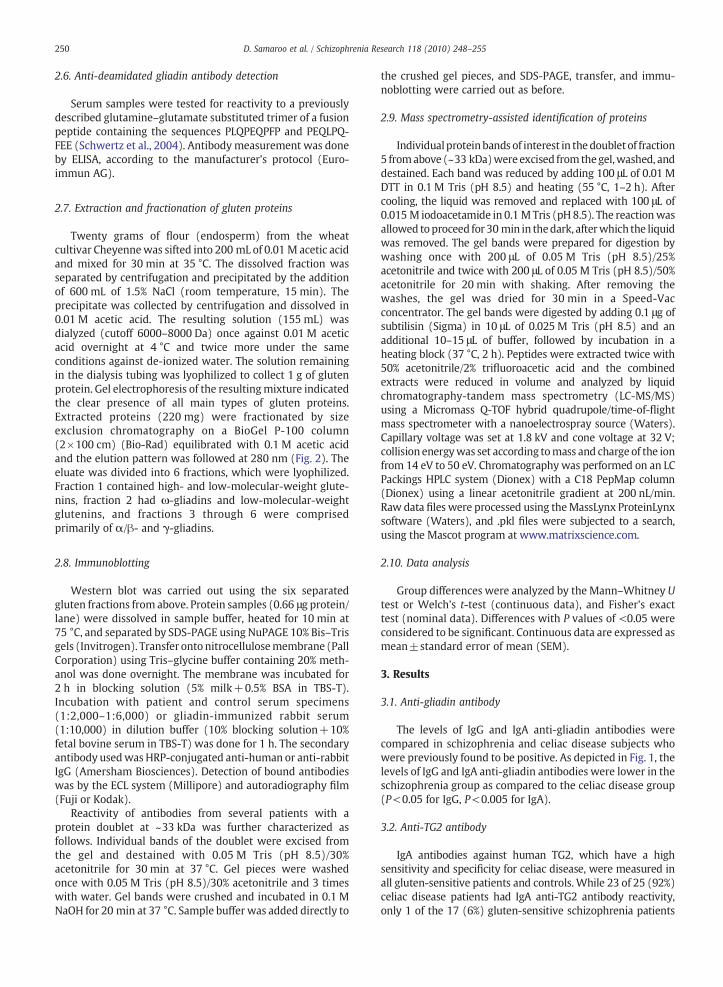
Twenty grams of flour (endosperm) from the wheatcultivar Cheyennewas sifted into 200 mL of 0.01 M acetic acidand mixed for 30 min at 35 °C. The dissolved fraction wasseparated by centrifugation and precipitated by the additionof 600 mL of 1.5% NaCl (room temperature, 15 min). Theprecipitate was collected by centrifugation and dissolved in0.01 M acetic acid. The resulting solution (155 mL) wasdialyzed (cutoff 6000–8000 Da) once against 0.01 M aceticacid overnight at 4 °C and twice more under the sameconditions against de-ionized water. The solution remainingin the dialysis tubing was lyophilized to collect 1 g of glutenprotein. Gel electrophoresis of the resultingmixture indicatedthe clear presence of all main types of gluten proteins.Extracted proteins (220 mg) were fractionated by sizeexclusion chromatography on a BioGel P-100 column(2×100 cm) (Bio-Rad) equilibrated with 0.1 M acetic acidand the elution pattern was followed at 280 nm (Fig. 2). Theeluate was divided into 6 fractions, which were lyophilized.Fraction 1 contained high- and low-molecular-weight glute-nins, fraction 2 had ω-gliadins and low-molecular-weightglutenins, and fractions 3 through 6 were comprisedprimarily of α/β- and γ-gliadins.
2.8. Immunoblotting
Western blot was carried out using the six separatedgluten fractions from above. Protein samples (0.66 μg protein/lane) were dissolved in sample buffer, heated for 10 min at75 °C, and separated by SDS-PAGE using NuPAGE 10% Bis–Trisgels (Invitrogen). Transfer onto nitrocellulosemembrane (PallCorporation) using Tris–glycine buffer containing 20% meth-anol was done overnight. The membrane was incubated for2 h in blocking solution (5% milk+0.5% BSA in TBS-T).Incubation with patient and control serum specimens(1:2,000–1:6,000) or gliadin-immunized rabbit serum(1:10,000) in dilution buffer (10% blocking solution+10%fetal bovine serum in TBS-T) was done for 1 h. The secondaryantibody usedwas HRP-conjugated anti-human or anti-rabbitIgG (Amersham Biosciences). Detection of bound antibodieswas by the ECL system (Millipore) and autoradiography film(Fuji or Kodak).
Reactivity of antibodies from several patients with aprotein doublet at ~33 kDa was further characterized asfollows. Individual bands of the doublet were excised fromthe gel and destained with 0.05 M Tris (pH 8.5)/30%acetonitrile for 30 min at 37 °C. Gel pieces were washedonce with 0.05 M Tris (pH 8.5)/30% acetonitrile and 3 timeswith water. Gel bands were crushed and incubated in 0.1 MNaOH for 20 min at 37 °C. Sample buffer was added directly to
the crushed gel pieces, and SDS-PAGE, transfer, and immu-noblotting were carried out as before.
2.9. Mass spectrometry-assisted identification of proteins
Individualproteinbands of interest in thedoublet of fraction5 fromabove (~33 kDa)were excised from thegel,washed, anddestained. Each band was reduced by adding 100 μL of 0.01 MDTT in 0.1 M Tris (pH 8.5) and heating (55 °C, 1–2 h). Aftercooling, the liquid was removed and replaced with 100 μL of0.015 M iodoacetamide in 0.1 M Tris (pH 8.5). The reactionwasallowed toproceed for 30 min in thedark, afterwhich the liquidwas removed. The gel bands were prepared for digestion bywashing once with 200 μL of 0.05 M Tris (pH 8.5)/25%acetonitrile and twice with 200 μL of 0.05 M Tris (pH 8.5)/50%acetonitrile for 20 min with shaking. After removing thewashes, the gel was dried for 30 min in a Speed-Vacconcentrator. The gel bands were digested by adding 0.1 μg ofsubtilisin (Sigma) in 10 μL of 0.025 M Tris (pH 8.5) and anadditional 10–15 μL of buffer, followed by incubation in aheating block (37 °C, 2 h). Peptides were extracted twice with50% acetonitrile/2% trifluoroacetic acid and the combinedextracts were reduced in volume and analyzed by liquidchromatography-tandem mass spectrometry (LC-MS/MS)using a Micromass Q-TOF hybrid quadrupole/time-of-flightmass spectrometer with a nanoelectrospray source (Waters).Capillary voltage was set at 1.8 kV and cone voltage at 32 V;collision energywas set according tomass and charge of the ionfrom 14 eV to 50 eV. Chromatography was performed on an LCPackings HPLC system (Dionex) with a C18 PepMap column(Dionex) using a linear acetonitrile gradient at 200 nL/min.Raw data files were processed using theMassLynx ProteinLynxsoftware (Waters), and .pkl files were subjected to a search,using the Mascot program at www.matrixscience.com.
2.10. Data analysis
Group differences were analyzed by the Mann–Whitney Utest or Welch's t-test (continuous data), and Fisher's exacttest (nominal data). Differences with P values of <0.05 wereconsidered to be significant. Continuous data are expressed asmean±standard error of mean (SEM).
3. Results
3.1. Anti-gliadin antibody
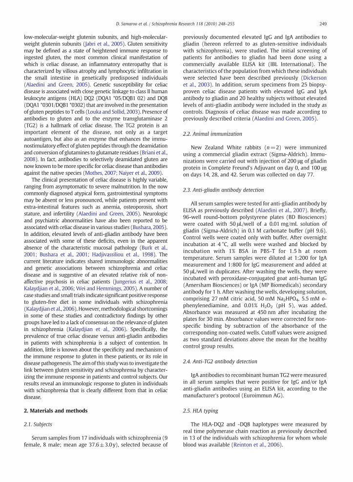
The levels of IgG and IgA anti-gliadin antibodies werecompared in schizophrenia and celiac disease subjects whowere previously found to be positive. As depicted in Fig. 1, thelevels of IgG and IgA anti-gliadin antibodies were lower in theschizophrenia group as compared to the celiac disease group(P<0.05 for IgG, P<0.005 for IgA).
3.2. Anti-TG2 antibody
IgA antibodies against human TG2, which have a highsensitivity and specificity for celiac disease, were measured inall gluten-sensitive patients and controls. While 23 of 25 (92%)celiac disease patients had IgA anti-TG2 antibody reactivity,only 1 of the 17 (6%) gluten-sensitive schizophrenia patients
Fig. 1. Comparison of mean antibody levels in celiac disease and gluten-sensitive schizophrenia patients. A) Antibodies to native gliadin. Individuals withschizophrenia that were positive for anti-gliadin antibodies had lower levels of IgG (P<0.05) and IgA (P<0.005) antibodies to gliadin than celiac disease patients.B) Antibodies to a deamidated (glutamine–glutamate substituted) trimer of a fusion gliadin peptide containing PLQPEQPFP and PEQLPQFEE sequences in the samepatients as in panel A. Individuals with schizophrenia whowere positive for anti-gliadin antibodies had significantly lower mean IgG and IgA antibody reactivity todeamidated gliadin than celiac disease patients (P<0.001). C) IgA antibodies to recombinant human TG2. Levels were significantly lower in the schizophreniagroup than the celiac disease group (P<0.001). Dotted lines indicate established cutoff values for positivity. Error bars represent the standard error of the mean.
251D. Samaroo et al. / Schizophrenia Research 118 (2010) 248–255
was positive (P<0.001). Comparison of normalized meanantibody reactivity for the two groups also revealed signifi-cantly lower levels of anti-TG2 IgA antibodies in schizophreniapatients (P<0.001) (Fig. 1C).Noneof thehealthy control serumsamples were positive.
3.3. HLA typing
There was not a significantly increased correlationbetween gluten sensitivity and HLA-DQ2/DQ8 haplotypes inpatients with schizophrenia as compared with the generalpopulation. 5/13 (38.5%) of the gluten-sensitive individualswith schizophrenia displayed HLA-DQ2 and/or DQ8 (1 DQ2, 4DQ8) haplotypes. In most populations, 90% or more of celiacdisease patients carry the DQ2 haplotype and the rest haveDQ8, compared to 20%–40% in healthy individuals havingeither (Alaedini and Green, 2005; Sollid, 2000).
3.4. Anti-deamidated gliadin antibody
IgG and IgA antibodies to a deamidated (glutamine–glutamate substituted) fusion gliadin peptide trimer weremeasured in all subjects. While 22 of 25 (88%) celiac diseasepatients exhibited IgG antibody reactivity to deamidatedgliadin, only 1 of the 17 (6%) gluten-sensitive individuals withschizophrenia was positive (P<0.001). Similarly, 23 of 25(92%) celiac disease patients and none of the 17 gluten-sensitive individuals with schizophrenia were positive for theIgA antibodies to deamidated gliadin (P<0.001). Comparisonof normalized mean antibody reactivity for the two groupsalso indicated significantly lower levels of IgG and IgAantibodies to the modified peptide in schizophrenia patients
(P<0.001) (Fig. 1B). None of the healthy control serumsamples were positive for antibodies to deamidated gliadin.
3.5. Immunoblotting
Following the measurement of antibody levels by ELISA,serum specimens from representative subjects were furthercharacterized with regard to reactivity towards fractionatedgluten proteins. The gluten proteins used in this study werepurified by extraction with acetic acid and size exclusionchromatography. The eluted six fractions were furtherseparated by SDS-PAGE (Fig. 2) and transferred to nitrocel-lulosemembrane for immunoblotting. The observed antibodyreactivity to the gluten proteins was heterogeneous inimmunized rabbits, celiac disease patients, and schizophreniapatients. However, while antibodies from celiac patients andimmunized rabbits bound to many gliadins and/or glutenins,antibodies from the individuals with schizophrenia bound toa relatively smaller set of gluten molecules (Fig. 3). Serumsamples from a few of these patients displayed an antibodyspecificity profile that was similar to celiac disease (Fig. 3,patient S4), but antibodies from most sera bound to a limitedrepertoire of proteins in the glutenin and gliadin fractions(Fig. 3, patients S1–S3). Antibodies from 6 individuals withschizophrenia reacted with a protein band of ~33 kDa ingluten fraction 5, either exclusively, or in conjunction withweaker binding to glutenin proteins in fractions 1 and 2. Noneof the celiac disease sera exhibited exclusive or prominentreactivity to this protein band. The band appeared as adoublet in Coomassie-stained gels. In order to determinewhich member of the protein doublet was targeted by theanti-gluten antibodies of patients with schizophrenia, proteinbands were individually excised from Coomassie-stained gel,extracted, and re-analyzed. Patient antibody reactivity was
Fig. 2. A) Chromatogram of gluten proteins eluted from the BioGel P-100 sizeexclusion column, following acetic acid extraction. B) SDS-PAGE profile(Coomassie-stained) of fractions 1–6 of gluten proteins collected from thecolumn.
252 D. Samaroo et al. / Schizophrenia Research 118 (2010) 248–255
found to be directed primarily at the upper band, with onlyresidual binding to the lower band (Fig. 4).
3.6. Identification of ~33 kDa target proteins
The protein bands of the ~33 kDa doublet were individuallyexcised, digested with subtilisin after chemical modification,and analyzed by LC/MS-MS. The detected peptide masses weresearched against theNCBI protein sequence database. Results ofthe database search revealed two γ-gliadins (accessionnumbers ABO37962 and AAQ63857) as the top two significantcandidate proteins in the immunoreactive upper band, with 15and 11 peptides matched and 42% and 32% sequence coverage,respectively. Similarly, the database search revealed two nearlyidentical γ-gliadins (accession numbers ABO37962 andAAK84776) as the top two significant candidate proteins inthe lower band of the doublet, with 34 peptides matched (bothproteins) and 58% and 57% sequence coverage, respectively.
4. Discussion
Schizophrenia is a complex disorder affecting approxi-mately 1% of the population and is caused by a combination ofgenetic and environmental factors. The immune system isbelieved to play a considerable role in the disease process.Recently-reported single nucleotide polymorphism (SNP)-based genome-wide studies in schizophrenia populations
show significant association with several markers spanningthe major histocompatibility complex (MHC) region onchromosome 6 (Purcell et al., 2009; Shi et al., 2009;Stefansson et al., 2009). Dating back to the 1950s, schizo-phrenia has been linked to anti-gliadin antibodies and celiacdisease in several studies (Bender, 1953; Cascella et al., 2009;Dohan et al., 1972; Graff and Handford, 1961; Jin et al., 2008;Reichelt and Landmark, 1995). A number of case reports andsmall trials also indicate significant positive response togluten-free diet in some individuals with schizophrenia(Kalaydjian et al., 2006). Considering the limited responseto traditional therapies by many individuals with schizophre-nia and the side effects of current treatments, identification ofa subset of patients who might benefit from an effectivedietary approach without significant side effects wouldrepresent an important step forward in the understandingand treatment of the disease. However, the currently limitedknowledge of the significance of anti-gliadin antibodies in thecontext of schizophrenia in general, combined with theresults of a number of studies that do not support a linkwith celiac disease, has cast doubt on the relevance of glutenand gluten sensitivity to schizophrenia (Lambert et al., 1989;Ludvigsson et al., 2007).
In this study, our aim was to address these issues byassessing the prevalence of celiac disease-associated biomar-kers and through the characterization of the anti-glutenimmune response at the molecular level in individuals withschizophrenia and elevated anti-gliadin antibodies and incontrol subjects. We found no correlation between elevatedanti-gliadin antibodies and the presence of increased anti-TG2 antibody levels or celiac-associated HLA genes inindividuals with schizophrenia. Considering the high nega-tive predictive value of the anti-TG2 antibody and HLAmarkers for celiac disease, it can be concluded with highcertainty that themajority of these patients do not have celiacdisease. We have also shown that the anti-gliadin antibodyresponses in celiac disease and gluten-sensitive individualswith schizophrenia are not necessarily directed at the samemolecules. Equally interesting is the finding that theantibodies in schizophrenia do not appear to show significantaffinity for deamidated gliadin peptides, again implying theabsence of celiac disease in the majority of gluten-sensitiveindividuals with schizophrenia. As such, the anti-glutenimmune response in schizophrenia patients is likely to beindependent of the TG2 enzyme and does not require HLA-DQ2 and/or -DQ8molecules for driving the process of antigenpresentation and antibody production.
It is important to note that the sample size in this study isnot large enough to exclude the possibility of celiac-associated markers (HLA-DQ2/DQ8 genes, anti-TG2 antibo-dies, anti-deamidated gliadin antibodies), and thereby celiacdisease, being more prevalent in schizophrenia patients thanin normal healthy individuals. However, it is adequately largeto determine with high certainty that unlike in celiac disease,the anti-gluten immune response in schizophrenia is notassociated with the above-mentioned biomarkers and there-fore does not imply celiac disease in the overwhelmingmajority of individuals. This agrees with the results of arecently published large study, which found that mostschizophrenia patients with elevated anti-gliadin antibodytiters do not have anti-TG2 and anti-endomysial antibodies
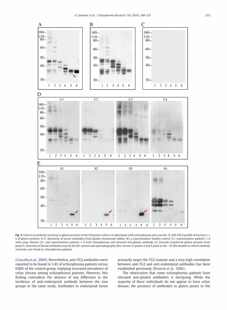
Fig. 3. Pattern of antibody reactivity to gluten proteins of the Cheyenne cultivar in individuals with schizophrenia and controls. A) SDS-PAGE profile of fractions 1–6 of gluten proteins. B–E) Reactivity of serum antibodies from gliadin-immunized rabbits (B), a representative healthy control (C), representative patients 1–4with celiac disease (D), and representative patients 1–4 with schizophrenia and elevated anti-gliadin antibody (E) towards transferred gluten proteins frompanel A. Detection of bound antibodies was by the ECL system and autoradiography film. Arrows in panels A and E point to the ~33 kDa doublet to which antibodyreactivity was found in schizophrenia patients.
253D. Samaroo et al. / Schizophrenia Research 118 (2010) 248–255
(Cascella et al., 2009). Nevertheless, anti-TG2 antibodies werereported to be found in 5.4% of schizophrenia patients versus0.80% of the control group, implying increased prevalence ofceliac disease among schizophrenia patients. However, thisfinding contradicts the absence of any difference in theincidence of anti-endomysial antibody between the twogroups in the same study. Antibodies to endomysial tissue
primarily target the TG2 enzyme and a very high correlationbetween anti-TG2 and anti-endomysial antibodies has beenestablished previously (Rostom et al., 2006).
The observation that some schizophrenia patients haveelevated anti-gliadin antibodies is intriguing. While themajority of these individuals do not appear to have celiacdisease, the presence of antibodies to gluten points to the
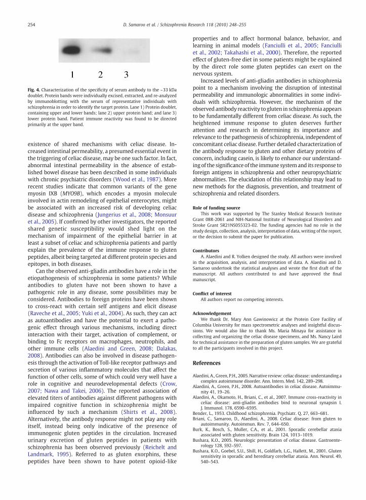
Fig. 4. Characterization of the specificity of serum antibody to the ~33 kDadoublet. Protein bands were individually excised, extracted, and re-analyzedby immunoblotting with the serum of representative individuals withschizophrenia in order to identify the target protein. Lane 1) Protein doublet,containing upper and lower bands; lane 2) upper protein band; and lane 3)lower protein band. Patient immune reactivity was found to be directedprimarily at the upper band.
254 D. Samaroo et al. / Schizophrenia Research 118 (2010) 248–255
existence of shared mechanisms with celiac disease. In-creased intestinal permeability, a presumed essential event inthe triggering of celiac disease, may be one such factor. In fact,abnormal intestinal permeability in the absence of estab-lished bowel disease has been described in some individualswith chronic psychiatric disorders (Wood et al., 1987). Morerecent studies indicate that common variants of the genemyosin IXB (MYO9B), which encodes a myosin moleculeinvolved in actin remodeling of epithelial enterocytes, mightbe associated with an increased risk of developing celiacdisease and schizophrenia (Jungerius et al., 2008; Monsuuret al., 2005). If confirmed by other investigators, the reportedshared genetic susceptibility would shed light on themechanism of impairment of the epithelial barrier in atleast a subset of celiac and schizophrenia patients and partlyexplain the prevalence of the immune response to glutenpeptides, albeit being targeted at different protein species andepitopes, in both diseases.
Can the observed anti-gliadin antibodies have a role in theetiopathogenesis of schizophrenia in some patients? Whileantibodies to gluten have not been shown to have apathogenic role in any disease, some possibilities may beconsidered. Antibodies to foreign proteins have been shownto cross-react with certain self antigens and elicit disease(Raveche et al., 2005; Yuki et al., 2004). As such, they can actas autoantibodies and have the potential to exert a patho-genic effect through various mechanisms, including directinteraction with their target, activation of complement, orbinding to Fc receptors on macrophages, neutrophils, andother immune cells (Alaedini and Green, 2008; Dalakas,2008). Antibodies can also be involved in disease pathogen-esis through the activation of Toll-like receptor pathways andsecretion of various inflammatory molecules that affect thefunction of other cells, some of which could very well have arole in cognitive and neurodevelopmental defects (Crow,2007; Nawa and Takei, 2006). The reported association ofelevated titers of antibodies against different pathogens withimpaired cognitive function in schizophrenia might beinfluenced by such a mechanism (Shirts et al., 2008).Alternatively, the antibody response might not play any roleitself, instead being only indicative of the presence ofimmunogenic gluten peptides in the circulation. Increasedurinary excretion of gluten peptides in patients withschizophrenia has been observed previously (Reichelt andLandmark, 1995). Referred to as gluten exorphins, thesepeptides have been shown to have potent opioid-like
properties and to affect hormonal balance, behavior, andlearning in animal models (Fanciulli et al., 2005; Fanciulliet al., 2002; Takahashi et al., 2000). Therefore, the reportedeffect of gluten-free diet in some patients might be explainedby the direct role some gluten peptides can exert on thenervous system.
Increased levels of anti-gliadin antibodies in schizophreniapoint to a mechanism involving the disruption of intestinalpermeability and immunologic abnormalities in some indivi-duals with schizophrenia. However, the mechanism of theobserved antibody reactivity to gluten in schizophrenia appearsto be fundamentally different from celiac disease. As such, theheightened immune response to gluten deserves furtherattention and research in determining its importance andrelevance to the pathogenesis of schizophrenia, independent ofconcomitant celiac disease. Further detailed characterization ofthe antibody response to gluten and other dietary proteins ofconcern, including casein, is likely to enhance our understand-ingof the significance of the immune systemand its response toforeign antigens in schizophrenia and other neuropsychiatricabnormalities. The elucidation of this relationship may lead tonew methods for the diagnosis, prevention, and treatment ofschizophrenia and related disorders.
Role of funding sourceThis work was supported by The Stanley Medical Research Institute
Grant 08R-2061 and NIH-National Institute of Neurological Disorders andStroke Grant 5R21NS055323-02. The funding agencies had no role in thestudy design, collection, analysis, interpretation of data, writing of the report,or the decision to submit the paper for publication.
ContributorsA. Alaedini and R. Yolken designed the study. All authors were involved
in the acquisition, analysis, and interpretation of data. A. Alaedini and D.Samaroo undertook the statistical analyses and wrote the first draft of themanuscript. All authors contributed to and have approved the finalmanuscript.
Conflict of interestAll authors report no competing interests.
AcknowledgementWe thank Dr. Mary Ann Gawinowicz at the Protein Core Facility of
Columbia University for mass spectrometric analyses and insightful discus-sions. We would also like to thank Ms. Maria Minaya for assistance incollecting and organizing the celiac disease specimens, and Ms. Nancy Lairdfor technical assistance in the preparation of gluten samples. We are gratefulto all the participants involved in this project.
References
Alaedini, A., Green, P.H., 2005. Narrative review: celiac disease: understanding acomplex autoimmune disorder. Ann. Intern. Med. 142, 289–298.
Alaedini, A., Green, P.H., 2008. Autoantibodies in celiac disease. Autoimmu-nity 41, 19–26.
Alaedini, A., Okamoto, H., Briani, C., et al., 2007. Immune cross-reactivity inceliac disease: anti-gliadin antibodies bind to neuronal synapsin I.J. Immunol. 178, 6590–6595.
Bender, L., 1953. Childhood schizophrenia. Psychiatr. Q. 27, 663–681.Briani, C., Samaroo, D., Alaedini, A., 2008. Celiac disease: from gluten to
autoimmunity. Autoimmun. Rev. 7, 644–650.Burk, K., Bosch, S., Muller, C.A., et al., 2001. Sporadic cerebellar ataxia
associated with gluten sensitivity. Brain 124, 1013–1019.Bushara, K.O., 2005. Neurologic presentation of celiac disease. Gastroente-
rology 128, S92–S97.Bushara, K.O., Goebel, S.U., Shill, H., Goldfarb, L.G., Hallett, M., 2001. Gluten
sensitivity in sporadic and hereditary cerebellar ataxia. Ann. Neurol. 49,540–543.
255D. Samaroo et al. / Schizophrenia Research 118 (2010) 248–255
Cascella, N.G., Kryszak, D., Bhatti, B. et al., 2009. Prevalence of Celiac Diseaseand Gluten Sensitivity in the United States Clinical Antipsychotic Trials ofIntervention Effectiveness Study Population. Schizophr Bull. [Epub aheadof print].
Crow, M.K., 2007. Type I interferon in systemic lupus erythematosus. In:Pitha, P.M. (Ed.), CTMI, Vol. 316. Springer Berlin Heidelberg, pp. 359–386.
Dalakas, M.C., 2008. B cells as therapeutic targets in autoimmune neurologicaldisorders. Nat. Clin. Pract. Neurol. 4, 557–567.
Dickerson, F.B., Boronow, J.J., Stallings, C., Origoni, A.E., Ruslanova, I., Yolken,R.H., 2003. Association of serum antibodies to herpes simplex virus 1with cognitive deficits in individuals with schizophrenia. Arch. Gen.Psychiatry 60, 466–472.
Dohan, F.C., Martin, L., Grasberger, J.C., Boehme, D., Cottrell, J.C., 1972.Antibodies to wheat gliadin in blood of psychiatric patients: possible roleof emotional factors. Biol. Psychiatry 5, 127–137.
Fanciulli, G., Dettori, A., Tomasi, P.A., et al., 2002. Prolactin and growthhormone response to intracerebroventricular administration of the foodopioid peptide gluten exorphin B5 in rats. Life Sci. 71, 2383–2390.
Fanciulli, G., Dettori, A., Demontis, M.P., Tomasi, P.A., Anania, V., Delitala, G.,2005. Gluten exorphin B5 stimulates prolactin secretion through opioidreceptors located outside the blood–brain barrier. Life Sci. 76, 1713–1719.
Graff, H., Handford, A., 1961. Celiac syndrome in the case histories of fiveschizophrenics. Psychiatr. Q. 35, 306–313.
Hadjivassiliou, M., Grunewald, R.A., Chattopadhyay, A.K., et al., 1998. Clinical,radiological, neurophysiological, and neuropathological characteristicsof gluten ataxia. Lancet 352, 1582–1585.
Jabri, B., Kasarda, D.D., Green, P.H., 2005. Innate and adaptive immunity: theyin and yang of celiac disease. Immunol. Rev. 206, 219–231.
Jin, S.Z., Xu, Q., Wu, N., et al., 2008. A study of gluten antibody levels in serumamong patients with schizophrenia. Schizophr. Res. 98, S145.
Jungerius, B.J., Bakker, S.C., Monsuur, A.J., Sinke, R.J., Kahn, R.S., Wijmenga, C.,2008. IsMYO9B themissing linkbetween schizophrenia and celiacdisease?Am. J. Med. Genet. B Neuropsychiatr. Genet. 147, 351–355.
Kalaydjian, A.E., Eaton,W., Cascella, N., Fasano, A., 2006. The gluten connection:the association between schizophrenia and celiac disease. Acta Psychiatr.Scand. 113, 82–90.
Lambert, M.T., Bjarnason, I., Connelly, J., et al., 1989. Small intestinepermeability in schizophrenia. Br. J. Psychiatry 155, 619–622.
Louka, A.S., Sollid, L.M., 2003. HLA in coeliac disease: unravelling the complexgenetics of a complex disorder. Tissue Antigens 61, 105–117.
Ludvigsson, J.F., Osby, U., Ekbom, A., Montgomery, S.M., 2007. Coeliac diseaseand risk of schizophrenia and other psychosis: a general populationcohort study. Scand. J. Gastroenterol. 42, 179–185.
Monsuur, A.J., de Bakker, P.I., Alizadeh, B.Z., et al., 2005. Myosin IXB variantincreases the risk of celiac disease and points toward a primary intestinalbarrier defect. Nat. Genet. 37, 1341–1344.
Mothes, T., 2007. Deamidated gliadin peptides as targets for celiac disease-specific antibodies. Adv. Clin. Chem. 44, 35–63.
Naiyer, A.J., Hernandez, L., Ciaccio, E.J., et al., 2009,. Comparison of commerciallyavailable serologic kits for thedetectionof celiacdisease. J. Clin.Gastroenterol.43, 225–232.
Nawa, H., Takei, N., 2006. Recent progress in animal modeling of immuneinflammatory processes in schizophrenia: implication of specificcytokines. Neurosci. Res. 56, 2–13.
Patterson, P.H., 2008. Immune involvement in schizophrenia and autism:etiology, pathology and animal models. Behav. Brain Res. [Epub ahead ofprint].
Purcell, S.M., Wray, N.R., Stone, J.L., et al., 2009. Common polygenic variationcontributes to risk of schizophrenia and bipolar disorder. Nature. [Epubahead of print].
Raveche, E.S., Schutzer, S.E., Fernandes, H., et al., 2005. Evidence of Borreliaautoimmunity-induced component of Lyme carditis and arthritis. J. Clin.Microbiol. 43, 850–856.
Reichelt, K.L., Landmark, J., 1995. Specific IgA antibody increases inschizophrenia. Biol. Psychiatry 37, 410–413.
Reinton, N., Helgheim, A., Shegarfi, H., Moghaddam, A., 2006. A one-step real-time PCR assay for detection of DQA1*05, DQB1*02 and DQB1*0302 toaid diagnosis of celiac disease. J. Immunol. Methods 316, 125–132.
Rostom, A., Murray, J.A., Kagnoff, M.F., 2006. American GastroenterologicalAssociation (AGA) Institute technical review on the diagnosis and manage-ment of celiac disease. Gastroenterology 131, 1981–2002.
Schwertz, E., Kahlenberg, F., Sack, U., et al., 2004. Serologic assay based ongliadin-related nonapeptides as a highly sensitive and specific diagnosticaid in celiac disease. Clin. Chem. 50, 2370–2375.
Shi, J., Levinson, D.F., Duan, J., et al., 2009. Common variants on chromosome6p22.1 are associated with schizophrenia. Nature [Epub ahead of print].
Shirts, B.H., Prasad, K.M., Pogue-Geile, M.F., Dickerson, F., Yolken, R.H.,Nimgaonkar, V.L., 2008. Antibodies to cytomegalovirus andHerpes SimplexVirus 1 associatedwith cognitive function in schizophrenia. Schizophr. Res.106, 268–274.
Sollid, L.M., 2000. Molecular basis of celiac disease. Annu. Rev. Immunol. 18,53–81.
Stefansson, H., Ophoff, R.A., Steinberg, S., et al., 2009. Common variantsconferring risk of schizophrenia. Nature. [Epub ahead of print].
Takahashi, M., Fukunaga, H., Kaneto, H., Fukudome, S., Yoshikawa, M., 2000.Behavioral and pharmacological studies on gluten exorphin A5, a newlyisolated bioactive food protein fragment, in mice. Jpn. J. Pharmacol. 84,259–265.
Torrey, E.F., Yolken, R.H., 2001. The schizophrenia–rheumatoid arthritis connec-tion: infectious, immune, or both? Brain Behav. Immun. 15, 401–410.
Wei, J., Hemmings, G.P., 2005. Gene, gut and schizophrenia: the meetingpoint for the gene−environment interaction in developing schizophre-nia. Med. Hypotheses 64, 547–552.
Wood, N.C., Hamilton, I., Axon, A.T., et al., 1987. Abnormal intestinalpermeability. An aetiological factor in chronic psychiatric disorders?Br. J. Psychiatry 150, 853–856.
Yuki, N., Susuki, K., Koga, M., et al., 2004. Carbohydrate mimicry between humanganglioside GM1 and Campylobacter jejuni lipooligosaccharide causesGuillain-Barre syndrome. Proc. Natl. Acad. Sci. U S A 101, 11404–11409.