Embed Size (px)
Citation preview

�
CLINICAL REPORT
Novel Homozygous DEAF1 Variant Suspected inCausing White Matter Disease, IntellectualDisability, and Microcephaly
Eissa A. Faqeih,1 Mohammed Al-Owain,2,3 Dilek Colak,4 Rosan Kenana,5 Yusra Al-Yafee,5Mazhor Al-Dosary,5 Abdulaziz Al-Saman,6 Fadwa Albalawi,5 Dalia Al-Sarar,5 Dalia Domiaty,5
Maha Daghestani,5,7 and Namik Kaya5*1Section of Medical Genetic, Pediatric Department, Children’s Hospital, King Fahad Medical City, Riyadh, Saudi Arabia2Department of Medical Genetics, Neuroscience Centre, King Fahad Medical City, Riyadh, Saudi Arabia3College of Medicine, Alfaisal University, Riyadh, Saudi Arabia4Department of Biostatistics, Epidemiology and Scientific Computing, Neuroscience Centre, King Fahad Medical City, Riyadh, Saudi Arabia5Department of Genetics, King Faisal Specialist Hospital and Research Centre, Neuroscience Centre, King Fahad Medical City, Riyadh,
Saudi Arabia6Department of Pediatric Neurology, Neuroscience Centre, King Fahad Medical City, Riyadh, Saudi Arabia7College of Science, King Saud University, Riyadh, Saudi Arabia
Manuscript Received: 30 July 2013; Manuscript Accepted: 12 January 2014
How to Cite this Article:Faqeih EA, Al-Owain M, Colak D, Kenana
R, Al-Yafee Y, Al-Dosary M, Al-Saman A,
Albalawi F, Al-Sarar D, Domiaty D,
Daghestani M, Kaya N. 2014. Novel
homozygous DEAF1 variant suspected in
causing white matter disease, intellectual
disability, and microcephaly.
Am J Med Genet Part A 164A:1565–1570.
Conflicts of interest: none.
Grant sponsor: King Faisal Specialist Hospital and Research Centre
(KFSHRC).�Correspondence to:
DEAF1 encodes a transcriptional binding factor and is a regulator of
serotonin receptor 1A. Its protein has a significant expression in the
neuronsofdifferentbrainregionsandis involvedinearlyembryonic
development. In addition, its role in neural tube development is
evident from the knockout mouse as many homozygotes have
exencephaly.Heterozygousmutations of this gene have been linked
to intellectual disability in addition to the gene’s involvement in
major depression, suicidal tendencies, and panic disorder. In this
clinical report, we describe two children from a consanguineous
familywithintellectualdisability,microcephaly,andhypotonia.The
brainMRI of both patients showed bilateral and symmetrical white
matter abnormalities, andoneof thepatientshada seizuredisorder.
Usingwhole exome sequencing combinedwith homozygositymap-
ping, a homozygous p.R226W (c.676C>T)mutation inDEAF1was
foundinbothpatients.Furthermore,sequencinganalysisconfirmed
complete segregation in tested family members and absence of the
mutation in control cohort (n¼ 650). The mutation is located in a
highlyconservedstructuraldomain thatmediatesDNAbindingand
therefore regulates transcriptional activity of its target molecules.
This study indicates, for thefirst time toourknowledge, ahereditary
role of DEAF1 in white matter abnormalities, microcephaly and
syndromic intellectual disability. � 2014 Wiley Periodicals, Inc.
Key words: DEAF1; homozygous p.R226W; syndromic in-
tellectual disability; white matter abnormality; microcephaly
Namik Kaya, Ph.D., Department of Genetics, King Faisal Specialist
Hospital and Research Center, MBC 03, Riyadh 11211, Kingdom of
Saudi Arabia. E-mail: [email protected]; [email protected]
Article first published online in Wiley Online Library
(wileyonlinelibrary.com): 25 March 2014
DOI 10.1002/ajmg.a.36482
INTRODUCTION
Neurodevelopmental disorders (NDs) comprise a group of highly
heterogeneous disorders mostly caused by alterations during early
2014 Wiley Periodicals, Inc.
brain development. Various NDs share similar features such as
brain dysfunction and cognitive impairment [van Loo and
Martens, 2007]. The recent advances inmassively parallel-sequenc-
ing techniques have led to numerous gene breakthroughs in
inherited and sporadic NDs related to intellectual disability. How-
ever, the full picture of pathogenesis of NDs is not fully understood.
1565

TABLE I. Clinical and Laboratory Findings
Patients II-2 II-5
Age 21/2 years 2 years
Gender Male Male
1566 AMERICAN JOURNAL OF MEDICAL GENETICS PART A
Deformed epidermal autoregulatory factor 1 (DEAF1)—also
known as “nuclear DEAF-1-related transcriptional regulator”—is a
trans-acting, trans-regulator element that regulates the serotonin 1A
receptor (5HT1A) in the brain. DEAF1 is known to be highly
conserved among different species with significant expression in
all the neurons of different brain regions. It is important in early
embryonic development and regulates immunity gene expression in
drosophila [Veraksa et al., 2002;Reed et al., 2008]. In addition, its role
in neural tube development is evident from the mouse knockout as
many homozygotes have exencephaly [Hahm et al., 2004]. The gene
longest transcript yields 565-amino acid-long polypeptide that is
critical for transcriptional regulation of serotonergic synapses.
5HT1A is widely expressed in serotonergic neurons and brain, and
co-localizeswithDEAF1 in serotonergic raphe cells andhippocampal
and cortical neurons [Lemonde et al., 2003]. DEAF1 selectively
suppresses somatodendric 5HT1A autoreceptor expression in sero-
tonergic synapses in an allele-specificmanner [Lemonde et al., 2004].
Besides itsproposedrole inpsychiatricdisorders [Mann,1999;Czesak
et al., 2006; Jans et al., 2007] and carcinoma[Carr et al., 1990; LeBoeuf
et al., 1990, 1998; Ban and LeBoeuf, 1994; Manne et al., 2001], the
action ofDEAF1 in regulating the serotonin system further suggests a
causative role in mental disorders [Czesak et al., 2012]. DEAF1 has
been previously linked to nonsyndromic sporadic intellectual dis-
ability (ID) [Vissers et al., 2010; Rauch et al., 2012] and is suggested to
have a role in major depression, suicidal tendencies, and panic
disorder [Albert and Lemonde, 2004; Lemonde et al., 2004].
Due to high consanguinity rate (reaching to nearly 80% in some
regions) and the large family size, countries like Saudi Arabia (SA)
allow great opportunities for geneticists who focus on inherited
NDs. Altogether, these disorders in SA are estimated to be around
3–5:100. Despite the discovery of numerous disease-causing genes,
many clinically suspected inherited NDs cases remain molecularly
unidentified. In order to identify novel genetic causes of hereditary
NDs, we performed autozygome analysis using high-density SNP
arrays combined with exome sequencing as a positional cloning
strategy that led to the identification of a novel missense mutation
in DEAF1 in a consanguineous Saudi family.
Age at presentation Neonatal 2 MonthsBirth weight 2.3 kg Not available
Hypotonia þ þFeeding difficulties þ þGrowth Poor Poor
OC 44 cm (21/2 years) 44 cm (at 22 months)
Vision Normal Normal
Hearing Normal Normal
Dysmorphism None None
Motor delay Moderate Moderate
Speech delay Severe Severe
Cognitive delay Severe Severe
Seizure þ �Hyperactivity � �Karyotype 46 XY 46 XY
Array CGH Normal Normal
Metabolic screen Unremarkable Unremarkable
Brain MRI Abnormal (see text) Abnormal (see text)
Electroencephalogram Normal Not availableMRI, magnetic resonance imaging; OC, occipitofrontal circumference.
METHODS
Two patients and their family members from a consanguineous
family were ascertained under KFSHRC IRB-approved protocols
(RAC#2120 022) after signing the written informed consents.
Whole blood samples were collected and DNA isolations were
performed using PureGene DNA Purification Kit (Gentra Systems,
Inc., Minneapolis, MN). Human Mapping Axiom arrays (Affyme-
trix, Inc., SantaClara, CA)were used for SNP genotyping. The assay
preparation, scanning, image processing, genotyping, and prelimi-
nary data analysis were all done according to manufacturers’
protocols and guidelines. Exon Capture and library construction
were done according to Agilent’s SureSelect Protocol Version 1.2
(Agilent, Inc.). Enrichment was carried out using SureSelect pro-
tocols. Sequencing was performed on the Illumina HiSeq2000
platform using TruSeq v3 chemistry (Illumina, San Diego, CA).
Runs of homozygous (ROH) blocks using SNP calls were deter-
minedby identifying loss of heterozygous regions (LOH) calculated
byAutoSNPa [Carr et al., 2006] andHomozygosityMapper [Seelow
et al., 2009]. Exon specific primers for DEAF1 were designed using
publicly available Primer3 algorithm [Rozen and Skaletsky, 2000].
PCRs were performed according to standard protocols using
BigDye Terminators (Thermo Fisher Scientific Inc., Waltham,
MA) on an ABI 3100 sequencer. Sequence chromatograms were
analyzed using SeqMAN software version 1.1 (DNASTAR, Inc.,
Madison, WI). Eleven different bioinformatics tools were utilized
topredict the functional consequencesof thep.R226W(Supporting
Information File 1). DEAF1 related sequences from various species
were obtained from Ensembl and multiple sequence alignment
(MSA) was performed using ClustalW and then visualized with
JalView. Protein Knowledgebase (UniprotKB) was searched for
DEAF1. Uniprot output O75398 (DEAF1_HUMAN) was used
for 3-D structural analysis for SAND domain. Array Comparative
Genomic Hybridization (aCGH) was performed as part of
routine diagnostic cytogenetic protocols using Agilent 180K arrays
(Agilent, Inc., Santa Clara, CA) according to manufacturer’s
instructions and guidelines.
CLINICAL REPORT
Phenotypic Analysis of the PatientsWe ascertained a two-branched consanguineous Saudi family from
a farming community in central Saudi Arabia. The clinical features
of the patients are given in Table I. The prominent clinical features
of the two patients include microcephaly, ID, and white matter
abnormalities. Index Patient II-5 had a sister who died at the age of
3 years with similar features (DNA was not available in this
research). Extensive metabolic investigation was performed on
the deceased patient (Patient II-4) including plasma amino acids,
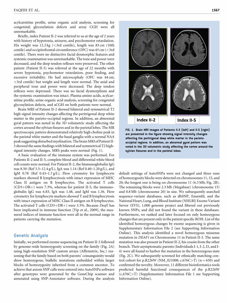
FIG. 1. Brain MRI images of Patients II-2 (left) and II-5 (right)
are presented in the figure showing signal intensity changes
affecting the peritrigonal deep white matter in the parieto-
occipital regions. In addition, an abnormal gyral pattern was
noted in the 3D volumetric study affecting the cortex around the
sylvian fissures and in the parietal lobes.
FAQEIH ET AL. 1567
acylcarnitine profile, urine organic acid analysis, screening for
congenital; glycosylation defects and array CGH were all
unremarkable.
Briefly, index Patient II-2 was referred to us at the age of 2 years
with history of hypotonia, seizures, and psychomotor retardation.
His weight was 12.2 kg (<3rd centile), length was 83 cm (10th
centile) and occipitofrontal circumference (OFC)was 43 cm (<3rd
centile). There were no distinctive facial dysmorphic features and
systemic examination was unremarkable. The tone and power were
decreased, and the deep tendon reflexes were preserved. The other
patient (Patient II-5) was referred at the age of 22 months with
severe hypotonia, psychomotor retardation, poor feeding, and
excessive irritability. He had microcephaly (OFC was 44 cm;
<3rd centile) but weight and length were normal. The axial and
peripheral tone and power were decreased. The deep tendon
reflexes were depressed. There was no facial dysmorphism and
the systemic examination was intact. Plasma amino acids, acylcar-
nitine profile, urine organic acid analysis, screening for congenital
glycosylation defects, and aCGH on both patients were normal.
Brain MRI of Patient II-2 showed bilateral and symmetrical T2
high-signal intensity changes affecting the peritrigonal deep white
matter in the parieto-occipital regions. In addition, an abnormal
gyral pattern was noted in the 3D volumetric study affecting the
cortex around the sylvian fissures and in the parietal lobes. TheMR
spectroscopic pattern demonstrated relatively high choline peak in
the parietal white matter and the basal ganglia with a normal NAA
peak suggestingdisturbedmyelination.ThebrainMRIofPatient II-
5 showed the same findingswith bilateral and symmetrical T2 high-
signal intensity changes. MRS peaks were normal (Fig. 1).
A basic evaluation of the immune system was performed. For
Patients II-2 and II-5, complete blood and differential white blood
cell counts were normal. For Patient II-2, the Immunoglobulin IgG
was 8.50 (Ref 3.5–12.4 g/L), IgA was 1.14 (Ref 0.40–1.20 g/L), and
IgM 0.78 (Ref 0.43–1.7 g/L). Flow cytometry for lymphocyte
markers showed B lymphocytosis with intact expression of MHC
Class II antigen on B lymphocytes. The activated T cells
(CD3þDRþ) were 7.3%, whereas for patient II-5, the immuno-
globulin IgG was 6.83, IgA was 1.48, and IgM was 1.36. Flow
cytometry for lymphocyte markers showed T and B lymphocytosis
with intact expression of MHC Class II antigen on B lymphocytes.
The activated T cells (CD3þDRþ) were 3.3%. Because Deaf1 has
been implicated in immune function [Yip et al., 2009], the mea-
sured indices of immune function were all in the normal range in
patients carrying the mutation.
Genetic AnalysisInitially, we performed exome sequencing on Patient II-5 followed
by genome-wide homozygosity screening on the family (Fig. 2A)
using high-resolution SNP custom arrays (Affymetrix, Inc.) rea-
soning that the family based on both parents’ consanguinity would
show homozygous, biallelic mutations embedded within larger
blocks of homozygosity inherited from a common ancestor. To
achieve that axiom SNP calls were entered into AutoSNPa software
after genotypes were generated by the GeneChip scanner and
annotated using SNP-Annotator software. During the analysis
default settings of AutoSNPa were not changed and three runs
of homozygosity blocks were detected on chromosomes 11, 15, and
20; the longest one is being on chromosome 11 (6.3Mb; Fig. 2B).
The remaining blocks were 2.3Mb (Megabase) (chromosome 15)
and 0.8Mb (chromosome 20) in size. We subsequently searched
sequence-variant databases, such as dbSNP, Ensembl, and the
NationalHeart, Lung, andBlood Institute (NHLBI)ExomeVariant
Server (EVS), 1,000 genome project and filtered out previously
known SNPs; and did not found the variant in these databases.
Furthermore, we ranked and later focused on only homozygous
changes that are present only in the patient specific ROH. List of the
identified homozygous changes by exome sequencing is given in
Supplementary Information File 2 (see Supporting Information
Online). This analysis identified a novel homozygous missense
mutation in DEAF1 on Chromosome 11 in Patient II-5. The same
mutation was also present in Patient II-2, his cousin from the other
branch. Their asymptomatic parents (Individuals I-1, I-2, I3, and I-
4) were all found to harbor the mutation in the heterozygous state
(Fig. 2C). We subsequently screened for ethnically matching con-
trol cohort for p.R226W (NM_021008; c.676C>T) (n¼ 650) and
confirmed the novelty. Moreover, 11 different bioinformatics tools
predicted harmful functional consequences of the p.R226W
(c.676C>T) (Supplementary Information File 1 see Supporting
Information Online).
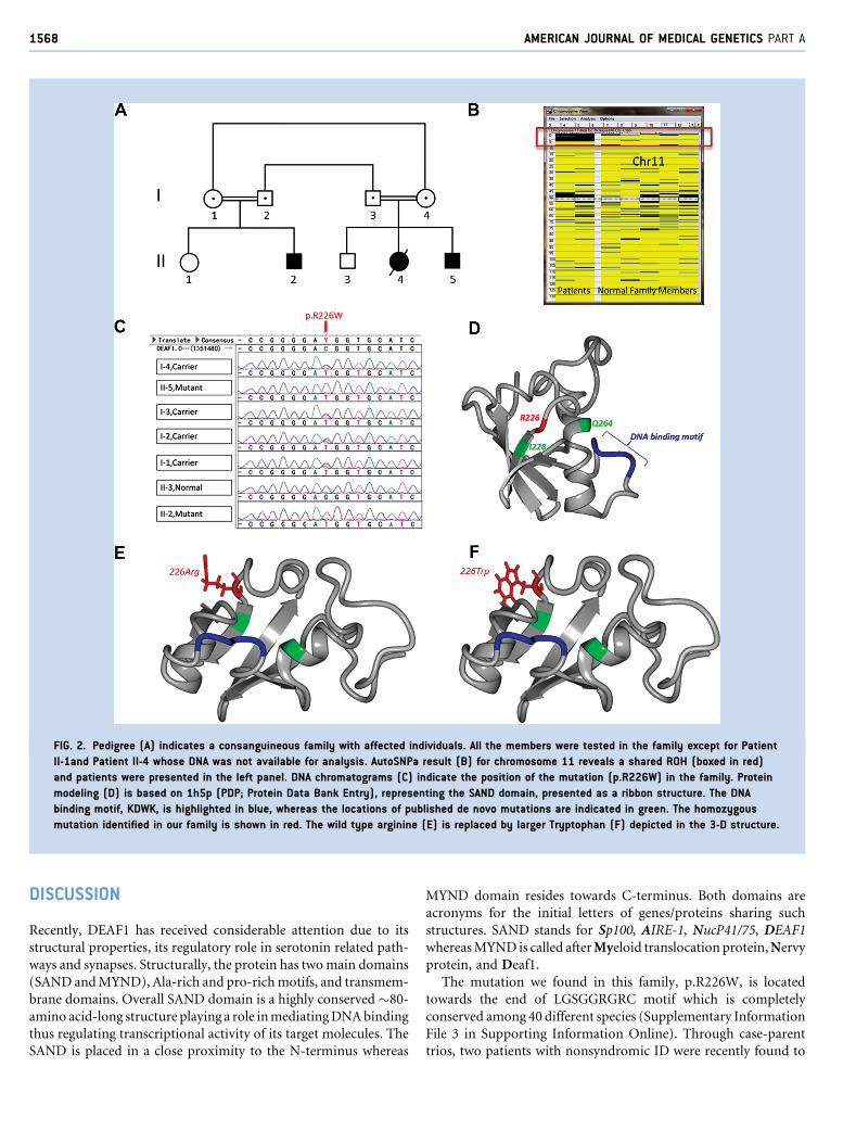
FIG. 2. Pedigree (A) indicates a consanguineous family with affected individuals. All the members were tested in the family except for Patient
II-1and Patient II-4 whose DNA was not available for analysis. AutoSNPa result (B) for chromosome 11 reveals a shared ROH (boxed in red)
and patients were presented in the left panel. DNA chromatograms (C) indicate the position of the mutation (p.R226W) in the family. Protein
modeling (D) is based on 1h5p (PDP; Protein Data Bank Entry), representing the SAND domain, presented as a ribbon structure. The DNA
binding motif, KDWK, is highlighted in blue, whereas the locations of published de novo mutations are indicated in green. The homozygous
mutation identified in our family is shown in red. The wild type arginine (E) is replaced by larger Tryptophan (F) depicted in the 3-D structure.
1568 AMERICAN JOURNAL OF MEDICAL GENETICS PART A
DISCUSSION
Recently, DEAF1 has received considerable attention due to its
structural properties, its regulatory role in serotonin related path-
ways and synapses. Structurally, the protein has twomain domains
(SAND andMYND), Ala-rich and pro-richmotifs, and transmem-
brane domains. Overall SAND domain is a highly conserved �80-
amino acid-long structure playing a role inmediatingDNAbinding
thus regulating transcriptional activity of its target molecules. The
SAND is placed in a close proximity to the N-terminus whereas
MYND domain resides towards C-terminus. Both domains are
acronyms for the initial letters of genes/proteins sharing such
structures. SAND stands for Sp100, AIRE-1, NucP41/75, DEAF1
whereasMYND is called afterMyeloid translocation protein,Nervy
protein, and Deaf1.
The mutation we found in this family, p.R226W, is located
towards the end of LGSGGRGRC motif which is completely
conserved among 40 different species (Supplementary Information
File 3 in Supporting Information Online). Through case-parent
trios, two patients with nonsyndromic ID were recently found to

FAQEIH ET AL. 1569
havedenovomutations (Thefirst is c.683T>G,p.Ile228Ser; and the
second is g.686871T>G, Gln264Pro) in DEAF1 in heterozygous
state that are likely to be pathogenic [Vissers et al., 2010; Rauch
et al., 2012]. The site of themutation in this report in relation to the
previously known de novo mutations is depicted on 3-D structure
(Fig. 2D). Previously known somatic mutations are given in
Supplementary Information File 4 (see Supporting Information
Online). The mutation, p.R226W, does not seem to directly inter-
fere with the DNA binding domain and located distantly from the
binding site; however, it is reasonable to consider that the large
tryptophan may stereotypically hinder normal protein folding
(Fig. 2E,F).
Interestingly, DEAF1 regulates its targets by a number of differ-
ent mechanisms. It is able to bind its own promoter as well as
promoters of other genes, and has capacity to regulate its targets
without promoter binding perhaps through protein–protein inter-
actions [Huggenvik et al., 1998; Michelson et al., 1999]. It is also
known that it acts on retinoic acid response element and may be
involved in arresting cells in the G0 or G1 phase [Manne
et al., 2001]. DEAF1 has also been shown to reduce activity of
the serotonin autoreceptor 5HT1A through allele-specific manner
[Lemonde et al., 2003, 2004; Albert and Lemonde, 2004].
Taken together, our results implicate an inheritedDEAF1 defect
in a syndromic form of ID and opens up new research possibilities
into the intricate involvement of SAND domain related defects in
human neurodevelopment, brain and synaptic function.
ACKNOWLEDGMENTS
We are very grateful to the family for their participation in this
study. This research was conducted through intramural funds
provided by King Faisal Specialist Hospital and Research Centre
(KFSHRC). We would like to thank KFSHRC/RAC for their kind
support. The authors have no conflict of interest to declare.
REFERENCES
Albert PR, Lemonde S. 2004. 5-HT1A receptors, gene repression, anddepression: Guilt by association. Neuroscientist 10:575–593.
BanEM, LeBoeuf RD. 1994. Suppressin: An endogenous negative regulatorof immune cell activation. Immunol Res 13:1–9.
Carr DJ, Blalock JE, Green MM, LeBoeuf RD. 1990. Immunomodulatorycharacteristics of a novel antiproliferative protein, suppressin. J Neuro-immunol 30:179–187.
Carr IM, Flintoff KJ, Taylor GR, Markham AF, Bonthron DT. 2006.Interactive visual analysis of SNP data for rapid autozygosity mappingin consanguineous families. Hum Mutat 27:1041–1046.
Czesak M, Le Francois B, Millar AM, Deria M, Daigle M, Visvader JE,Anisman H, Albert PR. 2012. Increased serotonin-1A (5-HT1A) autor-eceptor expression and reduced raphe serotonin levels in deformedepidermal autoregulatory factor-1 (Deaf-1) gene knock-out mice. JBiol Chem 287:6615–6627.
Czesak M, Lemonde S, Peterson EA, Rogaeva A, Albert PR. 2006. Cell-specific repressor or enhancer activities of Deaf-1 at a serotonin 1Areceptor gene polymorphism. J Neurosci 26:1864–1871.
HahmK, Sum EY, Fujiwara Y, Lindeman GJ, Visvader JE, Orkin SH. 2004.Defective neural tube closure and anteroposterior patterning in micelacking the LIMprotein LMO4or its interacting partnerDeaf-1.MolCellBiol 24:2074–2082.
Huggenvik JI, Michelson RJ, Collard MW, Ziemba AJ, Gurley P, MowenKA. 1998. Characterization of a nuclear deformed epidermal autoregu-latory factor-1 (DEAF-1)-related (NUDR) transcriptional regulatorprotein. Mol Endocrinol 12:1619–1639.
Jans LA, Riedel WJ, Markus CR, Blokland A. 2007. Serotonergic vulnera-bility and depression: Assumptions, experimental evidence and impli-cations. Mol Psychiatry 12:522–543.
LeBoeuf RD, BanEM,GreenMM, StoneAS, Propst SM, Blalock JE, TauberJD. 1998. Molecular cloning, sequence analysis, expression, and tissuedistribution of suppressin, a novel suppressor of cell cycle entry. J BiolChem 273:361–368.
LeBoeuf RD, Burns JN, Bost KL, Blalock JE. 1990. Isolation, purification,and partial characterization of suppressin, a novel inhibitor of cellproliferation. J Biol Chem 265:158–165.
Lemonde S, Du L, Bakish D, Hrdina P, Albert PR. 2004. Association of theC(-1019)G 5-HT1A functional promoter polymorphism with antide-pressant response. Int J Neuropsychopharmacol 7:501–506.
Lemonde S, Turecki G, Bakish D, Du L, Hrdina PD, Bown CD, SequeiraA, KushwahaN,Morris SJ, Basak A, OuXM, Albert PR. 2003. Impairedrepression at a 5-hydroxytryptamine 1A receptor gene polymorphismassociated with major depression and suicide. J Neurosci 23:8788–8799.
Mann JJ. 1999. Role of the serotonergic system in the pathogenesis ofmajordepression and suicidal behavior. Neuropsychopharmacology 21:99S–105S.
ManneU,GaryBD,OelschlagerDK,WeissHL,FrostAR,GrizzleWE.2001.Altered subcellular localization of suppressin, a novel inhibitor of cell-cycle entry, is an independent prognostic factor in colorectal adenocar-cinomas. Clin Cancer Res 7:3495–3503.
Michelson RJ, Collard MW, Ziemba AJ, Persinger J, Bartholomew B,Huggenvik JI. 1999. Nuclear DEAF-1-related (NUDR) protein containsa novel DNA binding domain and represses transcription of the hetero-geneous nuclear ribonucleoprotein A2/B1 promoter. J Biol Chem274:30510–30519.
Rauch A, Wieczorek D, Graf E, Wieland T, Endele S, Schwarzmayr T,Albrecht B, Bartholdi D, Beygo J, Di Donato N, Dufke A, Cremer K,Hempel M, Horn D, Hoyer J, Joset P, Ropke A, Moog U, Riess A, ThielCT, Tzschach A,Wiesener A,Wohlleber E, Zweier C, Ekici AB, Zink AM,RumpA,MeisingerC,GrallertH, StichtH, SchenckA,EngelsH,RappoldG, Schrock E,Wieacker P, Riess O,Meitinger T, Reis A, StromTM. 2012.Range of genetic mutations associated with severe non-syndromicsporadic intellectual disability: An exome sequencing study. Lancet380:1674–1682.
ReedDE,HuangXM,Wohlschlegel JA, LevineMS, SengerK. 2008.DEAF-1regulates immunity gene expression in Drosophila. Proc Natl Acad SciUSA 105:8351–8356.
Rozen S, SkaletskyH. 2000. Primer3 on theWWWfor general users and forbiologist programmers. Methods Mol Biol 132:365–386.
Seelow D, Schuelke M, Hildebrandt F, Nurnberg P. 2009. Homozygosi-tyMapper–an interactive approach to homozygosity mapping. NucleicAcids Res 37:W593–W599.
van Loo KM, Martens GJ. 2007. Genetic and environmental factors incomplex neurodevelopmental disorders. Curr Genomics 8:429–444.
Veraksa A, Kennison J, McGinnis W. 2002. DEAF-1 function is essentialfor the early embryonic development of Drosophila. Genesis 33:67–76.

1570 AMERICAN JOURNAL OF MEDICAL GENETICS PART A
Vissers LE, de Ligt J, Gilissen C, Janssen I, Steehouwer M, de Vries P, vanLier B, Arts P,WieskampN, del RosarioM, van Bon BW,Hoischen A, deVriesBB, BrunnerHG,Veltman JA. 2010.Adenovoparadigm formentalretardation. Nat Genet 42:1109–1112.
Yip L, Su L, Sheng D, Chang P, Atkinson M, Czesak M, Albert PR, CollierAR, Turley SJ, Fathman CG, Creusot RJ. 2009. Deaf1 isoforms controlthe expression of genes encoding peripheral tissue antigens in the
pancreatic lymph nodes during type 1 diabetes. Nat Immunol10:1026–1033.
SUPPORTING INFORMATION
Additional supporting information may be found in the online
version of this article at the publisher’s web-site.