Embed Size (px)
Citation preview
8/12/2019 Novel Hippocampal Neurogenic Agents.pdf
http://slidepdf.com/reader/full/novel-hippocampal-neurogenic-agentspdf 1/10
Discovery of Novel Hippocampal Neurogenic Agents by Usingan in Vivo Stable Isotope Labeling Technique
Mahalakshmi Shankaran, Chelsea King, Jean Lee, Robert Busch, Mary Wolff, andMarc K. Hellerstein
KineMed, Inc., Emeryville, California (M.S., C.K., J.L., R.B., M.W.); and Department of Nutritional Sciences and Toxicology,University of California Berkeley, Berkeley, California (M.K.H.)
Received July 11, 2006; accepted September 12, 2006
ABSTRACT
Neurogenesis occurs in discrete regions of adult mammalianbrain, including the subgranular zone of the hippocampus.Hippocampal neurogenesis is enhanced by different classes ofantidepressants, but screening for neurogenic actions of novelantidepressants has been inefficient because of limitations of5-bromo-2-deoxyuridine labeling techniques. We describe anefficient in vivo method for measuring hippocampal neurogen-esis involving incorporation of the stable isotope, 2H, intogenomic DNA during labeling with 2H2O (heavy water). Malerodents received 8 to 10% 2H2O in drinking water; DNA wasisolated from hippocampal progenitor cells or neurons. Labelincorporation into progenitor cells of Swiss-Webster mice re-vealed subpopulation kinetics: 16% divided with t 1/2 of 2.7weeks; the remainder did not divide over 1 year. Progenitor cellproliferation rates in mice were strain-dependent. Chronic an-tidepressant treatment for 3 weeks, with 2H2O administered
during the final week, increased progenitor cell proliferationacross all the strains tested. Fluoxetine treatment increased 2Hincorporation into DNA of gradient-enriched neurons or flow-sorted neuronal nuclei 4 weeks after 2H2O labeling, represent-ing the survival and differentiation of newly divided cells intoneurons. By screening 11 approved drugs for effects on pro-genitor cell proliferation, we detected previously unrecognized,dose-dependent enhancement of hippocampal progenitor cellproliferation by two statins and the anticonvulsant topiramate.We also confirmed stimulatory activity of other anticonvulsantsand showed inhibition of progenitor cell proliferation by isotreti-noin and prednisolone. In conclusion, stable isotope labeling isan efficient, high-throughput in vivo method for measuring hip-pocampal progenitor cell proliferation that can be used toscreen for novel neurogenic drugs.
In rodents, adult neurogenesis occurs in discrete regions of the brain, particularly in the subventricular zone, giving riseto granule cells in the olfactory bulb, and in the subgranularzone, generating new granule cells in the dentate gyrus of thehippocampus (Altman and Das, 1965; Kaplan and Hinds,1977; Kuhn et al., 1996). Hippocampal neuronal cells in theadult are formed through replication and differentiation frompluripotent neural progenitor cells (Gage et al., 1998). Imma-ture neurons migrate to the granule cell layer and mature
over a period of weeks into granule cells that form functionalconnections (van Praag et al., 2002).
Hippocampal neurogenesis has emerged as a central ther-apeutic target for antidepressant agents based on accumu-
lating evidence supporting the neurogenic theory of depres-sion (Jacobs et al., 2000; Kempermann, 2002). All the known
classes of clinical antidepressant drugs, including tricyclics,monoamine oxidase inhibitors, and selective serotonin re-
uptake inhibitors (SSRI), have been shown to increase cellproliferation in the hippocampus (Malberg et al., 2000; San-
tarelli et al., 2003). Moreover, Santarelli et al. (2003) pro- vided evidence, by irradiation of the hippocampus, that hip-pocampal neurogenesis is required to achieve the behavioral
effects of antidepressants in animal models. Hippocampalcell proliferation is decreased under conditions of chronic
stress, and this effect is reversed by antidepressant treat-ment (reviewed by Dranovsky and Hen, 2006; Warner-Schmidt and Duman, 2006). Imaging studies have also
shown that depressed human subjects exhibit volume loss
This study was supported in part by KineMed, Inc. and University of California-Industry Discovery Program (BioStar Grant No. Bio04-10445 toM.K.H.).
Part of this work was presented at the Annual Society of Neuroscience Meeting: Shankaran M, King C, Busch R, Gee T, Keifer K, Mahsut A, andHellerstein M (2005) In vivo heavy water (2H2O) labeling: a novel method formeasuring hippocampal cell proliferation and neurogenesis. Program 1024.9,in 2005 Abstract Viewer/Itinerary Planner; 2005 Nov 12–16; Washington, DC.Online, Society for Neuroscience, Washington, DC.
Article, publication date, and citation information can be found athttp://jpet.aspetjournals.org.
doi:10.1124/jpet.106.110510.
ABBREVIATIONS: SSRI, selective serotonin reuptake inhibitor(s); BrdU, 5-bromo-2-deoxyuridine; PBS, phosphate-buffered saline; TTX, tetanus
toxin C fragment; PI, propidium iodide; GC/MS, gas chromatography/mass spectrometry.
0022-3565/06/3193-1172–1182$20.00THE JOURNAL OF PHARMACOLOGY AND E XPERIMENTAL THERAPEUTICS Vol. 319, No. 3Copyright © 2006 by The American Society for Pharmacology and Experimental Therapeutics 110510/3154770JPET 319:1172–1182, 2006 Printed in U.S.A.
1172
8/12/2019 Novel Hippocampal Neurogenic Agents.pdf
http://slidepdf.com/reader/full/novel-hippocampal-neurogenic-agentspdf 2/10
and atrophy of the hippocampus (Sheline, 2003). In additionto a role in mood disorders, increased neurogenesis may be arepair mechanism in stroke (Lichtenwalner and Parent,2006) and traumatic brain injury (Emery et al., 2005) andmay also contribute to learning and memory (Shors et al.,2001).
A number of factors and intersecting pathways influencethe drug-induced stimulation of neurogenesis in the hip-
pocampus. For example, antidepressant drugs activate intra-cellular second messenger systems, leading to the activationof transcription factors and neurotrophic factors, finally cul-minating in increased numbers of new neurons in the hip-pocampus (Warner-Schmidt and Duman, 2006). The com-plexity of neurogenic regulation opens the possibility thatmultiple therapeutic targets may exist, on the one hand, butalso makes predicting the effect of any agent acting on aparticular target difficult and complicates the interpretationof in vitro screening approaches based on receptor binding ormodulation of enzyme activities. Therefore, it is important to validate in vivo the neurogenic activity of agents identifiedthrough in vitro screens; moreover, in vivo screening may
uncover novel mechanisms that contribute to neurogenesisarising from unanticipated connectivity relationships in thewhole organism.
Measurement of neurogenesis in vivo has been problem-atic, however. Early studies used [3H]thymidine to label di- viding cells (Altman and Das, 1965; Kaplan and Hinds,1977), whereas the most commonly used method currentlyfor measuring cell proliferation in the hippocampus involvesinjecting animals with 5-bromo-2-deoxyuridine (BrdU) 24 hbefore sacrifice (Kuhn et al., 1996; Cameron and McKay,
2001). The hippocampus is then serially sectioned for immu-nohistochemical detection of BrdU, as well as double-immu-nohistochemical labeling with neuronal markers, to establishthe phenotype of BrdU-positive cells. BrdU labeling has lim-itations, however, such as a short half-life and rapid clear-ance of BrdU from the brain, variable efficiency of BrdUentry into cellular precursor pools, and the requirement forhigh doses to accurately estimate the number of proliferating
cells (Cameron and McKay, 2001; Gould and Gross, 2002).Moreover, immunohistochemical enumeration of BrdU-la-beled cells in the entire hippocampus is labor-intensive, sothat throughput with BrdU labeling is not sufficient for usein broad screening or testing of potential neurogenic agents.
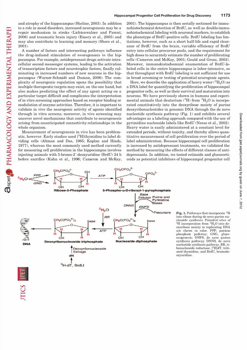
Here, we describe the application of heavy water (2H2O) asa DNA label for quantifying the proliferation of hippocampalprogenitor cells, as well as their survival and maturation intoneurons. We have previously shown in humans and experi-mental animals that deuterium (2H) from 2H2O is incorpo-rated constitutively into the deoxyribose moiety of purinedeoxyribonucleotides in genomic DNA through the de novonucleotide synthesis pathway (Fig. 1) and exhibits several
advantages as a labeling approach compared with the use of pyrimidine nucleoside labels like BrdU (Neese et al., 2002).Heavy water is easily administered at a constant level forextended periods, without toxicity, and thereby allows quan-titative measurement of cell proliferation over the period of label administration. Because hippocampal cell proliferationis increased by antidepressant treatments, we validated themethod by measuring the effects of different classes of anti-depressants. In addition, we tested retinoids and glucocorti-coids as potential inhibitors of hippocampal progenitor cell
Fig. 1. Pathways that incorporate 2Hinto ribose during de novo purine nu-cleoside synthesis. Potential sites of 2H incorporation from 2H2O into de-oxyribose moiety in replicating DNA are shown in color. PPP, pentosephosphate pathway; GNG, gluco-neogenesis; DNPS, de novo purine
synthesis pathway; DNNS, de novonucleotide synthesis pathway, RR, ri-bonucleoside reductase, [3H]dT, triti-ated thymidine, and BrdU, bromode-oxyuridine.
Hippocampal Progenitor Cell Proliferation for Drug Discovery 1173
8/12/2019 Novel Hippocampal Neurogenic Agents.pdf
http://slidepdf.com/reader/full/novel-hippocampal-neurogenic-agentspdf 3/10
proliferation. We then used this biomarker as a relativelyhigh-throughput in vivo screening tool to discover previouslyunrecognized neurogenic stimulatory actions of approveddrugs.
Materials and Methods
Animals. All of the animal studies were carried out within Na-
tional Institutes of Health guidelines for the care and use of labora-tory animals and received approval from the institutional animal usecommittee. Ten- to 12-week-old male C57Bl/6, Swiss-Webster, orBALB/c mice were obtained from Charles River Laboratories (Wil-mington, MA), and 10- to 12-week-old male 129SvEv mice wereobtained from Taconic (Oxnard, CA). The outbred Swiss-Websterstrain of mice was used for the initial drug-screening experiments.Subsequent experiments were performed in the inbred 129SvEvstrain because these showed the least interanimal variability. MaleSprague-Dawley rats (250 g) were obtained from Charles River Lab-oratories. Animals were housed in a climate-controlled environmentand fed standard rodent chow and water ad libitum.
2H2
O Labeling. For 2H2O labeling, animals received a priming i.p. bolusof 49 ml/kg 99% 2H2O (Spectra Stable Isotopes, Columbia,MD) containing 0.9% NaCl and were maintained on 10% 2H2O in
drinking water for the duration of the labeling period. In previousstudies (reviewed in Jones and Leatherdale, 1991), intake of up to20% 2H2O has no apparent phenotypic or behavioral effects. Forprotocols involving label incorporation in hippocampal progenitorcells, mice were labeled continuously for up to 1 year. In a separatestudy, total hippocampal tissue was isolated after 3, 7, or 14 days of labeling. Animals that received drug treatment were labeled during the final 7 to 10 days of treatment. For studies that assessed prolif-eration of mature neurons, animals were labeled with 10% 2H2O for3 weeks, after which time the label was discontinued, and animalswere sacrificed 4 weeks later.
Drug Treatments. Male rats and mice were treated with anti-depressants of different classes, such as an SSRI (fluoxetine), atricyclic (imipramine), or a serotonin-norepinephrine reuptake inhib-
itor (venlafaxine), administered in drinking water. The drug solu-tions were dissolved at a concentration of 100, 200, and 100 mg/l forfluoxetine, imipramine, and venlafaxine, respectively. These concen-trations were calculated to achieve a dose of 10 mg/kg/day for fluox-etine and venlafaxine and 20 mg/kg/day for imipramine, based on theaverage cage consumption of water (3 ml/30-g mouse or 25 ml/250-g rat). Treatment with antidepressant drugs was continued for 3 to 5weeks.
Various approved drugs were screened for neurogenic activity(Table 1). These agents were selected based on long-standing use inhumans and on the recognition of having several therapeutic actions(i.e., pleiotropic effects). This effort was intended to be a proof-of-concept study to show “indications discovery,” i.e., ability to findunexpected actions of agents with potential use in a new indication.The doses selected were based on use in published preclinical studies
with these agents, and the concentrations in food or water were
calculated based on the average cage consumption of food (3 g/30-g
mouse) and water (3 ml/30-g mouse). As a follow-up study after
initial screening, a potential “class” effect for one of the agents
(topiramate) tested in the initial screen was investigated by treating
mice p.o. for 3 weeks with 1 of 11 other anticonvulsants: valproate (1
g/kg), clonazepam (3 mg/kg), gabapentin (100 mg/kg), carbamazepine
(30 mg/kg), ethosuximide (300 mg/kg), levetiracetam (30 mg/kg),
oxcarbazepine (100 mg/kg), phenytoin (100 mg/kg), primidone (100
mg/kg), tiagabine (30 mg/kg), or zonisamide (50 mg/kg).
Dose-response studies were also done for topiramate (10, 30, 100,
and 150 mg/kg p.o.), atorvastatin (1, 3, 10, and 30 mg/kg in diet), and
simvastatin (1, 3, 10, and 30 mg/kg p.o.). In addition, mice received
chronic treatment with potential inhibitors of neurogenesis, such as
isotretinoin (1 and 3 mg/kg i.p.) or prednisolone (5 and 40 mg/kg in
diet).
Isolation of Hippocampal Progenitor Cells and Neurons.
Animals were euthanized by CO2 asphyxiation; brains were imme-
diately removed; and the hippocampus was dissected out. Progenitor
cells and neurons were isolated by a modification of methods de-
scribed previously (Palmer et al., 1999). In brief, tissues were finely
minced and digested in a solution of papain (4 U/ml) (Worthington
Biochemical Corporation, Lakewood, NJ) and DNase (250 U/ml)
(Roche Applied Science, Indianapolis, IN) dissolved in Hibernate-A (BrainBits LLC, Springfield, IL). The digested tissue was then me-
chanically triturated and thoroughly mixed with an equal volume of
Percoll solution, made by mixing nine parts of Percoll (Amersham
Biosciences, Piscataway, NJ) with one part 10 phosphate-buffered
saline (PBS). The cell suspension was fractionated by centrifugation
for 30 min, 18°C at 20,000 g. Density beads were run in parallel, and
the progenitor cells were fractionated between 1.064 and 1.075 g/ml,
whereas neuronal cells fractionated at densities 1.035 g/ml. The
progenitor and neuronal cell fractions were collected, washed free of
Percoll, and stored frozen at 20°C until isolation of DNA. A flow
chart of the method is shown in Fig. 1A.
Characterization of Progenitor Cells and Neurons by Flow
Cytometry. The gradient-purified progenitor cells were immun-
ofluorescently stained for intracellular markers, nestin and vimen-tin, and analyzed by flow cytometry. The cells were fixed and per-
meabilized with IntraCyte buffers (Orion Biosolutions, Vista, CA)
and incubated overnight at 4°C with mouse antinestin (1:50, Rat401;
BD Pharmingen, San Diego, CA) or mouse antivimentin (1:50; BD
Pharmingen) primary antibodies. Gradient-purified hippocampal
neurons were stained for tetanus toxin C fragment (TTX), a cell
surface marker for neurons. The cells were fixed with 4% parafor-
maldehyde and incubated with TTX followed by anti-TTX mouse
monoclonal antibody (Roche Applied Science). After washing, cells
were incubated with Alexa 488-conjugated goat anti-mouse IgG sec-
ondary antibody (Molecular Probes, Eugene, OR). Propidium iodide
(PI) was used to stain DNA, and Alexa 488 staining of PI-positive
single cells (gated on plots of forward scatter peak area versus peak
TABLE 1
Drug Class Dose Route
Aspirin NSAID 60 mg/kg/day Diet Atorvastatin HMG CoA reductase inhibitor 10 mg/kg/day Oral gavageCalcitriol Vitamin D analog 2.5 g/kg/day DietClofibrate PPAR- agonist 50 mg/kg/day DietEnalapril maleate ACE inhibitor 2.5 mg/kg/day Drinking waterEtiocholanedione Steroid analog 1.5 g/kg/day DietFlurbiprofen NSAID 50 mg/kg/day i.p.Ketoconazole Antifungal 100 mg/kg/day DietMethotrexate Folate inhibitor 20 mg/kg/2days i.p.Rosiglitazone PPAR- agonist 6 mg/kg/day DietTopiramate Anticonvulsant 100 mg/kg/day Diet
HMG CoA, hydroxyl-methylglutaryl CoA; NSAID, nonsteroidal anti-inflammatory drug; PPAR, peroxisome proliferator-activated receptor; ACE, angiotensin-converting
enzyme.
1174 Shankaran et al.
8/12/2019 Novel Hippocampal Neurogenic Agents.pdf
http://slidepdf.com/reader/full/novel-hippocampal-neurogenic-agentspdf 4/10
height) was analyzed on a Coulter EpicsXL cytometer (BeckmanCoulter, Fullerton, CA).
Isolation of Nuclei from Mature Neurons and Sorting
NeuN-Positive Nuclei by Flow Cytometry. Neuronal nuclei wereisolated from frozen hippocampal tissue by a modification of a re-cently described method (Spalding et al., 2005). In brief, tissue washomogenized in 1 ml of lysis buffer (0.32 M sucrose, 5 mM CaCl2, 3mM magnesium acetate, 0.1 mM EDTA, 10 mM Tris-HCl, pH 8.0,0.1% Triton X-100, and 1 mM DTT). Homogenized samples were
gently suspended in 1.8 ml of sucrose solution (1.8 M sucrose, 3 mMmagnesium acetate, 1 mM DTT, and 10 mM Tris-HCl, pH 8.0),layered onto a cushion of 1 ml of sucrose solution, and centrifuged at30,000 g for 2.5 h at 4°C. The isolated nuclei were resuspended in 1ml of PBS and stored overnight at 4°C.
To identify neuronal nuclei, anti-NeuN antibodies were directlyconjugated with Zenon mouse IgG labeling reagent (Alexa 488; Mo-lecular Probes) by mixing 10 l of Alexa 488 conjugate in 100 l of blocking buffer (PBS/0.5% bovine serum albumin/10% normal goatserum) with 300 l of NeuN antibody (1 mg/ml; diluted 1:250 in PBS)and incubating at room temperature for 5 min. The suspension of nuclei (1 ml) was added and incubated at 4°C for 1 h. The nuclei werethen washed twice with 3 ml of PBS by centrifuging at 1000 g for 10min. PI was used to stain DNA, and neuronal nuclei were sorted as
a homogeneous population of NeuN
bright
cells using a Coulter EpicsElite sorter (Beckman Coulter) with gates set for PI-positive singlenuclei.
Measurement of Cell Proliferation. Gas chromatography/massspectrometry (GC/MS) analyses were performed as described previ-ously (Neese et al., 2002; Busch et al., 2004) to measure 2H incorpo-ration from 2H2O into purine deoxyribonucleotides in genomic DNA.In brief, DNA was isolated from isolated progenitor cells, sortedneurons, or hippocampal tissue using a DNeasy tissue kit (Qiagen,
Valencia, CA) and hydrolyzed enzymatically to free deoxyribonucleo-sides. The deoxyribose moiety of purine deoxyribonucleosides wasconverted to the pentafluorobenzyl tetraacetate derivative and ana-
lyzed by GC/MS in the negative chemical ionization mode using an Agilent (Palo Alto, CA) model 5973 mass spectrometer and a 6890gas chromatograph fitted with a db-225 column. Selected ion moni-toring was performed with mass/charge ratios (m / z) of 435 for the M0and 436 for M1 mass isotopomer, respectively. Incorporation of 2Hfrom 2H2O into purine deoxyribose was quantified as the molarexcess fraction M1 (EM1), i.e., the increase over natural abundance(background) defined as the fractional M1 value for an unlabeledDNA standard from calf thymus.
EM1 abundance m / z 436sample
abundance m / z 435 436sample
abundance m / z 436standard
abundance m / z 435 436standard
The fraction of newly labeled cells was calculated as the ratio of excess 2H enrichment (EM1) in isolated cells to the corresponding
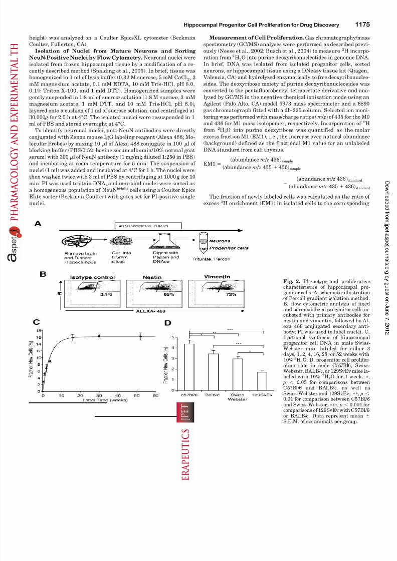
Fig. 2. Phenotype and proliferative
characteristics of hippocampal pro-genitor cells. A, schematic illustrationof Percoll gradient isolation method.B, flow cytometric analysis of fixedand permeabilized progenitor cells in-cubated with primary antibodies fornestin and vimentin, followed by Al-exa 488 conjugated secondary anti-body; PI was used to label nuclei. C,fractional synthesis of hippocampalprogenitor cell DNA in male Swiss-Webster mice labeled for either 3days, 1, 2, 4, 16, 28, or 52 weeks with10% 2H2O. D, progenitor cell prolifer-ation rate in male C57/Bl6, Swiss-Webster, BALB/c, or 129SvEv mice la-beled with 10% 2H2O for 1 week. ,
p 0.05 for comparisons betweenC57Bl/6 and BALB/c, as well as
Swiss-Webster and 129SvEv; , p 0.01 for comparison between C57Bl/6and Swiss-Webster; , p 0.001 forcomparisons of 129SvEv with C57Bl/6or BALB/c. Data represent mean S.E.M. of six animals per group.
Hippocampal Progenitor Cell Proliferation for Drug Discovery 1175
8/12/2019 Novel Hippocampal Neurogenic Agents.pdf
http://slidepdf.com/reader/full/novel-hippocampal-neurogenic-agentspdf 5/10
enrichment in bone marrow DNA (an essentially fully turned-overtissue after 7–10 days of labeling, thereby representing an asymp-totic enrichment value for comparison with other tissues) as de-scribed previously (Neese et al., 2002). For studies involving shorterlabeling periods than 7 to 10 days, 2H incorporation in fully turned-over tissue DNA was estimated from body water 2H enrichments atsacrifice based on previously established relationships between bodywater 2H enrichments and asymptotic labeling in tissues (Neese etal., 2002).
Statistical Analysis. For label incorporation curves, the datawere fit by nonlinear regression analysis (SigmaPlot). Student’s t
test was used with a 95% confidence interval for comparison betweentwo groups. For comparison between multiple groups, one-way anal-ysis of variance was used with a post-hoc Student-Newman-Keul testfor all the pairwise multiple comparisons or Dunnett’s test for com-parisons with a negative control (SigmaStat). Data were consideredsignificant at p 0.05.
Results
Flow Cytometric Analysis of Gradient-Enriched Hip-
pocampal Progenitor Cells. Measurement of 2H incorpo-ration into the DNA of hippocampal progenitor cells requires
isolation of this cell population from intact tissue. To thisend, progenitor cells were isolated from the hippocampus of Swiss-Webster mice by Percoll gradient fractionation (Fig.2A); the isolated cells were fixed and permeabilized, stainedfor specific intracellular markers of progenitor cells (nestinand vimentin), and analyzed by flow cytometry after gating on nucleated (PI-stained) cells and excluding doublets. A majority of cells stained positively for either nestin (65%positive in Fig. 2B) or vimentin (72% positive) compared withisotype controls (2.1% positive). A broad distribution of fluo-rescence intensity was observed, and the cells remaining inthe negative region of the dot plot appeared to be weaklystained.
Label Incorporation Kinetics in Progenitor Cells. Tocharacterize the proliferation kinetics of gradient-enrichedhippocampal progenitor cells, Swiss-Webster mice were la-beled continuously with 10% 2H2O in drinking water starting at 10 weeks of age; hippocampal progenitor cells were iso-lated after various labeling times, and 2H incorporation intoDNA, analyzed by GC/MS, was used to determine the frac-tion of cells that had incorporated the label through celldivision (Fig. 2C). Approximately 16% of cells incorporatedlabel at plateau, with a half-life within this dividing popula-tion of approximately 2.7 weeks; the majority of cells contin-ued to be unlabeled over the course of 1 year. This is consis-tent with a “kinetic subpopulation” pattern wherein apreponderance of nondividing precursors is present with a
subset of actively dividing cells that enters and exits the pool(by differentiation or death). The baseline rate of progenitorproliferation was strain-dependent (Fig. 2D). The initialrates of labeling, measured after 1 week of 2H2O intake, variedapproximately 3-fold among four mouse strains tested and weresignificantly ( p 0.001) different from each other. The highestproliferation rate was observed in C57Bl/6 mice, whereas the129SvEv mice had the lowest rate of proliferation.
The fraction of new cells in whole hippocampal tissue of C57Bl/6 mice after 3, 7, or 14 days of labeling was 0.7 0.01,1.3 0.3, and 2.9 0.6%, respectively. Thus, gradient iso-lation enriched for proliferating cells, and the consistency of the labeling results indicated that the cell isolation method
was highly consistent in this regard.
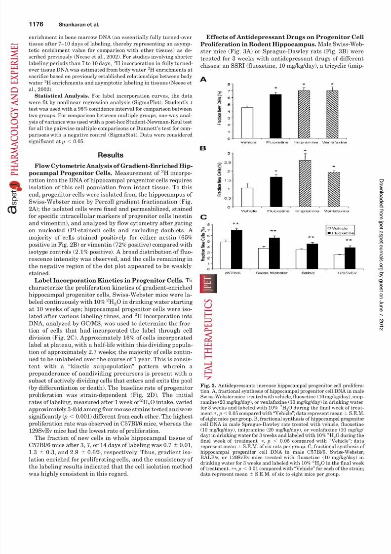
Effects of Antidepressant Drugs on Progenitor Cell
Proliferation in Rodent Hippocampus. Male Swiss-Web-ster mice (Fig. 3A) or Sprague-Dawley rats (Fig. 3B) weretreated for 3 weeks with antidepressant drugs of differentclasses: an SSRI (fluoxetine, 10 mg/kg/day), a tricyclic (imip-
Fig. 3. Antidepressants increase hippocampal progenitor cell prolifera-tion. A, fractional synthesis of hippocampal progenitor cell DNA in maleSwiss-Webster mice treated with vehicle, fluoxetine (10 mg/kg/day), imip-ramine (20 mg/kg/day), or venlafaxine (10 mg/kg/day) in drinking waterfor 3 weeks and labeled with 10% 2H2O during the final week of treat-ment. , p 0.05 compared with “Vehicle”; data represent mean S.E.M.of eight mice per group. B, fractional synthesis of hippocampal progenitorcell DNA in male Sprague-Dawley rats treated with vehicle, fluoxetine(10 mg/kg/day), imipramine (20 mg/kg/day), or venlafaxine (10 mg/kg/ day) in drinking water for 3 weeks and labeled with 10% 2H2O during thefinal week of treatment. , p 0.05 compared with “Vehicle”; datarepresent mean S.E.M. of six rats per group. C, fractional synthesis of hippocampal progenitor cell DNA in male C57Bl/6, Swiss-Webster,BALB/c, or 129SvEv mice treated with fluoxetine (10 mg/kg/day) indrinking water for 3 weeks and labeled with 10% 2H2O in the final weekof treatment. , p 0.01 compared with “Vehicle” for each of the strain;
data represent mean S.E.M. of six to eight mice per group.
1176 Shankaran et al.
8/12/2019 Novel Hippocampal Neurogenic Agents.pdf
http://slidepdf.com/reader/full/novel-hippocampal-neurogenic-agentspdf 6/10
ramine, 20 mg/kg/day), or a serotonin-norepinephrine re-uptake inhibitor (venlafaxine, 10 mg/kg/day). After labeling with 2H2O during the last week of treatment, antidepres-sant-treated animals from all the groups showed a signifi-cant increase in the progenitor cell proliferation rate in thehippocampus (Fig. 3, A and B). Both baseline proliferationand the magnitude of the drug effects were somewhat differ-ent between mice and rats, however. Fluoxetine treatment
produced a significant ( p 0.01) increase in the hippocampalprogenitor cell proliferation in C57Bl/6, 129SvEv, Swiss-Webster, and BALB/c mice (Fig. 3C). The percent stimulationof progenitor cell proliferation by fluoxetine was similaracross strains.
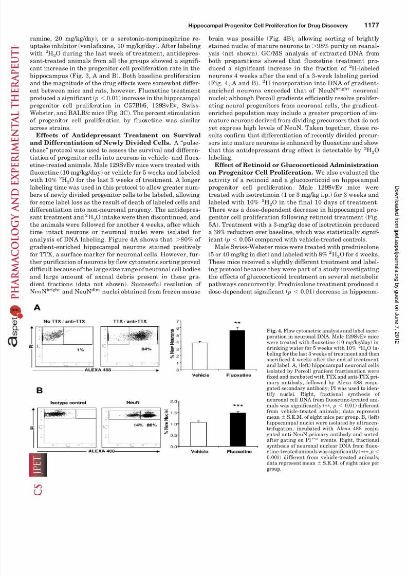
Effects of Antidepressant Treatment on Survival
and Differentiation of Newly Divided Cells. A “pulse-chase” protocol was used to assess the survival and differen-tiation of progenitor cells into neurons in vehicle- and fluox-etine-treated animals. Male 129SvEv mice were treated withfluoxetine (10 mg/kg/day) or vehicle for 5 weeks and labeledwith 10% 2H2O for the last 3 weeks of treatment. A longerlabeling time was used in this protocol to allow greater num-
bers of newly divided progenitor cells to be labeled, allowing for some label loss as the result of death of labeled cells anddifferentiation into non-neuronal progeny. The antidepres-sant treatment and 2H2O intake were then discontinued, andthe animals were followed for another 4 weeks, after whichtime intact neurons or neuronal nuclei were isolated foranalysis of DNA labeling. Figure 4A shows that 80% of gradient-enriched hippocampal neurons stained positivelyfor TTX, a surface marker for neuronal cells. However, fur-ther purification of neurons by flow cytometric sorting proveddifficult because of the large size range of neuronal cell bodiesand large amount of axonal debris present in these gra-dient fractions (data not shown). Successful resolution of NeuNbright and NeuNdim nuclei obtained from frozen mouse
brain was possible (Fig. 4B), allowing sorting of brightlystained nuclei of mature neurons to 98% purity on reanal-ysis (not shown). GC/MS analysis of extracted DNA fromboth preparations showed that fluoxetine treatment pro-duced a significant increase in the fraction of 2H-labeledneurons 4 weeks after the end of a 3-week labeling period(Fig. 4, A and B). 2H incorporation into DNA of gradient-enriched neurons exceeded that of NeuNbright neuronal
nuclei; although Percoll gradients efficiently resolve prolifer-ating neural progenitors from neuronal cells, the gradient-enriched population may include a greater proportion of im-mature neurons derived from dividing precursors that do notyet express high levels of NeuN. Taken together, these re-sults confirm that differentiation of recently divided precur-sors into mature neurons is enhanced by fluoxetine and showthat this antidepressant drug effect is detectable by 2H2Olabeling.
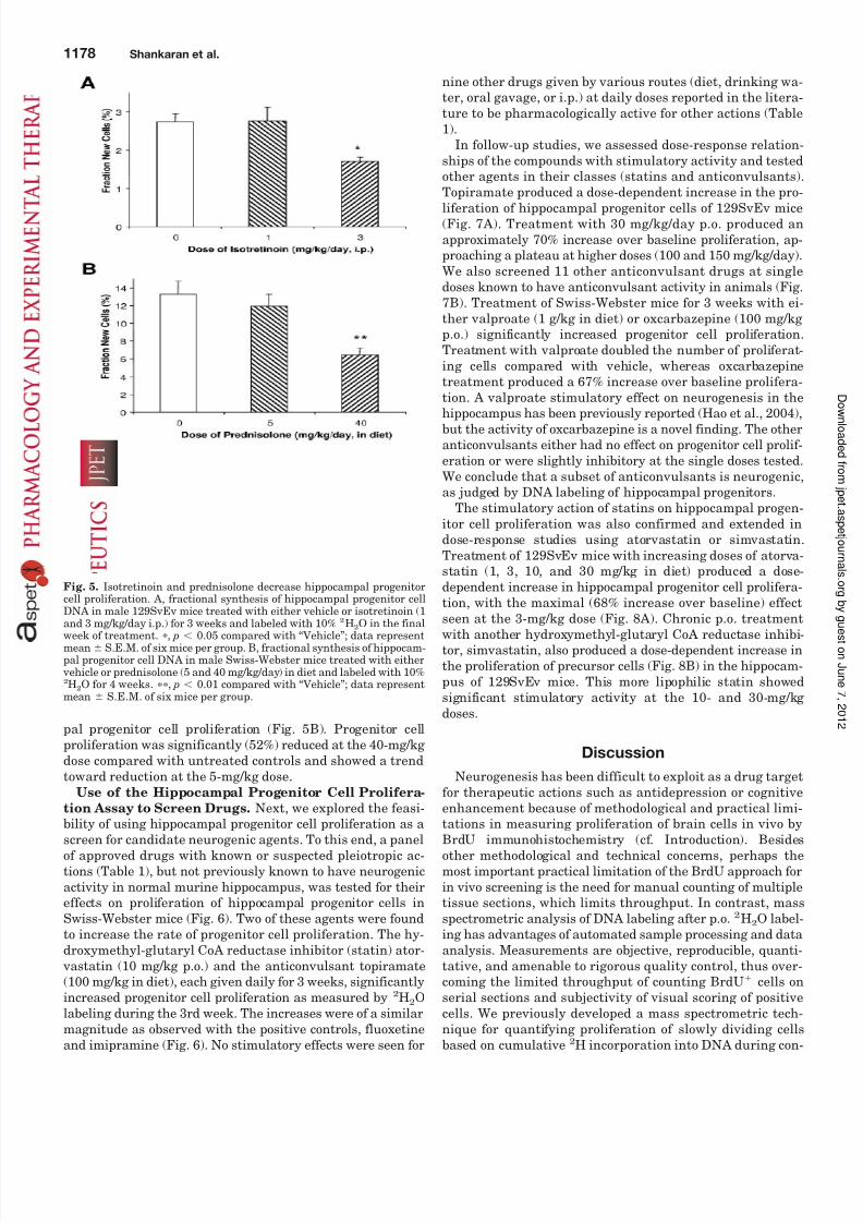
Effect of Retinoid or Glucocorticoid Administration
on Progenitor Cell Proliferation. We also evaluated theactivity of a retinoid and a glucocorticoid on hippocampalprogenitor cell proliferation. Male 129SvEv mice were
treated with isotretinoin (1 or 3 mg/kg i.p.) for 3 weeks andlabeled with 10% 2H2O in the final 10 days of treatment.There was a dose-dependent decrease in hippocampal pro-genitor cell proliferation following retinoid treatment (Fig.5A). Treatment with a 3-mg/kg dose of isotretinoin produceda 38% reduction over baseline, which was statistically signif-icant ( p 0.05) compared with vehicle-treated controls.
Male Swiss-Webster mice were treated with prednisolone(5 or 40 mg/kg in diet) and labeled with 8% 2H2O for 4 weeks.These mice received a slightly different treatment and label-ing protocol because they were part of a study investigating the effects of glucocorticoid treatment on several metabolicpathways concurrently. Prednisolone treatment produced adose-dependent significant ( p 0.01) decrease in hippocam-
Fig. 4. Flow cytometric analysis and label incor-poration in neuronal DNA. Male 129SvEv micewere treated with fluoxetine (10 mg/kg/day) indrinking water for 5 weeks with 10% 2H2O la-beling for the last 3 weeks of treatment and thensacrificed 4 weeks after the end of treatmentand label. A, (left) hippocampal neuronal cellsisolated by Percoll gradient fractionation werefixed and incubated with TTX and anti-TTX pri-mary antibody, followed by Alexa 488 conju-gated secondary antibody; PI was used to iden-
tify nuclei. Right, fractional synthesis of neuronal cell DNA from fluoxetine-treated ani-mals was significantly (, p 0.01) differentfrom vehicle-treated animals; data representmean S.E.M. of eight mice per group. B, (left)hippocampal nuclei were isolated by ultracen-trifugation, incubated with Alexa 488 conju-gated anti-NeuN primary antibody and sortedafter gating on PI ve events. Right, fractionalsynthesis of neuronal nuclear DNA from fluox-etine-treated animals was significantly (, p 0.001) different from vehicle-treated animals;data represent mean S.E.M. of eight mice pergroup.
Hippocampal Progenitor Cell Proliferation for Drug Discovery 1177
8/12/2019 Novel Hippocampal Neurogenic Agents.pdf
http://slidepdf.com/reader/full/novel-hippocampal-neurogenic-agentspdf 7/10
pal progenitor cell proliferation (Fig. 5B). Progenitor cellproliferation was significantly (52%) reduced at the 40-mg/kg dose compared with untreated controls and showed a trendtoward reduction at the 5-mg/kg dose.
Use of the Hippocampal Progenitor Cell Prolifera-
tion Assay to Screen Drugs. Next, we explored the feasi-bility of using hippocampal progenitor cell proliferation as a
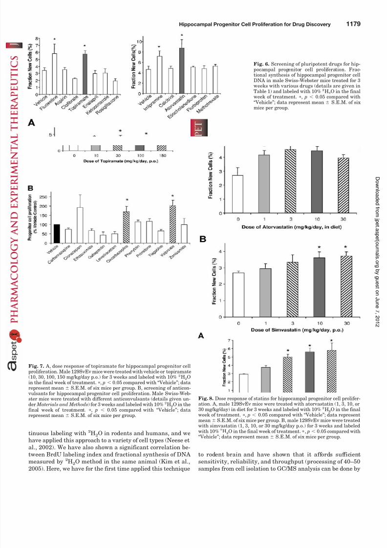
screen for candidate neurogenic agents. To this end, a panelof approved drugs with known or suspected pleiotropic ac-tions (Table 1), but not previously known to have neurogenicactivity in normal murine hippocampus, was tested for theireffects on proliferation of hippocampal progenitor cells inSwiss-Webster mice (Fig. 6). Two of these agents were foundto increase the rate of progenitor cell proliferation. The hy-droxymethyl-glutaryl CoA reductase inhibitor (statin) ator- vastatin (10 mg/kg p.o.) and the anticonvulsant topiramate(100 mg/kg in diet), each given daily for 3 weeks, significantlyincreased progenitor cell proliferation as measured by 2H2Olabeling during the 3rd week. The increases were of a similarmagnitude as observed with the positive controls, fluoxetine
and imipramine (Fig. 6). No stimulatory effects were seen for
nine other drugs given by various routes (diet, drinking wa-ter, oral gavage, or i.p.) at daily doses reported in the litera-ture to be pharmacologically active for other actions (Table1).
In follow-up studies, we assessed dose-response relation-ships of the compounds with stimulatory activity and testedother agents in their classes (statins and anticonvulsants).Topiramate produced a dose-dependent increase in the pro-
liferation of hippocampal progenitor cells of 129SvEv mice(Fig. 7A). Treatment with 30 mg/kg/day p.o. produced anapproximately 70% increase over baseline proliferation, ap-proaching a plateau at higher doses (100 and 150 mg/kg/day).We also screened 11 other anticonvulsant drugs at singledoses known to have anticonvulsant activity in animals (Fig.7B). Treatment of Swiss-Webster mice for 3 weeks with ei-ther valproate (1 g/kg in diet) or oxcarbazepine (100 mg/kg p.o.) significantly increased progenitor cell proliferation.Treatment with valproate doubled the number of proliferat-ing cells compared with vehicle, whereas oxcarbazepinetreatment produced a 67% increase over baseline prolifera-tion. A valproate stimulatory effect on neurogenesis in the
hippocampus has been previously reported (Hao et al., 2004),but the activity of oxcarbazepine is a novel finding. The otheranticonvulsants either had no effect on progenitor cell prolif-eration or were slightly inhibitory at the single doses tested.We conclude that a subset of anticonvulsants is neurogenic,as judged by DNA labeling of hippocampal progenitors.
The stimulatory action of statins on hippocampal progen-itor cell proliferation was also confirmed and extended indose-response studies using atorvastatin or simvastatin.Treatment of 129SvEv mice with increasing doses of atorva-statin (1, 3, 10, and 30 mg/kg in diet) produced a dose-dependent increase in hippocampal progenitor cell prolifera-tion, with the maximal (68% increase over baseline) effectseen at the 3-mg/kg dose (Fig. 8A). Chronic p.o. treatmentwith another hydroxymethyl-glutaryl CoA reductase inhibi-tor, simvastatin, also produced a dose-dependent increase inthe proliferation of precursor cells (Fig. 8B) in the hippocam-pus of 129SvEv mice. This more lipophilic statin showedsignificant stimulatory activity at the 10- and 30-mg/kg doses.
Discussion
Neurogenesis has been difficult to exploit as a drug targetfor therapeutic actions such as antidepression or cognitiveenhancement because of methodological and practical limi-tations in measuring proliferation of brain cells in vivo by
BrdU immunohistochemistry (cf. Introduction). Besidesother methodological and technical concerns, perhaps themost important practical limitation of the BrdU approach forin vivo screening is the need for manual counting of multipletissue sections, which limits throughput. In contrast, massspectrometric analysis of DNA labeling after p.o. 2H2O label-ing has advantages of automated sample processing and dataanalysis. Measurements are objective, reproducible, quanti-tative, and amenable to rigorous quality control, thus over-coming the limited throughput of counting BrdU cells onserial sections and subjectivity of visual scoring of positivecells. We previously developed a mass spectrometric tech-nique for quantifying proliferation of slowly dividing cells
based on cumulative 2
H incorporation into DNA during con-
Fig. 5. Isotretinoin and prednisolone decrease hippocampal progenitorcell proliferation. A, fractional synthesis of hippocampal progenitor cellDNA in male 129SvEv mice treated with either vehicle or isotretinoin (1
and 3 mg/kg/day i.p.) for 3 weeks and labeled with 10%
2
H2O in the finalweek of treatment. , p 0.05 compared with “Vehicle”; data representmean S.E.M. of six mice per group. B, fractional synthesis of hippocam-pal progenitor cell DNA in male Swiss-Webster mice treated with either
vehicle or prednisolone (5 and 40 mg/kg/day) in diet and labeled with 10%2H2O for 4 weeks. , p 0.01 compared with “Vehicle”; data representmean S.E.M. of six mice per group.
1178 Shankaran et al.
8/12/2019 Novel Hippocampal Neurogenic Agents.pdf
http://slidepdf.com/reader/full/novel-hippocampal-neurogenic-agentspdf 8/10
8/12/2019 Novel Hippocampal Neurogenic Agents.pdf
http://slidepdf.com/reader/full/novel-hippocampal-neurogenic-agentspdf 9/10
a technician in 5 days) to enable the discovery of novel neu-rogenic agents by in vivo screening.
Overall, our 2H2O labeling results are consistent with mea-surements of hippocampal neurogenesis by BrdU labeling. Assuming that the hippocampus has 2 106 total cells(Abusaad et al., 1999), the labeling rate of 0.2% per day intotal C57Bl/6 hippocampus that we observed with 2H2Owould be equivalent to 4000 labeled cells/day, a value com-
parable with estimates by 12- to 24-h saturation labeling with BrdU (Hayes and Nowakowski, 2002). Palmer et al.(1999) reported that 0.7% of progenitor cells were labeledafter daily BrdU injection for 6 days in a confocal analysis of BrdU incorporation into gradient-isolated progenitor cellsfrom rat hippocampus, a similar value as we measured by2H2O labeling on similarly isolated rat hippocampal progen-itors (1% per week). In mice, the hierarchy of baseline pro-genitor cell proliferation across different mouse strains in ourstudy agreed well with strain effects on total hippocampalcell proliferation obtained by BrdU labeling (C57Bl/6 BALB/c Swiss-Webster 129/Sv) (Kempermann et al.,1997; Hayes and Nowakowski, 2002) and further supports
the view that genetic background strongly influences hip-pocampal neurogenesis. Interestingly, genetic differences inhippocampal neurogenesis have been shown to correlate withhippocampal function (Kempermann et al., 1998), and re-cently, using recombinant inbred strain of mice, Kemper-mann et al. (2006) have identified several genes that controladult neurogenesis. In terms of survival and differentiationof proliferated cells, 1% new NeuN ve neurons were observedin the hippocampus of 129SvEv mice 1 month after a 3-week2H2O labeling protocol, similar to the estimation of BrdU-labeled hippocampal granule cells in the closely related 129/ SvJ strain of mice (Kempermann et al., 1997). Most impor-tantly for drug studies, 2H2O labeling qualitatively andquantitatively reproduced the known enhancing effects of antidepressants of various classes on hippocampal progeni-tor cell proliferation (Malberg et al., 2000; Santarelli et al.,2003), as well as the known inhibitory effect of isotretinoin onhippocampal progenitor cell proliferation (Crandall et al.,2004), and the proneurogenic activity of valproate (Hao et al.,2004). We conclude that 2H2O labeling generates similarresults and reveals the same neurogenic drug actions asBrdU labeling.
For screening purposes, we chose to analyze progenitorcells rather than mature neurons, even though labeling of both was increased by antidepressants. Progenitor cells com-prise the majority of proliferating cells in the hippocampus,and sufficient 2H label for quantification is incorporated into
DNA of gradient-enriched progenitors after only 7 to 10 daysof 2H2O exposure, whereas label detection in mature neuronstakes an additional 4 weeks, and the new neurons are dilutedinto a much larger number of nondividing cells. Becausestandard antidepressants exhibit a lag period of approxi-mately 10 days before stimulating neurogenesis in rodenthippocampus (Santarelli et al., 2003), we designed ourscreening experiments for 3 weeks of treatment, with 2H2Olabeling for the last 7 to 10 days. The lag between drug treatment and labeling can be shortened for detecting earlyonset of action of novel agents.
Using 2H2O labeling, we were able to explore aspects of hippocampal cell dynamics that would not have been readily
accessible by BrdU labeling. First, analysis of gradient-en-
riched hippocampal progenitors after up to 1 year of in vivo2H2O labeling revealed kinetic heterogeneity in this popula-tion: only 16% of these cells turned over during this time.This finding suggests that high-density hippocampal gradi-ent fractions harbor a mixture of proliferating progenitorsand resting cells. 2H2O labeling may be valuable in searching for cellular markers associated with the proliferating progen-itor cell subset.
Second, we were able to exploit the increased throughput of the 2H2O progenitor cell proliferation assay to screen a siz-able panel of approved drugs for previously unknown neuro-genic effects in the hippocampus. Two of the 11 agents testedinitially, atorvastatin and topiramate, increased progenitorcell proliferation, and these findings were robust in follow-updose-response studies. Moreover, the proliferative activity of these agents appeared to be shared by other drugs of theirclass: a second statin, simvastatin, showed stimulatory ac-tivity, as did another 2 of 12 structurally diverse anticonvul-sants tested, oxcarbazepine and valproate. This surprisinglyhigh “hit” rate supports our notion, discussed in detail else-where (Turner and Hellerstein, 2005), that unanticipated
cross-talk of drug actions on apparently off-target pathwaysin vivo might be more common than previously thought be-cause of unappreciated connectivity relationships in complexmetabolic networks. In the case of neurogenesis, this makessense in view of the recognized complex regulation of neuro-genesis through sensory input, neurotransmitters, hor-mones, and neurotrophic factors such as brain-derived neu-rotrophic factor, vascular endothelial growth factor, insulingrowth factor (Warner-Schmidt and Duman, 2006), and sev-eral other known and unknown pathways.
The drug activities that we uncovered also seem plausible,after the fact, based on published literature. Atorvastatin hasbeen reported to boost neurogenesis in the hippocampus andsubventricular zone after middle cerebral artery occlusion, amodel for stroke (Chen et al., 2003), but drug effects inuninjured brain were not measured in that study. Increasedexpression of vascular endothelial growth factor and brain-derived neurotrophic factor may mediate the neurogenic ef-fect of atorvastatin in both stroke-induced (Chen et al., 2005)and in unmanipulated animals. Moreover, statins also alterexpression of genes associated with apoptosis, cell growth,and signaling (Johnson-Anuna et al., 2005). The prolifera-tion-stimulating activities of oxcarbazepine and topiramateare plausible, given published results with valproate, al-though these results would have been difficult to predictbecause many anticonvulsants did not increase progenitorcell proliferation. Although the exact mechanism of neuro-
genic activation by some anticonvulsants remains unknown,these drugs influence several neurotransmitter systems, aswell as intracellular signaling cascades, such as the extracel-lular signal regulated kinase pathway, which has beenshown to be activated by the neurogenic anticonvulsant val-proate (Hao et al., 2004). The inhibition of hippocampal pro-genitor cell proliferation by the synthetic corticosteroidanalog, prednisolone, confirms the suppressive effect of glu-cocorticoids because similar effects have been reported forcorticosterone and dexamethasone (Cameron and Gould,1994; Kim et al., 2004).
Intriguingly, the novel proneurogenic and antineurogenicdrug effects that we discovered closely parallel the known
behavioral effects of these drugs. Retinoid therapy may cause
1180 Shankaran et al.
8/12/2019 Novel Hippocampal Neurogenic Agents.pdf
http://slidepdf.com/reader/full/novel-hippocampal-neurogenic-agentspdf 10/10
depression in humans as a side effect of treatment (Hull andD’Arcy, 2003). Likewise, conditions exhibiting hypercorti-solism (Cushing’s syndrome, stress, glucocorticoid therapy)are also associated with clinical depression (Mitchell andO’Keane, 1998). Conversely, anticonvulsants have been usedas augmentation therapy in depression (Hantouche et al.,2005); differential neurogenic activity may guide the choice of anticonvulsants for this therapeutic use. Moreover, there is
increasing evidence that statins may have utility in the treat-ment of neurological diseases (Menge et al., 2005). Long-termuse of statins has been shown to be associated with reducedrisk of anxiety, depression, and hostility (Young-Xu et al.,2003). In a trial for Alzheimer’s disease, patients treatedwith atorvastatin for 1 year showed significant improvementof depression (Sparks et al., 2005). Together with our obser- vations of progenitor cell stimulatory activity of these drugs,these findings provide further support for the neurogenictheory of depression and indicate that in vivo screens forneurogenesis may be useful in the discovery and preclinical validation of novel antidepressants.
Finally, hippocampal neurogenesis is involved in other bi-
ological processes besides depression. Because neurogenesisplays a significant role in synaptic plasticity, disorders of learning and memory may be amenable to discovery effortsusing this as a biomarker. Neurogenesis may also be a ther-apeutic target for other conditions, such as traumatic braininjury, stroke, and Alzheimer’s disease.
In conclusion, 2H2O labeling represents a quantitative,reproducible, and relatively high-throughput in vivo methodfor measuring hippocampal progenitor cell proliferation,which is closely linked to neurogenesis and is useful forscreening and discovering novel neurogenic stimulatorydrugs. Statins and certain anticonvulsant agents were dis-covered to have potent stimulating activity and may havetherapeutic uses based on this activity.
Acknowledgments
We thank Michael Swenson, Kerstin Kiefer, Michelle Goldfinger,Chancy Fessler, and Holly Turner for technical assistance.
References
Abusaad I, MacKay D, Zhao J, Stanford P, Collier DA, and Everall IP (1999)Stereological estimation of the total number of neurons in the murine hippocam-pus using the optical dissector. J Comp Neurol 408:560–566.
Altman J and Das GD (1965) Autoradiographic and histological evidence of postnatalhippocampal neurogenesis in rats. J Comp Neurol 124:319–335.
Busch R, Cesar D, Higuera-Alhino D, Gee T, Hellerstein MK, and McCune JM (2004)Isolation of peripheral blood CD4() T cells using RosetteSep and MACS forstudies of DNA turnover by deuterium labeling. J Immunol Methods 286:97–109.
Cameron HA and Gould E (1994) Adult neurogenesis is regulated by adrenal steroidsin the dentate gyrus. Neuroscience 61:203–209.
Cameron HA and McKay RD (2001) Adult neurogenesis produces a large pool of newgranule cells in the dentate gyrus. J Comp Neurol 435:406–417.Chen J, Zhang C, Jiang H, Li Y, Zhang L, Robin A, Katakowski M, Lu M, and Chopp
M (2005) Atorvastatin induction of VEGF and BDNF promotes brain plasticityafter stroke in mice. J Cereb Blood Flow Metab 25:281–290.
Chen J, Zhang ZG, Li Y, Wang Y, Wang L, Jiang H, Zhang C, Lu M, Katakowski M,Feldkamp CS, et al. (2003) Statins induce angiogenesis, neurogenesis, and synap-togenesis after stroke. Ann Neurol 53:743–751.
Crandall J, Sakai Y, Zhang J, Koul O, Mineur Y, Crusio WE, and McCaffery P (2004)13-cis-retinoic acid suppresses hippocampal cell division and hippocampal-dependent learning in mice. Proc Natl Acad Sci USA 101:5111–5116.
Dranovsky A and Hen R (2006) Hippocampal neurogenesis: regulation by stress andantidepressants. Biol Psychiatry 59:1136–1143.
Emery DL, Fulp CT, Saatman KE, Schutz C, Neugebauer E, and McIntosh TK (2005)Newly born granule cells in the dentate gyrus rapidly extend axons into thehippocampal CA3 region following experimental brain injury. J Neurotrauma22:978–988.
Gage FH, Kempermann G, Palmer TD, Peterson DA, and Ray J (1998) Multipotentprogenitor cells in the adult dentate gyrus. J Neurobiol 36:249–266.
Gould E and Gross CG (2002) Neurogenesis in adult mammals: some progress andproblems. J Neurosci 22:619–623.
Hantouche EG, Akiskal HS, Lancrenon S, and Chatenet-Duchene L (2005) Moodstabilizer augmentation in apparently “unipolar” MDD: predictors of response inthe naturalistic French national EPIDEP study. J Affect Disord 84:243–249.
Hao Y, Crecon T, Zhang L, Li P, Du F, Yuan P, Gould TD, Manji HK, and Chen G(2004) Mood stabilizer valproate promotes ERK pathway-dependent cortical neu-ronal growth and neurogenesis. J Neurosci 24:6590–6599.
Hayes NL and Nowakowski RS (2002) Dynamics of cell proliferation in the adultdentate gyrus of two inbred strains of mice. Brain Res Dev Brain Res 134:77–85.
Hull PRand D’ArcyC (2003)Isotretinoin useand subsequent depression andsuicide:presenting the evidence. Am J Clin Dermatol 4:493–505.
Jacobs BL, Praag H, and Gage FH (2000) Adult brain neurogenesis and psychiatry:a novel theory of depression. Mol Psychiatry 5:262–269.
Johnson-Anuna LN, Eckert GP, Keller JH, Igbavboa U, Franke C, Fechner T,Schubert-Zsilavecz M, Karas M, Muller WE, and Wood WG (2005) Chronic admin-istration of statins alters multiple gene expression patterns in mouse cerebralcortex. J Pharmacol Exp Ther 312:786–793.
Jones PJ and Leatherdale ST (1991) Stable isotopes in clinical research: safetyreaffirmed. Clin Sci (Lond) 80:277–280.
Kaplan MS and Hinds JW (1977) Neurogenesis in the adult rat: electron microscopicanalysis of light radioautographs. Science (Wash DC) 197:1092–1094.
Kempermann G (2002) Regulation of adult hippocampal neurogenesis—implicationsfor novel theories of major depression. Bipolar Disord 4:17–33.
Kempermann G, Brandon EP, and Gage FH (1998) Environmental stimulation of 129/SvJ mice causes increased cell proliferation and neurogenesis in the adultdentate gyrus. Curr Biol 8:939–942.
Kempermann G, Chesler EJ, Lu L, Williams RW, and Gage FH (2006) Natural variation and genetic covariance in adult hippocampal neurogenesis. Proc Natl
Acad Sci USA 103:780–785.Kempermann G, Kuhn HG, and Gage FH (1997) Genetic influence on neurogenesisin the dentate gyrus of adult mice. Proc Natl Acad Sci USA 94:10409–10414.
Kim JB, Ju JY, Kim JH, Kim TY, Yang BH, Lee YS, and Son H (2004) Dexameth-asone inhibits proliferation of adult hippocampal neurogenesis in vivo and in vitro.
Brain Res 1027:1–10.Kim SJ, Turner S, Killion S, and Hellerstein MK (2005) In vivo measurement of DNA
synthesis rates of colon epithelial cells in carcinogenesis. Biochem Biophys ResCommun 331:203–209.
Kuhn HG, Dickinson-Anson H, and Gage FH (1996) Neurogenesis in the dentategyrus of the adult rat: age-related decrease of neuronal progenitor proliferation.
J Neurosci 16:2027–2033.Lichtenwalner RJ and Parent JM (2006) Adult neurogenesis and the ischemic
forebrain. J Cereb Blood Flow Metab 26:1–20.Malberg JE, Eisch AJ, Nestler EJ, and Duman RS (2000) Chronic antidepressant
treatment increases neurogenesis in adult rat hippocampus. J Neurosci 20:9104–9110.
Menge T, Hartung HP, and Stuve O (2005) Statins—a cure-all for the brain? Nat Rev Neurosci 6:325–331.
Mitchell A and O’Keane V (1998) Steroids and depression. BMJ 316:244–245.
Neese RA, Misell LM, Turner S, Chu A, Kim J, Cesar D, Hoh R, Antelo F, Strawford A, McCune JM, et al. (2002) Measurement in vivo of proliferation rates of slowturnover cells by 2H2O labeling of the deoxyribose moiety of DNA. Proc Natl Acad
Sci USA 99:15345–15350.Palmer TD, Markakis EA, Willhoite AR, Safar F, and Gage FH (1999) Fibroblast
growth factor-2 activates a latent neurogenic program in neural stem cells fromdiverse regions of the adult CNS. J Neurosci 19:8487–8497.
Santarelli L, Saxe M, Gross C, Surget A, Battaglia F, Dulawa S, Weisstaub N, LeeJ, Duman R, Arancio O, et al. (2003) Requirement of hippocampal neurogenesis forthe behavioral effects of antidepressants. Science (Wash DC) 301:805–809.
Sheline YI (2003) Neuroimaging studies of mood disorder effects on the brain. Biol Psychiatry 54:338–352.
Shors TJ, Miesegaes G, Beylin A, Zhao M, Rydel T, and Gould E (2001) Neurogenesisin the adult is involved in the formation of trace memories. Nature (Lond) 410:372–376.
Spalding KL, Bhardwaj RD, Buchholz BA, Druid H, and Frisen J (2005) Retrospec-tive birth dating of cells in humans. Cell 122:133–143.
Sparks DL, Sabbagh MN, Connor DJ, Lopez J, Launer LJ, Browne P, Wasser D,Johnson-Traver S, Lochhead J, and Ziolwolski C (2005) Atorvastatin for the
treatment of mild to moderate Alzheimer disease: preliminary results. Arch Neurol62:753–757.Turner SM and Hellerstein MK (2005) Emerging applications of kinetic biomarkers
in preclinical and clinical drug development. Curr Opin Drug Discov Dev 8:115–126.
van Praag H, Schinder AF, Christie BR, Toni N, Palmer TD, and Gage FH (2002)Functional neurogenesis in the adult hippocampus. Nature (Lond) 415:1030–1034.
Warner-Schmidt JL and Duman RS (2006) Hippocampal neurogenesis: opposing effects of stress and antidepressant treatment. Hippocampus 16:239–249.
Young-Xu Y, Chan KA, Liao JK, Ravid S, and Blatt CM (2003) Long-term statin useand psychological well-being. J Am Coll Cardiol 42:690–697.
Address correspondence to: Dr. Mahalakshmi Shankaran, KineMed, Inc.,5980 Horton Street, Suite 400, Emeryville, CA 94608. E-mail: mshankaran@ kinemed.com
Hippocampal Progenitor Cell Proliferation for Drug Discovery 1181