Embed Size (px)
Citation preview
Acta Biomaterialia 3 (2007) 51–57www.actamat-journals.com
Brief communication
Novel composite Wber structures to provide drug/protein delivery for medical implants and tissue regeneration
Meital Zilberman ¤
Department of Biomedical Engineering, Faculty of Engineering, Tel-Aviv University, Tel-Aviv 69978, Israel
Received 11 April 2006; received in revised form 16 June 2006; accepted 27 June 2006
Abstract
A novel class of bioresorbable composite (core/shell) Wber structures loaded with bioactive agents was developed and studied. Theseunique polymeric structures are designed to combine good mechanical properties with a desired controlled release proWle, in order toserve as scaVolds for tissue regeneration applications and as basic elements of medical implants. These core/shell Wber structures wereformed by “coating” core polymer Wbers with drug/protein-containing poly(DL-lactic-co-glycolic acid) porous structures. The shell prepa-ration (“coating”) was performed by the freeze-drying of water-in-oil emulsions. Both water soluble and water insoluble agents can beincorporated in these structures and their activity is preserved, since the Wber fabrication requires neither high temperatures nor harsh sol-vents in the vicinity of the bioactive agents. Examples for release proWles of protein (horseradish peroxidase) and drug (paclitaxel) are pre-sented. We have demonstrated that appropriate selection of the emulsion’s parameters can yield a variety of new core/shell Wberstructures with desirable drug/protein release behavior. This will lead to the engineering of new implants and scaVolds, and will advancethe Weld of tissue regeneration and medical implants.© 2006 Acta Materialia Inc. Published by Elsevier Ltd. All rights reserved.
Keywords: Drug release; Horseradish peroxidase (HRP); Paclitaxel; Tissue regeneration; Medical implants
1. Introduction
The loss or failure of critical tissues is one of the mostfrequent and devastating problems in human health care.Tissue regeneration involves the preparation of polymericstructures that serve as degradable scaVolding for bioactivemolecules or cells as well as the study of their structure andproperties [1]. However, the key problem of how to incor-porate bioactive molecules into thin delicate structures thatconstruct devices and scaVolds remains, since they must beincorporated into dense polymeric structures withoutadversely aVecting either the scaVold’s properties or theagent’s activity. Conventional scaVolds for tissue regenera-tion are usually composed of bioresorbable Wbers that buildbulky porous structures. Biologically active molecules are
* Tel.: +972 3 6405842; fax: +972 3 6407939.E-mail address: [email protected].
1742-7061/$ - see front matter © 2006 Acta Materialia Inc. Published by Elsedoi:10.1016/j.actbio.2006.06.008
located in the pores, between adjacent Wbers. We havedeveloped and studied special implants, made of bioresorb-able Wbers, in which the biologically active molecules areincorporated within the Wbers. Such unique scaVolds areideal when thin, delicate structures are needed, but they arebeneWcial also as basic elements of conventional bulkystructures, due to better release proWle control. They canalso be used to build implants that combine drug releasewith other functions, such as mechanical support.
Few controlled-release Wber systems based on bioresorb-able polymers have been investigated to date [2–9]. The twobasic types of drug-loaded Wbers that have been reportedare monolithic Wbers and reservoir Wbers. In systems thatuse monolithic Wbers the drug is dissolved or dispersedthroughout the polymer Wber. For example, curcumin, pac-litaxel and dexamethasone have been melt spun withpoly(L-lactic acid) (PLLA) to generate drug-loaded Wbers[2] and aqueous drugs have been solution spun with PLLA
vier Ltd. All rights reserved.

52 M. Zilberman / Acta Biomaterialia 3 (2007) 51–57
[3]. Various steroid-loaded Wber systems have demonstratedthe expected Wrst order release kinetics [4–6]. In systemsthat use hollow reservoir Wbers, drugs such as dexametha-sone and methotrexane have been added to the internal sec-tion of the Wber [7–9]. Advantages of drug-loaded Wbersinclude their ease of fabrication, high surface area for con-trolled release, wide range of possible physical structuresincluding monolithic and reservoir devices, and localizeddelivery of bioactive agents to their target. Their disadvan-tages include poor mechanical properties due to drug incor-poration and limitations in drug loading. Furthermore,many drugs and all proteins do not tolerate melt processingand organic solvents.
In the current study we present a new concept of core/shell Wber structures which successfully meets these chal-lenges. In these Wbers the drug or protein is located in a sep-arate compartment (a “shell”) around a melt spun “core”Wber. The shell is prepared using the “water-in-oil” emul-sion freeze-drying technique [10]. This results in goodmechanical properties as well as in the desired drug releaseproWle. Two types of systems were investigated, Wbersloaded with the water soluble model enzyme horseradishperoxidase (HRP) and Wbers loaded with the water insolu-ble drug paclitaxel. The eVects of the emulsion’s composi-tion on the release proWle of the two active agents arepresented in this study, as well as microstructure and tensilemechanical properties.
2. Materials and methods
2.1. Materials
2.1.1. PolymersBioresorbable Wbers were made of relatively high molec-
ular weight poly(L-lactic acid) (PLLA), RESOMER L210(inherent viscosityD 3.6 dl/g in CHCl3 at 30 °C), BoehringerIngelheim, Germany. Ethilon™ W597 nylon sutures werepurchased from Johnson & Johnson.
Bioresorbable porous structures (coatings for theWbers) were made of 75/25 poly(DL-lactic-co-glycolic acid)(PDLGA) (inherent viscosityD 0.65 dl/g in CHCl3 at30 °C, approximately 118,000 g/mole), obtained fromAbsorbable Polymer Technologies, Inc., USA.
2.1.2. Bioactive agentsHorseradish peroxidase (HRP) with an initial enzymatic
activity of 500 U/mg, Aldrich, served as a protein model.Paclitaxel (Genexol™) was purchased from Sam YangCorp, Seoul, Korea.
2.1.3. OthersA BCA™ Protein Assay Kit was used for measuring
the protein content of solutions with a relatively high(20–2000 �g/ml) protein content, and a Micro BCA™ Pro-tein Assay Kit was used for measuring the protein contentof solutions with a relatively low (0.5–40 �g/ml) proteincontent.
2.2. Preparation of core/shell Wber structures
2.2.1. Core WbersPLLA Wbers were melt spun at 190 °C in a batch mode
(Alex James, Greer, SC) and then drawn at 70 °C to a drawratio of approximately 4:1 and used as core Wbers for HRP-eluting Wbers. Nylon (Ethilon™) sutures were used as coreWbers for paclitaxel-eluting Wbers. The diameters of bothWbers were approximately 200 �m.
2.2.2. EmulsionsPDLGA was dissolved in chloroform to form an
organic solution. Double distilled water was poured intothe organic solution (in a test tube) and homogenizationof the emulsion was performed using a hand-held homog-enizer (OMNI TH, 7 mm rotor) operated at 5000 rpm for3 min. We have previously found that these processingconditions are optimal [11]. For HRP-eluting Wbers HRPwas incorporated in the water to give an aqueous solu-tion, while for paclitaxel-eluting Wbers paclitaxel wasincorporated in the organic solution. It should be men-tioned that the HRP-containing emulsions are diVerentfrom paclitaxel-containing emulsions. This is attributedmainly to the fact that HRP is a big molecule and actu-ally acts as a surfactant which stabilizes the emulsion.Also, HRP is a water soluble agent, while paclitaxel ispractically water insoluble. Hence, in order to get stableemulsions and also desirable release proWles of bothactive agents, we had to use diVerent ranges of emulsionformulations (components). For example, relatively highorganic:aqueous (O:A) ratios are relevant for HRP-release with relatively small burst eVects, while relativelylow O:A ratios are relevant for paclitaxel release withfeasible rates of release.
2.2.3. Core/shell Wber structuresThe core Wbers were slightly stretched on special holders
and then dip-coated in fresh emulsions and frozen immedi-ately in a liquid nitrogen bath. The holders + samples werethen placed in a pre-cooled freeze dryer (Virtis 101equipped with a liquid nitrogen trap) capable of workingwith organic solvents (freezing temperature of the con-denser, approximately ¡105 °C), and freeze-dried in orderto preserve the microstructure of the emulsion-based core/shell Wber structures. The samples were stored in desicca-tors until use. The nylon Wbers were dipped in 75/25 (v/v)formic acid/ethanol solution for 15 s prior to their coatingprocess, in order to remove their original coat and enable ahigh quality interface with the shell.
2.3. Morphological characterization
The morphology of the composite core/shell Wber struc-tures (cryogenically fractured surfaces) was observed usinga JEOL JSM-6300 scanning electron microscope (SEM) atan accelerating voltage of 5 kV. The SEM samples were Ausputtered prior to observation.
M. Zilberman / Acta Biomaterialia 3 (2007) 51–57 53
The images were processed using the “Sigma Scan Pro 5”image analysis software. SPSS 10 statistic analysis softwarewas used for the statistical analysis. Three representativespecimen images were analyzed for each sample, as follows:The image was improved using an intensity histogram stretchfunction and 100 pores were manually circled and their areawas measured. For each specimen an average and standarddeviation was calculated using the SPSS 10 software.ANOVA (Tukey-Kramer) was used for group comparison.
2.4. In vitro drug release studies
The composite core/shell Wber structures were immersedin sterile double-distilled water at 37 °C (static incubation)for at least 90 days in order to determine the release kineticsof the bioactive agents from these structures. The releasestudies were conducted in closed glass vessels containing 1 mlaqueous medium and sodium azide as preservative (0.05% w/w). The medium was taken out (completely) periodically andfresh medium was introduced. The HRP content of the rele-vant medium samples was determined by the micro BCAassay method, by measuring absorbance at 595 nm, using aSpectraMax 340 PC384 plate reader spectrophotometer. Thepaclitaxel content of the relevant medium samples was deter-mined using Agilent 1100 high performance liquid chroma-tography (HPLC). In both cases, cumulative release proWleswere determined relative to the initial amount of the bioac-tive agent in the composite Wbers (released quantity duringthe incubation period +the residue remaining in the Wbers).All experiments were performed in triplicate. Results are pre-sented as means§ standard deviations. The eVects of theemulsion’s composition on the release proWle of the bioactiveagents from the composite Wbers were studied by examiningthe following parameters: O:A phase ratio, polymer contentin the organic phase (% w/v), and drug content (relative topolymer content, % w/w).
2.5. Residual drug recovery from composite Wbers
Residual protein recovery from composite Wber samplesused for in vitro release experiments was conducted as fol-lows: the Wbers were placed in 1 ml sodium dodecyl sulfate(SDS)/NaOH 5%/0.1 M solution for 48 h at 37 °C. Follow-ing extraction, the HRP concentration was estimated usinga micro BCA assay method as described above.
Residual paclitaxel recovery from the composite Wberswas measured as follows: the Wbers were placed in 1 mlmethylene chloride and 3 ml of 50/50 acetonitrile/watersolution was added. The methylene chloride evaporationwas performed under a nitrogen stream and then the paclit-axel concentration was estimated using HPLC. Extractionfactor was used for correction.
2.6. Estimation of HRP activity
Estimation of the enzymatic activity of HRP extractedfrom the composite Wbers was performed similarly to a pre-
viously described method [12]. Samples containing 15% w/vpolymer, 5% w/w HRP and O:A phase ratios of 4:1, 8:1 and16:1 were tested in triplicates. In short, an HRP calibrationcurve was obtained using an HRP source solution withconcentrations ranging from 0.1 to 10 �g/ml. Activity wasestimated using a slow tetramethylbenzidine (TMB)reagent (Pierce) as the substrate solution and 1 N sulfuricacid (H2SO4) as the stop solution. 0.4 ml TMB reagent wasplaced in a 2 ml Eppendorf tube. The addition of 5 �l HRPsolution initiated the enzymatic reaction. After 2 min, 0.4 mlH2SO4 solution was added to the tube to terminate thereaction. Absorbance was measured at 450 nm using aSpectraMax 340PC384 plate reader spectrophotometer. ThespeciWc enzymatic activity of unknown samples was esti-mated in using the HRP calibration curve. Three sampleswere tested for each point. Mean values and standarderrors are presented.
2.7. Mechanical property measurements
The mechanical properties of the Wbers were measured atroom temperature in unidirectional tension at a rate of50 mm/min (ASTM D 3379), using a 5500 Instron machine.The tensile strength was deWned as the maximum strengthin the stress–strain curve; the maximal strain as the break-ing strain; Young’s modulus as the slope of the stress–strain curve in the elastic (linear) region. Five samples weretested for each point. The average and standard deviationwere calculated using the SPSS 10 software. ANOVA(Tukey-Kramer) was used for group comparison.
3. Results and discussion
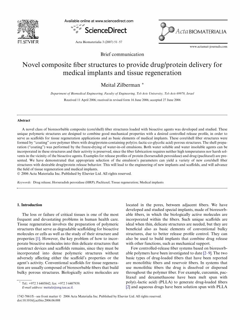
The structure of the new core/shell Wbers is presented inFig. 1. A general view indicates that these Wbers actuallyresemble sutures (Fig. 1(a)). The concept of dense core Wbersurrounded by porous drug/protein-containing shell isschematically depicted in Fig. 1(b). Using this concept, pro-teins and water-soluble drugs are designed to be incorpo-rated in the aqueous phase of the emulsion; thus afterfreeze-drying they will be entrapped in the pores of theshell. It should be noted that by using this technique ofcomposite Wber preparation the active agent is almost notexposed to the organic solvent. In our initial studies theshell was loaded with the model enzyme HRP, which is verysensitive to both solvents and elevated temperature andwhose activity is a sensitive monitor of damage during pro-cessing. We found that under mild processing conditionsHRP preserved 98–100% of its content and 94–100% of itsactivity. The speciWc enzymatic activities (%) of the 4:1, 8:1and 16:1 samples are 94.5§ 2.7, 97.5§ 2.9 and 100§ 1.5,respectively. However, high rates and durations of emulsionhomogenization (mixing) should be avoided. Good adhe-sion was observed between the dense core PLLA Wber andthe porous 75/25 PDLGA shell. Since both parts are madeof aliphatic poly(�-hydroxy acids), their similar surface ten-sions contribute to good adhesion at the interface. Since the
54 M. Zilberman / Acta Biomaterialia 3 (2007) 51–57
incorporated proteins remain highly active, such Wbers canbe loaded with growth factors and formed into bioactivescaVolds to promote tissue regeneration.
Composite Wber structures could also be used as basicelements of biodegradable endovascular stents thatmechanically support blood vessels, while delivering drugssuch as paclitaxel directly to the blood vessel wall. Exam-ples of Wber-based stents for blood vessel support andother biomedical applications are described in our previ-ous studies [13–17]. Paclitaxel is an anti-proliferative drug,used to prevent restenosis by inhibiting the proliferation ofvessel smooth muscle cells [18]. We investigated paclitaxel-
loaded core/shell Wber structures in which paclitaxel, ahydrophobic drug, was incorporated in the polymericphase of the emulsion rather than in the aqueous phase (aswe did with HRP loaded systems). The paclitaxel-loadedemulsion-based coatings can be used also on stable coreWbers. Therefore, in order to simulate a system of stablestent coated with a degradable coating, nylon sutures werechosen as core Wbers. Since the core nylon Wbers under-went surface treatment prior to their coating with emul-sion, good adhesion between core and shell was achievedalso for the paclitaxel-eluting Wbers, as presented inFig. 1(c). Other potential applications for our new core/
Fig. 1. The structure of the composite core/shell Wber structures: (a) general view (photograph) of the Wber; (b) schematic representation showing the coredense Wber and the porous drug-loaded shell; (c) SEM fractograph of part of a core Wber coated with drug-loaded porous PDLGA shell (cross section).Good adhesion between core and shell is demonstrated.
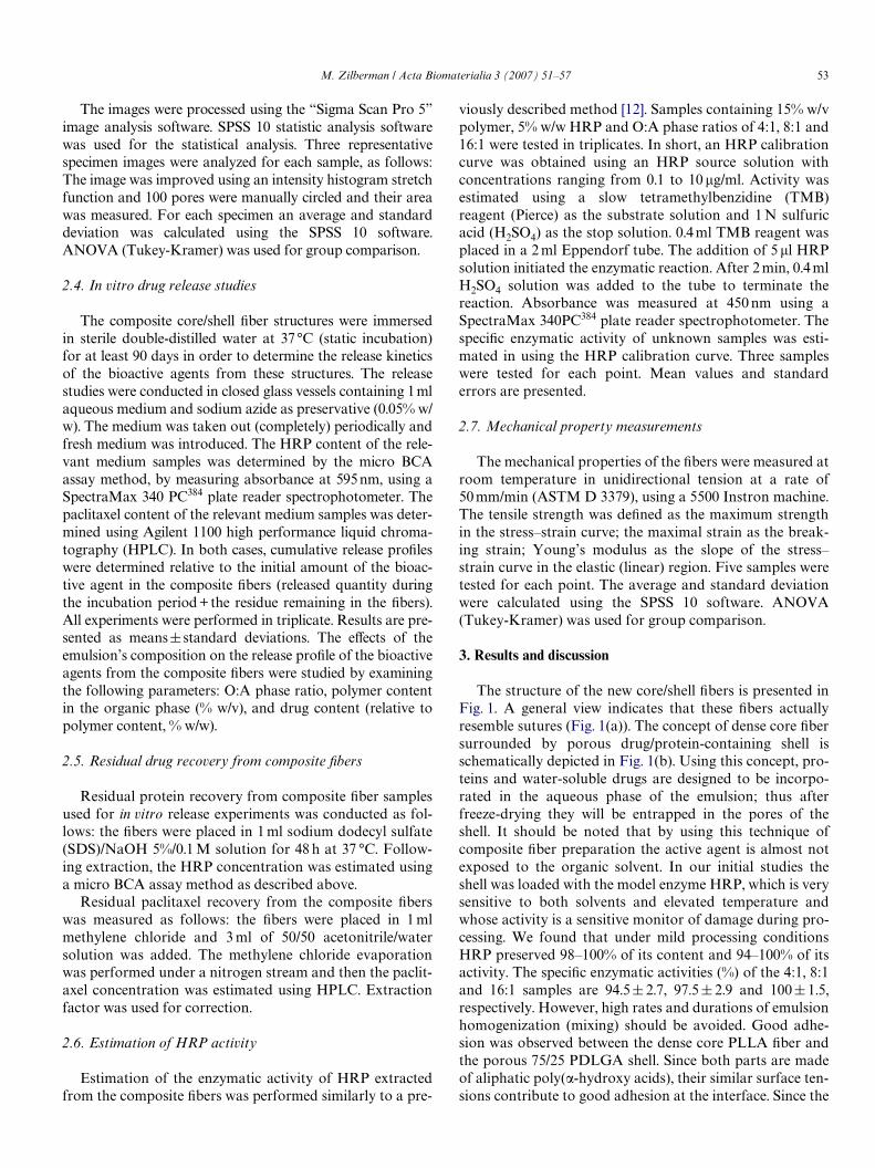
Fig. 2. Cumulative release proWles of bioactive agents from core/shell Wber structures: (a) Bioactive agent: HRP (5% w/w), shell polymer content: 19% w/v,O:A phase ratio: – 4:1, – 8:1, – 16:1. (b) Bioactive agent: paclitaxel (1.4% w/w), O:A phase ratio: 2:1, shell polymer content: – 15% w/v, –22.5% w/v.
0
20
40
60
80
100
0 20 40 60 80 100
HR
P C
umul
ativ
e R
elea
se (
%)
Time (days)
0
5
10
15
20
25
30
35
0 20 40 60 80 100 120
Pacl
itaxe
l Cum
ulat
ive
Rel
ease
(%
)
Time (days)

M. Zilberman / Acta Biomaterialia 3 (2007) 51–57 55
shell Wber structures include bioresorbable burn andwound dressings loaded with anti-microbial agents andbioresorbable antibiotic-loading sutures and meshes forhernia repair. Although the pores on the surface are muchsmaller than those in the bulk, in applications where asmooth surface is needed, an additional thin and smoothbioresorbable coat can be used.
The eVect of emulsion composition on the shell’s micro-structure and on the resulting bioactive agent release proWlefrom the composite Wbers was studied for both types ofWbers, HRP-loaded Wbers and paclitaxel-loaded Wbers. Themost important cumulative release results are presented inFig. 2. In both cases, the release proWles generally exhibitedan initial burst eVect accompanied by a decrease in therelease rate with time, as is typical for diVusion-controlledsystems. It appeared that the O:A emulsion’s phase ratiohas a major eVect on the HRP release proWle, while theemulsion’s polymer content has a major eVect on the paclit-axel release proWle. For both systems the release rate can beenhanced by increasing the active agent content, due to ahigher driving force for diVusion.
HRP release proWles from composite Wbers containing5% w/w HRP and 19% w/v polymer with various O:Aphase ratios are presented in Fig. 2(a), All samples releasedat least 90% of the active agent during the 90-day experi-ment. An increase in the O:A phase ratio resulted in a sig-niWcant decrease in the initial burst release as well as in amore moderate release curve. These trends in the cumula-tive release proWles are attributed mainly to changes in theshell microstructure. Fig. 3 indicates that the structurechanges from a dense and partially interconnected porepopulation for the 4:1 O:A phase ratio formulations to aless dense population with a closed pore pattern in the 16:1O:A phase ratio. Formulations of 5% (w/w) HRP and 19%w/v polymer exhibited pore sizes of 3.82§ 1.13, 2.40§1.1and 1.50§0.78, for coatings with O:A ratios of 4:1, 8:1 and16:1, respectively. These results are signiWcant. Both com-parisons, between 4:1 and 8:1 samples and between 4:1 and16:1 samples show a p-value smaller than 0.001. A compari-son between 8:1 and 16:1 samples shows a p-value of 0.012.This change in the shell’s microstructure resulted in a sharpdecrease in HRP diVusion from the porous coating, andtherefore dramatically reduced the burst eVect from 70% to80% for the 4:1 samples to only about 10–20% for the 16:1samples. Thus, manipulation of the emulsion’s O:A phaseratio served as a powerful tool for achieving a variety ofprotein release proWles, while preserving a constant HRPload (Fig. 2(a)). These fundamental changes in the proteinrelease proWles enable these novel composite Wbers toaddress a variety of future therapeutic demands. Althoughthe polymer content determines the emulsion’s viscosity, inour system it had a minor eVect on the shell microstructureand on the HRP release proWle.
Since paclitaxel is a hydrophobic drug and was incorpo-rated in the shell’s polymer, relatively high water contents(i.e., low O:A phase ratios such as 2:1 and 4:1) were neces-sary in order to exhibit a highly porous structure, which
resulted in desired drug release proWles. The paclitaxelrelease proWle obtained for most studied structures duringthe test period showed a very low initial burst eVect (lessthan 3%), and most of the release occurred within the Wrst30 days of the study. For this system the emulsion’s poly-mer content had a dominant eVect on the release proWle(Fig. 2(b)). The release of paclitaxel from the porous shell isrelatively slow, mainly due to the extremely hydrophobicnature of paclitaxel. Moreover, as the release continues itsrate decreases, since the drug molecules have a longer dis-tance to pass and lower driving force for diVusion. Thetotal release quantity does not exceed 40% of the incorpo-rated drug. Hence, part of the drug remains within the coat-ing shell. It is therefore clear that a second release phase
Fig. 3. SEM fractographs (cross section) of composite Wbers showing theeVect of emulsion organic: aqueous phase ratio on the shell’s micro-structure of Wbers loaded with 5% w/w HRP. Emulsion phase ratio (v/v):(a) 4:1, (b) 8:1 and (c) 16:1.

56 M. Zilberman / Acta Biomaterialia 3 (2007) 51–57
0
200
400
600
800
1000
0 10 20 30 40 50 60 70
Stre
ss (
MPa
)
Strain (%)
a
0
100
200
300
400
500
600
0 10 20 30 40 50
Stre
ss (
MPa
)
Strain (%)
b1
34
1
2
4
3
should occur, i.e., during the Wrst release phase most of thedrug is released within the Wrst month, and a second releasephase should occur later on, while the polymer is degradedinto very small fragments. Such a release proWle can bebeneWcial for our application, especially when it is knownthat restenosis may occur within 6 months after theprocedure [19]. Our results show that a decrease in polymercontent within the organic phase increases the drug releaserate (Fig. 2(b)). Since the pore size almost did not changewith the emulsion’s polymer content [20], but less dense“polymeric walls” were probably created between adjacentpores, it is suggested that a relatively low polymer contentreduces the binding region between the matrix and paclit-axel, resulting in a higher diVusion coeYcient which enablesa more eVective drug release.
The tensile stress–strain curves of the neat PLLA andnylon Wbers and their corresponding core/shell Wber struc-tures are presented in Fig. 4. The evaluated tensile mechani-cal properties are presented in Table 1. Two methods wereused for the evaluation of the mechanical properties of thecore/shell Wbers: (a) by using the total diameter of the Wber,(b) by using an eVective diameter which is practically thecore Wber. The PLLA Wber lost 25% of its strength and 35%of its Young’s modulus due to its coating. However, itshould be noted that the highly porous shell cannot practi-cally carry the load. Therefore the second method of evalu-ation gives the real eVect of coating, which is a 14%decrease in tensile strength and a 15% decrease in Young’s
modulus for PLLA Wbers (Table 1). All tensile strength andmodulus values of PLLA Wbers are statistically signiWcant.
Composite Wbers of core nylon Wber coated with PDLGAshell are more complicated, since surface treatment is neededin order to get good adhesion between core and shell. Thistreatment results in a decrease in tensile strength and modu-lus and some increase in strain (Fig. 4(b)). A comparisonbetween the treated core nylon Wber and the core/shell Wberstructures shows that the coating process results in adecrease of 18% in tensile strength and 20% in modulus, ifthe evaluation is based on the eVective diameter of the com-posite Wber (Table 1). Most results for tensile strength andmodulus of nylon Wbers are statistically signiWcant. Only thediVerence between modulus based on real diameter andmodulus based on eVective diameter is not statisticallysigniWcant. For both systems, PLLA-based Wbers and nylon-based Wbers, the maximal strain does not change signiW-cantly when the coating is applied.
The mechanical properties are very important sincedevices such as stents, scaVolds and wound dressings areproduced from these Wbers. Our results indicate that the newcomposite Wbers are strong and Xexible enough to be used asbasic elements for various medical devices. Compared tomonolithic or reservoir Wbers, the decrease in the mechanicalproperties due to the coating remains within acceptablelimits, i.e., according to our experience with devices such asstents with good radial compression strength [12–15], thesecan be constructed from our composite Wbers.
Fig. 4. Tensile stress–strain curves of (a) PLLA Wbers and (b) nylon Wbers. Fiber type: 1 – neat core Wber, 2 – surface treated core Wber, 3 – core/shell Wberstructure (total diameter is considered), 4 – core/shell Wber structure (eVective diameter is considered).
Table 1Tensile mechanical properties of Wbers
a Evaluation is based on total Wber diameter.b Evaluation is based on the eVective Wber diameter.
Fiber type Strength (MPa) Modulus (MPa) Strain (%)
Core PLLA Wber 833 § 54 4.09 § 0.21 56.2 § 5.9PLLA/PDLGA composite Wbera 629 § 44 2.65 § 0.28 67.2 § 4.7PLLA/PDLGA composite Wberb 718 § 51 3.50 § 0.46 63.7 § 3.1Treated core nylon Wber 396 § 50 0.88 § 0.15 48.0 § 5.5Nylon/PDLGA composite Wbera 267 § 32 0.59 § 0.07 47.4 § 4.8Nylon/PDLGA composite Wberb 325 § 40 0.70 § 0.12 47.9 § 5.0

M. Zilberman / Acta Biomaterialia 3 (2007) 51–57 57
4. Conclusion
The concept of composite core/shell Wber structures,composed of a dense core and porous drug/protein loadedshell prepared using the “water-in-oil” emulsion freeze-drying technique, is presented here for the Wrst time. Bothwater soluble and water insoluble bioactive agents can beincorporated in the shell of these Wbers, without aVectingtheir activity. We have shown that appropriate selection ofthe emulsion’s parameters can yield a wide variety of newcore/shell Wber structures with desirable mechanical anddrug/protein release behaviors. This will lead to the engi-neering of new implants and scaVolds, and will advancethe Weld of tissue regeneration and medical implants.
Acknowledgements
This work was supported by a grant from the Tel-AvivUniversity Internal Fund and by the Israel Ministry ofHealth. The author would like to thank Mr. Yair Lavy andMr. Amir Kraitzer (Department of Biomedical Engineer-ing, Tel-Aviv University, Israel) for the controlled releasemeasurements.
References
[1] Thomson RC, Shung AK, Yaszemski MJ, Mikos AG. Polymer scaV-old processing. In: Lanza RP, Langer R, Vacanti J, editors. Principlesof tissue engineering. New York: Academic Press; 2000. p. 251–62.
[2] Su SH, Landau CL, Chao RY, Timmons RB, Meidell RS, Tang L, etal. Expandable bioresorbable endovascular stent with anti-plateletand anti-inXammation treatments. Circulation 2001;104(II):500–7.
[3] Alikacem N, Yoshizawa T, Wilson C, Nelson KD. Quantitative MRimaging study of intravitreal sustained release of VEGF in rabbits.Invest Ophthalmol Vis Sci 2000;41:1561–9.
[4] Dunn RL, Lewis DH, Goodson JM. Monolithic Wbers for controlleddelivery of tetracycline. Proc Int Symp Control Rel Bioact Mater1982;9:157–63.
[5] Dunn RL, English JP, Stoner WC, Potter AG, Perkins BH. Biode-gradable Wbers for the controlled release of tetracycline in treatment
of peridontal disease. Proc Int Symp Control Rel Bioact Mater1987;14:289–94.
[6] Dunn RL, Lewis DH, Beck LR. Fibrous polymer for the delivery ofcontraceptive steroids to the female reproductive track. In: Lewis DH,editor. Controlled release of pesticides and pharmaceuticals. NewYork: Plenum Press; 1981. p. 125–46.
[7] Eenink MDJ, Feijen J, Oligslanger J, Albers JHM, Rieke JC, Greid-onus PJ. Biodegradable hollow Wbers for the controlled release of hor-mones. J Control Rel 1987;6:225–37.
[8] Polacco G, Cascone MG, Lazzeri L, Ferrara S, Giusti P. Biodegrad-able hollow Wbers containing drug-loaded nanoparticles as controlledrelease systems. Polym Int 2002;51(12):1464–72.
[9] Lazzeri L, Cascone MG, Quiriconi S, Morabito L, Giusti P. Biode-gradable hollow microWbers to produce bioactive scaVolds. Polym Int2005;54:101–7.
[10] Whang K, Thomas KG, Healy KE. A biodegradable polymer scaVoldfor delivery of osteotropic factors. Biomaterials 2000;21:2545–51.
[11] Levy Y. MSc thesis, Tel-Aviv University; 2005.[12] Woo BH, Jiang G, Jo YW, Delucas PP. Preparation and characteriza-
tion of a composite PLGA and Poly(acryloyl hydroxyethyl starch)microsphere systems for protein delivery. Pharm Res2001;18(11):1600–5.
[13] Zilberman M, Schwade ND, Eberhart RC. Protein-loaded bioresorb-able Wbers and expandable stents: mechanical properties and proteinrelease. J Biomed Mater Res Part B: Appl Biomater 2004;69:1–10.
[14] Zilberman M, Nelson KD, Eberhart R. Mechanical properties andin-vitro degradation of bioresorbable Wbers and expandable Wber-based stents. J Biomed Mater Res Part B: Appl Biomater 2005;74:792–9.
[15] Zilberman M, Eberhart RC. Drug-eluting bioresorbable stents forvarious applications. Annu Rev Biomed Eng 2006;8:153–80.
[16] Nguyen K, Su SH, Zilberman M, Bohluli P, Frenkel P, et al. Biomate-rials and stent technology. In: Yaszemski M, Trantolo D, Lewand-rowski KU, Hasirci V, Altobelli D, Wise D, editors. Tissueengineering and novel delivery systems, vol. 5. New York: MarcelDekker; 2004. p. 107–30.
[17] Su S, Chao RY, Landau CL, Nelson KD, Timmons RB, et al.Expandable bioresorbable endovascular stent. I. Fabrication andproperties. Ann Biomed Eng 2003;31:667–77.
[18] Woods TC, Marks AR. Drug-eluting stents. Annu Rev Med2004;55:169–78.
[19] Kimura T, Yokoi H, Nakagawa Y. Three-year follow-up afterimplantation of metallic coronary artery stents. N Engl J Med1996;334:561–6.
[20] Kraitzer A. MSc thesis, Tel-Aviv University; 2006.