Embed Size (px)
Citation preview

Zika virus (ZIKV) is a positive sense RNA virus of the Flaviviridae family, which includes sev-
eral medically notable arboviruses such as dengue (DENV), yellow fever, Japanese encephalitis virus, and West Nile virus. Before the 2015 epidemic in the Americas that spread to >40 countries and infected >1 million people (1), ZIKV was considered rare and responsible for minor epidemics in East Africa and parts of Asia. Although most ZIKV infections are as-
ymptomatic or clinically mild, the epidemic in the Americas revealed that the virus can cause serious neurologic problems in some persons and severe ter-atogenic effects in pregnant women (2–4). Because ZIKV and DENV-1–4 share the same Aedes mosqui-to vectors, areas in the Americas most affected by ZIKV also experience endemic DENV transmission. The ZIKV pandemic in the Americas led to novel observations and questions about its epidemiology and pathogenesis in regions with endemic DENV transmission. Recent studies indicate that cross-re-active immunity between ZIKV and DENV can lead to protection or to more severe disease depending on the context (5–7).
Although sporadic transmission of Asian lin-eages of ZIKV in Southeast Asia and Pacifi c islands is well-documented (8,9), its prevalence in the region has been diffi cult to estimate using current serologic assays because of intense transmission of multiple DENV serotypes and antibody cross-reactivity be-tween DENV and ZIKV. Most serologic assays for fl aviviruses measure antibodies binding to viral-en-velope glycoprotein (E protein) because this antigen is a major target of human antibodies. The FlavivirusE protein contains immunodominant antibody epi-topes that are conserved (cross-reactive) between different fl aviviruses or unique to each virus (type-specifi c) (10–12).
Traditional Flavivirus serologic assays exhibit poor specifi city in distinguishing DENV from ZIKV infections because these assays use whole virions or E proteins containing conserved epitopes as antigens (13–15). More recently, the ZIKV epidemic in the Americas spurred the development of recombinant viral antigens and serologic assays for distinguishing ZIKV from DENV (16–18). We previously described a serologic assay using domain III of the ZIKV E
Novel Assay to Measure Seroprevalence of Zika Virus
in the PhilippinesCameron Adams, Ramesh Jadi, Bruno Segovia-Chumbez, Jedas Daag, Michelle Ylade,
Freddy A. Medina, Tyler M. Sharp, Jorge L. Munoz-Jordan, In-Kyu Yoon, Jacqueline Deen, Anna Lena Lopez,1 Aravinda M. de Silva, Lakshmanane Premkumar
Emerging Infectious Diseases • www.cdc.gov/eid • Vol. 27, No. 12, December 2021 3073
Author affi liations: University of North Carolina School of Medicine, Chapel Hill, North Carolina, USA (C. Adams, R. Jadi, B. Segovia-Chumbez, A.M. de Silva, L. Premkumar); University of the Philippines, Manila, Philippines (J. Daag, M. Ylade, J. Deen, A.L. Lopez); Centers for Disease Control and Prevention, San Juan, Puerto Rico, USA (F.A. Medina, T.M. Sharp, J.L. Munoz-Jordan); Coalition for Epidemic Preparedness Innovations, Washington, DC, USA (I.-K. Yoon)
DOI: https://doi.org/10.3201/eid2712.211150 1Deceased..
Zika virus (ZIKV) is a member of the Flaviviridae family, which includes other clinically notable viruses such as the 4 dengue virus serotypes (DENV-1–4). Distinguish-ing DENVs from ZIKV using the established serologic assays widely used for monitoring DENV transmission is diffi cult because of antibody cross-reactivity between these closely related fl aviviruses. We describe a modifi ed and improved recombinant envelope domain III–based serologic assay for detecting ZIKV type-specifi c antibod-ies in regions with endemic DENV transmission. When the assay was used to measure ZIKV seroprevalence in 2017 among children 9–14 years of age living in a re-gion of the Philippines with endemic DENV transmission, we observed a ZIKV seroprevalence of 18%. Investiga-tors should consider using the ZIKV envelope domain III–based assay, which is simple and readily adaptable for use in standard clinical and public health laboratories, to assess ZIKV seroprevalence in areas with endemic DENV transmission.

RESEARCH
protein (EDIII) to detect ZIKV type-specific antibod-ies among persons in areas with DENV and ZIKV co-circulation (19). Here we describe development of a second-generation ZIKV EDIII–based serologic assay and its use to measure the seroprevalence of ZIKV among children 9–14 years of age in the Cebu Prov-ince of the Philippines.
Materials and Methods
ZIKV EDIII Antigen ProductionWe expressed a codon-optimized gene encoding for EDIII from ZIKV strain H/PF/2013 in Expi293 cells as a fusion protein containing a human serum albumin signal peptide for secretion, a polyhisti-dine tag (his-tag) for affinity purification, and a HaloTag (Promega, https://www.promega.com) for biotinylation (20). We deposited the nucleotide sequence of the construct into Genbank (accession no. MZ592925). HaloTag enables single, site-specific biotinylation distant from the folded EDIII protein. We purified recombinant EDIII antigen from the culture supernatant using nickel-nitrilotriacetic acid agarose (QIAGEN, https://www.qiagen.com) and biotinylated it using HaloTag PEG biotin ligand (Promega), according to manufacturer protocol. We analyzed the identity and purity of the biotinylated EDIII antigen using SDS-PAGE (sodium dodecyl-sulfate polyacrylamide gel electrophoresis) mobili-ty-shift analysis.
ZIKV EDIII ELISAWe coated a 96-well high binding microtiter plate (Greiner Bio-One, https://www.gbo.com) with 50 µL of streptavidin at 4 µg/mL in tris-buffered saline (TBS, pH 7.4) for 1 h at 37°C. We captured the bioti-nylated EDIII at 2 µg/mL in TBS, washed the plate 3 times with wash buffer (TBS containing 0.2% Tween 20), and then blocked it with 100 µL of blocking so-lution (3% milk in TBS containing 0.05% Tween 20) for 1 h at 37°C. After removing the blocking solution, we added 50 µL of heat-inactivated (56°C for 30 min) serum sample at 1:20 or indicated dilutions in block-ing buffer and incubated for 1 hour at 37°C. After washing the plate in the wash buffer, we added 50 µL of alkaline phosphatase-conjugated secondary goat anti-human secondary IgG (Sigma) at 1:2,500 dilu-tion for 1 hour at 37°C. We washed the plate, added 50 µL of p-nitrophenyl phosphate substrate (Sigma, https://www.sigmaaldrich.com), and measured absorbance at 405 nm using an Epoch plate reader (Biotek, https://www.biotek.com). For analyzing the 547 serum samples from the Cebu cohort using
ELISA assays performed over several days, we used human monoclonal antibody ZKA190 (21), which binds to highly accessible regions of ZIKV EDIII as a control to standardize EDIII ELISA optical density (OD) values across assays. Each plate was developed until wells with ZKA190 generated an OD value within a 1.0–1.3 range. If the signal was outside this range, we considered the assay invalid and repeated the process. We divided all OD values for human samples by the ZKA190 OD value on the same plate before determining the ZIKV-immune status of each participant (>0.34 cutoff).
Human Serum Panel Used to Validate the EDIII AssayTo validate the EDIII assay as described elsewhere (19), we used a panel of 142 archived convalescent serum samples from 15 participants who received a licensed Flavivirus (yellow fever/Japanese encepha-litis virus) vaccine, 27 serum samples from Flavivi-rus-naive participants, 33 from participants with immunity to ZIKV (including some with both ZIKV and DENV immunity), and 67 from participants with DENV immunity but no immunity to ZIKV. The DENV- and ZIKV-immunity status of conva-lescent specimens in the panel was based on par-ticipants in febrile illness study cohorts in Nicara-gua and Sri Lanka with laboratory-confirmed acute DENV or ZIKV infections or tests for the presence of neutralizing DENV or ZIKV antibodies in single se-rum samples from healthy persons. We designated samples with no detectable neutralizing DENV and ZIKV antibodies or 50% plaque reduction neutral-ization test (PRNT50) results <10 as naive. Collection, storage, and use of these convalescent serum sam-ples for research was approved by the Institutional Review Board of the University of North Carolina at Chapel Hill (protocol 08–0895).
Centers for Disease Control and Prevention ZIKV Persistence SamplesThe Centers for Disease Control and Prevention (CDC) and the Ponce Medical School Foundation monitored DENV and ZIKV patients seeking treat-ment at 2 hospitals in southern Puerto Rico. During triage, we identified case-patients with >1 signs or symptoms: fever (temperature >38.0°C or >100.5°F) or reporting fever lasting <7 days, rash, arthralgia or arthritis, or conjunctivitis and offered them participa-tion in the study. We identified ZIKV cases through test results from the CDC Dengue Branch laboratory in San Juan, Puerto Rico, where reverse transcrip-tion (RT) PCR testing for DENV, ZIKV, CHIKV, and other respiratory infectious diseases is performed and
3074 Emerging Infectious Diseases • www.cdc.gov/eid • Vol. 27, No. 12, December 2021

Seroprevalence of Zika Virus, the Philippines
followed ZIKV cases longitudinally as described else-where (22). For our study, we selected and prospec-tively followed 27 ZIKV RT-PCR–positive cases for up to 2.5 years. Specimen collection was approved by institutional review boards at CDC and Ponce Medi-cal School Foundation.
Pediatric Samples from Cebu ProvinceWe published the Cebu study protocol and approvals elsewhere (23). In brief, our study used baseline se-rum samples that were collected from 2,996 children 9–14 years of age enrolled in a postlicensure DENV vaccine study. Cohort residence was equally split between Bogo and Balamban, both semiurban areas. We collected samples and demographic information before participants were vaccinated at the same visit.
DENV and ZIKV Focus Reduction Neutralization TestWe determined neutralization titers against DENV and ZIKV by focus-reduction neutralization test (FRNT) in a 96-well format described elsewhere (24). Serially diluted serum was mixed with 50–100 focus-forming units of the virus in Dulbecco modified Eagle medium with 2% fetal bovine serum. We incubated the antibody and virus complexes (1 h, 37°C), then transferred them to a monolayer of Vero-81 cells for infection. After 1 additional hour of incubating an-tibody and virus complex on Vero-81 monolayer, we overlaid cells with GIBCO Opti-MEM (https://www.thermofisher.com), a modified Eagle medium containing 2% fetal bovine serum and 1% carboxy-methylcellulose. After allowing the predetermined time required to form viral foci, we fixed Vero-81 cells and immunostained them with Flavivirus-spe-cific monoclonal antibodies. For neutralization as-says, we calculated 50% inhibitory concentration by using the sigmoidal dose-response (variable slope) equation in Prism 6 (GraphPad Software, https://www.graphpad.com). For the study, we included only reported values with an R2 (coefficient of deter-mination) >0.75, hill slope >0.5, and 50% inhibitory concentration within the assay range. We performed FRNT for Flavivirus strains WP74 (DENV-1), S16803 (DENV-2), CH53489 (DENV-3), TVP-376 (DENV-4), and H/PF/2013 (ZIKV).
Depletion of DENV binding AntibodiesWe obtained purified viral antigens for antibody depletions by infecting Vero-81 cell cultures in 850 cm2 roller bottles (Greiner Bio-One, https://www.gbo.com) as described elsewhere (25). We conjugated Flavivirus-specific antibody 1M7 to Tosyl-activated Dynabeads (Thermo Fisher) magnetic beads and
incubated purified DENV-1–4 viral antigens (1 h at 37°C) with the beads. To deplete DENV-specific anti-bodies, we incubated serum samples (3 h at 37°C) with DENV-conjugated Dynabeads and incubated serum samples with Dynabeads conjugated to an equivalent bovine serum albumin concentration. We confirmed cross-reactive and DENV antibody depletion using whole virion capture ELISA against DENV-1–4. We measured serotype-specific ZIKV antibodies in serum samples using ZIKV whole-virion capture ELISA af-ter depletion, as described elsewhere (24).
Receiver Operator Characteristic AnalysisWe used SPSS Statistics for Macintosh version 27.0 (https://www.ibm.com) to report the performance of the EDIII ELISA based on the receiver operating characteristic (ROC) curve, which presents test per-formance as true-positive (sensitivity) versus false-positive (1 – specificity). We calculated the optimal assay cutoff value, which maximizes sensitivity and specificity, from the ROC curve using the same soft-ware. According to the test, sensitivity is the fraction of total confirmed positive samples with true posi-tives, and specificity is total confirmed negative sam-ples with true negatives.
Results
Development of Immunoassay for Detecting ZIKV Antibodies in Patient SerumWe previously demonstrated that a serologic assay using the ZIKV EDIII fused to maltose-binding pro-tein (MBP) reliably differentiated persons with past ZIKV infections from those with DENV infections (19). However, we observed a high background sig-nal in some specimens, originating from human anti-bodies binding to MBP or the mouse antigen capture antibody used in the assay. To overcome this prob-lem in the earlier version of the assay, we produced the ZIKV EDIII antigen fused to a HaloTag (Figure 1, panel A), derived from the haloalkane dehalogenase enzyme from Rhodococcus rhodochrous bacteria (26). By using a biotin HaloTag ligand (27), we added a single biotin molecule for each protein molecule at a site distant from the EDIII antigen (Figure 1, panel B). Using streptavidin-coated ELISA plates to capture the antigen, we established an assay for detecting ZIKV EDIII–binding antibodies in human clinical samples (Figure 1, panel C).
Performance of the Second-Generation ZIKV EDIII ELISAWe performed an ROC analysis to determine the diagnostic performance of the new EDIII-capture
Emerging Infectious Diseases • www.cdc.gov/eid • Vol. 27, No. 12, December 2021 3075

RESEARCH
ELISA in a panel of serum samples collected >12 weeks after symptom onset among persons with lab-oratory-confirmed ZIKV or DENV infections or who received a licensed Flavivirus vaccine. The panel in-cluded serum samples from DENV-naive and DENV-immune participants who received positive ZIKV RT-PCR test results, archived serum samples from persons who had primary (only 1 serotype) or sec-ondary (>2 serotypes) DENV infections, control sam-ples from persons who had not experienced DENV or ZIKV infections, and samples from travelers who had received Japanese encephalitis virus vaccine or yel-low fever virus vaccine or both. The EDIII assay was positive for 32 of 33 ZIKV-immune, 7 of 67 DENV-im-mune, and 2 of 42 DENV- and ZIKV-naive or yellow fever/Japanese encephalitis virus vaccine recipients (Figure 2, panel A). The ROC analysis demonstrated an area under the curve of 0.97 (95% CI of 0.94–0.99) (Figure 2, panel B). At a cutoff value of OD 0.34, the sensitivity of the EDIII-capture ELISA was 97% and the specificity was 92%.
Durability of ZIKV EDIII Antibodies in Persons Exposed to ZIKV InfectionsNext, we assessed the durability of EDIII antibodies using a panel of 98 longitudinal samples collected 1–600 days after symptom onset from 27 residents
of Puerto Rico with PCR-confirmed ZIKV infections. By testing the acute specimens for DENV-specific antibodies, we stratified the DENV-immune status at the time of ZIKV infection as 12 DENV-naive and 15 DENV-immune cases. We measured endpoint ti-ters at each time point to follow the kinetics of serum ZIKV EDIII antibodies (Figure 3). The ZIKV EDIII antibody titers reached peak levels by 1 month af-ter initial infection and stayed well above detection level for ≥20 months in DENV- naive and DENV-immune participants.
Seroprevalence of ZIKV in Cebu ProvinceHaving established the performance of the ZIKV EDIII ELISA among participants exposed to multiple DENV serotypes, we used the assay to estimate the seroprevalence of ZIKV among children living in Balamban and Bogo City in Cebu Province. In 2017, we collected baseline blood samples from a DENV vaccine study of 2,996 children 9–14 years of age. Elsewhere we reported that 89.3% of the children were DENV-immune at baseline, demonstrating the high endemicity of the virus in this population (23). We selected a representative sample of 547 chil-dren on the basis of DENV-immune status from the baseline cohort to determine the seroprevalence of ZIKV (Table 1). The children selected for the study
3076 Emerging Infectious Diseases • www.cdc.gov/eid • Vol. 27, No. 12, December 2021
Figure 1. ZIKV Biotinylated-EDIII antigen capture ELISA in study of novel assay to measure ZIKV seroprevalence in the Philippines. A) Schematic of the ZIKV EDIII construct with an N-terminal human albumin secretion signal, a HaloTag (Promega, https://www.promega.com) for site-specific biotinylation and a C-terminal 6-histidine residue tag for affinity purification. The EDIII construct design also included a TEV protease cleavage site between HaloTag and EDIII. B) Biotinylated ZIKV EDIII displays an electrophoretic mobility shift with streptavidin. A site-specific biotinylated ZIKV EDIII was prepared using HaloTag biotin ligand. Electrophoretic gel shift analysis was performed in sodium dodecyl sulphate–polyacrylamide gel electrophoresis with biotinylated EDIII antigen in the presence and absence of streptavidin. C) Schematic of second-generation ZIKV EDIII ELISA using streptavidin-biotin interaction. Biotinylated EDIII antigen is captured by plate immobilized streptavidin. The antibody bound to EDIII is detected by a secondary anti–human IgG conjugated to alkaline phosphatase. EDIII, E protein domain III; IgG-AP, secondary IgG antibody conjugated to alkaline phosphatase; SA, streptavidin; SP, N terminal human albumin secretion signal; TEV, tobacco etch virus protease cleavage site; ZIKV, Zika virus.

Seroprevalence of Zika Virus, the Philippines
consisted of 60 (11%) with no immunity to DENV, 43 (8%) with immunity to 1 DENV serotype, and 444 (81%) with immunity to >2 serotypes (Table 1; Fig-ure 4, panel B) (23). We observed that 98/547 (18%) children were ZIKV antibody positive in the ZIKV EDIII assay (Figure 4, panel A). The number of chil-dren who tested positive or negative for ZIKV anti-bodies did not differ by sex, location, or prior DENV-immune status (Table 1).
Comparison of ZIKV EDIII ELISA and Virus Neutralizing Antibody AssaysThe current standard for estimating the seropreva-lence of a particular flavivirus is based on measuring virus neutralizing antibodies in cell culture systems. Although neutralizing antibody assays are more spe-cific than assays that measure antibody binding to fla-viviruses or whole recombinant proteins, persons ex-posed to multiple DENV serotypes can develop ZIKV cross-neutralizing antibodies as reported elsewhere (10,24,28,29). Given the large number of children in our cohort with a history of >2 DENV serotype infec-tions, we compared the performance for estimating ZIKV seroprevalence of the EDIII ELISA using FRNT.
From the 547 baseline samples tested by ZIKV EDIII assay, we tested 495 samples in a single dilu-tion (1:40) ZIKV neutralization assay. We have dem-onstrated elsewhere that at 1:40 dilution, neutraliza-tion of >70% of the input virus is a reliable screening criterion for previous DENV infection (23). When we
used the same criterion for the single-dilution ZIKV neutralization assay, we observed a much higher seroprevalence (39%) compared with the estimate (18%) based on the EDIII ELISA (Appendix Table 1, https://wwwnc.cdc.gov/EID/article/27/12/21-1150-App1.pdf). Most of the discordant samples (positive in the ZIKV neutralization assay and nega-tive in the ZIKV EDIII assay) were from children with preexisting multitypic immunity to DENVs (105/110 participants) (Appendix Table 1), raising the possibility that high levels of DENV antibodies cross-neutralize ZIKV.
To evaluate whether the discordant results were because of the poor specificity of the ZIKV neutral-ization assay or poor sensitivity of the ZIKV EDIII ELISA, we selected 24 samples that had been classi-fied by full curve neutralization testing against the 4 DENV serotypes and ZIKV as primary DENV-immune only (n = 3), primary ZIKV-immune only (n = 4), both primary DENV- and primary ZIKV-immune (n = 6), multitypic DENV-immune only (n = 2), or both multitypic DENV- and ZIKV-immune (n = 9) participants (Appendix Table 2, Figure 2) for further study. By selectively removing all antibod-ies to DENVs in a sample before testing for ZIKV binding antibodies, it is possible to detect ZIKV type-specific antibodies indicative of a past ZIKV infection (11). We incubated all 24 samples with magnetic beads coated with the 4 DENV serotypes to remove all DENV-binding antibodies. After
Emerging Infectious Diseases • www.cdc.gov/eid • Vol. 27, No. 12, December 2021 3077
Figure 2. Performance evaluation of ZIKV EDIII assay in study of novel assay to measure ZIKV seroprevalence in the Philippines. Shown are the binding (A) and ROC (B) curve analysis of ZIKV EDIII ELISA using human convalescent serum samples. A panel of convalescent serum samples collected >12 weeks after onset of symptoms from primary and secondary ZIKV infections (n = 33), primary and multitypic DENV infections (n = 67), and serum samples collected >12 weeks after vaccination with a licensed Flavivirus vaccine or serum samples from Flavivirus-naive participants (n = 42) were tested by ZIKV EDIII ELISA. ROC demonstrated 0.966 (95% CI 0.94–0.99) area under the curve. The sensitivity of the EDIII capture ELISA was 97% (32/33) and the specificity 92% (100/109) at a cutoff value of 0.34. Red line indicates a random classifier and represents data points with equal true-positive rate and false-positive rate. The blue line is the ROC curve showing high performance of the ZIKV EDIII assay because the blue line is above and further away from a random classifier. DENV, dengue virus; EDIII, E protein domain III; OD405, optical density at 405 nm; ROC, receiver operating characteristic; ZIKV, Zika virus.

RESEARCH
confirming removal of all DENV-binding antibodies, we tested the samples for ZIKV-binding antibodies. After depleting DENV-binding antibodies, we ob-served ZIKV-binding antibodies for the participants
designated as immune to primary Zika only and primary DENV and primary ZIKV (Appendix Table 2, Figure 1, panel A). In contrast, 6/9 participants designated by neutralization testing as multitypic DENV- and ZIKV-immune showed no ZIKV-bind-ing antibodies (Appendix Figure 1, panel A). All participants that retained ZIKV-binding antibodies after DENV-binding antibody depletion also tested positive in the ZIKV EDIII ELISA and all partici-pants with no detectable ZIKV-specific antibodies tested negative in the EDIII assay (Appendix Table 2, Figure 1, panel B). These results suggest that the ZIKV EDIII ELISA is more reliable than neutraliza-tion testing for estimating ZIKV seroprevalence in-dependent of DENV status.
DiscussionThe 2015 ZIKV pandemic in the Americas mostly af-fected communities with high DENV endemicity be-cause these viruses share the same mosquito vector. Efforts to monitor the spread of ZIKV and the effect of cross-reactive immunity on viral pathogenesis were severely hampered by the inability of conventional serologic assays to accurately distinguish DENV from ZIKV seropositivity. Several groups developed ZIKV recombinant antigen–based assays or antigen-anti-body competition assays to improve the specificity of serologic assays (16–18).
In a previous study, we documented develop-ment of an ELISA to detect antibodies in persons who had recovered from ZIKV infections, using ZIKV EDIII fused to Escherichia coli MBP as an antigen (19). We observed in some persons high background levels of antibodies to the MBP fusion protein alone, most likely from natural exposure to bacterial pro-teins, highlighting the need for modifying the fusion
3078 Emerging Infectious Diseases • www.cdc.gov/eid • Vol. 27, No. 12, December 2021
Figure 3. Durability of ZIKV EDIII antibodies in study of novel assay to measure ZIKV seroprevalence in the Philippines. Longitudinal samples from patients with PCR-confirmed ZIKV infections were collected 1–600 days after symptom onset and tested for ZIKV EDIII–binding antibodies. DENV serostatus was determined by DENV focus reduction neutralization test. Primary ZIKV serostatus indicates ZIKV infection in DENV-naive participants; secondary ZIKV serostatus indicates ZIKV infection in participants previously infected with DENV. DENV, dengue virus; EDIII, E protein domain III; ZIKV, Zika virus.
Figure 4. ZIKV positivity in Cebu, Philippines, in study of novel assay to measure ZIKV seroprevalence in the Philippines. A) Distribution of ZIKV EDIII antibody reactivity among the 547 participants tested; 18% of the participants tested positive. B) Distribution of ZIKV EDIII–positive participants by DENV serostatus. Horizontal dotted red lines indicate positive threshold of EDIII assay. DENV, dengue virus; EDIII, E protein domain III; OD, optical density; ZIKV, Zika virus.
Seroprevalence of Zika Virus, the Philippines
protein used for antigen production. We describe a ZIKV EDIII antigen improved by replacing the MBP fusion tag with a HaloTag amenable to site-specific biotinylation. Using streptavidin-biotin chemistry to capture the antigen, we developed an ELISA with 97% sensitivity and 92% specificity for detecting past ZIKV infections.
A strength of our study was the use of serum samples from participants living in Flavivirus-endem-ic regions with well-defined exposures to DENVs or ZIKV or both to determine the performance of the as-say when testing specimens with high levels of cross-reactive antibodies. A potential problem with using modular domains such as EDIII instead of the full-length envelope protein is a loss in assay sensitivity over time. However, our analysis of the longitudinal samples from patients with documented ZIKV infec-tion demonstrated that ZIKV EDIII antibodies robust-ly developed and remained at high levels even after 2.7 years. This finding suggests that serologic assays based on EDIII will have the necessary sensitivity to characterize past ZIKV circulation at individual and population levels.
The severity of the epidemic in the Americas renewed interest in the epidemiology and patho-genesis of ZIKV in Asia. Asian lineages of the virus have been circulating for decades if not longer. The virus has been detected by molecular methods or virus isolation by cell culture in many countries, in-cluding Thailand, the Philippines, Vietnam, Malay-sia, Cambodia, and India (30–35). One study of 600 migrant workers in Taiwan (predominantly from Indonesia, the Philippines, Thailand, and Vietnam) reported 37% seroprevalence using a ZIKV IgG as-say (36,37). A similar study of Taiwan residents (n = 212) identified 4.2% of all participants with ZIKV IgG but confirmed only 1 participant by neutraliza-tion test (36). Investigators tested a cohort of chil-dren 1–4 years of age (n = 662) in Indonesia and estimated 9.1% seroprevalence using 90% neu-tralization as a cutoff value (38). A 2017 study of healthy adults (n = 801) in Vietnam estimated 1.1% seroprevalence using a ZIKV neutralization assay (39). An additional study in Guangxi Province, China, in March 2019 found 6% of 273 participants bound to ZIKV NS1 using IgG ELISA testing and neutralized ZIKV in cell cultures (40). Those past studies, especially those relying on tests of whole Zika virions and full-length antigens, had poor specificity, mainly because of antibodies induced by DENV infections cross-reacting with ZIKV an-tigens. In our study, we used a recombinant ZIKV antigen with a high specificity for distinguishing
ZIKV-induced from DENV-induced antibodies; results showed that 18% of children 9–14 years of age living in Cebu Province had previously experi-enced a ZIKV infection. Although ZIKV is current-ly not considered by public health agencies to be a common infection in the Philippines, our results indicate otherwise.
Our results also highlight a problem with using accepted standards for neutralizing antibody testing to identify ZIKV infections in populations heavily exposed to DENVs. We observed ZIKV neutraliz-ing antibodies in 39% of the children in this study, which was more than double the estimate based on the EDIII assay results. By depleting all DENV-binding antibodies from a subset of samples and then measuring ZIKV antibodies, we demonstrated that some children exposed to >2 DENV serotypes have low levels of ZIKV cross-neutralizing antibod-ies that can lead to false-positive results. In contrast, the ZIKV EDIII assay was not impacted by DENV-immune status because, even after removing all DENV-binding antibodies, the children maintained ZIKV EDIII–binding antibodies.
The novel assay we describe, which uses a bio-tinylated EDIII antigen to detect ZIKV type-specif-ic antibodies, is simple and readily adaptable for use in standard clinical and public health labora-tories to monitor ZIKV transmission at both the individual and population levels. Given that the 2015 ZIKV epidemic spread to >40 countries and infected more than a million people, many in re-gions with endemic transmission of DENVs, and that DENVs demonstrate cross-reactive immunity with ZIKV, the improved specificity of this assay provides a necessary resource for accurately moni-toring ZIKV transmission in regions with high DENV endemicity.
Emerging Infectious Diseases • www.cdc.gov/eid • Vol. 27, No. 12, December 2021 3079
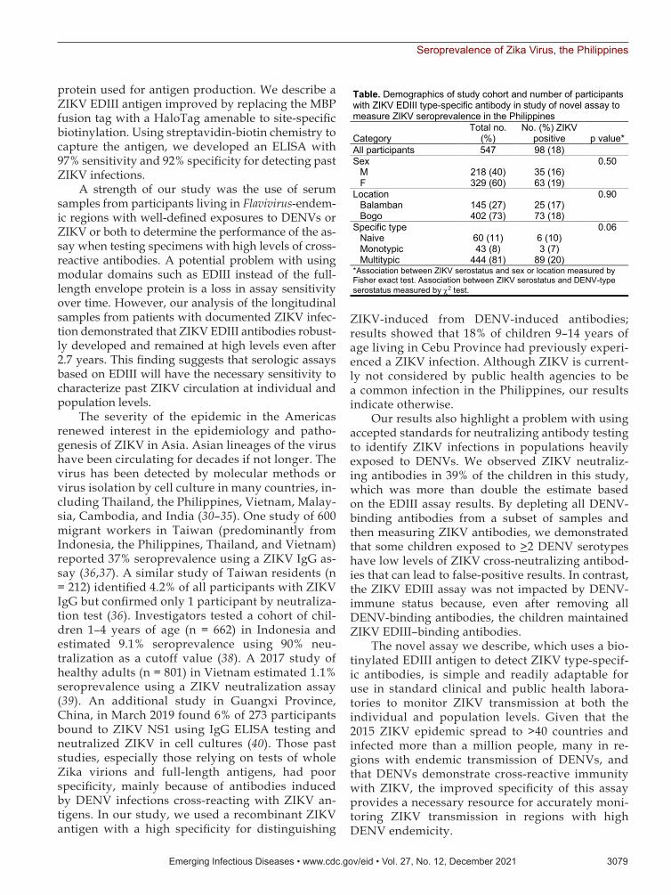
Table. Demographics of study cohort and number of participants with ZIKV EDIII type-specific antibody in study of novel assay to measure ZIKV seroprevalence in the Philippines
Category Total no.
(%) No. (%) ZIKV
positive p value* All participants 547 98 (18)
Sex 0.50 M 218 (40) 35 (16) F 329 (60) 63 (19) Location 0.90 Balamban 145 (27) 25 (17) Bogo 402 (73) 73 (18) Specific type 0.06 Naive 60 (11) 6 (10) Monotypic 43 (8) 3 (7) Multitypic 444 (81) 89 (20) *Association between ZIKV serostatus and sex or location measured by Fisher exact test. Association between ZIKV serostatus and DENV-type serostatus measured by 2 test.

RESEARCH
AcknowledgmentsWe thank the children and their families in Balamban and Bogo City who volunteered for this study and the field staff who assisted with the study. We also thank Laura White and Alena Markmann for providing specimens for validating the assay and for supervising sample processing, inventory, and storage and Matt Collins for providing clinical samples for validating the assay.
The field component of the study in the Philippines was supported by the Department of Health (Philippines), the Hanako Foundation, the World Health Organization, and the Swedish International Development Cooperation Agency in partnership with the International Vaccine Institute in Seoul, South Korea. The laboratory studies at the University of North Carolina were supported by grants from the US Centers for Disease Control and Prevention (BAA 2017-N-18041; PI AdeS) and the US National Institutes of Health (U24 AI152170-01 and R21 AI134073 PIs: Ades and LP). C.A. was supported by NIH Predoctoral Training Grant T32 AI007419: “Molecular Biology of Viral Disease.” US patent application no. 20200300855 on the method incorporating the EDIII antigen for serodiagnosis of Zika infection has been filed under the title “Methods and Compositions for Zika Virus Detection.”
About the AuthorMr. Adams is an MD/PhD student at the University of North Carolina School of Medicine. His main research and clinical interests involve global health, infectious diseases, and vaccinology.
References 1. Hills SL, Fischer M, Petersen LR. Epidemiology of Zika
virus infection. J Infect Dis. 2017;216(suppl_10):S868–74. https://doi.org/10.1093/infdis/jix434
2. Ventura CV, Maia M, Bravo-Filho V, Góis AL, Belfort R Jr. Zika virus in Brazil and macular atrophy in a child with microcephaly. Lancet. 2016;387:228. https://doi.org/ 10.1016/S0140-6736(16)00006-4
3. Johansson MA, Mier-y-Teran-Romero L, Reefhuis J, Gilboa SM, Hills SL. Zika and the risk of microcephaly. N Engl J Med. 2016;375:1–4. https://doi.org/10.1056/NEJMp1605367
4. Hoen B, Schaub B, Funk AL, Ardillon V, Boullard M, Cabié A, et al. Pregnancy outcomes after ZIKV infec-tion in French territories in the Americas. N Engl J Med. 2018;378:985–94. https://doi.org/10.1056/NEJMoa1709481
5. Borchering RK, Huang AT, Mier-Y-Teran-Romero L, Rojas DP, Rodriguez-Barraquer I, Katzelnick LC, et al. Impacts of Zika emergence in Latin America on endemic dengue transmission. Nat Commun. 2019;10:5730. https://doi.org/10.1038/s41467-019-13628-x
6. Gordon A, Gresh L, Ojeda S, Katzelnick LC, Sanchez N, Mercado JC, et al. Prior dengue virus infection and risk of Zika: A pediatric cohort in Nicaragua. PLoS Med.
2019;16:e1002726. https://doi.org/10.1371/journal.pmed.1002726
7. Katzelnick LC, Narvaez C, Arguello S, Lopez Mercado B, Collado D, Ampie O, et al. Zika virus infection enhances future risk of severe dengue disease. Science. 2020;369:1123–8. https://doi.org/10.1126/science.abb6143
8. Duong V, Dussart P, Buchy P. Zika virus in Asia. Int J Infect Dis. 2017;54:121–8. https://doi.org/10.1016/j.ijid.2016.11.420
9. Hu T, Li J, Carr MJ, Duchêne S, Shi W. The Asian lineage of Zika virus: transmission and evolution in Asia and the Americas. Virol Sin. 2019;34:1–8. https://doi.org/10.1007/s12250-018-0078-2
10. Priyamvada L, Quicke KM, Hudson WH, Onlamoon N, Sewatanon J, Edupuganti S, et al. Human antibody responses after dengue virus infection are highly cross-reactive to Zika virus. Proc Natl Acad Sci U S A. 2016;113:7852–7. https://doi.org/10.1073/pnas.1607931113
11. Andrade P, Gimblet-Ochieng C, Modirian F, Collins M, Cárdenas M, Katzelnick LC, et al. Impact of pre-existing dengue immunity on human antibody and memory B cell responses to Zika. Nat Commun. 2019;10:938. https://doi.org/10.1038/s41467-019-08845-3
12. Rogers TF, Goodwin EC, Briney B, Sok D, Beutler N, Strubel A, et al. Zika virus activates de novo and cross- reactive memory B cell responses in dengue-experienced donors. Sci Immunol. 2017;2:eaan6809. https://doi.org/ 10.1126/sciimmunol.aan6809
13. Chua CL, Chan YF, Andu ESGS, Rovie-Ryan JJ, Sitam FT, Verasahib K, et al. Little evidence of Zika virus infection in wild long-tailed Macaques, peninsular Malaysia. Emerg Infect Dis. 2019;25:374–6. https://doi.org/10.3201/eid2502.180258
14. Beck C, Leparc-Goffart I, Desoutter D, Debergé E, Bichet H, Lowenski S, et al. Serological evidence of infection with dengue and Zika viruses in horses on French Pacific Islands. PLoS Negl Trop Dis. 2019;13:e0007162. https://doi.org/10.1371/journal.pntd.0007162
15. Netto EM, Moreira-Soto A, Pedroso C, Höser C, Funk S, Kucharski AJ, et al. High Zika virus seroprevalence in Salvador, Northeastern Brazil limits the potential for further outbreaks. MBio. 2017;8:e01390–17. https://doi.org/10.1128/mBio.01390-17
16. Balmaseda A, Stettler K, Medialdea-Carrera R, Collado D, Jin X, Zambrana JV, et al. Antibody-based assay discrimi-nates Zika virus infection from other flaviviruses. Proc Natl Acad Sci U S A. 2017;114:8384–9. https://doi.org/10.1073/pnas.1704984114
17. Rockstroh A, Moges B, Barzon L, Sinigaglia A, Palù G, Kumbukgolla W, et al. Specific detection of dengue and Zika virus antibodies using envelope proteins with mutations in the conserved fusion loop. Emerg Microbes Infect. 2017;6:e99. https://doi.org/10.1038/emi.2017.87
18. Denis J, Attoumani S, Gravier P, Tenebray B, Garnier A, Briolant S, et al. High specificity and sensitivity of Zika EDIII-based ELISA diagnosis highlighted by a large human reference panel. PLoS Negl Trop Dis. 2019;13:e0007747. https://doi.org/10.1371/journal.pntd.0007747
19. Premkumar L, Collins M, Graham S, Liou GA, Lopez CA, Jadi R, et al. Development of envelope protein antigens to serologically differentiate Zika virus infection from dengue virus infection. J Clin Microbiol. 2018;56:e01504–17. https://doi.org/10.1128/JCM.01504-17
20. Los GV, Encell LP, McDougall MG, Hartzell DD, Karassina N, Zimprich C, et al. HaloTag: a novel protein labeling technology for cell imaging and protein analysis. ACS Chem Biol. 2008;3:373–82. https://doi.org/10.1021/cb800025k
3080 Emerging Infectious Diseases • www.cdc.gov/eid • Vol. 27, No. 12, December 2021

Seroprevalence of Zika Virus, the Philippines
21. Wang J, Bardelli M, Espinosa DA, Pedotti M, Ng TS, Bianchi S, et al. A human bi-specific antibody against Zika virus with high therapeutic potential. Cell. 2017;171:229–241.e15. https://doi.org/10.1016/j.cell.2017.09.002
22. Paz-Bailey G, Rosenberg ES, Sharp TM. Persistence of Zika virus in body fluids—final report. N Engl J Med. 2019;380:198–9. https://doi.org/10.1056/NEJMc1814416
23. Lopez AL, Adams C, Ylade M, Jadi R, Daag JV, Molloy CT, et al. Determining dengue virus serostatus by indirect IgG ELISA compared with focus reduction neutralisation test in children in Cebu, Philippines: a prospective population-based study. Lancet Glob Health. 2021;9:e44–51. https://doi.org/10.1016/S2214-109X(20)30392-2
24. Collins MH, McGowan E, Jadi R, Young E, Lopez CA, Baric RS, et al. Lack of durable cross-neutralizing antibodies against Zika virus from dengue virus infection. Emerg Infect Dis. 2017;23:773–81. https://doi.org/10.3201/eid2305.161630
25. Putnak R, Barvir DA, Burrous JM, Dubois DR, D’Andrea VM, Hoke CH, et al. Development of a purified, inactivated, dengue-2 virus vaccine prototype in Vero cells: immunogenicity and protection in mice and rhesus monkeys. J Infect Dis. 1996;174:1176–84. https://doi.org/10.1093/infdis/174.6.1176
26. Benink HA, Urh M. HaloTag technology for specific and covalent labeling of fusion proteins. Methods Mol Biol. 2015; 1266:119–28. https://doi.org/10.1007/978-1-4939-2272-7_8
27. Dubey M, Emoto K, Takahashi H, Castner DG, Grainger DW. Affinity-based protein surface pattern formation by ligand self-selection from mixed protein solutions. Adv Funct Mater. 2009;19:3046–55. https://doi.org/10.1002/adfm.200900809
28. Dussupt V, Sankhala RS, Gromowski GD, Donofrio G, De La Barrera RA, Larocca RA, et al. Potent Zika and dengue cross-neutralizing antibodies induced by Zika vaccination in a dengue-experienced donor. Nat Med. 2020;26:228–35. https://doi.org/10.1038/s41591-019-0746-2
29. Fernandez E, Dejnirattisai W, Cao B, Scheaffer SM, Supasa P, Wongwiwat W, et al. Human antibodies to the dengue virus E-dimer epitope have therapeutic activity against Zika virus infection. [Erratum in Nat Immunol. 2020;21:354.] Nat Immunol. 2017;18:1261–9. https://doi.org/10.1038/ni.3849
30. Woon YL, Lim MF, Tg Abd Rashid TR, Thayan R, Chidambaram SK, Syed Abdul Rahim SS, et al. Zika virus infection in Malaysia: an epidemiological, clinical and virological analysis. BMC Infect Dis. 2019;19:152. https://doi.org/10.1186/s12879-019-3786-9
31. Ho ZJM, Hapuarachchi HC, Barkham T, Chow A, Ng LC, Lee JMV, et al.; Singapore Zika Study Group. Outbreak of Zika virus infection in Singapore: an epidemiological, entomological, virological, and clinical analysis. Lancet
Infect Dis. 2017;17:813–21. https://doi.org/10.1016/ S1473-3099(17)30249-9
32. Buathong R, Hermann L, Thaisomboonsuk B, Rutvisuttinunt W, Klungthong C, Chinnawirotpisan P, et al. Detection of Zika virus infection in Thailand, 2012–2014. Am J Trop Med Hyg. 2015;93:380–3. https://doi.org/10.4269/ajtmh.15-0022
33. Ruchusatsawat K, Wongjaroen P, Posanacharoen A, Rodriguez-Barraquer I, Sangkitporn S, Cummings DAT, et al. Long-term circulation of Zika virus in Thailand: an observational study. [Erratum in Lancet Infect Dis. 2019;19:e109.] Lancet Infect Dis. 2019;19:439–46. https://doi.org/10.1016/S1473-3099(18)30718-7
34. Alera MT, Hermann L, Tac-An IA, Klungthong C, Rutvisuttinunt W, Manasatienkij W, et al. Zika virus infection, Philippines, 2012. Emerg Infect Dis. 2015;21:722–4. https://doi.org/10.3201/eid2104.141707
35. Buerano CC, Pangilinan LS, Dimamay MTA, Mapua CA, Dimamay MPS, Matias RR, et al. Zika virus infection, Philippines, 2012. Emerg Infect Dis. 2020;26:2300–1. https://doi.org/10.3201/eid2609.190896
36. Chien YW, Ho TC, Huang PW, Ko NY, Ko WC, Perng GC. Low seroprevalence of Zika virus infection among adults in Southern Taiwan. BMC Infect Dis. 2019;19:884. https://doi.org/10.1186/s12879-019-4491-4
37. Perng GC, Ho TC, Shih HI, Lee CH, Huang PW, Chung CH, et al. Seroprevalence of Zika and dengue virus antibodies among migrant workers, Taiwan, 2017. Emerg Infect Dis. 2019;25:814–6. https://doi.org/10.3201/eid2504.181449
38. Sasmono RT, Dhenni R, Yohan B, Pronyk P, Hadinegoro SR, Soepardi EJ, et al. Zika virus seropositivity in 1–4-year-old children, Indonesia, 2014. Emerg Infect Dis. 2018;24:1740–3. https://doi.org/10.3201/eid2409.180582
39. Nguyen CT, Moi ML, Le TQM, Nguyen TTT, Vu TBH, Nguyen HT, et al. Prevalence of Zika virus neutralizing antibodies in healthy adults in Vietnam during and after the Zika virus epidemic season: a longitudinal population-based survey. BMC Infect Dis. 2020;20:332. https://doi.org/ 10.1186/s12879-020-05042-2
40. Zhou CM, Liu JW, Qi R, Fang LZ, Qin XR, Han HJ, et al. Emergence of Zika virus infection in China. PLoS Negl Trop Dis. 2020;14:e0008300. https://doi.org/10.1371/journal.pntd.0008300
Address for correspondence: Aravinda de Silva or Lakshmanane Premkumar, Department of Microbiology and Immunology, University of North Carolina School of Medicine, Chapel Hill, NC 27599-7292, USA; email: [email protected] or [email protected]
Emerging Infectious Diseases • www.cdc.gov/eid • Vol. 27, No. 12, December 2021 3081