Embed Size (px)
Citation preview
~ ) Pergamon Bioorganic & Medicinal Chemistry Letters, VoL 6, No. 19, pp. 2311-2316, 1996
Copydght 0 1996 Elsevier Science Ltd Printed in Great Britain. All rights reserved
0960-894X/96 $15.00 + 0.00 pll: S0960-894X(96)00428-3
NOVEL ANALOGS OF CYCLIC-ADP-RIBOSE: 9-CYCLIC ETHENO-ADP-RIBOSE AND CYCLIC ETHENO-CDP-RIBOSE
Fang-Jie Zhang and Charles J. Sih* School o f Pharmacy, University o f Wisconsin-Madison,
425 N. Charter Street, Madison, WI 53706-1515
A b s t r a c t : Two novel analogs of cyclic ADP-ribose (cADPR), 9-cyclic etheno-ADP-ribose (1) and cyclic etheno-CDP-ribose (2) were synthesized. We have shown for the first time that enzymatic and biomimetic methods may proceed by different reaction pathways. 1,N6-Etheno-nicotinamide-adenine dinucleotide (3) was converted into 1 using the biomimetic procedure, whereas ADP-ribosyl cyclase transformed 3 into 4. The unique fluorescence property and the strong Ca 2+ mobilizing activity of 1 provide investigators with a useful probe for the study of cADPR-binding proteins. Copyright © 1996 Elsevier Science Ltd
Cyclic ADP-ribose (cADPR) is a novel metabolite of NAD and its synthesizing enzyme, ADP-ribosyl
cyclase, has been shown to be widely distributed among mammalian and invertebrate tissues.1 Currently, there
is considerable interest in defining the biological roles of cADPR as a second messenger candidate in Ca 2+
signaling 2 and insulin release) The structure and absolute stereochemistry of cADPR have now been firmly
established by X-ray crystallography 4 and by synthesis. 5
New structural analogs of cADPR can now be prepared using either the biomimetic route 5b or the ADP-
ribosyl cyclase from Aplysia californica. 6 For the most part, both procedures give similar product profiles, but
higher yields are generally obtained with the enzymatic method. These synthetic methodologies have allowed
the transformation of a variety of structurally modified analogs of NAD into their corresponding cyclic
nucleotides with the newly formed glycosyl bonds attached to the N-1 nitrogen of the purine rings as in
cADPR. 7 However, when the adenine ring in NAD was replaced by guanine or hypoxanthine, an alternative
mode of cyclization was observed wherein the N-7 nitrogen of the purine rings were used to form the glycosyl
bonds. 8
Our continuing interest in the synthesis of more active and stable analogs of cADPR as fluorescent
affinity probes for investigations of cADPR-binding proteins led us to the preparation of two novel analogs.
Herein, we describe the experimental details used in the synthesis and characterization of 9-cyclic etheno-ADP-
ribose, 1 and cyclic etheno-CDP-ribose, 2.
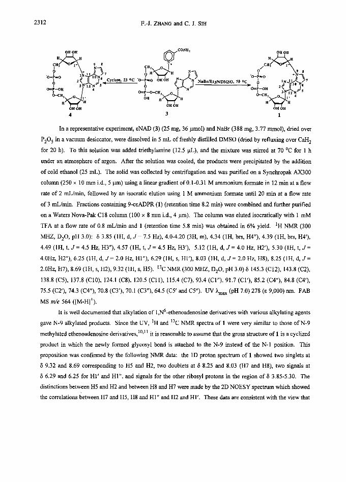
We have previously reported that ADP-ribosyl cyclase catalyzed the conversion of 1,N6-etheno -
Nicotinamide-adenine dinueleotide (3) into the cyclic nucleotide (4) whose N-glycosyl bond is attached onto the
N-1 position of the l,N6-etheno-adenine nucleus corresponding to the N-7 position of the adenine ring as
shown. 9 However, when 3 was subjected to the conditions of biomimetic synthesis, 5b we were unable to detect
the formation of 4. Instead, a novel fluorescent cyclic nucleotide was isolated in 6% yield.
2311
2312 F.-J. ZHANG and C. J. SIH
OH OH ~E'~'~f cONH" OH OH
H ~ E ~N(~J H ~ CH 9 $ J N 0 9
I 2 ~ II I ° Cyclase, ~ oc "o- 7 ? LN.~.2 N~5, ? ~'~"N ~ N~e~/Et3N/V~fSO, 70 oc
H" ~ -H OH OH OH OH OH OH
4 3 1
In a representative experiment, eNAD (3) (25 mg, 36 ~tmol) and NaBr (388 mg, 3.77 mmol), dried over
P205 in a vacuum desiccator, were dissolved in 5 mL of freshly distilled DMSO (dried by refluxing over Call 2
for 20 h). To this solution was added triethylamine (12.5 ~tL), and the mixture was stirred at 70 °C for 1 h
under an atmosphere of argon. After the solution was cooled, the products were precipitated by the addition
of cold ethanol (25 mL). The solid was collected by centrifugation and was purified on a Synchropak AX300
column (250 x l0 mm i.d., 5 btm) using a linear gradient of 0.1-0.31 M ammonium formate in 12 min at a flow
rate of 2 mL/min, followed by an isocratic elution using 1 M ammonium formate until 20 min at a flow rate
of 3 mL/min. Fractions containing 9-ceADPR (1) (retention time 8.2 min) were combined and further purified
on a Waters Nova-Pak C18 column (100 x 8 mm i.d., 4 btm). The column was eluted isocratically with 1 mM
TFA at a flow rate of 0.8 mL/min and 1 (retention time 5.8 min) was obtained in 6% yield. IH NMR (300
MHZ, DE0 , pH 3.0): ~5 3.85 (1H, d, J = 7.5 Hz), 4.0-4.20 (3H, m), 4.34 (1H, brs, H4"), 4.39 (1H, brs, H4'),
4.49 (1H, t, J = 4.5 Hz, H3"), 4.57 (1H, t, J = 4.5 Hz, H3'), 5.12 (1H, d, J = 4.0 Hz, H2'), 5.30 (IH, t, J =
4.0Hz, H2"), 6.25 (1H, d, J = 2.0 Hz, HI"), 6.29 (1H, s, HI'), 8.03 (1H, d, J = 2.0 Hz, H8), 8.25 (1H, d, 3" =
2.0Hz, H7), 8.69 (1H, s, H2), 9.32 (1H, s, HS). 13C NMR (300 MHZ, DE0 , pH 3.0) 8 145.3 (C12), 143.8 (C2),
138.8 (C5), 137.8 (C10), 124.1 (C8), 120.5 (Cll) , 115.4 (C7), 93.4 (CI"), 91.7 (CI'), 85.2 (C4"), 84.8 (C4'),
75.5 (C2'), 74.3 (C4"), 70.8 (C3'), 70.1 (CY'), 64.5 (C5' and C5"). UV ~'max (pU 7.0) 278 (e 9,000) nm. FAB
MS m/e 564 ([M-H]+).
It is well documented that alkylation of 1,N6-ethenoadenosine derivatives with various alkylating agents
gave N-9 alkylated products. Since the UV, IH and 13C NMR spectra of 1 were very similar to those of N-9
methylated ethenoadenosine derivatives, I°'11 it is reasonable to assume that the gross structure of 1 is a cyclized
product in which the newly formed glycosyl bond is attached to the N-9 instead of the N-I position. This
proposition was confirmed by the following NMR data: the 1D proton spectrum of I showed two singlets at
8 9.32 and 8.69 corresponding to H5 and H2, two doublets at 8 8.25 and 8.03 (H7 and HS), two signals at
5 6.29 and 6.25 for HI ' and HI", and signals for the other ribosyl protons in the region of ~5 3.85-5.30. The
distinctions between H5 and H2 and between H8 and H7 were made by the 2D NOESY spectrum which showed
the correlations I~etween H7 and H5, H8 and HI" and H2 and Hl' . These data are consistent with the view that
Novel analogs of cyclic-ADP-ribose 2313
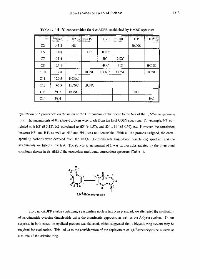
Table 1. IH-13C connectivities for 9-ceADPR established by HMBC spectrum
C2 143.8 HC
C5 138.8
C7 115.4
C8 124.1
C10 137.8
C11 120.5 HCNC
C12 145.3 HCNC
CI ' 91.7 HCNC
CI" 93.4
HC
HCNC
HCNC
HCNC
HCNC
HC HCC
HCC HC HCNC
HCNC HCNC HCNC
HC
HC
cyclization of 3 proceeded via the union of the C 1" position of the ribose to the N-9 of the 1, N6-ethenoadenine
ring. The assignments of the ribosyl protons were made from the H-H COSY spectrum. For example, HI ' cor-
related with H2' (5 5.12), H2' correlated to H3' (6 4.57), and H3' to I-I4' (5 4.39), etc. However, the correlation
between H5' and H4', as well as H5" and H4", was not detectable. With all the protons assigned, the corre-
sponding carbons were deduced from the HSQC (Heteronuclear single-bond correlation) spectrum and the
assignments are listed in the text. The structural assignment of 1 was further substantiated by the three-bond
couplings shown in its HMBC (heteronuclear multibond correlation) spectrum (Table 1).
3,N4-Ethenocytosine
Since no cADPR analog containing a pyrimidine nucleus has been prepared, we attempted the cyclization
of nicotinamide cytosine dinucleotide using the biomimetic approach, as well as the Aplysia cyclase. To our
surprise, in both cases, no cyclized product was detected, which suggested that a bicyclic ring system may be
required for cyclization. This led us to the consideration of the deployment 4 of 3,N -ethenocytosine nucleus as
a mimic of the adenine ring.
2314 F.-J. ZHANG and C. J. SIH
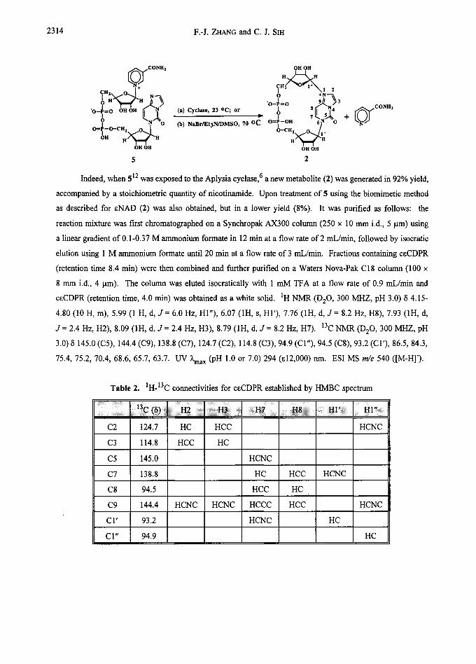
~ CONH2 OH OH H ~
H2 O O +N"~ I 9~ 3 ? -o-~--o ,.L
"O--P----O+ OH OH ~.N,~O~ (a) Cyclase, 23 oc; or ~ ~)l 7 1[~,5~8 .f "N4
Offil~--O--CH2. O. [ (b) NaBr/Et3N/DMSO, 70 OC O=P--OH 6? O O--CH2 ~,tO...~1,
OH H ~ I ~ H H / ~ # ¢ -H OH OH OH OH
5 2
+ ~ ' ~ CONH2
Indeed, when 512 was exposed to the Aplysia cyclase, 6 a new metabolite (2) was generated in 92% yield,
accompanied by a stoichiometric quantity of nicotinamide. Upon treatment of 5 using the biomimetic method
as described for cNAD (2) was also obtained, but in a lower yield (8%). It was purified as follows: the
reaction mixture was first chromatographed on a Synchropak AX300 column (250 x 10 mm i.d., 5 lam) using
a linear gradient of 0.1-0.37 M ammonium formate in 12 min at a flow rate of 2 mL/min, followed by isocratic
elution using 1 M ammonium formate until 20 min at a flow rate of 3 mL/min. Fractions containing ceCDPR
(retention time 8.4 min) were then combined and further purified on a Waters Nova-Pak C18 column (I00 x
8 mm i.d., 4 ~tm). The column was eluted isocratically with 1 mM TFA at a flow rate of 0.9 mL/min and
ceCDPR (retention time, 4.0 min) was obtained as a white solid. 1H NMR (D20, 300 MHZ, pH 3.0) 8 4.15-
4.80 (10 H, m), 5.99 (1 H, d, J = 6.0 Hz, HI"), 6.07 (1H, s, HI'), 7.76 (1H, d, J = 8.2 Hz, H8), 7.93 (1H, d,
d = 2.4 Hz, H2), 8.09 (1H, d, d = 2.4 Hz, H3), 8.79 (1H, d, d = 8.2 Hz, H7). 13C NMR (D20 , 300 MHZ, pH
3.0) 8 145.0 (C5), 144.4 (C9), 138.8 (C7), 124.7 (C2), 114.8 (C3), 94.9 (CI"), 94.5 (C8), 93.2 (CI'), 86.5, 84.3,
75.4, 75.2, 70.4, 68.6, 65.7, 63.7. UV ~'max (pH 1.0 or 7.0) 294 (el2,000) nm. ESI MS m/e 540 ([M-H]').
Table 2. 1H-13C connectivities for ceCDPR established by HMBC spectrum
C2 124.7 HC HCC
C3 114.8 HCC HC
C5 145.0
C7 138.8
C8 94.5
C9 144.4 HCNC HCNC
CI' 93.2
CI" 94.9
HCNC
HC
HCC
HCCC
HCNC
HCC
HC
HCC
HCNC
HC
HCNC
HCNC
HC
Novel analogs of cyclic-ADP-ribose 2315
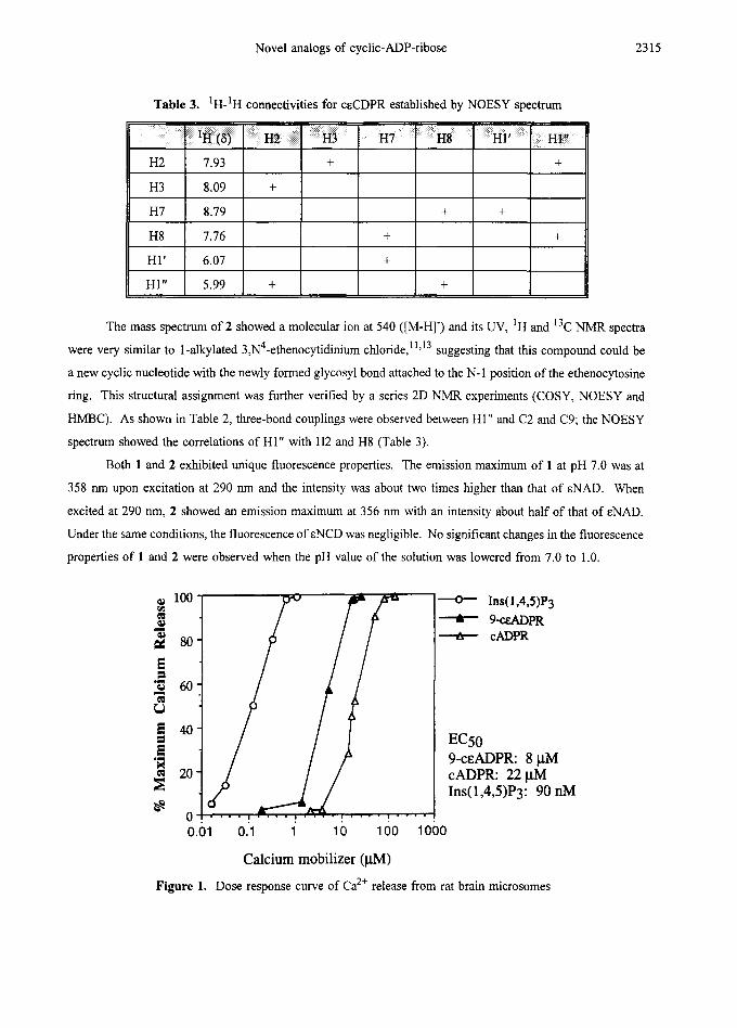
Table 3. ‘H-‘H cormectivities for CECDPR established by NOESY spectrum
The mass spectrum of 2 showed a molecular ion at 540 ([M-H]) and its UV, ‘H and 13C NMR spectra
were very similar to 1-alkylated 3,N4-ethenocytidinium chloride,’ “13 suggesting that this compound could be
a new cyclic nucleotide with the newly formed glycosyl bond attached to the N- 1 position of the ethenocytosine
ring. This structural assignment was further verified by a series 2D NMR experiments (COSY, NOESY and
HMBC). As shown in Table 2, three-bond couplings were observed between Hl” and C2 and C9; the NOESY
spectrum showed the correlations of Hl” with H2 and H8 (Table 3).
Both 1 and 2 exhibited unique fluorescence properties. The emission maximum of 1 at pH 7.0 was at
358 nm upon excitation at 290 nm and the intensity was about two times higher than that of ENAD. When
excited at 290 nm, 2 showed an emission maximum at 356 run with an intensity about half of that of ENAD.
Under the same conditions, the fluorescence of ENCD was negligible. No significant changes in the fluorescence
properties of 1 and 2 were observed when the pH value of the solution was lowered from 7.0 to 1.0.
~ 100 ]* WL4S)P3
- @ ti 80 -
--t- [email protected] + cADPR
E a
‘0
z B E ‘ir u E
60 -
EC50 9-CEADPR: 8 pM cADPR: 22 j.d4 Ins( 1,4,5)Py 90 nM
0.01 0.1 1 10 100 1000
Calcium mobilizer (PM)
Figure 1. Dose response curve of Ca2+ release from rat brain microsomes
2316 F.-J. ZHANG and C. J. SIH
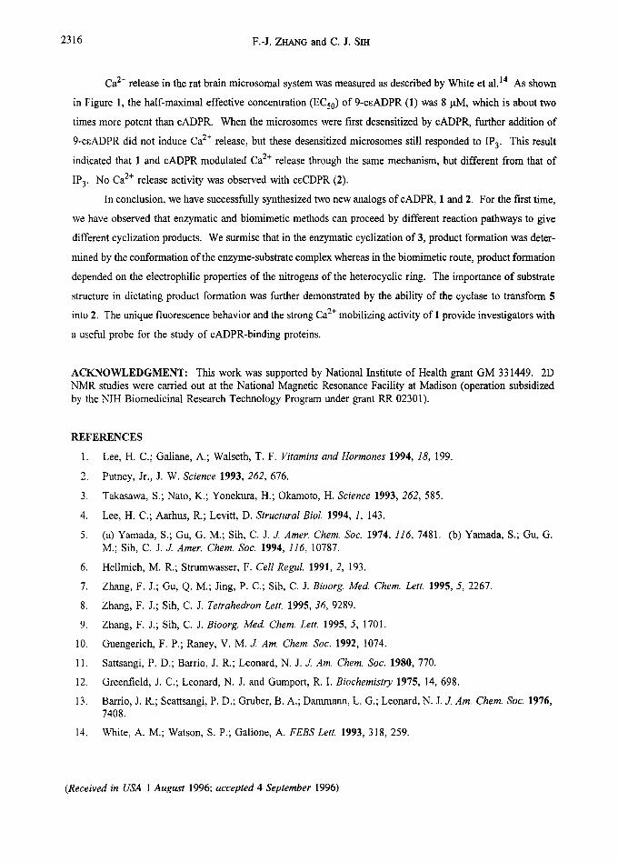
Ca 2÷ release in the rat brain microsomal system was measured as described by White et al. 14 As shown
in Figure 1, the half-maximal effective concentration (ECs0) of 9-ceADPR (1) was 8 ~tM, which is about two
times more potent than cADPR. When the microsomes were first desensitized by cADPR, further addition of
9-ceADPR did not induce Ca 2+ release, but these desensitized microsomes still responded to IP 3. This result
indicated that 1 and cADPR modulated Ca 2+ release through the same mechanism, but different from that of
IP 3. No Ca 2+ release activity was observed with ceCDPR (2).
In conclusion, we have successfully synthesized two new analogs of cADPR, 1 and 2. For the first time,
we have observed that enzymatic and biomimetic methods can proceed by different reaction pathways to give
different cyclization products. We surmise that in the enzymatic cyclization of 3, product formation was deter-
mined by the conformation of the enzyme-substrate complex whereas in the biomimetic route, product formation
depended on the electrophilic properties of the nitrogens of the heterocyclic ring. The importance of substrate
structure in dictating product formation was further demonstrated by the ability of the cyclase to transform 5
into 2. The unique fluorescence behavior and the strong Ca 2+ mobilizing activity of 1 provide investigators with
a useful probe for the study of cADPR-binding proteins.
ACKNOWLEDGMENT: This work was supported by National Institute of Health grant GM 331449. 2D NMR studies were carried out at the National Magnetic Resonance Facility at Madison (operation subsidized by the NIH Biomedicinal Research Technology Program under grant RR 02301).
REFERENCES
1, Lee, H. C.; Galiane, A.; Walseth, T. F. Vitamins and Hormones 1994, 18, 199.
2, Putney, Jr., J. W. Science 1993, 262, 676.
3. Takasawa, S.; Nato, K.; Yonekura, H.; Okamoto, H. Science 1993, 262, 585.
4. Lee, H. C.; Aarhus, R.; Levitt, D. Structural Biol. 1994, 1, 143.
5. (a) Yamada, S.; Gu, G. M.; Sih, C. J. J. Amer Chem. Soc. 1974, 116, 7481. (b) Yamada, S.; Gu, G. M.; Sih, C. J. d. Amer. Chem. Soc. 1994, 116, 10787.
6. Hellmich, M. R.; Strumwasser, F. Cell Regul. 1991, 2, 193.
7. Zhang, F. J.; Gu, Q. M.; Jing, P. C.; Sih, C. J. Bioorg. Meal Chem. Lett. 1995, 5, 2267.
8, Zhang, F. J.; Sih, C. J. Tetrahedron Lett. 1995, 36, 9289.
9. Zhang, F. J.; Sih, C. J. Bioorg. Med. Chem. Lett. 1995, 5, 1701.
10. Guengerich, F. P.; Raney, V. M. d. Am. Chem. Soc. 1992, 1074.
11. Sattsangi, P. D.; Barrio, J. R.; Leonard, N. J. J. Am. Chem. Soc. 1980, 770.
12. Greenfield, J. C.; Leonard, N. J. and Gumport, R. I. Biochemistry 1975, 14, 698.
13. Barrio, J. R.; Scattsangi, P. D.; Gruber, B. A.; Dammann, L. G.; Leonard, N. J. J. Am. Chem. Soc. 1976, 7408.
14, White, A. M.; Watson, S. P.; Galione, A. FEBSLett. 1993, 318, 259.
(Received in USA 1 August 1996; accepted 4 September 1996)