Embed Size (px)
Citation preview
Vol. 56, No. 6INFECTION AND IMMUNITY, June 1988, p. 1647-16510019-9567/88/061647-05$02.00/0Copyright C) 1988, American Society for Microbiology
NOTES
Serological Studies of Oral Bacteroides intermediusF. NAKAZAWA,' J. J. ZAMBON,2* H. S. REYNOLDS,2 AND R. J. GENCO2
Department of Oral Microbiology, School of Dentistry, Niigata University, Niigata 951, Japan,' and Departments of OralBiology and Periodontology, School of Dental Medicine, State University ofNew York at Buffalo,
Buffalo, New York 14214 2
Received 21 October 1987/Accepted 18 February 1988
Bacteroides intermedius is a gram negative, anaerobic microorganism associated with certain forms of humanperiodontal disease, including adult periodontitis and acute necrotizing ulcerative gingivitis. Previous studieshave indicated the presence of two DNA homology groups which could be distinguished by analysis of proteinpatterns on polyacrylamide gel electrophoresis, as well as at least two serogroups within B. intermedius. Thepresent study examined the serology of B. intermedius and determined the distribution of B. intermediusserogroups in clinical isolates and patient plaque samples. Serological reactions with unabsorbed rabbitantisera and antisera immunoabsorbed with B. intermedius strains demonstrated a previously unreportedantigenic group within B. intermedius, serogroup C, in both immunodiffusion and immunofluorescence assays.
Of 79 B. intermedius isolates from 68 subjects examined with specific antisera, 55% of the isolates and 52% ofthe subjects were categorized in serogroup C, 40% of the isolates and 46% of the subjects were in serogroup
B, and 5% of the isolates and 6% of the subjects were in serogroup A. In 31 samples of subgingival dentalplaque from adolescents known to harbor B. intermedius, 81% demonstrated serogroup B, 16% had serogroup
A, and 3% had serogroup C.
The black-pigmented Bacteroides species are gram nega-tive, anaerobic, nonmotile bacilli which appear as brown- toblack-pigmented colonies when grown on medium contain-ing blood. The black-pigmented Bacteroides species of theoral cavity can cause medically important extraoral infec-tions, including brain abscess (17), lung abscess (1), medias-tinitis (20, 32), and podiatric infections in diabetics (23). Inthe oral cavity, black-pigmented Bacteroides species havebeen implicated in the etiology of human periodontal dis-ease, odontogenic abscesses, and endodontic lesions (6, 33).Among the black-pigmented Bacteroides species, Bacte-roides intermedius has been associated with several forms ofperiodontal disease, including adult periodontitis (21, 25, 30,34, 36), acute necrotizing ulcerative gingivitis (2, 12, 15, 27),and pregnancy gingivitis (11).
B. intermedius, however, appears to represent a hetero-geneous group of microorganisms with possibly differinglevels of virulence. DNA-DNA hybridization studies indi-cate the presence of two distinct DNA homology groupswithin B. intermedius (10), while previous antigenic studieshave demonstrated the presence of two serologically distinctgroups within oral B. intermedius (19). Heterogeneity withinthis species is also suggested by studies of patient antibodyresponses to B. intermedius infection. Patients with acutenecrotizing ulcerative gingivitis, for example, develop highlevels of serum antibody to only certain representativestrains of B. intermedius (2).
In the study presented here, the serology ofB. intermediuswas examined and the distribution of B. intermedius sero-groups was determined for both pure isolates and patientsubgingival plaque samples.The strains of B. intermedius examined in this study
included 49 laboratory strains and 30 fresh isolates. The
* Corresponding author.
laboratory strains included ATCC 25611T (type strain) andATCC 25261 (American Type Culture Collection, Rockville,Md.); NCTC 9336 (National Collection of Type Cultures,London, United Kingdom); 13025, D25B-1, D16B-7, 4203,D28D-12, D22B-23, 13044, D11B-5, 9042, D1OA-24, 13042,D16A-32, 9849, and 13029 (courtesy of L. V. H. Moore,Virginia Polytechnic Institute, Blacksburg); M86-688, M86-449, M86-596, M86-607, M86-675, M86-678, M86-679, M86-692, M86-698, M86-699, M86-714, M86-730, M86-734, M86-735, and Ball (courtesy of J. Slots, School of DentalMedicine, University of Pennsylvania, Philadelphia); 39,564, 333, 334, 158, 96, 85, 73, 135, and 308 (courtesy of G.Bowden, University of Manitoba, Winnipeg, Canada); 5W2,520-2, 525-1, M22-4, MR45B-2, and PD1SM-1 (courtesy ofG. Bourgeau, Universitd Laval, Quebec, Canada); andSUNYaB20-3 (School of Dental Medicine, State Universityof New York at Buffalo).The fresh isolates, SUNYaB FL8-2, SUNYaB M2D2K-1,
SUNYaB 17-9K-3, SUNYaB 16-12K-1, SUNYaB 17-12K-1,SUNYaB BLX-28B, SUNYaB BLX-28BK-1, SUNYaBBLX-83B-1, SUNYaB BLX-29BK-1, SUNYaB A9A2-13,SUNYaB A6A2-3, SUNYaB A6A2-30, SUNYaB G8-9K-3,SUNYaB T19M-1. SUNYaB T8M-BM-2, SUNYaB 16-5K-8, SUNYaB (L2, -3, -5, -6, -7, and -8), SUNYaB (A9A1-4,-5, -9, and -10), and SUNYaB (A6A1-1, -3, -5, and -7), wereobtained from samples of human dental plaque taken withpaper points and anaerobically cultured on tryptic soy agar(Difco Laboratories, Detroit, Mich.) containing 5% rabbitblood and with 5.0 jig of hemin and 0.5 pug of vitamin K1 perml, as previously described (35, 36). Strains of gram-nega-tive anaerobic rods were classified as B. intermedius if they(i) fermented glucose, fructose, maltose, and sucrose, butnot cellobiose or lactose; (ii) were indole positive andcatalase negative; (iii) did not hydrolyze starch or esculin;and (iv) produced succinic, acetic, isovaleric, and sometimes
1647

INFECT. IMMUN.
formic, isobutyric, and propionic acids as metabolic acid endproducts (7, 9).
Serological studies of B. intermedius used rabbit polyclo-nal antisera produced in female New Zealand White rabbits.Bacterial cells for immunization and for sonication in thepreparation of antigen extracts were anaerobically culturedin brain heart infusion broth for 36 h, harvested by centrif-ugation, washed, and suspended at a concentration of 10 mg(wet weight) per ml in sterile saline. Twelve 1.0-ml sampleswere intravenously injected via the marginal ear vein by theprotocol of McCarty and Lancefield (18). Trial bleedingswere obtained from the central ear artery, and the antibodytiter was determined by serial dilution in immunodiffusionassays. Once a satisfactory antibody titer was achieved, therabbits were exsanguinated by cardiac puncture. Antiserumsamples were heated to 56°C for 30 min and stored in smallportions at -70°C.The reactivity and pattern of precipitin bands was exam-
ined in double-immunodiffusion assays carried out in gelscontaining 1.2% agarose (SeaKem; FMC Corp., MarineColloids Div., Rockland, Maine) in 0.33 M Veronal buffer,pH 8.2, by the method of Ouchterlony (22).
Sonic extracts of 10 strains of B. intermedius, includingATCC 25611T, ATCC 25261, NCTC 9336, SUNYaB F L8-2,SUNYaB G 8-9K-3, SUNYaB T 19M-1, SUNYaB T 8M-BM-2, SUNYaB 20-3, SUNYaB 16-5K-8, and SUNYaB17-9K-3, at a protein concentration of 10 mg/ml (16) were
examined by double gel diffusion against the correspondingunabsorbed rabbit antisera. Three immunoprecipitin reac-
tion patterns were demonstrated, which suggested threeserogroups: serogroup A, designated by ATCC 25611T andSUNYaB FL8-2; serogroup B, designated by NCTC 9336,ATCC 25261, and SUNYaB 17-9K-3; and serogroup C,designated by SUNYaB G 8-9K-3, SUNYaB T 19M-1,SUNYaB T 8M-BM-2, SUNYaB 20-3, and SUNYaB 16-5K-8 (Table 1). The only cross-reactions noted were between thetwo serogroup A antisera and the five serogroup C sonicates.
Serogroup-specific antiserum was prepared by immunoab-sorption of serum samples representative of the threegroups, including ATCC 25611T (serogroup A), NCTC 9336(serogroup B), and SUNYaB G 8-9K-3 (serogroup C). Anti-serum to B. intermedius ATCC 25611T was absorbed withwhole cells of strains NCTC 9336 and SUNYaB G 8-9K-3 toproduce a serogroup-A-specific antiserum. Likewise, antise-rum to strain NCTC 9336 was absorbed with cells of ATCC25611T and SUNYaB G 8-9K-3 to produce a serogroup-B-
specific antiserum, and antiserum to SUNYaB G 8-9K-3 wasabsorbed with strain ATCC 25611T and NCTC 9336 toproduce serogroup-C-specific antiserum. Immunoabsorp-tions were performed by adding 100 mg (wet weight) ofwhole bacterial cells to 1 ml of rabbit antiserum. The mixturewas placed in a shaker for 1 h at 37°C and then at 4°C for 12h. After centrifugation at 12,000 x g for 60 min, theantiserum supernatant was removed, and the absorption wasrepeated with bacterial cells from the other heterologousserogroup. Gel filtration chromatography was then used toeliminate nonprecipitating particulates and antigen-antibodycomplexes. The immunoabsorbed antisera were applied toBio-Gel ASm columns (Bio-Rad Laboratories, Richmond,Calif.) and eluted with phosphate-buffered saline (PBS)containing 0.02% sodium azide. Fractions were monitored indouble-gel-diffusion assays with bacterial sonicates fromrepresentatives of each of the serogroups. Reacting fractionswere pooled and concentrated to the original volume ofserum using a Minicon B15 clinical sample concentrator(Amicon Division of W. R. Grace & Co., Danvers, Mass.).The three serogroup-specific antisera were then reacted in
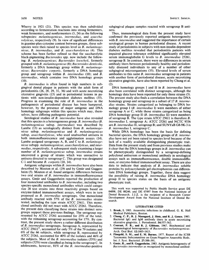
double-immunodiffusion assays against the same 10 cellsonicates. Precipitin bands formed between the absorbedantisera and sonicates from each B. intermedius strain in thatserogroup, but there were no bands formed against sonicatesfrom B. intermedius strains in the other serogroups. Inaddition, the precipitin bands demonstrated reactions ofnonidentity. The precipitin line formed by the reaction of theserogroup-A-specific antiserum with sonic extracts fromATCC 25611 shows a reaction of nonidentity with thatformed by the reaction of the serogroup-B-specific antiserumreacting with sonic extracts from NCTC 9336 and with thatformed by serogroup-C-specific antiserum reacting withsonic extracts from SUNYaB G 8-9K-3 (Fig. 1). Similarly,precipitin lines formed in the B-specific and C-specific reac-
tions also exhibit nonidentity (Fig. 1).To verify the serogroup specificity of the absorbed anti-
sera and to determine the serogroup distribution of B.intermedius isolates and patient plaque samples, immunoflu-orescence assays were performed as described by Mouton etal. (19). Pure B. intermedius isolates were cultured on trypticsoy agar (Difco) supplemented with 5% sheep blood (CraneLaboratories, Inc., Syracuse, N.Y.) and with 5 t.g of heminand 0.5 ,ug of vitamin K1 per ml. Cultures were incubated for48 h at 37°C in an anaerobic chamber (Forma Scientific,Marietta, Ohio) containing an atmosphere of 85% N2, 5%
TABLE 1. Immunoprecipitin reaction patterns in Ouchterlony gel diffusion assays
Antigen reaction'
Antiseruma strain Serogroup A Serogroup B Serogroup C
ATCC SUNYaB NCTC ATCC SUNYaB SUNYaB SUNYaB SUNYaB SUNYaB SUNYaB25611T FL8-2 9336 25261 17-9K-3 G 8-9K-3 T19M-1 T8M-BM-2 20-3 16-5K-8
ATCC 25611T + + - - - + + + + +SUNYaB FL8-2 + + - - - + + +/- + +/-NCTC 9336 - - + + + - - - - -ATCC 25261 - - + + + - - - - -SUNYaB 17-9K-3 - - + + +SUNYaB G8-9K-3 - - - - - + + + + +SUNYaB T19M-1 - - - - - + + + + +SUNYaB T8M-BM-2 - - - - - + + + + +SUNYaB 20-3 - - - - - + + + + +SUNYaB 16-5K-8 - - - - - + + + + +
a Unabsorbed rabbit antiserum.b Sonic extracts from bacterial cells at 10 mg (dry weight) per ml in PBS.
1648 NOTES

NOTES 1649
(i;j/!/\ ../ \..
FIG. 1. Serogroup reactions demonstrated in double-immunodif-fusion assays. Wells: A, serogroup-A-specific antiserum (rabbitantiserum to strain ATCC 25611T absorbed with whole bacteria cellsof strain NCTC 9336 and SUNYaB G8-9K-3); B, serogroup-B-specific antiserum (rabbit antiserum to strain NCTC 9336 absorbedwith whole bacterial cells of strains ATCC 25611T and SUNYaBG8-9K-3); C, serogroup-C-specific antiserum (rabbit antiserum tostrain SUNYaB G8-9K-3 absorbed with whole bacterial cells ofstrains ATCC 25611T and NCTC 9336); 1, sonicates from serogroupsA (ATCC 25611T) and B (NCTC 9336); 2, sonicates from serogroupsB and C (SUNYaB G8-9K-3); 3, sonicates from serogroups C(SUNYaB G8-9K-3) and A (ATCC 25611T). The bacterial sonicextracts were used at a concentration of 10 mg (dry weight) per mlin PBS, with equal volumes in each well.
C02, and 10% H2. The bacterial cells were harvested fromthe surface of the agar plates with sterile cotton swabs anddispersed in sterile Ringer solution to an optical density of0.7 at 540 nm. The serotype distribution of B. intermedius insubgingival plaque samples obtained from 31 adolescentsshown by previous immunofluorescence assays to harbor B.intermedius was also determined with serogroup-specificantisera in immunofluorescence assays. Subgingival plaquewas obtained from the mesial surface of the first four molarteeth with sterile paper points. Samples were then pooled in1.0 ml of sterile Ringer solution with 2% Formalin anddispersed by a vortex mixer.Samples (10 RI each) of bacterial suspension or patient
plaque were placed on glass slides, air dried, and gently heatfixed. Working titer concentrations (the highest twofoldserial dilution still giving 4+ fluorescence) of serogroup-specific antiserum and fluorescein isothiocyanate-conjugatedantisera were determined by checkerboard titration. Serum(10 RI) at a working titer concentration in PBS (pH 7.2)containing 0.05% Tween 20 (PBS-T) was applied to eachbacterial smear. After 10 min, the specimens were gentlyrinsed with PBS-T, washed in PBS, and rinsed with distilledwater. Slides were then incubated with 25 pl1 of affinity-purified goat anti-rabbit immunoglobulin G conjugated tofluorescein isothiocyanate (isomer I, fluorescein-to-proteinratio of 25 pLg/ml; BBL Microbiology Systems, Cockeysville,Md.). After 20 min of incubation at 37°C in 100% humidity,the slides were washed as described above and glass coverslips were mounted with glycerol in PBS (2:1 [vol/vol], pH9.0).The stained bacterial smears were examined with a Zeiss
standard 14 microscope equipped for phase-contrast illumi-nation and for incident light fluorescence. The light sourcewas a 100-W halogen lamp with a BP 450 to 490 interferencefilter in its excitation pathway and with an LP 520 barrierfilter. Fluorescence was graded from 0 to 4+ according tothe following scheme: 0, no fluorescence; 1+, bare fluores-cence, with single cells not distinguishable; 2+, faint fluo-rescence, with single cells visible but no definition of cellshape; 3+, moderate fluorescence, with good cell envelopedefinition and a dark cell center; 4+, brilliant fluorescence,with good cell envelope definition and a dark cell center.Grades 3+ and 4+ were considered positive reaction results.
Like double-diffusion assays, immunofluorescence assaysdemonstrated the presence of three distinct serogroupswithin B. intermedius. Each serogroup-specific antiserum atworking titer (1:512, 1:512, and 1:64 for serogroups A, B,and C, respectively) gave positive fluorescence reactionsonly with B. intermedius strains of the same serogroup.The serogroup distribution of 78 B. intermedius isolates
from 68 subjects was determined by indirect immunofluores-cence (Table 2). The vast majority of isolates were inserogroups B and C. Serogroup A, represented by the typestrain, ATCC 25611, accounted for 5% of the isolates and 6%of the subjects, while serogroup B, represented by ATCC25261, accounted for 40% of the isolates and 46% of thesubjects. Most of the B. intermedius isolates (55%) andsubjects (52%) belonged to serogroup C.There appeared to be a strong correlation between the B.
intermedilus serogroups and DNA homology groups as re-ported by Johnson and Holdeman (10). Of the 17 B. inter-mediius isolates for which DNA homology data were avail-able, B. intermedius isolates in DNA homology group I wereall found in either serogroup A or C, while B. intermediusisolates in DNA homology group II were all found inserogroup B. The close relationship between serogroups Aand C is also reflected in the serological studies with unab-sorbed antisera. There was cross-reaction between unab-sorbed serogroup A antisera to strains 25611T and SUNYaBFL8-2 with sonicate to the serogroup C strains in immuno-diffusion assays (Table 1). This reaction was eliminated byabsorption of the serogroup A antisera with bacteria fromserogroup C. Neither unabsorbed nor absorbed antisera tothe serogroup C strains, however, reacted with the sero-group A microorganisms in these assays.The distribution of B. intermedius serogroups was also
determined in subgingival plaque samples from 31 adoles-cent patients. Of the samples, 81% reacted with serogroup Bantiserum, 16% reacted with serogroup A antiserum, and 3%reacted with serogroup C antiserum. None of the patientplaque samples reacted with more than one of the B.intermedius-serogroup-specific reagents.The black-pigmented Bacteroides species were first de-
scribed as Bacterium melaninogenicum by Oliver and
TABLE 2. B. intermedius serogroup distribution by indirectimmunofluorescence assay with serogroup-specific antisera
Serogroup Strains"
A. ATCC 25611 (I), SUNYaB FL8-2, M86-688, 2D2K-1
B. NTC 9336 (II), ATCC 25261 (II), SUNYaB 17-9K-3,SUNYaB 16-12K-1, SUNYaB 17-12K-1, 13025(Il), D25B-1 (II), D16B-7 (II), 4203 (II), D28D-12(II), D22B-23 (II), SUNYaB BLX-228, SUNYaBBLX-28BK-1, SUNYaB BLX-83B-1, SUNYaBBLX-29BK-1, SUNYaB A9A2-13, SUNYaBA6A2-3, SUNYaB A6A2-30, -39, -564, -333, -334,-158, -96, -85, -73, 5W2, 520-2, 525-1, MM22-4,MR45B-2, Ball
C. SUNYaB G8-9K-3, SUNYaB T19M-1, SUNYaBT8M-BM-2, SUNYaB 16-SK-8, SUNYaB 20-3,SUNYaB (L2, -3, -5, -6, -7, and -8), 13044 (1),D11B-5 (I), 9042 (I), DlOA-24 (1), 13042 (I), D16A-32 (I), 9849 (I), 13029 (I), PD15M-1, 35, 308, M86-449, M86-596, M86-607, M86-675, M86-678,M86-679, M86-692, M86-698, M86-699, M86-714,M86-730, M86-734, M86-735, SUNYaB (A9A1-4,-5, -9, and -10), SUNYaB (A6A1-1, -3, -5, and -7)
" DNA homology groups I and 11 reported by Johnson and Holdeman (10).
VOL. 56, 1988
'iI I
NI
1650 NOTES
Wherry in 1921 (21). This species was then subdividedaccording to fermentation reactions into strong fermenters,weak fermenters, and nonfermenters (3, 24) as the followingsubspecies: melaninogenicus, intermedius, and asaccha-rolyticus, respectively (9). In view of significant differencesin guanine-plus-cytosine content between groups, these sub-species were then raised to species level as B. melaninoge-nicus, B. intermedius, and B. asaccharolyticus (4). Thisscheme has been further refined so that the saccharolyticblack-pigmenting Bacteroides spp. now include the follow-ing: B. melaninogenicus; Bacteroides loescheii, formerlygrouped with B. melaninogenicus (8); Bacteroides denticola,formerly a DNA homology group within B. melaninoge-nicus; Bacteroides corporis, formerly a DNA homologygroup and serogroup within B. intermedius (10); and B.intermedius, which contains two DNA homology groups(10).
B. intermedius is often found in high numbers in sub-gingival dental plaque in patients with the adult form ofperiodontitis (26, 28, 29, 31, 36) and with acute necrotizingulcerative gingivitis (15) and in periodontal lesions in pa-tients with non-insulin-dependent diabetes mellitus (35).Progress in examining the role of B. intermedius in thepathogenesis of periodontal disease has been hampered,however, by the presence of major antigenic and DNAhomology subgroups within this species which may, them-selves, have differing pathogenic potential.
Serological studies of B. intermedius have also revealedthat this species is a heterogeneous group. B. intermedius (B.melaninogenicus subsp. intermedius) was described byLambe (13) as serologically distinct from B. melaninoge-nicus subsp. melaninogenicus and B. melaninogenicussubsp. asaccharolyticus, who used unabsorbed antisera inboth immunofluorescence and agglutination assays (14).Lambe assigned serogroups A, B, and C to B. melaninoge-nicus subspp. melaninogenicus, asaccharolyticus, and inter-medius, respectively. A subsequent study examining a largernumber of B. melaninogenicus subsp. intermedius strainsuncovered a group of strains which was unreactive toantisera directed to serogroup C. This group was designatedC-1 and became B. corporis (10, 14).
Antigenic subgroups within B. intermedius have also beendescribed by Mouton et al. (19) and by Gmur and Guggen-heim (5). Mouton et al. found antigenic differences betweentwo oral strains of B. intermedius in immunofluorescenceassays. Gmur and Guggenheim reported the production offour nmonoclonal antibodies to B. intermedius, including twospecies-specific monoclonal antibodies which could catego-rize 28 test strains into three reactivity groups based onenzyme-linked immunosorbent assays, which were in turnproposed as B. intermedius serogroups. One monoclonalantibody reacted with 57% of the B. intermedius strainstested, including the type strain ATCC 25611. This mono-clonal antibody did not react with ATCC 25261. Therefore,the present data classifying these strains into distinct sero-groups support these previous findings. The serogroup rep-resented by ATCC 25261 accounted for 25% of the total,with the remaining serogroup accounting for 17%. By con-trast, the present study shows a rather different distributionof B. intermedius serogroups. Serogroup A, represented byATCC 25611T, accounted for only 5% of the 79 isolates and6% of the 66 subjects, while serogroup B, represented byATCC 25261, accounted for 40% of the isolates and 46% ofthe subjects. Most of the B. intermedius isolates (55%) andsubjects (52%) were classified as being in the serogroup C. Inadolescents, however, 81% of the B. intermedius-positive
subgingival plaque samples reacted with serogroup B anti-sera.Thus, immunological data from the present study have
confirmed the previously reported antigenic heterogeneitywith B. intermedius and suggested the importance of certainserological groups in human periodontal disease. A recentstudy of periodontitis in subjects with non-insulin-dependentdiabetes mellitus revealed that periodontitis patients withimpaired glucose tolerance exhibited significantly elevatedserum immunoglobulin G levels to B. intermedius 25261,serogroup B. In contrast, there were no differences in serumantibody titers between periodontally healthy and periodon-tally diseased individuals to any of a number of othersubgingival microorganisms (35). Elevated levels of serumantibodies to this same B. intermedius serogroup in patientswith another form of periodontal disease, acute necrotizingulcerative gingivitis, have also been reported by Chung et al.(2).DNA homology groups I and II in B. intermedius have
also been correlated with distinct serogroups, although thehomology data have been reported for only three strains (5).The present study also finds a correlation between the DNAhomology group and serogroup in a subset of 17 B. interme-dius strains. Strains categorized as belonging to DNA ho-mology group I (B. intermedius I) were members of eitherserogroup A or C, while those strains categorized as beingDNA homology group II (B. intermedius II) were membersof serogroup B. The type strain ATCC 25611 is therefore B.intermedius I, serogroup A; ATCC 25261 and NCTC 9336are B. intermedius II, serogroup B; and SUNYaB G8-9K-3 isB. intermedius I, serogroup C.
While DNA homology has been the basis for definingbacterial species, the DNA homology groups of B. interme-dius have not yet been raised to species level because of thelack of a readily available distinguishing phenotypic trait.Data from the present study and from previous studies makeit clear that the DNA homology groups in B. intermedius canbe phenotypically distinguished by a serogroup-definingpolyclonal antiserum or monoclonal antibody in serologicalassays such as immunofluorescence, double immunodiffu-sion, or enzyme-linked immunosorbent assay. There are alsodata to indicate that analysis of B. intermedius solubleproteins by polyacrylamide gel electrophoresis can differen-tiate DNA homology groups. Together, these data suggestthe possibility of raising B. intermedius DNA homologygroup II to species status on the basis of an antigenicphenotypic trait.
This work was supported by Public Health Service grant DE04898, DE 08240, and DE 07497 from the National Institute ofDental Research. J.J.Z. is the recipient of a Research CareerDevelopment Award from the National Institute of Dental Re-search.
LITERATURE CITED1. Brook, I. 1983. Anaerobic infections in childhood. G. K. Hall
Medical Publishers, Boston.2. Chung, C. P., R. J. Nisengard, J. Slots, and R. J. Genco. 1983.
Bacterial IgG and IgM antibody titers in acute necrotizingulcerative gingivitis. J. Periodontol. 54:557-562.
3. Courant, P. R., and R. J. Gibbons. 1967. Biochemical andimmunological heterogeneity of Bacteroides melaninogenicus.Arch. Oral Biol. 12:1605-1613.
4. Finegold, S. M., and E. M. Barnes. 1977. Report of the ICSBTaxonomic Subcommittee on Gram-Negative Anaerobic Rods.Int. J. Syst. Bacteriol. 27:388-391.
5. Gmur, R., and B. Guggenheim. 1983. Antigenic heterogeneity ofBacteroides intermedius as recognized by monoclonal antibod-
INFECT. IMMUN.

NOTES 1651
ies. Infect. Immun. 42:459-470.6. Griffee, M. B., S. S. Patterson, C. H. Miller, A. H. Kafrawy, and
J. J. deObarrio. 1982. Bacteroides inelaninogeniclus and dentalinfections: some questions and answers. Oral Surg. Oral Med.Oral Pathol. 54:486-489.
7. Holdeman, L. V., E. P. Cato, and W. E. C. Moore (ed.). 1977.Anaerobic laboratory manual, 4th ed. Anaerobe Laboratory,Virginia Polytechnic Institute and State University, Blacksburg.
8. Holdeman, L. V., and J. L. Johnson. 1982. Description ofBacteroides loescheii sp. nov. and emendation of the descrip-tions of Bacteroides melaninogenicus (Oliver and Wherry) Royand Kelly 1939 and Bacteroides denticola Shah and Collins1981. Int. J. Syst. Bacteriol. 32:399-409.
9. Holdeman, L. V., and W. E. C. Moore. 1970. Bacteroides, p.
33-44. In E. P. Cato, C. S. Cummis, L. V. Holdeman, J. L.Johnson, W. E. C. Moore, R. M. Smibert, and L. D. S. Smith(ed.), Outline of clinical methods in anaerobic bacteriology.Anaerobe Laboratory, Virginia Polytechnic Institute and StateUniversity, Blacksburg.
10. Johnson, J. L., and L. V. Holdeman. 1983. Bacteroides inter-inedius comb. nov. and descriptions of Bacteroides corporis sp.
nov. and Bacteroides lei'ii sp. nov. Int. J. Syst. Bacteriol. 33:15-25.
11. Kornman, K. S., and W. J. Loesche. 1980. The subgingivalmicrobial flora during pregnancy. J. Periodontal Res. 15:111-122.
12. Kristoffersen, T., and T. Lie. 1984. Necrotizing gingivitis, p.
202-217. In J. Lindhe (ed.), Textbook of clinical periodontol-ogy. Munksgaard, Copenhagen.
13. Lambe, D. W., Jr. 1974. Determination of Bacteroides mnelani-nogeniiculs serogroups by fluorescent antibody staining. Appl.Microbiol. 28:561-567.
14. Lambe, D. W., Jr., and R. C. Jerris. 1976. Description of a
polyvalent conjugate and a new serogroup of Bacteroides me-
laninogenicus by fluorescent antibody staining. J. Clin. Micro-biol. 3:506-512.
15. Loesche, W. J., S. A. Syed, B. E. Laughon, and J. Stoll. 1982.The bacteriology of acute necrotizing ulcerative gingivitis. J.Periodontol. 53:223-230.
16. Lowry, 0. H., N. J. Rosebrough, A. L. Farr, and R. J. Randall.1951. Protein measurement with the Folin phenol reagent. J.Biol. Chem. 193:265-275.
17. Mathisen, G. E., R. D. Meyer, W. Lance George, D. M. Citron,and S. M. Finegold. 1984. Brain abscess and cerebritis. Rev.Infect. Dis. 6(Suppl.):S101-S106.
18. McCarty, M., and R. C. Lancefield. 1955. Variation in thegroup-specific carbohydrates of variant strains. J. Exp. Med.102:1-28.
19. Mouton, C., P. G. Hammond, J. Slots, M. J. Reed, and R. J.Genco. 1981. Identification of Bacteroides gingi'alis by fluores-
cent antibody staining. Ann. Microbiol. (Paris) 132B:69-83.20. Murray, P. M., and S. M. Finegold. 1984. Anaerobic mediasti-
nitis. Rev. Infect. Dis. 6:123-127.21. Oliver, W. W., and W. B. Wherry. 1921. Notes on some
bacterial parasites of the human mucous membranes. J. Infect.Dis. 28:341-345.
22. Ouchterlony, 0. 1958. Diffusion in gel methods for immunolog-ical analysis. Prog. Allergy 5:1-9.
23. Sapico, F. L., J. L. Witte, H. N. Canawati, J. Z. Montgomerie,and A. N. Bessman. 1984. The infected foot of the diabeticpatient: quantitative microbiology and analysis of clinical fea-tures. Rev. Infect. Dis. 6:171-176.
24. Saywer, S. J., J. B. MacDonald, and R. J. Gibbons. 1962.Biochemical characterization of Bacteroides melaninogenicius.Arch. Oral Biol. 7:685-691.
25. Slots, J. 1977. The predominant cultivable microflora of ad-vanced periodontitis. Scand. J. Dent. Res. 85:114-121.
26. Slots, J. 1979. Subgingival microflora and periodontal disease. J.Clin. Periodontol. 6:351-382.
27. Slots, J. 1982. Importance of black-pigmented Bacteroides inhuman periodontal disease, p. 27-45. In R. J. Genco and S. E.Mergenhagen (ed.), Host-parasite interactions in periodontaldiseases. American Society for Microbiology, Washington,D.C.
28. Socransky, S. S. 1977. Microbiology of periodontal disease-present status and future considerations. J. Periodontol. 48:497-504.
29. Socransky, S. S. 1979. Criteria for the infectious agents in dentalcaries and periodontal disease. J. Clin. Periodontol. 6:16-21.
30. Spiegel, C. A., S. E. Hayduk, G. E. Minah, and G. N. Krywolap.1979. Black-pigmented Bacteroides from clinically periodontalsites. J. Periodontal Res. 14:376-382.
31. Tanner, A. C. R., C. Haffer, G. T. Bratthall, R. A. Visconti, andS. S. Socransky. 1979. A study of the bacteria associated withadvancing periodontitis in man. J. Clin. Periodontol. 6:278-307.
32. Toews, A., and A. G. de la Rocha. 1980. Oropharyngeal sepsiswith endothoracic spread. Can. J. Surg. 23:265-268.
33. Van Winkelhoff, A. J., A. W. Carlee, and J. de Graaff. 1985.Bacttetroides endodontalis and other black-pigmented Bacte-roides species in odontogenic abscesses. Infect. Immun. 49:494-497.
34. White, D., and D. Mayrand. 1981. Association of oral Bacte-roides with gingivitis and adult periodontitis. J. Periodontal Res.16:259-265.
35. Zambon, J. J., H. S. Reynolds, J. G. Fisher, M. Shlossman, R.Dunford, and R. J. Genco. 1988. Microbiological and immuno-logical studies of adult periodontitis in patients with non-insulindependent diabetes mellitus. J. Periodontol. 59:23-31.
36. Zambon, J. J., H. S. Reynolds, and J. Slots. 1981. Black-pig-mented Bacteroides spp. in the human oral cavity. Infect.Immun. 32:198-203.
VOL. 56, 1988