Embed Size (px)
Citation preview
Notch controls embryonic Schwann cell differentiation,postnatal myelination and adult plasticity
Ashwin Woodhoo1, Maria B Duran Alonso1, Anna Droggiti1, Mark Turmaine1, Maurizio D’Antonio2,David B Parkinson3, Daniel K Wilton1, Raya Al-Shawi4, Paul Simons4, Jie Shen5, Francois Guillemot6,Freddy Radtke7, Dies Meijer8, M Laura Feltri2, Lawrence Wrabetz2, Rhona Mirsky1 & Kristjan R Jessen1
Notch signaling is central to vertebrate development, and analysis of Notch has provided important insights into pathogenetic
mechanisms in the CNS and many other tissues. However, surprisingly little is known about the role of Notch in the development
and pathology of Schwann cells and peripheral nerves. Using transgenic mice and cell cultures, we found that Notch has complex
and extensive regulatory functions in Schwann cells. Notch promoted the generation of Schwann cells from Schwann cell
precursors and regulated the size of the Schwann cell pool by controlling proliferation. Notch inhibited myelination, establishing
that myelination is subject to negative transcriptional regulation that opposes forward drives such as Krox20. Notably, in the
adult, Notch dysregulation resulted in demyelination; this finding identifies a signaling pathway that induces myelin breakdown
in vivo. These findings are relevant for understanding the molecular mechanisms that control Schwann cell plasticity and underlie
nerve pathology, including demyelinating neuropathies and tumorigenesis.
Schwann cells associate with axons in peripheral nerve trunks. Theycontrol neuronal survival in the embryo, provide myelin that isessential for normal movement and sensation in the adult, and controlregeneration and repair in injured nerves1–3.
Notch signaling is integral to the development of the main types ofglial cell in the CNS, including astrocytes, Muller cells, radial glia andoligodendrocytes4,5, and Notch dysfunction has been implicated in arange of CNS diseases, including tumorigenesis and neurodegenera-tion6,7. In view of this, it has become important to understand the roleof Notch in the development and pathology of Schwann cells, a systemin which the involvement of Notch is surprisingly poorly understood.
Here, we describe the role of Notch in the generation and amplifica-tion of Schwann cells in embryonic nerves; in myelination in perinatalnerves; and in the dramatic response of Schwann cells to injury inadult nerves.
For these studies we generated several mouse mutants with condi-tional activation or inactivation of Notch signaling at different stages ofthe Schwann cell lineage (see Supplementary Fig. 1 online for theNotch pathway and our strategy). We focused on limb nerves in whichthe key transitions of the lineage have been unambiguously established(Supplementary Fig. 2 online)1. First, Schwann cell precursors (SCPs),which occupy mouse limb nerves at embryo day (E) 12/13 (E14/15 inthe rat), are formed from neural crest cells. Second, immature Schwanncells, which occupy nerves from E15/16 (E17/18 in the rat), aregenerated. At this stage, axon/Schwann cell numbers are matched by
regulation of Schwann cell proliferation and death. Third, immaturecells diverge to form myelinating and non-myelinating cells. Beforemyelination, which starts around birth, axons and promyelin Schwanncells establish a 1:1 relationship from the irregular groups of axons andSchwann cells (Schwann cell families) that characterize embryonicnerves. This process is known as radial sorting2. Remarkably, in injuredadult nerves, mature Schwann cells can dedifferentiate to a phenotyperelated to that of the immature Schwann cell8–10.
We find that in embryonic nerves Notch accelerates the generation ofSchwann cells from SCPs and regulates the size of the Schwann cell poolby controlling proliferation, without affecting apoptosis. In perinatalnerves, Notch acts as a brake to delay myelination, without affectingradial sorting. This finding establishes in vivo that myelination is subjectto negative transcriptional regulation that opposes forward drivers ofthe process such as the key myelin transcription factor Krox20. Sig-nificantly, in adult nerves, Notch dysregulation results in demyelination.This represents, to the best of our knowledge, the first identification of asignaling pathway that induces myelin breakdown in vivo. These dataestablish that Notch regulates every developmental step of the Schwanncell lineage and controls pathological conditions in adult nerves.
RESULTS
Notch signaling and gliogenesis
Notch signaling in Schwann cells has mainly been studied in the contextof initial gliogenesis from the neural crest or from crest-like cells2. Even
©20
09 N
atu
re A
mer
ica,
Inc.
All
rig
hts
res
erve
d.
Received 20 January; accepted 30 March; published online 14 June 2009; doi:10.1038/nn.2323
1Department of Cell and Developmental Biology, University College London, London, UK. 2DIBIT, San Raffaele Scientific Institute, Milan, Italy. 3Peninsula Medical School,Plymouth, UK. 4Department of Medicine, University College London, London, UK. 5Center for Neurologic Diseases, Brigham & Women’s Hospital, Harvard Medical School,Boston, Massachusetts, USA. 6National Institute for Medical Research, London, UK. 7Ludwig Institute for Cancer Research, University of Lausanne, Epalinges, Switzerland.8Department of Cell Biology and Genetics, Erasmus MC, Rotterdam, The Netherlands. Correspondence should be addressed to K.R.J. ([email protected]).
NATURE NEUROSCIENCE ADVANCE ONLINE PUBLICATION 1
ART ICLES
here, the role of Notch remains unclear, as Notch activation in culturedcrest cells promotes glial differentiation in rat11, but not chick12,whereas Notch activation in the neural crest in ovo fails to promotethe generation of Schwann cells13.
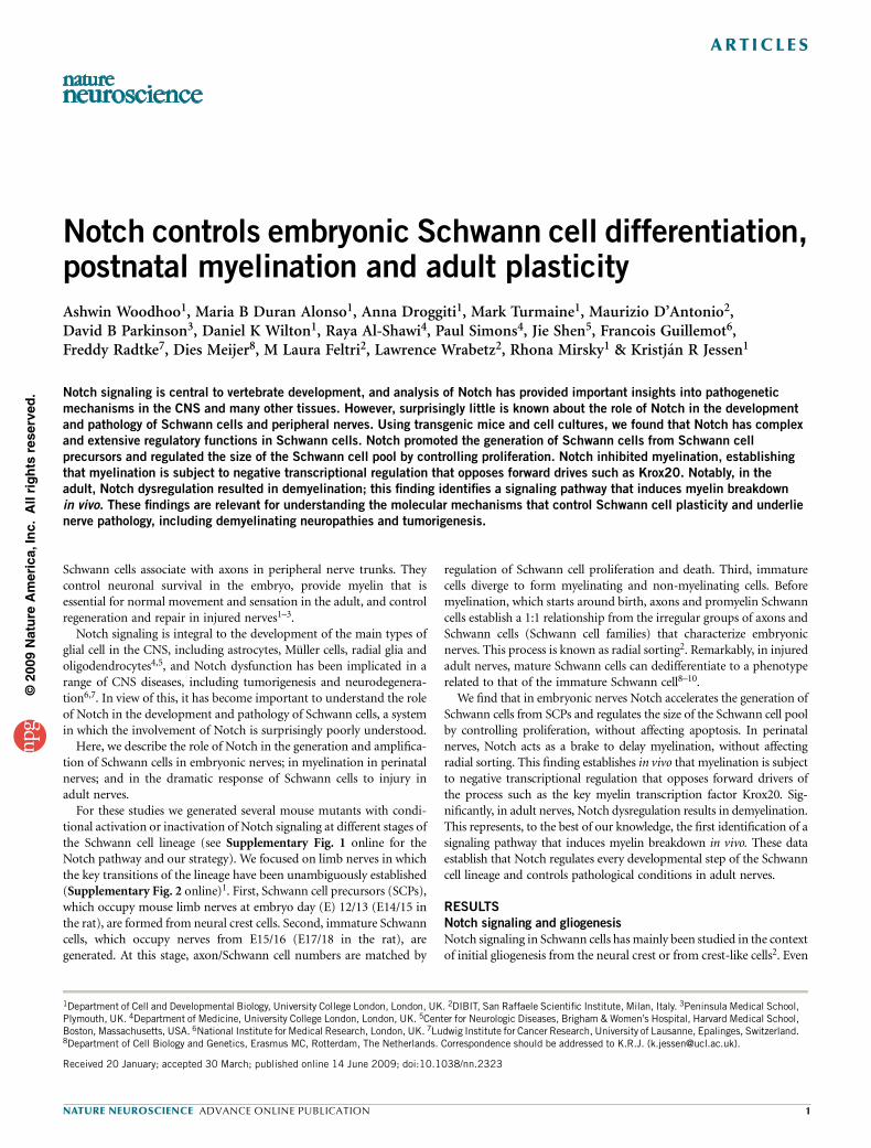
When we examined spinal nerves in mouse embryos lacking Hes 1and/or Hes 5, the effectors of canonical Notch signaling14, we found nosignificant effect on SCP formation (Fig. 1a and Supplementary Table 1online). Consistent with this, Schwann cell generation is reported tooccur in mice with neural crest-selective ablation of RBPJ15, the keytranscriptional mediator of canonical Notch signaling14, although thedevelopment of satellite cells and ganglia is impaired.In vitro, we failed to induce expression of the early glial (SCP) marker
protein zero (P0) by infecting crest cells with an adenovirus expressingNICD, the active intracellular portion of the Notch receptor14 (Ad-NICD). In parallel experiments, P0 was readily induced by the gliogenicsignal retinoic acid (Fig. 1b). These experiments indicate that the firststep in the generation of Schwann cells, namely the appearance of SCPs,does not depend on canonical Notch signals.
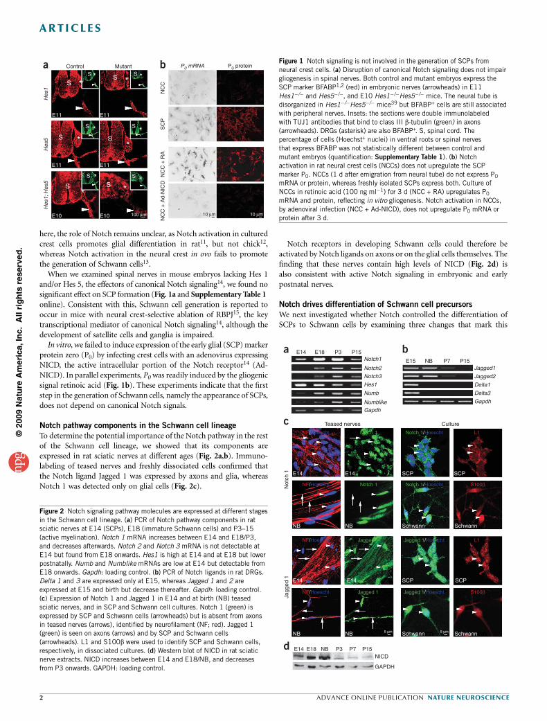
Notch pathway components in the Schwann cell lineage
To determine the potential importance of the Notch pathway in the restof the Schwann cell lineage, we showed that its components areexpressed in rat sciatic nerves at different ages (Fig. 2a,b). Immuno-labeling of teased nerves and freshly dissociated cells confirmed thatthe Notch ligand Jagged 1 was expressed by axons and glia, whereasNotch 1 was detected only on glial cells (Fig. 2c).
Notch receptors in developing Schwann cells could therefore beactivated by Notch ligands on axons or on the glial cells themselves. Thefinding that these nerves contain high levels of NICD (Fig. 2d) isalso consistent with active Notch signaling in embryonic and earlypostnatal nerves.
Notch drives differentiation of Schwann cell precursors
We next investigated whether Notch controlled the differentiation ofSCPs to Schwann cells by examining three changes that mark this
©20
09 N
atu
re A
mer
ica,
Inc.
All
rig
hts
res
erve
d.
Controla b
Hes
1H
es5
Hes
1; H
es5
SS
S
E11 E11
S
* * **
S
E11
S
**
S
E11
S
*
*
S
E10
S
*
*S
E10 100 µm
S
**
Mutant P0 mRNA P0 protein
10 µm 10 µm
NC
CS
CP
NC
C +
RA
NC
C +
Ad-
NIC
D
Figure 1 Notch signaling is not involved in the generation of SCPs from
neural crest cells. (a) Disruption of canonical Notch signaling does not impair
gliogenesis in spinal nerves. Both control and mutant embryos express the
SCP marker BFABP1,2 (red) in embryonic nerves (arrowheads) in E11
Hes1�/� and Hes5�/�, and E10 Hes1�/�Hes5�/� mice. The neural tube is
disorganized in Hes1�/�Hes5�/� mice39 but BFABP+ cells are still associated
with peripheral nerves. Insets: the sections were double immunolabeled
with TUJ1 antibodies that bind to class III b-tubulin (green) in axons(arrowheads). DRGs (asterisk) are also BFABP+. S, spinal cord. The
percentage of cells (Hoechst+ nuclei) in ventral roots or spinal nerves
that express BFABP was not statistically different between control and
mutant embryos (quantification: Supplementary Table 1). (b) Notch
activation in rat neural crest cells (NCCs) does not upregulate the SCP
marker P0. NCCs (1 d after emigration from neural tube) do not express P0
mRNA or protein, whereas freshly isolated SCPs express both. Culture of
NCCs in retinoic acid (100 ng ml�1) for 3 d (NCC + RA) upregulates P0
mRNA and protein, reflecting in vitro gliogenesis. Notch activation in NCCs,
by adenoviral infection (NCC + Ad-NICD), does not upregulate P0 mRNA or
protein after 3 d.
E14a
c
d
b
E14 E18 NB P3 P7 P15NICD
GAPDH
E18
Teased nerves
Not
ch 1
Jagg
ed 1
NF/Hoescht L1Notch 1 Notch 1/Hoescht
NF/Hoescht S100βNotch 1 Notch 1/Hoescht
NF/Hoescht L1Jagged 1 Jagged 1/Hoescht
NF/Hoescht S100βJagged 1 Jagged 1/Hoescht
E14 E14 SCP SCP
NB NB Schwann Schwann
NB
E14 E14
NB 5 µm 5 µmSchwann
SCP SCP
Schwann
Culture
P3 P15
E15 NB P7 P15Notch1
Jagged1
Jagged2
Delta1
Delta3
Gapdh
Notch2
Notch3
Hes1
Numb
NumblikeGapdh
Figure 2 Notch signaling pathway molecules are expressed at different stages
in the Schwann cell lineage. (a) PCR of Notch pathway components in rat
sciatic nerves at E14 (SCPs), E18 (immature Schwann cells) and P3–15
(active myelination). Notch 1 mRNA increases between E14 and E18/P3,
and decreases afterwards. Notch 2 and Notch 3 mRNA is not detectable at
E14 but found from E18 onwards. Hes1 is high at E14 and at E18 but lower
postnatally. Numb and Numblike mRNAs are low at E14 but detectable from
E18 onwards. Gapdh: loading control. (b) PCR of Notch ligands in rat DRGs.
Delta 1 and 3 are expressed only at E15, whereas Jagged 1 and 2 are
expressed at E15 and birth but decrease thereafter. Gapdh: loading control.
(c) Expression of Notch 1 and Jagged 1 in E14 and at birth (NB) teased
sciatic nerves, and in SCP and Schwann cell cultures. Notch 1 (green) is
expressed by SCP and Schwann cells (arrowheads) but is absent from axons
in teased nerves (arrows), identified by neurofilament (NF; red). Jagged 1(green) is seen on axons (arrows) and by SCP and Schwann cells
(arrowheads). L1 and S100b were used to identify SCP and Schwann cells,
respectively, in dissociated cultures. (d) Western blot of NICD in rat sciatic
nerve extracts. NICD increases between E14 and E18/NB, and decreases
from P3 onwards. GAPDH: loading control.
2 ADVANCE ONLINE PUBLICATION NATURE NEUROSCIENCE
ART ICLES
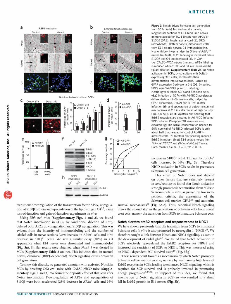
transition: downregulation of the transcription factor AP2a, upregula-tion of S100b protein and upregulation of the lipid antigen O41,2, usingloss-of-function and gain-of-function experiments in vivo.
Using Dhh-cre+ mice (Supplementary Figs. 1 and 2), we foundthat Notch inactivation in SCPs, by conditional deletion of RBPJ,delayed both AP2a downregulation and S100b upregulation. This wasevident from the intensity of immunolabeling and the number oflabeled cells in nerve sections (24% increase in AP2a+ cells and 30%decrease in S100b+ cells). We saw a similar delay (40%) in O4appearance when E14 nerves were dissociated and immunolabeled(Fig. 3a). Similar results were obtained when Notch 1 was deleted inSCPs (Supplementary Table 2 online). This indicates that, in normalnerves, canonical (RBPJ-dependent) Notch signaling drives Schwanncell generation.
To show this directly, we generated a mutant with activated Notch inSCPs by breeding Dhh-cre+ mice with CALSL-NICD mice (Supple-mentary Figs. 1 and 2). We found the opposite effect of that seen afterNotch inactivation. Downregulation of AP2a and upregulation ofS100b were both accelerated (28% decrease in AP2a+ cells and 35%
increase in S100b+ cells). The number of O4+
cells increased by 46% (Fig. 3b). ThereforeNICD activation in SCPs results in prematureSchwann cell generation.
This effect of Notch does not dependon other factors that are selectively presentin vivo, because we found that Notch activationstrongly promoted the transition from SCPs toSchwann cells in vitro as judged by two inde-pendent criteria, the appearance of theSchwann cell marker GFAP16 and autocrine
survival mechanisms17 (Fig. 3c–e). Thus, canonical Notch signalingdrives the second step in the generation of Schwann cells from neuralcrest cells, namely the transition from SCPs to immature Schwann cells.
Notch elevates erbB2 receptors and responsiveness to NRG1
We have shown previously that the transition from SCPs to immatureSchwann cells in vitro is also promoted by neuregulin-1 (NRG1)18. Wetherefore sought a link between Notch and NRG1 signaling, as seen inthe development of radial glia4,5. We found that Notch activation inSCPs selectively upregulated the ErbB2 receptors for NRG1 andincreased the sensitivity of SCPs to NRG1. This was measured usingan NRG1-dependent SCP survival assay19 (Fig. 3f,g).
These results point towards a mechanism by which Notch promotesSchwann cell generation in vivo, namely by maintaining high levels ofErbB2 receptors in SCPs, leading to enhanced NRG1 signaling, which isrequired for SCP survival and is probably involved in promotinglineage progression2,19,20. In support of this idea, we found thatinactivation of Notch signaling in SCPs in vivo resulted in a sharpfall in ErbB2 protein in E14 nerves (Fig. 3h).
©20
09 N
atu
re A
mer
ica,
Inc.
All
rig
hts
res
erve
d.
RBPJ inactivation Enforced NICD expressionControl
a
c
e
h
f g
d
b
Control 3T3
Nestin D3 D3Nestin
GFAP GFAP
Delta1 3T3100
Control 3T3Delta1 3T3
Ad-GFPAd-NICD
Ad-GFPAd-NICD
Ad-GFP
Ad-NICDAd-
GFP
Ad-NIC
D
75
50
**
*
*
*
*
*
*
25
0D1
ErbB2
ErbB3p-ERK1/2p-JNK1/2
p-p38
β-tubulin
NICD
D2 D3 D4 D5 D2 D4
Per
cent
age
of G
FAP
+
per
Nes
tin+ c
ells
100
75
50
25
0125cells
Con Mut
Con Mut
ErbB2
GAPDH
RBPJinactivation
Notch 1inactivation
10,000cells
Per
cent
age
surv
ival
Per
cent
age
surv
ival
100
150
125
100
75
50
25
0 2 4 6Neuregulin-1 (ng ml–1)
8 10
80
60
40
20
0
Per
cent
age
of G
FAP
+
per
Nes
tin+ c
ells
AP
2αS
100β
O4
AP
2αS
100β
O4
S
S SE14 E14
E14 E14
E14 E14
50 µm
50 µm
10 µm
20 µm
S S
S SE14 E14
E14 E14
E14 E14
SMutant
Notch activation in cultured SCPs
Control Mutant
Figure 3 Notch drives Schwann cell generation
from SCPs. (a,b) Top and middle panels,
longitudinal sections of E14 hind limb nerves
immunolabeled for TUJ1 (inset; red), AP2a or
S100b (DAB). Insets, spinal cord (S), DRG
(arrowheads). Bottem panels, dissociated cells
from E14 sciatic nerves; 04 immunolabeling.
Nuclei (blue): Hoechst dye. In Dhh–cre+RBPJf/f
nerves (mutant), AP2a labeling is increased, while
S100b and O4 are decreased (a). In Dhh–
cre+CALSL–NICD nerves (mutant), AP2a labeling
is reduced while S100 and 04 are increased (b)
(quantification: Supplementary Table 2). (c) Notch
activation in SCPs, by co-culture with Delta1-
expressing 3T3 cells, accelerates their
differentiation into Schwann cells, judged by
GFAP expression (red) over a 5-d (D1–5) period.
SCPs were 94–99% pure (L1 labeling)1,2.
Nestin (green) labels SCPs and Schwann cells.
(d,e) Infection of SCPs with Ad-NICD accelerates
differentiation into Schwann cells, judged by
GFAP expression, 2 (D2) and 4 (D4) d after
infection (d), and appearance of autocrine survival
mechanisms at 2 d in cells plated at high density
(10,000 cells; e). (f) Western blot showing that
ErbB2 receptors are elevated in Ad-NICD-infected
SCP cultures. Phospho-p38 levels are alsoelevated. (g) The NRG1 concentration needed for
50% survival of Ad-NICD-infected SCPs is only
about half that needed for control Ad-GFP-
infected cells. (h) Western blot showing reduced
ErbB2 in mutant (Mut) E14 sciatic nerves from
Dhh-cre+RBPJf/f and Dhh-cre+Notch1f/f mice.
Data: mean ± s.e.m., n ¼ 3, *P o 0.01.
NATURE NEUROSCIENCE ADVANCE ONLINE PUBLICATION 3
ART ICLES
Notch controls Schwann cell proliferation and numbers
Schwann cell generation from SCPs occurs during a period ofincreasing cell proliferation2,21,22 that peaks at the immatureSchwann cell stage, a time of maximal NICD expression (Fig. 2d),indicating that Notch might drive Schwann cell proliferation. Whenwe conditionally deleted RBPJ or Notch 1 using P0-cre
+ mice(Supplementary Figs. 1 and 2), we found a marked reduction inproliferation (incorporation of 5-bromodeoxyuridine (BrdU) andphosphohistone (PH3) labeling) in both mouse lines (Fig. 4a–c andSupplementary Table 3 online).
To confirm this, we examined proliferation in mice with activatedNICD in Schwann cells (P0-cre
+ mice bred with CALSL-NICD mice;Supplementary Figs. 1 and 2). Even though cells in control nervesproliferated rapidly, both BrdU incorporation and PH3 labelingwere significantly increased in this mutant (Fig. 4d,e and Supplemen-tary Table 3).
When we counted Schwann cell nuclei in ultra-thin sections of P2nerves, we found that in all three mutants Schwann cell numberschanged in line with expectations. There was a reduction of 30% and18% in P0cre
+RBPJ f/f mice and P0cre+Notch1f/f mice, respectively, and
an increase of 13% in P0cre+CALSL-NICD mice (Fig. 4c,f and Supple-
mentary Table 3). These findings indicate that the canonical Notchpathway is mitogenic for Schwann cells and that NICD elevation is partof the mechanism that controls proliferation and numbers indeveloping nerves.
To show this directly, we examined proliferation in immunopurifiedcultured Schwann cells after Notch activation. We found that BrdUincorporation was strongly stimulated by either infection withAd-NICD (Fig. 4g) or co-culture with the immortalized cell line3T3, expressing the Notch ligand, Delta 1 (data not shown).
To confirm that Notch stimulates proliferation, we used cells thatlack RBPJ (RBPJnull; RBPJ f/f cells treated in vitro with Cre adenovirus).Ad-NICD did not induce DNA synthesis in these cells, unlike NRG1(Fig. 4h,i). The reduced proliferation observed in vivo in mice that lackRBPJ (P0-cre
+RBPJ f/f mice) therefore reflects reduced Notch, but notNRG1, signaling.
Enforced NICD expression also upregulated the cell cycle markerscyclin D1 and cdk2 (Fig. 4j). The mitogenic effect of NICD on Schwanncells involved and depended on phosphorylation of the kinases ERK1/2and JNK, but not p38 kinase (Fig. 4k,l). Apoptotic cell death2,23
(measured by TdT-mediated dUTP nick end (TUNEL) labeling)was unaffected in E17 nerves of the three Notch mutants (Supplemen-tary Fig. 3 online). In embryonic nerves, therefore, Notch signalingcontrols Schwann cell proliferation and numbers, but not apoptoticcell death.
Krox20 suppresses Notch signaling in myelinating cells
We next investigated Notch signaling in perinatal nerves duringmyelination. This process, which starts around birth, is incompatiblewith proliferation, and most Schwann cells have stopped dividing asthey reach the pro-myelin 1:1 stage and start significant myelingene activation2,21,24.
Using fluorescence activated cell sorting (FACS) to separate myeli-nating cells from cells that are not making myelin in P5 nerves, wefound that Notch signaling (which drives proliferation) is inactivatedin myelinating cells in vivo (Fig. 5a). This is consistent with thedownregulation of NICD protein at the start of myelination inpostnatal nerves (Fig. 2d).
The transcription factor Krox20 activates myelin genes andsuppresses genes associated with immature Schwann cells1,25–27.We found that Krox20 suppressed Notch signaling in myelinatingcells, as in culture, enforced Krox20 expression by adenoviralinfection (Ad-K20) suppressed NICD protein, and in vivo, NICDlevels remained high in P7 Krox20–/– animals, but low in normalnerves (Fig. 5b,c).
©20
09 N
atu
re A
mer
ica,
Inc.
All
rig
hts
res
erve
d.
Control
a b c
d e f
g h i
j k l
Hoescht
BrdU
Hoescht
BrdU
Hoescht
BrdU
Hoescht
BrdU
Hoescht
BrdU
Hoescht
BrdU
20 µm
20 µm
Mutant
Control
Ad-GFP Ad-NICD
Cyclin D1
cdk2
NICD
β-tubulin
p-ERK1/2
p-JNK1/2
NICD
β-tubulin
Ad-GFP
Ad-NIC
D
Ad-GFP
Ad-NIC
D
Mutant
Control
50
40
*
*
* *
**
*
*
*
*
*30
Per
cent
age
Brd
U+ c
ells
Per
cent
age
PH
3+ c
ells
20
10
0
50
40
30
Per
cent
age
Brd
U+ c
ells
20
10
0
100
75
Per
cent
age
Brd
U+ c
ells
50
25
0
100
75
Per
cent
age
Brd
U+ c
ells
50
25
0
100
75
Per
cent
age
Brd
U+ c
ells
50
25
0
Contro
l
RBPJnu
ll
Contro
l
RBPJnu
ll
4
3
2
1
0
Per
cent
age
PH
3+ c
ells 4
3
2
1
0
SC
nuc
lei p
er n
erve
400
300
200
100
0
SC
nuc
lei p
er n
erve
400
300
200
100
0
MutantControlMutant
ControlMutant
ControlMutant
Ad-GFP Ad-NICD
Control
SB202190
JNK peptideUO126
–NRG +NRG
Figure 4 Notch signaling controls the proliferation of immature Schwann
cells. (a–c) RBPJ inactivation. (a) E17 nerve sections showing fewer BrdU+
Schwann cells (green) in P0-cre+RBPJf/f nerves. Hoechst (): nuclei. (b) RBPJ
inactivation significantly reduces BrdU+ and PH3+ cells in E17 nerves.
(c) The number of Schwann cell (SC) nuclei in P2 nerves is decreased in
mutants. (d–f) Enforced NICD expression. (d) E17 nerve sections showing
increased BrdU+ Schwann cells (green) in P0-cre+CALSL-NICD nerves.
(e) NICD significantly increases BrdU+ and PH3+ cells in E17 nerves.(f) The number of Schwann cell nuclei in P2 nerves is increased in mutants.
(g-l) Notch activation in cultured Schwann cells. (g) Ad-NICD-infected
Schwann cells have more BrdU+ cells (red) than controls (73.9 ± 2.0
compared to 4.6 ± 1.3). (h,i) BrdU+ cells in control or RBPJnull Schwann
cells after infection with Ad-NICD (h), or in the presence of absence of NRG1
(i), showing that Ad-NICD, unlike NRG1, fails to induce DNA synthesis in the
absence of RBPJ. (j,k) Western blot shows upregulation of cyclin D1 and
cdk2 (j) and phospho-ERK 1/2 and phospho-JNK 1/2 (k), in Ad-NICD-
infected Schwann cells 2 d after infection. NICD demonstrates enforced
NICD expression. b-tubulin: loading control. (l) Ad-NICD-induced
proliferation in Schwann cells in vitro is inhibited by JNK peptide (to block
JNK phosphorylation) or UO126 (to block ERK1/2 phosphorylation) but not
by SB202190 (to block p38 kinase phosphorylation). Data: mean ± s.e.m.,
n ¼ 3, *P o 0.01.
4 ADVANCE ONLINE PUBLICATION NATURE NEUROSCIENCE
ART ICLES
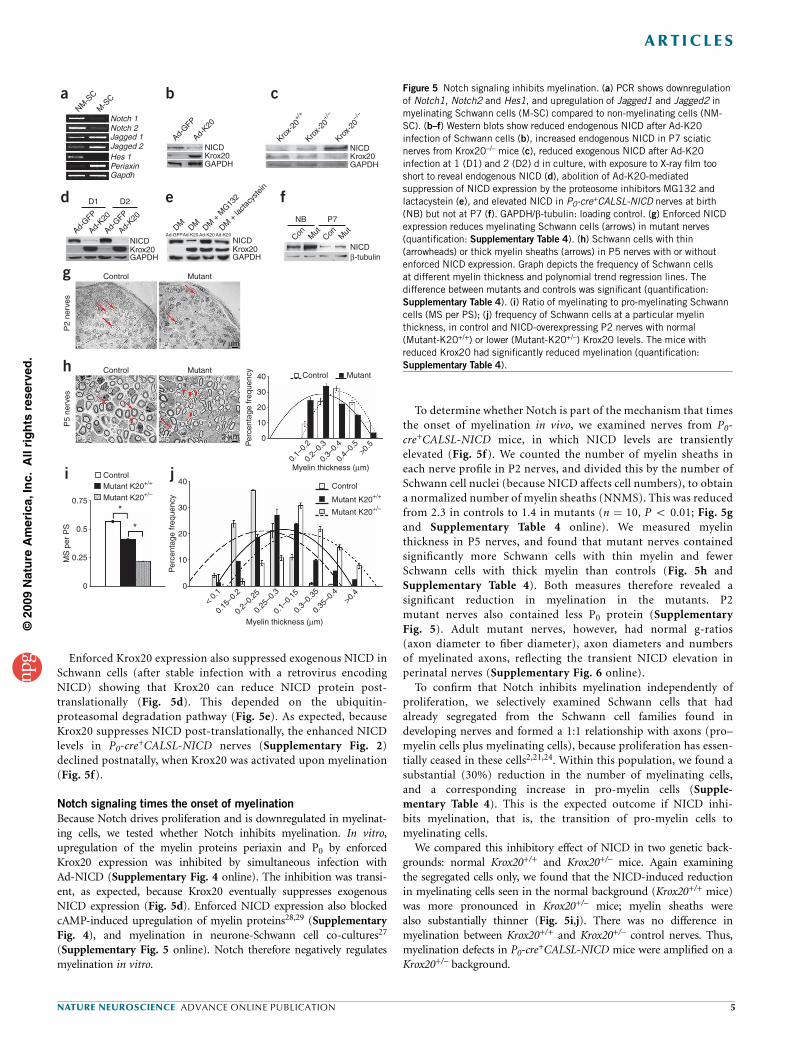
Enforced Krox20 expression also suppressed exogenous NICD inSchwann cells (after stable infection with a retrovirus encodingNICD) showing that Krox20 can reduce NICD protein post-translationally (Fig. 5d). This depended on the ubiquitin-proteasomal degradation pathway (Fig. 5e). As expected, becauseKrox20 suppresses NICD post-translationally, the enhanced NICDlevels in P0-cre
+CALSL-NICD nerves (Supplementary Fig. 2)declined postnatally, when Krox20 was activated upon myelination(Fig. 5f).
Notch signaling times the onset of myelination
Because Notch drives proliferation and is downregulated in myelinat-ing cells, we tested whether Notch inhibits myelination. In vitro,upregulation of the myelin proteins periaxin and P0 by enforcedKrox20 expression was inhibited by simultaneous infection withAd-NICD (Supplementary Fig. 4 online). The inhibition was transi-ent, as expected, because Krox20 eventually suppresses exogenousNICD expression (Fig. 5d). Enforced NICD expression also blockedcAMP-induced upregulation of myelin proteins28,29 (SupplementaryFig. 4), and myelination in neurone-Schwann cell co-cultures27
(Supplementary Fig. 5 online). Notch therefore negatively regulatesmyelination in vitro.
To determine whether Notch is part of the mechanism that timesthe onset of myelination in vivo, we examined nerves from P0-cre+CALSL-NICD mice, in which NICD levels are transientlyelevated (Fig. 5f). We counted the number of myelin sheaths ineach nerve profile in P2 nerves, and divided this by the number ofSchwann cell nuclei (because NICD affects cell numbers), to obtaina normalized number of myelin sheaths (NNMS). This was reducedfrom 2.3 in controls to 1.4 in mutants (n ¼ 10, P o 0.01; Fig. 5gand Supplementary Table 4 online). We measured myelinthickness in P5 nerves, and found that mutant nerves containedsignificantly more Schwann cells with thin myelin and fewerSchwann cells with thick myelin than controls (Fig. 5h andSupplementary Table 4). Both measures therefore revealed asignificant reduction in myelination in the mutants. P2mutant nerves also contained less P0 protein (SupplementaryFig. 5). Adult mutant nerves, however, had normal g-ratios(axon diameter to fiber diameter), axon diameters and numbersof myelinated axons, reflecting the transient NICD elevation inperinatal nerves (Supplementary Fig. 6 online).
To confirm that Notch inhibits myelination independently ofproliferation, we selectively examined Schwann cells that hadalready segregated from the Schwann cell families found indeveloping nerves and formed a 1:1 relationship with axons (pro–myelin cells plus myelinating cells), because proliferation has essen-tially ceased in these cells2,21,24. Within this population, we found asubstantial (30%) reduction in the number of myelinating cells,and a corresponding increase in pro-myelin cells (Supple-mentary Table 4). This is the expected outcome if NICD inhi-bits myelination, that is, the transition of pro-myelin cells tomyelinating cells.
We compared this inhibitory effect of NICD in two genetic back-grounds: normal Krox20+/+ and Krox20+/– mice. Again examiningthe segregated cells only, we found that the NICD-induced reductionin myelinating cells seen in the normal background (Krox20+/+ mice)was more pronounced in Krox20+/– mice; myelin sheaths werealso substantially thinner (Fig. 5i,j). There was no difference inmyelination between Krox20+/+ and Krox20+/– control nerves. Thus,myelination defects in P0-cre
+CALSL-NICD mice were amplified on aKrox20+/– background.
©20
09 N
atu
re A
mer
ica,
Inc.
All
rig
hts
res
erve
d.
NM-S
C
M-S
C
Notch 1
NICDKrox20GAPDH
NICD
Control
P2 P2
P5 P5
P2
nerv
esP
5 ne
rves
Mutant
7 µm
2 µm
Control Mutant40
0.75
ControlMutant K20+/+
Mutant K20+/–Control
Mutant K20+/+
Mutant K20+/–
40
30
20
10
Myelin thickness (µm)
0
< 0.
1
0.15
–0.2
0.2–
0.25
0.25
–0.3
0.1–
0.15
0.3–
0.35
0.35
–0.4
>0.4
0.5
*
*
MS
per
PS
Per
cent
age
freq
uenc
y
0.25
0
Control Mutant
30
Per
cent
age
freq
uenc
y
20
10
0
0.1–
0.2
0.2–
0.3
0.3–
0.4
0.4–
0.5
Myelin thickness (µm)
>0.5
Krox20GAPDH
NICD
NB
Con Mut
Con Mut
P7
Krox20GAPDH
NICDβ-tubulin
NICDKrox20GAPDH
Ad-GFP
Ad-GFPDM DM DM
+ M
G132
DM +
lacta
cyste
in
Ad-K20 Ad-K20 Ad-K20
Ad-K20
Ad-GFP
Ad-K20
Ad-GFP
Ad-K20
Krox-
20+/
+
Krox-
20+/
–
Krox-
20–/
–
Hes 1PeriaxinGapdh
D1
a b c
d
g
h
i j
e fD2
Notch 2Jagged 1Jagged 2
Figure 5 Notch signaling inhibits myelination. (a) PCR shows downregulation
of Notch1, Notch2 and Hes1, and upregulation of Jagged1 and Jagged2 in
myelinating Schwann cells (M-SC) compared to non-myelinating cells (NM-
SC). (b–f) Western blots show reduced endogenous NICD after Ad-K20
infection of Schwann cells (b), increased endogenous NICD in P7 sciatic
nerves from Krox20–/– mice (c), reduced exogenous NICD after Ad-K20
infection at 1 (D1) and 2 (D2) d in culture, with exposure to X-ray film too
short to reveal endogenous NICD (d), abolition of Ad-K20-mediatedsuppression of NICD expression by the proteosome inhibitors MG132 and
lactacystein (e), and elevated NICD in P0-cre+CALSL-NICD nerves at birth
(NB) but not at P7 (f). GAPDH/b-tubulin: loading control. (g) Enforced NICD
expression reduces myelinating Schwann cells (arrows) in mutant nerves
(quantification: Supplementary Table 4). (h) Schwann cells with thin
(arrowheads) or thick myelin sheaths (arrows) in P5 nerves with or without
enforced NICD expression. Graph depicts the frequency of Schwann cells
at different myelin thickness and polynomial trend regression lines. The
difference between mutants and controls was significant (quantification:
Supplementary Table 4). (i) Ratio of myelinating to pro-myelinating Schwann
cells (MS per PS); (j) frequency of Schwann cells at a particular myelin
thickness, in control and NICD-overexpressing P2 nerves with normal
(Mutant-K20+/+) or lower (Mutant-K20+/–) Krox20 levels. The mice with
reduced Krox20 had significantly reduced myelination (quantification:
Supplementary Table 4).
NATURE NEUROSCIENCE ADVANCE ONLINE PUBLICATION 5
ART ICLES
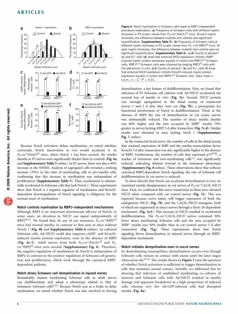
Because Notch activation delays myelination, we tested whether,conversely, Notch inactivation in vivo would accelerate it. InP0-cre
+Notch1f/f mice, where Notch 1 has been excised, the myelinsheaths in P5 nerves were significantly thicker than in controls (Fig. 6aand Supplementary Table 5 online). In P2 nerves, there was also a 40%increase in the NNMS. Analysis of segregated cells revealed a strikingincrease (70%) in the ratio of myelinating cells to pro-myelin cells,confirming that this increase in myelination was independent ofproliferation (Supplementary Table 5). Thus, myelination is substan-tially accelerated in Schwann cells that lack Notch 1. These experimentsshow that Notch is a negative regulator of myelination and Krox20-dependent downregulation of Notch signaling is obligatory for thenormal onset of myelination.
Notch controls myelination by RBPJ–independent mechanisms
Although RBPJ is an important downstream effector of Notch, insome cases, an elevation in NICD can signal independently ofRBPJ30,31. We found that, by any of our measures, P0-cre
+RBPJ f/f
mice had normal myelin, in contrast to that seen in mice withoutNotch 1 (Fig. 6b and Supplementary Table 6 online). In culturedSchwann cells, Ad-NICD could also suppress cAMP- and Krox20-induced myelin protein expression, even in the absence of RBPJ(Fig. 6c–f). Adult nerves from both P0-cre
+Notch1 f/f and P0-cre+RBPJ f/f mice were normal (Supplementary Fig. 6). Therefore,the negative regulation of myelination by Notch is independent ofRBPJ, in contrast to the positive regulation of Schwann cell genera-tion and proliferation, which work through the canonical RBPJ-dependent pathway.
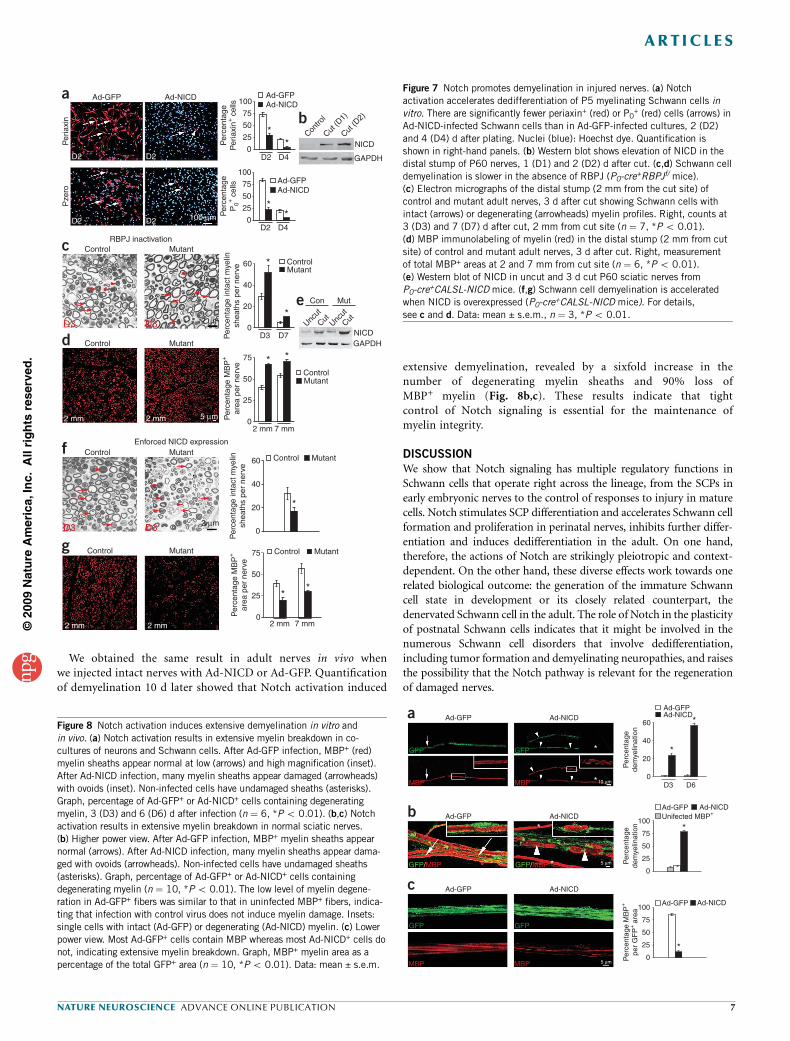
Notch drives Schwann cell demyelination in injured nerves
Remarkably, mature myelinating Schwann cells in adult nervescan dedifferentiate and adopt a phenotype related to that ofimmature Schwann cells8–10. Because Notch acts as a brake to delaymyelination, we tested whether Notch was also involved in driving
demyelination, a key feature of dedifferentiation. First, we found thatinfection of P5 Schwann cell cultures with Ad-NICD accelerated thenormal loss of myelin in vitro (Fig. 7a). Second, NICD proteinwas strongly upregulated in the distal stump of transectednerves 1 and 2 d after they were cut (Fig. 7b), a prerequisite forfunctional involvement of Notch in dedifferentiation. Third, in theabsence of RBPJ the rate of demyelination in cut sciatic nerveswas substantially reduced. The number of intact myelin sheathswas 80% higher and the area occupied by MBP+ myelin 70%greater in nerves lacking RBPJ 3 d after transection (Fig. 7c,d). Similarresults were obtained in mice lacking Notch 1 (SupplementaryFig. 7 online).
In the transected facial nerve, the number of cells in the distal stumpthat retained expression of MBP and the myelin transcription factorKrox20 3 d after transection was also significantly higher in the absenceof RBPJ. Furthermore, the number of cells that expressed p75NTR, amarker of immature and non-myelinating cells1,2, was significantlyreduced, indicating delayed reversal to the immature phenotype(Supplementary Fig. 8 online). These observations show that withoutcanonical RBPJ-dependent Notch signaling, the rate of Schwann celldedifferentiation in cut nerves is reduced.
To show directly that Notch can accelerate demyelination in vivo, weexamined myelin disappearance in cut nerves of P0-cre
+CALSL-NICDmice. First, we confirmed that nerve transection in these mice elevatedNICD when compared with cut control nerves (Fig. 7e). This wasexpected because nerve injury will trigger expression of both theendogenous NICD (Fig. 7b) and the CALSL-NICD transgene, bothof which are suppressed in intact nerves through a Krox-20-dependentmechanism (Fig. 5e,f). This increase in NICD resulted in accelerateddedifferentiation. The P0-cre
+CALSL-NICD nerves contained 50%fewer intact myelinating Schwann cells and the area occupied byMBP+ myelin was 50% smaller than in cut control nerves 3 d aftertransection (Fig. 7f,g). These experiments show that Notchsignaling drives demyelination in injured nerves through an RBPJ-dependent mechanism.
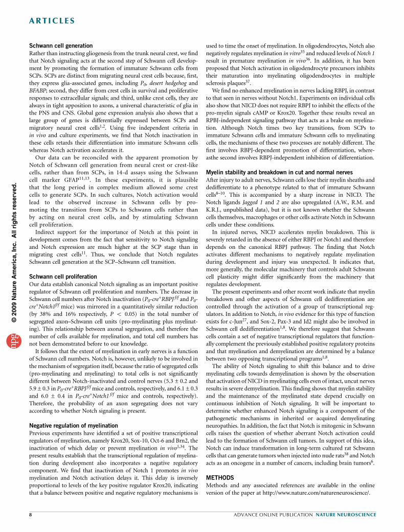
Notch initiates demyelination even in uncut nerves
In demyelinating neuropathies, demyelination occurs even thoughSchwann cells remain in contact with axons until the later stageswhen axons die32,33. The results shown in Figure 7 raise the questionof whether Notch activation is sufficient to trigger demyelination incells that maintain axonal contact. Initially, we addressed this byshowing that infection of established myelinating co-cultures ofneurons and Schwann cells with Ad-NICD resulted in myelindamage and apparent breakdown in a high proportion of infectedcells, whereas very few Ad-GFP-infected cells had disruptedmyelin (Fig. 8a).
©20
09 N
atu
re A
mer
ica,
Inc.
All
rig
hts
res
erve
d.
50
a b
d
f* *
* *
Control RBPJnull
Control RBPJnull
75
100
Per
cent
age
P0+
cel
ls
50
25
0
75P
erce
ntag
eP
0+ c
ells 50
25
0
Notch 1 inactivation RBPJ inactivation
Control Mutant Control Mutant
40
30
Per
cent
age
freq
uenc
y
20
10
0
50
40
30
Per
cent
age
freq
uenc
y
20
10
0
0.1–
0.2
0.2–
0.3
c
e
75
*
* *
*
Per
cent
age
Per
iaxi
n+ c
ells
cAM
P-in
duce
dm
yelin
diff
eren
tiatio
nK
rox2
0-in
duce
dm
yelin
diff
eren
tiatio
n
50
25
Control RBPJnull
Control RBPJnull
0
75
100
Per
cent
age
Per
iaxi
n+ c
ells
50
25
0
Ad-GFP Ad-NICD
Ad-K20/Ad-GFPAd-K20/Ad-NICD
Ad-K20/Ad-GFPAd-K20/Ad-NICD
Ad-GFP Ad-NICD
Myelin thickness (µm)0.
3–0.
4
0.4–
0.5
0.5–
0.6
>0.6
0.1–
0.2
0.2–
0.3
Myelin thickness (µm)0.
3–0.
4
0.4–
0.5
>0.5
Figure 6 Notch inactivation in Schwann cells leads to RBPJ-independent
premature myelination. (a) Frequency of Schwann cells with different myelin
thickness in P5 sciatic nerves from P0-cre+Notch1f/f mice. At each myelin
thickness, the difference between mutants and controls was significant
(quantification: Supplementary Table 5). (b) Frequency of Schwann cells at
different myelin thickness in P5 sciatic nerves from P0�cre+RBPJf/f mice. At
each myelin thickness, the difference between mutants and controls was not
significant (quantification: Supplementary Table 6). (c,d) Counts of periaxin+
(c) and P0+ cells (d) show that enforced NICD expression inhibits cAMP-
induced myelin protein expression equally in control and RBPJnull Schwann
cells. RBPJnull Schwann cells were obtained by treating RBPJf/f cells with
Cre adenovirus in vitro. (e,f) Counts of periaxin+ (e) and P0+ cells (f) show
that enforced NICD expression inhibits Krox20-induced myelin protein
expression equally in control and RBPJnull Schwann cells. Data: mean ±
s.e.m., n ¼ 3, *P o 0.01.
6 ADVANCE ONLINE PUBLICATION NATURE NEUROSCIENCE
ART ICLES
We obtained the same result in adult nerves in vivo whenwe injected intact nerves with Ad-NICD or Ad-GFP. Quantificationof demyelination 10 d later showed that Notch activation induced
extensive demyelination, revealed by a sixfold increase in thenumber of degenerating myelin sheaths and 90% loss ofMBP+ myelin (Fig. 8b,c). These results indicate that tightcontrol of Notch signaling is essential for the maintenance ofmyelin integrity.
DISCUSSION
We show that Notch signaling has multiple regulatory functions inSchwann cells that operate right across the lineage, from the SCPs inearly embryonic nerves to the control of responses to injury in maturecells. Notch stimulates SCP differentiation and accelerates Schwann cellformation and proliferation in perinatal nerves, inhibits further differ-entiation and induces dedifferentiation in the adult. On one hand,therefore, the actions of Notch are strikingly pleiotropic and context-dependent. On the other hand, these diverse effects work towards onerelated biological outcome: the generation of the immature Schwanncell state in development or its closely related counterpart, thedenervated Schwann cell in the adult. The role of Notch in the plasticityof postnatal Schwann cells indicates that it might be involved in thenumerous Schwann cell disorders that involve dedifferentiation,including tumor formation and demyelinating neuropathies, and raisesthe possibility that the Notch pathway is relevant for the regenerationof damaged nerves.
©20
09 N
atu
re A
mer
ica,
Inc.
All
rig
hts
res
erve
d.
Ad-GFPa
b
c
GFP
MBP
GFP * *
*
*
*
*
*MBP
GFP
MBP
GFP
MBP
10 µm
5 µm
5 µm
Ad-NICD60
Ad-GFPAd-NICD
Ad-GFPUnifected MBP+
Ad-NICD
Ad-GFP Ad-NICD
40
Per
cent
age
dem
yelin
atio
n
20
0
100
75
Per
cent
age
dem
yelin
atio
n
25
50
0
100
75
Per
cent
age
MB
P+
per
GF
P+
area
25
50
0*
D3 D6
Ad-GFP
GFP/MBP GFP/MBP
Ad-NICD
Ad-GFP Ad-NICD
Ad-GFPa
c
d
e
f
g
b
Control MutantRBPJ inactivation
Enforced NICD expression
Control Mutant
Control Mutant
Control Mutant
D2 D2
100 µm
5 µm
2 µm
2 µm
D2
2 mm 2 mm
2 mm 2 mm
D2
Per
iaxi
nP
zero
Ad-NICD 100Ad-GFP
Contro
l
Uncut
Cut Uncut
Cut
Cut (D
1)
Cut (D
2)
Ad-GFP
NICD
GAPDH
NICDGAPDH
Ad-NICD
Ad-NICD
ControlMutant
ControlMutant
Control Mutant
Control Mutant
Per
cent
age
Per
iaxi
n+ c
ells
75
*
*
*
*
*
*
**
*
*
*
50
25
0
100
60
40
Per
cent
age
inta
ct m
yelin
shea
ths
per
nerv
eP
erce
ntag
e M
BP
+
area
per
ner
ve
20
0
60
40
Per
cent
age
inta
ct m
yelin
shea
ths
per
nerv
e
20
0
75
50
25
02 mm 7 mm
Per
cent
age
MB
P+
area
per
ner
ve
75
50
25
02 mm 7 mm
Per
cent
age
P0+
cel
ls 75
50
25
0
D2 D4
D2 D4
D3 D7
Con Mut
Figure 7 Notch promotes demyelination in injured nerves. (a) Notch
activation accelerates dedifferentiation of P5 myelinating Schwann cells in
vitro. There are significantly fewer periaxin+ (red) or P0+ (red) cells (arrows) in
Ad-NICD-infected Schwann cells than in Ad-GFP-infected cultures, 2 (D2)
and 4 (D4) d after plating. Nuclei (blue): Hoechst dye. Quantification is
shown in right-hand panels. (b) Western blot shows elevation of NICD in the
distal stump of P60 nerves, 1 (D1) and 2 (D2) d after cut. (c,d) Schwann cell
demyelination is slower in the absence of RBPJ (P0-cre+RBPJf/ mice).
(c) Electron micrographs of the distal stump (2 mm from the cut site) of
control and mutant adult nerves, 3 d after cut showing Schwann cells with
intact (arrows) or degenerating (arrowheads) myelin profiles. Right, counts at
3 (D3) and 7 (D7) d after cut, 2 mm from cut site (n ¼ 7, *P o 0.01).
(d) MBP immunolabeling of myelin (red) in the distal stump (2 mm from cut
site) of control and mutant adult nerves, 3 d after cut. Right, measurement
of total MBP+ areas at 2 and 7 mm from cut site (n ¼ 6, *P o 0.01).
(e) Western blot of NICD in uncut and 3 d cut P60 sciatic nerves from
P0-cre+CALSL-NICD mice. (f,g) Schwann cell demyelination is accelerated
when NICD is overexpressed (P0-cre+CALSL-NICD mice). For details,
see c and d. Data: mean ± s.e.m., n ¼ 3, *P o 0.01.
Figure 8 Notch activation induces extensive demyelination in vitro and
in vivo. (a) Notch activation results in extensive myelin breakdown in co-
cultures of neurons and Schwann cells. After Ad-GFP infection, MBP+ (red)
myelin sheaths appear normal at low (arrows) and high magnification (inset).
After Ad-NICD infection, many myelin sheaths appear damaged (arrowheads)
with ovoids (inset). Non-infected cells have undamaged sheaths (asterisks).
Graph, percentage of Ad-GFP+ or Ad-NICD+ cells containing degenerating
myelin, 3 (D3) and 6 (D6) d after infection (n ¼ 6, *P o 0.01). (b,c) Notch
activation results in extensive myelin breakdown in normal sciatic nerves.
(b) Higher power view. After Ad-GFP infection, MBP+ myelin sheaths appear
normal (arrows). After Ad-NICD infection, many myelin sheaths appear dama-
ged with ovoids (arrowheads). Non-infected cells have undamaged sheaths
(asterisks). Graph, percentage of Ad-GFP+ or Ad-NICD+ cells containing
degenerating myelin (n ¼ 10, *P o 0.01). The low level of myelin degene-ration in Ad-GFP+ fibers was similar to that in uninfected MBP+ fibers, indica-
ting that infection with control virus does not induce myelin damage. Insets:
single cells with intact (Ad-GFP) or degenerating (Ad-NICD) myelin. (c) Lower
power view. Most Ad-GFP+ cells contain MBP whereas most Ad-NICD+ cells do
not, indicating extensive myelin breakdown. Graph, MBP+ myelin area as a
percentage of the total GFP+ area (n ¼ 10, *P o 0.01). Data: mean ± s.e.m.
NATURE NEUROSCIENCE ADVANCE ONLINE PUBLICATION 7
ART ICLES
Schwann cell generation
Rather than instructing gliogenesis from the trunk neural crest, we findthat Notch signaling acts at the second step of Schwann cell develop-ment by promoting the formation of immature Schwann cells fromSCPs. SCPs are distinct from migrating neural crest cells because, first,they express glia-associated genes, including P0, desert hedgehog andBFABP; second, they differ from crest cells in survival and proliferativeresponses to extracellular signals; and third, unlike crest cells, they arealways in tight apposition to axons, a universal characteristic of glia inthe PNS and CNS. Global gene expression analysis also shows that alarge group of genes is differentially expressed between SCPs andmigratory neural crest cells1,2. Using five independent criteria inin vivo and culture experiments, we find that Notch inactivation inthese cells retards their differentiation into immature Schwann cellswhereas Notch activation accelerates it.
Our data can be reconciled with the apparent promotion byNotch of Schwann cell generation from neural crest or crest-likecells, rather than from SCPs, in 14-d assays using the Schwanncell marker GFAP11,13. In these experiments, it is plausiblethat the long period in complex medium allowed some crestcells to generate SCPs. In such cultures, Notch activation wouldlead to the observed increase in Schwann cells by pro-moting the transition from SCPs to Schwann cells rather thanby acting on neural crest cells, and by stimulating Schwanncell proliferation.
Indirect support for the importance of Notch at this point indevelopment comes from the fact that sensitivity to Notch signalingand Notch expression are much higher at the SCP stage than inmigrating crest cells11. Thus, we conclude that Notch regulatesSchwann cell generation at the SCP–Schwann cell transition.
Schwann cell proliferation
Our data establish canonical Notch signaling as an important positiveregulator of Schwann cell proliferation and numbers. The decrease inSchwann cell numbers after Notch inactivation (P0-cre
+RBPJ f/f and P0-cre+Notch1f/f mice) was mirrored in a quantitatively similar reduction(by 38% and 16% respectively, P o 0.05) in the total number ofsegregated axon–Schwann cell units (pro-myelinating plus myelinat-ing). This relationship between axonal segregation, and therefore thenumber of cells available for myelination, and total cell numbers hasnot been demonstrated before to our knowledge.
It follows that the extent of myelination in early nerves is a functionof Schwann cell numbers. Notch is, however, unlikely to be involved inthe mechanism of segregation itself, because the ratio of segregated cells(pro-myelinating and myelinating) to total cells is not significantlydifferent between Notch-inactivated and control nerves (5.3 ± 0.2 and5.9 ± 0.3 in P0-cre
+RBPJ f/f mice and controls, respectively, and 6.1 ± 0.3and 6.0 ± 0.4 in P0-cre
+Notch1 f/f mice and controls, respectively).Therefore, the probability of an axon segregating does not varyaccording to whether Notch signaling is present.
Negative regulation of myelination
Previous experiments have identified a set of positive transcriptionalregulators of myelination, namely Krox20, Sox-10, Oct-6 and Brn2, theinactivation of which delay or prevent myelination in vivo1,34. Thepresent results establish that the transcriptional regulation of myelina-tion during development also incorporates a negative regulatorycomponent. We find that inactivation of Notch 1 promotes in vivomyelination and Notch activation delays it. This delay is inverselyproportional to levels of the key positive regulator Krox20, indicatingthat a balance between positive and negative regulatory mechanisms is
used to time the onset of myelination. In oligodendrocytes, Notch alsonegatively regulates myelination in vitro35 and reduced levels of Notch 1result in premature myelination in vivo36. In addition, it has beenproposed that Notch activation in oligodendrocyte precursors inhibitstheir maturation into myelinating oligodendrocytes in multiplesclerosis plaques37.
We find no enhanced myelination in nerves lacking RBPJ, in contrastto that seen in nerves without Notch1. Experiments on individual cellsalso show that NICD does not require RBPJ to inhibit the effects of thepro-myelin signals cAMP or Krox20. Together these results reveal anRPBJ-independent signaling pathway that acts as a brake on myelina-tion. Although Notch times two key transitions, from SCPs toimmature Schwann cells and immature Schwann cells to myelinatingcells, the mechanisms of these two processes are notably different. Thefirst involves RBPJ-dependent promotion of differentiation, where-asthe second involves RBPJ-independent inhibition of differentiation.
Myelin stability and breakdown in cut and normal nerves
After injury to adult nerves, Schwann cells lose their myelin sheaths anddedifferentiate to a phenotype related to that of immature Schwanncells8–10. This is accompanied by a sharp increase in NICD. TheNotch ligands Jagged 1 and 2 are also upregulated (A.W., R.M. andK.R.J., unpublished data), but it is not known whether the Schwanncells themselves, macrophages or other cells activate Notch in Schwanncells under these conditions.
In injured nerves, NICD accelerates myelin breakdown. This isseverely retarded in the absence of either RBPJ or Notch1 and thereforedepends on the canonical RBPJ pathway. The finding that Notchactivates different mechanisms to negatively regulate myelinationduring development and injury was unexpected. It indicates that,more generally, the molecular machinery that controls adult Schwanncell plasticity might differ significantly from the machinery thatregulates development.
The present experiments and other recent work indicate that myelinbreakdown and other aspects of Schwann cell dedifferentiation arecontrolled through the activation of a group of transcriptional reg-ulators. In addition to Notch, in vivo evidence for this type of functionexists for c-Jun27, and Sox-2, Pax-3 and Id2 might also be involved inSchwann cell dedifferentiation1,8. We therefore suggest that Schwanncells contain a set of negative transcriptional regulators that function-ally complement the previously established positive regulatory proteinsand that myelination and demyelination are determined by a balancebetween two opposing transcriptional programs1,8.
The ability of Notch signaling to shift this balance and to drivemyelinating cells towards demyelination is shown by the observationthat activation of NICD in myelinating cells even of intact, uncut nervesresults in severe demyelination. This finding shows that myelin stabilityand the maintenance of the myelinated state depend crucially oncontinuous inhibition of Notch signaling. It will be important todetermine whether enhanced Notch signaling is a component of thepathogenetic mechanisms in inherited or acquired demyelinatingneuropathies. In addition, the fact that Notch is mitogenic in Schwanncells raises the question of whether aberrant Notch activation couldlead to the formation of Schwann cell tumors. In support of this idea,Notch can induce transformation in long-term cultured rat Schwanncells that can generate tumors when injected into nude rats38 and Notchacts as an oncogene in a number of cancers, including brain tumors6.
METHODS
Methods and any associated references are available in the onlineversion of the paper at http://www.nature.com/natureneuroscience/.
©20
09 N
atu
re A
mer
ica,
Inc.
All
rig
hts
res
erve
d.
8 ADVANCE ONLINE PUBLICATION NATURE NEUROSCIENCE
ART ICLES
Note: Supplementary information is available on the Nature Neuroscience website.
ACKNOWLEDGMENTSThis work was funded by a Wellcome Trust Programme Grant to K.R.J., R.M.and D.B.P., a Wellcome Trust Project grant to K.R.J. and R.M. and grants fromthe US National Institutes of Health to M.L.F. and L.W.
AUTHOR CONTRIBUTIONSA.W. carried out all the experiments with the exception of cAMP myelinationassays and PCR analyses, which were performed by M.B.D.A. A.W. was helpedby A.D. in in situ hybridization experiments, by M.T. in EM sectioning, by M.D.in FACS, by D.B.P. in in vitro inhibitor experiments and by D.K.W. in animalhusbandry. R.A.-S. and P.S. generated Hes1–/–Hes5–/– cells from frozen embryos.J.S., F.G., F.R., D.M., M.L.F. and L.W. provided the mice. A.W. generated all thefigures. A.W., R.M. and K.R.J. designed the experiments. K.R.J., A.W. and R.M.wrote the manuscript.
Published online at http://www.nature.com/natureneuroscience/
Reprints and permissions information is available online at http://npg.nature.com/
reprintsandpermissions/
1. Jessen, K.R. & Mirsky, R. The origin and development of glial cells in peripheral nerves.Nat. Rev. Neurosci. 6, 671–682 (2005).
2. Woodhoo, A. & Sommer, L. Development of the Schwann cell lineage: from the neuralcrest to the myelinated nerve. Glia 56, 1481–1490 (2008).
3. Chen, Z.L., Yu, W.M. & Strickland, S. Peripheral regeneration. Annu. Rev. Neurosci. 30,209–233 (2007).
4. Louvi, A. & Artavanis-Tsakonas, S. Notch signalling in vertebrate neural development.Nat. Rev. Neurosci. 7, 93–102 (2006).
5. Yoon, K. & Gaiano, N. Notch signaling in the mammalian central nervous system:insights from mouse mutants. Nat. Neurosci. 8, 709–715 (2005).
6. Lasky, J.L. & Wu, H. Notch signaling, brain development and human disease. Pediatr.Res. 57, 104R–109R (2005).
7. Bothwell, M. & Giniger, E. Alzheimer’s disease: neurodevelopment converges withneurodegeneration. Cell 102, 271–273 (2000).
8. Jessen, K.R. & Mirsky, R. Negative regulation of myelination: relevance for development,injury and demyelinating disease. Glia 56, 1552–1565 (2008).
9. Scherer, S.S. & Salzer, J.L. Axonal-Schwann cell interactions during peripheral nervedegeneration and regeneration. in Glial Cell Development: Basic Principles and ClinicalRelevance (eds. Jessen, K.R. & Richardson, W.D.) 299–330 (Oxford, New York, 2001).
10. Muller, H.W. & Stoll, G. Nerve injury and regeneration: basic insights and therapeuticinterventions. Curr. Opin. Neurol. 11, 557–562 (1998).
11. Kubu, C.J. et al. Developmental changes in Notch1 and numb expression mediated bylocal cell-cell interactions underlie progressively increasing delta sensitivity in neuralcrest stem cells. Dev. Biol. 244, 199–214 (2002).
12. Wakamatsu, Y., Maynard, T.M. & Weston, J.A. Fate determination of neural crest cells byNOTCH-mediated lateral inhibition and asymmetrical cell division during ganglio-genesis. Development 127, 2811–2821 (2000).
13. Morrison, S.J. et al. Transient Notch activation initiates an irreversible switch fromneurogenesis to gliogenesis by neural crest stem cells. Cell 101, 499–510 (2000).
14. Bray, S.J. Notch signalling: a simple pathway becomes complex.Nat. Rev.Mol. Cell Biol.7, 678–689 (2006).
15. Taylor, M.K., Yeager, K. & Morrison, S.J. Physiological Notch signaling promotesgliogenesis in the developing peripheral and central nervous systems. Development134, 2435–2447 (2007).
16. Jessen, K.R., Morgan, L., Stewart, H.J.S. & Mirsky, R. Three markers of adult non-myelin-forming Schwann cells, 217c (Ran-1), A5E3 and GFAP: development andregulation by neuron–Schwann cell interactions. Development 109, 91–103 (1990).
17. Meier, C., Parmantier, E., Brennan, A., Mirsky, R. & Jessen, K.R. Developing Schwanncells acquire the ability to survive without axons by establishing an autocrine circuitinvolving insulin-like growth factor, neurotrophin-3 and platelet-derived growth factor-BB. J. Neurosci. 19, 3847–3859 (1999).
18. Brennan, A. et al. Endothelins control the timing of Schwann cell generation in vitro andin vivo. Dev. Biol. 227, 545–557 (2000).
19. Dong, Z. et al. Neu differentiation factor is a neuron-glia signal and regulates survival,proliferation and maturation of rat Schwann cell precursors. Neuron 15, 585–596(1995).
20. Birchmeier, C. & Nave, K.A. Neuregulin-1, a key axonal signal that drives Schwann cellgrowth and differentiation. Glia 56, 1491–1497 (2008).
21. Stewart, H.J., Morgan, L., Jessen, K.R. & Mirsky, R. Changes in DNA synthesisrate in the Schwann cell lineage in vivo are correlated with the precursor-Schwann cell transition and myelination. Eur. J. Neurosci. 5, 1136–1144(1993).
22. Yu, W.M., Feltri, M.L., Wrabetz, L., Strickland, S. & Chen, Z.L. Schwann cell–specificablation of laminin gamma1 causes apoptosis and prevents proliferation. J. Neurosci.25, 4463–4472 (2005).
23. Winseck, A.K. & Oppenheim, R.W. An in vivo analysis of Schwann cell programmed celldeath in embryonic mice: the role of axons, glial growth factor, and the pro-apoptoticgene Bax. Eur. J. Neurosci. 24, 2105–2117 (2006).
24. Webster, H.D., Martin, R. & O’Connell, M.F. The relationships between interphaseSchwann cells and axons before myelination: a quantitative electron microscopic study.Dev. Biol. 32, 401–416 (1973).
25. Topilko, P. et al. Krox-20 controls myelination in the peripheral nervous system. Nature371, 796–799 (1994).
26. Parkinson, D.B. et al. Regulation of the myelin gene periaxin provides evidence forKrox-20–independent myelin-related signaling in Schwann cells. Mol. Cell. Neurosci.23, 13–27 (2003).
27. Parkinson, D.B. et al. c-Jun is a negative regulator of myelination. J. Cell Biol. 181,625–637 (2008).
28. Lemke, G. & Chao, M. Axons regulate Schwann cell expression of the major myelin andNGF receptor genes. Development 102, 499–504 (1988).
29. Morgan, L., Jessen, K.R. & Mirsky, R. The effects of cAMP on differentiation of culturedSchwann cells: progression from an early phenotype (04+) to a myelin phenotype (P0+,GFAP�, N-CAM�, NGF receptor�) depends on growth inhibition. J. Cell Biol. 112,457–467 (1991).
30. Luo, D., Renault, V.M. & Rando, T.A. The regulation of Notch signaling in musclestem cell activation and postnatal myogenesis. Semin. Cell Dev. Biol. 16, 612–622(2005).
31. Martinez Arias, A., Zecchini, V. & Brennan, K. CSL-independent Notch signaling: acheckpoint in cell fate decisions during development. Curr. Opin. Genet. Dev. 12,524–533 (2002).
32. Nave, K.A., Sereda, M.W. & Ehrenreich, H. Mechanisms of disease: inherited demye-linating neuropathies from basic to clinical research. Nat. Clin. Pract. Neurol. 3,453–464 (2007).
33. Suter, U. & Scherer, S.S. Disease mechanisms in inherited neuropathies. Nat. Rev.Neurosci. 4, 714–726 (2003).
34. Svaren, J. & Meijer, D. The molecular machinery of myelin gene transcription inSchwann cells. Glia 56, 1541–1551 (2008).
35. Wang, S. et al. Notch receptor activation inhibits oligodendrocyte differentiation.Neuron 21, 63–75 (1998).
36. Givogri, M.I. et al. Central nervous system myelination in mice with deficient expressionof Notch1 receptor. J. Neurosci. Res. 67, 309–320 (2002).
37. John, G.R. et al. Multiple sclerosis: re-expression of a developmental pathway thatrestricts oligodendrocyte maturation. Nat. Med. 8, 1115–1121 (2002).
38. Li, Y. et al. Notch and Schwann cell transformation. Oncogene 23, 1146–1152(2004).
39. Hatakeyama, J. et al. Hes genes regulate size, shape and histogenesis of the nervoussystem by control of the timing of neural stem cell differentiation. Development 131,5539–5550 (2004).
©20
09 N
atu
re A
mer
ica,
Inc.
All
rig
hts
res
erve
d.
NATURE NEUROSCIENCE ADVANCE ONLINE PUBLICATION 9
ART ICLES
ONLINE METHODSAnimals. We used a cre/lox strategy to inactivate Notch signaling. To delete
RBPJ in SCPs, mice homozygous for the RBPJf/f locus40 were crossed with mice
in which Cre expression is controlled by desert hedgehog regulatory sequences
and directed to SCPs around E12 (Dhh-cre+ mice)41. The Dhh-cre+RBPJ f/+
offspring were backcrossed with RBPJ f/f mice to generate Dhh-cre+RBPJ f/f
(mutants) and Dhh-cre–RBPJ f/f (controls). To delete Notch1, we used mice
homozygous for the Notch1 f/f locus42.
For Notch activation in SCPs, we crossed CALSL-NICD transgenic mice, in
which transcription of an NICD transgene is blocked by an upstream floxed
‘stop–cassette’43, with Dhh-cre+ mice to generate Dhh-Cre+NICD (mutants)
and Dhh-cre�NICD (controls). To activate or inactivate Notch signaling in
Schwann cells, the same strategy was used, except that mice in which Cre
expression is controlled by the P0 gene and activated selectively in Schwann cells
around E15 (P0-cre+ mice)44, were used in place of Dhh-cre+ mice to generate
the various mutants. Supplementary Figure 2 shows cre expression and
recombination efficiency in mutants.
Hes 1, Hes 5 and Krox20 null mice, and Hes1Hes5 double null mice have been
described25,39. Primers for genotyping and recombination experiments are in
Supplementary Table 7 online. Time-mated females, postnatal and adult rats
and imprinting control region (ICR) mice were from the Biological Services
Unit at UCL.
Electron microscopy and morphometric analyses. Processing of nerves and
analyses of morphometry, G–ratios and axon diameters in adults were as
described27,45,46. For early myelination studies, images of whole P2 and P5
nerves were acquired and analyzed using NIH ImageJ. At P2, the number of
myelin sheaths (MS) per nerve profile was counted andeither divided by the
number of Schwann cells, identified by their nuclei and association with axons,
to obtain the normalized number of myelin sheaths (NNMS), or expressed as a
ratio of the number of pro-myelinating Schwann cells (PS; non-myelinating
Schwann cells in a 1:1 relationship with an axon) (MS per PS). In P5 nerves, we
determined the axon diameter and myelin thickness of about 500 sheaths
and calculated the proportion of Schwann cells within defined myelin
thickness ranges.
Nerve transection. Nerve transection was performed as described, according to
UK Home Office guidelines27,47. Distal stumps and the control uninjured
contralateral nerves were processed for western blotting, immunohisto-
chemistry (IHC) or electron microscopy (EM) analysis, or dissociated and
plated on coverslips for immunolabeling. For EM analysis, images of the whole
nerve were acquired and the number of intact myelin sheaths counted and
expressed as a percentage of the total number of myelinating Schwann cells in
the uninjured contralateral nerve. For IHC, paraffin sections were immunola-
beled for MBP and confocal pictures of the whole nerve acquired. The MBP+
myelin area was calculated and expressed as a percentage of MBP+ myelin in the
uninjured contralateral nerve using NIH ImageJ.
In vivo adenoviral infections. We exposed sciatic nerves of anaesthetized P60
ICR mice and injected 1 ml of Ad-NICD or Ad-GFP (1012 virions ml�1) into
the endoneurium (ten animals per group). The nerves were dissected out 10 d
later, teased onto microscope slides and immunolabeled for MBP. Confocal
pictures of the infected Schwann cells (GFP expression) and myelin (MBP
expression) were acquired and analyzed. To determine the extent of Notch–
induced myelin breakdown, we counted the number of GFP+ Schwann cells
with damaged myelin profiles. We also determined the area of MBP+ myelin
occupied by GFP+ cells using NIH Image J.
Neural crest cell cultures. Neural tubes from E11 rat embryos were dissected
out and plated on a PDL-fibronectin coated 35-mm Petri dish containing 2 ml
of defined medium (DM) (1:1 mixture of DMEM and Ham’s F12, supple-
mented with transferrin (100 mg ml�1), progesterone (60 ng ml–1), putrescine
(16 mg ml–1), thyroxine (0.4 mg ml–1), tri-iodothyronine (10.1 ng ml–1),
dexamethasone (38 ng ml–1), selenium (160 ng ml–1), BSA (0.3 mg ml–1),
penicillin (100 IU ml–1), streptomycin (100 IU ml–1) and glutamine (2 mM))
supplemented with NRG1 (10 ng ml–1), IGF1 (100 ng ml–1), N-acetyl cysteine
(1 mM), FGF2 (3 ng ml–1) and insulin (10�9 M). Explants were cultured for
24 h at 37 1C and 5% CO2 after which the neural tubes were excised with a
needle using an inverted microscope, leaving the neural crest cells attached to
the dish. The cells were then dissociated by incubating for 3 min in 200 ml
versene (0.2 mg ml–1 EDTA, 0.01% PBS, 0.005% Phenol Red in ultra
pure deionized water) containing three drops of enzyme cocktail (collagenase
(2 mg ml–1), hyaluronidase (1.2 mg ml–1) and trypsin inhibitor (0.3 mg ml–1)
in DMEM). After centrifugation the cells were resuspended in the relevant
medium and plated.
Schwann cell precursor cultures. We dissected sciatic nerves from E14 rat
embryos and incubated them for 1 h in enzyme cocktail at 37 1C. The cell
suspension was centrifuged and resuspended in DM supplemented with NRG1
(10 ng ml–1) and insulin (10�9 M).
Serum-purified Schwann cell cultures. Sciatic nerves were dissected from
newborn or postnatal day 3 (P3) rats and dissociated by digestion in 0.25%
trypsin, 0.4% collagenase in DMEM at 37 1C and 5%CO2 and 95% air for
35 min with trituration at the end. The cell suspension was then centrifuged
and cultured for 3 d in DMEM containing 10% FCS (FCS) and cytosine
arabisnoside (AraC) (10�3 M), which kills contaminating fibroblasts. After 3 d
in culture, more than 95% of the cells are Schwann cells.
Neuron/Schwann cell co-cultures. We prepared co-cultures of neurons and
myelinating Schwann cells by adding together purified E15 rat DRG neurons
and purified neonatal Schwann cells27 stably infected with an NICD-expressing
retrovirus. Myelination was induced by adding 50 mg ml–1 ascorbic acid to the
medium, which contained DM, 10% FCS and NGF (50 ng ml–1). We used P0
antibodies to immunolabel myelin segments.
For demyelination assays, organotypic DRG/SC co-cultures were set up and
myelination induced with ascorbic acid over two weeks. Thereafter, the cultures
were infected with Ad-GFP or Ad-NICD adenovirus. At 3 and 6 d after
infection, the cultures were fixed and immunolabeled with MBP antibodies.
The number of GFP+ cells with damaged myelin profiles was counted and
expressed as a percentage of the total number of myelin segments.
Viral constructs. Adenoviral constructs expressing GFP/Krox20 (Ad-K20) and
its matched GFP control (Ad-GFP) were a gift from J. Milbrandt (Washington
University, St. Louis, Missouri)26,27. Adenoviral constructs expressing GFP/
NICD (Ad-NICD) and its matched GFP control (Ad-GFP) were a gift from
G.P. Dotto (University of Lausanne, Epaninges, Switzerland)48. The cDNAs for
NICD, obtained by PCR from the full-length human Notch1 cDNA (amino
acids 1760–2566) and the cDNAs for mouse full length Delta 1 (amino acids
14–2190) were cloned into the retroviral plasmid vector pBABEpuro, and the
GP+E ectotropic packaging cell line was then stably transfected with the
plasmid DNA. We used plasmids without the transgene as a control vector.
Retroviral supernatant obtained from the infected GP+E cells was used to infect
confluent cells, followed by puromycin (1 mg ml–1) selection.
Inhibitors. Specific inhibitors used were: UO126 (ERK 1/2 phosphorylation
inhibitor; 10 mM, Calbiochem), SB202190 (p38 phosphorylation inhibitor;
10 mM, Calbiochem), JNK peptide (JNK 1/2 phosphorylation inhibitor;
10 mM, a gift from H. Mehmet (Imperial College, London, UK27)), MG132
(10 mM, Calbiochem) and lactacystein (10 mM, Calbiochem).
cAMP myelination assay. A cAMP analog, dibutryl cAMP (10�3 M), was
added to cultures, as mentioned in the text. The cells were then fixed after 1 d
for Krox20 ICC, 2 d for periaxin ICC or 3 d for P0 ICC26,27.
Fluorescence-activated cell sorting (FACS). We separated immunopanned P5
Schwann cells into myelinating and non-myelinating populations by FACS47.
Schwann cells were isolated from 10 P5 rat pups, purified by immunopanning
with OX-7 antibodies, immunolabeled with antibodies against GalC, which at
this age is expressed by myelinating Schwann cells but not by non-myelinating
Schwann cells49, and then sorted using a Becton-Dickinson FACSCalibur
machine (BD), which first sorted the live cells from the dead cells and then
separated the fluorescent (GalC+) from the non-fluorescent cells (GalC–).
The two populations were collected and the mRNA extracted and processed
for RT-PCR.
©20
09 N
atu
re A
mer
ica,
Inc.
All
rig
hts
res
erve
d.
NATURE NEUROSCIENCE doi:10.1038/nn.2323
Immunohistochemistry (IHC) and immunocytochemistry (ICC). For frozen
sections, samples were fixed for 4 h in 4% PFA, placed in 30% sucrose
overnight, frozen in OCT and sectioned (10 mm) using a cryostat. For paraffin
sections, samples were fixed in PFA in PBS at 4 1C overnight, dehydrated and
embedded in paraffin wax. Paraffin sections (5 mm) were rehydrated and
subjected to antigen retrieval in 10 mM sodium citrate buffer (pH 6.0). For
teased nerves, nerves were dissected out, immediately fixed in 4% PFA in PBS
for 10 min, teased on microscope slides and allowed to dry.
The samples were incubated in 0.2% triton in blocking solution (BS: PBS
containing 10% FCS, 0.1% lysine and 0.02% sodium azide) followed by
overnight incubation at 4 1C with the following primary antibodies: BFABP
(rabbit, 1:5000); TUJ1 (mouse, 1:5000, Covance); S100b (rabbit, 1:1000,
Dakopatts); AP2a (rabbit, 1:1000; Santa Cruz Biotechnology); MBP (mouse,
1:500, Sternberger Monoclonals); Jagged 1 (rabbit, 1:200, Santa Cruz Biotech-
nology); Notch 1 (hamster, 1:100, Upstate); and neurofilament (mouse, 1:50).
For S100b and AP2a IHC, HRP-conjugated secondary antibodies were used
followed by HRP substrate, diaminobenzidine. For other IHC, secondary
antibodies for detection of rabbit or mouse antibodies were labeled by
fluorescence (FITC or Cy3; Cappel MP biomedicals or Jackson Immunore-
search Labs).
For GFAP and nestin ICC, cultures were fixed with 4% PFA in PBS for 10
min, permeabilized with methanol (�20 1C) for 10 min and the following
primary antibodies applied overnight at 4 1C: GFAP (rabbit, 1:200, Dakopatts);
nestin (mouse, 1:2000, Developmental Studies Hybridoma Bank). Krox20,
periaxin and P0 ICC procedures were as described26,27. For O4 antigen/L1
ICC or p75NTR ICC, unfixed cells were incubated with O4 hybridoma super-
natant (mouse, 1:1), and L1 hybridoma supernatant (rat, 1:1) or p75NTR
antibody (rabbit, 1:200, Cell Signaling Technology) diluted in MEM-Hepes/
10% FCS for 1 h, followed by application of appropriate secondary antibodies
and post-fixation with 4% PFA in PBS for 5 min.
Apoptotic cells were detected by TUNEL labeling. Proliferative cells were
detected in vivo in E17 sciatic nerves by BrdU incorporation and immunolabel-
ing or PH3 immunolabeling and in vitro were detected by BrdU incorpora-
tion46. P0 in situ hybridization has been described50.
Western Blotting. Protein extracts were prepared and blotted as described26,27.
The following antibodies were used: b-tubulin (mouse, 1:2000, Sigma), Cdk 2
(rabbit, 1:500, Santa Cruz Biotechnology), Cyclin D1 (mouse, 1:500, Santa
Cruz Biotechnology), ErbB2 (rabbit, 1:500, Santa Cruz Biotechnology), ErbB3
(rabbit, 1:500, Santa Cruz Biotechnology), GAPDH (mouse, 1:5000 Abcam),
Krox20 (rabbit, 1:2500, Covance), NICD (mouse, 1:1000, Chemicon), phos-
pho-ERK 1/2 (mouse, 1:2000, Sigma), phospho-JNK 1/2 (rabbit, 1:1000, Cell
Signaling Technology), phospho-p38 (rabbit, 1:1000, Cell Signaling Technol-
ogy), P0 (mouse, 1:2000, Astexx). HRP-conjugated secondary antibodies were
used and developed with ECL reagent (Amersham Biosciences). Experi-
ments were repeated three times with fresh samples and representative pictures
are shown. For in vitro experiments, fresh cultures were used each time. For
in vivo experiments, embryonic nerves were isolated from several embryos
from at least two litters and postnatal nerves from at least two animals
were used. Uncropped pictures of western blots are in Supplementary
Figure 9 online.
Semi-quantitative PCR. Protocols for RNA extraction from nerve samples and
cDNA synthesis have been described47 and the primers used are found in
Supplementary Table 7. Experiments were repeated three times with fresh
samples and representative pictures are shown. For each experiment, embryo-
nic nerves or DRGs were isolated from several embryos. For isolation of
postnatal nerves, at least two animals were used.
Statistical analysis. All values are shown as mean ± s.e.m. from at least three
independent experiments and considered significant if P o 0.01. Significance
between groups was calculated using Student’s t-tests.
40. Tanigaki, K. et al. Notch-RBP-J signaling is involved in cell fate determination ofmarginal zone B cells. Nat. Immunol. 3, 443–450 (2002).
41. Jaegle, M. et al. The POU proteins Brn-2 and Oct-6 share important functions inSchwann cell development. Genes Dev. 17, 1380–1391 (2003).
42. Radtke, F. et al. Deficient Tcell fate specification in mice with an induced inactivation ofNotch1. Immunity 10, 547–558 (1999).
43. Yang, X. et al. Notch activation induces apoptosis in neural progenitor cells through ap53-dependent pathway. Dev. Biol. 269, 81–94 (2004).
44. Feltri, M.L. et al. P0-Cre transgenic mice for inactivation of adhesion molecules inSchwann cells. Ann. NY Acad. Sci. 883, 116–123 (1999).
45. Sharghi-Namini, S. et al. The structural and functional integrity of peripheral nervesdepends on the glial-derived signal desert hedgehog. J. Neurosci. 26, 6364–6376(2006).
46. D’Antonio, M. et al. TGFbeta type II receptor signaling controls Schwann cell death andproliferation in developing nerves. J. Neurosci. 26, 8417–8427 (2006).
47. D’Antonio, M. et al.Gene profiling and bioinformatic analysis of Schwann cell embryonicdevelopment and myelination. Glia 53, 501–515 (2006).
48. Rangarajan, A. et al.Notch signaling is a direct determinant of keratinocyte growth arrestand entry into differentiation. EMBO J. 20, 3427–3436 (2001).
49. Jessen, K.R., Morgan, L., Brammer, M. & Mirsky, R. Galactocerebroside is expressedby non-myelin-forming Schwann cells in situ. J. Cell Biol. 101, 1135–1143 (1985).
50. Morgan, L., Jessen, K.R. & Mirsky, R. Negative regulation of the P0 gene in Schwanncells: suppression of P0 mRNA and protein induction in cultured Schwann cells by FGF2and TGF beta 1, TGF beta 2 and TGF beta 3. Development 120, 1399–1409 (1994).
©20
09 N
atu
re A
mer
ica,
Inc.
All
rig
hts
res
erve
d.
doi:10.1038/nn.2323 NATURE NEUROSCIENCE


![Oligodendroglial myelination requires astrocyte … 5...Accordingly, genetic impairment of endogenous lipid synthesis in Schwann cells (SC) interferes with the acute phase of PNS myelination[5]](https://img.pdfslide.us/doc/110x75/5ca0fba988c9932f098b64ec/oligodendroglial-myelination-requires-astrocyte-5accordingly-genetic-impairment.jpg)