Embed Size (px)
Citation preview

Not everything that can be counted counts, and not everything that counts can be counted.
Albert Einstein

List of Papers
This thesis is based on the following papers, which are referred to in the text by their Roman numerals.
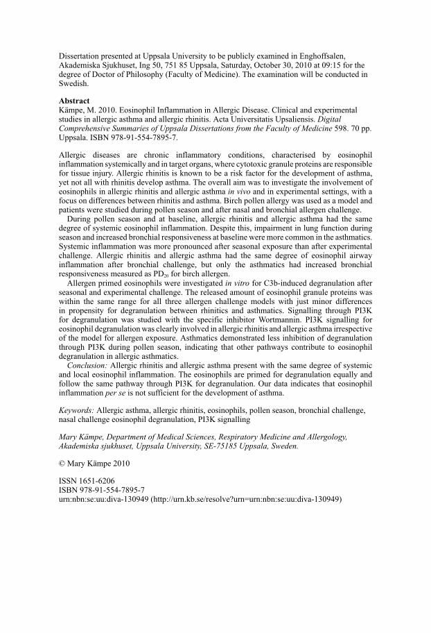
I. Kämpe M, Stålenheim G, Janson C, Stolt I, Carlson M. Systemic and
local eosinophil inflammation during the birch pollen season in allergic
patients with predominant rhinitis or asthma. Clin Mol Allergy
2007;(5)4:1-8.
II. Kämpe M, Janson C, Stålenheim G, Stolt I, Carlson M. Experimental
and seasonal exposure to birch pollen in allergic rhinitis and allergic
asthma with regard to the inflammatory response. Clin Resp J
2010;4:37-44.
III. Kämpe M, Stolt I, Lampinen M, Janson C, Stålenheim G, Carlson M.
Patients with allergic rhinitis and allergic asthma share the same pat-
tern of eosinophil and neutrophil degranulation after allergen chal-
lenge. Submitted for publication.
IV. Kämpe M, Lampinen M, Stolt I, Janson C, Stålenheim G, Carlson M.
PI3-kinase regulates eosinophil and neutrophil degranulation in pa-
tients with allergic rhinitis and allergic asthma irrespective of allergen
challenge model. Submitted for publication.
Reprints were made with permission from the respective publishers.

Contents
Introduction...................................................................................................11 Background ..............................................................................................11 Atopy, IgE and allergic disease................................................................11 Allergic asthma ........................................................................................12 Allergic rhinitis ........................................................................................13 The united airways concept......................................................................13 Treatment of allergic rhinitis and allergic asthma....................................15 Allergens and birch pollen allergy ...........................................................16 Seasonal allergen exposure vs. experimental allergen challenge .............17 The hygiene hypothesis ............................................................................17 Hypersensitivity reactions ........................................................................18 Sensitisation and the allergic cascade ......................................................18 Toll-like receptors ....................................................................................19 Mast cells and basophils...........................................................................20 B cells and isotype class-switch to IgE ....................................................20 T cells .......................................................................................................21
CD4+ T cell lineage..............................................................................22 Eosinophils ...............................................................................................23
Activation ............................................................................................24 Effector functions and released mediators...........................................25
Neutrophils ...............................................................................................26 Release of granule proteins and signalling through PI3K ........................27
Aims of the present investigations ................................................................29 Overall aim...............................................................................................29 Specific aims ............................................................................................29
Subjects .........................................................................................................30 Subjects ....................................................................................................30 Control group ...........................................................................................30
Methods ........................................................................................................32 Study design .............................................................................................32 Total pollen count.....................................................................................32 Diary.........................................................................................................33 Skin prick tests .........................................................................................33 Spirometry................................................................................................34
Nasal lavage .............................................................................................34 Induced sputum ........................................................................................34 Nasal allergen challenge test ....................................................................35 Bronchial allergen challenge test .............................................................35 Inflammatory cell counts and preparation of serum samples ...................35 Specific IgE ..............................................................................................36 Isolation of blood granulocytes ................................................................36 Measurement of eosinophil and neutrophil degranulation .......................36 Inhibitor....................................................................................................37 Inhibition of PI3K pathway......................................................................37 Radioimmunoassay (RIA)........................................................................37 Calculations of released amounts of granule proteins..............................37 Statistical analyses....................................................................................38 Ethical approval ........................................................................................38
Results...........................................................................................................39 Paper I. Systemic and local allergic inflammation during pollen season.39 Paper II. Comparison of experimental and seasonal allergen exposure ...41 Paper III. C3b-induced in vitro degranulation from primed eosinophils and neutrophils after seasonal and experimental allergen exposure.........42 Paper IV. Signalling through PI3K for in vitro degranulation from primed eosinophils and neutrophils after seasonal and experimental allergen exposure......................................................................................44
General discussion ........................................................................................46 Classification of allergic rhinitis and allergic asthma ..............................46 Eosinophil inflammation ..........................................................................46 Neutrophil inflammation ..........................................................................48 Concluding remarks and future perspectives ...........................................49
Conclusions...................................................................................................51
Swedish summary .........................................................................................52
Acknowledgements.......................................................................................54
References.....................................................................................................56
Abbreviations
Akt Protein kinase B, PKB APC Antigen presenting cell ATP Adenosine-5´-triphophate BCR B cell receptor BHR Bronchial hyperresponsiveness C3b Complement factor 3 CC Chemokine CCR Chemokine receptor COPD Chronic obstructive pulmonary disease CR1 Complement receptor type I, CD11b/CD18 CTL Cytotoxic T lymphocytes, CD8+ ECP Eosinophil cationic protein EDTA Ethylene-diamine tetra-acetic acid EPO Eosinophil peroxidase EPX/EDN Eosinophil protein X/ Eosinophil derived neurotoxin FEV1 Forced expiratory volume in one second FVC Forced vital capacity Fc Constant part of immunoglobulin FcεRI High-affinity IgE receptor FcεRII Low-affinity IgE receptor (CD23) FOXP3 Forkhead box P3, transcription factor GINA Global initiative for asthma GM-CSF Granulocyte-macrophage colony-stimulating factor HNL Human neutrophil lipocalin IC50 Half maximal inhibitory concentration IFN Interferon Ig Immunoglobulin IL Interleukin LT Leukotriene MBP Major basic protein MHC Major histocompatability complex MPO Myeloperoxidase PAF Platelet activating factor PD20 Provocation dose, cumulative dose causing 20% decrease
in FEV1 PEFR Peak expiratory flow rate
PI3K Phospho-inositide-3 kinase PIP3 Phosphatidylinositol (3,4,5) - triphosphate RANTES Chemochine ligand-5, CCL5 RIA Radioimmunoassay RT Room temperature SQ-U Standard quality unit TCR T cell receptor TGF Transforming growth hormone Th cell T helper cell TLR Toll-like receptor TNF Tumour necrosis factor TReg T regulatory cell, CD4+CD25+FOXP3+ TReg
11
Introduction
Background Allergy has been increasing world-wide over the past decades, especially in the western developed countries, and is today a major health problem with a prevalence of over a third of the population in many regions (1, 2). Allergic diseases manifest as hyperresponsiveness in the target organ, whether skin, nose, lung or the gastrointestinal tract; the hyperresponsiveness may or may not be IgE-mediated. It is generally accepted that chronic inflammation un-derlies the manifestations of the various allergic conditions. Exposure to environmental allergens is one of the most important stimuli for the initiation of allergic inflammation, especially in the airways, and thus the most impor-tant factor associated with development of asthma and rhinitis in the western world (3, 4, 5).
Atopy, IgE and allergic disease IgE, the fifth class of human antibodies, was discovered in the 1960s (6, 7), a major breakthrough for understanding the underlying mechanisms of allergy that has had a major impact on both diagnosis and treatment of allergic dis-eases. The term atopy, derived from the Greek word atopia (strangeness), was first introduced in 1923 by Coca and Cook to describe an inherited ten-dency to develop immediate-type hypersensitivity reactions against common environmental allergens (8). The definition of atopy thus is a genetic predis-position to produce IgE-antibodies against common environmental and harmless antigens and during the sensitisation period, the latent asympto-matic phase, IgE antibodies can be detected (9). However, on re-exposure to allergen the “atopic march” proceeds in some individuals to a clinically symptomatic allergic disease. In most literature, atopic sensitisation is de-fined as the presence of specific IgE by in vitro tests or skin prick test in a symptomless subject (9, 10, 11). Allergy, on the other hand, is the clinical expression of an IgE-mediated disease, the symptoms depending on the af-fected organs. Hence, allergy is an immunological disorder, but is often re-ferred to by many people when meaning any uncomfortable experience.
12
Allergic asthma Bronchial asthma is a widespread disease globally. Its prevalence has almost doubled over the last 50 years and is now approaching 10%, although preva-lence rates differ greatly depending on geographical area, and is more fre-quent in childhood than in adulthood (12, 13). Asthma is clinically character-ized by reversible airway obstruction, variable over time, with the cardinal sign of bronchial hyperresponsiveness associated with chronic eosinophil inflammation of the airway wall (14). Asthma is thus a disorder of the air-ways, which contract too much and too easily to a wide range of exogenous and endogenous stimuli (15). The typical asthma symptoms are wheeze, cough, mucus production and dyspnoea. Classification of asthma can be based on age, etiology, associated characteristics and severity. Asthma in children is mostly IgE-mediated (16) but after the age of 20 years it is more complex and more difficult to classify because of heterogeneity and overlap-ping entities due to gene-environment interactions. In new-onset asthma during adulthood, there appear to be entities independent of atopy in most cases (17). These non-allergic forms of the disease have been subject to care-ful comparative investigations, but so far no clear pathogenic cause has been identified (18, 19). The detection of IgE isotype-switching in bronchial air-way biopsies in both atopic and non-atopic asthma has provided some evi-dence that local IgE mechanisms are involved (20). The overall pathology in atopic and non-atopic asthma seems to be quite similar (21, 22, 23). On the other hand, Amin et al. have reported contradictory results, with less eosino-phils and more neutrophils in bronchial biopsies in non-atopic asthma than atopic asthma (24).
Thus, allergic asthma is a chronic inflammatory disease caused by re-peated immediate-hypersensitivity reactions and late-phase reactions leading to intermittent and initially reversible airway obstruction. In the chronic stage the Th2-driven disease is characterized by chronic eosinophilic in-flammation, smooth muscle hypertrophy, goblet cell hyperplasia and epithe-lial remodelling (25). Tissue eosinophilia and eosinophil degranulation is commonly associated with fibrotic diseases and eosinophils have been iden-tified as a significant source of transforming growth factor-β1 (TGF-ß1), one of the most fibrinogenic factors known (26). TGF-ß1 is also involved in air-way remodelling in asthma with increased deposition of collagen and ex-tracellular matrix proteins (27, 28, 29, 30). Zagai et al. have reported from a series of in vitro investigations that eosinophils via released ECP mediate the remodelling of extracellular matrix by influence on human lung fibroblasts (31, 32, 33). The recognition that asthma is a highly heterogeneous disorder in terms of clinical expression, response to different therapies, natural his-tory and association with environmental conditions has opened up the dis-ease beyond atopic sensitization. It has been suggested that the cause of the disease may lie in the airway epithelium (34). A defective barrier function
13
may activate the epithelial-mesenchymal-trophic-unit (EMTU) in the air-ways, leading to sensitisation and ongoing disease with persistent hyperre-sponsiveness to irritative stimuli (35, 36).
Allergic rhinitis Allergic rhinitis is the most frequent manifestation of allergic disease affect-ing the airways and its development depends on the interaction between genes, environment and immunological factors (37). The diagnosis of rhini-tis is based on the report of subjective nasal complaints (nasal blockage, itching, sneezing and increased secretions), increased nasal responsiveness and increased nasal airway resistance. To date, the different tests for rhinitis have low sensitivity and specificity and the diagnosis is therefore predomi-nately made on the basis of clinical history (38, 39). The physiological func-tion of the nose is to condition the inhaled air, filter small airborne particles and maintain defence mechanisms against the environment (40).
Airway inflammation is also present in the upper airways, but with little collagen deposition and absence of myofibroblasts in the nasal mucosa (41). There is however, evidence of remodelling in the nasal mucosa (42). The inflammation in the nasal mucosa is dominated by esoinophils, with accumu-lation in the reticular basement membrane and epithelial shedding, though not to the same degree as in the bronchi of patients with allergic asthma (43, 44). It has also been suggested that neural pathways may contribute to the pathophysiology of allergic rhinitis (45). Neurotrophins, and nerve-growth-factor (NGF) expressed in the esoinophils in the nasal mucosa has been sug-gested as candidates for the nasal hyperresonsiveness (46). Nasal obstruction is mostly the result of dilatation of capillary vessels, whereas bronchial ob-struction is mainly caused by smooth muscle contraction.
Seasonal allergic rhinitis to birch pollen is primary diagnosed by exclud-ing a history of asthma/respiratory complaints and chronic nasal disease, with specific IgE for birch and with no need for treatment outside pollen season. But some authors claim that this is not consistent with real life and that mixtures are common (47).
The united airways concept Allergic rhinitis and allergic asthma are considered to be manifestations of “the allergic syndrome” and it has been demonstrated that allergic rhinitis is a strong risk factor for the onset of asthma (42, 43, 48, 49), even independent of allergy (50, 51). The majority of patients with allergic asthma present with symptoms of seasonal or perennial rhinitis and in epidemiological stud-ies rhinitis were found in 70-80% of patients with asthma (52, 53). The risk
14
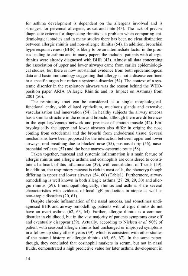
for asthma development is dependent on the allergens involved and is strongest for perennial allergens, as cat and mite (43). The lack of precise diagnostic criteria for diagnosing rhinitis is a problem when comparing epi-demiological studies and in many studies there has been no clear distinction between allergic rhinitis and non–allergic rhinitis (54). In addition, bronchial hyperresponsiveness (BHR) is likely to be an intermediate factor in the proc-ess leading to asthma and in many papers the included patients with allergic rhinitis were already diagnosed with BHR (43). Almost all data concerning the association of upper and lower airways came from earlier epidemiologi-cal studies, but there is now substantial evidence from both epidemiological data and basic immunology suggesting that allergy is not a disease confined to a specific organ but rather a systemic disorder (54). The context of a sys-temic disorder in the respiratory airways was the reason behind the WHO-position paper ARIA (Allergic Rhinitis and its Impact on Asthma) from 2001 (50).
The respiratory tract can be considered as a single morphological-functional entity, with ciliated epithelium, mucinous glands and extensive vascularisation and innervation (54). In healthy subjects the airway mucosa has a similar structure in the nose and bronchi, although there are differences in the capillary/venous network and presence of smooth muscle (42). Em-bryologically the upper and lower airways also differ in origin; the nose coming from ectodermal and the bronchi from endodermal tissue. Several mechanisms have been proposed for the interaction between upper and lower airways; oral breathing due to blocked nose (55), postnasal drip (56), naso-bronchial reflexes (57) and the bone marrow-systemic route (58).
Taken together, mucosal and systemic inflammation is a main feature of allergic rhinitis and allergic asthma and eosinophils are considered to consti-tute a hallmark of this inflammation (39), with contribution of T-cells (59). In addition, the respiratory mucosa is rich in mast cells, the phenotyp though differing in upper and lower airways (54, 60) (Table1). Furthermore, airway remodelling is well known in both allergic asthma (27, 28, 29, 30) and aller-gic rhinitis (59). Immunopathologically, rhinitis and asthma share several characteristics with evidence of local IgE production in atopic as well as non-atopic disorders (20, 61).
Despite chronic inflammation of the nasal mucosa, and sometimes undi-agnosed BHR and airway remodelling, patients with allergic rhinitis do not have an overt asthma (62, 63, 64). Further, allergic rhinitis is a common disorder in childhood, but in the vast majority of patients symptoms ease off and eventually disappear (39). Actually, according to Nielsen et al. 90% of patient with seasonal allergic rhinitis had unchanged or improved symptoms in a follow-up study after 6 years (39), which is consistent with other studies of the natural history of allergic rhinitis (65, 66, 67). In the same paper, though, they concluded that eosinophil markers in serum, but not in nasal fluids, demonstrated a high predictive value for later asthma development in
15
patients with allergic rhinitis (39). In conclusion, this indicates that eosino-phil inflammation is not sufficient to cause asthma and that additional factors contribute to whether or not a patient with rhinitis develops asthma.
Table 1. Similarities and differences in allergic rhinitis and allergic asthma [Modi-fied after Braunstahl et al. (59)].
Nose Bronchi
Epithelium Basement mem-brane Submucosal cells
Shedding Metaplasia Pseudo-thickening Collagen deposition Eosinophils Lymphocytes (CD4+) Vascular network Smooth muscle (Myo)fibroblasts Mast cells
0 to + 0 to +
0 to + 0 to +
+++
+ to ++ +++
0 0 to +
MCTC*
+++ 0
++ to +++ ++ to +++
+++
+ to ++ +
++ ++ to +++
MCT**
MCTC*: tryptase/chymase-positive mast cells, MCT**: tryptase-positive mast cells
Treatment of allergic rhinitis and allergic asthma Allergic diseases constitute a substantial global health problem with increas-ing socioeconomic impact and impaired quality of life. In addition to phar-macological treatment it is important to control contributing factors, i.e. en-vironmental allergens and triggers, gastro-oesophageal reflux, sinus disease, smoking history etc. The basis for treatment of mild seasonal allergic asthma is rapid-acting β2-agonists as reliever medication in addition to low-dose inhaled corticosteroids as monotherapy. According to GINA guidelines this therapy is recommended as first-line maintenance therapy for most mild asthmatics, reducing exacerbations and improving quality of life (68). Mild seasonal rhinitis is treated with antihistamines (per oral and topical) and nasal steroids, where the nasal steroids produce the greatest improvements in nasal symptoms in patients with seasonal allergic rhinitis according to a re-cent Cochrane review (69).
Leukotriene receptor antagonists may be added for further symptom im-provement in both allergic rhinitis as well as in allergic asthma. For the more severe cases allergen-specific immunotherapy may be considered and occa-
16
sionally the novel anti-IgE therapy, that recently has been proved to be affec-tive not only in allergic asthma but also in allergic rhinitis (70, 71).
Allergens and birch pollen allergy Pollen allergy amounts to approximately 20 % of community allergy (72). Air pollution is known to exaggerate pollen allergen reactions, probably through acting as hapten/adjuvant or by direct toxicity and damage of the airway mucosa, facilitating the penetration of allergens (72, 73). The most common allergen sources are pollen, fungi, pet dander and house dust mite.
Allergens are antigens giving an IgE response, instead of an IgG re-sponse, to harmless peptides or proteins (74, 75), eliciting immediate hyper-sensitivity and late-phase reactions in different target organs. Most allergens are proteins or glycoproteins with masses ranging from 5-100 kD and as the size of the allergen increases, the number of potential epitopes increase. The major allergenic pollens (grasses, trees and weeds) are wind-pollinated rather than insect-pollinated, they are soluble and with a size of 5 - 200µm (76). Birch pollen allergies are increasing and approximately 20% of the population in the Northern European countries is sensitized to birch pollen, the proteins responsible belonging to the Betula verrucosa (Bet v) family. In over 90% the allergy is due to the major and most important allergen Bet v 1 (77), belonging to the pathogenesis-related protein family 10 (PR-10 family) (78).
Many common allergens are enzymes, particularly proteases, and it has been suggested that in human defence against invading helminths the host secretes proteolytic enzymes which promote Th2 responses (79). This hy-pothesis is supported by the fact that the major house dust mite allergen Dermatophagoides pteronyssinus I (Der p I) is a cysteine protease that cleaves the intracellular tight junctions, and so gains access to the subepithe-lial antigen-presenting cells (80). However, even if many common allergens are enzymes, most are not and in a systematic overview of the structural biology of 40 common allergens no characteristic structural feature could be recorded (81). The structural components recognized by the innate immune system, the route of entry, particle size and adjuvants as pollutants are as-sumed to be important in determining if the antigen is recognized as an al-lergen by the host (82).
17
Seasonal allergen exposure vs. experimental allergen challenge Allergen exposure during pollen season is a low-dose challenge over a long period and is more like natural course of allergy development compared to a single high-dose allergen challenge. It is known, from both experimental studies and real life, that very high doses of allergen can elicit asthma symp-toms in non-asthmatics, as in epidemic asthma linked to thunderstorms dur-ing grass-pollen season and epidemic soybean asthma in Barcelona in the 1980s (83, 84). The Swedish birch pollen season is very convenient to study, as it is relatively short and defined in time, coming after a long cold winter with no pollen prevalence. Studies that depend on the natural variation of allergens are though time consuming and involve a level of uncertainty as the concentration of allergens may vary regionally and also from year to year. In contrast, studies employing experimental allergen exposure such as bronchial and nasal challenge are easier to perform (85, 86, 87). However, there are also several issues related to bronchial challenge methods such as establishing relevant doses and correct lung deposition (88, 89). In addition, the results of nasal and bronchial allergen challenge may be more difficult to interpret as the time course and dose of exposure is fundamentally different from seasonal exposure. Other experimental models such as repeated low-dose regimens for a range of days (90, 91) or different models of experimen-tal environmental settings (92, 93) may simulate natural exposure to a larger degree.
The hygiene hypothesis Allergic diseases are inflammatory disorders that develop on the basis of complex gene-environment interactions. The incidence is steadily increasing, which seems to be associated with a modern lifestyle where evolutionary adaptation has not had time to catch up (95). The “hygiene hypothesis”, or the “Old Friends” hypothesis, was originally proposed by Strachan in 1989, pointing out that allergies increased in society with reduction in the number of family members in the household (96). Epidemiological studies demon-strate an explosion of allergic diseases over the past decades (97, 98) parallel to a tremendous decrease in both the incidence and prevalence of bacterial, viral and helminth infections during the same time period (95, 99). This has been achieved by vaccination strategies, antibiotic treatment and improved living conditions as high standards of water supply and sewage systems.
The current view of the cellular and molecular mechanisms responsible for these phenomena includes changes in the fine balance of Th1, Th2 and regulatory T-cell responses, which are triggered by altered or missing innate immune cell activation (100). The fetus is exposed to a Th2 environment in
18
the uterus and the immune system is therefore Th2-polarized at birth. It is believed that immune deviation towards Th1 responses normally occurs in response to bacterial infections in the neonate, resulting in suppression of the Th2 skewness (89). It is well accepted that Th2 responses, above all have evolved in order to efficiently combat helminth infections (99, 101).
Though the “hygiene hypothesis” originally was an attempt to explain the rising incidence of allergic diseases in the developed countries, today it is also suggested to be applicable to the coinciding increase of several other chronic inflammatory disorders and autoimmune diseases (102, 103, 104, 105). It has been speculated that infections of historical importance might have shaped our immune system during evolution and may have played a major role in down regulating allergic and autoimmune responses (106).
Hypersensitivity reactions Hypersensitivity reactions are undesirable responses of the adaptive immune system to innocuous antigens sometimes causing severe disease due to tissue damage. Already in 1963 Gell and Coombs stated the scheme for classifica-tion of hypersensitivity reactions (type I-IV), requiring a pre-sensitized status of the host (107). The classification is still applicable for both allergic and other immune reactions after minor modifications. The immune system has many built-in feedback loops and amplification mechanisms and once a pathologic response starts it is often difficult to control and to terminate, which explains why hypersensitivity reactions often tend to be chronic. Al-lergic reactions can be divided into immediate and late-phase reactions. The immediate reaction (type I) is caused by vasoactive mediators released pri-marily from mast cells and the late-phase reaction (type IV) is cell-mediated due to recruitment of inflammatory cells to the target organ, the latter phase not always preceded by a detectable type I reaction. The immediate reaction can subside but generally proceeds to the late-phase reaction, giving rise to chronic inflammation with more serious long-term illness in the affected tissue (108).
Sensitisation and the allergic cascade Dentritic cells, mast cells, basophils, esoinophils and IgE are essential com-ponents of the allergic reaction, as well as T-cells and B-cells (109). The dentric cells, mast cells and basophils together with epithelial cells are cru-cially located at body surfaces ensuring first defence against environmental pathogens (110). The first step in the allergic sensitisation is the uptake and presentation of the allergen/antigen by antigen presenting cells (APC), the most important being the dentritic cells. The dentritic cells are professional
19
APCs, i.e. they express MHC class II molecules as well as co-stimulatory factors necessary for T-cell activation and initiation of the adaptive immune response. After allergen uptake and processing, the dentritic cells mature and migrate to the lymph nodes were they present the peptide fragments for na-ïve T-cells, thereby directing them in favour of a Th2-phenotype (111). The antigen-specific Th2-cells migrate to the target tissue where they orchestrate the immune response by releasing Th2-cytokines, starting cross-talk between immune cells and an amplification cascade takes place (112).
The crucial role in the sensitisation process is played by the dentritic cells (110), but the B-cells are also important for allergen capture and processing when small allergens are involved (113). In the presence of co-stimulation the Th2-cells upregulate the expression of IL-4 and IL-13, which are neces-sary for initiation of class-switching to IgE synthesis (114). Class-switch is generally thought to occur in the lymph nodes, but has recently been re-ported to take place locally in nasal and bronchial mucosa and also in the gastrointestinal tract in patients with food allergy (115). The Th2-cytokines IL-4 and IL-13 also act on epithelial cells, smooth muscle cells and goblet cells in the airways and are suggested to be responsible for airway hyperre-sponsiveness (112). The released Th2-cytokines are involved in recruitment of mast cells (IL-4, IL-9 and IL-13), basophils (IL-3 and IL-4) and eosino-phils (IL-3, IL-5 and GM-CSF), the main mediator cells involved in the al-lergic tissue response (116). Once sensitised to an allergen, on re-exposure the allergen cause cross-linking of the IgE bound to high-affinity IgE-receptors on mast cells, stimulating release of preformed histamine and newly generated lipid mediators responsible for the immediate allergic reac-tion. In addition, the released mediators and cytokines from the mast cells recruit eosinophils, basophils and macrophages responsible for the late aller-gic reaction (109).
Toll-like receptors The first line of defence against pathogens is the constitutive innate immune system, immediately mobilised upon infection or any tissue damage. The immune cells have Pattern Recognition Receptors (PRRs) on their surface, which recognise the highly conserved set of molecular structures specific for microbes, the Pathogen Associated Molecular Patterns (PAMPs) (117). The best understood and perhaps the most important subgroup of PRRs is the family of Toll-like receptors (TLRs), natural ligands for the PAMPs of bac-teria, viruses and fungi (118). Signalling through Toll-like receptors, ex-pressed on the epithelium of all body barriers, initiates acute inflammatory responses and plays a crucial role for the early shaping of the immune sys-tem and the suppression of Th2-driven allergic immune responses (118). Recent evidence suggests that the TLRs play an important role in controlling
20
the adaptive immune responses through “the IgE-Toll like receptor network” (115, 119, 120, 121, 122).
Mast cells and basophils Mast cells and basophils share many features and they are thought to have similar functions in protecting against helminth infections and in IgE-mediated allergic inflammation. Both cells are highly conserved through evolution, traditionally considered to be cells of the innate immune system, but have proved to be able to modulate adaptive immune responses as well (123,124,125). Mast cells and basophils express TLRs, MHC class II mole-cules and the high-affinity IgE receptor (FcεRI) and can act as professional APCs (108,123,126,127). Both cells are considered to maintain the sensi-tised state of the mucosa and to be initiators of the allergic reaction (125).
Mast cell precursors circulate in the blood and mature when entering the tissue, localizing near blood vessels and at epithelial surfaces (128), whereas basophils are primary encountered in the circulation (129). The homing propensity of mast cells to mucosal tissues is essential for the allergic reac-tions to take place in particular organs and the microenvironment of mucosal tissues favours the local synthesis of IgE ahead of IgG in atopic subjects (126). The human mast cells are divided into two types according to the con-tent of tryptase (MCT cells) or tryptase and chymase (MCTC cells) (108).
Basophils constitute less than 1% of the peripheral blood leukocytes and have previously gained very little interest, even though it has previously been unravelled that they play a critical role both in chronic allergic inflam-mation and in systemic anaphylaxis (125, 129, 130). Basophils are involved in IgG-mediated anaphylaxis, often requiring larger amounts of allergens than IgE-mediated, by releasing the mediator Platelet-activating factor (PAF) (125). In addition, the activated basophils release histamine, serine proteases and leukotriens, but not prostaglandins, as well as a cytokines, chemokines and complement receptors (108).
B cells and isotype class-switch to IgE Adaptive immunity is mediated through numerous genetic and cellular proc-esses to generate antigen-binding immunoglobulins and T-cell receptors (TRCs) through recombination of variable (V), diversity (D) and joining (J) segments (131). All antibody molecules share the same basic structure with two identical light chains, two identical heavy chains and a constant C-region, the Fc-region, on the heavy chain. The light and heavy chains have variable regions that participate in antigen recognition and the constant re-gion determines Ig-class isotype; different isotypes (IgA, IgD, IgE, IgG and
21
IgM) performing different effector functions when antigen is bound to their Fc-receptor (131). B-cells can act as professional APCs and via their recep-tor (BCR) internalize and process small antigens for Th-cell presentation (113).
All naïve B cells express both IgM and IgD but on encountering antigen, depending on the cytokine signals, a class-switch to either IgG, IgA or IgE will take place in the secondary lymph nodes (132, 133). The Th2-derived cytokines IL-4 and IL-13 specify class switch in the lymph nodes and in the target tissue basophils are responsible for class-switch, which protect and re-sensitise FcεRI on mast cells in a positive feedback mechanism giving fur-ther amplification of the IgE production (108, 128). The process of mast cell and APC recruitment and IgE production in the mucosal tissues is central to the function of the IgE network (89), and also in protecting the host against systemic anaphylaxis (115). The B-cells express two types of IgE-receptors on their surface: the high- and the low-affinity IgE-receptors (FcεRI and FcεRII/CD23). IgE is usually measured as total serum IgE, but there are also in vitro assays for detection of free IgE (108).
T cells Naïve T lymphocytes migrate from the bone marrow to the thymus were they traditionally are considered to mature into two main lineages: T-helper cells and T-cytotoxic cells (CTL). The lineage choice goes by way of double thymocytes, i.e., CD4+CD8+ cells expressing a fully developed T-cells recep-tor composed of α and ß chains with immense variability (134,135). The basis for selection of TCRs in the thymus is their ability to discriminate self from non-self, i.e., the TCR is MHC-restricted and self-tolerant (136). T-helper cells express CD4 protein on their cell surface and are activated by antigen bound to MHC class II molecules, whereas CTLs are CD8+ and acti-vated by antigen associated to MHC class I (137). The division of T cells into two functional subsets based on their cytokine production was already described in 1986, though recently this concept has been re-evaluated point-ing in the direction of functional plasticity of the T cell subsets with ability to convert to another phenotype (138, 139). CTLs are potent in defence against pathogens but can also be directed against self-tissues; in autoim-mune diseases, graft rejection and graft-versus-host-disease (GVHD) (140). T-helper cells play critical roles in adaptive immune responses and the di-verse functions are determined by their cytokine secretion patterns and tissue location, though it is known today that they rather represent polarised forms of the highly heterogeneous CD4+ T cell-mediated immune responses (140, 141). So far, the generally recognized CD4+ lineages are Th1, Th2, Th17 and TReg cells, but further subunits have been proposed (142, 143, 144).
22
CD4+ T cell lineage Th1 cells are characterised by INF-γ production and are primarily involved in cellular immunity against intracellular bacteria (142). Th17 cells are char-acterised by expression of IL-17 and IL-22, mainly located at barrier sur-faces (145). The regulatory T cells are crucial for the maintenance of self tolerance and immune homeostasis, dysfunction causing autoimmune dis-eases, immunopathology and allergy (146). TReg cells, expressing CD4+CD25+ and the transcription factor FOXP3 (forkhead box P3) are natu-rally present in the immune system (nTregs) and developmentally deter-mined in the thymus (147). In addition, there are induced T regulatory cells (iTreg) generated in the periphery in the presence of antigen and cytokines, especially TGF-ß (146). At mucosal surfaces the iTregs are in close connec-tion with Th17 cells suggesting overlapping development and dynamic in-terplay in peripheral tolerance (148, 149). The Th17 cells have especially been considered to have a crucial roll in the development of allergy (144).
The Th2 cells control immunity to extracellular parasites and all forms of allergic responses by way of class switch to IgE (112). The products of al-lergens and helminths are strong activators of Th2 responses locally in the airways, sensing these products and activating the Th2 response in the tissue (112). However, as mentioned, not only Th2 cells decide the outcome of immune responses to allergen encountered in the tissues, but also the inter-play and cross-regulation with other T cells, above all Tregs and possibly Th17 cells (150, 151).
23
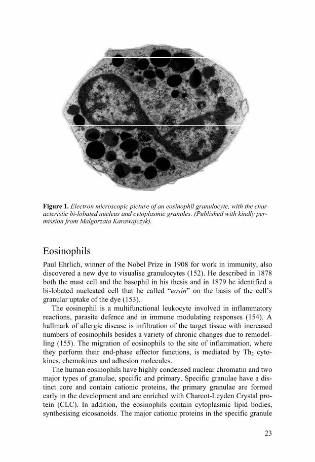
Figure 1. Electron microscopic picture of an eosinophil granulocyte, with the char-acteristic bi-lobated nucleus and cytoplasmic granules. (Published with kindly per-mission from Malgorzata Karawajczyk).
Eosinophils Paul Ehrlich, winner of the Nobel Prize in 1908 for work in immunity, also discovered a new dye to visualise granulocytes (152). He described in 1878 both the mast cell and the basophil in his thesis and in 1879 he identified a bi-lobated nucleated cell that he called “eosin” on the basis of the cell’s granular uptake of the dye (153).
The eosinophil is a multifunctional leukocyte involved in inflammatory reactions, parasite defence and in immune modulating responses (154). A hallmark of allergic disease is infiltration of the target tissue with increased numbers of eosinophils besides a variety of chronic changes due to remodel-ling (155). The migration of eosinophils to the site of inflammation, where they perform their end-phase effector functions, is mediated by Th2 cyto-kines, chemokines and adhesion molecules.
The human eosinophils have highly condensed nuclear chromatin and two major types of granulae, specific and primary. Specific granulae have a dis-tinct core and contain cationic proteins, the primary granulae are formed early in the development and are enriched with Charcot-Leyden Crystal pro-tein (CLC). In addition, the eosinophils contain cytoplasmic lipid bodies, synthesising eicosanoids. The major cationic proteins in the specific granule
24
are major basic protein (MBP), eosinophil cationic protein (ECP), eosinophil peroxidase (EPO) and eosinophil protein X [(EPX)/eosinophil derived neu-rotoxin (EDN)] all of which are extremely toxic to tissues (156, 157).
The eosinophils express an array of cell-surface proteins, including Ig-receptor for IgG, IgA, complement receptors, leukotriene receptors, pros-taglandin receptors, PAF receptor and TLRs as well as several inhibitory receptors. In addition, the eosinohpils express receptors for cytokines (IL-3, IL-5, GM-CSF and IL-1α, IL-4, INF-α, TNF- α), chemokines (CCR1 and CCR3) and for adhesion molecules (VLA-4, α4ß7 and siglec-8) (108). Fur-thermore, when activated for degranulation the eosinophils express both high-affinity IgE-receptors (FcεRI), low-affinity IgE-receptors (FcεRII/CD23), high-affinity IgG-receptors and complement receptors (154, 158). FcεRII/CD23 facilitates antigen presentation in the highly activated eosinophil (154). They also express MHC class II molecules and co-stimulatory factors, e.g. they can act as professional APCs (159). Develop-ment and differentiation of eosinophils is promoted by IL-3, IL-5 and GM-CSF, but only IL-5 is considered specific for eosinophils. Eosinophils are released from the bone marrow to the circulation after stimulation with IL-5, produced at the site of allergic inflammation, and migrating to the inflamma-tory tissue (160). The half-life of eosinophils in the circulation is 8-18 hours, but in the tissues they can survive for up to several weeks (161).
Activation The activation of eosinophils is strictly regulated; an inappropriate activation would be harmful to the individual and in healthy conditions the eosinophils are inactive with a high threshold for release of their granule proteins (162,163). There is no consensus on the major signalling mechanism for eosinophil activation, but they are known to be activated by cross-linking of IgG and IgA Fc-receptors and a number of mediators (IL-3, IL-5 and GM-CSF, CC chemokines and PAF), the role of FcεRI is however unclear (108).
The granule proteins can be released by exocytosis, compound exocyto-sis, piecemeal exocytosis by transport vesicles or cytolysis (164, 165). In vitro studies have demonstrated selective release of the individual granule proteins (166, 167) and interestingly, different eosinophilic diseases are characterised by a marked heterogeneity in degranulation levels (168). Pre-vious studies have implicated that the priming degree of the blood eosino-phils is related to the degranulation status of the tissue-residing eosinophils and corresponds to the activity of the eosinophilic disease (169). Hence, the eosinophil can be activated in different ways and by different antigens and furthermore there are four ways of eosinophil degranulation reported, the outcome of activation can thus be variable.
25
Effector functions and released mediators The eosinophil stores a vast array of mediators, cytokines and chemokines with different target activities. Depending on the triggering stimuli a differ-ential release of proinflammatory mediators takes place in the target tissue (170). At the site of inflammation the primed eosinophil rapidly secretes its preformed granule proteins (MBP, ECP, EPO, EPX/EDN) in addition to different cytokines (IL-2, IL-4, IL-5, IL-10, IL-12, IL-13, Il-16 and IL-18), chemokines (eotaxin-1 and RANTES), growth factors (TGF-β) and newly synthesised eicosanoids [platelet-activating factor (PAF) and leukotriene C4 (LTC4)] (161). These molecules have pro-inflammatory effects in up-regulation of adhesion systems, modulation of cellular trafficking and in inducing tissue damage; above all ECP is involved (160, 161). MBP ac-counts for more than half of the granule protein mass, is highly cationic but lacks enzymatic activity and is believed to act through enhanced membrane permeability (161). It has in vitro activity against parasites but its role in allergy is unclear (108). EPO, also highly cationic and localised to the matrix of the specific granule, makes up approximately 25% of the granule proteins (161) and is considered to be the most specific of the eosinophil granule proteins (171) and also the most potent, on a molar basis, to kill helminths (172).
In many papers it has been observed that EPO is more difficult to mobi-lize than ECP (168,173,174) and it has been postulated that the very low release in degranulation assays depends on the sticky nature of the protein, due to a very high cationic charge and therefore EPO is adhering to the labo-ratory tubes (175, 176). This difference in degranulation could also be ex-plained by selective granule release in response to different stimuli for de-granulation (168).
ECP and EPX/EDN have RNAse activity and are localized to the matrix of the specific granule. Although both have in vitro toxicity against patho-gens, the RNAse activity of EDN is much more potent than that of ECP (156, 161). Genes coding for ECP and EPX/EDN show extremely high mu-tation rates, suggesting extraordinarily selective pressure due to rapid evolu-tion of pathogens (108). The gene family expressing ECP has one of the highest rates of mutations in the primate genome, suggesting specialised biological activities for the subgroups (177, 178) and involvement in adverse reactions eliciting allergic diseases (180). ECP in particular has gained inter-est in the pathogenesis of allergic diseases, and being a ribonuclease it facili-tates entry of other cytotoxic molecules as well (181, 182). Eosinophils are highly cytotoxic to human airway epithelial cells and are considered to have a major impact on airway remodelling by promoting fibrotic processes and increased vascularity, as well as modulating mesenchymal functions directly (61, 183, 184). It has been shown that ECP stimulates migration of human
26
lung fibroblasts, stimulates TGF-ß1 release from the fibrobalsts and also interact with mesenchymal cells in vitro (31, 32, 33).
Taken together, there is much evidence suggesting that eosinophils have an important effector role in chronic allergic inflammation.
Neutrophils Neutrophils are the most abundant leukocytes in circulation and play a fun-damental role in the innate immune response against pathogens. They are normally not present in healthy tissue, but are rapidly recruited to the site of inflammation. Neutrophils are phagocytic cells and a major source of pro-inflammatory cytokines, contributing to the onset and early orchestration of the inflammatory response in many diseases. Recently it has been unravelled that neutrophils also are important in shaping immune responses and are able to suppress T-cell activation (185). Furthermore, the neutrophils do not only destroy tissue, they are also involved in tissue repair (185).
Neutrophil granulae are divided into primary (azurophilic), secondary (specific) and tertiary granulae (gelatinase) (186,187,188), though a modern view is to regard granulae as a continuum over time as the neutrophil precur-sors mature in the bone marrow (187, 188). The primary source of the highly cytotoxic MPO is from the primary neutrophil granule (186, 189) and after activation it will be secreted together with other inflammatory mediators (169). Human neutrophil lipocalin, HNL, a recently discovered unique neu-trophil mediator, is stored in the secondary granule and measurement of this protein seems to be of particular clinical relevance for studying neutrophil involvement (190, 191).
The eosinophil’s role in inflammation is well established in the patho-genesis of asthma, but the role of the neutrophils is less understood in aller-gic airway inflammation except in more severe forms of chronic asthma and COPD (192, 193, 194, 195). It has recently been reported that neutrophils are the first cells recruited to the site of the allergic reaction where they also may take part in the resolution process of the allergic response (196). This is in line with studies of remodelling in mild atopic asthma after allergen chal-lenge (94) and the finding of neutrophils in induced sputum of non-atopic asthmatic children (197). Additionally, the recent advances in studies of anti-IL-5 therapy also indicate an involvement of other inflammatory cells than just the eosinophils (198).
Altogether, this implies that there might not be a clear-cut difference be-tween mild and severe asthma with regard to the neutrophil involvement, and thus eosinophilic and neutrophilic asthma might not be mutually exclu-sive subtypes of asthma.
27
Release of granule proteins and signalling through PI3K Effector functions of the eosinophils are based on secretory pathways for release of pre-stored pools of cationic granule proteins, cytokines and chemokines. The eosinophil is known to have four different forms of de-granulation, although piecemeal degranulation have been demonstrated to be the pathway of choice for selective granule release in allergic responses and also for IL-4 release (164, 165, 168, 169). Recently, piecemeal degranulation has been reported to be the secretory pathway for release of MBP from eosi-nophils after stimulation with eotaxin in healthy donors and in patients with hypereosinophilic syndrome (HES) (199, 200).
There is no consensus as to the major signalling mechanism for eosino-phil activation, but they are known to be activated by cross-linking of IgG and IgA Fc-receptors. It has long been known that binding of eosinophils to a surface by complement receptors induces a strong signal for degranulation, i.e. involving the receptor for complement factor 3 (C3b-receptor, CR1, CD11b/CD18) (201, 202, 203). Experimental settings adjusted to elucidate the degranulation process in more detail in eosinophils and neutrophils have been developed (67, 203, 204, 205). Using serum opsonised Sephadex parti-cles in vitro enhances this C3b-induced degranulation of the eosinophils in allergic diseases as well as in infection (203).
The mechanism behind ECP release after addition of serum opsonised Sephadex-particles is that of frustrated phagocytosis, as the opsonised sur-face is non-phagocytable, representative for the mechanism involved in ex-tracellular killing of parasites (202). Furthermore the cationic proteins are only cytotoxic at high local concentrations, i.e. in close contact with the parasite, for which reason granule must be released in a closed compartment onto the surface of the parasite (202). This implies that soluble secre-tagogues, such as cytokines, are of less importance for degranulation in eosi-nophils. The main signal for degranulation comes from cross-linked Fc-receptors and C3-derivates, and proinflammatory cytokines (IL-3, IL-5 and GM-CSF) enhance rather than act themselves in the degranulation process. This indicates a role for complement receptors (mainly CR1) and the eosino-phil IgG receptor in ECP release induced by serum opsonised Sephadex particles (202, 205).
The chemotaxis and recruitment of the eosinophils to the site of inflam-mation is fairly well studied, but the downstream signalling leading to the degranulation of eosinophils in allergic diseases is less understood and still much unexplored. The phosphoinositide-3-kinase family (PI3-kinases, PI3K) regulates membrane trafficking and controls many fundamental biological functions such as cell growth, survival and proliferation by downstream acti-vation (206, 207). PI3Ks are divided into three major classes based on struc-tural features and lipid substrate preference, of which class I is the most studied (206, 208). Little is also known about the tissue distribution of the
28
different PI3Ks, as there are no available antibodies for immunohistochemis-try to date (208). PI3Ks generate 3-phosphorylated phosphoinositide lipids and for class I the end-product is PIP3 [Phosphatidylinositol (3,4,5)-trisphosphate, PtdIns(3,4,5)P3]. Class I generated PIP3 acts as a second mes-senger and promote downstream activation through a phosphorylation cas-cade of pleckstrin homology domain-containing proteins, e.g. activation of primarily the Akt pathway which in turn phosphorylates numerous protein targets (206, 209, 210). Class I PI3Ks can be activated by external stimuli (211) and are thought to be involved in mast cell and basophil degranulation in allergy (212, 213), but the role in eosinophil degranulation in answer to allergic reactions is so far not elucidated.
Inhibitors have been useful for characterising the PI3Ks and Wortmannin, a fungal metabolite, has been widely used for studying the PI3K pathway in leukocytes, although implications mainly have been drawn to class I PI3Ks (214). Wortmannin is a very potent universal PI3K inhibitor with IC50 of ~2-10nM depending on the stimulus activating PI3K (215, 216). It has been regarded as a selective PI3K inhibitor, although recent evidence suggests that Wortmannin can compete with additional kinases and ATP, but only at much higher concentrations than are usually used in assays (211, 216). In recent years the biology and signalling through the PI3K axis and further downstream has been the subject of intense investigations and the basic framework of PI3K signalling has been unravelled, especially for class I, although less is known of the dynamic regulation and stimuli needed for their relative functional output.
29
Aims of the present investigations
Overall aim The overall aim of the present thesis was to study eosinophil inflammation in allergic rhinitis and allergic asthma with regard to the systemic and local tissue inflammation as well as similarities and differences between the two allergic disorders. The investigations were performed during birch pollen season and after bronchial and nasal allergen challenge for assessing differ-ences in long-term low-dose allergen exposure compared to single high-dose allergen exposure. The neutrophil inflammation was studied concurrently for comparison.
Specific aims • To study differences in systemic and local inflammation in allergic rhini-
tis and allergic asthma during birch pollen season (Paper I).
• To examine differences in systemic and local inflammatory responses after experimental allergen challenge and seasonal allergen exposure in allergic rhinitis and allergic asthma (Paper II).
• To assess stimulated in vitro degranulation from systemically allergen primed eosinophils in allergic rhinitis and allergic asthma after experi-mental allergen challenge and seasonal allergen exposure (Paper III).
• To examine signalling through PI3-kinase for in vitro degranulation of systemically allergen primed eosinophils in allergic rhinitis and allergic asthma after experimental allergen challenge and seasonal allergen ex-posure (Paper IV).
30
Subjects
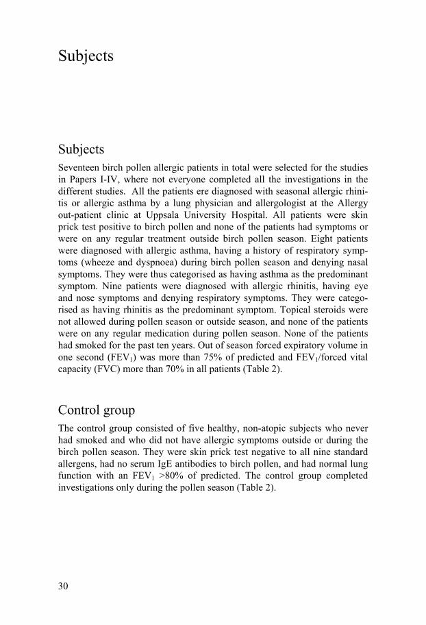
Subjects Seventeen birch pollen allergic patients in total were selected for the studies in Papers I-IV, where not everyone completed all the investigations in the different studies. All the patients ere diagnosed with seasonal allergic rhini-tis or allergic asthma by a lung physician and allergologist at the Allergy out-patient clinic at Uppsala University Hospital. All patients were skin prick test positive to birch pollen and none of the patients had symptoms or were on any regular treatment outside birch pollen season. Eight patients were diagnosed with allergic asthma, having a history of respiratory symp-toms (wheeze and dyspnoea) during birch pollen season and denying nasal symptoms. They were thus categorised as having asthma as the predominant symptom. Nine patients were diagnosed with allergic rhinitis, having eye and nose symptoms and denying respiratory symptoms. They were catego-rised as having rhinitis as the predominant symptom. Topical steroids were not allowed during pollen season or outside season, and none of the patients were on any regular medication during pollen season. None of the patients had smoked for the past ten years. Out of season forced expiratory volume in one second (FEV1) was more than 75% of predicted and FEV1/forced vital capacity (FVC) more than 70% in all patients (Table 2).
Control group The control group consisted of five healthy, non-atopic subjects who never had smoked and who did not have allergic symptoms outside or during the birch pollen season. They were skin prick test negative to all nine standard allergens, had no serum IgE antibodies to birch pollen, and had normal lung function with an FEV1 >80% of predicted. The control group completed investigations only during the pollen season (Table 2).
31
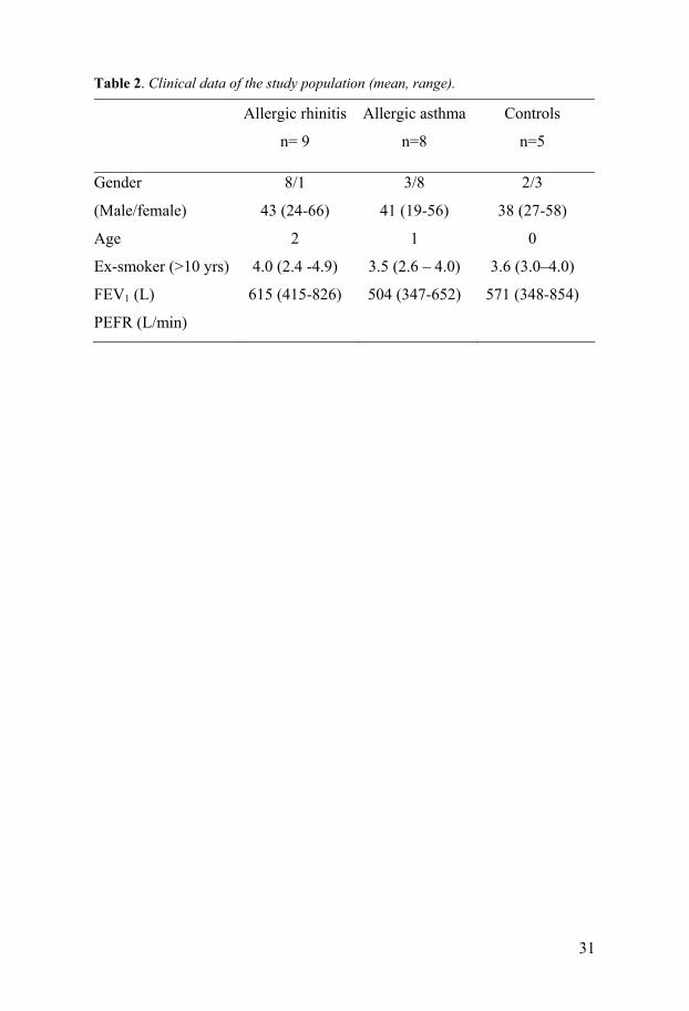
Table 2. Clinical data of the study population (mean, range).
Allergic rhinitis
n= 9
Allergic asthma
n=8
Controls
n=5
Gender
(Male/female)
Age
Ex-smoker (>10 yrs)
FEV1 (L)
PEFR (L/min)
8/1
43 (24-66)
2
4.0 (2.4 -4.9)
615 (415-826)
3/8
41 (19-56)
1
3.5 (2.6 – 4.0)
504 (347-652)
2/3
38 (27-58)
0
3.6 (3.0–4.0)
571 (348-854)
32
Methods
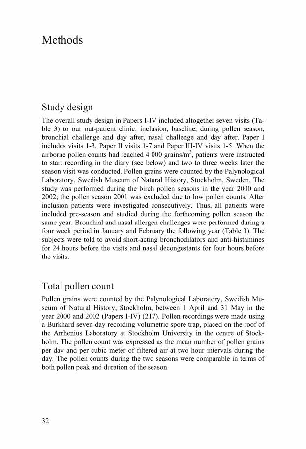
Study design The overall study design in Papers I-IV included altogether seven visits (Ta-ble 3) to our out-patient clinic: inclusion, baseline, during pollen season, bronchial challenge and day after, nasal challenge and day after. Paper I includes visits 1-3, Paper II visits 1-7 and Paper III-IV visits 1-5. When the airborne pollen counts had reached 4 000 grains/m3, patients were instructed to start recording in the diary (see below) and two to three weeks later the season visit was conducted. Pollen grains were counted by the Palynological Laboratory, Swedish Museum of Natural History, Stockholm, Sweden. The study was performed during the birch pollen seasons in the year 2000 and 2002; the pollen season 2001 was excluded due to low pollen counts. After inclusion patients were investigated consecutively. Thus, all patients were included pre-season and studied during the forthcoming pollen season the same year. Bronchial and nasal allergen challenges were performed during a four week period in January and February the following year (Table 3). The subjects were told to avoid short-acting bronchodilators and anti-histamines for 24 hours before the visits and nasal decongestants for four hours before the visits.
Total pollen count Pollen grains were counted by the Palynological Laboratory, Swedish Mu-seum of Natural History, Stockholm, between 1 April and 31 May in the year 2000 and 2002 (Papers I-IV) (217). Pollen recordings were made using a Burkhard seven-day recording volumetric spore trap, placed on the roof of the Arrhenius Laboratory at Stockholm University in the centre of Stock-holm. The pollen count was expressed as the mean number of pollen grains per day and per cubic meter of filtered air at two-hour intervals during the day. The pollen counts during the two seasons were comparable in terms of both pollen peak and duration of the season.
33
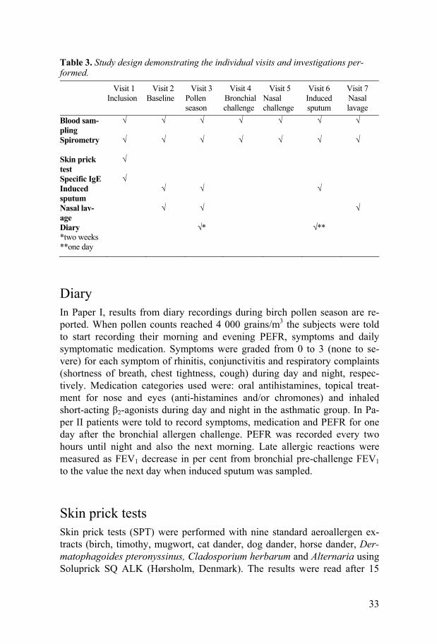
Table 3. Study design demonstrating the individual visits and investigations per-formed.
Visit 1 Inclusion
Visit 2 Baseline
Visit 3 Pollen season
Visit 4 Bronchial challenge
Visit 5 Nasal challenge
Visit 6 Induced sputum
Visit 7 Nasal lavage
Blood sam-pling
√ √ √ √ √ √ √
Spirometry
√ √ √ √ √ √ √
Skin prick test
√
Specific IgE √ Induced sputum
√ √ √
Nasal lav-age
√ √ √
Diary *two weeks **one day
√* √**
Diary In Paper I, results from diary recordings during birch pollen season are re-ported. When pollen counts reached 4 000 grains/m3 the subjects were told to start recording their morning and evening PEFR, symptoms and daily symptomatic medication. Symptoms were graded from 0 to 3 (none to se-vere) for each symptom of rhinitis, conjunctivitis and respiratory complaints (shortness of breath, chest tightness, cough) during day and night, respec-tively. Medication categories used were: oral antihistamines, topical treat-ment for nose and eyes (anti-histamines and/or chromones) and inhaled short-acting β2-agonists during day and night in the asthmatic group. In Pa-per II patients were told to record symptoms, medication and PEFR for one day after the bronchial allergen challenge. PEFR was recorded every two hours until night and also the next morning. Late allergic reactions were measured as FEV1 decrease in per cent from bronchial pre-challenge FEV1 to the value the next day when induced sputum was sampled.
Skin prick tests Skin prick tests (SPT) were performed with nine standard aeroallergen ex-tracts (birch, timothy, mugwort, cat dander, dog dander, horse dander, Der-matophagoides pteronyssinus, Cladosporium herbarum and Alternaria using Soluprick SQ ALK (Hørsholm, Denmark). The results were read after 15
34
minutes measuring the largest diameter of the wheal and its perpendicular diameter, and the product was expressed in mm2. Skin reactions were con-sidered positive when larger than 9 mm2.
Spirometry Lung function tests were performed with a Vitalograph-Compact spirometer (Vitalograph Ltd., Buckingham, England). FEV1, FVC, FEV1/FVC% and PEFR were recorded. The reference values were those from the European Community for Coal and Steel (218). Spirometry was performed before and 90 sec after inhalation of physiologic saline. FEV1 was measured before and after inhalation of hypertonic saline solution and the magnitude of the FEV1 decrease was used as a marker of bronchial responsiveness. Morning and evening PEFR were measured during pollen season (Paper I) and for one day after bronchial challenge (Paper II), using a mini-Wright Peak Flow Meter (Clement Clarke International Ltd., Essex, England) and recorded in the diaries, respectively.
Nasal lavage Lavage of the nasal mucosa was performed according to Wålinder et al (219) with a 20 mL syringe attached to a nose olive; the subjects standing with their heads flexed 30° forward. Each nostril was lavaged with 5 mL of 0.9% sterile saline solution at RT and flushed back and forth five times via the syringe at intervals of three seconds. The recovered fluid was weighed and the amounts obtained were comparable in all subjects. The fluid was transferred into 10 mL polypropylene centrifuge tubes, kept on ice and within 30 min centrifuged at 337 g (1500 rpm) for 10 min. The supernatant was immediately frozen in small aliquots at -70°C for later analyses of HNL. Differential cell counts were calcu-lated on the remaining suspension, using a cytospin preparation (Cytospin, Shandon, Southern Instruments, Sewickley, PA, USA), stained with May-Grünewald and Giemsa and examined under light microscope (Paper I-II).
Induced sputum Sputum samples were obtained by inhalation of hypertonic saline according to Pizzichini et al. (220) except that the subjects were not pre-treated with inhaled bronchodilators. An ultrasonic nebuliser (OMRON U 1, Sonesta Tamro no 28 36 06, Stockholm, Sweden) was used for the inhalations. After inhalation of physiologic saline solution for negative control the subjects inhaled (4.5%) hypertonic saline solution in five inhalation steps; 0.5, 1, 4, 8
35
and 16 min. After each inhalation step the subjects were instructed to “huff” and cough into the container. The mucus clods were aspirated and collected with a 2 mL syringe, then weighed and immediately transported to the labo-ratory. The sputum sample was kept on ice, incubated at 22°C for 15 min with equal amounts of 0.2% dithiothreitol (DTT) in phosphate buffer and sputolysin (CalbioChem, SputolysinReagent, art no 56000) before centrifu-gation. The supernatant was then frozen at -70°C for subsequent analysis of ECP and HNL (Paper I-II).
Nasal allergen challenge test The experimental nasal challenge test was performed by instillation in the same nostril of 0.3 mL diluent for negative control followed by instillation of birch pollen extract (Aquagen ® SQ, ALK-Abelló, Hørsholm, Denmark) every 15 min in three steps: 1 000 SQ-U/mL, 10 000 SQ-U/mL and 100 000 SQ-U/mL. The symptom score was estimated; if pronounced local symptoms and sneezing occurred the challenge test was stopped. The response to the allergen challenge was categorized into four groups: no response or response to each of the three allergen doses used. Blood samples and nasal lavage were taken 18 hr (±1 hr) after the challenge test was completed (Paper II-IV).
Bronchial allergen challenge test The experimental bronchial challenge test was performed using a DeVilbiss-40 nebuliser [(particle size 0.5 to 5.5 μm, output 0.175 ± 0.3 ml/min, mean ±SD) (Devillbiss Co, Somerset, PA)] (221). Bronchial challenge with birch pollen extract (Aquagen ® SQ, ALK-Abelló, Hørsholm, Denmark) was per-formed in three steps with the doses 1 000 SQ-U, 10 000 SQ-U and 100 000 SQ-U, starting with inhalation of a diluent for negative control. The response to the allergen provocation was calculated as the cumulative dose that caused at least 20% decrease in FEV1 (allergen provocation dose, PD20). In cases when no significant fall in FEV1 occurred, allergen PD20 was arbitrarily given the value 150 000 SQ-U. Blood and sputum samples were taken after 18 hr (±1 hr) after the challenge test was completed (Paper II-IV).
Inflammatory cell counts and preparation of serum samples Four ml of blood was collected in EDTA tubes for routine laboratory tests of eosinophil and neutrophil counts (Cell-Dyn 4000, Abbott Laboratories, Ab-
36
bot Park, Illinois, USA) at the accredited laboratory at the Department of Clinical Chemistry, Uppsala University Hospital. Differential cell counts were obtained using a cytospin preparation (Cytospin, Shandon, Southern Instruments, Sewickley, PA, USA) stained with May-Grünewald and Giemsa and examined under light microscope. For analyses of serum ECP and HNL four ml of blood was collected in SST tubes (Becton Dickinson AB), kept for 60 min in RT and then centrifuged for 10 min at 1942 g (3600 rpm). The serum was frozen to -70°C. Measurements were performed in duplicates of 50 µl of the supernatants. Inter- and intra assay coefficients of variation were less than 10% for all tests.
Specific IgE Specific IgE was determined by RAST (ImmunoCAP, Pharmacia Diagnos-tics AB, Uppsala, Sweden) at the Department of Clinical Immunology, Upp-sala University Hospital (normal <0.35 kU/L).
Isolation of blood granulocytes Granulocytes were isolated from heparinised blood. The mononuclear leuko-cytes were separated by percoll gradient centrifugation (222). The granulo-cyte mixture obtained by this procedure had a purity of 99.8% ± 0.2% (SD) (Paper III-IV).
Measurement of eosinophil and neutrophil degranulation The release assay for C3b-mediated degranulation by Sephadex particles, was performed according to Winquist et al. (223), with some minor modifications previously described (169). The final concentration of granulocytes in the assay was 1.0 x 10⁹/L. The cells were pre-incubated for 10 min with assay buffer. Incu-bation was then performed at 37ºC for 0 and 20 min with either assay buffer for spontaneous granule release or with washed, serum-treated Sephadex G-15 parti-cles (83.5 g/L) [GE Healthcare (formerly Amersham Biosciences) NJ, USA] for stimulated release. Hank´s solution supplemented with 0.74 mM Ca2+ and 0.1% human serum albumin (HSA) was used as assay buffer. All incubations were performed in duplicate. For measurement of total cell content of granule proteins; 300 mL of granulocytes (3.0 x 10⁹/L) was mixed with 1.5mL of 0.5% N-acetyl-N,N,N-trimethylammonium bromide [cetyl-trimethyl- ammoniumbromide (CTAB)] in 0.15 mM NaCl and incubated for 1 hr at RT followed by centrifuga-
37
tion at 600 g for 10 min at 4ºC. A volume of 1.5 mL of supernatant was removed and stored for later measurement of granule proteins (Paper III).
Inhibitor The PI3K pathway inhibitor Wortmannin (Calbiochem-Novabiochem Corp, La Jolla, CA, USA) was dissolved in dimethyl sulfoxide (DMSO) (Sigma Chemical Company, St. Louise, Mo, USA) and kept in the dark at -18ºC. For negative controls, granulocytes were incubated with DMSO in dilutions cor-responding to the stated concentrations of the inhibitor. On the day of use, dilutions from the stock material were performed in assay buffer [Hank´s solution supplemented with 0.74 mM Ca2+ and 0.1% human serum albumin (HSA)] to the stated concentrations of 10-6 to 10-9M of Wortmannin (Paper IV).
Inhibition of PI3K pathway Granulocytes were preincubated with Wortmannin (10-6 to 10-9M) for 10 min at 37°C, before induction of granule protein release. The cell viability after this procedure was 99.0-99.5%, determined by Tryptan blue staining. All incubations were made in duplicates (Paper IV).
Radioimmunoassay (RIA) The released amounts of ECP and MPO from the eosinophils and neutro-phils, respectively, were assayed by means of specific RIA (Pharmacia Di-agnostics AB, Uppsala, Sweden) and EPO, in the supernatant, was deter-mined using an EPO CAP-FEIA prototype (Pharmacia Diagnostics AB, Uppsala, Sweden). HNL was assayed by a double-antibody RIA (224).
Calculations of released amounts of granule proteins The released amounts of ECP, EPO and MPO were expressed as percent of total cellular content calculated from a standard curve of serial dilutions of respective cell extracts. Results were calculated by regression analysis.
38
Statistical analyses The Kruskal-Wallis, ANOVA and Mann-Whitney U test were used to evaluate statistical differences between patient groups. For paired analyses, we used Friedman’s ANOVA and Wilcoxon’s matched pairs test. Correlations were investigated with Spearman’s test (rho). A p-value of < 0.05 was considered significant. All the calculations were performed using the statistical software package Statistica® (Statsoft Inc, Tulsa, Oklahoma, USA).
Ethical approval The study was performed in accordance with the Declaration of Helsinki and the approval of the Ethics committee at the Medical Faculty at Uppsala Uni-versity. Patients and controls were included in the study only after informed written consent was obtained.
39
Results
Paper I. Systemic and local allergic inflammation during pollen season Nine birch pollen allergic rhinitics and seven with allergic asthma as well as five controls completed the investigations during birch pollen season. Pa-tients with allergic rhinitis and allergic asthma were comparable with regard to allergic parameters. No significant differences in pre-seasonal lung func-tion measured as FEV1 was seen between allergic rhinitis, allergic asthma and the control group.
Clinical diary data Patients with allergic rhinitis and allergic asthma recorded the same degree of symptoms, and medication used for rhinitis and conjunctivitis in the diary during pollen season. Both the rhinitic and asthmatic patients also reported the same rate of symptom scores for respiratory complaints, but only the asthmatics were using ß2-agonists (p=0.006).
Lung function and bronchial responsiveness Bronchial responsiveness at baseline, measured by inhalation of (4.5%) hy-pertonic saline solution was significantly higher in the asthmatic patients than in the rhinitic patients (p=0.017). The median decrease in FEV1 after inhalation of hypertonic saline solution was 7.0% in the asthmatics, 0.4% in the rhinitics and 1.1% in the control group [range (-9.4 -1.6%), (-4.1 - 4.0%) and (-0.5- 8.2%), respectively].
There were no significant changes in FEV1 during pollen season in any of the allergic groups compared to the pre-season values. However, patients with aller-gic asthma recorded a significantly lower morning and evening PEFR in the diary compared to the rhinitic patients (p=0.002 and p=0.005, respectively).
Eosinophil and neutrophil inflammation At baseline no significant differences in inflammatory markers in blood, nasal lavage or induced sputum were found between allergic rhinitis, allergic asthma and the controls, except for significantly higher ECP amounts in nasal lavage in the rhinitic patients compared to the controls (p=0.045).
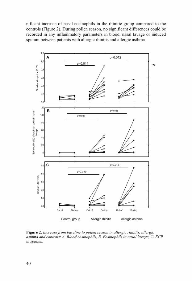
During pollen season there were significant increases in blood eosinophils and sputum-ECP in both the rhinitic and asthmatic patients, but only a sig-
40
nificant increase of nasal-eosinophils in the rhinitic group compared to the controls (Figure 2). During pollen season, no significant differences could be recorded in any inflammatory parameters in blood, nasal lavage or induced sputum between patients with allergic rhinitis and allergic asthma. Figure 2. Increase from baseline to pollen season in allergic rhinitis, allergic asthma and controls: A. Blood eosinophils, B. Eosinophils in nasal lavage, C. ECP in sputum.
0,0
0,2
0,4
0,6
0,8
1,0
1,2
Blo
od e
osin
ophi
l x 1
0-9
/L
0
20
40
60
80
100
120
Eos
inop
hils
(%) o
f tot
al c
ell c
ount
in n
asal
lava
ge
Out of During Out of During Out of During
0.0
1.0
2.0
3.0
4.0
5.0
Spu
tum
EC
P m
g/L
A
p=0.007
p=0.055B
p=0.019
p=0.018C
Control group Allergic rhinitis Allergic asthma
p=0.014
p=0.012
41
Paper II. Comparison of experimental and seasonal allergen exposure Fifteen birch pollen allergic patients, eight with allergic rhinitis and seven with allergic asthma, and five controls were studied during pollen season and after nasal and bronchial allergen challenge. After bronchial challenge PEFR registration, symptoms and medication needed were recorded in a diary for one day. In this paper, we also studied the two allergic groups combined as we, in the previous paper, had observed very little differences between pa-tients with allergic rhinitis and allergic asthma with regard to the systemic and local inflammatory response.
Clinical data There was no significant difference in symptoms, medication needed or late allergic reactions measured as PEFR decrease in the diary between patients with allergic rhinitis and allergic asthma after bronchial allergen challenge.
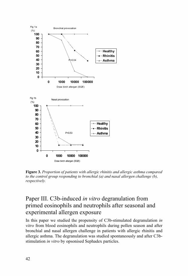
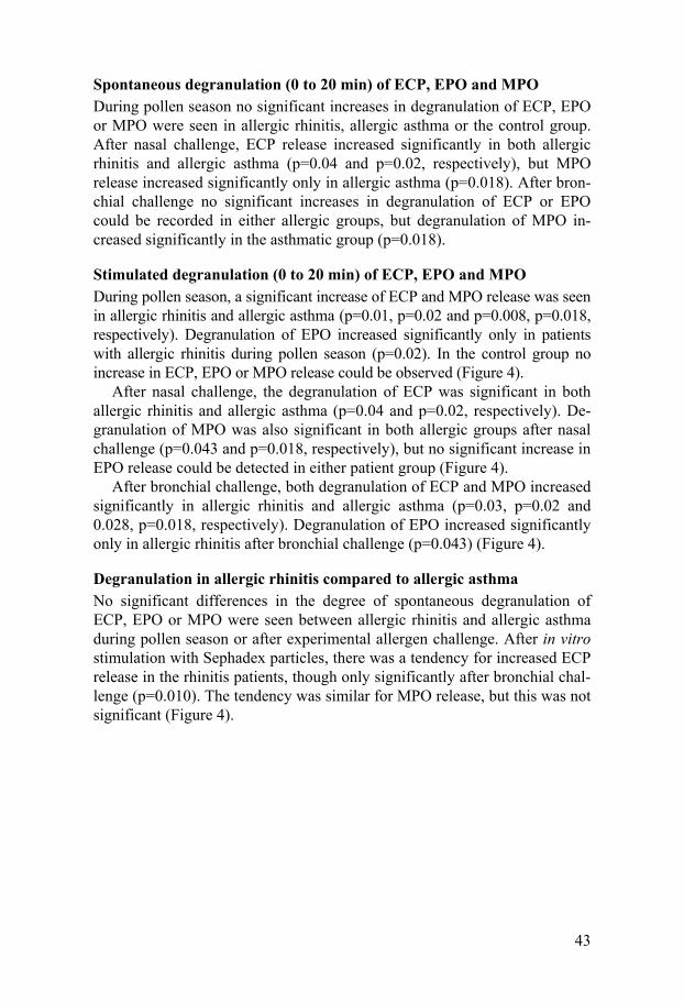
Bronchial and nasal responsiveness to allergen challenge Patients with allergic asthma were more responsive, measured by FEV1 de-cline, to bronchial challenge than patients with rhinitis, and the FEV1 decline was significantly lower in asthmatics 30 min after bronchial challenge com-pared to the rhinitics (p=0.018). Increased bronchial responsiveness, meas-ured as PD20 for birch allergen, was also recorded in allergic asthma com-pared to allergic rhinitis [PD20 = 3 700 SQ-U (2 450-7 700) vs. 34 500 SQ-U (3 850-150 000), p=0.04] (Figure 3). After nasal challenge no differences in allergen responsiveness were found between the rhinitic and asthmatic pa-tients (Figure 3). In the control group no reaction could be recorded after either allergen challenge test (Figure 3).
Eosinophil inflammation The increase in blood eosinophils was significantly higher during pollen season than after bronchial and nasal allergen challenge when combining the allergic groups compared to the controls (p=0.03 and p=0.003, respectively).
After nasal challenge no significant inflammatory reactions could be found either systemically or locally in either patients with allergic rhinitis or allergic asthma, but seasonal exposure was associated with a significant in-crease of nasal-ECP in the allergic groups combined (p=0.04).
The eosinophil inflammatory response in the airways was low both during pollen season and after bronchial challenge. However, a significant correla-tion was found in the change of sputum-ECP between bronchial challenge and seasonal exposure when combining the allergic patients (rho=0.62, p=0.02).
42
Figure 3. Proportion of patients with allergic rhinitis and allergic asthma compared to the control group responding to bronchial (a) and nasal allergen challenge (b), respectively.
Paper III. C3b-induced in vitro degranulation from primed eosinophils and neutrophils after seasonal and experimental allergen exposure In this paper we studied the propensity of C3b-stimulated degranulation in vitro from blood eosinophils and neutrophils during pollen season and after bronchial and nasal allergen challenge in patients with allergic rhinitis and allergic asthma. The degranulation was studied spontaneously and after C3b-stimulation in vitro by opsonised Sephadex particles.
0102030405060708090
100
0 1000 10000 100000
Healthy Rhinitis Asthma
Nasal provocation(%)
P=0.53
Dose birch allergen (SQE)
Fig 1b
0102030405060708090
100
0 1000 10000 100000
Healthy Rhinitis Asthma
Bronchial provocation(%)
P=0.04
Dose birch allergen (SQE)
Fig 1a
43
Spontaneous degranulation (0 to 20 min) of ECP, EPO and MPO During pollen season no significant increases in degranulation of ECP, EPO or MPO were seen in allergic rhinitis, allergic asthma or the control group. After nasal challenge, ECP release increased significantly in both allergic rhinitis and allergic asthma (p=0.04 and p=0.02, respectively), but MPO release increased significantly only in allergic asthma (p=0.018). After bron-chial challenge no significant increases in degranulation of ECP or EPO could be recorded in either allergic groups, but degranulation of MPO in-creased significantly in the asthmatic group (p=0.018).
Stimulated degranulation (0 to 20 min) of ECP, EPO and MPO During pollen season, a significant increase of ECP and MPO release was seen in allergic rhinitis and allergic asthma (p=0.01, p=0.02 and p=0.008, p=0.018, respectively). Degranulation of EPO increased significantly only in patients with allergic rhinitis during pollen season (p=0.02). In the control group no increase in ECP, EPO or MPO release could be observed (Figure 4).
After nasal challenge, the degranulation of ECP was significant in both allergic rhinitis and allergic asthma (p=0.04 and p=0.02, respectively). De-granulation of MPO was also significant in both allergic groups after nasal challenge (p=0.043 and p=0.018, respectively), but no significant increase in EPO release could be detected in either patient group (Figure 4).
After bronchial challenge, both degranulation of ECP and MPO increased significantly in allergic rhinitis and allergic asthma (p=0.03, p=0.02 and 0.028, p=0.018, respectively). Degranulation of EPO increased significantly only in allergic rhinitis after bronchial challenge (p=0.043) (Figure 4).
Degranulation in allergic rhinitis compared to allergic asthma No significant differences in the degree of spontaneous degranulation of ECP, EPO or MPO were seen between allergic rhinitis and allergic asthma during pollen season or after experimental allergen challenge. After in vitro stimulation with Sephadex particles, there was a tendency for increased ECP release in the rhinitis patients, though only significantly after bronchial chal-lenge (p=0.010). The tendency was similar for MPO release, but this was not significant (Figure 4).
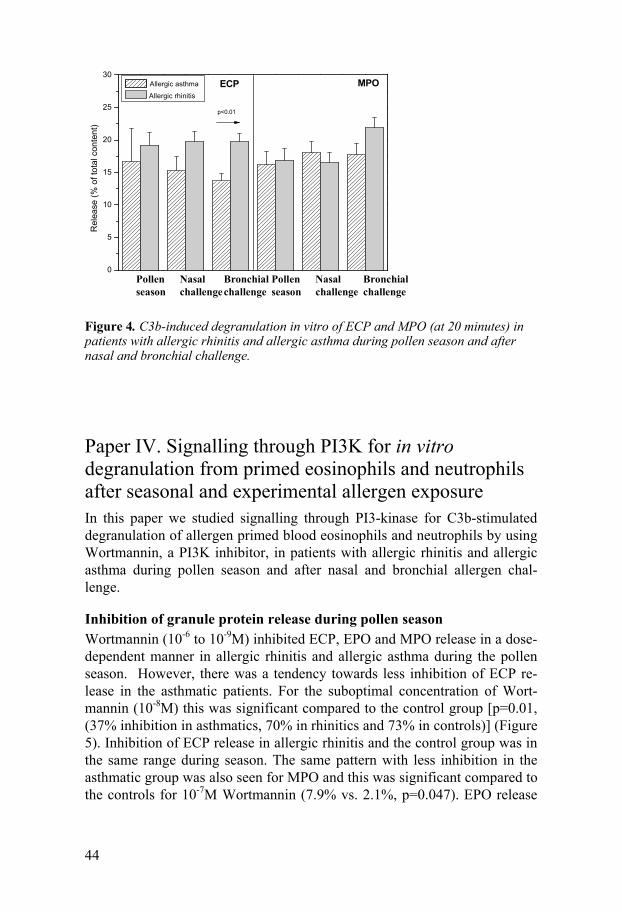
44
0
5
10
15
20
25
30
Rel
ease
(% o
f tot
al c
onte
nt)
Allergic asthma
Allergic rhinitisECP MPO
p<0.01
Pollenseason
Pollenseason
Bronchialchallenge
Nasalchallenge
Nasalchallenge
Bronchialchallenge
Figure 4. C3b-induced degranulation in vitro of ECP and MPO (at 20 minutes) in patients with allergic rhinitis and allergic asthma during pollen season and after nasal and bronchial challenge.
Paper IV. Signalling through PI3K for in vitro degranulation from primed eosinophils and neutrophils after seasonal and experimental allergen exposure In this paper we studied signalling through PI3-kinase for C3b-stimulated degranulation of allergen primed blood eosinophils and neutrophils by using Wortmannin, a PI3K inhibitor, in patients with allergic rhinitis and allergic asthma during pollen season and after nasal and bronchial allergen chal-lenge.
Inhibition of granule protein release during pollen season Wortmannin (10-6 to 10-9M) inhibited ECP, EPO and MPO release in a dose-dependent manner in allergic rhinitis and allergic asthma during the pollen season. However, there was a tendency towards less inhibition of ECP re-lease in the asthmatic patients. For the suboptimal concentration of Wort-mannin (10-8M) this was significant compared to the control group [p=0.01, (37% inhibition in asthmatics, 70% in rhinitics and 73% in controls)] (Figure 5). Inhibition of ECP release in allergic rhinitis and the control group was in the same range during season. The same pattern with less inhibition in the asthmatic group was also seen for MPO and this was significant compared to the controls for 10-7M Wortmannin (7.9% vs. 2.1%, p=0.047). EPO release
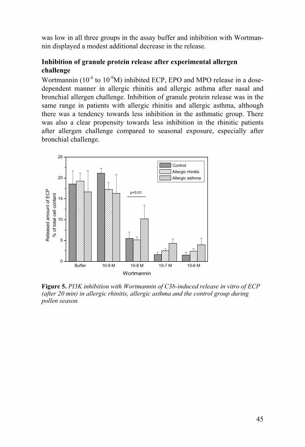
45
was low in all three groups in the assay buffer and inhibition with Wortman-nin displayed a modest additional decrease in the release.
Inhibition of granule protein release after experimental allergen challenge Wortmannin (10-6 to 10-9M) inhibited ECP, EPO and MPO release in a dose-dependent manner in allergic rhinitis and allergic asthma after nasal and bronchial allergen challenge. Inhibition of granule protein release was in the same range in patients with allergic rhinitis and allergic asthma, although there was a tendency towards less inhibition in the asthmatic group. There was also a clear propensity towards less inhibition in the rhinitic patients after allergen challenge compared to seasonal exposure, especially after bronchial challenge.
Buffer 10-9 M 10-8 M 10-7 M 10-6 M0
5
10
15
20
25
Rel
ease
d am
ount
of E
CP
% o
f tot
al c
ell c
onte
nt
Wortmannin
Control Allergic rhinitis Allergic asthma
p=0.01
Figure 5. PI3K inhibition with Wortmannin of C3b-induced release in vitro of ECP (after 20 min) in allergic rhinitis, allergic asthma and the control group during pollen season.
46
General discussion
Classification of allergic rhinitis and allergic asthma The primarily aim of this thesis was to study similarities and differences in eosinophil airway inflammation between patients with seasonal allergic rhinitis and patients with allergic asthma. These two conditions have tradi-tionally been regarded as two separate diseases, but during the last decade the concept of “one airway – one disease” has been discussed (59,225). There have also been discussions about the issue of “the allergic march”, e.g. whether or not rhinitis always precedes asthma and whether asthma can be prevented by early and intensive intervention for rhinitis; with allergen spe-cific immunotherapy and environmental remediation in addition to medical treatment (15, 116, 226). Evidently, there remains a group of patients with rhinitis who do not develop asthma and patients with mild asthma who do not have nasal symptoms. In spite of difficulties categorizing patients with allergic rhinitis and allergic asthma in two separate groups, we have made a serious attempt to include only patients who have a clear predominance of symptoms of either rhinitis or asthma during the birch pollen season in the studies presented in Paper I-IV.
At baseline no significant differences in systemic and local eosinophil in-flammation could be recorded between allergic rhinitis and allergic asthma, but there was a significant increase in bronchial responsiveness to inhalation of hypertonic saline in the asthmatic patients compared to the rhinitic pa-tients (Paper I). Furthermore, patients with allergic asthma had a greater decrease in both morning and evening PEFR during pollen season (Paper I) and a greater responsiveness expressed as allergen PD20 for birch after bron-chial challenge compared to the rhinitic patients (Paper II). Taken together, this indicates a correct classification of the two allergic patient groups in the studies.
Eosinophil inflammation Out of pollen season no significant differences in either the systemic or local eosinophil inflammation could be recorded between patients with allergic rhinitis and allergic asthma. During pollen season the pattern between the groups was the same with comparable levels of eosinophil inflammatory
47
markers in blood, nasal fluids and induced sputum (Paper I). The symptom score for rhinoconjunctivitis during pollen season was within the same range in the two allergic groups, but impaired lung function was more common in the patients with asthma (Paper I). Our data is in contrast to previous reports of early impairment of lung function during pollen season in patients with allergic rhinitis, but in those studies patients with perennial allergy and es-tablished bronchial hyperresponsiveness were included (43). In Paper II a more marked systemic inflammation during pollen season compared to ex-perimental challenge was recorded. This is in accordance with reports from other groups reporting lack of systemic inflammatory response after bron-chial challenge (227, 228). However, the eosinophil inflammatory response in the nose and bronchi was within the same range after nasal and bronchial challenge as during seasonal exposure in patients with allergic rhinitis and allergic asthma.
In Paper III the hypothesis was that differences in the pattern of eosino-phil degranulation accounted for the distinct clinical manifestations in rhini-tis and asthma. However, the main finding was that all three allergen chal-lenge models primed eosinophils for degranulation after in vitro stimulation and the release pattern was similar for ECP and EPO in allergic rhinitis and allergic asthma. The only recorded discrepancy was a significantly greater release of ECP in the rhinitic patients after bronchial allergen challenge. One interpretation could be that the eosinophils in allergic asthma are easier to activate, particularly after bronchial allergen challenge, and therefore already have released their granule proteins. This hypothesis is supported by a slightly higher amount of ECP per eosinophil cell prior to the C3b-induced granule release in the rhinitic patients and is also in accordance with results from other groups, having observed hypodense blood eosinophils after aller-gen exposure (164). It has been suggested that signalling through the intra-membrane enzyme PI3K is involved in eosinophil degranulation (212, 213) and dysregulation of PI3K may contribute to respiratory disorders and aller-gies (229). Recently it has been demonstrated that the PI3K pathway is in-volved in eosinophil degranulation in healthy blood donors (230). In Paper IV we studied signalling through PI3K by using the inhibitor Wortmannin in order to evaluate if signalling in allergen primed eosinophils followed the same pathway in the two allergic groups. Our data clearly points toward involvement of the PI3K pathway in eosinophil degranulation in allergic rhinitis and allergic asthma irrespective of allergen challenge model. How-ever, patients with asthma demonstrated less inhibition of ECP release through PI3K than the rhinitics during pollen season. This may indicate that pathways other than PI3K may play a larger role in eosinophil degranulation in allergic asthma than allergic rhinitis.
48
Neutrophil inflammation Eosinophil inflammation is well established in the pathogenesis of asthma, but neutrophils have traditionally been connected with more severe forms of asthma and chronic obstructive pulmonary disease. In this thesis the main objective was to study eosinophil inflammation in allergic rhinitis and aller-gic asthma during different conditions, in order to evaluate differences be-tween the two allergic groups. We selected the neutrophil as a comparative cell for the investigations and did not expect to discover any major influ-ences of allergen priming on neutrophil inflammation in these patients with seasonal allergic rhinitis and mild seasonal allergic asthma. However, previ-ous papers have reported increased degranulation of MPO after in vitro stimulation with GM-CSF in asthmatic patients, pointing in the direction of allergen priming of the neutrophils as well (169, 205). At baseline, an in-crease in neutrophil inflammatory markers was observed in nasal lavage and during pollen season a significant decrease in HNL was recorded in nasal fluids and sputum in the rhinitic patients (Paper I). In addition, in the control group there was a tendency towards an increase in systemic and nasal neu-trophil inflammation during seasonal allergen exposure (Paper I). Further-more, after nasal and bronchial challenge there was a slight tendency to-wards increased amounts of HNL in nasal lavage and sputum compared to pollen season in the allergic patients (Paper II). Though, we have no expla-nation for these somewhat surprising and opposed findings. In the study presented in Paper III it was seen that all three allergen challenge models could prime neutrophils to an increased degranulation of MPO after in vitro stimulation with C3b, following the same pattern as the eosinophils. In the last study, Wortmannin inhibited MPO release in a dose-dependent manner in both allergic rhinitis and allergic asthma indicating signalling through PI3 for neutrophil, as well as eosinophil, degranulation after allergen exposure (Paper IV).
HNL, which originates from the specific granule, was measured as marker of neutrophil inflammation in Paper I – II. However, in Paper III - IV MPO from the primary neutrophil granule was measured. Differences in interpre-tation of the data of neutrophil involvement may in part be due to the choice of marker, as there may be different physiological functions for the different granule proteins. Furthermore, in Paper I - II spontaneous neutrophil de-granulation of HNL in vivo were measured, whereas in vitro stimulation of the neutrophils was performed in Paper III – IV. Taken together, the data from our studies indicate that the neutrophil is involved in the allergic in-flammation.
49
Concluding remarks and future perspectives According to our studies, with minor discrepancies, patients with allergic rhinitis and allergic asthma had the same degree of eosinophil inflammation both systemically and locally and yet they presented with different clinical pictures. In addition, no differences in eosinophil degranulation patterns or signalling through PI3K could be observed in the two allergic groups. Thus, the difference in bronchial response can not be explained by just the ongoing eosinophil inflammation. However, bronchial airway inflammation was measured by inflammatory markers in induced sputum, reflecting mainly the central airways. Previous reports have highlighted the issue of the contribu-tion of inflammation in the small airways in asthma, since peripheral ob-struction is not generally detectable by routine lung function studies (231). It has been proposed that a major difference in allergic rhinitis and allergic asthma may be due to the spread of inflammation to the small airways (232, 233). Furthermore, the recent advances using anti-IL-5 therapy did not show any efficacy in the late cell-mediated phase reaction or on non-specific hy-perresponsiveness, despite depletion of eosinophils from the circulation and the basement membrane in the airways (234). This could imply that IL-5 not is as important as suggested for eosinophil recruitment (160) or that eosino-phils not are as important as proposed in chronic asthma (235). On the other hand, Rådinger et al. have speculated in the possibility of eosinophil in situ differentiation in the lung tissue to mature cells, persisting in the tissue inde-pendent of IL-5 and perhaps influenced by other cytokines or chemokines, as eotaxin (236).
The former view of asthma as being simply a disease of hyperresponsive airways changed many years ago in favour of a model of chronic systemic inflammation, and extensive research during the recent decades has concen-trated on allergic inflammation. However, recent studies on the concept of united airways have opened up new approaches for the underlying causes as to why not all patients with allergic rhinitis present with asthma. The studies in this thesis evaluated the two allergic groups simultaneously in vivo as well as in vitro under different conditions and for different parameters influencing eosinophil inflammation, as most previous studies have investigated either allergic rhinitis or allergic asthma separately. Airway remodelling per se may be one of the mechanisms that explain why some allergic patients de-velop asthma. Acute inflammatory reactions, regardless of the type of trigger stimuli, often tend to be exaggerated due to many feedback loops and ampli-fication mechanisms in the immune system and can eventually give rise to tissue injury and aberrant tissue repair. Although airway remodelling has been considered to be important in the more severe forms of asthma for many years (237), defective epithelial function has only recently regained attention. The airway epithelium, being a barrier to the external environment, has been proposed to be fundamentally abnormal in asthmatics with their
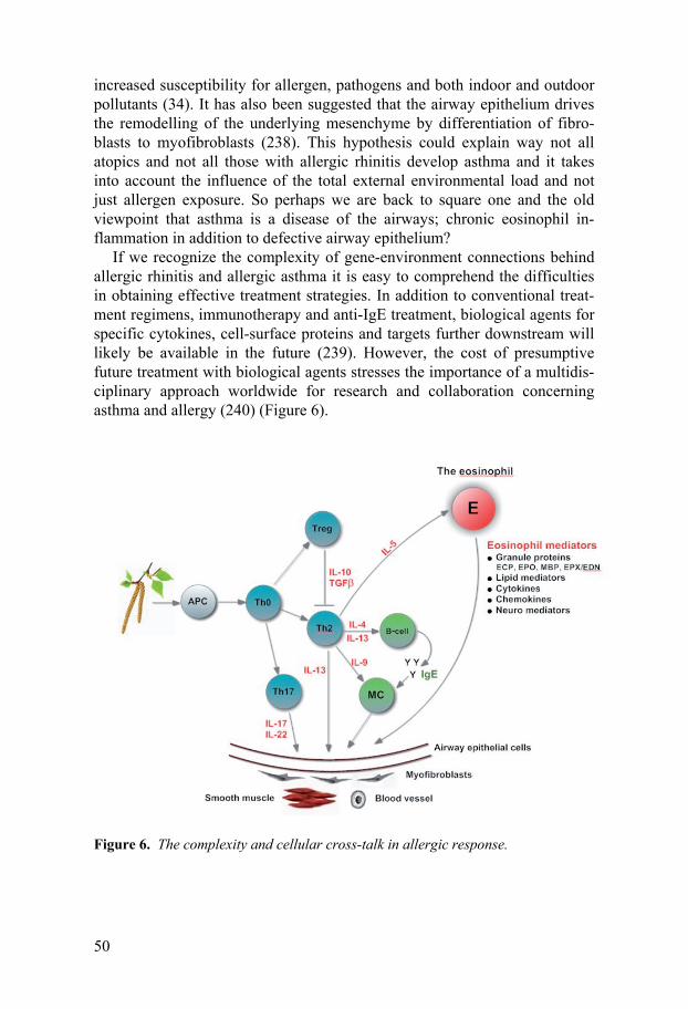
50
increased susceptibility for allergen, pathogens and both indoor and outdoor pollutants (34). It has also been suggested that the airway epithelium drives the remodelling of the underlying mesenchyme by differentiation of fibro-blasts to myofibroblasts (238). This hypothesis could explain way not all atopics and not all those with allergic rhinitis develop asthma and it takes into account the influence of the total external environmental load and not just allergen exposure. So perhaps we are back to square one and the old viewpoint that asthma is a disease of the airways; chronic eosinophil in-flammation in addition to defective airway epithelium?
If we recognize the complexity of gene-environment connections behind allergic rhinitis and allergic asthma it is easy to comprehend the difficulties in obtaining effective treatment strategies. In addition to conventional treat-ment regimens, immunotherapy and anti-IgE treatment, biological agents for specific cytokines, cell-surface proteins and targets further downstream will likely be available in the future (239). However, the cost of presumptive future treatment with biological agents stresses the importance of a multidis-ciplinary approach worldwide for research and collaboration concerning asthma and allergy (240) (Figure 6).
Figure 6. The complexity and cellular cross-talk in allergic response.
51
Conclusions
I. Allergic rhinitis and allergic asthma have the same degree of eosinophil inflammation systemically and locally at baseline and during pollen season. Despite this similarity, impairment in lung function during pollen season and increased bronchial responsiveness at baseline were more common in asth-matics.
II. Allergic rhinitis and allergic asthma have the same degree of airway eosi-nophil inflammation after bronchial allergen challenge, but allergic asthma has increased bronchial responsiveness measured as PD20 for birch allergen. Systemic eosinophil inflammation was more pronounced after seasonal ex-posure than after experimental allergen challenge in allergic rhinitis and allergic asthma.
III. Systemically allergen primed eosinophils have similar patterns of stimu-lated degranulation in vitro in allergic rhinitis and allergic asthma. The re-leased amounts of ECP and EPO were in the same range in all allergen chal-lenge models in allergic rhinitis and allergic asthma.
IV. Signalling through PI3K is involved in eosinophil in vitro degranulation of ECP and EPO in allergic rhinitis and allergic asthma irrespective of the model of allergen exposure. Patients with allergic asthma demonstrated less inhibition of ECP release via this pathway during pollen season, indicating that other pathways apart from signalling through PI3K play a greater role in eosinophil degranulation in allergic asthma than in allergic rhinitis.
52
Swedish summary
De allergiska sjukdomarna har kraftigt ökat de senaste decennierna i den industrialiserade delen av världen och är idag ett stort hälsoproblem med ökande socioekonomiska konsekvenser. Epidemiologiska undersökningar har visat att drygt 30 % av befolkningen i västvärlden har någon form av allergi. De allergiska sjukdomarna kan yttra sig som rhinit (hösnuva), astma, eksem och nässelutslag, men praktiskt taget alla organ kan drabbas.
Allergi är en immunologisk reaktion mot ett s.k. allergen, som är helt harmlöst och oskadligt för kroppen, men som kroppen producerar antikrop-par mot. Benägenheten att producera dessa allergiantikroppar (IgE-antikroppar) är ärftlig och kallas atopi. Inte alla som är atopiker utvecklar dock allergi, utan detta beror på en kombination av olika faktorer, som t.ex. luftföroreningar, yrke, rökning men även övervikt har visat sig spela roll.
Hösnuva och astma är vanligast i barndomen och flertalet förbättras med åren, men framför allt förekomsten av astma har ökat bland vuxna på senare år . Epidemiologiska undersökningar har visat att närmare 10 % av befolk-ningen lider av astma i många urbaniserade geografiska områden i Sverige och andra delar av den industrialiserade delen av världen. De allergiska sjukdomarna karakteriseras som kroniskt inflammatoriska systemsjukdomar med lokalengagemang och man vet att allergisk rhinit är en riskfaktor för utvecklande av astma. Flertalet astmapatienter har vidare rhinitsymptom, men inte alla med rhinit utvecklar astma.
Den allergiska inflammationen i vävnaden kännetecknas av eosinofila cel-ler, som är vita blodkroppar. Eosinofilerna finns i blodet, men vandrar ut i vävnaden om allergen penetrerat kroppens försvarsbarriärer. Detta är vanligt under björkpollensäsongen, då björkpollen fastnar i nässlemhinnan och i luftvägarna när vi andas. Den eosinofila cellen uppfattar björkpollenkornen som sjukdomsframkallande och bekämpar dessa genom att frisätta en rad starkt vävnadstoxiska produkter. Vid upprepad allergenexponering fås en ond cirkel med kronisk eosinofil inflammation och åtföljande vävnadsskada. Det har närmast varit vedertaget under många år att den eosinofila inflamma-tionen i luftvägarna är en viktig orsak till astmautveckling.
Det övergripande syftet med detta avhandlingsarbete var att studera likhe-ter och olikheter i den eosinofila inflammationen vid allergisk rhinit och allergisk astma och frågan varför inte alla rhinitiker får astma. Studierna utfördes under björkpollensäsongen och efter experimentell björkpollenpro-vokation i näsa respektive luftvägar. I de första två delarbetena mättes eosi-
53
nofila markörer i blod, nässköljvätska och luftvägar efter respektive aller-genprovokationsmodell, medan däremot arbete tre och fyra var rent experi-mentella. I arbete tre studerades de eosinofila cellernas förmåga till frisätt-ning av sina vävnadstoxiska produkter efter stimulering in vitro efter respek-tive allergenprovokationsmodell hos de två allergiska grupperna. I det sista arbetet studerades mekanismen för cellsignalering för frisättandet av de eo-sinofila markörerna.
Våra studier har visat att den eosinofila inflammationen i såväl blod, som näsa och luftvägar är av samma storleksordning hos patienter med allergisk rhinit och allergisk astma både under björkpollensäsongen och utanför pol-lensäsong. Trots samma grad av inflammation uppvisar endast astmatikerna en ökad känslighet i luftvägarna med försämring av lungfunktionen under pollensäsongen. Vid bronkialprovokation med björkpollenallergen ser man vidare en signifikant ökad känslighet i luftvägarna hos astmatikerna, men även här ses samma grad av eosinofil inflammation i luftvägarna hos de två patientgrupperna. Däremot är den systemiska inflammationen mer uttalad under pollensäsongen än efter både bronk- och nasalprovokation hos både rhiniker och astmatiker. I det tredje arbetet kunde vi visa att de eosinofila cellerna var likvärdigt aktiverade av björkpollenallergen för frisättning av sina inflammatoriska markörer vid in vitro stimulering. Detta gällde både under pollensäsongen och efter de experimentella provokationerna hos både rhinitiker och astmatiker. Vi har även visat att signalering över cellmembra-net via enzymet PI3K för frisättning av eosinofila markörer ej skiljer sig åt mellan de två allergiska grupperna. Här kunde man dock se en tendens till högre frisättning hos astmatikerna vid blockering av enzymet, vilket skulle kunna tala för involvering av andra signaltransduktionsvägar hos astmatiker-na.
Sammanfattningsvis kan sägas att allergisk rhinit och allergisk astma uppvisar samma grad av eosinofil inflammation i såväl blod som näsa och luftvägar. De eosinofila cellerna är vidare likvärdigt aktiverade för frisätt-ning och följer samma signalleringsväg över cellmembranet. Våra data talar för att den eosinofila inflammationen i sig inte är tillräcklig för att patienter med allergisk rhinit ska utveckla allergisk astma, utan att det finns ytterligare bakomliggande faktorer av vikt för sjukdomsutvecklingen. Sannolikt före-ligger ett komplext samspel av genetiska faktorer och miljöpåverkan bakom utvecklandet av de olika symptombilderna vid de allergiska sjukdomarna.
54
Acknowledgements
Marie Carlson, my supervisor, for her deep knowledge in eosinophil re-search and unfailing scientific enthusiasm, for her expert guidance and in-valuable encouragement, for patience with my impatience, and for good friendship.
Christer Janson, co-supervisor and my professor, for providing excellent research facilities and for sharing his deep knowledge in scientific work, for his constant guidance and patience, and for friendship throughout the years.
Gunnemar Stålenheim, co-supervisor and tutor, for introducing me into the field of clinical allergology and leading me into allergic research, generously sharing his deep knowledge, and for good friendship over the years.
Maria Lampinen, co-author, for her deep knowledge in scientific work and eosinophil research, for her valuable views on the manuscript, help with the figures and improving the English language, and for pleasant co-operation.
Ingrid Stolt, co-author, for her excellent and experienced knowledge in labo-ratory work, for carrying out the assays, and for patiently guiding and intro-ducing me to the laboratory methods used in this thesis.
Signe Svedberg-Brandt and Katarina Göthberg, research nurses, for their skilful work, excellent care of the patients and for their helpfulness.
Eva Lindberg, former head of the Department of Lung medicine and Aller-gology, for encouraging me and providing facilities for the re-start of the work with this thesis and for good friendship.
Inger Dahlén, present head of the Department of Lung medicine and Aller-gology, for providing excellent working facilities and valuable support, for creating a pleasant atmosphere at work and for good friendship.
Gunnar Boman, professor emeritus, for kindly introducing me in research education. Lavinia Machado Boman, colleague, for friendship and much laughter over the years.
55
Gun-Marie Bodman Lund, member of the Lung Research Group and my personal IT-support, for excellent assistance, practical help and for drawing the last picture in the thesis.
All the members of the Lung Research Group, for their support and friend-ship, and for their ability of creating a friendly atmosphere.
My colleagues throughout many years, Kristina Lamberg Lundström and Carl-Axel Karlsson, for friendship and sharing their deep clinical knowl-edge, always being helpful and for much laughter and joyful companionship. María Gudbjörnsdottír, for support, good collaboration and friendship over the years.
All the staff and my colleagues at the Department of Lung medicine and Allergology, for their interest and support in my work and for all good times working together.
Steven Lucas for excellent linguistic revision of this thesis.
All the patients who so willingly and with interest participated in the studies.
My parents, Karin and my late father, for love and encouragement, their never-ending caring for me and my sister Anne throughout the years. Anne, my dear sister, and her family, for so much love, fun and many good times.
Finally, to Anders and Johan for just being there and making it worthwhile.
This work was supported by grants from the Swedish Association against Asthma and Allergy, the Swedish Heart and Lung Foundation, Bror Hjerpstedt’s Foundation, the Uppsala County Against Heart and Lung dis-eases and the Medical Faculty of Uppsala University.
56
References
1. Johansson SG, Bieber T, Dahl R, Friedmann PS, Lanier BQ, Lockkey RF, Mo-
tala C, Ortega Martell JA, Platts-Mills TA, Ring J, Thien F, Van Cauwenberge P, Williams HC. Revised nomenclature for allergy for global use: Report of the Nomenclature Review Committee of the World Allergy Organisation, October 2003. J Allergy Clin Immunol 2004;113:832 -836.
2. ISAAC Steering Committee. Worldwide variations in the prevalence of symp-toms of asthma, allergic rhinoconjuctivities and atopic eczema: The interna-tional Study of Asthma and Allergies in Childhood (ISAAC). Lancet 1998:351:1225-1232.
3. Platts-Mills TA, Tovey ER, Mitchell BE, Moszoro H, Nock P. Reduction of bronchial hyperreactivity during prolonged allergen avoidance. Lancet 1982:675-78.
4. Sears MR, Herbison GP, Holdaway MD, Hewitt CJ, Flannery EM, Silva PA. The relative risks of sensitivity to grass pollen, house dust mite and cat dander in the development of childhood asthma. Clin Exp Allergy 1989;19:419-24.
5. Sibbald B, Rink E. Epidemiology of seasonal and perennial rhinitis: clinical presentation and medical history. Thorax 1991;46:895-901.
6. Johansson SG, Bennich H, Wide L. A new class of immunoglobulin in human serum. Immunology 1967;14:265-72.
7. Ishizaka K, Ishizaka T. Identification of gamma-E-antibodies as a carrier of reaginic activity. J Immunol 1967;99:1187-98.
8. Coca AF, Cooke RA. On the classification of the phenomenon of hypersensi-tivness. J Immunol 1923;8:163.
9. Johansson SG, Hourihane JO, Bousquet J, Bruijnzeel-Koomen C, Dreborg S, Haahtela T, Kowalski ML, Mygind N, Ring J, van Cauwenberge P, van Hage-Hamsten M, Wüthrich B. A revised nomenclature for allergy: An EAACI posi-tion statement from EAACI nomenclature task force. Allergy 2001;56:813-24.
10. Bousquet PJ, Chatzi L, Jarvis D, Burney P. Assessing skin prick test reliability in ECRHS-I. Allergy 2008;63:341-46.
11. Witteman AM, Stapel SO, Perdok GJ, Sjamsoedin DH, Jansen HM, Aalberse RC, van der Zee JS. The relationsship between RAST and skin test results in pa-tients with asthma or rhinitis: A quantitative study with purified major allergens. J Allergy Clin Immunol 1996;1:16-25
12. Accordini S, Corsico A, Cerveri A, Gislason D, Gulsvik A, Janson C, Jarvis D, Marcon A, Pin I, Vermeire P, Almar E, Bugiani M, Cazzoletti L, Duren, Taule-ria E, Jögi R, Marinoni A, Martinez-Mortalla J, Leynaert B, de Marco R. Ther-apy and Health Economics Working Group of the European Community Respi-ratory Health Survey II. The socio-economic burden of asthma is substential in Europe. Allergy 2008;63:116-24.
57
13. Anandan C, Nurmatov U, van Schayck OC, Sheikh A. Is the prevalence of asthma declining? Systematic review of epidemiological studies. Allergy 2010;65:152-67.
14. National Heart, Lung and Blood Institute: National Asthma Education Program; Expert Panel on the Management of Asthma. Guidelines for the Diagnosis and Management of Asthma: Expert Panel Report. Betheseda: National Asthma Education Program, Office of Prevention, Education, and Control. National Heart, Lung, and Blood Institute, National Institutes of Health, U.S. Department of Health and Human Services, Public Health Service; 1991: Publ. No. 91 3042A.
15. Lemanske RF, Busse WW. Asthma: Clinical expression and molecular macha-nisms. J Allergy Clin Immunol 2010;125:S95-102.
16. Asher MI, Montefort S, Björksten B, Lai CK, Strachen DP, Weiland SK, Wil-liams H. Worldwide time trends in the prevalence of symptoms of asthma, al-lergic rhinoconjuctivities and excema in childhood: ISAAC Phase One and Three repeat multicountry cross-sectional surveys. Lancet 2006;368:733-43.
17. Humbert M. Does intrinsic asthma exist? Rev Mal Respir 2000;17:245-54. 18. Corrigan C. Mechanisms of intrinsic asthma. Curr Opin Allergy Clin Immunol
2004;4:53-56. 19. Humbert M, Menz G, Ying S, Corrigan CJ, Robinson DS, Durham SR, Kay
AB. The immunopathology of extrinsic (atopic) and instrinsic (non-atopic) asthma: more similarities than differences. Immunol Today 1999;20:528-33.
20. Ying S, Humbert M, Meng Q, Pfister R, Menz G, Gould HJ, Kay AB, Durham SR. Local expression of epsilon germline gene transcripts and RNA for the ep-silon heavy chain of IgE in the bronchial mucosa in atopic and nonatopic asthma. J Allergy Clin Immunol 2001;107:686-92.
21. Ying S, Humbert M, Barkans J, Corrigan CJ, Pfister R, Menz G, Larché M, Robinson DS, Durham SR, Kay AB. Expression of IL-4 and IL-5 mRNA and protein product by CD4+ and CD8+T cells, eosinophils and mast cells in bron-chial biopsies obtained from atopic and nonatopic (intrinsic) asthmatics. J Im-munol 1997;158:3539-44.
22. Ying S, Meng Q, Zeibecoglou K, Robinson DS, Macfarlane A, Humbert M, Kay AB. Eosinophil chemotactic chemokines (eotaxin, eotaxin-2, RANTES, monocyte chemoattractant protein-3 and 4) and C-C chemokine receptor 3 ex-pression in bronchial biopsies from atopic and nonatopic (intrinsic) asthmatics. J Immunol 1999;163:6321-29.
23. Menz G, Ying S, Durham SR, Corrigan CJ, Robinson DS, Hamid Q, Pfister R, Humbert M, Kay AB. Molecular concepts of IgE-initiated inflammation in atopic and nonatopic asthma. Allergy 1998;53(S):15-21.
24. Amin K, Lúdvíksdóttir D, Janson C, Nettelbladt O, Björnsson E, Roomans GM, Boman G, Sevéus L, Venge P. Inflammation and structural changes in the air-ways of patients with atopic and nonatopic asthma. BHR Group. Am J Respir Crit Care Med 2000;162:2295-301.
25. Kariyawasam HH, Robinson DS. The eosinophil: the cell and its weapons, the cytokines, its location. Semin Respir Crit Care Med 2006;27:117-27.
26. Minshall EM, Leung DY, Martin RJ, Song YL, Cameron L, Ernst P, Hamid Q. Eosinophil-associated TGF-β1 mRNA expression and airways fibrosis in bron-chial asthma. Am J Respir Cell Mol Biol 1997:17:326–333.
27. Kariyawasam HH, Robinson DS. The role of tissue remodelling in asthma. Curr Opin Immunol 2007;19:681-86.
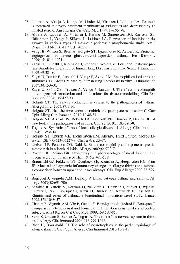
58
28. Laitinen A, Altraja A, Kämpe M, Linden M, Virtanen I, Laitinen LA. Tenascin is increased in airway basement membrane of asthmatics and decreased by an inhaled steroid. Am J Respir Crit Care Med 1997;156:951-8.
29. Altraja A, Laitinen A, Virtanen I, Kämpe M, Simonsson BG, Karlsson SE, Håkansson L, Venge P, Sillastu H, Laitinen LA. Expression of laminins in the airways in various types of asthmatic patients: a morphometric study. Am J Respir Cell Mol Biol 1996;15:482-8.
30. Vrugt B, Wilson S, Bron A, Holgate ST, Djukanovic R, Aalbers R. Bronchial angiogenesis in severe glucocorticoid-dependent asthma, Eur Respir J 2000;15:1014–1021.
31. Zagai U, Lundahl J, Klominek J, Venge P, Sköld CM. Eosinophil cationic pro-tein stimulates migration of human lung fibroblasts in vitro. Scand J Immunol. 2009;69:381-6.
32. Zagai U, Dadfar E, Lundahl J, Venge P, Sköld CM. Eosinophil cationic protein stimulates TGF-beta1 release by human lung fibroblasts in vitro. Inflammation 2007;30:153-60.
33. Zagai U, Sköld CM, Trulson A, Venge P, Lundahl J. The effect of eosinophils on collagen gel contraction and implications for tissue remodelling. Clin Exp Immunol 2004;135:427-33.
34. Holgate ST. The airway epithelium is central to the pathogenesis of asthma. Allergol Inter 2008;57:1-10.
35. Holgate ST. Has the time come to rethink the pathogenesis of asthma? Curr Opin Allerg Clin Immunol 2010;10:48-53.
36. Holgate ST, Arshad HS, Roberts GC, Howarth PH, Thurner P, Davies DE. A new look at the pathogenesis of asthma. Clin Sci 2010;118:439-50.
37. Togias A. Systemic effects of local allergic disease. J Allergy Clin Immunol 2004;113:S8-14.
38. Holgate ST, Church MK, Lichtenstein LM. Allergy, Third Edition. Mosby El-sevier. ISBN 0-323-03227-8. Chaper 4, p 55-67.
39. Nielsen LP, Peterson CG, Dahl R. Serum eosinophil granule proteins predict asthma risk in allergic rhinitis. Allergy 2009;64:733-7.
40. Proctor DF, Adams GK. Physiology and pharmacology of nasal function and mucus secretion. Pharmacol Ther 1976;2:493-509.
41. Braunstahl GJ, Fokkens WJ, Overbeek SE, KleinJan A, Hoogsteden HC, Prins JB. Mucosal and systemic inflammatory changes in allergic rhinitis and asthma: a comparison between upper and lower airways. Clin Exp Allergy 2003;33:579-87.
42. Bousquet J, Vignola A.M, Demoly P. Links between asthma and rhinitis. Al-lergy 2003;58:691-706.
43. Shaaban R, Zureik M, Soussan D, Neukirch C, Heinrich J, Sunyer J, Wjst M, Cerveri I, Pin I, Bousquet J, Jarvis D, Burney PG, Neukirch F, Leynaert B. Rhinitis and onset of asthma: a longitudinal population-based study. Lancet 2008;372:1049-57.
44. Chanez P, Vignola AM, Vic P, Guddo F, Bonsignore G, Godard P, Bousquet J. Comparison between nasal and bronchial inflammation in asthmatic and control subjects. Am J Respir Crit Care Med 1999;159:588-95.
45. Sarin S, Undem B, Sanico A, Togias A. The role of the nervous system in rhini-tis. J Allergy Clin Immunol 2006;118:999-1016.
46. Raap U, Braunstahl GJ. The role of neurotrophins in the pathophysiology of allergic rhinitis. Curr Opin Allergy Clin Immunol 2010;10:8-13.
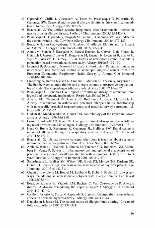
59
47. Ciprandi G, Cirillo I, Vizzaccaro A, Tosca M, Passalacqua G, Pallestrini E, Canonica GW. Seasonal and perennial allergic rhinitis: is this classification ad-herent to real life? Allergy 2005;60:882-7.
48. Braunstahl GJ.The unified system: Respiratory tract-nasobronchial interaction mechanisms in allergic disease. J Allergy Clin Immunol 2005;115:142-48.
49. Passalacqua G, Ciprandi G, Pasquali M, Guerra L, Canonica GW. An update on the asthma-rhinitis link. Curr Opin Allergy Clin Immunol 2004;4:177-183.
50. Bousquet J, van Cauwenberge P, Khaltaev N. Allergic Rhinitis and its Impact on Asthma. J Allergy Clin Immunol 2001;108:S147-334.
51. Antó JM, Sunyer J, Basagaña X, Garcia-Esteban R, Cerveri I, de Marco R, Heinrich J, Janson C, Jarvis D, Kogevinas M, Kuenzli N, Leynaert B, Svanes C, Wjst M, Gislason T, Burney P. Risk factors of new-onset asthma in adults: a population-based international cohort study. Allergy 2010;65:1021-30.
52. Leynaert B, Bousquet J, Neukirch C, Liard R, Neukirch F. Perennial rhinitis: an independent risk factor for asthma in nonatopic subjects: results from The European Community Respiratory Health Survey. J Allergy Clin Immunol 1999;104:301-304.
53. Linneberg A, Henrik Nielsen N, Frølund L, Madsen F, Dirksen A, Jørgensen T. The link between allergic rhinitis and allergic asthma: a prospective population-based study. The Copenhagen Allergy Study. Allergy 2002;57:1048-52.
54. Passalacqua G, Canonica GW. Impact of rhinitis on airway inflammation: bio-logical and therapeutic implications. Respir Res 2001;:320-23.
55. Alvarez MJ, Olaguibel JM, García BE, Rodríquez A, Tabar AI, Urbiola E. Airway inflammation in asthma and perennial allergic rhinitis. Relationship with nonspecific bronchial responsiveness and maximal airway narrowing. Al-lergy 2000;55:355-62
56. Undem BJ, McAlexander M, Hunter DD. Neurobiology of the upper and lower airways. Allergy 1999;54:81-93.
57. Corren J, Adinoff AD, Irvin CG. Changes in bronchial responsiveness follow-ing nasal provocation with allergen. J Allergy Clin Immunol 1992;89:611-18.
58. Hens G, Bobic S, Reekmans K, Ceuppens JL, Hellings PW. Rapid systemic uptake of allergens through the respiratory mucosa. J Allergy Clin Immunol 2007;120:472-4.
59. Braunstahl GJ. United airways concept: what does it teach us about systemic inflammation in airways disease? Proc Am Thorac Soc 2009;6:652-4.
60. Amin K, Rinne J, Haahtela T, Simola M, Peterson CG, Roomans GM, Malm-berg H, Venge P, Sevéus L. Inflammatory cell and epithelial characteristics of perennial allergic and nonallergic rhinitis with a symptom history of 1 to 3 years' duration. J Allergy Clin Immunol 2001;107:249-57.
61. Smurthwaite L, Walker SN, Wilson DR, Birch DS, Merrett TG, Durham SR, Gould HJ. Persistent IgE synthesis in the nasal mucosa of hayfever patients. Eur J Immunol 2001;31:3422-31.
62. Chakir J, Laviolette M, Boutet M, Laliberté R, Dubé J, Boulet LP. Lower air-ways remodelling in nonasthmatic subjects with allergic rhinitis. Lab Invest 1996:75:735-44.
63. Bousquet J, Jacot W, Vignola AM, Bachert C, Van Cauwenberge P. Allergic rhinitis: A disease remodeling the upper airways? J Allergy Clin Immunol 2004;113: 43-49.
64. Cirillo I, Pistorio A, Tosca M, Ciprandi G. Impact of allergic rhinitis on asthma: effects on bronchial hyperreactivity. Allergy 2009;64:439-44.
65. Danielsson J, Jessen M. The natural course of allergic rhinitis during 12 years of follow-up. Allergy 1997;52:331–334.
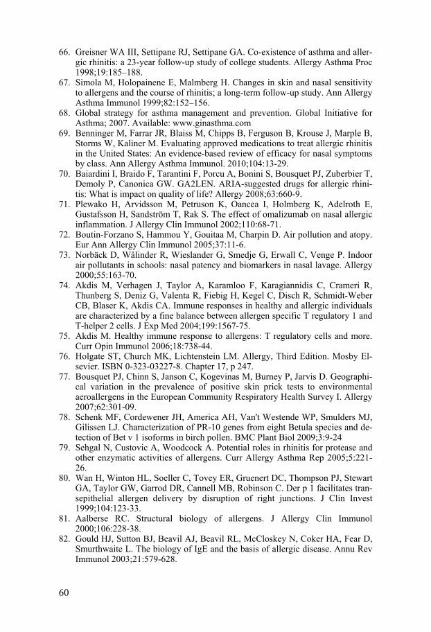
60
66. Greisner WA III, Settipane RJ, Settipane GA. Co-existence of asthma and aller-gic rhinitis: a 23-year follow-up study of college students. Allergy Asthma Proc 1998;19:185–188.
67. Simola M, Holopainene E, Malmberg H. Changes in skin and nasal sensitivity to allergens and the course of rhinitis; a long-term follow-up study. Ann Allergy Asthma Immunol 1999;82:152–156.
68. Global strategy for asthma management and prevention. Global Initiative for Asthma; 2007. Available: www.ginasthma.com
69. Benninger M, Farrar JR, Blaiss M, Chipps B, Ferguson B, Krouse J, Marple B, Storms W, Kaliner M. Evaluating approved medications to treat allergic rhinitis in the United States: An evidence-based review of efficacy for nasal symptoms by class. Ann Allergy Asthma Immunol. 2010;104:13-29.
70. Baiardini I, Braido F, Tarantini F, Porcu A, Bonini S, Bousquet PJ, Zuberbier T, Demoly P, Canonica GW. GA2LEN. ARIA-suggested drugs for allergic rhini-tis: What is impact on quality of life? Allergy 2008;63:660-9.
71. Plewako H, Arvidsson M, Petruson K, Oancea I, Holmberg K, Adelroth E, Gustafsson H, Sandström T, Rak S. The effect of omalizumab on nasal allergic inflammation. J Allergy Clin Immunol 2002;110:68-71.
72. Boutin-Forzano S, Hammou Y, Gouitaa M, Charpin D. Air pollution and atopy. Eur Ann Allergy Clin Immunol 2005;37:11-6.
73. Norbäck D, Wålinder R, Wieslander G, Smedje G, Erwall C, Venge P. Indoor air pollutants in schools: nasal patency and biomarkers in nasal lavage. Allergy 2000;55:163-70.
74. Akdis M, Verhagen J, Taylor A, Karamloo F, Karagiannidis C, Crameri R, Thunberg S, Deniz G, Valenta R, Fiebig H, Kegel C, Disch R, Schmidt-Weber CB, Blaser K, Akdis CA. Immune responses in healthy and allergic individuals are characterized by a fine balance between allergen specific T regulatory 1 and T-helper 2 cells. J Exp Med 2004;199:1567-75.
75. Akdis M. Healthy immune response to allergens: T regulatory cells and more. Curr Opin Immunol 2006;18:738-44.
76. Holgate ST, Church MK, Lichtenstein LM. Allergy, Third Edition. Mosby El-sevier. ISBN 0-323-03227-8. Chapter 17, p 247.
77. Bousquet PJ, Chinn S, Janson C, Kogevinas M, Burney P, Jarvis D. Geographi-cal variation in the prevalence of positive skin prick tests to environmental aeroallergens in the European Community Respiratory Health Survey I. Allergy 2007;62:301-09.
78. Schenk MF, Cordewener JH, America AH, Van't Westende WP, Smulders MJ, Gilissen LJ. Characterization of PR-10 genes from eight Betula species and de-tection of Bet v 1 isoforms in birch pollen. BMC Plant Biol 2009;3:9-24
79. Sehgal N, Custovic A, Woodcock A. Potential roles in rhinitis for protease and other enzymatic activities of allergens. Curr Allergy Asthma Rep 2005;5:221-26.
80. Wan H, Winton HL, Soeller C, Tovey ER, Gruenert DC, Thompson PJ, Stewart GA, Taylor GW, Garrod DR, Cannell MB, Robinson C. Der p 1 facilitates tran-sepithelial allergen delivery by disruption of right junctions. J Clin Invest 1999;104:123-33.
81. Aalberse RC. Structural biology of allergens. J Allergy Clin Immunol 2000;106:228-38.
82. Gould HJ, Sutton BJ, Beavil AJ, Beavil RL, McCloskey N, Coker HA, Fear D, Smurthwaite L. The biology of IgE and the basis of allergic disease. Annu Rev Immunol 2003;21:579-628.
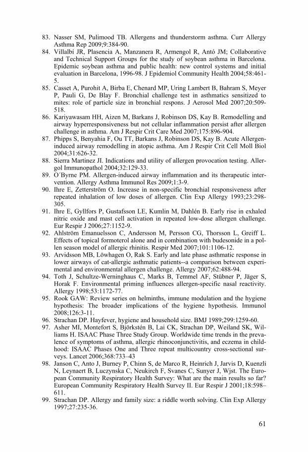
61
83. Nasser SM, Pulimood TB. Allergens and thunderstorm asthma. Curr Allergy Asthma Rep 2009;9:384-90.
84. Villalbí JR, Plasencia A, Manzanera R, Armengol R, Antó JM; Collaborative and Technical Support Groups for the study of soybean asthma in Barcelona. Epidemic soybean asthma and public health: new control systems and initial evaluation in Barcelona, 1996-98. J Epidemiol Community Health 2004;58:461-5.
85. Casset A, Purohit A, Birba E, Chenard MP, Uring Lambert B, Bahram S, Meyer P, Pauli G, De Blay F. Bronchial challenge test in asthmatics sensitized to mites: role of particle size in bronchial respons. J Aerosol Med 2007;20:509-518.
86. Kariyawasam HH, Aizen M, Barkans J, Robinson DS, Kay B. Remodelling and airway hyperresponsiveness but not cellular inflammation persist after allergen challenge in asthma. Am J Respir Crit Care Med 2007;175:896-904.
87. Phipps S, Benyahia F, Ou TT, Barkans J, Robinson DS, Kay B. Acute Allergen-induced airway remodelling in atopic asthma. Am J Respir Crit Cell Moll Biol 2004;31:626-32.
88. Sierra Martinez JI. Indications and utility of allergen provocation testing. Aller-gol Immunopathol 2004;32:129-33.
89. O´Byrne PM. Allergen-induced airway inflammation and its therapeutic inter-vention. Allergy Asthma Immunol Res 2009;1:3-9.
90. Ihre E, Zetterström O. Increase in non-specific bronchial responsiveness after repeated inhalation of low doses of allergen. Clin Exp Allergy 1993;23:298-305.
91. Ihre E, Gyllfors P, Gustafsson LE, Kumlin M, Dahlén B. Early rise in exhaled nitric oxide and mast cell activation in repeated low-dose allergen challenge. Eur Respir J 2006;27:1152-9.
92. Ahlström Emanuelsson C, Andersson M, Persson CG, Thorsson L, Greiff L. Effects of topical formoterol alone and in combination with budesonide in a pol-len season model of allergic rhinitis. Respir Med 2007;101:1106-12.
93. Arvidsson MB, Löwhagen O, Rak S. Early and late phase asthmatic response in lower airways of cat-allergic asthmatic patients--a comparison between experi-mental and environmental allergen challenge. Allergy 2007;62:488-94.
94. Toth J, Schultze-Werninghaus C, Marks B, Temmel AF, Stübner P, Jäger S, Horak F. Environmental priming influences allergen-specific nasal reactivity. Allergy 1998;53:1172-77.
95. Rook GAW: Review series on helminths, immune modulation and the hygiene hypothesis: The broader implications of the hygiene hypothesis. Immunol 2008;126:3-11.
96. Strachan DP. Hayfever, hygiene and household size. BMJ 1989;299:1259-60. 97. Asher MI, Montefort S, Björkstén B, Lai CK, Strachan DP, Weiland SK, Wil-
liams H. ISAAC Phase Three Study Group. Worldwide time trends in the preva-lence of symptoms of asthma, allergic rhinoconjunctivitis, and eczema in child-hood: ISAAC Phases One and Three repeat multicountry cross-sectional sur-veys. Lancet 2006;368:733–43
98. Janson C, Anto J, Burney P, Chinn S, de Marco R, Heinrich J, Jarvis D, Kuenzli N, Leynaert B, Luczynska C, Neukirch F, Svanes C, Sunyer J, Wjst. The Euro-pean Community Respiratory Health Survey: What are the main results so far? European Community Respiratory Health Survey II. Eur Respir J 2001;18:598–611.
99. Strachan DP. Allergy and family size: a riddle worth solving. Clin Exp Allergy 1997;27:235-36.
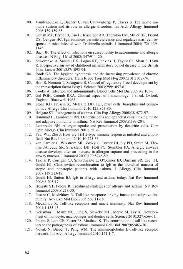
62
100. Vandenbulcke L, Bachert C, van Cauwenberge P, Claeys S. The innate im-mune system and its role in allergic disorders. Int Arch Allergy Immunol 2006;139:159-65.
101. Gurish MF, Bryce PJ, Tao H. Kisselgof AB, Thornton EM, Miller HR, Friend DS, Oettgen HC. IgE enhances parasite clereance and regulates mast cell re-sponse in mice infected with Trichinella spiralis. J Immunol 2004;172:1139-1145.
102. Bach JF. The effect of infections on susceptibility to autoimmune and allergic diseases. N Engl J Med 2002; 347:911–20.
103. Sawczenko A, Sandhu BK, Logan RF, Jenkins H, Taylor CJ, Mian S, Lynn R. Prospective survey of childhood inflammatory bowel disease in the British Isles. Lancet 2001;357:1093-94.
104. Rook GA. The hygiene hypothesis and the increasing prevalence of chronic inflammatory disorders. Trans R Soc Trop Med Hyg 2007;101:1072-74.
105. Hori S, Nomura T, Sakaguchi S. Control of regulatory T cell development by the transcription factor Foxp3. Science 2003;299:1057-61.
106. Cooke A. Infection and autoimmunity. Blood Cells Mol Dis 2009;42:105-7. 107. Gel PGH, Coomb RRA. Clinical aspect of Immunology. 1 st ed. Oxford,
England, Blackwell:1963. 108. Stone KD, Prussin K, Metcalfe DD. IgE, mast cells, basophils and eosino-
phils. J Allergy Clin Immunol 2010;125:S73-80. 109. Holgate ST. Pathogenesis of asthma. Clin Exp Allergy 2008;38 :872-97. 110. Hammad H, Lambrecht BN. Dendritic cells and epithelial cells: linking innate
and adaptive immunity in asthma. Nat Rev Immunol 2008;8:193-204. 111. Lambrecht BN. Allergen uptake and presentation by dendritic cells. Curr
Opin Allergy Clin Immunol 2001;1:51-9. 112. Paul WE, Zhu J. How are T(H)2-type immune responses initiated and ampli-
fied? Nat Rev Immunol 2010;10:225-35. 113. von Garnier C, Wikstrom ME, Zosky G, Turner DJ, Sly PD, Smith M, Tho-
mas JA, Judd SR, Strickland DH, Holt PG, Stumbles PA. Allergic airways disease develops after an increase in allergen capture and processing in the airway mucosa. J Immunol 2007;179:5748-59.
114. Takhar P, Corrigan CJ, Smurthwaite L, O'Connor BJ, Durham SR, Lee TH, Gould HJ. Class switch recombination to IgE in the bronchial mucosa of atopic and nonatopic patients with asthma. J Allergy Clin Immunol 2007;119:213-18.
115. Gould HJ, Sutton BJ. IgE in allergy and asthma today. Nat Rev Immunol 2008;8:205-17.
116. Holgate ST, Polosa R. Treatment strategies for allergy and asthma. Nat Rev Immunol 2008;8:218-30.
117. Pasare C, Medzhitov R. Toll-like receptors: linking innate and adaptive im-munity. Adv Exp Mol Biol 2005;560:11-18.
118. Medzhitov R. Toll-like receptors and innate immunity. Nat Rev Immunol 2001;1:135-45.
119. Geissman F, Manz MG, Jung S, Sieweke MH, Merad M, Ley K. Develop-ment of monocyte, macrophages and dentric cells. Science 2010;327:656-61.
120. Phipps S, Lam CE, Foster PS, Matthaei K. The contribution of toll-like recep-tors to the pathogenesis of asthma. Immunol Cell Biol 2007;85:463-70.
121. Novak N, Bieber T, Peng WM. The immunoglobulin E-Toll-like receptor network. Int Arch Allergy Immunol 2010;151:1-7.
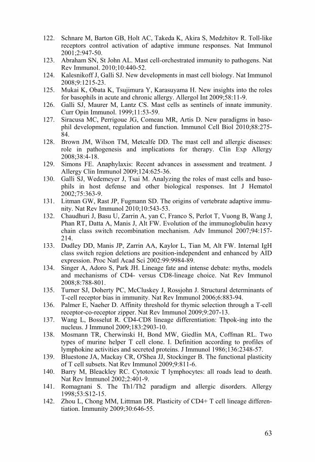
63
122. Schnare M, Barton GB, Holt AC, Takeda K, Akira S, Medzhitov R. Toll-like receptors control activation of adaptive immune responses. Nat Immunol 2001;2:947-50.
123. Abraham SN, St John AL. Mast cell-orchestrated immunity to pathogens. Nat Rev Immunol. 2010;10:440-52.
124. Kalesnikoff J, Galli SJ. New developments in mast cell biology. Nat Immunol 2008;9:1215-23.
125. Mukai K, Obata K, Tsujimura Y, Karasuyama H. New insights into the roles for basophils in acute and chronic allergy. Allergol Int 2009;58:11-9.
126. Galli SJ, Maurer M, Lantz CS. Mast cells as sentinels of innate immunity. Curr Opin Immunol. 1999;11:53-59.
127. Siracusa MC, Perrigoue JG, Comeau MR, Artis D. New paradigms in baso-phil development, regulation and function. Immunol Cell Biol 2010;88:275-84.
128. Brown JM, Wilson TM, Metcalfe DD. The mast cell and allergic diseases: role in pathogenesis and implications for therapy. Clin Exp Allergy 2008;38:4-18.
129. Simons FE. Anaphylaxis: Recent advances in assessment and treatment. J Allergy Clin Immunol 2009;124:625-36.
130. Galli SJ, Wedemeyer J, Tsai M. Analyzing the roles of mast cells and baso-phils in host defense and other biological responses. Int J Hematol 2002;75:363-9.
131. Litman GW, Rast JP, Fugmann SD. The origins of vertebrate adaptive immu-nity. Nat Rev Immunol 2010;10:543-53.
132. Chaudhuri J, Basu U, Zarrin A, yan C, Franco S, Perlot T, Vuong B, Wang J, Phan RT, Datta A, Manis J, Alt FW. Evolution of the immunoglobulin heavy chain class switch recombination mechanism. Adv Immunol 2007;94:157-214.
133. Dudley DD, Manis JP, Zarrin AA, Kaylor L, Tian M, Alt FW. Internal IgH class switch region deletions are position-independent and enhanced by AID expression. Proc Natl Acad Sci 2002:99:9984-89.
134. Singer A, Adoro S, Park JH. Lineage fate and intense debate: myths, models and mechanisms of CD4- versus CD8-lineage choice. Nat Rev Immunol 2008;8:788-801.
135. Turner SJ, Doherty PC, McCluskey J, Rossjohn J. Structural determinants of T-cell receptor bias in immunity. Nat Rev Immunol 2006;6:883-94.
136. Palmer E, Naeher D. Affinity threshold for thymic selection through a T-cell receptor-co-receptor zipper. Nat Rev Immunol 2009;9:207-13.
137. Wang L, Bosselut R. CD4-CD8 lineage differentiation: Thpok-ing into the nucleus. J Immunol 2009;183:2903-10.
138. Mosmann TR, Cherwinski H, Bond MW, Giedlin MA, Coffman RL. Two types of murine helper T cell clone. I. Definition according to profiles of lymphokine activities and secreted proteins. J Immunol 1986;136:2348-57.
139. Bluestone JA, Mackay CR, O'Shea JJ, Stockinger B. The functional plasticity of T cell subsets. Nat Rev Immunol 2009;9:811-6.
140. Barry M, Bleackley RC. Cytotoxic T lymphocytes: all roads lead to death. Nat Rev Immunol 2002;2:401-9.
141. Romagnani S. The Th1/Th2 paradigm and allergic disorders. Allergy 1998;53:S12-15.
142. Zhou L, Chong MM, Littman DR. Plasticity of CD4+ T cell lineage differen-tiation. Immunity 2009;30:646-55.

64
143. Bonneville M, O'Brien RL, Born WK. Gammadelta T cell effector functions: a blend of innate programming and acquired plasticity. Nat Rev Immunol 2010;10:467-78.
144. Cua DJ, Tato CM. Innate IL-17-producing cells: the sentinels of the immune system. Nat Rev Immunol 2010;10:479-89.
145. Weaver CT, Harrington LE, Mangan PR, Gavrieli M, Murphy KM. Th17: an effector CD4 T cell lineage with regulatory T cell ties. Immunity 2006;24:677-88.
146. Sakaguchi S, Yamaguchi T, Nomura T, Ono M. Regulatory T cells and im-mune tolerance. Cell 2008;133:775-87.
147. Sakaguchi S, Miyara M, Costantino CM, Hafler DA. FOXP3+ regulatory T cells in the human immune system. Nat Rev Immunol 2010;10:490-500.
148. Weaver CT, Hatton RD. Interplay between the TH17 and TReg cell lineages: a (co-) evolutionary perspective. Nat Rev Immunol 2009;9:883-9.
149. Zheng W, Flavell RA. The transcription factor GATA-3 is necessary and sufficient for Th2 cytokine gene expression in CD4 T cells. Cell 1997;89:587-96.
150. Langrish CL, Chen Y, Blumenschein WM, Mattson J, Basham B, Sedgwick JD, McClanahan T, Kastelein RA, Cua DJ. IL-23 drives a pathogenic T cell population that induces autoimmune inflammation. J Exp Med 2005;201:233-40.
151. Molet S, Hamid Q, Davoine F, Nutku E, Taha R, Pagé N, Olivenstein R, Elias J, Chakir J. IL-17 is increased in asthmatic airways and induces human bron-chial fibroblasts to produce cytokines. J Allergy Clin Immunol 2001;108:430-438.
152. Paul Ehrlich the Nobel Prize in Medicin 1908. www.nobelprize.org. 153. Wenzel SE. Eosinophils in asthma–closing the loop or opening the door?
NEJM 2009;360:1026-28. 154. Hogan SP, Rosenberg HF, Moqbel R, Phipps S, Foster PS, Lacy P, Kay AB,
Rothenberg ME. Eosinophils: biological properties and role in health and dis-ease. Clin Exp Allergy 2008;38:709-50.
155. Venge P. The eosinophil and airway remodelling in asthma. Clin Respir J 2010;4 (S1):15-9.
156. Blanchard C, Rothenberg ME. Biology of the eosinophil. Adv Immunol 2009;101:81-121.
157. Rothenberg ME, Hogan SP. The eosinophil. Annu Rev Immunol 2006;24:147-74.
158. Venge P, Bystrom J, Carlson M, Hakansson L, Karawacjzyk M, Peterson C, Seveus L, Trulson A. Eosinophil cationic protein (ECP): molecular and bio-logical properties and the use of ECP as a marker of eosinophil activation in disease. Clin Exp Allergy 1999;29:1172-86.
159. Akuthotam P, Wang H, Weller PF. Eosinophils as antigen-presenting cells in allergic upper airway disease. Curr Opin Allergy Clin Immunol 2010;10:14-19.
160. Rosenberg HF, Phipps S, Foster PS. Eosinophil trafficking in allergy and asthma. J Allergy Clin Immunol 2007;119:1303-10.
161. Hogan SP. Recent advances in eosinophil biology. Int Arch Allergy Immunol 2007;143 (S1) 3-14.
162. Kato M, Kephart GM, Talley NJ, Wagner JM, Sarr MG, Bonno M, McGovern TW, Gleich GJ. Eosinophil infiltration and degranulation in nor-mal human tissue. Anat Rec 1998;252:418-25.
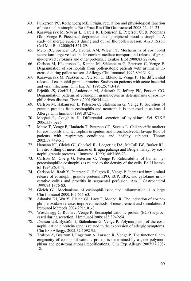
65
163. Fulkerson PC, Rothenberg ME. Origin, regulation and physiological function of intestinal eosinophils. Best Pract Res Clin Gastroenerol 2008;22:411-23.
164. Karawajczyk M, Sevéus L, Garcia R, Björnsson E, Peterson CGB, Roomans GM, Venge P. Piecemeal degranulation of peripheral blood eosinophils: A study of allergic subjects during and out of the pollen season. Am J Respir Cell Mol Biol 2000;34:521-29.
165. Melo RC, Spencer LA, Dvorak AM, Wleer PF. Mechanisms of eosinophil sectretion: large vesicotubular carriers mediate transport and release of gran-ule-derived cytokines and other proteins. J Leukoc Biol 2008;83:229-36.
166. Carlson M, Håkansson L, Kämpe M, Stålenheim G, Peterson C, Venge P. Degranulation of eosinophils from pollen-atopic patients with asthma is in-creased during pollen season. J Allergy Clin Immunol 1992;89:131-9.
167. Karawajczyk M, Pauksen K, Peterson C, Eklund E, Venge P. The differential release of eosinophil granule proteins. Studies on patients with acute bacterial and viral infections. Clin Exp All 1995;25:713-19.
168. Erjefält JS, Greiff L, Andersson M, Ädelroth E, Jeffery PK, Persson CG. Degranulation patterns of eosinophil granulocytes as determinants of eosino-phil driven disease. Thorax 2001;56:341-44.
169. Carlson M, Håkansson L, Peterson C, Stålenheim G, Venge P. Secretion of granule proteins from eosinophils and neutrophils is increased in asthma. J Allergy Clin Immunol 1991;87:27-33.
170. Moqbel R, Coughlin JJ. Differential secretion of cytokines. Sci STKE 2006;338:pe26.
171. Metso T, Venge P, Haahtela T, Peterson CG, Sevéus L. Cell specific markers for eosinophils and neutrophils in sputum and bronchoalveolar lavage fluid of patients with respiratory conditions and healthy subjects. Thorax 2002;57:449-51.
172. Hamann KJ, Gleich GJ, Checkel JL, Loegering DA, McCall JW, Barker RL. In vitro killing of microfilariae of Brugia pahangi and Brugia malayi by eosi-nophil granule proteins. J Immunol 1990;144:3166-73.
173. Carlson M, Oberg G, Peterson C, Venge P. Releasability of human hy-pereosinophilic eosinophils is related to the density of the cells. Br J Haema-tol 1994;86:41-7.
174. Carlson M, Raab Y, Peterson C, Hällgren R, Venge P. Increased intraluminal release of eosinophil granule proteins EPO, ECP, EPX, and cytokines in ul-cerative colitis and proctitis in segmental perfusion. Am J Gastroenterol 1999;94:1876-83.
175. Gleich GJ. Mechanisms of eosinophil-associated inflammation. J Allergy Clin Immunol 2000;105:651-63.
176. Adamko DJ, Wu Y, Gleich GJ, Lacy P, Moqbel R. The induction of eosino-phil peroxidase release: improved methods of measurement and stimulation. J Immunol Methods 2004;291:101-8.
177. Woschnagg C, Rubin J, Venge P. Eosinophil cationic protein (ECP) is proc-essed during secretion. J Immunol 2009;183:3949-54.
178. Jönsson UB, Byström J, Stålenheim G, Venge P. Polymorphism of the eosi-nophil cationic protein-gene is related to the expression of allergic symptoms. Clin Exp Allergy. 2002;32:1092-95.
179. Trulson A, Byström J, Engström A, Larsson R, Venge P. The functional het-erogeneity of eosinophil cationic protein is determined by a gene polymor-phism and post-translational modifications. Clin Exp Allergy 2007;37:208-18.
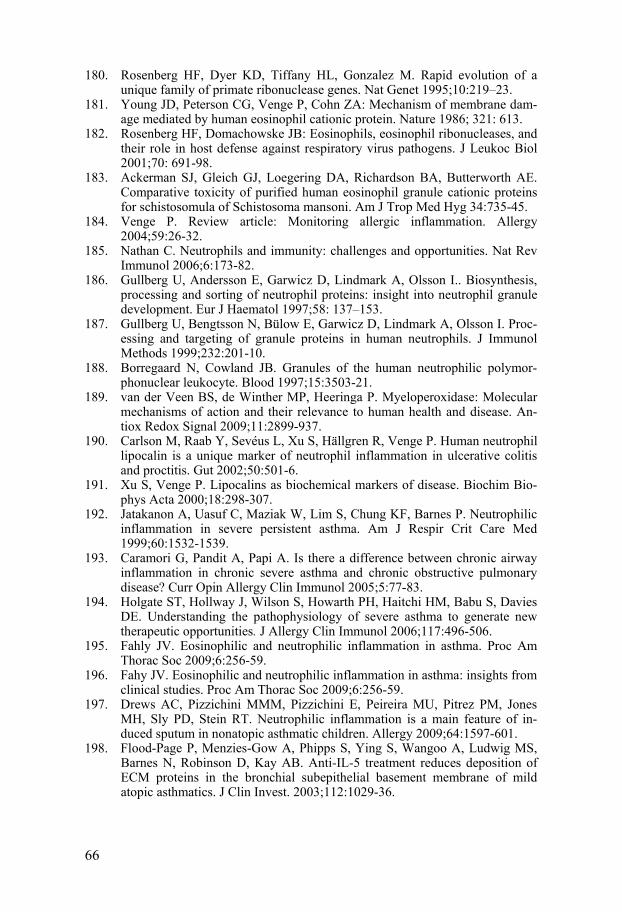
66
180. Rosenberg HF, Dyer KD, Tiffany HL, Gonzalez M. Rapid evolution of a unique family of primate ribonuclease genes. Nat Genet 1995;10:219–23.
181. Young JD, Peterson CG, Venge P, Cohn ZA: Mechanism of membrane dam-age mediated by human eosinophil cationic protein. Nature 1986; 321: 613.
182. Rosenberg HF, Domachowske JB: Eosinophils, eosinophil ribonucleases, and their role in host defense against respiratory virus pathogens. J Leukoc Biol 2001;70: 691-98.
183. Ackerman SJ, Gleich GJ, Loegering DA, Richardson BA, Butterworth AE. Comparative toxicity of purified human eosinophil granule cationic proteins for schistosomula of Schistosoma mansoni. Am J Trop Med Hyg 34:735-45.
184. Venge P. Review article: Monitoring allergic inflammation. Allergy 2004;59:26-32.
185. Nathan C. Neutrophils and immunity: challenges and opportunities. Nat Rev Immunol 2006;6:173-82.
186. Gullberg U, Andersson E, Garwicz D, Lindmark A, Olsson I.. Biosynthesis, processing and sorting of neutrophil proteins: insight into neutrophil granule development. Eur J Haematol 1997;58: 137–153.
187. Gullberg U, Bengtsson N, Bülow E, Garwicz D, Lindmark A, Olsson I. Proc-essing and targeting of granule proteins in human neutrophils. J Immunol Methods 1999;232:201-10.
188. Borregaard N, Cowland JB. Granules of the human neutrophilic polymor-phonuclear leukocyte. Blood 1997;15:3503-21.
189. van der Veen BS, de Winther MP, Heeringa P. Myeloperoxidase: Molecular mechanisms of action and their relevance to human health and disease. An-tiox Redox Signal 2009;11:2899-937.
190. Carlson M, Raab Y, Sevéus L, Xu S, Hällgren R, Venge P. Human neutrophil lipocalin is a unique marker of neutrophil inflammation in ulcerative colitis and proctitis. Gut 2002;50:501-6.
191. Xu S, Venge P. Lipocalins as biochemical markers of disease. Biochim Bio-phys Acta 2000;18:298-307.
192. Jatakanon A, Uasuf C, Maziak W, Lim S, Chung KF, Barnes P. Neutrophilic inflammation in severe persistent asthma. Am J Respir Crit Care Med 1999;60:1532-1539.
193. Caramori G, Pandit A, Papi A. Is there a difference between chronic airway inflammation in chronic severe asthma and chronic obstructive pulmonary disease? Curr Opin Allergy Clin Immunol 2005;5:77-83.
194. Holgate ST, Hollway J, Wilson S, Howarth PH, Haitchi HM, Babu S, Davies DE. Understanding the pathophysiology of severe asthma to generate new therapeutic opportunities. J Allergy Clin Immunol 2006;117:496-506.
195. Fahly JV. Eosinophilic and neutrophilic inflammation in asthma. Proc Am Thorac Soc 2009;6:256-59.
196. Fahy JV. Eosinophilic and neutrophilic inflammation in asthma: insights from clinical studies. Proc Am Thorac Soc 2009;6:256-59.
197. Drews AC, Pizzichini MMM, Pizzichini E, Peireira MU, Pitrez PM, Jones MH, Sly PD, Stein RT. Neutrophilic inflammation is a main feature of in-duced sputum in nonatopic asthmatic children. Allergy 2009;64:1597-601.
198. Flood-Page P, Menzies-Gow A, Phipps S, Ying S, Wangoo A, Ludwig MS, Barnes N, Robinson D, Kay AB. Anti-IL-5 treatment reduces deposition of ECM proteins in the bronchial subepithelial basement membrane of mild atopic asthmatics. J Clin Invest. 2003;112:1029-36.
67
199. Melo RC, Weller PF. Piecemeal degranulation in human eosinophils: a dis-tinct secretion mechanism underlying inflammatory responses. Histol Histo-pathol 2010;25:1341-54.
200. Melo RC, Spencer LA, Perez SA, Neves JS, Bafford SP, Morgan ES, Dvorak AM, Weller PF. Vesicle-mediated secretion of human eosinophil granule-derived major basic protein. Lab Invest 2009;89:769-81.
201. Winqvist I, Olofsson T, Olsson I. Mechanisms for eosinophil degranulation; release of the eosinophil cationic protein. Immunology 1984;51:1-8.
202. Egesten A, Malm J. Eosinophil leukocyte degranulation in response to serum opsonized beads: C5a and platelet-activating factor enhance ECP release, with roles for protein kinases A and C. Allergy 1998;53:1066-73
203. Tai PC, Spry CJ. The effect of recombinant granulocyte-macrophage colony-stimulating factor (GM-CSF) and interleukin 3 on the secretory capacity of human blood eosinophils. Clin Exp Immunol 1990;80:426-434.
204. Tomassini M, Tsicopoulos A, Tai PC, Gruart V, Tonnel AB, Prin L, Capron A, Capron M. Release of granule proteins by eosinophils from allergic and non-allergic patients with eosinophilia on immunoglobulin-dependent activa-tion. J Allergy Clin Immunol 1991;88:365-75.
205. Carlson M, Peterson C, Venge P. The influence of IL-3, IL-5, and GM-CSF on normal human eosinophil and neutrophil C3b-induced degranulation. Al-lergy 1993, 8:437-442.
206. Hawkins PT, Anderson KE, Davidson K, Stephens LR. Signalling through Class I PI3Ks in mammalian cells. Biochem Soc Trans 2006;34:647-62.
207. Welch HC, Coadwell WJ, Stephens LR, Hawkins PT. Phosphoinositide 3-kinase-dependent activation of Rac. FEBS Lett 2003;546:93-7.
208. Kok K, Geering B, Vanhaesebroeck B. Regulation of phosphoinositide 3-kinase expression in health and disease. Trends Biochem Sci 2009;34:115-27.
209. Amzel LM, Huang CH, Mandelker D, Lengauer C, Gabelli SB, Vogelstein B. Structural comparisons of class I phosphoinositide 3-kinases. Nat Rev Cancer 2008;8:665-69.
210. Medina-Tato DA, Ward SG, Watson ML. Phosphoinositide 3-kinase signal-ling in lung disease: leucocytes and beyond. Immunology 2007;121:448-61.
211. Lindmo K, Stenmark H. Regulation of membrane traffic by phosphoinositide 3-kinases. J Cell Sci 2006;119:605-14.
212. Ali K, Bilancio A, Thomas M, Pearce W, Gilfillan AM, Tkaczyk C, Kuehn N, Gray A, Giddings J, Peskett E, Fox R, Bruce I, Walker C, Sawyer C, Ok-kenhaug K, Finan P, Vanhaesebroeck B. Essential role for the p110delta phosphoinositide 3-kinase in the allergic response. Nature 2004;431:1007-11.
213. Ito N, Yokomizo T, Sasaki T, Kurosu H, Penninger J, Kanaho Y, Katada T, Hanaoka K, Shimizu T. Requirement of phosphatidylinositol 3-kinase activa-tion and calcium influx for leukotriene B4-induced enzyme release. J Biol Chem 2002;277:44898-904.
214. Pinho V, Souza DG, Barsante MM, Hamer FP, De Freitas MS, Rossi AG, Teixeira MM. Phosphoinositide-3 kinases critically regulate the recruitment and survival of eosinophils in vivo: importance for the resolution of allergic inflammation. J Leukoc Biol 2005;77:800-10.
215. Woscholski R, Kodaki T, McKinnon M, Waterfield MD, Parker PJ. A com-parison of demethoxyviridin and wortmannin as inhibitors of phosphatidy-linositol 3-kinase. FEBS Lett 1994;342:109-14.
216. Powis G, Bonjouklian R, Berggren MM, Gallegos A, Abraham R, Ashendel C, Zalkow L, Matter WF, Dodge J, Grindey G, et al. Wortmannin, a potent
68
and selective inhibitor of phosphatidylinositol-3-kinase. Cancer Res 1994;54:2419-23.
217. Ogden EC, Raynor GS, Haynes JV, Lewis DM, Haine JH. Manual for sam-pling airborne pollen. New York: Haftner Press, 1974:1-182.
218. Quanjer PH, Tammeling GJ, Cotes JE, Pedersen OF, Peslin R, Yernault JC. Lung volumes and forced ventilatory flows. Report Working Party Standardi-zation of Lung Function Tests, European Community for Steel and Coal. Of-ficial Statement of the European Respiratory Society. Eur Respir J 1993;(S)16:5-40.
219. Wålinder R, Norbäck D, Wieslander G, Smedje G, Erwall C, Venge P. Nasal patency and biomarkers in nasal lavage--the significance of air exchange rate and type of ventilation in schools. Int Arch Occup Environ Health 1998;71:479-86.
220. Pizzichini MM, Pizzichini E, Clelland L, Efthimiadis A, Mahony J, Dolovich J, Hargreave FE. Sputum in severe exacerbations of asthma: kinetics of in-flammatory indices after prednisone treatment. Am J Respir Crit Care Med 1997;155:1501-8.
221. Machado L. Increased bronchial hypersensitivity after early and late bron-chial reacations provoked by allergen inhalation. Allergy.1985;40:580-85.
222. Hansell TT, De Vries IJ, Iff T, Rihs S, Wandzalik M, Betz S, Blaser K, Walker C. An improved immunomagnetic procedure for the isolation of highly purified human blood eosinophils. J Immunol Methods 1991, 145:105-110.
223. Winqvist I, Olofsson T, Olsson I. Mechanisms for eosinophil degranulation; release of the eosinophil cationic protein. Immunology. 1984;51:1-8.
224. Xu SY, Petersson CG, Carlson M, Venge P. The development of an assay for human neutrophil lipocalin (HNL)--to be used as a specific marker of neutro-phil activity in vivo and vitro. J Immunol Methods 1994;171:245-52.
225. Compalati E, Ridolo E, Passalacqua G, Braido F, Villa E, Canonica GW. The link between allergic rhinitis and asthma: the united airways disease. Expert Rev Clin Immunol 2010;6:413-23.
226. Holgate ST. A look at the pathogenesis of asthma: the need for a change in direction. Discov Med 2010;9:439-47.
227. Roquet A, Ihre E, van Hage-Hamsten M, Halldén G, Zettterström O. Aller-gen-induced inflammation in the nose: a comparison of acute and repeated low-dose allergen exposure. Allergy1996;51:42-8.
228. Keen C, Johansson S, Reinholdt J, Benson M, Wennergren G. Bet v 1-specifik IgA increases during the pollen season but not after a single allergen challenge in children with birch pollen-induced intermittent allergic rhinitis. Ped Allergy Immunol 2005;16:209-16.
229. Thomas M, Owen C. Inhibition of PI-3 kinase for treating respiratory disease: good idea or bad idea? Curr Opin Pharmacol 2008;8:267-74.
230. Carlson M, Venge P, Lampinen M. C3b-induced eosinophil degranulation involves PI3-kinases and is inhibited by PKC activity. Revised and resubmit-ted 2010.
231. Bjermer L. Time for a paradigm shift in asthma treatment: from relieving bronchospasm to controlling systemic inflammation. J Allergy Clin Immunol 2007;120:1269-75.
232. Aronsson D, Tufvesson E, Ankerst J, Bjermer L. Allergic rhinitis with hyper-responsiveness differ from asthma in degree of peripheral obstruction during metacholine challenge test. Clin Physiol Funct Imaging 2008;28:81-5.
69
233. Tufvesson E, Aronsson D, Ankerst J, George SC, Bjermer L. Peripheral nitric oxide is increased in rhinitic patients with asthma compared to bronchial hy-perresponsiveness. Respir Med 2007;101:2321-26.
234. Flood-Page PT, Menzies-Gow AN, Kay AB, Robinson DS. Eosinophil's role remains uncertain as anti-interleukin-5 only partially depletes numbers in asthmatic airway. Am J Respir Crit Care Med 2003;167:199-204.
235. O'Byrne PM. The demise of anti IL-5 for asthma, or not. Am J Respir Crit Care Med 2007;176:1059-60.
236. Rådinger M, Lötvall J. Eosinophil progenitors in allergy and asthma - do they matter? Pharmacol Ther 2009;12:174-84.
237. Janson C. The importance of airway remodelling in the natural course of asthma. Clin Respir J 2010;4(S1):28-34.
238. Davies DE. The role of the epithelium in airway remodeling in asthma. Proc Am Thorac Soc 2009;6:678-82.
239. Bousquet J, Chiron R, Humbert M. Biologics in asthma: difficulties and drawbacks. Expert Opin Biol Ther 2008;8:1921-8.
240. Bousquet J, Burney PG, Zuberbier T, Cauwenberge PV, Akdis CA, Bindslev-Jensen C, Bonini S, Fokkens WJ, Kauffmann F, Kowalski ML, Lodrup-Carlsen K, Mullol J, Nizankowska-Mogilnicka E, Papadopoulos N, Toskala E, Wickman M, Anto J, Auvergne N, Bachert C, Bousquet PJ, Brunekreef B, Canonica GW, Carlsen KH, Gjomarkaj M, Haahtela T, Howarth P, Lenzen G, Lotvall J, Radon K, Ring J, Salapatas M, Schünemann HJ, Szczecklik A, Todo-Bom A, Valovirta E, von Mutius E, Zock JP. GA2LEN (Global Allergy and Asthma European Network) addresses the allergy and asthma 'epidemic'. Allergy 2009;64:969-77.