Embed Size (px)
Citation preview
RESULTS All 143 patients of this study were male veterans,
with a mean age of 61 years. The patients were extracted from all hospital services to provide a comprehensive view of the problem of I-VASC device, blood-borne infection. Eighty-seven patients from the Medical Ser- vice accounted for 94 episodes of infection; 55 Surgical Service patients had 64 episodes of infection; and a single patient with a single episode of infection was the Spinal Cord Injury Service. Among medical patients, 30 episodes (32%) occurred in the medical intensive care unit, whereas, in surgical patients, 30 episodes (47%) occurred in the surgical intensive care unit. Among patients who had multiple episodes (two different de- vices at separate times with separate blood cultures), 10 patients had 2 separate episodes of infection, and 3 patients had 3 episodes.
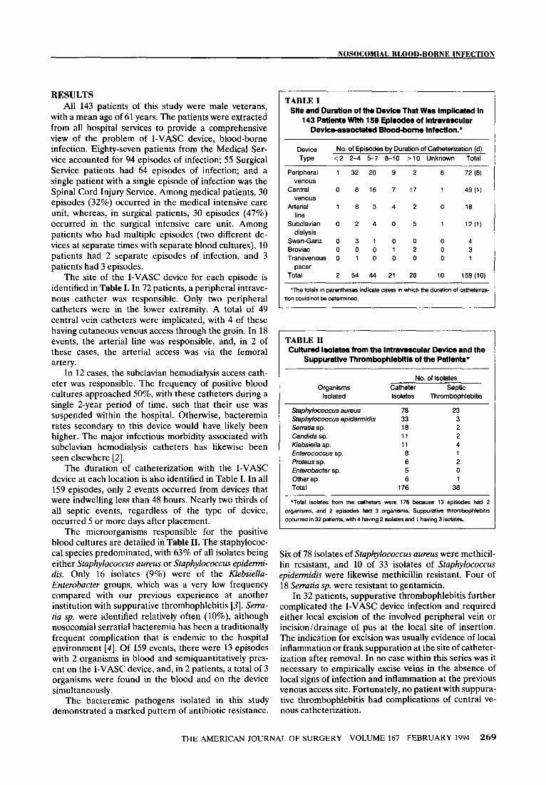
The site of the I-VASC device for each episode is identified in Table I. In 72 patients, a peripheral intrave- nous catheter was responsible. Only two peripheral catheters were in the lower extremity. A total of 49 central vein catheters were implicated, with 4 of these having cutaneous venous access through the groin. In 18 events, the arterial line was responsible, and, in 2 of these cases, the arterial access was via the femoral artery.
In 12 cases, the subclavian hemodialysis access cath- eter was responsible. The frequency of positive blood cultures approached 50%, with these catheters during a single 2-year period of time, such that their use was suspended within the hospital. Otherwise, bacteremia rates secondary to this device would have likely been higher. The major infectious morbidity associated with subclavian hemodialysis catheters has likewise been seen elsewhere [2].
The duration of catheterization with the I-VASC device at each location is also identified in Table I. In all 159 episodes, only 2 events occurred from devices that were indwelling less than 48 hours. Nearly two thirds of all septic events, regardless of the type of device, occurred 5 or more days after placement.
The microorganisms responsible for the positive blood cultures are detailed in Table II. The staphylococ- cal species predominated, with 63% of all isolates being either Staphylococcus aureus or Staphylococcus epidemi- dis. Only 16 isolates (9%) were of the Klebsiella- Enterobacter groups, which was a very low frequency compared with our previous experience at another institution with suppurative thrombophlebitis [3]. Serra- tia sp. were identified relatively often (lo%), although nosocomial serratial bacteremia has been a traditionally frequent complication that is endemic to the hospital environment [4]. Of 159 events, there were 13 episodes with 2 organisms in blood and semiquantitatively pres- ent on the I-VASC device, and, in 2 patients, a total of 3 organisms were found in the blood and on the device simultaneously.
The bacteremic pathogens isolated in this study demonstrated a marked pattern of antibiotic resistance.
TABLE I Site and Duration of the Device That Was Implicated In
143 Patients With 159 Episodes of intravascular Device-associated Blood&one InfectIon.*
Device No. of Episodes by Duration of Catheterization (d)
Type <2 24 5-7 6-10 >I0 Unknown Total
Peripheral venous
Central
venous
Arterial line
Subclavian dialysis
Swan-Ganz
Broviac Transvenous
pacer
Total
1 32 20 9 2 8
0 6 16 7 17 1
i a 3 4 2 0
0 2 4 0 5 1
0 3 1 0 0 0 00012 0
0 1 0 0 0 0
2 54 44 21 28 IO
72 (6)
49 (1)
ia
12 (1)
4
3 1
159 (IO)
‘The totals in parentheses indicate cases in which the duration of catheteriza-
tion could not be determined.
I
TABLE II Cultured isolates from the Intravascular Devlce and the
Suppurative Thrombophlebltls of the Patlen&*
Organisms
Isolated
Staphylococcus aureus Staphylococcus epidermidis
Serratia sp.
Candida sp.
Klebsiella sp. Enterococcus sp.
Proteus sp.
Enterobacter sp.
Other sp. Total
No. of isolates
Catheter Septic
Isolates Thrombophlebitis
78 23 33 3 ia 2
11 2 11 4
a 1 6 2
5 0 6 1
176 38
*Total isolates from the catheters were 176 because 13 episodes had 2
organisms, and 2 episodes had 3 organisms. Suppurative thrombophlebiiis
occurred in 32 patbnts, with 4 having 2 isolates and 1 having 3 isolates.
1
1
I
Six of 78 isolates of Staphylococcus aureus were methicil- lin resistant, and 10 of 33 isolates of Staphylococcus epidemidis were likewise methicillin resistant. Four of 18 Serratia sp. were resistant to gentamicin.
In 32 patients, suppurative thrombophlebitis further complicated the I-VASC device infection and required either local excision of the involved peripheral vein or incision/drainage of pus at the local site of insertion. The indication for excision was usually evidence of local inflammation or frank suppuration at the site of catheter- ization after removal. In no case within this series was it necessary to empirically excise veins in the absence of local signs of infection and inflammation at the previous venous access site. Fortunately, no patient with suppura- tive thrombophlebitis had complications of central ve- nous catheterization.
THE AMERICAN JOURNAL OF SURGERY VOLUME 167 FEBRUARY 1994 269
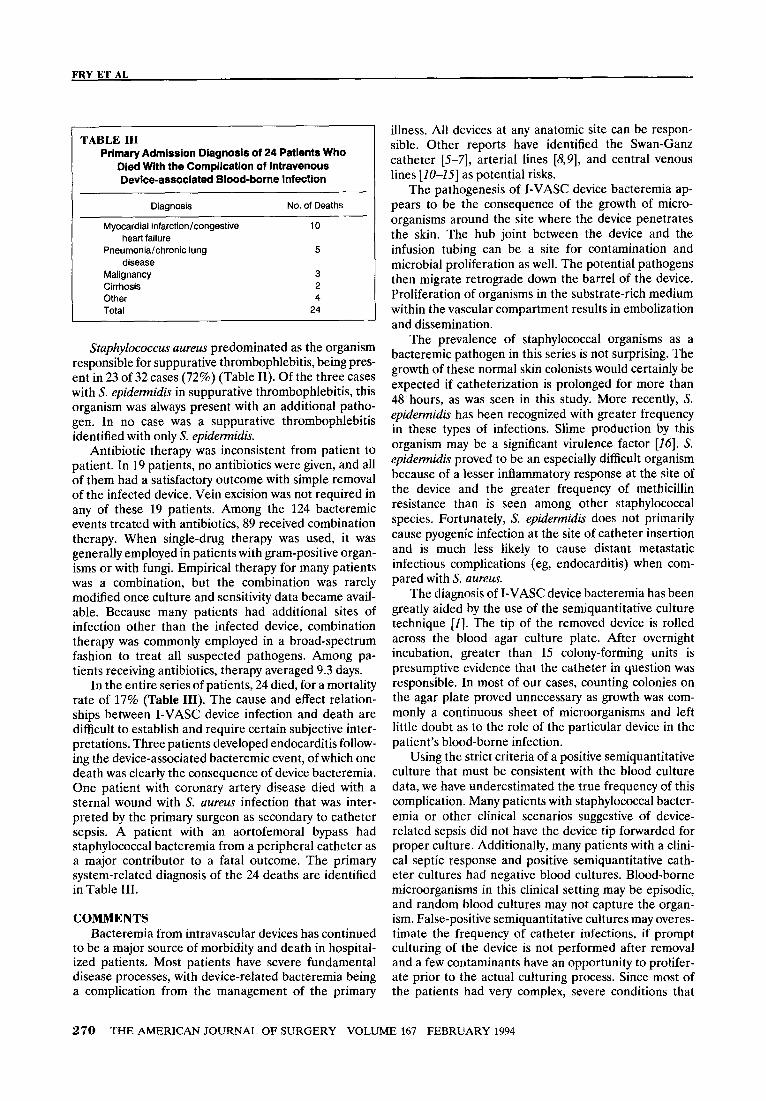
TABLE III Primary Admission Diagnosis of 24 Patients Who
Died With the Complication of Intravenous Device-associated Blood-borne Infection
Diagnosis
Myocardial infarction/congestive
heart failure Pneumonia/chronic lung
disease Malignancy Cirrhosis
Other
Total
No. of Deaths
10
5
3
2 4
24
Staphylococcus aureus predominated as the organism responsible for suppurative thrombophlebitis, being pres- ent in 23 of 32 cases (72%) (Table II). Of the three cases with S. epidemidis in suppurative thrombophlebitis, this organism was always present with an additional patho- gen. In no case was a suppurative thrombophlebitis identified with only S. epidetmidis.
Antibiotic therapy was inconsistent from patient to patient. In 19 patients, no antibiotics were given, and all of them had a satisfactory outcome with simple removal of the infected device. Vein excision was not required in any of these 19 patients. Among the 124 bacteremic events treated with antibiotics, 89 received combination therapy. When single-drug therapy was used, it was generally employed in patients with gram-positive organ- isms or with fungi. Empirical therapy for many patients was a combination, but the combination was rarely modified once culture and sensitivity data became avail- able. Because many patients had additional sites of infection other than the infected device, combination therapy was commonly employed in a broad-spectrum fashion to treat all suspected pathogens. Among pa- tients receiving antibiotics, therapy averaged 9.3 days.
In the entire series of patients, 24 died, for a mortality rate of 17% (Table III). The cause and effect relation- ships between I-VASC device infection and death are difficult to establish and require certain subjective inter- pretations. Three patients developed endocarditis follow- ing the device-associated bacteremic event, of which one death was clearly the consequence of device bacteremia. One patient with coronary artery disease died with a sternal wound with S. aureus infection that was inter- preted by the primary surgeon as secondary to catheter sepsis. A patient with an aortofemoral bypass had staphylococcal bacteremia from a peripheral catheter as a major contributor to a fatal outcome. The primary system-related diagnosis of the 24 deaths are identified in Table III.
COMMENTS Bacteremia from intravascular devices has continued
to be a major source of morbidity and death in hospital- ized patients. Most patients have severe fundamental disease processes, with device-related bacteremia being a complication from the management of the primary
illness. All devices at any anatomic site can be respon- sible. Other reports have identified the Swan-Ganz catheter [5-q, arterial lines [8,9], and central venous lines [20-251 as potential risks.
The pathogenesis of I-VASC device bacteremia ap- pears to be the consequence of the growth of micro- organisms around the site where the device penetrates the skin. The hub joint between the device and the infusion tubing can be a site for contamination and microbial proliferation as well. The potential pathogens then migrate retrograde down the barrel of the device. Proliferation of organisms in the substrate-rich medium within the vascular compartment results in embolization and dissemination.
The prevalence of staphylococcal organisms as a bacteremic pathogen in this series is not surprising. The growth of these normal skin colonists would certainly be expected if catheterization is prolonged for more than 48 hours, as was seen in this study. More recently, S. epidemidis has been recognized with greater frequency in these types of infections. Slime production by this organism may be a significant virulence factor [26]. S. epidemidis proved to be an especially difficult organism because of a lesser inflammatory response at the site of the device and the greater frequency of methicillin resistance than is seen among other staphylococcal species. Fortunately, S. epidermidis does not primarily cause pyogenic infection at the site of catheter insertion and is much less likely to cause distant metastatic infectious complications (eg, endocarditis) when com- pared with S. aureus.
The diagnosis of I-VASC device bacteremia has been greatly aided by the use of the semiquantitative culture technique [I]. The tip of the removed device is rolled across the blood agar culture plate. After overnight incubation, greater than 15 colony-forming units is presumptive evidence that the catheter in question was responsible. In most of our cases, counting colonies on the agar plate proved unnecessary as growth was com- monly a continuous sheet of microorganisms and left little doubt as to the role of the particular device in the patient’s blood-borne infection.
Using the strict criteria of a positive semiquantitative culture that must be consistent with the blood culture data, we have underestimated the true frequency of this complication. Many patients with staphylococcal bacter- emia or other clinical scenarios suggestive of device- related sepsis did not have the device tip forwarded for proper culture. Additionally, many patients with a clini- cal septic response and positive semiquantitative cath- eter cultures had negative blood cultures. Blood-borne microorganisms in this clinical setting may be episodic, and random blood cultures may not capture the organ- ism. False-positive semiquantitative cultures may overes- timate the frequency of catheter infections, if prompt culturing of the device is not performed after removal and a few contaminants have an opportunity to prolifer- ate prior to the actual culturing process. Since most of the patients had very complex, severe conditions that
270 THE AMERICAN JOURNAL OF SURGERY VOLUME 167 FEBRUARY 1994
NOSOCOMIAL BLOOD-BORNE INFECTION
were commonly associated with infections, a positive semiquantitative catheter culture, but without a positive blood culture, could not be assumed to be responsible for signs of infection that may have been identified in the patient.
Obtaining a direct Gram’s stain of the catheter itself and examination under oil immersion microscopy has been advocated as a rapid method for diagnosis of device sepsis [17]. This method is probably quite effec- tive for those patients in whom semiquantitative culture shows overwhelming growth. For more subtle cases when colony counts are significantly less, this method will be less sensitive. Regardless of the diagnostic modality employed, a surgical patient with positive staphylococcal blood cultures (whether S. aureus or S. epidennidis) in the absence of a gram-positive soft tissue infection must be considered as having a device-related bacteremia until proven otherwise. All indwelling de- vices need to be removed, culture must be obtained, and appropriate systemic antibiotic therapy needs to be initiated.
reduce catheter-associated infection [23]. A silver im- pregnated cuff of the catheter may also be useful [24]. In the final analysis, the prevention of I-VASC device sepsis is aseptic placement of the device, 72-hour rota- tion of peripheral catheters, and meticulous day-to-day care of arterial and central lines. The alternative meth- ods that employ antibiotic bonding, topical antiseptic agents, and silver bonding to the catheter will need further evaluation.
The duration of catheterization among our patients was certainly the single most important variable in this complication. The routine removal and replacement of peripheral intravenous devices every 72 hours would have been a cost-effective means to eliminate this infectious complication [18]. Arterial line bacteremia likewise was a consequence of these catheters remaining in-place for more than 1 week in several patients. Because the arterial line is used for frequent blood sampling, it is subject to contamination far more com- monly than other devices. Although changing the arte- rial line in those patients truly in need of continuous pressure monitoring is not always practical, the arterial line must be managed with meticulous aseptic care if this complication is to be avoided. Similarly, central venous and pulmonary artery catheters must have dressings changed every 24 hours, and the skin about the insertion site must be meticulously cleansed and redressed in an aseptic fashion.
The management of catheter sepsis requires removal of the infected catheter, selected excision of suppurative thrombophlebitis, and specific antibiotic therapy. Re- moval of the catheter is of paramount importance. Careful inspection of the catheter site will usually provide sufficient evidence to proceed with vein exci- sion. Failure of the blood-borne infection to resolve after device removal suggests that either a suppurative vein needs to be excised or that a metastatic focus of infection already exists (eg, endocarditis). The routine excision of all intravenous sites implicated in catheter bacteremia is not necessary if local signs of inflammation are not present and if prompt clinical defervescence of the septic response attends catheter removal.
A more difficult problem involves the use of antibi- otic agents. Antibiotic coverage during the actual bacte- remit episode is obviously desirable with a drug to which the pathogen is sensitive. Our impression has been that antibiotic coverage for gram-negative or fungal species need only be for 48 to 72 hours provided clinical resolution of the infection is evident following removal of the device. Because of the propensity of S. aureus to be associated with distant metastatic infection, a full 7 days of systemic therapy is warranted for these infec- tions. The drug therapy must be tailored to the appropri- ate sensitivities of the pathogen. Sensitivities are espe- cially important in those patients with 5’. epidemidis because of the high frequency of methicillin resistance. In this latter group of patients, vancomycin therapy is currently the drug of choice.
Several innovative methods to prevent catheter sep- sis have been advocated. Antibiotic bonding to the catheter has been recommended for prevention [19,20] and may prove to be an effective method. The use of metal “butterfly” catheters rather than plastic catheters has been suggested as another method to prevent peripheral intravenous catheter sepsis, but, in a prospec- tive, randomized study, this was not proven to be valid [21]. All infected peripheral catheters in this study were synthetic indwelling catheters and were not the “butterfly” type of small metal devices.
The microbiology of the ill surgical patient in the 1990s continues to evolve. It seems that every patient is a forest of lines, and some of those lines do get infected. The special role of staphylococcal bacteremia in this setting means that presumptive intravascular device removal should be the second step in the diagnosis and treatment of the problem. A graded but conscious sequence of treatments seem best.
Numerous topical substances applied to the skin entrance site are employed to enhance antisepsis. A comparison of saline versus neosporin versus iodophor in a large prospective, randomized trial demonstrated fewer positive catheter cultures in those patients receiv- ing topical antibiotics [22]. Unfortunately, no difference in catheter-related sepsis was seen. More recent data indicate that chlorhexidine as a topical antiseptic may
REFERENCES 1. Maki DG, Weise CE, Sarafin HW. A semiquantitative culture method for identifying catheter-related infection. N Engl J Med 1977; 296: 1305-9. 2. Sherertz RJ, Falk RJ, Huffman KA, et al. Infections associated with subclavian Uldall catheters. Arch Intern Med 1983; 143: 52-6. 3. Garrison RN, Richardson JD, Fry DE. Catheter-associated
septic thrombophlebitis. South Med J 1982; 75: 917-9. 4. Fry DE, Fry RV, Shlaes DM. Serratial bacteremia in the
surgical patient. Am Surg 1987; 53: 438-41. 5. Elliott CG, Zimmerman GA, Clemmer TP. Complications of
THE AMERICAN JOURNAL OF SURGERY VOLUME 167 FEBRUARY 1994 271
pulmonary artery catheterization in the care of critically-ill pa- tients. Chest 1979; 76: 647-52. 6. Michel L, Marsh HM, McMichan JC, et al. Infection of pulmonary artery catheters in critically ill patients. JAMA 1981; 245: 1032-6. 7. Prachar H, Dittel M, Jobst C, el al. Bacterial contamination of pulmonary artery catheters. Int Care Med 1978; 4: 79-82. 8. Shinozaki T, Deane RS, Mazuzan JE, et al. Bacterial contami- nation of arterial lines. JAMA 1983; 249: 223-5. 9. Gardner RM, Schwartz R, Wong HC, et al. Percutaneous indwelling radial-artery catheters for monitoring cardiovascular function. N Engl J Med 1974; 290: 1227-31. 10. Ryan J, Abel R, Abbott W, et al. Catheter complications in total parenteral nutrition: a prospective study of 200 consecutive patients. N Engl J Med 1974; 290: 75761. 11. Sitzmann JV, Townsend TR, Siler MC, Bartlett JG. Septic and technical complications of central venous catheterization: a prospective study of 200 consecutive patients. Ann Surg 1985; 202: 766-70. 12. Bozzetti F. Central venous catheter sepsis. Surg Gynecol Obstet 1985; 161: 293-301. 13. Benezra D, Kiehn TE, Gold JWM, et al. Prospective study of infections in indwelling central venous catheters unsing quantita- tive blood cultures. Am J Med 1988; 85: 495-8. 14. Toltzis P, Goldmann DA. Current issues in central venous catheter infection. Annu Rev Med 1990; 41: 169-76. 15. Clarke DE, Raffin TA. Infectious complications of indwelling long-term central venous catheters. Chest 1990; 97: 966-72. 16. Kaebnick HW, Bandyk DF, Bergamini TW, Towne JB. The
microbiology of explanted vascular prostheses. Surgery 1987; 102: 756-62. 17. Cooper GL, Hopkins CC. Rapid diagnosis of intravascular catheter-associated infection by direct gram staining of catheter segments. N Engl J Med 1985; 312: 1142-7. 18. Maki DG, Botticelli JT, LeRoy ML, Thielke TS. Prospective study of replacing administration sets for intravenous therapy at 48- versus 72-hour intervals: 72 hours is safe and cost-effective. JAMA 1987; 258: 1777-81. 19. Trooskin SZ, Donetz AP, Harvey RA, Greco RS. Prevention of catheter sepsis by antibiotic bonding. Surgery 1985; 97: 547-51. 20. Kamal GD, Pfaller MA, Rempe LE, Jebson PJR. Reduced intravascular catheter infection by antibiotic bonding. JAMA 1991; 265: 2364-8. 21. Tully JL, Friedland GH, Baldini LM, Goldmann DA. Compli- cations of intravenous therapy with steel needles and teflon catheters. Am J Med 1981; 70: 702-6. 22. Maki DG, Band JD. A comparative study of polyantibiotic and iodophor ointments in prevention of vascular catheter-related infection. Am J Med 1981; 70: 739-44. 23. Maki DG, Ringer M, Alvarado CJ. Prospective randomised trial of povidone-iodine, alcohol, and chlorhexidine for prevention of infection associated with central venous and arterial catheters. Lancet 1991; 338: 33943. 24. Maki DG, Cobb I, Garman JK, et al. An attachable siver- impregnated cuff for prevention of infection with central venous catheters: a prospective randomised multi-center trial. Am J Med 1988; 85: 307-14.
272 THE AMERICAN JOURNAL OF SURGERY VOLUME 167 FEBRUARY 1994