Embed Size (px)
Citation preview

RESEARCH Open Access
NOS1 inhibits the interferon response ofcancer cells by S-nitrosylation of HDAC2Pengfei Xu1†, Shuangyan Ye1†, Keyi Li1, Mengqiu Huang1, Qianli Wang1, Sisi Zeng1, Xi Chen1, Wenwen Gao1,Jianping Chen1, Qianbing Zhang1, Zhuo Zhong2, Ying Lin1, Zhili Rong1, Yang Xu1, Bingtao Hao1, Anghui Peng1,Manzhao Ouyang3 and Qiuzhen Liu1,3*
Abstract
Background: The dysfunction of type I interferon (IFN) signaling is an important mechanism of immune escapeand metastasis in tumors. Increased NOS1 expression has been detected in melanoma, which correlated withdysfunctional IFN signaling and poor response to immunotherapy, but the specific mechanism has not beendetermined. In this study, we investigated the regulation of NOS1 on the interferon response and clarified therelevant molecular mechanisms.
Methods: After stable transfection of A375 cells with NOS1 expression plasmids, the transcription and expression ofIFNα-stimulated genes (ISGs) were assessed using pISRE luciferase reporter gene analysis, RT-PCR, and westernblotting, respectively. The effect of NOS1 on lung metastasis was assessed in melanoma mouse models. A biotin-switch assay was performed to detect the S-nitrosylation of HDAC2 by NOS1. ChIP-qPCR was conducted tomeasure the binding of HDAC2, H4K16ac, H4K5ac, H3ac, and RNA polymerase II in the promoters of ISGs after IFNαstimulation. This effect was further evaluated by altering the expression level of HDAC2 or by transfecting theHDAC2-C262A/C274A site mutant plasmids into cells. The coimmunoprecipitation assay was performed to detectthe interaction of HDAC2 with STAT1 and STAT2. Loss-of-function and gain-of-function approaches were used toexamine the effect of HDAC2-C262A/C274A on lung metastasis. Tumor infiltrating lymphocytes were analyzed byflow cytometry.
Results: HDAC2 is recruited to the promoter of ISGs and deacetylates H4K16 for the optimal expression of ISGs inresponse to IFNα treatment. Overexpression of NOS1 in melanoma cells decreases IFNα-responsiveness and inducesthe S-nitrosylation of HDAC2-C262/C274. This modification decreases the binding of HDAC2 with STAT1, therebyreducing the recruitment of HDAC2 to the ISG promoter and the deacetylation of H4K16. Moreover, expression of amutant form of HDAC2, which cannot be nitrosylated, reverses the inhibition of ISG expression by NOS1 in vitroand decreases NOS1-induced lung metastasis and inhibition of tumor infiltrating lymphocytes in a melanomamouse model.
Conclusions: This study provides evidence that NOS1 induces dysfunctional IFN signaling to promote lungmetastasis in melanoma, highlighting NOS1-induced S-nitrosylation of HDAC2 in the regulation of IFN signaling viahistone modification.
Keywords: NOS1, S-nitrosylation, Melanoma, HDAC2, IFNα, H4K16ac, Metastasis
© The Author(s). 2019 Open Access This article is distributed under the terms of the Creative Commons Attribution 4.0International License (http://creativecommons.org/licenses/by/4.0/), which permits unrestricted use, distribution, andreproduction in any medium, provided you give appropriate credit to the original author(s) and the source, provide a link tothe Creative Commons license, and indicate if changes were made. The Creative Commons Public Domain Dedication waiver(http://creativecommons.org/publicdomain/zero/1.0/) applies to the data made available in this article, unless otherwise stated.
* Correspondence: [email protected]†Pengfei Xu and Shuangyan Ye contributed equally to this work.1Cancer Research Institute, Guangdong Provincial Key Laboratory of CancerImmunotherapy, Guangzhou key laboratory of tumor immunology research,School of Basic Medical Sciences, Southern Medical University, Guangzhou510515, China3Center for medical transformation, Shunde Hospital, Southern MedicalUniversity, Foshan 528308, ChinaFull list of author information is available at the end of the article
Xu et al. Journal of Experimental & Clinical Cancer Research (2019) 38:483 https://doi.org/10.1186/s13046-019-1448-9

BackgroundType I interferon (IFN) plays a pivotal role in suppressingneoplastic growth and shaping tumor immunogenicity.Both IFNs produced by malignant cells and tumor-infiltrating dendritic cells may underlie cancer immuno-surveillance [1]. Tumor cells express type I IFN receptorsand can produce IFNs, which not only optimally activatethe antitumor response in immune cells but also directlyinduce the expression of tumor antigens and affect tumorcell growth, survival, and sensitivity to some chemicaltreatments [1, 2]. Dysfunction in IFN signaling is involvedin tumorigenesis, tumor progression and cancer immuneescape [3, 4]. In addition, the therapeutic effects of chemo-therapeutic agents, targeted anticancer agents, are largelydependent on intact type I IFN signaling in cancer cells[5]. The intratumoural expression levels of IFNs or ofIFN-stimulated genes (ISGs) correlate with favorable dis-ease outcome [6, 7]. In contrast, the absence of type I IFNsignaling leads to rapid tumor growth and shortened sur-vival in animal models [8]. Moreover, evidence has indi-cated that restoration of IFN signaling in breast cancercells leads to reduced bone metastasis and prolonged sur-vival time [9]. Recently, two studies have also indicated akey role of the functional IFN pathway in melanoma pa-tients for sensitivity to PD-1 or CTLA-4 blockade im-munotherapy [10, 11]. These studies highlight the criticalrole of IFN signaling in melanoma cell immune surveil-lance, consistent with the dysregulation of the IFN signal-ing pathway that promotes melanoma progression.Considering the prevalence of nonresponse of IFNα inmelanoma cells and tissues [12, 13], uncovering the mech-anism of IFN dysfunction may be helpful for improvingthe therapeutic effect of the IFNα-based approach and im-proving the efficacy of chemotherapy and immunotherapyfor tumor control in patients.The biological effects of IFNs are mediated by signaling
through IFN receptors and the activation of ISGs that en-code effector proteins. IFNα binding to its transmembranereceptor induces the phosphorylation of STAT1 andSTAT2, which, together with IRF9, form the transcriptionfactor complex known as IFN-stimulated gene factor 3(ISGF3). This complex translocates into the nucleus, andbinds to the interferon-sensitive response element (ISRE)sequence of the promoter, leading to the expression of ISGs[14]. Histone modifications emerge as critical mechanismsfor the regulation of IFNα signaling. In contrast to the com-mon role of histone deacetylases (HDACs) in gene repres-sion, HDAC activity provides a required positive functionfor the IFNα response. Generally, blocking HDAC activitywith inhibitors prevents the induction of ISGs and the in-nate antiviral response [15]. HDAC activity has been foundto be required between ISGF3 promoter occupation andRNA polymerase II (RNA pol II) recruitment [15–17].These observations suggest that the regulatory effect of
HDACs on IFN signaling occurs primarily at the transcrip-tional level. Additionally, previous studies reported that his-tone H4 becomes deacetylated in ISG54 promoters inresponse to IFNα, suggesting that it may be a target forHDACs [16]. However, there is no evidence that HDACdirectly promotes ISG expression by deacetylating histoneH4. Importantly, the contribution of individual HDACs tothis phenomenon remained unclear until recently. In par-ticular, HDAC2, a class I HDAC, has been shown to be re-quired for type I and type II IFN signaling [18]. Inhibitionof HDAC2 by small interfering RNA (siRNA) decreasesIFNα responsiveness, demonstrating that HDAC2 modu-lates IFNα-induced transcription [19]. Furthermore, theSin3A complex that interacts with HDAC2 primarily in-hibits transcription but is required for ISG transcriptionalelongation [20]. Nevertheless, the transcriptional regulationof ISG is a rather complex and dynamic process, and themechanisms governing this process have not been thor-oughly elucidated to date.Nitric oxide, a signaling molecule synthesized by three
isoforms of NO synthase (NOS1, NOS2 and NOS3), in-creases in multiple cancers and participates in various can-cer processes such as formation, progression and metastasis[21]. Investigations have demonstrated multiple roles forNO in melanoma pathology, and elevated levels of NOprognosticate a poor outcome for melanoma patients [22].NO/NOSs primarily participate in the regulation of cellularfunction, and gene transcription through important targetmolecules of S-nitrosylation modification [23]. NumerousS-nitrosylated proteins have been reported to be involvedin various cancer-related events, such as p53, PTEN, Bcl-2,Caspases, and EGFR [24]. Our previous studies demon-strated that NOS1 was highly expressed in melanoma cellsand involved in inhibiting the reactivity of PBMCs to IFNα,revealing the critical role of NOS1 in tumor immune es-cape, but the specific mechanism governing this role hasnot been determined [25]. NOS1 can selectively induce S-nitrosylation of HDAC2 at specific cysteine residues (Cys-262, Cys-274). S-nitrosylation of HDAC2 does not affectdeacetylase activity but inhibits its association with targetgenes, which leads to chromatin remodeling during neur-onal development, thereby promoting dendritic growth andbranching via CREB activation [26]. Whether NOS1 partici-pates in the regulation of the IFNα response through S-nitrosylation of HDAC2 in tumor cells has not beendetermined. To address this question, we investigated theregulation of NOS1 on the interferon response and clarifiedthe relevant molecular mechanisms, which suggested a newmeans of targeting NOS1 in the treatment of melanoma.
Materials and methodsCell line culture, plasmids, antibodies, and reagentsThe human melanoma cell lines A375, human colorectalcancer cell lines SW480, human ovarian cancer cell lines
Xu et al. Journal of Experimental & Clinical Cancer Research (2019) 38:483 Page 2 of 16

SKOV3 and mouse melanoma cell lines B16F10 werepurchased from American Type Cell Collection (ATCC,Manassas, VA, USA). Cells were maintained in DMEMor RPMI 1640 (Gibco, Gaithersburg, MD, USA) supple-mented with 10% fetal bovine serum (BI, Salt Lake City,UT, USA) and 1% penicillin/streptomycin solution in ahumidified 37 °C incubator. Stable NOS1 overexpressionand nontargeted control cell lines were generated accordingto a previously reported method [27]. pcDNA3.1–HDAC2(WT, C262A/C274A-MUT, Flag-tagged, human) andpLVX-mCherry-C1-HDAC2 (WT, C262A/C274A-MUT,Flag-tagged, mouse) were designed and synthesized fromSynbio Technologies (Suzhou, China). The luciferasereporter plasmids pISRE-TA-luciferase and pRL-SV40-Renilla-luciferase were purchased from Beyotime Biotech-nology (Shanghai, China). The chemicals GSNO, N-PLA,L-NAME, and 1400W were obtained from Cayman Chem-ical (Ann Arbor, MI, USA). Human and mouse IFNα(Sigma-Aldrich, St. Louis, MO, USA) was used to treat thecells for the indicated duration of time at a concentrationof 1000 units per ml. In the in vivo experiment, tumor-bearing mice were given an intraperitoneal injection of 30,000 U/day IFNα 3 times before sacrifice. The primary anti-bodies against HDAC2, NOS1, STAT1, STAT2 and acetylhistone H4K16 were provided by Cell Signaling Technology(CST, Beverly, MA, USA). Antibodies recognizing acetylhistone H4K5 and Rpb1 were purchased from Abcam(Cambridge, MA, USA). The normal rabbit IgG antibodyand acetyl histone H3 antibody were obtained from Milli-pore (Boston, MA, USA). The anti-GAPDH, anti-Flag, anti-H4, and goat anti-rabbit secondary antibodies werepurchased from Proteintech (Wuhan, China). DAPI andAlexa Fluor 488-conjugated goat anti-rabbit antibodieswere purchased from Invitrogen (Carlsbad, CA, USA).
RNA extraction and quantitative PCR (qPCR)Total RNA was extracted from cultured cells or tumortissue, and cDNA was synthesized using RNAiso Plus re-agent (Takara, Shiga, Japan) and PrimeScript RT kit(Takara), respectively. qPCR was performed on a Light-Cycler 96 System (Roche Life Science) using TB GreenPremix Ex Taq II (Takara) and the primer pairs listed inAdditional file 1: Table S1. The reactions were per-formed in 30 s at 95 °C for initial denaturation and in 5 sat 95 °C, 30 s at 55 °C, and 30 s at 72 °C for 45 cycles. Allsamples were normalized to the endogenous controlGAPDH, and relative fold expression levels were calcu-lated using the 2−ΔΔCt method [28]. All experimentswere performed independently at least three times, withall samples being analyzed in triplicate.
Small interfering RNA (siRNA) and plasmid transfectionThe siRNA against HDAC2 and scrambled control se-quences were synthesized by Synbio Technologies (Suzhou,
China), and are listed in Additional file 1: Table S2. Cellswere seeded into 6-well plates (5 × 10⁵/well) and culturedwithout penicillin and streptomycin overnight. The nextday, cells were transfected with Opti-MEM medium (Invi-trogen, Gibco, China), lipofectamine 3000 (Invitrogen), 100nM siRNA or 2 μg plasmids according to the manufac-turer’s recommendations. Six hours after transfection, themedium was replaced with fresh growth medium. Afterculturing for 24–72 h, cells were used for further experi-ments. RT-PCR and immunoblotting were used to verifythe transfection efficiency.
Western blottingTotal protein was extracted by lysing with RIPA buffercontaining PMSF (1 mM) and phosphatase inhibitor (1mM) mixture. Nuclear and cytoplasmic extracts wereprepared using a kit (Cat. No. P0028, Beyotime Biotech-nology) according to the manufacturer’s protocol. Theexpression of each protein was analyzed using westernblotting according to a previously reported method [29].Briefly, 30 μg of protein per well was detached by SDS-PAGE. The sample was transferred to PVDF membranes(Millipore). After blocking with 5% BSA for 1–2 h, themembranes were incubated with the diluted appropriateprimary (1:1000) and HRP-conjugated IgG secondary (1:10000) antibodies. Signals were visualized using the ECLWestern Blot Kit (Millipore).
Immunofluorescence assayImmunofluorescence was performed as previously de-scribed [29]. A375 cells were seeded on coverslips andtreated with GSNO (100 μM) for 30min. The cells werethen fixed and permeabilized with 4% paraformaldehydeand 0.1% Triton X-100, respectively. After blocking with5% BSA, cells were probed with the primary anti-HDAC2antibody overnight at 4 °C and the corresponding AlexaFluor 488 antibody, followed by counterstaining withDAPI solution. Analysis was performed using a flores-cence microscope (Nikon Eclipse Ti-U, Japan).
ChIP-qPCRChromatin immunoprecipitation (ChIP) assays were per-formed using the SimpleChIP Plus Sonication Chromatin IPKit (Cat. No. 56383, CST) by following the manufacturer’sguidelines. Briefly, A375 cells were fixed with formaldehydeto crosslink DNA and protein, and sonicated to yield 150-bp to 900-bp fragments. The protein-DNA complexes wereprecipitated using the normal rabbit IgG antibody and poly-clonal antibodies. For each immunoprecipitation, 10 μg ofantibody was added to the lysate and incubated overnight at4 °C with rotation. Then, 30 μl of protein G magnetic beadswere added and incubated at 4 °C for 2 h with rotation. Pre-cipitin G beads were precipitated and washed sequentiallywith low-salt and high-salt wash buffer. The protein-DNA
Xu et al. Journal of Experimental & Clinical Cancer Research (2019) 38:483 Page 3 of 16

complex was reversed at 65 °C overnight followed by DNApurification. Enrichment of the DNA sequences was de-tected using qPCR as described above with the primerslisted in Additional file 1: Table S3. The data were normal-ized and analyzed using the percent input method asfollows:
Percent Input ¼ 2%� 2 C T½ � 2%Input Sample�C T½ � IP Sampleð Þ:
C T½ � ¼ Threshold cycle of PCR:
Dual-luciferase reporter gene assayA375 cells were cotransfected with 1 μg of pISRE-TA-luciferase and 0.01 μg of pRL-SV40-Renilla-luciferaseplasmids for 24 h using lipofectamine 3000 reagent (Invi-trogen), and then treated with or without IFNα for 6 h.A Dual-Luciferase Reporter Gene Assay Kit (Beyotime)was used to measure the luciferase activities. To deter-mine the effect of HDAC2 on IFNα-induced transcrip-tional activity, reporter genes were cotransfected withplasmids expressing HDAC2 (1 μg) or siRNAs (100 nM)specific for HDAC2. The plasmids and siRNAs weretransfected into A375 cells using the method describedabove. Data were normalized for transfection efficiencyby comparing the firefly luciferase (LUC) activity withthat of Renilla luciferase (REN).
Coimmunoprecipitation (co-IP)Immunoprecipitates were obtained using the Co-Immunoprecipitation Kit (Cat. No.26149, ThermoFisher), as we described previously [27]. The assay wasperformed according to standard procedures.
S-nitrosylation detection assayS-nitrosylated protein detection assays were performedas described previously [29]. Briefly, 100–250 μg proteinlysates were extracted from A375 and SKOV3 cellstreated with IFNα (1000 U/ml), GSNO (100 μM), L-NAME (1 mM), N-PLA (100 μM), or 1400W(100 μM).Biotinylated proteins can be easily detected by biotinwestern blot or streptavidin precipitation followed bywestern blotting.
Histone deacetylase activity assayFor the HDAC2 activity assay, immunoprecipitated pro-teins were obtained using an IP kit (Cat. No.26149,Thermo Fisher), and then assessed using a fluorogenicHDAC activity assay kit (Cat. No. 13601, AAT Bioquest),as described previously [26]. Briefly, the extracts weretransferred to a black 96-well plate and the fluorescenceintensity of Ex/Em = 490/525 was monitored using a
multifunction microplate reader. All experiments wererepeated three times.
CRISPR-Cas9-mediated genome editing, Lentivirusproduction and cell line selectionHDAC2-KO cells were obtained using the CRISPR-Cas9system as described previously [30]. The guide RNA (tar-get sequences: TGAGTCATCCGGATTCTATGAGG)was cloned into the Cas9 vector (NEWMOL, SynbioTechnologies). Guide RNA-encoding plasmids were trans-fected into B16F10 cells for 48 h as described above.Transfected cells were selected with G418 (300 ng/ml) togenerate stable clonal lines from single cells, and individ-ual clones were picked and cultured. Gene defects wereidentified by RT-PCR and immunoblotting. Lentiviral pro-duction was performed based on a previously describedprotocol [31]. Stable HDAC2-WT/MUT expression celllines were generated by HDAC2-KO cells infected withlentivirus vector encoding HDAC2-WT/MUT. Stableclones were selected with puromycin (1.5 μg/ml).
Tumor modelsAll animal experiments in this study were approved bythe Medical Ethics Committee of Southern Medical Uni-versity and conducted in strict accordance with theguidelines from the Ministry of Science and Technologyof China. C57BL/6 mice and BALB/c-nu mice (Female,6–8 weeks old) were all purchased from GuangdongMedical Laboratory Animal Center. To construct a lungmetastasis model of melanoma, 1–3 × 10⁶ B16F10 cellswere intravenously injected into mice. After cell injec-tion, the mice were randomly assigned to the experi-mental and control groups (5–14 mice per group), andthey were then housed in SPF facilities on a 12-h light/dark cycle until the end of the experiment. Mice wereeuthanized during days 11–17 postinjection, and lungtissue was isolated, photographed and then fixed with4% formaldehyde for histological and morphometricmeasurements. In some cases, mice were sacrificed indi-vidually upon signs of metastatic distress and lung me-tastasis confirmed via histology and lung weight. Thenumber of visible tumors in the lungs was counted sep-arately, and fixed murine lungs were routinely processedand embedded in paraffin. Paraffin sections (5 μm) werestained with H&E according to standard protocols, ex-amined by microscopy and photographed.
Flow cytometryFor analysis of tumor infiltrating lymphocytes, resectedtumor tissues were cut into small pieces and thendigested in collagenase I (1 mg/ml) and 13.3 μl DNase I(50 U/ml) at 37 °C for 30 min. The mixture was filteredthrough a 70-μm strainer to prepare a single cell suspen-sion. Cells were then washed twice with PBS and re-
Xu et al. Journal of Experimental & Clinical Cancer Research (2019) 38:483 Page 4 of 16

suspended in PBS, and 1 × 10⁶ cells were incubated with3 μl antibody for 30 min at 4 °C in darkness. Wash thecells twice and perform the analysis on the FACSCalibur(BD Biosciences, USA). The anti-mouse CD3-PE-Cy7,CD8-FITC, CD45-APC-Cy7, F4/80-PE, CD25- PerCP-Cy5.5 and CD11b-BV650 antibodies were all purchasedfrom BD Biosciences. Data are represented as the per-centage of lymphocytes as indicated.
Statistical analysisGeneration of all graphs and statistical analyses was per-formed with GraphPad Prism 7.0 software (San Diego,California, USA). Each experiment was repeated at leastthree times independently. The results are expressed asthe mean values ± SD; the comparisons between groupswere analyzed using Student’s t-test. Kaplan–Meier sur-vival plots were compared using a log-rank test. A Pvalue < 0.05 was considered to be statistically significant.
ResultsNOS1 blocks IFNα-stimulated gene induction andpromotes lung metastasis of melanomaIn initial experiments, we examined the role of NO inIFNα-stimulated gene (IFNα-ISG) transcription. We firstinvestigated the response to NO donor GSNO in the mel-anoma cell line A375 by testing the expression of 10 ISGs,including IRF7, ISG15, ISG54, ISG56, SOCS1, IFI27, MX1,IFITM3, OAS3, and IRF3, by RT-PCR. Treatment of A375cells with GSNO blocked ISG induction compared to cellstreated with IFNα alone (Fig. 1a), and similar ISG suppres-sion was observed in the other two human cancer cell linesSW480 and SKOV3 (Additional file 2: Figure S1a). To con-firm that NOS1 inhibited the expression of ISGs and to ruleout nonspecific effects of the compound, we stably overex-pressed NOS1 (Over-NOS1) in A375, SKOV3 and SW480cells by lentivirus transfection. The results showed thatoverexpression of NOS1 significantly reduced the expres-sion of ISGs that we tested compared to nontargeted con-trol cells (Fig. 1b, Additional file 2: Figure S1b). In addition,treatment with a NOS1-specific inhibitor (N-PLA) in-creased ISG induction of 1–2 ford, and similar ISG expres-sion was observed in a pan-NOS inhibitor (L-NAME)tested in A375 cells (Fig. 1c, d). These results suggest anegative role for NO/NOS1 in the induction of ISGs.Next, an interferon-sensitive response element (ISRE)
luciferase reporter gene assay was carried out to testwhether NOS1 is involved in the general transcriptionalregulation of ISGF3. Stimulation with IFNα resulted inincreased luciferase activity, but Over-NOS1 preventedreporter gene induction (Fig. 1e). The transcription fac-tor IRF7 was considered the master regulator of the typeI interferon response; therefore, we further tested the ef-fect of NOS1 on its protein expression. Western blottingresults showed that NOS1 reduced the expression of
these proteins after IFN stimulation at each time pointthat we tested (Fig. 1f). These findings support that en-dogenous NO derived from NOS1 downregulatesISGF3-dependent transcription and gene expression, in-dependent of specific cell lines and genes.To confirm the results, we also performed a similar
study in mouse melanoma cells B16F10, and RT-PCR re-sults showed that NOS1 inhibited the induction of ISGsin both in vivo and in vitro experiments (Fig. 1g, h).Studies have shown that dysfunction of IFN signaling isassociated with tumor metastasis [9]. To investigate therole of NOS1-downregulation of IFN signaling in themetastasis of melanoma, we successfully constructed ananimal model of melanoma lung metastasis by injectingB16F10-(Control/Over-NOS1) cells into C57BL/6 micevia the tail vein and sacrificed mice on the 11th day afterinjection to count the number of lung nodules. Asshown in Fig. 1i, the number of lung nodules in theOver-NOS1 mice was higher than that in the controlmice. On day 17, both lung nodules of each group weresignificantly increased and could not be counted. On theother hand, we further confirmed that the number ofnodules in Over-NOS1 mice was still higher than that incontrol mice by H&E staining of lung tissue (Fig. 1i).We also measured the lung nodules and lung weight onthese 2 days, and the quantification of lung metastasesconfirmed many metastases in Over-NOS1 mice,whereas in control mice, a few metastases were observed(Fig. 1j, k). There were still more lung nodules in theOver-NOS1 mice when the mice were sacrificed indi-vidually upon signs of metastatic distress (Fig. 1l). More-over, the mean survival times of the control and Over-NOS1mice were 19 days and 16 days, respectively, and there was asignificant difference in survival rates (Fig. 1m). Taken to-gether, these results indicate that NOS1 may promote lungmetastasis of melanoma by inhibiting IFN signaling.
NOS1 inhibits the recruitment of HDAC2 at the promoterof ISGsPrevious studies have shown that HDAC2 regulates theexpression of ISGs, but the mechanism has not been de-termined. We further studied this mechanism in melan-oma cells. To determine the requirement for HDAC2for the optimal transcriptional activity of ISGF3, wemodified the level of HDAC2 expression by siRNAknockdown or overexpression in A375 cells. The threesiRNAs specific for HDAC2 exhibited varied knockdownefficiencies at the mRNA and protein levels by RT-PCRand western blotting, respectively (Fig. 2a). After theknockdown of HDAC2 expression by siRNAs, the mRNAlevels of all eight ISGs induced by IFNα were reduced inthe HDAC2 siRNA-treated A375 and SW480 cells com-pared with the scramble-treated control cells (Fig. 2b, c).Because of the difference in interference efficiency, we
Xu et al. Journal of Experimental & Clinical Cancer Research (2019) 38:483 Page 5 of 16

chose the No.3 siRNA to continue the next experiment. Incontrast, the overexpression of HDAC2 by vector trans-fection significantly increased the transcription of ISGs(Fig. 2d, e). To determine whether HDAC2 is involved inthe transcriptional regulation of ISGs, we also performedan ISRE luciferase reporter gene assay. The results showedthat the increase of luciferase activity by IFNα stimulationwas inhibited by siRNA knockdown of HDAC2 and in-creased by the overexpression of HDAC2 (Fig. 2f). Theseresults suggested that HDAC2 promotes IFNα-inducedtranscription in tumor cells.To understand the mechanism by which HDAC2 pro-
motes the expression of ISGs, we investigated therecruitment of HDAC2 to the promoters of ISGs byChIP-qPCR with an anti-HDAC2 antibody. The ChIP
assay indicated that the binding of HDAC2 with the pro-moters of ISGs was increased by IFNα stimulation com-pared to the basal level, suggesting that HDAC2 wasrecruited to the promoters (Fig. 2g). A prior study re-ported that activated STAT1 and STAT2 are recruitedand bind to HDAC1 to regulate ISG expression [16]. Wenext investigated the association of HDAC2 with IFNα-activated STAT1 and STAT2 using a co-IP assay. Asshown in Fig. 2h, STAT1 or STAT2 expression wasfound in the cell lysates immunoprecipitated withHDAC2 antibody, which indicates that HDAC2 interactswith both STAT1 and STAT2 (Fig. 2h). This interactionappears unaffected by IFN stimulation, while overexpres-sion of NOS1 in A375 cells inhibited the interaction ofHDAC2 with STAT1 but not STAT2 (Fig. 2i). Moreover,
Fig. 1 NOS1 blocks IFNα-stimulated gene induction and promotes lung metastasis of melanoma. a A375 cells were stimulated with IFNα (1000 U/ml) for 6 h in the presence or absence of simultaneous GSNO (100 μM). The mRNA expression of ISGs was analyzed by RT-PCR. b Control/NOS1(A375) cells were treated with IFNα (1000 U/ml) for 6 h, followed by RT-PCR analysis. c, d Similar to a, but c N-PLA (100 μM), d L-NAME (1 mM)was used. e Control/NOS1 (A375) cells were cotransfected with pISRE-luc and Renilla-luc reporter plasmids for 24 h, treated with IFNα (1000 U/ml)for 6 h, and analyzed by luciferase assay. f Control/NOS1 (A375) cells were incubated with IFNα (1000 U/ml) for the indicated times, and westernblotting was used to detect the protein expression of IRF7. g, h The effect of Over-NOS1 on the expression of IFNα-ISGs in g B16 cells and htumor tissues was detected by RT-PCR. i Representative images of lung tissue and lung sections stained with H&E from each group are shown. j,k The j tumor nodules and k lung weight of each group (n = 4). l Mice were sacrificed individually upon signs of metastatic distress and lungmetastasis confirmed via histology. m Long rank analysis of mouse survival rates (n = 8). ns, not significant; *p < 0.05; **p < 0.01; ***p < 0.001;and ****p < 0.0001
Xu et al. Journal of Experimental & Clinical Cancer Research (2019) 38:483 Page 6 of 16

overexpression of NOS1 inhibited IFNα-induced recruit-ment of HDAC2 to the ISG promoters (Fig. 2j).We asked whether NOS1 inhibits HDAC2 expression
or subcellular localization. Therefore, we examined themRNA and protein expression of HDAC2 in cells over-expressing NOS1. The results showed that NOS1 didnot affect the expression of HDAC2 in the presence orabsence of IFNα stimulation (Fig. 2k, l, Additional file 2:Figure S2a). The subcellular location of HDAC2 was ex-amined by immunofluorescence assay. Consistent withother reports, HDAC2, stained with FITC, was mostlylocated in the nucleus of control cells and was not af-fected by GSNO treatment (Fig. 2m). In addition, we
also detected HDAC2 in the cytoplasm and nucleus bywestern blotting. As Fig. 2n shows, HDAC2 was mainlyexpressed in the nucleus and was not inhibited byNOS1. These results strongly implicate HDAC2 as acritical positive coactivator for ISGF3-dependent tran-scriptional responses, and NOS1 inhibits STAT1-mediated recruitment of HDAC2.
NOS1 induces S-nitrosylation of HDAC2-C262/C274Previous studies reported that HDAC2 was S-nitrosylatedby NO in nerve cells, and the S-nitrosylation site wasC262/C274 [26]. Therefore, we used the biotin-switchassay to test whether NOS1 directly modifies HDAC2 by
Fig. 2 NOS1 inhibits interferon-induced recruitment of HDAC2. a Negative control siRNA (si-NC) or an siRNA specific for HDAC2 (si-HDAC2) wasused to transfect A375 cells for 48 h, and then the knockdown efficiency was determined by RT-PCR and an immunoblotting assay. b, c siRNAwas used to transfect b A375 cells and c SW480 cells for 24 h, followed by stimulation with IFNα (1000 U/ml) for 6 h and RT-PCR analysis. dHDAC2 expression plasmids were used to transfect A375 cells for 24 h, and the transfection efficiency was determined by immunoblotting assay.e Similar to b, HDAC2 expression vectors were used to transfect A375 cells for 24 h before IFNα stimulation. f pISRE-luc and Renilla-luc reporterplasmids were used to cotransfect A375 cells in the presence of si-NC (Con), si-HDAC2 (HD2), or HDAC2 expression vectors as indicated.Luciferase activity was measured 24 h after transfection in untreated cells and cells treated with IFNα (1000 U/ml) for 6 h. g A375 cells wereincubated with or without IFNα (1000 U/ml, 6 h). ChIP-qPCR was used to detect the binding of HDAC2 to the ISG promoters. h A375 cells werestimulated with (+) or without (−) IFNα (1000 U/ml) for 6 h, and the lysates were precipitated with HDAC2 or IgG antibodies. Western blottingwas performed using the indicated antibody. i Similar to h, but Co-IP assays were performed in Control/NOS1 (A375) cells. j Similar to g, ChIPassays were performed in Control/NOS1 (A375) cells by stimulation with IFNα (1000 U/ml) for 6 h. k, l Control/NOS1 (A375) cells were treated withIFNα (1000 U/ml) for 6 h and 12 h, the expression of HDAC2 was detected by k western blotting and l RT-PCR. m The subcellular localization ofHDAC2 was visualized by immunofluorescence staining in A375 cells after treatment with or without GSNO for 6 h. Nuclei were stained with DAPI(blue), original magnification: 100×. n A375 cells were treated with or without IFNα (1000 U/ml) for 12 h, and the expression of nuclear andcytoplasmic proteins of HDAC2 was detected by western blotting. ns, not significant; *p < 0.05; **p < 0.01; ***p < 0.001; and ****p < 0.0001. Flag-HD2, Flag-tagged HDAC2 expression vectors; End-HD2, endogenous HDAC2
Xu et al. Journal of Experimental & Clinical Cancer Research (2019) 38:483 Page 7 of 16

means of S-nitrosylation in tumor cells. Under basal con-ditions, S-nitrosylation of HDAC2 was detected in A375and SKOV3 cell extracts. Stimulation of cells with IFNαresulted in a small reduction of S-nitrosylation, whileoverexpression of NOS1 increased this level, even beforeIFNα stimulation (Fig. 3a, Additional file 2: Figure S2b).To determine whether other NOS subtypes were involvedin the S-nitrosylation of HDAC2, A375 cells were treatedwith NOS1- and NOS2-specific inhibitors (N-PLA, 1400W) and a pan-NOS inhibitor (L-NAME), respectively. Asshown in Fig. 3b, N-PLA and L-NAME were found to in-duce reduced S-nitrosylation of HDAC2 but not 1400W(Fig. 3b). This finding suggests that NOS1 but not NOS2could induce the S-nitrosylation of HDAC2. To further in-vestigate the nitrosylation site of HDAC2, we transferredwild-type HDAC2 (HDAC2-WT) and Cys 262, Cys274double-mutant (HDAC2-C262A/C274A) plasmids intocells. Compared to transfection of wild-type plasmids, thedouble mutant form of HDAC2 completely abolished S-
nitrosylation of HDAC2 under conditions of GSNOexposure (Fig. 3c), suggesting that C262/C274 is the mainS-nitrosylation site of HDAC2. We further conducted asimilar study in cells overexpressing NOS1. The resultsclearly showed that Over-NOS1 significantly increased theS-nitrosylation level of HDAC2 in A375 cells transfectedwith HDAC2-WT plasmids but not HDAC2-C262A/C274A plasmids (Fig. 3d), indicating that C262/C274 ofHDAC2 was the S-nitrosylation site modified by NOS1.S-nitrosylation of critical cysteine residues may influ-
ence HDAC2 enzymatic activity. Therefore, wemeasured the deacetylase activity of HDAC2 in A375-Control/NOS1 cells. As shown in Fig. 3e, there was nodifference in HDAC2 activity between Control andOver-NOS1 cells (Fig. 3e). Moreover, HDAC2 activitywas not significantly affected in Over-NOS1 cells express-ing HDAC2-WT or HDAC2-C262A/C274A (Fig. 3f). Todetermine whether the S-nitrosylation of HDAC2 medi-ates IFNα-dependent transcriptional activation of ISGs in
Fig. 3 NOS1 induces S-nitrosylation of HDAC2-C262/274. a Control/NOS1 (A375) cells were treated with or without IFNα (1000 U/ml) for 6 h.Protein extracts were subjected to the biotin-switch assay. b Similar to a, but A375 cells were treated with or without L-NAME (1 mM), N-PLA(100 μM), 1400 W (100 μM) for 24 h. c Biotin-switch assay of A375 cells transfected with HDAC2-WT, HDAC2-C262A/C274A or empty vector(Control) for 24 h and then treated with GSNO (100 μM, 30 min). d Similar to c, but Control/NOS1 (A375) cells were used. e Control/NOS1(A375)cells were immunoprecipitated HDAC2 was subjected to HDAC activity assay. f Over-NOS1 (A375) cells were transfected with the indicatedHDAC2 vectors. Flag antibody immunoprecipitates were subjected to HDAC assay. Shown are the averages and SEM (n = 3). g HDAC2 vectorswere transfected into Over-NOS1 (A375) cells, followed by stimulation with IFNα (1000 U/ml) for 6 h and RT-PCR analysis. h HDAC2 vectors weretransfected into Over-NOS1 (A375) cells and then treated with IFNα (1000 U/ml) for 6 h. HDAC2 immunoprecipitation was followed by ChIP-qPCRanalysis. *p < 0.05; **p < 0.01; ***p < 0.001; ****p < 0.0001. Flag-HD2, Flag-tagged HDAC2 expression vectors; End-HD2, endogenous HDAC2
Xu et al. Journal of Experimental & Clinical Cancer Research (2019) 38:483 Page 8 of 16

tumor cells, Over-NOS1 (A375) cells were transfectedwith HDAC2-WT, HDAC2-C262A/C274A plasmids andstimulated with IFNα for 6 h. RT-PCR analysis showedthat HDAC2-C262A/C274A increased the induction ofISG expression compared to cells transfected with wild-type plasmids, suggesting that it reversed the inhibitoryeffect of NOS1 on ISG expression (Fig. 3g). We next ana-lyzed the effect of HDAC2-C262A/C274A on the bindingof HDAC2 to the ISG promoter in Over-NOS1 cells. As ex-pected, recruitment of HDAC2-C262A/C274A to chroma-tin was increased during IFNα stimulation (Fig. 3h). Theseresults indicate that NOS1-dependent S-nitrosylation ofHDAC2 on the critical cysteine residues Cys 262 and Cys274 is necessary to induce the inhibition of ISG expressionand recruitment of HDAC2 to chromatin.
NOS1 inhibits the deacetylation of H4K16 by S-nitrosylation of HDAC2The regulation of the specific-site acetylation status ofhistone is critical for maintaining IFN signal integrity.Histone H4 becomes deacetylated in the ISGF3 targetpromoter ISG54 in response to IFNα [16], and theacetylation of H4K16 was inconsistent with that ofthree other residues (H4K5, H4K8, and H4K12) [32].H4K16 acetylation (H4K16ac) plays a distinct role ingene silencing [33]. Therefore, we analyzed the acetyl-ation status of histone H4K16 in response to IFNαstimulation by ChIP assays. As shown in Fig. 4a, basalH4K16 acetylation at the promoters of the ISGs wasdetected readily but was reduced significantly afterIFNα stimulation (Fig. 4a). Consistent with other pub-lished reports, the acetylation levels of H4K5 and H3were significantly increased by IFNα stimulation asmeasured by ChIP assay (Fig. 4b, c). RNA pol II is acrucial regulator of initiation of transcription, and itwas previously reported that HDAC activity is re-quired for recruitment of RNA pol II to the promoterof ISGs. Therefore, we performed ChIP analysis withan anti-RNA pol II antibody (Rpb1) to evaluate thetranscriptional initiation of ISGs. As Fig. 4d shows,the binding of RNA pol II to the promoters was in-creased after IFNα stimulation (Fig. 4d). This indicatesthat in contrast to the increased acetylation levels ofH4K5 and H3, H4K16 is deacetylated in response to IFNαstimulation, which might be beneficial to the recruitmentof RNA polymerase II for the expression of ISGs.We next evaluated the involvement of HDAC2 in
H4K16 deacetylation at the promoters of ISGs afterIFNα stimulation. The levels of H4K16 acetylation in-creased by HDAC2 siRNA compared with the “scram-bled” control (Fig. 4e). In contrast with this result, theoverexpression of HDAC2 decreased the binding of acet-ylated H4K16 at the ISG promoters (Fig. 4f). Further-more, the protein levels of acetylated H4K16 were
decreased by IFNα treatment, while si-HDAC2 furtherincreased the level of H4K16ac (Fig. 4g, Additional file 2:Figure S3a). si-HDAC2 also reduced the recruitment ofRNA pol II to all ISG promoters (Fig. 4h). However, theacetylation statuses of H4K5 and H3 bound to the pro-moters of ISGs were not altered by si-HDAC2 (Fig. 4i,Additional file 2: Figure S3b). These results show thatHDAC2 regulates the acetylation status of H4K16 at theISG promoters, which may facilitate the recruitment ofRNA pol II to ISG promoters.We further analyzed the involvement of NOS1 in the
H4K16ac status at the ISG promoter by ChIP assay. The re-sults showed that Over-NOS1 increased the acetylation ofH4K16 in the promoter of all ISGs we detected, and inhibitedthe recruitment of RNA pol II to the promoter (Fig. 4j, k).When Over-NOS1 cells were transfected with HDAC2-C262A/C274A plasmids, IFNα failed to induce H4K16acetylation of the chromatin surrounding all 8 ISG promoters(Fig. 4l). This finding indicates that NOS1 inhibits the deace-tylation of H4K16 by Snitrosylation of HDAC2-C262/C274.
NOS1 promotes melanoma lung metastasis by S-nitrosylation of HDAC2-C262/274In previous experiments, we could not rule out the effectof endogenous HDAC2 on lung metastasis; therefore, wefurther constructed a mouse melanoma (B16F10-NOS1)cell line that knocked out HDAC2 using the CRISPR/Cas9 gene editing method. The protein and mRNA ex-pression of HDAC2 in knockout cells were detected bywestern blotting and RT-PCR. As shown in Fig. 5a, theexpression of HDAC2 in the KO cells was significantlyreduced, but it had no significant effect on the expres-sion of HDAC1 and HDAC3 (Fig. 5a). This resultindicates that we have successfully constructed theB16F10-HDAC2-KO cell lines. Subsequently, we alsoconstructed cell lines stably expressing HDAC2-WT andHDAC2-C262A/C274-MUT in HDAC2-KO cells, andthe results showed that the protein and mRNA expres-sion of HDAC2 was restored (Fig. 5b).We next investigated the metastasis of melanoma in vivo.
An animal model was constructed by injecting 3 × 10⁶ cellsinto the mice by tail vein, and the mice were divided intofour groups: B16-WT, B16-KO, B16-KO+HDAC2-WTand B16-KO+HDAC2-MUT. The mice were sacrificed onthe 11th day after injection to obtain lung tissue. As Fig. 5cshows, the number of lung nodules in the B16-KO micewas significantly lower than that of B16-WT mice, and noobvious tumor was observed. In contrast, after restoring theexpression of HDAC2, the lung nodules in the B16-KO+HDAC2-WT mice were similar to that of B16-WT mice,but significantly increased compared with B16-KO mice(Fig. 5c). This finding suggests that knockout of HDAC2 inB16 cells significantly inhibited lung metastasis, whereasstable expression of HDAC2-WT/MUT in KO cells
Xu et al. Journal of Experimental & Clinical Cancer Research (2019) 38:483 Page 9 of 16

restored lung metastasis of melanoma. Compared withHDAC2-WT, the mice of HDAC2-MUT had a lower lungmetastasis, and the lung weight was significantly decreased(Fig. 5d). The survival time of HDAC2-MUT mice was lon-ger than that of HDAC2-WT mice, indicating that theHDAC2 mutation partially reversed the promotion ofNOS1 on lung metastasis of melanoma (Fig. 5e). These dataimply that NOS1 promotes melanoma lung metastasis byS-nitrosylation of HDAC2-C262/274.
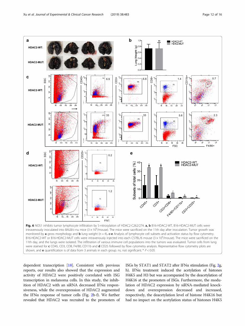
NOS1 inhibits tumor lymphocyte infiltration by S-nitrosylation of HDAC2-C262/274IFNα has been reported to have both anti-proliferativeand immunomodulatory effects. Because we found thatHDAC2-MUT partially reversed the role of NOS1 inpromoting lung metastasis, we further investigatedwhether HDAC2-MUT directly inhibited tumor growth.In the in vitro experiment, we did not observe differ-ences in the growth of HDAC2-WT and HDAC2-MUT
Fig. 4 NOS1 inhibits the deacetylation of H4K16 by S-nitrosylation of HDAC2. a-d A375 cells were treated with or without IFNα (1000 U/ml) for 1h. ChIP was performed using specific a anti-H4K16ac, b anti-H4K5ac, c anti-H3ac and d anti-Rbp1 antibodies. e HDAC2 siRNA or the control siRNAwere transfected into A375 cells for 24 h, followed by stimulation with IFNα (1000 U/ml) for 1 h. ChIP was performed using H4K16ac antibodies. fSimilar to e, but HDAC2 expression vectors were used to transfect A375 cells for 24 h before IFNα stimulation. g HDAC2 siRNA or control siRNAwas used to transfect A375 cells for 24 h and then stimulated with or without IFNα (1000 U/ml) for 6 h. Western blotting was performed usingthe indicated antibodies. h, i Using the same protocol as in e, ChIP assays were performed using specific h anti-Rpb1 and i anti-H4K5acantibodies. j, k Control/NOS1 (A375) cells were treated with IFNα (1000 U/ml) for 1 h. ChIP assays were performed using specific j anti-H4K16acand k anti-Rpb1 antibodies. l Over-NOS1 (A375) cells were transfected with HDAC2-WT or HDAC2-MUT vectors for 24 h and then treated withIFNα (1000 U/ml) for 1 h. ChIP assays were performed using specific anti-H4K16ac antibodies. ns, not significant; *p < 0.05; **p < 0.01;***p < 0.001; ****p < 0.0001
Xu et al. Journal of Experimental & Clinical Cancer Research (2019) 38:483 Page 10 of 16

cells; therefore, we injected the cells into BALB/c-numice. Lung metastasis was observed on the 11th dayafter cell injection, and the results showed that there wasno significant difference between the two groups (Fig. 6a,b). This result indicates that HDAC2-MUT does not dir-ectly inhibit tumor growth. A possible explanation is thatHDAC2-MUT reduces lung metastasis by regulating anti-tumor immunity. Therefore, we injected HDAC2-WTand HDAC2-MUT cells into C57BL/6 mice, and theisolated lung tissues were detected by flow cytometry.As shown in Fig. 6c, the lung from HDAC2-WT micecontained significantly reduced numbers of CD45+,CD3+, CD3 + CD8+ T cells and F4/80 + CD11b + mac-rophages compared to lung from HDAC2-MUT mice(Fig. 6c, e). We also examined the number of acti-vated T cells in the lungs of both groups. Similarly,the lungs of HDAC2-MUT mice contained elevatednumbers of CD3 + CD25+ T cells (Fig. 6d, e). Takentogether, these results indicate that NOS1-induced S-nitrosylation of HDAC2 promotes lung metastasis pri-marily by inhibiting tumor lymphocyte infiltration.
DiscussionThe expression of NOS1 in melanoma is closely corre-lated with dysfunctional type I IFN signaling and poorprognosis of patients. However, the underlying mechan-ism governing this role has not been determined. In thisstudy, we investigated the regulation of NOS1 on IFNsignaling and lung metastasis. Our findings are as fol-lows: (i) HDAC2 upregulates ISG expression by deacety-lating H4K16, increasing the recruitment of RNApolymerase II to the promoter; and (ii) NOS1 reducesSTAT1-mediated recruitment of HDAC2 to the ISGpromoter and deacetylation of H4K16 by S-nitrosylationof HDAC2-C262/C274, which results in inhibition ofIFN signaling and tumor lymphocyte infiltration, therebypromoting lung metastasis of melanoma.ISGs remain silent under normal conditions, but de
novo transcription is initiated by activated transcriptionfactors after stimulation by IFNα. The class I HDAC fam-ily has often been associated with the suppression of genetranscription via repressive complexes, while it acts asactivators of gene expression in IFN-induced STAT1-
Fig. 5 NOS1 promotes melanoma lung metastasis by S-nitrosylation of HDAC2-C262/274. a The mRNA expression of HDACs (1, 2, 3) and proteinexpression of HDAC2 in HDAC2-KO cells were determined by RT-PCR and western blotting. b The same method as a was used to detect themRNA and protein expression levels of HDAC2 of stably transfected HDAC2-WT and HDAC2-MUT plasmids in HDAC2-KO cells. c-e 3 × 10⁶ B16-WT, B16-HDAC2-KO, B16-HDAC2-WT, B16-HDAC2-MUT cells were intravenously injected into C57BL/6 mice. The mice were sacrificed on the 11thday after tumor cell inoculation. Tumor growth was monitored by c gross morphology, d lung weight (n = 4), and e survival rate (n = 8). ns, notsignificant; **p < 0.01; ***p < 0.001; ****p < 0.0001
Xu et al. Journal of Experimental & Clinical Cancer Research (2019) 38:483 Page 11 of 16
dependent transcription [18]. Consistent with previousreports, our results also showed that the expression andactivity of HDAC2 were positively correlated with ISGtranscription in melanoma cells. In this study, the inhib-ition of HDAC2 with an siRNA decreased IFNα respon-siveness, while the overexpression of HDAC2 augmentedthe IFNα response of tumor cells (Fig. 2b-f). We furtherrevealed that HDAC2 was recruited to the promoters of
ISGs by STAT1 and STAT2 after IFNα stimulation (Fig. 2g,h). IFNα treatment induced the acetylation of histonesH4K5 and H3 but was accompanied by the deacetylation ofH4K16 at the promoters of ISGs. Furthermore, the modu-lation of HDAC2 expression by siRNA-mediated knock-down and overexpression decreased and increased,respectively, the deacetylation level of histone H4K16 buthad no impact on the acetylation status of histones H4K5
Fig. 6 NOS1 inhibits tumor lymphocyte infiltration by S-nitrosylation of HDAC2-C262/274. a, b B16-HDAC2-WT, B16-HDAC2-MUT cells wereintravenously inoculated into BALB/c-nu mice (3 × 10⁶/mouse). The mice were sacrificed on the 11th day after inoculation. Tumor growth wasmonitored by a gross morphology and b lung weight (n = 4). c-e Analysis of lymphocyte cell subsets and activation status by flow cytometry.B16-HDAC2-WT or B16-HDAC2-MUT cells were intravenously injected into each C57BL/6 mouse (3 × 10⁶/mouse). The mice were sacrificed on the11th day, and the lungs were isolated. The infiltration of various immune cell populations into the tumors was evaluated. Tumor cells from lungwere stained for c CD45, CD3, CD8, F4/80, CD11b and d CD25 followed by flow cytometry analysis. Representative flow cytometry plots areshown, and e quantification is of data from 3 animals in each group. ns, not significant; * P < 0.05
Xu et al. Journal of Experimental & Clinical Cancer Research (2019) 38:483 Page 12 of 16
and H3. In addition, IFNα also induced the recruitment ofRNA pol II to the promoter, which was inhibited by si-HDAC2, indicating that HDAC2 is required for the re-cruitment of RNA pol II (Fig. 4).Acetylation of lysine residues of histone H3 and his-
tone H4 are critical for maintaining the integrity of IFNsignals [34]. Specifically, acetylation of H3 is considereda marker of general transcriptional activation. STAT2has been reported to recruit the histone acetyltransferaseGCN5 to promote acetylation of H3 after IFNα stimula-tion [35]. There are four acetyl-lysine residues, K5, K8,K12 and K16, in H4, but the generation and propertiesof acetylated histone H4K16 are distinct from those ofother acetylated sites in H4 (i.e., K5, K8, and K12) [36].For instance, the mutation of K5, K8 or K12 results insimilar effects and positively correlates with gene tran-scription, and these three sites complement each other.In contrast, H4K16 acetylation is not correlated with theother sites and is negatively associated with gene expres-sion [32]. Acetylated H4K16 is a key epigenetic markerinvolved in gene regulation and chromatin remodeling[37, 38]. Although this marker is known to be essentialfor embryonic development and heterochromatin forma-tion [33, 39], its role during ISG expression has not beendetermined. Previous studies confirmed that viral infec-tion induces significant H4K8 and H4K12 acetylation atthe IFNβ promoter, while H4K16 is not acetylated dur-ing transcriptional activation [40]. In this study, we showevidence of H4K16 deacetylation by HDAC2 under IFNαstimulation conditions in melanoma cells (Fig. 4e-g),providing a molecular mechanism of HDAC2 involve-ment in the regulation of ISG expression through his-tone modification. The observed H4K16 deacetylation,accompanied by an increase in the recruitment of RNApol II (Fig. 4d, h), suggests that HDAC2 is a transcrip-tional regulator for ISGs through chromatin remodeling,transcription-activating complex recruitment, and tran-scription initiation. HDAC1 has been reported to playan essential role in IFNα-induced transcription, butwhether HDAC1 works similarly to HDAC2 to deacety-late H4K16 for ISG expression was not investigated inthis study. Our study reveals the positive role of HDAC2in the IFNα response and provides new insights into theepigenetic regulation of IFNα signaling. Nevertheless, theinduced expression process of ISGs is regulated by a seriesof factors, and a comprehensive understanding of its regu-latory mechanisms requires more in-depth research.In recent years, the role of nitric oxide in tumor biology
has received increasing attention [41, 42]. NOS1 producesconstitutively low levels of NO, which generally promotetumor growth, such as cell proliferation, anti-apoptosisand migration, in many cellular processes [21]. Mean-while, it has been observed that NOS1 expression and in-creased NO production correlate with a poor prognosis in
melanoma patients [43]. Furthermore, NOS1 was closelyrelated to dysfunctional IFN signaling, and an inhibitor ofNOS1 resulted in reduced proliferation of melanoma cells[25, 44]. In this study, we found that endogenous NOfrom NOS1 downregulates ISGF3-dependent transcrip-tion and gene expression. On the other hand, animalmodels indicate that NOS1 promotes lung metastasis ofmelanoma (Fig. 1). These results further provide evidencethat NOS1 is involved in dysfunctional IFN signaling andmelanoma metastasis. NO is a molecule capable of modi-fying cysteines by S-nitrosylation, which affects proteinfunction by altering the interaction between proteins, sub-cellular localization or catalytic activity [24]. Previousstudies have shown that S-nitrosylation of HDAC2-C262/C274 does not alter the enzyme’s catalytic activity but in-duces its release from chromatin [26, 45]. HDACs exist ascomponents of multiprotein complexes amd are then tar-geted to specific genomic regions by interactions withDNA binding factors such as transcription factors [46].Furthermore, histone hyperacetylation is not always theresult of a loss of HDAC activity, but it could be due to aloss of HDAC targeted to specific DNA sequences [47].We further demonstrated that NOS1-induced S-nitrosylation reduces the recruitment of HDAC2 to theISG promoter by inhibiting the interaction of HDAC2with STAT1 (Fig. 2i, j, and Fig. 3h). This effect resulted inincreased acetylation of H4K16 (Fig. 4l) and transcrip-tional inhibition of ISGs (Fig. 3g). Thus, our study demon-strated a linkage between the S-nitrosylation of HDAC2and the regulation of ISG expression, providing an en-dogenous NO-mediated mechanism for the dysfunction ofthe IFNα response in melanoma cells.The loss of tumor cell type I IFN production, and hence
immune signalling, was associated with an increased risk ofmetastasis [48]. Downregulation of IRF7 has been describedin breast cancer and contributes to tumor metastasis, indi-cating that IFN signaling is involved in the control of meta-static spread [9]. Our study confirmed that NOS1 promoteslung metastasis at least partly through S-nitrosylation ofHDAC2-C262/C274 and deregulation of the IFNα response.Interestingly, in animal models, lung metastasis was signifi-cantly reduced after knockout of HDAC2 in melanoma cells(Fig. 5c), suggesting that HDAC2 is a tumor-promoting fac-tor, and other studies also support this finding [49–51].There is more evidence for HDAC2 overexpression in cer-tain cancers compared with normal tissues [52]. HDAC2regulates the cell cycle and apoptosis of cancer cells, andgene editing of HDAC2 resulted in a more differentiatedphenotype and increased apoptosis caused by augmentedlevels of p21 [53]. HDAC2 also plays a role in controlling cellsurvival by regulating p53 and its target genes [54]. Trans-formation of cells could be caused by elevated HDAC2, forexample via inactivation of p53 or regulation of p53-DNAbinding activity [55]. Thus, HDAC2 appears to represent a
Xu et al. Journal of Experimental & Clinical Cancer Research (2019) 38:483 Page 13 of 16

therapeutic target, and the development of anticancer drugsspecific for HDAC2 may inhibit tumor metastasis and pre-vent the side effects from the current pan-HDACi treatment.Importantly, HDAC2 bearing a mutation of Cys262 andCys274 (HDAC2-C262A/274A) cannot be nitrosylated andacts as a potent transcriptional activator in the IFNα path-way, thereby reversing the inhibition of ISGs and promotionof lung metastasis by NOS1.Type I IFNs have emerged as central coordinators of
tumor–immune-system interactions, including stimulationof anti-tumor effector cells (T cells, NK cells, dendritic cells),and negative regulation of suppressive cells (MDSCs andTreg cells) [56]. Recent studies have shown that the expres-sion and secretion of tumor-inherent IFNs is a key player inthe anti-tumor immune cascade, influencing the immuno-genicity, progression and therapeutic response of tumors[57]. Unfortunately, impaired interferon signaling has beenreported to be a common defect in human cancer [3], butthe mechanisms underlying tumor-inherent IFN dysfunctionhave not been determined. In this study, we found thatNOS1 modifies HDAC2 by S-nitrosylation, resulting in in-creased tumor metastasis in B16F10 mice. However, we didnot find evidence that NOS1 promotes proliferation in vitro,and the effect of NOS1 was completely abolished in im-munocompromised nude mice (Fig. 6a). This finding sug-gests that the mechanism of metastasis promotion by NOS1expression in tumor cells is caused by inhibition of tumorimmune surveillance. Notably, mice bearing HDAC2-C262A/C274A tumors had an increase in the number oftumor infiltrating lymphocytes (TILs), such as CD8+ T cellsand macrophages, which were linked with the type I IFNpathway (Fig. 6c). TILs are known to be key players in anti-tumor immunity, and their intratumoral accumulation is as-sociated with favorable outcomes in many cancers [58]. Inthis study, we report for the first time that S-nitrosylation ofHDAC2 inhibits IFN signaling and the accumulation of im-mune cells, impeding the suppression of metastases by Tlymphocytes and macrophages. These results suggest thatthe detection of HDAC2-C262/C274 S-nitrosylation can beused as a marker to determine the IFN treatment responseand cancer prognosis.
ConclusionsWe report the mechanism by which HDAC2 regulatesthe expression of ISGs in tumor cells, and NOS1 inducesepigenetic changes through S-nitrosylation of HDAC2,thereby leading to dysfunctional IFN signaling and pro-moting lung metastasis. These results show that by inhi-biting the S-nitrosylation of HDAC2 in tumor cells, IFNsignaling can be restored to inhibit metastasis. Our datawill prompt future research to target NOS1 or combinedimmunotherapy to control metastasis in melanomapatients.
Supplementary informationSupplementary information accompanies this paper at https://doi.org/10.1186/s13046-019-1448-9.
Additional file 1: Sequences of siRNA and primer used in this study.Table S1. Primer sequences for RT-PCR. Table S2. The sequences ofsiRNA. Table S3. Primer sequences for ChIP-qPCR
Additional file 2: Figure S1. NOS1 blocks IFNα-stimulated gene induc-tion. a SKOV3 and SW480 cells were treated with IFNα for 6 h in the pres-ence or absence of simultaneous GSNO. The mRNA expression of ISGswere analyzed by RT-PCR. b Control/NOS1 (SKOV3, SW480) cells were in-cubated with IFNα for 6 h, followed RT-PCR analysis. Figure S2. S-nitrosyl-tion of HDAC2 does not affect its expression. a Control/NOS1 (SKOV3)cells were treated with IFNα (1000 U/ml) for 6 h and 12 h, the expressionof HDAC2 was detected by RT-PCR and western blotting. b Control/NOS1(SKOV3, B16) cells were stimulated with or without IFNα for 6 h. Proteinextracts were subjected to the biotin-switch assay. Figure S3. HDAC2regulates the acetylation status of H4K16. a Densitometric analysis of thedata in Fig. 4g (n=3). b A375 cells were transfected si-RNA for 24 h andtreatment with IFNα for 1 h. ChIP assays were performed after chromatinwas immunoprecipitated with an anti-H3ac antibody. IP chromatin wassubjected to qPCR. ns, not significant.
AbbreviationsIFN: Interferon; NOS1: Nitric Oxide Synthase 1; ISGs: Interferon-stimulatedgenes; HDAC2: Histone deacetylase 2; H4K16ac: Acetylated H4 lysine 16;H4K5ac: Acetylated H4 lysine 5; H3ac: Acetylated H3; RNA pol II: RNAPolymerase II; RNA: Polymerase II; ISGF3: Interferon -stimulated gene factor 3;ISRE: Interferon-sensitive response element; NO: Nitric oxide;PBMCs: Peripheral blood mononuclear cells; STAT: Signal transducer andactivator of transcription; IRF: Interferon regulatory factor; ChIP: Chromatinimmunoprecipitation; TILs: Tumor infiltrating lymphocytes
AcknowledgmentsWe thank for the support from the Cancer Research Institute of SouthernMedical University, Guangdong provincial key laboratory of cancerimmunotherapy research, Guangzhou key laboratory of tumor immunologyresearch and the Shunde Hospital of Southern Medical University.
Author’s contributionsPerformed experiments and data analysis: PX and SY; Designed experimentsand wrote the manuscript: PX and QL; Data analysis and provided criticaltechnical and scientific discussion: PX, SY, KY, MH, QW, SZ, XC, WG, JC, QZ,ZZ, YL, ZR, YX, BH, AP, MO and QL. All authors read and approved the finalmanuscript.
FundingThis work was supported by grants from the National Natural ScienceFoundation of China (Grant No. 81472834).
Availability of data and materialsThe datasets used and/or analysed during the current study are availablefrom the corresponding author on reasonable request.
Ethics approvalAll animal experiments in this study were approved by the Medical EthicsCommittee of Southern Medical University and conducted in strictaccordance with the guidelines from the Ministry of Science and Technologyof China.
Consent for publicationNot application.
Competing interestsThe authors declare that they have no competing interests.
Author details1Cancer Research Institute, Guangdong Provincial Key Laboratory of CancerImmunotherapy, Guangzhou key laboratory of tumor immunology research,School of Basic Medical Sciences, Southern Medical University, Guangzhou
Xu et al. Journal of Experimental & Clinical Cancer Research (2019) 38:483 Page 14 of 16

510515, China. 2Department of Oncology, Guangzhou Hospital of IntegratedTraditional and Western Medicine, Guangzhou 510800, China. 3Center formedical transformation, Shunde Hospital, Southern Medical University,Foshan 528308, China.
Received: 19 July 2019 Accepted: 15 October 2019
References1. Laurence Z, Lorenzo G, Oliver K, Smyth MJ, Guido K. Type I interferons in
anticancer immunity. Nat Rev Immunol. 2015;15(7):405–14.2. Minn AJ. Interferons and the immunogenic effects of Cancer therapy.
Trends Immunol. 2015;36(11):725–37.3. Critchley-Thorne RJ, Simons DL, Yan N, Miyahira AK, Dirbas FM, Johnson DL,
et al. Impaired interferon signaling is a common immune defect in humancancer. Proc Natl Acad Sci U S A. 2009;106(22):9010–5.
4. Dunn GP, Koebel CM, Schreiber RD. Interferons, immunity and cancerimmunoediting. Nat Rev Immunol. 2006;6:836.
5. Antonella S, Takahiro Y, Erika V, Kariman C, Enot DP, Julien A, et al. Cancercell-autonomous contribution of type I interferon signaling to the efficacyof chemotherapy. Nat Med. 2014;20(11):1301–9.
6. Brockwell NK, Rautela J, Owen KL, Gearing LJ, Deb S, Harvey K, et al. Tumorinherent interferon regulators as biomarkers of long-term chemotherapeuticresponse in TNBC. NPJ Precision Oncol. 2019;3:21.
7. Katlinski KV, Gui J, Katlinskaya YV, Ortiz A, Chakraborty R, Bhattacharya S, et al.Inactivation of interferon receptor promotes the establishment of immuneprivileged tumor microenvironment. Cancer Cell. 2017;31(2):194–207.
8. Picaud S, Bardot B, De Maeyer E, Seif I. Enhanced tumor development inmice lacking a functional type I interferon receptor. J Interferon CytokineRes. 2002;22(4):457–62.
9. Bidwell BN, Slaney CY, Withana NP, Sam F, Yuan C, Sherene L, et al.Silencing of Irf7 pathways in breast cancer cells promotes bone metastasisthrough immune escape. Nat Med. 2012;18(8):1224–31.
10. Zaretsky JM, Garcia-Diaz A, Shin DS, Escuin-Ordinas H, Hugo W, Hu-Lieskovan S, et al. Mutations associated with acquired resistance to PD-1blockade in melanoma. N Engl J Med. 2016;375(9):819–29.
11. Gao J, Shi LZ, Zhao H, Chen J, Xiong L, He Q, et al. Loss of IFN-gammaPathway Genes in Tumor Cells as a Mechanism of Resistance to Anti-CTLA-4Therapy. Cell. 2016;167(2):397–404.e9.
12. Jackson DP, Watling D, Rogers NC, Banks RE, Kerr IM, Selby PJ, et al. The JAK/STATpathway is not sufficient to sustain the antiproliferative response in an interferon-resistant human melanoma cell line. Melanoma Res. 2003;13(3):219–29.
13. Di Trolio R, Simeone E, Di Lorenzo G, Buonerba C, Ascierto PA. The use ofinterferon in melanoma patients: a systematic review. Cytokine GrowthFactor Rev. 2015;26(2):203–12.
14. Nusinzon I, Horvath CM. Histone deacetylases as transcriptional activators?Role reversal in inducible gene regulation. Sci STKE. 2005;2005(296):re11.
15. Chang H-M, Paulson M, Holko M, Rice CM, Williams BRG, Marié I, et al. Inductionof interferon-stimulated gene expression and antiviral responses require proteindeacetylase activity. Proc Natl Acad Sci U S A. 2004;101(26):9578–83.
16. Nusinzon I, Horvath CM. Interferon-stimulated transcription and innateantiviral immunity require deacetylase activity and histone deacetylase 1.Proc Natl Acad Sci. 2003;100(25):14742–7.
17. Sakamoto S, Potla R, Larner AC. Histone deacetylase activity is required torecruit RNA polymerase II to the promoters of selected interferon-stimulatedearly response genes. J Biol Chem. 2004;279(39):40362–7.
18. Klampfer L, Huang J, Swaby LA, Augenlicht L. Requirement of histonedeacetylase activity for signaling by STAT1. J Biol Chem. 2004;279(29):30358–68.
19. Icardi L, Lievens S, Mori R, Piessevaux J, Cauwer LD, Bosscher KD, et al.Opposed regulation of type I IFN-induced STAT3 and ISGF3 transcriptionalactivities by histone deacetylases (HDACS) 1 and 2. FASEB J. 2012;26(1):240–9.
20. Marie IJ, Chang HM, Levy DE. HDAC stimulates gene expression throughBRD4 availability in response to IFN and in interferonopathies. J Exp Med.2018;215(12):3194–212.
21. Fukumura D, Kashiwagi S, Jain RK. The role of nitric oxide in tumourprogression. Nat Rev Cancer. 2006;6(7):521–34.
22. Yarlagadda K, Hassani J, Foote IP, Markowitz J. The role of nitric oxide inmelanoma. Biochim Biophys Acta Rev Cancer. 2017;1868(2):500–9.
23. Stamler JS, Lamas S, ., Fang FC. Nitrosylation. The prototypic redox-basedsignaling mechanism. Cell. 2001;106(6):675–683.
24. Aranda E, Lopez-Pedrera C, De La Haba-Rodriguez JR, Rodriguez-Ariza A.Nitric oxide and cancer: the emerging role of S-nitrosylation. Curr Mol Med.2012;12(1):50–67.
25. Liu Q, Tomei S, Ascierto ML, De Giorgi V, Bedognetti D, Dai C, et al.Melanoma NOS1 expression promotes dysfunctional IFN signaling. J ClinInvest. 2014;124(5):2147–59.
26. Nott A, Watson PM, Robinson JD, Crepaldi L, Riccio A. S-nitrosylation ofhistone deacetylase 2 induces chromatin remodelling in neurons. Nature.2008;455(7211):411–5.
27. Zhu L, Li L, Zhang Q, Yang X, Zou Z, Hao B, et al. NOS1 S-nitrosylates PTENand inhibits autophagy in nasopharyngeal carcinoma cells. Cell Death Dis.2017;3:17011.
28. Livak KJ, Schmittgen TD. Analysis of relative gene expression data usingreal-time quantitative PCR and the 2(−Delta Delta C(T)) Method. Methods(San Diego, Calif). 2001;25(4):402–8.
29. Li L, Zhu L, Hao B, Gao W, Wang Q, Li K, et al. iNOS-derived nitric oxidepromotes glycolysis by inducing pyruvate kinase M2 nuclear translocationin ovarian cancer. Oncotarget. 2017;8(20):33047–63.
30. Ran FA, Hsu PD, Wright J, Agarwala V, Scott DA, Zhang F. Genomeengineering using the CRISPR-Cas9 system. Nat Protoc. 2013;8:2281.
31. Gustavo T, Oded S, Verma IM. Production and purification of lentiviralvectors. Nat Protoc. 2006;1(1):241–5.
32. Millar CB, Kurdistani SK, Grunstein M. Acetylation of yeast histone H4 lysine16: a switch for protein interactions in heterochromatin and euchromatin.Cold Spring Harb Symp Quant Biol. 2004;69:193–200.
33. Oppikofer M, Kueng S, Martino F, Soeroes S, Hancock SM, Chin JW, et al. Adual role of H4K16 acetylation in the establishment of yeast silentchromatin. EMBO J. 2011;30(13):2610–21.
34. Chen K, Liu J, Cao X. Regulation of type I interferon signaling in immunityand inflammation: a comprehensive review. J Autoimmun. 2017;83:1–11.
35. Paulson M, Press C, Smith E, Tanese N, Levy DE. IFN-stimulated transcriptionthrough a TBP-free acetyltransferase complex escapes viral shutoff. Nat CellBiol. 2002;4(2):140–7.
36. Ma XJ, Wu J, Altheim BA, Schultz MC, Grunstein M. Deposition-related sitesK5/K12 in histone H4 are not required for nucleosome deposition in yeast.Proc Natl Acad Sci U S A. 1998;95(12):6693–8.
37. Zhang R, Erler J, Langowski J. Histone acetylation regulates chromatinaccessibility: role of H4K16 in inter-nucleosome interaction. Biophys J. 2017;112(3):450–9.
38. Urdinguio RG, Lopez V, Bayon GF. Diaz de la Guardia R, sierra MI, Garcia-Torano E, et al. chromatin regulation by histone H4 acetylation at lysine 16during cell death and differentiation in the myeloid compartment. NucleicAcids Res. 2019;47(10):5016–37.
39. Copur O, Gorchakov A, Finkl K, Kuroda MI, Muller J. Sex-specific phenotypesof histone H4 point mutants establish dosage compensation as the criticalfunction of H4K16 acetylation in drosophila. Proc Natl Acad Sci U S A. 2018;115(52):13336–41.
40. Agalioti T, Chen G, Thanos D. Deciphering the transcriptional histoneacetylation code for a human gene. Cell. 2002;111(3):381–92.
41. Vannini F, Kashfi K, Nath N. The dual role of iNOS in cancer. Redox Biol.2015;6:334–43.
42. Simone M, Vincenzo B, Donato N. Nitric oxide, a double edged sword in cancerbiology: searching for therapeutic opportunities. Med Res Rev. 2010;27(3):317–52.
43. Ahmed B, Van Den Oord JJ. Expression of the neuronal isoform of nitricoxide synthase (nNOS) and its inhibitor, protein inhibitor of nNOS, inpigment cell lesions of the skin. Br J Dermatol. 1999;141(1):12–9.
44. Yang Z, Misner B, Ji H, Poulos TL, Silverman RB, Meyskens FL, et al. Targeting nitricoxide signaling with nNOS inhibitors as a novel strategy for the therapy andprevention of human melanoma. Antioxid Redox Signal. 2013;19(5):433–47.
45. Nott A, Nitarska J, Veenvliet JV, Schacke S, Derijck AAHA, Sirko P, et al. S-nitrosylation of HDAC2 regulates the expression of the chromatin-remodeling factor Brm during radial neuron migration. Proc Natl Acad Sci US A. 2013;110(8):3113–8.
46. Vaquero A, Scher M, Reinberg D. Biochemistry of Multiprotein HDACComplexes; 2006.
47. Ropero S, Esteller M. The role of histone deacetylases (HDACs) in humancancer. Mol Oncol. 2007;1(1):19–25.
48. Rautela J, Baschuk N, Slaney CY, Jayatilleke KM, Xiao K, Bidwell BN, et al. Lossof host type-I IFN signaling accelerates metastasis and impairs NK-cellantitumor function in multiple models of breast Cancer. Cancer ImmunolRes. 2015;3(11):1207–17.
Xu et al. Journal of Experimental & Clinical Cancer Research (2019) 38:483 Page 15 of 16

49. Lin CL, Tsai ML, Lin CY, Hsu KW, Hsieh WS, Chi WM, et al. HDAC1 andHDAC2 Double Knockout Triggers Cell Apoptosis in Advanced ThyroidCancer. Int J Mol Sci. 2019;20(2):454.
50. Stojanovic N, Hassan Z, Wirth M, Wenzel P, Beyer M, Schafer C, et al. HDAC1and HDAC2 integrate the expression of p53 mutants in pancreatic cancer.Oncogene. 2017;36(13):1804–15.
51. Li L, Mei DT, Zeng Y. HDAC2 promotes the migration and invasion of non-small cell lung cancer cells via upregulation of fibronectin. BiomedPharmacother. 2016;84:284–90.
52. Weichert W. HDAC expression and clinical prognosis in humanmalignancies. Cancer Lett. 2009;280(2):168–76.
53. Huang BH, Laban M, Leung CH, Lee L, Lee CK, Salto-Tellez M, et al. Inhibition ofhistone deacetylase 2 increases apoptosis and p21Cip1/WAF1 expression,independent of histone deacetylase 1. Cell Death Differ. 2005;12(4):395–404.
54. Wagner T, Brand P, Heinzel T, Kramer OH. Histone deacetylase 2 controlsp53 and is a critical factor in tumorigenesis. Biochim Biophys Acta. 2014;1846(2):524–38.
55. Harms KL, Chen X. Histone deacetylase 2 modulates p53 transcriptionalactivities through regulation of p53-DNA binding activity. Cancer Res. 2007;67(7):3145–52.
56. Parker BS, Rautela J, Hertzog PJ. Antitumour actions of interferons:implications for cancer therapy. Nat Rev Cancer. 2016;16(3):131–44.
57. Brockwell NK, Parker BS. Tumor inherent interferons: impact on immunereactivity and immunotherapy. Cytokine. 2019;118:42–7.
58. Mahmoud SM, Paish EC, Powe DG, Macmillan RD, Grainge MJ, Lee AH, et al.Tumor-infiltrating CD8+ lymphocytes predict clinical outcome in breastcancer. J Clin Oncol. 2011;29(15):1949–55.
Publisher’s NoteSpringer Nature remains neutral with regard to jurisdictional claims inpublished maps and institutional affiliations.
Xu et al. Journal of Experimental & Clinical Cancer Research (2019) 38:483 Page 16 of 16